Article

Seroepidemiology and the Molecular Detection of Animal Brucellosis in Punjab, Pakistan

1
Wildlife Epidemiology and Molecular Microbiology Laboratory (One Health Research Group), Discipline of Zoology, Department of Wildlife & Ecology, University of Veterinary and Animal Sciences, Lahore, Ravi Campus, Pattoki 55300, Pakistan
2
Institute of Pharmaceutical Science, University of Veterinary and Animal Sciences, Lahore 54000, Pakistan
3
Friedrich-Loeffler-Institute, Institute of Bacterial Infections and Zoonoses, Naumburger Str. 96a, 07743 Jena, Germany
4
Faculty Medicine of Veterinary, Kafrelsheikh University, Kafr El-Sheikh 33511, Egypt
5
Department of Pathobiology, College of Veterinary and Animal Sciences, Jhang 35200, Pakistan
6
Department of Biological Sciences, University of Veterinary and Animal Sciences, Lahore, Ravi Campus, Pattoki 55300, Pakistan
*
Author to whom correspondence should be addressed.
Microorganisms 2019, 7(10), 449; https://doi.org/10.3390/microorganisms7100449
Submission received: 29 August 2019 / Revised: 9 October 2019 / Accepted: 12 October 2019 / Published: 13 October 2019
(This article belongs to the Section Public Health Microbiology)

Abstract

:
Brucellosis is an infectious disease caused by bacteria of the genus Brucella (B.), affecting both animals and humans, causing severe economic loses and severe illness, respectively. The objective of the present study was to determine the seroprevalence and the risk factors associated with caprine, ovine, and bovine brucellosis in selected districts of Punjab, Pakistan. A total of 1083 blood samples were randomly collected from animals (goats = 440, sheep = 203, cows = 206, and buffaloes = 234). Questionnaires were used to collect data on risk factors associated with brucellosis on the sampling day. All samples were initially screened for anti-Brucella antibodies using the rose bengal plate test (RBPT). The seropositive serum samples were confirmed by a quantitative real-time polymerase chain reaction (PCR) assay for the detection of the Brucella genus- and Brucella species-specific DNA (B. abortus and B. melitensis). Univariant and binary logistic regression were used to identify important risk factors of brucellosis. Anti-Brucella antibodies and DNA were detected in 35 (3.23%) serum samples. Thirty-four (97.1%) DNA samples were confirmed as B. melitensis by qRT-PCR. Abortion history and natural mating were found to be potential risk factors. Brucella melitensis was identified as the causative agent of caprine, ovine, and bovine brucellosis in the selected districts of Punjab, Pakistan. Diseased animals may act as a source of infection for other animals. The elimination of positive seroreactors, development of control strategies for brucellosis, and education programs regarding the control of zoonotic disease are highly needed in developing countries like Pakistan.

1. Introduction

Brucellosis is one of the most devastating zoonotic diseases that has spread all over the world. It affects a wide range of animal species and humans [1,2,3,4]. Brucellosis is considered the most critical zoonotic disease after rabies and tuberculosis. The main symptoms of brucellosis in animals are abortion, reduction of milk production, hygroma, neonatal mortality, infertility, epididymitis, and orchitis. The typical symptoms of brucellosis in males are orchitis and epididymitis, and abortion is typically the first recognized clinical sign in pregnant females [5]. Risk factors associated with brucellosis can be categorized into management, animal, and environmental factors. The screening of new arrivals, hygiene, vaccination, size of herd, breeding practices, and production system are management risk factors. Animal risk factors include age, breed, sex, abortion history, and milking method. The agro-ecological location of animals is categorized as an environmental factor [6,7].
Most of the animals which have recovered from brucellosis spontaneously might be shedding the bacteria in urine, milk, and vaginal secretion. When animals are slaughtered, Brucella species are also shed outside [8,9]. Brucellosis may lead to serious economic losses through the death of young stock, still birth, or abortion, and efforts for improved breeding are hindered [10]. Livestock provides food, skins, fibers, manure (fertilizer or fuel), and draught power. In developing countries, livestock is the basis of livelihood for about 95% of the rural population [11].
There are several draw backs to diagnose brucellosis using isolation and culture. Brucella shows slow growth, which may delay diagnosis for up to 7 days or more. The sensitivity of the culture is also low (50 to 90%), depending on the given species, stage of disease, culture media, quantity of bacteria, and culture technique used. Here, culturing is not economical and is time consuming. Thus, the most important diagnostic screening technique and confirmation method are serological methods [12]. However, no species diagnostic is feasible. The Brucella antigen shows cross reactions with some other bacteria species such as, Vibrio cholera, Bordetella bronchiseptica, Yersinia enterocolitica, and the Salmonella species [13]. Molecular methods, like polymerase chain reaction (PCR), have proven to be fast, specific, reliable, and safe methods. The most important advantage of RT-PCR is its analytical sensitivity, as can detect the few bacteria present in a sample [4].
In small ruminants, B. melitensis is typically the causative agent of brucellosis. However, the most frequent causative agent of bovine brucellosis is B. abortus. Infrequently B. suis or B. melitensis can cause brucellosis in bovines if kept in close contact to pigs, goats, or sheep [14]. Only few studies are available relating to animal brucellosis in Pakistan. The overall seroprevalence of brucellosis reported for Pakistan was 3.25–4.4% in livestock. Based on serum agglutination testing, the seroprevalence was 5.06% and 5.49% in cattle and buffaloes, respectively. The seroprevalence of brucellosis in goats and sheep were found to be 1.94% and 1.47%, respectively [15]. Keeping in view the economic importance of animal brucellosis, the present study was designed to determine the seroprevalence and risk factors associated with caprine, ovine, and bovine brucellosis in the selected districts of Punjab, Pakistan.

2. Materials and Methods

2.1. Sampling Sites

The study was conducted in Kasur, Okara, Lahore, and Faisalabad in the Punjab province of Pakistan (Figure 1). The Kasur District (31.1165° N, 74.4494° E) is south of Lahore and borders India along the Ganda Singh border. Here, important livestocks are cattle (Sahiwal), buffalo (Nili Ravi), goats (Beetal, Teddy), and sheep (Lohi, Kajli). According to the 9211 Virtual Governance System, the livestock of this district includes large animals (1,060,994), poultry (243,141), and small ruminants (319,048). Different infectious or metabolic diseases are reported from this area, including ketosis, milk fever, foot and mouth disease, hemorrhagic septicemia, etc. Vaccination for these diseases is given to animals [16]. The use of animals for research purposes was performed according to the Institutional Ethical Review Committee for Animal Care and Use, the University of Veterinary and Animal Sciences, Lahore, Pakistan via approval number DR/750.
The name of the Okara city (30.8090° N, 73.4508° E) is derived from the name of the tree “Okaan”. Here, the livestock population and production is very high. The city is located by the river Ravi. Okara is also famous for its cotton mills and various military dairy farms, and for the cheese of these farms. Important livestock breeds of the Okara district are cattle (Sahiwal, Cholistani), buffalo (Nili Ravi), goats (Beetal, Teddy), and sheep (Kajli). According to the 9211 Virtual Governance System, the livestock of this district includes poultry (296,097), large animals (1,312,106), and small ruminants (474,062). The prevalent diseases of this area related to livestock are milk fever, mastitis, and foot and mouth disease. Vaccination for these diseases is given to animals [16].
The capital of the province Punjab is Lahore (31.5204° N, 74.1350° E), which is the second densest city of Pakistan. It is located near the Indian border Wagah and river Ravi. In Lahore, famous livestock breeds are cattle (Sahiwal) and buffalo (Nili Ravi). According to the 9211 Virtual Governance System, the livestock of this district includes small ruminants (141,651), poultry (122,379), and large animals (604,565). Different initiatives have been taken to improve the livestock sector in this area, such as reducing calf and feed lot fattening, disease control compartment, milk awareness campaigns, poultry unit distribution, distribution of heifer cows/buffaloes, sheep and goats. The prevalent diseases of this area related to livestock are milk fever, mastitis, the herpes simplex virus, bovine ephemeral fever, and foot and mouth disease. Here, vaccinations are applied to animals [16].
Faisalabad (301.4504° N, 73.1350° E) is the third largest city of the province Punjab. In District Faisalabad, important livestock breeds are goats (Beetal), buffalo (Nili Ravi), cattle (Desi, cross breed), and sheep (Lohi). According to the 9211 Virtual Governance System, the livestock of this area includes goats (528,203), cattle (534,499), sheep (87,691), buffalo (999,087), rural poultry (285,492), and pet/captive birds (125,250). Here, the prevalent diseases related to livestock include black quarter red metabolic water, milk fever, foot and mouth disease, and herpes simplex virus infection. Here, vaccinations are applied to animals [16].

2.2. Epidemiological Data Collection

The demographic (district and tehsil) and epidemiological data related to risk factors of brucellosis (i.e., species, gender, breed, animal’s age, herd size, abortion history, have their own sire, cleaning up the coral, body condition and stock replacement) were collected using a questionnaire.

2.3. Blood Sampling

A total of 1083 blood samples from goats (n = 440), sheep (n = 203), buffaloes (n = 234) and cows (n = 206) were collected randomly from the districts Kasur, Okara, Lahore, and Faisalabad. Blood samples (4 mL) were collected into non-EDTA tubes from each animal aseptically from jugular venipuncture with disposable needle. These samples were immediately stored at 4 °C in ice boxes and transported to the Epidemiology and Microbiology Laboratory (One Health Research Group), Department of Wildlife and Ecology, University of Veterinary and Animal Sciences, Lahore, Ravi Campus, Pattoki.

2.4. Serum Separation

After the transportation of blood samples to the laboratory, they were kept upright at 4 °C for a maximum of 24 h. All tubes with blood were centrifuged at 5000 rpm for 5 min for serum separation. After centrifugation, the supernatants were collected in sterile Eppendorf tubes (1.5 mL) by pipettes and stored at −20 °C for further analysis.

2.5. Rose Bengal Plate Test (RBPT)

Serum samples were analyzed with the rose bengal test (RBT) antigen for the detection Brucella antibodies (IDvet France). Brucellosis positive and negative control sera were provided by the Friedrich Loeffler Institute, Jena, Germany, for checking the RBT antigen before analyzing the serum samples. For the serological analysis of the serum samples, equal volumes (30µL) of serum samples were mixed with the RBT antigen. If agglutination was observed (after 4 min), samples were considered positive, otherwise they were considered negative for brucellosis [16].

2.6. DNA Extraction

A total of 35 seropositive samples were used to extract DNA. A high purity PCR template preparation kit (Roche Diagnostic, Mannheim, Germany) was used for the extraction of DNA according to manufacturer’s instructions. A ND-1000 UV visible spectrophotometer (Nano-Drop technologies, Wilmington, DE) was used for checking the purity of DNA and its concentration. Then, the DNA samples were stored at −20 °C for further analysis.

2.7. Quantitative Real-Time (RT) PCR

Serum samples positive in serology were further confirmed for the presence of Brucella DNA using real-time PCR. Two species specific (B. abortus and B. melitensis) and one Brucella genus specific qRT-PCR assays were used [17]. The detailed procedure has been previously described [18]. The primers and probes used here were supplied by TIB MOLBIOL (Berlin, Germany) (Table 1).
The PCR reaction and analysis were performed using the Mx3000P Thermocycler (Stratagene, Canada). The samples scored positive by the instrument were additionally confirmed by visual inspection of the graphical plots, which show cycle numbers versus fluorescence values [19]. Reference strains of B. abortus S-99 (ATCC 23448) and B. melitensis 16M (ATCC 23456) were provided by the National Reference Laboratory (NRL) as a positive control. A non-Brucella gram-negative strain was used in the present study to evaluate the specificity of the primers and real-time PCR reaction, namely, E. coli (ATCC 10538). A sample with a fluorescence signal 30 times greater than the mean standard deviation in all wells over cycles 2 through 10 was considered as a positive result, whereas a sample yielding a fluorescence signal less than this threshold value was considered negative. Cycle threshold values below 38 cycles were interpreted as positive. The threshold was set automatically by the instrument. The samples scored positive by the instrument were additionally confirmed by visual inspection of the graphical plots, which show cycle numbers versus fluorescence values.

2.8. Statistical Analysis

The Statistical Package for Social Sciences (SPSS), version 21.0, software package was used to perform the statistical analysis of all collected data. The chi square test was used to assess the association between each risk factor and the outcome variable. To estimate the prevalence, binomial logistic regression was ran to calculate the confidence interval (CI). To determine the association between risk factors and the prevalence of brucellosis, odds ratios, along with a 95% CI, were calculated. Binary logistic regression was used to estimate the association between the seropositive and explanatory variables. P-values ≤ 0.05 were considered statistically significant for all analyses here.

3. Results

Thirty-five (3.2%) serum samples were positive for Brucella antibodies using the RBPT. The seroprevalence of Brucella antibodies was higher (11.3%) in the Faisalabad District as compared to the Okara (4.1%), Lahore (2.0%), and Kasur districts (1.3%) (Table 2). The seroprevalence of brucellosis significantly (p < 0.05) varied from one tehsil to another, where it was highest in Chunian (16.7%) and lowest in Pattoki (1%) (Table 2).
The prevalence of Brucella antibodies based on the animal species level were higher in buffaloes (5.1%) than cows (3.8%), sheep (3.4%), and goats (1.8%) (Table 3). The seroprevalence of brucellosis was much higher in males (7.4%) as compared to females (2.5%). The seroprevalence of different breeds of buffaloes and cows was 5.1% in Nili Ravi and 3.8% in Rojhan, respectively. In different breeds of sheep and goats, it was 3.4%, 1.9%, and 1.8% in the Kajli, Teddy, and Beetle breeds, respectively. However, the prevalence of brucellosis was not statistically significant here (p > 0.05). In term of age groups, a positive sample was not found in offspring, but Brucella antibodies were found in 3.3% of adults. Here, age group was not statistically significant (p > 0.05).
Overall, 19 (12.75%) herds were found to be positive, among these, six were goat herds, five were buffalo herds, four were cow herds, and four were sheep herds. The seroprevalence of Brucella antibodies was higher in large herds (26.4%) than in smaller herds (5.2%). The association of infection with increasing herd size was statistically significant (p < 0.05).
A higher seroprevalence (9.0%) was reported in animals that had a history of abortion when compared to animals which had no history of abortion (2.4%). Abortion history was a significant potential risk factor (Table 3). The seroprevalence of brucellosis was higher in those animals which were not sharing sires (5.4%) when compared to animals which had their own sire (2.1%). The sharing of sires was found to be a potential risk factor (p < 0.05) (Table 3). Interestingly, herds reared on cleaned corals show higher prevalence (14.2%) when compared to those which were reared on uncleaned corals (8.1%), but this finding was statistically insignificant (p > 0.05).
A high seroprevalence was observed in healthy animals (7.4%) than in medium (2.3%) and weak (0%) ones. This finding was statistically significant (p < 0.05). The seroprevalence regarding stock replacement practice was also determined. Seroprevalence was higher in those herds which purchased replacements (31.5%) as compared to those which reared the replacements on their own (10.0%). This finding was statistically significant (p < 0.05). Finally, based on the binary logistic regression analysis, abortion history and the sharing of sires were found to be a potential risk factors (p < 0.05) for the seropositivity of animal brucellosis (Table 4).
All seropositive samples (n = 35) were also positive in the Brucella genus-specific (bcsp31) RT-PCR. Out of the 35 seropositive serum samples, DNA of Brucella melitensis was detected in all (100%) samples by species-specific RT-PCR. However, no seropositive serum samples were positive for B. abortus species-specific RT-PCR (Table 5).

4. Discussion

Brucellosis is a zoonotic infectious bacterial disease, spreading worldwide and affecting animals or humans in developed and developing countries. This disease has negative effects on the economic prosperity of states and has major effects on human health and animal industry [20]. Loss of offspring, temporary or permanent infertility, and reduction in milk production are the adverse effects of brucellosis on livestock production. This disease is found to be more common in developing countries because consumers and farmers of developing countries are unaware of this disease, the possible risk, and appropriate countermeasures [21,22].
In this study, the overall seroprevalence was recorded to be 3.23%, which is higher than in a previous study conducted in the Punjab region of Pakistan, where it was recorded to be 1.6% [15]. However, our findings in this study are lower as compared to the results recorded (8.6%) in the Potohar Plateau, Pakistan [23]. Lower seroprevalence in the present study might be a geographical trend of brucellosis in the Potohar Plateau area.
A considerably higher prevalence of disease was recorded for camels (9.03%) for the Faisalabad district [24]. The possible reason for the seropositivity of other livestock species (i.e., buffalo, cows, goats, and sheep) might be that the same geographical area was included in present study (i.e., Faisalabad).
These results can be compared with these developing countries with cases of brucellosis in livestock. In Kampala, Uganda, 5% of animals were found to be seropositive [25]. A considerably higher number of goats and sheep, i.e., 9.7% and 16%, were found to be seropositive for brucellosis in the Afar and Somali regions of Ethiopia [26]. It can be suspected that poor local herd management might be an important factor for the spread of disease.
The seroprevalence of animal brucellosis was higher in the Faisalabad district (11.3%) than in the other districts (Okara, 4.1%; Lahore, 2.0%; Kasur, 1.3%). A comparatively lower prevalence (5 to 8.5%) was recorded in previous studies conducted in different regions of Pakistan [24,27]. A possible reason for the high seroprevalence of brucellosis in the study area is that livestock holders do not have the ability to find positively tested animals. Most of the livestock farmers are poor and they cannot afford to cull positive animals. So, the mixing of positive animals to healthy herds might spread the disease.
Seroprevalence was higher in buffaloes (5.1%) as compared to cattle (3.8%), sheep (3.4%), and goats (1.8%). A previous study in Nigeria found a higher prevalence of brucellosis in goats (2.83%) [28]. In Egypt, the prevalence was slightly higher in cattle (5.44%), but in buffaloes the prevalence was low (4.11%) [29]. A considerably higher seroprevalence was observed in sheep (5.94%) and goats (6.19%) in Mymensingh, Bangladesh [30]. A possible reason for the observed variations might be the different local herd management systems.
Seroprevalence was higher in male animals (7.4%) than in female animals (2.5%), which was quite the opposite to the situation in Michoacan, Mexico, for small ruminants (male 5%, female 9%) [31]. The data are also in contrast to a previous study conducted in small ruminants in the Potohar Plateau, (Islamabad, Rawat, and Kherimurat) where higher seroprevalence was recorded in females (10.4%) than males (3.03%) [23]. Males are considered less susceptible to infection, due to the absence of erythritol in their reproductive organs [32]. Again, the most plausible reason for the high seroprevalence in males is that farmers do not cull or dispose these animals with brucellosis, where instead they use them for breeding purpose or sell them to other farmers.
Two breeds of goats (Beetle, 1.8%; Teddy, 1.9%) were investigated in this study. Differences in breed prevalences were observed, which is a finding that was observed before in Mexico [31] and in Pakistan [21]. In this study, only one breed of cattle, buffalo, and sheep was observed to be positive to brucellosis (Nili Ravi 5.1%, Rojhan 3.8%, Kajli 3.4%), which was in accordance to earlier findings observed in Pakistan [33]. The possible reason for this result is that the local breed in Pakistan may be more resistant to infection.
This study indicates that sexually mature animals were mainly infected (3.2%). A similar finding was also described previously [34]. Most of the brucellosis infected animals are sexually mature. The multiplication and growth of Brucella is stimulated by erythritol and sex hormones, which increase with age and sexual maturity [35].
The prevalence of brucellosis was higher in large herds (24.4%) as compared to small herds (5.2%). Similarly, higher prevalence (56.3%) in large herds was observed in Uganda [25] and in the Potohar Plateau, Pakistan [33]. It is a well-known fact that animals of larger herds have a higher probability of getting into contact with infected animals.
The major symptom of brucellosis in breeding animals is abortion at an advanced stage of pregnancy [36]. In the present study, those animals which have had an abortion history were found to be associated with a higher prevalence of infection (9.0%) than those which had no history of abortion (2.4%). This finding is comparable to the findings conducted in Uganda, [25] Pakistan [33], and Kenya for cattle [37]. The higher rate of infection in aborted animals might be due to no disposal of Brucella-seropositive animals, thus enabling infected animals to transmit their infection to other healthy animals.
A higher prevalence of brucellosis was present in those herds with the sharing of sires (5.4%) compared to those with no sharing practice (2.1%). The possible reason of this is that infected males may transfer the disease during mating with females. Interestingly, higher prevalences were shown in animals which were kept in cleaned corals (14.2%) compared to those animals which were kept in uncleaned corals (2.0%). The reasonable explanation of this observation that infected animals were already present in those large herds which were kept in cleaned corals.
An interesting finding of this study is that the prevalence of brucellosis was higher in those animals which were healthy (7.4%) than in medium and weak ones. Our findings are in contrast to the findings of a previous study conducted in Punjab, Pakistan, in which the prevalence of brucellosis was higher in animals which were weak (6.67%) [23]. Also, another study conducted in Ethiopia in small ruminants reported a higher prevalence in weak (6.60%) animals as in medium (0.67%) or healthy ones [38]. The possible reason of controversy between our findings and the previous study might be a poor management system adopted in study area, which could be why a high prevalence was also noticed in healthy livestock.
In this study, stock replacement at the herd level was a potential risk factor of brucellosis. Higher prevalence was recorded in purchased animals (31.5%) than those self-reared (10.0%), which is in contrast to the finding of the prevalence of bovine brucellosis in stock replacement (self-reared 7.6% and purchased 5.9%) in the Potohar region, Pakistan [33]. A comparable result to the present study was observed in other farm animals [38,39]. Controlling the spread of brucellosis is only possible when positive animals are culled and disposed. Brucella-positive trade animals must be prohibited and official resources like reimbursement need to be set in force.
The molecular detection of Brucella genus-specific DNA confirmed the results obtained with RBT positive samples of goats, sheep, buffaloes, and cows. In Pakistan and in other countries, Bcsp-31 qRT-PCR and other PCR assays were used to amplify Brucella DNA from serum samples [20,23]. With the species-specific PCR assay, B. melitensis was confirmed as the causative agent of brucellosis in cattle, sheep, and goats. Similar PCR-based detection of B. melitensis from buffaloes, cattle, and goats has been reported from other regions of Pakistan [39,40] and neighboring countries i.e., China, India, and Iran [41,42,43]. An interesting finding is that no evidence for of B. abortus was found as the causative agent of brucellosis. However, B. abortus was identified as the causative agent of brucellosis in sheep and goats by different authors at the national and international level [21,44]. B. abortus was earlier confirmed as the causative agent of brucellosis in sheep, using the bacteriology technique [45].

5. Conclusions

In present study, only one species of brucellosis, i.e., Brucella melitensis, was identified as the causative agent of animal brucellosis in the Punjab, Pakistan. Diseased animals may act as sources of infection for other animals. The elimination of positive seroreactors, the development of control strategies for brucellosis, and education programs regarding the control of zoonotic disease are highly in need in developing countries like Pakistan.

Author Contributions

Conceptualization, U.S., F.M., H.N. and S.A.; Methodology, U.S., S.A. and H.N.; Formal analysis, U.S. and T.M.K.; Investigation, U.S., A.I. and A.U.K.; Resources, S.A., F.M., H.N. and H.E.-A.; Data curation, U.S.; Writing—original draft preparation, U.S., S.A. and T.M.K.; Writing—review and editing, U.S., F.M., H.N., A.U.K., A.I., H.E.-A. and S.A.; Visualization, S.A. and H.E.-A.; Supervision, S.A.; Project administration, H.N., F.M., H.E.-A. and S.A.; Funding acquisition, H.E.-A., F.M. and H.N.

Funding

This research was funded by Federal Foreign Office, Germany “German Biosecurity Programme”.

Acknowledgments

We wish to thank Syed Ghulam Mohayudin, Lecturer, Geographic Information System (GIS) Laboratory, Department of Wildlife & Ecology, University of Veterinary and Animal Sciences, Lahore, Pattoki, Ravi Campus, Punjab, Pakistan for help in construction of map of study area.

Conflicts of Interest

The authors declare no conflict of interest.

References

  1. Islam, M.A.; Khatum, M.M.; Were, S.R.; Sriranganathan, N.; Boyle, S.M. A review of Brucella seroprevalence among humans and animals in Bangladesh with special emphasis on epidemiology risk factors and control opportunities. Vet. Microbiol. 2013, 166, 317–326. [Google Scholar] [CrossRef] [PubMed]
  2. Schelling, E.; Diguimbave, C.; Daoud, S.; Nicolet, J.; Boerlin, P.; Tanner, M.; Zinsstag, J. Brucellosis and Q-fever seroprevalence of nomadic pastoralists and their livestock in Chad. Prev. Vet. Med. 2003, 61, 279–293. [Google Scholar] [CrossRef] [PubMed]
  3. Ali, S.; Ali, Q.; Neubauer, H.; Melzer, F.; Elschner, M.; Khan, I.; Abatih, E.N.; Ullah, N.; Irfan, M.; Akhter, S. Seroprevalence and risk factors associated with brucellosis as a professional hazard in Pakistan. Foodborne Path. Dis. 2013, 10, 1–6. [Google Scholar] [CrossRef] [PubMed]
  4. Gupta, V.K.; Verma, D.K.; Rout, P.K.; Singh, S.V.; Vihan, V.S. Polymerase chain reaction for detection of B. melitensis in goat milk. Small Rumin. Res. 2006, 65, 79–84. [Google Scholar] [CrossRef]
  5. Tasaime, W.; Emikpe, B.; Folitse, R.; Fofie, C.; Burimuah, V.; Johnson, S. The prevalence of brucellosis in cattle and their handle in North Tongu district, Volta region Ghana. Afr. J. Infect. Dis. 2016, 10, 111–117. [Google Scholar] [CrossRef] [PubMed]
  6. Ogugua, A.J.; Akinseye, V.O.; Ayoola, M.C.; Stack, J.; Babalola, C.S.I. Risk factors associated with brucellosis among slaughtered cattle: Epidemiological insight from two metropolitan abattoirs in south western Nigeria. Asian Pacific J. Trop. Dis. 2015, 5, 747–753. [Google Scholar]
  7. Zeng, J.; Duoji, C.; Yuan, Z.; Yuzhen, S.; Fan, W.; Tian, L. Seroprevalence and risk factors for bovine brucellosis in domestic Yaks (Bos grunniens) in Tibet, China. Trop. Anim. Health Pro. 2017, 49, 1339–1344. [Google Scholar] [CrossRef] [PubMed]
  8. Slack, M.P.; Cohen, J.; Powderly, W.G.; Opal, S.M. Gram-negative coccobacilli. Brucella species. In Infectious Diseases, 2nd ed.; Cohen, J., Powderly, W.G., Opal, S.M., Eds.; Mosby Elsevier: Edinburg, UK, 2004; pp. 2–8. [Google Scholar]
  9. Winn, W.; Allen, S.; Janda, W.; Koneman, W. Koneman’s Color Atlas and Textbook of Diagnostic Microbiology, 6th ed.; Lippincott Williams and Wilkins: New York, NY, USA, 2006; pp. 482–491. [Google Scholar]
  10. Maadi, H.; Moharamnejad, M.; Haghi, M. Prevalence of brucellosis in cattle in Urmia, Iran. Pak. Vet. J. 2011, 31, 81–82. [Google Scholar]
  11. Hussain, I.; Arshad, M.I.; Mahmood, M.S. Seroprevalence of brucellosis in human, cattle and buffalo population in Pakistan. Turk. J. Vet. Anim. Sci. 2008, 32, 315–318. [Google Scholar]
  12. Amen, K.M.R.; Rahman, M.B.; Rahman, M.S.; Han, J.C.; Park, J.H.; Chae, J.S. Prevalence of Brucella antibodies in sera of cows in Bangladesh. J. Vet. Sci. 2005, 6, 223–226. [Google Scholar] [CrossRef]
  13. Kara, R.; Akkaya, L. Investigation of B. abortus and B. melitensis at cheeses in Afyonkarahisar, Turkey. Br. J. Dairy Sci. 2013, 3, 5–8. [Google Scholar]
  14. Godfroid, J.; Scholz, H.C.; BArbier, T.; Nicolas, C.; Wattiau, P.; Fretin, D. Brucellosis at the animal/ecosystem/human interface at the beginning of the 21st century. Prev. Vet. Med. 2011, 102, 118–131. [Google Scholar] [CrossRef] [PubMed]
  15. Nasir, A.A.M.; Shah, M.A.; Rashid, M. Prevalence of antibody to Brucella in sheep and goats of Punjab region. Pak. J. Biol. Sci. 2000, 3, 196–198. [Google Scholar]
  16. 9211 Virtual Governance System. Available online: http://www.livestockpunjab.gov.pk/page/pages/9211_virtual_governance (accessed on 20 June 2019).
  17. Alton, G.G.; Jones, L.M.; Angus, R.D.; Verges, J.M. Techniques for the brucellosis laboratory Paris Institute National de la Recherche Agronomique. J. Clin. Microbiol. 1988, 33, 3198–3200. [Google Scholar]
  18. Probert, W.S.; Schrader, K.N.; Khuong, N.Y. Real time multiplies PCR assay for detection of Brucella spp, B. abortus and B. melitensis. J. Clin. Microbiol. 2004, 42, 1290–1293. [Google Scholar] [CrossRef]
  19. Fatima, S.; Khan, I.; Nasir, A.; Younas, M.; Saqib, M.; Melzer, F.; Neubauer, H.; El-Adawy, H. Serological, molecular detection and potential risk factors associated with camel brucellosis in Pakistan. Trop. Anim. Health Pro. 2016, 48, 1711–1718. [Google Scholar] [CrossRef] [PubMed]
  20. Gwida, M.M.; Abdel, H.E.G.; Falk, M. Comparison of diagnostic tests for the detection of Brucella spp, in camel sera. BMC Res. Note 2011, 4, 525. [Google Scholar] [CrossRef]
  21. Charachon, M.S.; Foulongne, V.; Callaghan, O.D.; Ramuz, M. Brucella at the dawn of the third millennium: Genomic organization and pathogenesis. Thol. Biol. 2002, 50, 401–412. [Google Scholar]
  22. Thakur, S.D.; Kumar, R.; Thapliyal, D.C. Human brucellosis: A review of an under-diagnosed animal transmitted disease. J. Com. Dis. 2002, 34, 287–301. [Google Scholar]
  23. Ali, S.; Akther, S.; Neubauer, H.; Melzer, F.; Khan, I.; Qurban, A.; Irfan, M. Serological, cultural, and molecular evidences of Brucella infection in small ruminants in Pakistan. J. Infect. Dev. Countr. 2015, 9, 470–475. [Google Scholar] [CrossRef]
  24. Shahzad, A.; Khan, A.; Khan, M.Z.; Saqib, M. Seroprevalence and molecular investigation of brucellosis in camels of selected districts of Punjab, Pakistan. Thai J. Vet. Med. 2017, 47, 207–215. [Google Scholar]
  25. Makita, K.; Fever, E.; Waiswa, C.; Eisler, M.; Thrusfield, M.; Welburn, S. Herd prevalence of bovine brucellosis and analysis of risk factor in cattle in urban and peri-urban areas of the Kampala economic zone, Uganda. BMC Vet. Res. 2011, 7, 1–8. [Google Scholar] [CrossRef] [PubMed]
  26. Yibeltal, M. A Seroprevalence Study of Small Ruminant’s Brucellosis in Selected Sites of the Afar and Somali Region, Ethiopia. DVM Thesis, Faculty of Veterinary Medicine, Addis Ababa University, Debre Zeit, Ethiopia, 2005. [Google Scholar]
  27. Shafee, M.; Rabbani, M.; Sheik, A.; Ahmad, M.; Razzaq, A. Prevalence of bovine brucellosis in organized dairy farms, using milk ELISA, in Quetta city, Baluchistan, Pakistan. Vet. Med. Int. 2011, 2011, 1–3. [Google Scholar] [CrossRef] [PubMed]
  28. Ogugua, A.J.; Akinseye, V.O.; Ayoola, M.C.; Oyesola, O.O.; Shima, F.K.; Tijjani, A.O.; Aderemi, N.A.; Adesokan, H.K.; Lorraine, P.; Andrew, T.; et al. Seroprevalence and risk factors for brucellosis in goats in selected in Nigeria and the public health implication. Afr. J. Med. Med. Sci. 2014, 43, 121–129. [Google Scholar] [PubMed]
  29. Hegazy, Y.; Moqwad, A.; Osman, S.; Ridler, A.; Guitian, J. Ruminant Brucellosis in the Kafr El Sheikh Governorate of the Nile Delta, Egypt: Prevalence of a neglected Zoonosis. PLoS Negl. Trop. Dis. 2011, 5, e944. [Google Scholar] [CrossRef] [PubMed]
  30. Akthar, L.; Islam, M.A.; Das, S.; Khatun, M.; Islam, A. Seroprevalence of Brucellosis and its associated risk factors in sheep and goat in the farms and slaughter house in Mymensingh, Bangladesh. Micro. Health 2014, 3, 25–28. [Google Scholar]
  31. Rivera, J.L.S.; Correa, J.C.S.; Gil, L.G.S. Seroprevalence of and risk factors for brucellosis of goats in herds of Michoacan, Mexico. Prev. Vet. Med. 2007, 82, 282–290. [Google Scholar] [CrossRef]
  32. Jain, N.; Boyle, S.M.; Sriranganathan, N. Effect of exogenous erythritol on growth and survival of Brucella. Vet. Microbiol. 2012, 160, 513–516. [Google Scholar] [CrossRef]
  33. Ali, S.; Akthar, S.; Neubauer, H.; Melzer, F.; Khan, I.; Abatih, E.N.; Adawy, H.E.; Irfan, M.; Muhammad, A.; Akbar, M.W.; et al. Seroprevalence and risk factors associated with bovine brucellosis in the Potohar Plateau, Pakistan. BMC Res. Notes 2017, 10, 1–11. [Google Scholar] [CrossRef]
  34. Radostits, O.M.; Gay, C.C.; Blood, D.C.; Hinchclif, K.W. Veterinary Medicine, Textbook of the Disease of Cattle, Sheep, Pigs, Goats and Horses, 10th ed.; ELBS Baillière-Tindall: London, UK, 2007; pp. 963–994. [Google Scholar]
  35. Radostits, O.M.; Gay, C.C.; Blood, D.C.; Hinchcliff, K.W. Veterinary Medicine, A Textbook of Diseases of Cattle, Sheep, Goats, Pigs and Horses, 9th ed.; ELBS Baillière-Tindall: London, UK, 2000; pp. 870–871. [Google Scholar]
  36. Harbord, R.; Whiting, P.M. Metandi: Meta-analysis of diagnostic accuracy using hierarchical logistic regression. Stata J. 2009, 9, 211–229. [Google Scholar] [CrossRef]
  37. Muendo, E.; Mbatha, P.; Macharia, J.; Abdoel, T.; Janszen, P.; Pastoor, R.; Smits, H. Infection of cattle in Kenya with Brucella abortus biovar 3 and Brucella melitensis biovar 1 genotypes. Trop. Anim. Health Prod. 2011, 44, 17–20. [Google Scholar] [CrossRef] [PubMed]
  38. Tsegay, A.; Tuli, G.; Kassa, T.; Kebede, N. Seroprevalence and risk factors of brucellosis in small ruminants slaughtered at Debra Ziet and Mojdjo export abattoirs, Ethiopia. J. Infect. Dev. Countr. 2014, 9, 373–380. [Google Scholar] [CrossRef] [PubMed]
  39. Mahmood, R.; Waheed, U.; Ali, T.; Gopaul, K.K.; Dainty, A.C.; Muchowski, J.K.; Koylass, M.S.; Brew, S.D.; Perrett, L.L.; Whatmore, A.M.; et al. Serological and nucleic acid based detection of brucellosis in livestock species and molecular characterization of Brucella melitensis strains isolated from Pakistan. Int. J. Agric. Biol. 2016, 18, 311–318. [Google Scholar] [CrossRef]
  40. Khan, M.Z.; Usman, T.; Sadique, U.; Qureshi, M.S.; Hassan, M.F.; Shahid, M.; Khan, A. Molecular characterization of Brucella abortus and Brucella melitensis in cattle and humans at the North West of Pakistan. Pak. Vet. J. 2017, 37, 360–363. [Google Scholar]
  41. Cao, X.; Shang, Y.; Liu, Y.; Li, Z.; Jing, Z. Genetic Characterization of Animal Brucella Isolates from Northwest Region in China. BioMed Res. Int. 2018. [Google Scholar] [CrossRef] [PubMed]
  42. Nagalingam, M.; Shome, R.; Balamurugan, V.; Shome, B.R.; NarayanaRao, K.; Vivekananda; Isloor, S.; Prabhudas, K. Molecular typing of Brucella species isolates from livestock and human. Trop. Anim. Health Prod. 2012, 44, 5–9. [Google Scholar] [CrossRef] [PubMed]
  43. Pishva, E.; Salehi, R.; Hoseini, A.; Kargar, A.; Taba, F.E.; Hajiyan, M.; Fadaei, R.; Ramezanpour, J. Molecular typing of Brucella species isolates from Human and livestock bloods in Isfahan province. Adv. Biomed Res. 2015, 4, 104. [Google Scholar] [PubMed]
  44. Matope, G.; Bhebhe, E.; Muma, J.; Oloya, J.; Madekurozwa, R.; Lund, A.; Skjerve, E. Seroprevalence of brucellosis and its associated risk factors in cattle from small holder’s dairy farm in Zimbabwe. Trop. Anim. Health Pro. 2011, 43, 975–982. [Google Scholar] [CrossRef]
  45. Dehkordi, F.S.; Saberian, S.; Momtaz, H. Detection and segregation of Brucella abortus and Brucella melitensis in aborted bovine, ovine, caprine, Buffalo and camelid Fetuses by application of conventional and real time PCR. Thai J. Vet. Med. 2013, 42, 13–20. [Google Scholar]
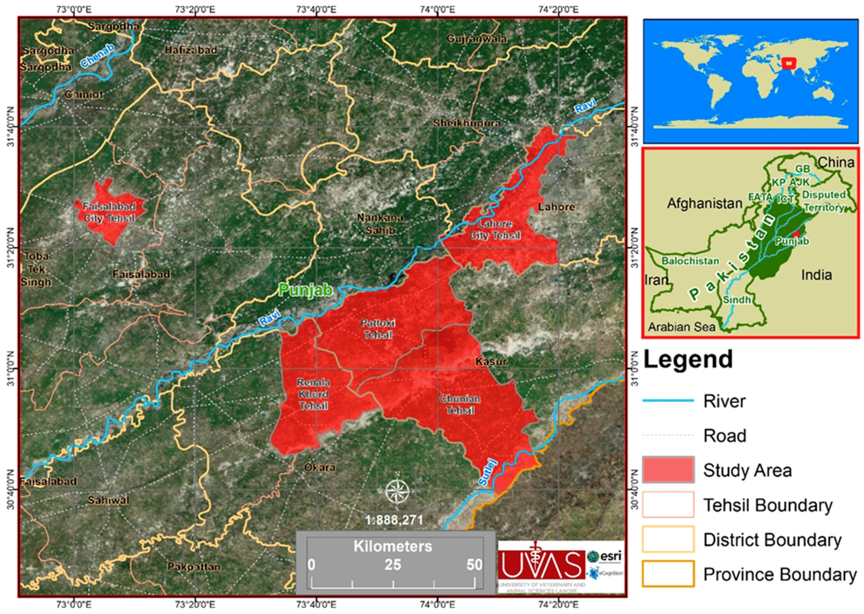
Figure 1. Study area map consisting of five tehsils (Pattoki, Chunian, Renala Khurd, Lahore, and Faisalabad) and four districts (Kasur, Lahore, Okara, and Faisalabad).
Microorganisms 07 00449 g001
Table 1. Primers and probes for the Brucella genus and the species-specific quantitative real-time polymerase chain reaction (qRT-PCR).
Primers and ProbesBrucella genusBrucella abortusBrucella melitensis
Forward 5′GCTCGGTTGCCAATATCAATGC3′5′GCGGCTTTTCTATCACGGTATTC3′5′AACAAGCGGCACCCCTAAAA3′
Reverse5′GGGTAAAGCGTCGCCAGAAG3′5′CATGCGCTATGATCTGGTTACG3′5′CATGCGCTATGATCTGGTTACG3′
Probe5′AAATCTTCCACCTTGCCCCTTGCCATCA3′5′CGCTCATGCTCGCCAGACTTCAATG3′5′CAGGAGTGTTTCGGCTCAGAATAATCCACA3′
5′Flourophore/3′ Quencher6-Fam/BHQ_1HEX/BHQ_1Cy5/BHQ_2
Target Genebcsp31IS711 element downstream of the alkBIS711 element downstream of BMEI1162
Table 2. District and tehsil-wise seroprevalence of brucellosis.
VariablesCategoriesSample ExaminedSample Positive (%)Statistical Analysis
Chi Square ValueP-Value
DistrictFaisalabad15017 (11.3)39.9470.000
Okara1697 (4.1)
Lahore1523 (2.0)
Kasur6128 (1.3)
TehsilChunian122 (16.7)49.1810.000
Faisalabad15017 (11.3)
Renala Khurd1697 (4.1)
Lahore1523 (2.0)
Pattoki6006(1.0)
Table 3. Risk factors associated with ovine, caprine, and bovine brucellosis in the four districts of Punjab, Pakistan, on the basis of Chi square analysis.
VariablesFactorsSample ExaminedPositive for RBPT (%)Statistical Analysis
Chi Square ValueP-Value
SpeciesBuffalo23412 (5.1)5.8130.121
Cow2068 (3.8)
Sheep2037 (3.4)
Goat4408 (1.8)
GenderMale14811 (7.4)9.6730.000
Female93524 (2.5)
BreedNili Ravi23412 (5.1)5.8150.213
Rojhan2068 (3.8)
Kajli2037 (3.4)
Teddy521 (1.9)
Beetle3887 (1.8)
AgeAdult106535 (3.3)0.6110.434
Young180 (0)
Herd size
(n = 149)
≤10965 (5.2)13.8030.000
>105314 (26.4)
Abortion historyNo95023 (2.4)16.2580.000
Yes13312 (9.0)
Have their own SireNo36820 (5.4)8.6500.000
Yes71515 (2.1)
Cleaning up the coral in herd (n = 149)No373 (8.1)0.9540.329
Yes11216 (14.2)
Body conditionWeak200 (0)14.3040.001
Medium86120 (2.3)
Healthy20215 (7.4)
Stock replacement in herd (n = 149)Self-reared13013 (10.0)6.9380.008
Purchased196 (31.5)
Table 4. Final model with associated risk factors for animal brucellosis-binary logistic regression analysis.
VariablesBinary Logistic RegressionP-Value
OR(95% CI)
LowerUpper
District1.4840.9872.2310.058
Tehsil1.4070.9762.0290.068
Herd Size0.6390.1472.7710.550
Species0.8710.5181.4650.604
Gender0.9180.2862.9540.886
Breed1.1130.7881.5740.543
Age21763518.140.000 0.999
Abortion history4.6031.85311.4300.001
Have their own sire0.2300.0860.6120.003
Cleaning up the coral0.4690.0942.3380.356
Body condition1.4400.9502.1810.085
Stock replacement1.5420.3097.6850.597
OR: Odds ratio.
Table 5. Seroprevalence and molecular detection of Brucella DNA in tested serum samples.
Case No. HostSerological AssayPCR
RBPTBrucella GenusB. abortusB. melitensis
94Goat+++
160Goat+++
167Goat+++
192Goat+++
198Sheep+++
278Goat+++
309Goat+++
315Goat+++
316Goat+++
319Sheep+++
320Sheep+++
322Sheep+++
324Buffalo+++
334Buffalo+++
342Cow+++
348Cow+++
350Cow+++
440Sheep+++
456Buffalo+++
463Buffalo+++
470Buffalo+++
473Buffalo+++
479Cow+++
489Cow+++
491Cow+++
499Cow+++
503Buffalo+++
504Buffalo+++
506Buffalo+++
513Buffalo+++
517Buffalo+++
553Buffalo+++
569Cow+++
578Sheep+++
580Sheep+++