Article

Targeting the 3C Protease of Hepatitis A Virus Subgenotype IB: Virtual Screening and Identification of Potent Lead Candidates


by
Tatsuo Kanda
1,*,
Reina Sasaki-Tanaka
2,
Hiroaki Okamoto
3,
Shuji Terai
2,
Cole D. Cwiklowski
4 and
Kalyan C. Nagulapalli Venkata
4,5,*
1
Division of Gastroenterology and Hepatology, Uonuma Institute of Community Medicine, Niigata University Medical and Dental Hospital, 4132 Urasa, Minamiuonuma 949-7302, Japan
2
Division of Gastroenterology and Hepatology, Niigata University Graduate School of Medicine, Dentistry and Health Sciences (Medicine), Niigata University, Niigata 951-8510, Japan
3
Division of Virology, Department of Infection and Immunity, Jichi Medical University School of Medicine, Shimotsuke 329-0498, Japan
4
Department of Medicinal Chemistry, University of Health Sciences and Pharmacy, St. Louis, MO 63010, USA
5
Department of Pharmaceutical and Administrative Sciences, Saint Louis College of Pharmacy, University of Health Sciences and Pharmacy, St. Louis, MO 63010, USA
*
Authors to whom correspondence should be addressed.
Microorganisms 2026, 14(9), 1987; https://doi.org/10.3390/microorganisms14091987
Submission received: 14 August 2026 / Revised: 3 September 2026 / Accepted: 6 September 2026 / Published: 8 September 2026
(This article belongs to the Special Issue Infective Liver Diseases)

Abstract

Hepatitis A virus (HAV) infection remains a global public health concern in both developing and developed countries. In the present study, we identified anti-HAV drugs, using AutoDock Vina Modeling software, and evaluated the compounds in vitro. Following cytotoxicity for Huh7 cells, 5 out of 10 compounds were selected. We evaluated effective HAV 3C protease inhibitors with activity against both HAV genotype IB HM175/18f and HAV genotype IIIA HA11-1299-infected human hepatoma cells. Among the five compounds, we identified only one (KCN-A-12), which had an inhibitory effect on both HAV genotype IB HM175/18f and HAV genotype IIIA HA11-1299 replication in human hepatoma Huh7 cells. KCN-A-12 has more effective inhibitory effects on the replication of HAV genotype IB HM175/18f than that of HAV genotype IIIA HA11-1299. This difference may be attributable to the fact that our discovery system depends on crystal structures from HAV 3C protease based on the HAV genotype IB HM175 strain. In conclusion, we observed that KCN-A-12 was able to inhibit HAV replication. In silico screening for HAV 3C protease inhibitors may be useful for further discovery of anti-HAV drugs.

1. Introduction

In developing and developed countries, hepatitis A virus (HAV) infection is a life-threatening disease [1]. In 2021, the estimated global incidence of and deaths from hepatitis A were 56,100,000 and 27,400 persons/year, respectively; the global incidence of and deaths from foodborne hepatitis A were 20,000,000 and 11,900 persons/year, respectively. The proportion of illnesses that are foodborne is about 0.356 [1]. We also reported a hepatitis A outbreak associated with a revolving sushi bar in Japan [2]. Thus, HAV infection is a serious foodborne non-diarrheal enteric disease.
Recently, HAV infection has been linked to “person-to-person contact” or sexually transmitted diseases [3]. In Japan, a male-dominant hepatitis A outbreak has been observed among men who have sex with men (MSM), both among those who have been infected with HIV and among those who have not [4]. MSM have a high risk for HAV infection [5].
To prevent HAV infection, HAV vaccines should be administered. In particular, travelers who go to areas endemic for hepatitis A should be vaccinated [6]. In Korea, similarly to the situation in Japan, young adults seem not to have a protective antibody against HAV, although a universal vaccination program for newborns has been underway since 2015 [7]. A total of 10,083 confirmed cases were reported in seven months in 2019 according to the Korean Centers for Disease Control and Prevention (KCDC). Therefore, it is important to develop anti-HAV drugs and to disseminate HAV vaccines.
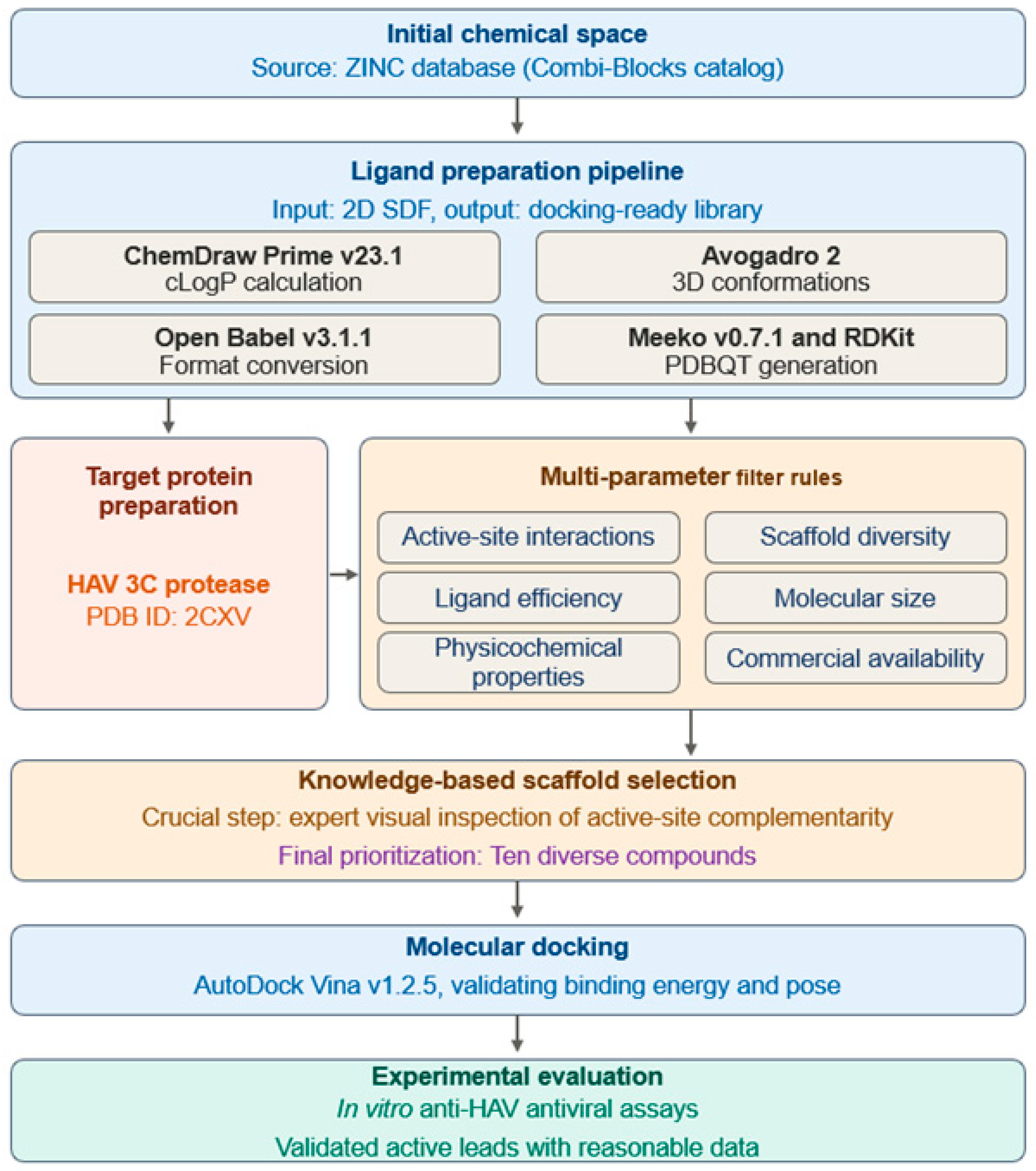
We previously reported the in silico screening of anti-HAV compounds targeting the 3C protease enzyme using the Schrödinger Modeling software from an antiviral library of 25,000 compounds and identified HAV 3C protease inhibitor Z10325150 [8]. Artificial intelligence could identify anti-HAV drugs more effectively [8,9]. In the present study, we developed anti-HAV drugs, using the AutoDock Vina Modeling software, and evaluated them in vitro. Compared to our previous study [8], this work broadened both the chemical space and screening strategy. Instead of relying mainly on docking scores from an antiviral-focused Enamine library, we screened a larger and more diverse ZINC-based library [10]. We prioritized candidates using a multiparameter approach that considered active-site interactions, ligand efficiency, scaffold diversity, physicochemical properties, molecular size, and commercial availability.

2. Materials and Methods

2.1. Software, Database, and Molecular Docking

AutoDock Vina v1.2.5 [11] was used to perform molecular docking. The crystal structure of hepatitis A virus (HAV) 3C protease was obtained from the Protein Data Bank (PDB ID: 2CXV) [12]. A virtual screening library comprising commercially available small molecules was compiled from the ZINC database and commercially sourced from Combi-Blocks (San Diego, CA, USA). Chemical structures were converted to three-dimensional conformations with Avogadro 2 and further processed using Open Babel v3.1.1. Ligand preparation was performed using Meeko v0.7.1 and RDKit v2025.03.6 to generate docking-ready structures in PDBQT format. Lipophilicity values (cLogP) were calculated using ChemDraw Prime v23.1.1.3.

2.2. Receptor Preparation

The search grid was positioned over the substrate-binding groove between the two β-barrel domains of the protease, encompassing the catalytic residues His44, Asp84, and Cys172, the oxyanion-hole region, and adjacent substrate-recognition residues. The grid box was centered at x = −1.344, y = −4.165, and z = 31.408, with dimensions of 22 × 22 × 22 Å. Docking utilized an exhaustiveness value of 32, retained 20 poses per ligand, and applied an energy range of 4 kcal mol−1. All ligands were docked against the same receptor using identical grid coordinates, box dimensions, and search parameters. For each compound, the most favorable Vina score among the generated poses was recorded. Batch docking was automated using a custom Python v2.7.11. script that processed the ligand set sequentially under these conditions.

2.3. Selection of Compounds for Antiviral Activity Evaluation

We commercially sourced prioritized compounds from Combi-Blocks (San Diego, CA, USA) and carried them forward for biological evaluation using a multiparameter approach rather than relying solely on docking scores (Figure 1).
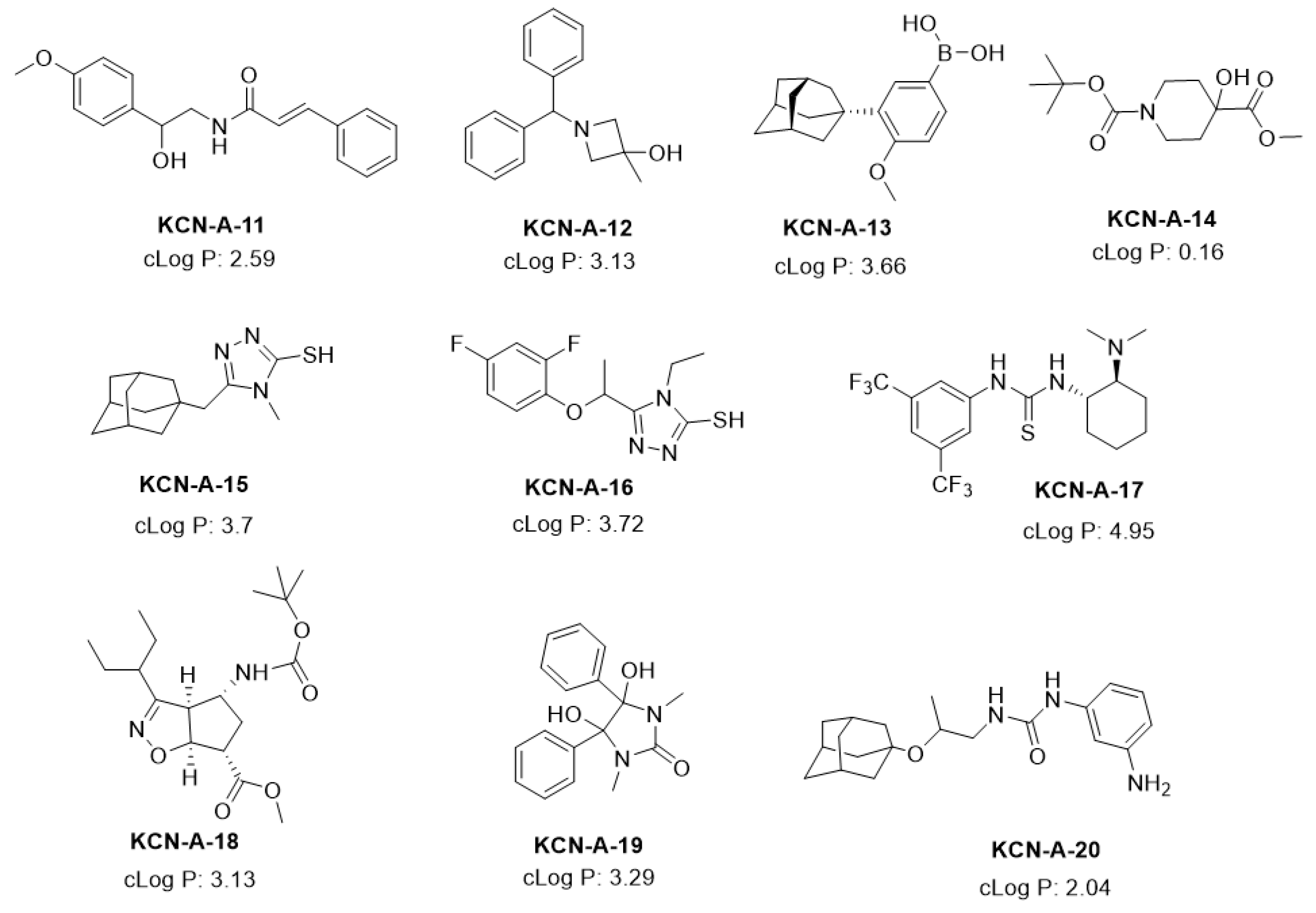
Selection criteria included the ability of each ligand to adopt a plausible orientation within the catalytic region; predicted interactions with His44, Asp84, Cys172, the oxyanion-hole region, and neighboring residues; scaffold diversity; calculated physicochemical properties such as cLogP; and commercial availability. Ten compounds, designated KCN-A-11 to KCN-A-20 (Figure 2), were procured and assessed for anti-HAV activity and cytotoxicity.

2.4. Analysis of Predicted Binding Modes and Ligand Efficiency

Docked complexes were analyzed and superimposed using UCSF ChimeraX v1.12rc. Protein–ligand contacts were characterized with BIOVIA Discovery Studio Visualizer 2025 (v25.1.0.24284) to assess hydrogen bonding, hydrophobic and π–alkyl contacts, π–sulfur interactions, and van der Waals contacts. To account for the influence of molecular size on empirical docking scores, ligand efficiency (LE) was calculated for each compound as LE = −S/N, where S represents the best AutoDock Vina score and N is the number of non-hydrogen atoms [13], enabling size-independent comparison across the series.
Figure 2. Chemical structures of KCN-A-11 to KCN-A-20 selected for antiviral activity evaluation.
Microorganisms 14 01987 g002

2.5. Cell Lines and Reagents

Human hepatoma cell line Huh7, which was kindly provided by Prof. Bartenschlager [14], was kept in Roswell Park Memorial Institute medium (RPMI; Sigma-Aldrich, St. Louis, MO, USA) containing 10% heat-inactivated fetal bovine serum (FBS; Sigma-Aldrich), 100 units/mL penicillin, and 100 μg/mL streptomycin (Sigma-Aldrich) under a 5% CO2 atmosphere at 37 °C. HAV HM175 18f genotype IB and HAV HA11-1299 genotype IIIA were used for HAV infection in the present study [15]. HAV HM175/18f genotype IB was provided by Prof. Stanley M. Lemon (University of North Carolina at Chapel Hill, Chapel Hill, NC, USA) and Dr. Asuka Hirai-Yuki (National Institute of Infectious Diseases, Tokyo, Japan).

2.6. Cell Viability Assays

For the evaluation of cell viability, dimethylthiazol carboxymethoxyphenyl sulfophenyl tetrazolium (MTS) assays were performed using the CellTiter 96 Aqueous One-Solution cell proliferation assay (Promega, Madison, WI, USA). The MTS assay was performed on a Bio-Rad iMark microplate reader (Bio-Rad, Hercules, CA, USA) at a 490 nm wavelength [15]. Trypan blue dye exclusion assays were also performed with 0.4% trypan blue solution (Sigma-Aldrich), and both live and dead cells were counted under phase contrast microscopy (CKX53, Olympus, Tokyo, Japan) [16].

2.7. Half-Maximal Inhibitory Concentration (IC50)

IC50 indicates the concentration of each compound that produces 50% of the maximal inhibitory effects on HAV replication. IC50 was calculated using a previously described formula [17].

2.8. Infection of Huh7 Cells with HAV Genotype IB HM175/18f or HAV Genotype IIIA HA11-1299

HAV genotype IB HM175/18f and HAV genotype IIIA HA11-1299 were used for HAV infection in the present study. Before 24 h of infection, Huh7 cells were seeded at a density of 5 × 10⁵ cells/well on 6-well plates (VIOLAMO, AS ONE, Osaka, Japan). Cells were washed twice with phosphate-buffered saline (PBS) (FUJIFILM Wako Chemicals, Osaka, Japan) and infected with HAV genotype IB HM175/18f or HAV genotype IIIA HA11-1299 at a multiplicity of infection (MOI) of 0.01 in serum-free medium. HAV inoculum was incubated with hepatocytes for 6 h, and we added 1 mL of medium containing 2% FBS. After 24 h of incubation, cells were washed once with PBS, followed by the addition of 1 mL of RPMI containing 5% FBS [15]. Then, several concentrations of compounds were added to HAV-infected Huh7 cells according to the results of cell viability assays. After 72 h of infection, cellular RNA was extracted using the RNeasy Mini Kit (Qiagen, Hilden, Germany), and HAV RNA levels were determined using real-time RT-PCR

2.9. Extraction of Cellular RNA, cDNA Synthesis, and Real-Time RT-PCR

Total cellular RNA was extracted by the QIAshredder (Qiagen) and RNeasy Mini Kit (Qiagen), according to the manufacturer’s instructions. Then, cDNA synthesis was performed using PrimeScript RT reagent (Perfect Real Time, TaKaRa Bio, Kusatsu, Shiga, Japan) at 37 °C for 15 min, followed by inactivation at 85 °C for 5 s and incubation at 4 °C. Real-time PCR was performed on a StepOnePlus real-time PCR system (Applied Biosystems, Tokyo, Japan), using Power SYBR Green PCR Master Mix (Applied Biosystems). The PCR conditions were as follows: 95 °C for 10 min, followed by 40 cycles of 95 °C for 15 s and 60 °C for 1 min. Real-time PCR was performed in triplicate. These data were analyzed by the ddCt method. For the quantitation of HAV RNA or β-actin mRNA, the following oligo primers were used: sense primer 5′-AGGCTACGGGTGAAACCTCTTAG-3′ and antisense primer 5′-GCCGCTGTTACCCTATCCAA-3′ for the former and sense primer 5′-CAGCCATGTACGTTGCTATCCAGG-3′ and antisense primer 5′-AGGTCCAGACGCAGGATGGCATG-3′ for the latter [15].

2.10. Statistical Analysis

Data are expressed as the means ± standard deviations (SDs). Statistical analyses were performed with Student’s t-test. p-values < 0.05 were considered significant. All assays were performed in at least triplicate. Statistical analysis was performed using DA Stats software (O. Nagata, Nifty Serve: PAF01644).

3. Results

3.1. Docking of the Selected Compound Series

The ten prioritized compounds (Figure 2) were docked into the catalytic region of the HAV 3C protease under identical conditions, and all compounds were accommodated within the targeted site. The best predicted Vina scores ranged from −7.612 to −5.752 kcal mol−1 (Table 1). Molecular weights ranged from 253.35 to 413.42 g mol−1, and the compounds contained 18 to 27 heavy atoms. The calculated physicochemical properties indicated general consistency with conventional Lipinski thresholds; however, KCN-A-13 exhibited a cLogP of 5.59, which is slightly above the conventional lipophilicity threshold of 5. The ligand efficiency (LE) values clustered within a relatively narrow range of 0.247 to 0.328 kcal mol−1 per heavy atom. No direct correlation was observed between the predicted docking scores and antiviral activity. Notably, KCN-A-12 demonstrated the strongest anti-HAV activity despite a comparatively modest Vina score of −5.835 kcal mol−1. These findings support the use of docking primarily to evaluate plausible active-site binding modes rather than to quantitatively predict cellular antiviral potency.

3.2. Effects of 10 Compounds on Survival of Huh7 Cells

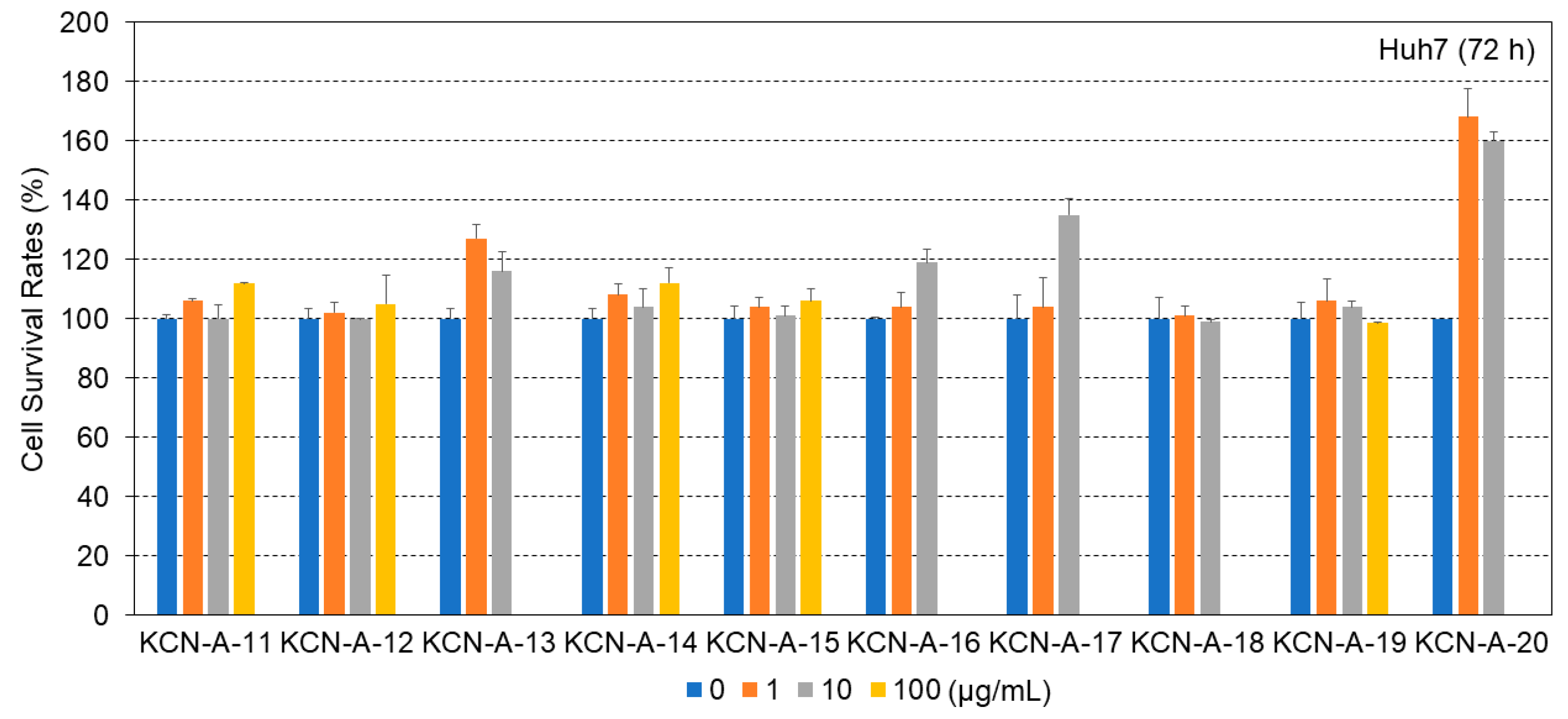
First, before we examined the effects of these 10 compounds on HAV replication in human hepatoma Huh7 cells, we studied their effects on the induction of cell death in Huh7 cells. The sensitivities of Huh7 cells to these 10 compounds were monitored for 72 h. It was shown that 100 μg of five compounds (KCN-A-13, KCN-A-16, KCN-A-17, KCN-A-18, and KCN-A-20) markedly reduced Huh7 cell viability, and that 100 μg of the other five compounds (KCN-A-11, KCN-A-12, KCN-A-14, KCN-A-15, and KCN-A-19) did not increase susceptibility in Huh7 cells (Figure 3). The half-maximal inhibitory concentration of five compounds (KCN-A-13, KCN-A-16, KCN-A-17, KCN-A-18, and KCN-A-20) on the Huh7 cells was < 100 μg/mL; and that of the other five compounds (KCN-A-11, KCN-A-12, KCN-A-14, KCN-A-15, and KCN-A-19) was >100 μg/mL. Therefore, we chose a concentration of 100 μg of these five compounds for experiments to screen the inhibition of HAV replication.

3.3. Effects of Five Compounds on HAV Genotype IB HM175/18f Replication in Huh7 Cells

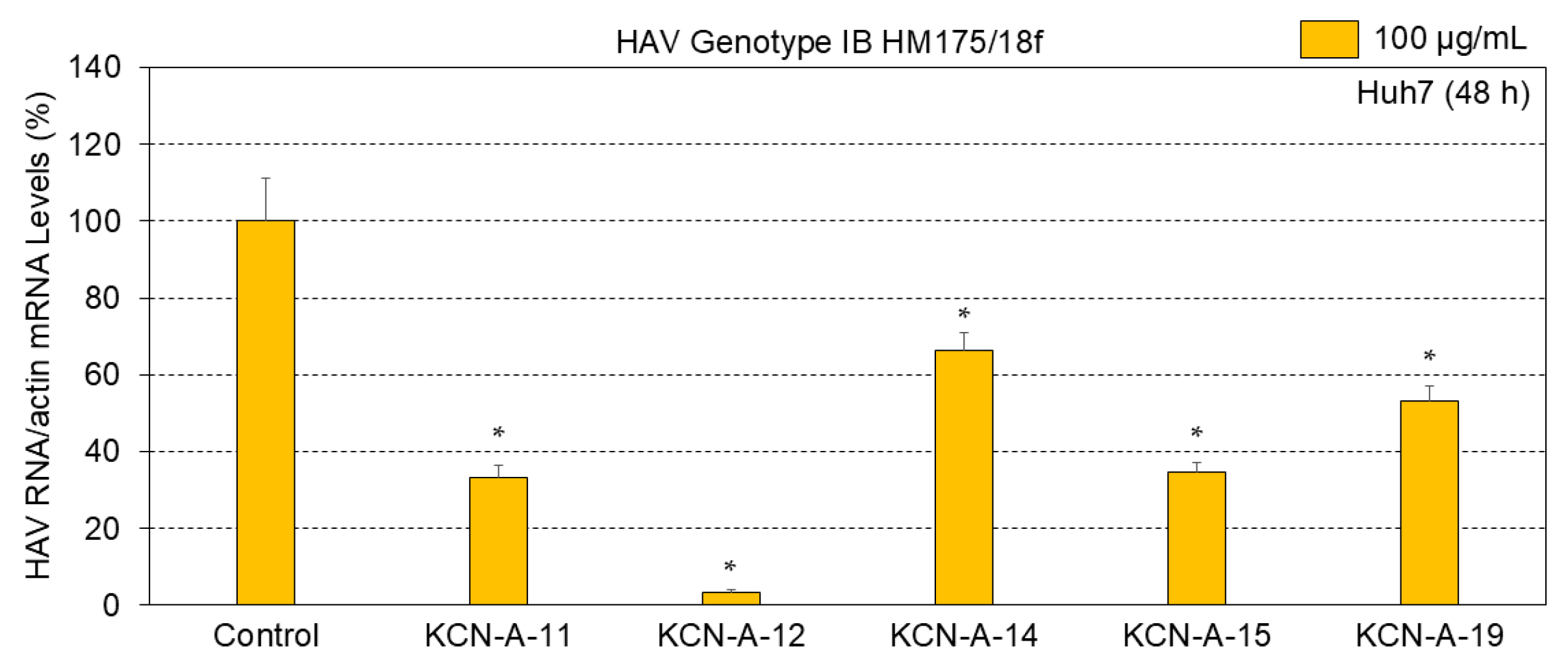
After 24 h of HAV genotype IB HM175/18f infection, Huh7 cells were incubated with 100 μg of each compound—KCN-A-11, KCN-A-12, KCN-A-14, KCN-A-15, or KCN-A-19—for 48 h. After 72 h of HAV genotype IB HM175/18f infection, cellular RNA was extracted, and HAV RNA levels were determined using real-time RT-PCR. Five compounds (KCN-A-11, KCN-A-12, KCN-A-14, KCN-A-15, and KCN-A-19) could significantly inhibit HAV genotype IB HM175/18f replication (each: n = 3, p < 0.05). Among these five compounds, KCN-A-12 treatment resulted in 3.4% of HAV genotype IB HM175/18f replication in Huh7 cells treated with the control (Figure 4). The IC50 of KCN-A-12 is 37.5 μg/mL.
When the selectivity index (SI) was calculated for each compound using the formula of the half-maximal inhibitory concentration of each compound on the Huh7 cells/IC50 of each compound for HAV genotype IB HM175/18f replication, all five compounds (KCN-A-11, KCN-A-12, KCN-A-14, KCN-A-15, or KCN-A-19) showed greater efficacy against HAV genotype IB HM175/18f replication than the cytotoxicity against Huh7 cells (favorable SI > 1.0) (Figure 3 and Figure 4).

3.4. Effects of KCN-A-12 on HAV Genotype IIIA HA11-1299 Replication in Huh7 Cells

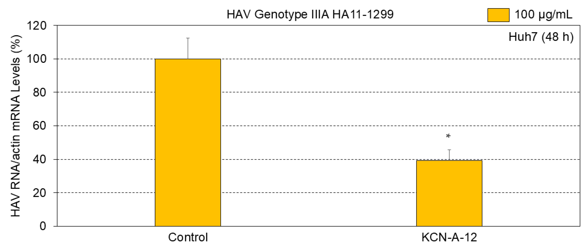
We next examined the effects of KCN-A-12 on HAV genotype IIIA HA11-1299 replication in Huh7 cells. Similarly, after 24 h of HAV genotype IIIA HA11-1299 infection, Huh7 cells were incubated with 100 μg of KCN-A-12 for 48 h. After 72 h of HAV genotype IIIA HA11-1299 infection, cellular RNA was extracted, and HAV RNA levels were determined using real-time RT-PCR. KCN-A-12 was able to significantly inhibit HAV genotype IIIA HA11-1299 replication compared to the control (Figure 5). The KCN-A-12 with efficiency against HAV genotype IIIA HA11-1299 replication greater than the cytotoxicity against Huh7 cells (favorable SI > 1.0) was indicated (Figure 3 and Figure 5). Thus, KCN-A-12 significantly inhibited the replication of both HAV genotype IB HM175/18f and HAV genotype IIIA HA11-1299 in Huh7 cells.

3.5. Predicted Binding Mode of the Lead Compound KCN-A-12

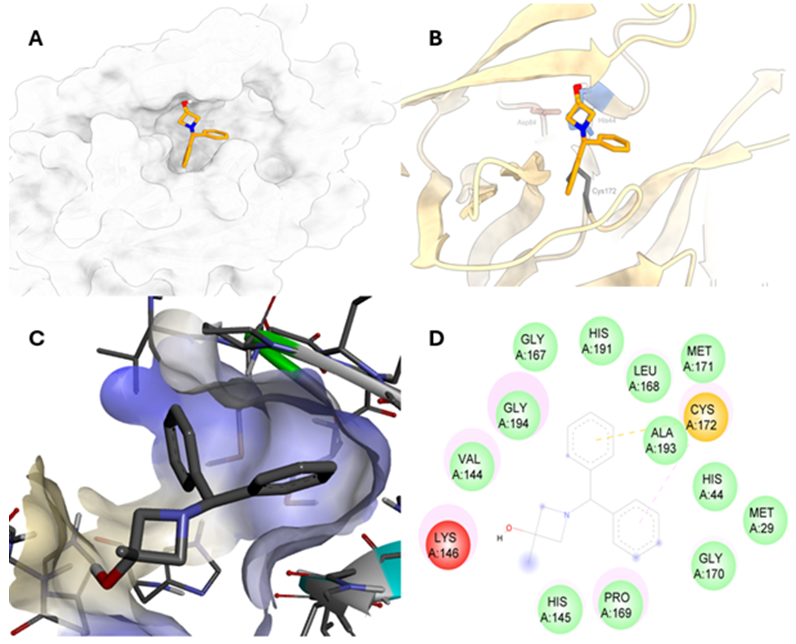
Prediction models binding between KCN-A-12 and the HAV 3C protease active site are shown in Figure 6. KCN-A-12, which exhibited the strongest anti-HAV activity, was predicted to bind within the proteolytic groove rather than at a peripheral surface site (Figure 6A,C). The ligand occupies a substantial proportion of the substrate-binding cavity, indicating favorable shape complementarity, and is positioned adjacent to the catalytic residues. One aromatic ring is oriented towards Cys172 and His44 (Figure 6B).
Interaction analysis identified a predicted π–sulfur contact between an aromatic ring of KCN-A-12 and the sulfur atom of the catalytic Cys172. Additional hydrophobic and π–alkyl contacts involve Val144, Lys146, Pro169, Ala193, and Gly194. The binding site is further lined by van der Waals contacts with Met29, His44, His145, Gly167, Leu168, Gly170, Met171, and His191 (Figure 6D). Contacts extending towards the Gly170–Met171–Cys172 segment position the ligand near the oxyanion-hole region. The proximity to His191 orients part of the scaffold towards the S1 specificity pocket, which is associated with the enzyme’s preference for glutamine at the P1 position.
The predicted pose is dominated by hydrophobic and van der Waals contacts. No conventional hydrogen bond is formed between the azetidinol hydroxyl group and active-site residues in the selected pose. Overall, the predicted binding mode is consistent with steric occlusion of the substrate-binding groove and interference with substrate access to the catalytic machinery.

4. Discussion

HAV infections are still associated with morbidity and mortality, despite the fact that HAV vaccines are widely administered for their prevention [5]. To identify novel inhibitors of HAV 3C protease [18,19,20], an in silico study was performed, and its anti-HAV efficacies were examined in vitro. First, after the examination of the cytotoxicity of Huh7 cells, 5 out of 10 compounds were selected. Second, after examining the effects on HAV genotype IB HM175/18f replication in Huh7 cells, among the five compounds, KCN-A-12 was identified as having an inhibitory effect < 10%. Third, after examining the effects on HAV genotype IIIA HA11-1299 replication in Huh7 cells, we also identified KCN-A-12 as having an inhibitory effect.
Because most of the crystal structures of HAV 3C protease were based on the HAV genotype IB HM175 strain [18,19,20,21], we initially examined the effects on HAV genotype IB HM175/18f replication in Huh7 cells. Compared to the control (100%), a treatment of 100 μg/mL KCN-A-12 for 48 h reduced HAV genotype IB HM175/18f replication in Huh7 cells to 3.4%. This suggests that KCN-A-12 had a stronger inhibitory effect on HAV genotype IB HM175/18f replication in Huh7 cells.
A previous study demonstrated that Z10325150 could inhibit HAV genotype IIIA HA11-1299 replication [8]. Compared to the control (100%), a treatment of 100 μg/mL Z10325150 for 72 h reduced HAV genotype IIIA HA11-1299 replication to 69.6% [8]. In the present study, compared to the control (100%), a treatment of 100 μg/mL KCN-A-12 for 48 h reduced HAV genotype IIIA HA11-1299 replication in Huh7 cells to 39.3%, suggesting that KCN-A-12 seems to have an inhibitory effect on HAV genotype IIIA HA11-1299 replication in Huh7 cells.
Of interest, it seems that KCN-A-12 has a stronger inhibitory effect on the replication of HAV genotype IB HM175/18f than that of HAV genotype IIIA HA11-1299 in Huh7 cells. The nucleotide length of the HAV 3C coding region from HAV genotype IB HM175/18f [22] is shorter than that from HAV genotype IIIA HA11-1299 (660 nt. vs. 657 nt.). Compared nucleotide substitutions between HAV genotype IB HM175/18f and HAV genotype IIIA HA11-1299, those are 16% (105/657) and 15.9% (105/660). Thus, as a difference in nucleotide sequences of the HAV 3C protease coding region exists between the two strains, structural comparison of HAV 3C proteases will be needed between HAV genotype IB HM175/18f and HAV genotype IIIA HA11-1299.
The primary screening experiments were performed at 100 μg/mL. This appears to be a relatively high concentration. The 100 μg/mL of KCN-A-11, KCN-A-12, KCN-A-13, KCN-A-14, KCN-A-15, KCN-A-16, KCN-A-17, KCN-A-18, KCN-A-19, and KCN-A-20 are equal to 336 μM, 394 μM, 349 μM, 385 μM, 379 μM, 350 μM, 241 μM, 282 μM, 335 μM, and 291 μM, respectively. The IC50 of KCN-A-12 is 148 μM.
MTS assays might measure metabolic activity rather than cell death directly. When trypan blue dye exclusion assays were also performed [16], the viabilities of Huh7 cells were 95.3 ± 0.5, 95.3 ± 0.5, 94 ± 1.4, 94.6 ± 0.5, 82.3 ± 2.0, and 45.3 ± 5.2 after 72 h of treatment of KCN-A-12 at 0, 1, 10, 100, 150, and 200 μg/mL, respectively. The 1–100 μg/mL KCN-A-12 did not significantly reduce the viabilities of Huh7 cells. The half-maximal inhibitory concentration of KCN-A-12 on the Huh7 cells was 192 μg/mL (759 μM). When the SI was calculated for each compound using the formula for the half-maximal inhibitory concentration of KCN-A-12 on the Huh7 cells by trypan blue dye exclusion assay/IC50 of KCN-A-12 for HAV genotype IB HM175/18f replication, KCN-A-12 also indicated a drug with greater efficacy against HAV genotype IB HM175/18f replication than the cytotoxicity against Huh7 cells (favorable SI 5.12).
Immunity or vaccination against HAV infection is useful as pre-exposure prophylaxis (PrEP) [23]. Postexposure prophylaxis (PEP) with hepatitis A vaccine or immune globulin effectively prevents HAV infection within 2 weeks of exposure [24]. There are no anti-HAV drugs that are useful for PrEP or PEP for HAV infection. From the viewpoint of public health, drug discovery in virology, including in HAV infection, is important [1].
The process of drug discovery for HAV infection starts with the identification of the target (such as HAV 3C protease), followed by target validation, hit discovery, lead optimization, and preclinical/clinical development [25]. In the present study, we targeted HAV 3C protease inhibitors, identified several drugs by in silico screening, and finally found an effective compound, KCN-A-12, which had a stronger inhibitory effect on both HAV genotype IB HM175/18f and HAV genotype IIIA HA11-1299 replication in vitro.
There are several limitations in the present study: (1) we may not measure the reduction in HAV productive replication. Orthogonal validation, such as measurement of extracellular viral RNA/infectious virus and viral protein expression, may be needed; (2) the impact of compounds on viral replication under varying MOI conditions should be assessed; (3) crystal structures of HAV 3C protease based on the HAV genotype IB HM175 strain were used for the in silico study—we may use those derived from other HAV strains. Structural comparison of HAV 3C proteases between HAV genotype IB and HAV genotype IIIA should be needed; (4) our study of KCN-A-12 lacks protease assay, molecular dynamics analysis or in vivo validation, although protease assay may not always be needed [26]; (5) their safety and effects could be confirmed by an in vivo study; and (6) compound KCN-A-12 could be further modified to develop more effective drugs with fewer adverse events.

5. Conclusions

We evaluated more effective HAV 3C protease inhibitors with activity against both HAV genotype IB HM175/18f and HAV genotype IIIA HA11-1299-infected human hepatoma cell lines. In particular, we showed that KCN-A-12 suppressed HAV replication and is predicted to inhibit the protease activity. In silico screening for HAV 3C protease inhibitors may be useful for further discovery of anti-HAV drugs.

Author Contributions

Conceptualization, T.K., R.S.-T., C.D.C. and K.C.N.V.; methodology, T.K., R.S.-T. and K.C.N.V.; software, T.K., R.S.-T. and K.C.N.V.; validation, T.K., R.S.-T. and K.C.N.V.; formal analysis, T.K. and K.C.N.V.; investigation, T.K. and K.C.N.V.; resources, T.K., C.D.C. and K.C.N.V.; data curation, T.K. and K.C.N.V.; writing—original draft preparation, T.K., R.S.-T. and K.C.N.V.; writing—review and editing, T.K., R.S.-T., H.O., S.T., C.D.C. and K.C.N.V.; visualization, T.K.; supervision, H.O. and S.T.; project administration, T.K., R.S.-T. and K.C.N.V.; funding acquisition, T.K. and K.C.N.V. All authors have read and agreed to the published version of the manuscript.

Funding

This research was funded by the Japan Agency for Medical Research and Development (AMED), grant number JP25fk0210132 (T.K.), and JP26fk0210198 (T.K.). Faculty Research Incentive Fund (FRIF-20-134) of University of Health Sciences and Pharmacy, St. Louis, MO-63011 (K.C.N.V.).

Institutional Review Board Statement

Not applicable.

Informed Consent Statement

Not applicable.

Data Availability Statement

The original contributions presented in this study are included in the article. Further inquiries can be directed to the corresponding authors.

Acknowledgments

We would like to thank Ratna Ray (Saint Louis University, MO, USA), Ranjit Ray (Saint Louis University, MO, USA), Ralf Bartenschlager (Heidelberg University, Heidelberg, Germany), Verena Gauss-Müller (University of Lübeck, Lübeck, Germany), and Stanley M. Lemon (The University of North Carolina at Chapel Hill, Chapel Hill, NC, USA) for generously providing the cell lines, plasmids, and HAV HM175-18f.

Conflicts of Interest

The authors declare no conflicts of interest.

Abbreviations

The following abbreviations are used in this manuscript:
HAVHepatitis A virus
MSMMen who have sex with men
KCDCKorean Centers for Disease Control and Prevention
RPMIRoswell Park Memorial Institute medium
FBSFetal bovine serum
PBSPhosphate-buffered saline
MOIMultiplicity of infection
MTSDimethylthiazol carboxymethoxyphenyl sulfophenyl tetrazolium
IC50Half-maximal inhibitory concentration
SDsStandard deviations
PrEPPre-exposure prophylaxis
PEPPostexposure prophylaxis

References

  1. Majowicz, S.E.; Scallan Walter, E.; Pires, S.M.; Minato, Y.; Founou, L.L.; Devleesschauwer, B.; di Bari, C.; Havelaar, A.H.; Vaes, L.; Estrada-Garcia, T.; et al. WHO estimates of the global, regional, and national burden of eight foodborne non-diarrhoeal enteric disease hazards, 2000–2021: An updated data synthesis. Lancet Glob. Health 2026, 14, 103981. [Google Scholar] [CrossRef] [Scilit] [PubMed]
  2. Tominaga, A.; Kanda, T.; Akiike, T.; Komoda, H.; Ito, K.; Abe, A.; Aruga, A.; Kaneda, S.; Saito, M.; Kiyohara, T.; et al. Hepatitis A outbreak associated with a revolving sushi bar in Chiba, Japan: Application of molecular epidemiology. Hepatol. Res. 2012, 42, 828–834. [Google Scholar] [CrossRef] [Scilit] [PubMed]
  3. Lin, K.Y.; Huang, Y.C.; Huang, M.H.; Tsai, T.Y.; Chen, G.J.; Huang, Y.S.; Huang, S.H.; Sun, H.Y.; Hung, C.C. Hepatitis A to E in People with HIV: A Virus-Specific Review of Prevention and Treatment. Infect. Dis. Ther. 2026, 15, 391–415. [Google Scholar] [CrossRef] [Scilit] [PubMed]
  4. Honda, M.; Asakura, H.; Kanda, T.; Somura, Y.; Ishii, T.; Yamana, Y.; Kaneko, T.; Mizutani, T.; Takahashi, H.; Kumagawa, M.; et al. Male-Dominant Hepatitis A Outbreak Observed among Non-HIV-Infected Persons in the Northern Part of Tokyo, Japan. Viruses 2021, 13, 207. [Google Scholar] [CrossRef] [Scilit] [PubMed]
  5. Shiferaw, W.; Martin, B.M.; Dean, J.A.; Mills, D.; Lau, C.; Paterson, D.; Koh, K.; Eriksson, L.; Furuya-Kanamori, L. A systematic review and meta-analysis of sexually transmitted infections and blood-borne viruses in travellers. J. Travel. Med. 2024, 31, taae038. [Google Scholar] [CrossRef] [Scilit] [PubMed]
  6. Kanda, T.; Sasaki-Tanaka, R.; Ishii, K.; Suzuki, R.; Inoue, J.; Tsuchiya, A.; Nakamoto, S.; Abe, R.; Fujiwara, K.; Yokosuka, O.; et al. Recent advances in hepatitis A virus research and clinical practice guidelines for hepatitis A virus infection in Japan. Hepatol. Res. 2024, 54, 4–23. [Google Scholar] [CrossRef] [Scilit] [PubMed]
  7. Kang, S.H.; Kim, M.Y.; Baik, S.K. Perspectives on Acute Hepatitis A Control in Korea. J. Korean Med. Sci. 2019, 34, e230. [Google Scholar] [CrossRef] [Scilit] [PubMed]
  8. Sasaki-Tanaka, R.; Nagulapalli Venkata, K.C.; Okamoto, H.; Moriyama, M.; Kanda, T. Evaluation of Potential Anti-Hepatitis A Virus 3C Protease Inhibitors Using Molecular Docking. Int. J. Mol. Sci. 2022, 23, 6044. [Google Scholar] [CrossRef] [Scilit] [PubMed]
  9. Kanda, T.; Sasaki, R.; Masuzaki, R.; Moriyama, M. Artificial intelligence and machine learning could support drug development for hepatitis A virus internal ribosomal entry sites. Artif. Intell. Gastroenterol. 2021, 2, 1–9. [Google Scholar] [CrossRef] [Scilit]
  10. Tingle, B.I.; Tang, K.G.; Castanon, M.; Gutierrez, J.J.; Khurelbaatar, M.; Dandarchuluun, C.; Moroz, Y.S.; Irwin, J.J. ZINC-22A Free Multi-Billion-Scale Database of Tangible Compounds for Ligand Discovery. J. Chem. Inf. Model. 2023, 63, 1166–1176. [Google Scholar] [CrossRef] [Scilit] [PubMed]
  11. Eberhardt, J.; Santos-Martins, D.; Tillack, A.F.; Forli, S. AutoDock Vina 1.2.0: New Docking Methods, Expanded Force Field, and Python Bindings. J. Chem. Inf. Model. 2021, 61, 3891–3898. [Google Scholar] [CrossRef] [Scilit] [PubMed]
  12. Yin, J.; Bergmann, E.M.; Cherney, M.M.; Lall, M.S.; Jain, R.P.; Vederas, J.C.; James, M.N. Dual modes of modification of hepatitis A virus 3C protease by a serine-derived beta-lactone: Selective crystallization and formation of a functional catalytic triad in the active site. J. Mol. Biol. 2005, 354, 854–871. [Google Scholar] [CrossRef] [Scilit] [PubMed]
  13. Hopkins, A.L.; Keserü, G.M.; Leeson, P.D.; Rees, D.C.; Reynolds, C.H. The role of ligand efficiency metrics in drug discovery. Nat. Rev. Drug Discov. 2014, 13, 105–121. [Google Scholar] [CrossRef] [Scilit] [PubMed]
  14. Lohmann, V.; Körner, F.; Koch, J.; Herian, U.; Theilmann, L.; Bartenschlager, R. Replication of subgenomic hepatitis C virus RNAs in a hepatoma cell line. Science 1999, 285, 110–113. [Google Scholar] [CrossRef] [Scilit] [PubMed]
  15. Kanda, T.; Sasaki-Tanaka, R.; Abe, H.; Yokoo, T.; Sakamaki, A.; Hayashi, K.; Kamimura, H.; Tsuchiya, A.; Masuzaki, R.; Kogure, H.; et al. Zinc Acetate Inhibits Hepatitis A Virus Replication: Possible Treatment for Patients with Type A Acute-on-Chronic Liver Failure. Pathogens 2025, 14, 882. [Google Scholar] [CrossRef] [Scilit] [PubMed]
  16. Strober, W. Trypan Blue Exclusion Test of Cell Viability. Curr. Protoc. Immunol. 2015, 111, A3.B.1–A3.B.3. [Google Scholar] [CrossRef] [Scilit] [PubMed]
  17. Sasaki-Tanaka, R.; Shibata, T.; Okamoto, H.; Moriyama, M.; Kanda, T. Favipiravir Inhibits Hepatitis A Virus Infection in Human Hepatocytes. Int. J. Mol. Sci. 2022, 23, 2631. [Google Scholar] [CrossRef] [Scilit] [PubMed]
  18. Allaire, M.; Chernaia, M.M.; Malcolm, B.A.; James, M.N. Picornaviral 3C cysteine proteinases have a fold similar to chymotrypsin-like serine proteinases. Nature 1994, 369, 72–76. [Google Scholar] [CrossRef] [Scilit] [PubMed]
  19. Bergmann, E.M.; Mosimann, S.C.; Chernaia, M.M.; Malcolm, B.A.; James, M.N. The refined crystal structure of the 3C gene product from hepatitis A virus: Specific proteinase activity and RNA recognition. J. Virol. 1997, 71, 2436–2448. [Google Scholar] [CrossRef] [Scilit] [PubMed]
  20. Bergmann, E.M.; Cherney, M.M.; Mckendrick, J.; Frormann, S.; Luo, C.; Malcolm, B.A.; Vederas, J.C.; James, M.N. Crystal structure of an inhibitor complex of the 3C proteinase from hepatitis A virus (HAV) and implications for the polyprotein processing in HAV. Virology 1999, 265, 153–163. [Google Scholar] [CrossRef] [Scilit] [PubMed]
  21. Cohen, J.I.; Ticehurst, J.R.; Purcell, R.H.; Buckler-White, A.; Baroudy, B.M. Complete nucleotide sequence of wild-type hepatitis A virus: Comparison with different strains of hepatitis A virus and other picornaviruses. J. Virol. 1987, 61, 50–59. [Google Scholar] [CrossRef] [Scilit] [PubMed]
  22. Lemon, S.M.; Murphy, P.C.; Shields, P.A.; Ping, L.H.; Feinstone, S.M.; Cromeans, T.; Jansen, R.W. Antigenic and genetic variation in cytopathic hepatitis A virus variants arising during persistent infection: Evidence for genetic recombination. J. Virol. 1991, 65, 2056–2065. [Google Scholar] [CrossRef] [Scilit] [PubMed]
  23. Le Turnier, P.; Charreau, I.; Gabassi, A.; Carette, D.; Cotte, L.; Pialoux, G.; Tremblay, C.; Spire, B.; Chaix, M.L.; Meyer, L.; et al. Hepatitis A and B vaccine uptake and immunisation among men who have sex with men seeking PrEP: A substudy of the ANRS IPERGAY trial. Sex. Transm. Infect. 2023, 99, 140–142. [Google Scholar] [CrossRef] [Scilit] [PubMed]
  24. Nelson, N.P.; Link-Gelles, R.; Hofmeister, M.G.; Romero, J.R.; Moore, K.L.; Ward, J.W.; Schillie, S.F. Update: Recommendations of the Advisory Committee on Immunization Practices for Use of Hepatitis A Vaccine for Postexposure Prophylaxis and for Preexposure Prophylaxis for International Travel. MMWR Morb. Mortal. Wkly. Rep. 2018, 67, 1216–1220. [Google Scholar] [CrossRef] [Scilit] [PubMed]
  25. Al Khzem, A.H.; Shoaib, T.H.; Mukhtar, R.M.; Alturki, M.S.; Gomaa, M.S.; Hussein, D.; Mostafa, A.; Alrumaihi, L.A.; Alansari, F.A.; Laabei, M. In Silico Lead Identification of Staphylococcus aureus LtaS Inhibitors: A High-Throughput Computational Pipeline Towards Prototype Development. Int. J. Mol. Sci. 2025, 26, 12038. [Google Scholar] [CrossRef] [Scilit] [PubMed]
  26. Huitema, C.; Zhang, J.; Yin, J.; James, M.N.; Vederas, J.C.; Eltis, L.D. Heteroaromatic ester inhibitors of hepatitis A virus 3C proteinase: Evaluation of mode of action. Bioorganic Med. Chem. 2008, 16, 5761–5777. [Google Scholar] [CrossRef] [Scilit] [PubMed]
Figure 1. Multi-parameter approach to in silico molecular modeling.
Microorganisms 14 01987 g001
Figure 3. Effects of 10 compounds on cell viabilities of Huh7 cells. Huh7 cells were incubated with each compound for 72 h. Cell viabilities were determined via dimethylthiazol carboxymethox-yphenyl sulfophenyl tetrazolium (MTS) assay. Data are expressed as means and standard deviations of triplicate determinations from three independent experiments. Statistical significance was analyzed using the two-tailed Student’s t-test.
Microorganisms 14 01987 g003
Figure 4. Effects of five compounds on HAV genotype IB HM175/18f replication in Huh7 cells. Cells were seeded before 24 h of HAV genotype IB HM175/18f infection at 0.01 MOI. After 24 h of infection, cells were incubated with each compound at 100 μg/mL for 48 h. Cellular RNA was collected at 72 h after infection, and HAV RNA/actin mRNA levels were measured by real-time RT-PCR. Data are expressed as means and standard deviations of triplicate determinations from three independent experiments. Statistical significance was analyzed using the two-tailed Student’s t-test. * p < 0.05.
Microorganisms 14 01987 g004
Figure 5. Effects of KCN-A-12 on HAV genotype IIIA HA11-1299 replication in Huh7 cells. Cells were seeded before 24 h of HAV genotype IIIA HA11-1299 infection at 0.01 MOI. After 24 h of infection, cells were incubated with each compound at 100 μg/mL for 48 h. Cellular RNA was collected at 72 h after infection, and HAV RNA/actin mRNA levels were measured by real-time RT-PCR. Data are expressed as means and standard deviations of triplicate determinations from three independent experiments. Statistical significance was analyzed using the two-tailed Student’s t-test. * p < 0.05.
Microorganisms 14 01987 g005
Figure 6. Predicted binding mode of KCN-A-12 within the HAV 3C protease active site. (A) Surface view of KCN-A-12 in the substrate-binding pocket. (B) Ribbon view showing its orientation relative to His44, Asp84, and Cys172. (C) Close-up view of KCN-A-12 within the active-site cavity. (D) Two-dimensional interaction map showing catalytic and surrounding pocket residues.
Microorganisms 14 01987 g006
Table 1. Molecular formulas, physicochemical properties, best AutoDock Vina scores, and ligand efficiencies for compounds KCN-A-11 to KCN-A-20 docked against the HAV 3C protease.
CompoundMolecular
Formula
Molecular Weights Heavy Atoms (N) aVina Score
(kcal mol−1)
Ligand
Efficiency (LE) b
KCN-A-11C18H19NO3297.3522−6.8250.310
KCN-A-12C17H19NO253.3519−5.8350.307
KCN-A-13C17H23BO3286.1821−6.8090.324
KCN-A-14C12H21NO5259.3018−5.7520.320
KCN-A-15C14H21N3S263.4018−5.9070.328
KCN-A-16C12H13F2N3OS285.3119−5.9380.313
KCN-A-17C17H21F6N3S413.4227−7.6120.282
KCN-A-18C18H28N2O5354.4525−6.1690.247
KCN-A-19C17H18N2O3298.3422−6.6860.304
KCN-A-20C20H29N3O2343.4725−6.8400.274
a N = number of non-hydrogen (heavy) atoms. b Ligand efficiency, LE = −S/N, reported in kcal mol−1 per heavy atom, S = docking score.
Disclaimer/Publisher’s Note: The statements, opinions and data contained in all publications are solely those of the individual author(s) and contributor(s) and not of MDPI and/or the editor(s). MDPI and/or the editor(s) disclaim responsibility for any injury to people or property resulting from any ideas, methods, instructions or products referred to in the content.