Article

Spores Among Feathers: Evaluating Fungal Diversity in Taxidermized Birds and the Discovery of a New Species


1
Centre for Functional Ecology (CFE)—Science for People & the Planet, Department of Life Sciences, University of Coimbra, Calçada Martim de Freitas, 3000-456 Coimbra, Portugal
2
Centre for Innovative Biomedicine and Biotechnology (CIBB), University of Coimbra, 3000-548 Coimbra, Portugal
3
Coimbra Institute for Clinical and Biomedical Research (iCBR), Clinic Academic Centre of Coimbra (CACC), Faculty of Medicine, University of Coimbra, 3000-548 Coimbra, Portugal
4
Instituto de Histologia e Embriologia, Faculty of Medicine, University of Coimbra, Rua Larga, Edifício da FMUC, Pólo 1, 2º piso, 3004-504 Coimbra, Portugal
5
TERRA—Associate Laboratory for Sustainable Land Use and Ecosystem Services, Department of Life Sciences, University of Coimbra, Calçada Martim de Freitas, 3000-456 Coimbra, Portugal
6
FitoLab—Laboratory for Phytopathology, Instituto Pedro Nunes, Rua Pedro Nunes, 3030-199 Coimbra, Portugal
*
Author to whom correspondence should be addressed.
These authors contributed equally to this work.
Microorganisms 2026, 14(9), 1894; https://doi.org/10.3390/microorganisms14091894
Submission received: 29 July 2026 / Revised: 23 August 2026 / Accepted: 25 August 2026 / Published: 26 August 2026
(This article belongs to the Section Environmental Microbiology)

Abstract

Fungal organisms play a major role in the contamination and biodeterioration of natural history museum collections and cultural heritage as a whole, leading to the potential loss of key knowledge preserved by these. As part of the ongoing efforts to study the fungal contaminations observed in the Science Museum of the University of Coimbra (Portugal) collections, eight taxidermized birds were sampled and 27 fungal isolates were retrieved. A comprehensive analysis of these isolates led to the identification of 14 different species belonging to 13 different genera, with Aspergillus accounting for 37% (1 species), Talaromyces for 11.11% (1 species); Cladosporium, Paramicrodochium, and Periconia accounting for 7.4% each (2, 1, and 1 species, respectively); and Arcopilus, Beauveria, Coprinellus, Hyphodermella, Mycoaciella, Neokalmusia, Paraeutypella, and Penicillium collectively accounting for 3.7% each, all represented by a single species. Among these were two isolates of an unknown Periconia species. These isolates were thoroughly studied through an integrative analysis based on a multi-locus phylogeny of a combined dataset of ITS rDNA, LSU, SSU, TEF1α, and RPB2, along with morphological characteristics. Based on the data obtained from this study, we propose a new addition to this genus, Periconia callaina sp. nov. This discovery provides further insight into the fungal diversity present in natural history museums and the communities that colonize and threaten our cultural heritage.

1. Introduction

Natural History Museums accommodate important repositories of cultural heritage from fields as diverse as minerology, anthropology and zoology. Zoology collections are composed of a variety of animal specimens preserved via taxidermy, skeleton preparation and other forms, and can be used both as datasets for biodiversity studies [1,2], as well as genetic material repositories for studies targeting genomics, paleogenomics and paleoproteomics to provide valuable insights into the evolutionary history of the species represented in these collections [2,3]. With an estimate of 3 billion specimens housed in natural history collections worldwide, these have been instrumental in demonstrating the effects of climate change and human activity in animal populations by comparing modern specimens to those present in these collections [4]. As such, the ever-present threat of biodeterioration that all forms of cultural heritage face is of particular concern in this context.
While biodeterioration can be caused by the activity of a wide range of living organisms, including, but not limited to, archaea, algae, bacteria and fungi, that through chemical and/or mechanical processes affect the appearance and the integrity of the materials [5], in the context of museums and historic collections such as those found in Natural History Museums, insects and fungi pose the greatest threats [6].
Fungi in particular are a major concern, due to their ubiquitous nature, complex and varied metabolic pathways and ability to thrive even in the most extreme environments [7]. These characteristics allow fungi to colonize and deteriorate the various materials that make up historically relevant artifacts [8]. This colonization often leads to physical and chemical deterioration processes on the surface and/or on the interior of the colonized object, resulting in significant aesthetic and structural damage [9,10]. Chemical processes are often associated with the colonizer’s metabolism, both by substrate digestion and by the excretion of potentially damaging metabolites to the contaminated area. Excretion of metabolites and enzymes such as cellulases, proteases and keratinases can lead to material dissolution, the formation of soluble or insoluble salts and the deterioration of artifacts [11,12,13]. While a larger number of studies has provided insight into the role of fungi in the deterioration of stone monuments [10,14,15,16,17], books and parchments [11,18,19], and wooden artefacts [20,21], fungal based deterioration of zoology specimens in Natural History Museums is largely unexplored. Nevertheless, some work has been conducted in this field already. Pinzari and colleagues have conducted extensive research regarding the fungal contamination of bone and demonstrated that this substrate provides a niche for specialized fungi in museum environments [7]. Aspergillus, Chaetomium and Mucor species are often associated with Natural History collections and were found to be responsible for the contamination of taxidermized bat specimens and pinned entomological collections [22]. These genera are commonly associated with museum environments alongside Alternaria, Cladosporium and Penicillium [8]. In addition to their potential for causing biodeterioration, the presence of these fungi in museum environments may also pose health risks to staff and visitors, with genera such as Aspergillus and Penicillium being recognized for their potential to cause respiratory infections and other adverse health effects [23].
The Science Museum of the University of Coimbra (MCUC), part of the “Universidade de Coimbra, Alta e Sofia” UNESCO World Heritage Site, houses a collection of over 650,000 artifacts and objects of cultural significance, with a strong emphasis on preserved animal specimens, and is currently facing severe issues related to fungal contamination of its collections [24]. As part of the ongoing efforts to understand, characterize and control fungal populations threatening the MCUC collections, a set of taxidermized birds showing clear signs of contamination was selected for sampling and study.
Thus, the goal of our work was to isolate and characterize fungal contaminants present in the specimens, as well as determining the taxonomic relationships of two identical isolates obtained during the course of this study. Multi-locus phylogenetic and morphological analyses showed that these isolates represent an undescribed species belonging to the Periconia genus (Periconiaceae) that is taxonomically resolved here.

2. Materials and Methods

2.1. Site Description and Samples Collection

Currently undergoing a severe reform, contending with limited resources for conservation, and facing challenges in maintaining optimal environmental conditions, the MCUC finds itself with most of its collections closed to the public, stored in archives or closed rooms. All this has led to outbreaks of fungal contamination in different collections, rooms and archives. One such outbreak was observed in a room currently closed to the public, containing mostly taxidermized animals, primarily in a long cabinet with hundreds of taxidermized birds (Figure 1). Eight bird specimens of the African Birds collection displaying macroscopically visible fungal growth were selected for sampling. They represented Turdoides jardineii tamalakanei Meyer de Schauensee, R 1932 (ZOO.0038791, ZOO.0038792); Nectarinia bocagii Shelley, GE 1879 (ZOO.0000395); Chalcomitra senegalensis saturatior Reichenow, A 1891 (ZOO.0039018, ZOO.0039012, ZOO.AVE.AFR.0182.05); and Melaenornis pammelaina pammelaina Stanley, ES 1814 subspecies (ZOO.0038991, ZOO.0038992). All specimens originate from the Benguela region in Angola, dating back to the late 19th century.
Sampling was performed using non-invasive methods by swabbing sterile nitrocellulose membranes on areas showing clear evidence of fungal growth, under semi-aseptic conditions. The obtained samples were kept in sealed, otherwise empty and sterile Petri plates and stored in the dark at room temperature and were processed within 6 h from the time of collection. A digital thermohygrometer was used to measure the temperature (T) and relative humidity (RH) at the beginning and the end of the sampling procedure. The average values were T 18 °C and RH 71%.
Given the historical and fragile nature of the specimens, sampling was designed to minimise physical disturbance while targeting areas with visible evidence of fungal growth. Sampling was therefore restricted to accessible, visibly affected areas, which may not fully represent the overall fungal diversity associated with each specimen. Furthermore, the heterogeneous nature of taxidermized specimens and the uneven distribution of fungal growth across their surfaces were considered when selecting sampling sites.

2.2. Culturable Fungal Isolation, Identification and Analysis

Each nitrocellulose membrane used for sampling was washed with 2 mL of sterile 0.9% (w/v) NaCl solution, and the resulting suspension was collected. Per resulting sample, aliquots of 100 µL were plated on Potato Dextrose Agar (PDA; Difco, Sparks, MD, USA); Malt Extract Agar (MEA; Difco, Sparks, MD, USA); Dichloran-glycerol Agar (DG-18; Oxoid, Basingstoke, UK); and Blood Agar Base (BA, Sigma-Aldrich, St. Louis, MO, USA), in 5 replicates. Streptomycin was added to all media at a concentration of 0.5 gL−1 to prevent bacterial growth. Inoculated media plates were incubated at 25 ± 2 °C aerobically and left in the dark for 3 months. All emerging colonies in each culture medium with significantly different morphology were further isolated to axenic cultures onto both PDA and the original culture medium they were isolated from and incubated at 25 ± 2 °C aerobically. When significant mycelium growth was observed, DNA extraction of the pure cultures was performed with the REDextract-N.AmpTM Plant PCR Kit (Sigma Aldrich, St. Louis, MO, USA), with several modifications, as described by Paiva et al. [15]. DNA obtained was used for PCR amplification of the ITS, LSU, SSU, RPB2, BTUB, CAM and TEF1α regions, using the ITS1-F/ITS4 [25,26], LSU1-Fd/LR5 [27,28,29], NS1/NS4 [26], RPB2-5f/RPB2-7cR [30,31], Bt2a/Bt2b [32], CMD5/CMD6 [33], and Tef1-1018F/Tef1-1620R [34] primer pairs, respectively. An initial molecular analysis based on ITS sequencing was performed for all isolates. Based on the preliminary ITS results, additional loci were selected according to established literature for the respective genera, considering commonly used barcoding regions as well as markers demonstrated to provide phylogenetic resolution and support species identification and delimitation. Accordingly, LSU was amplified for species of Neokalmusia, Cladosporium, Talaromyces, Penicillium, Arcopilus, Coprinellus, Paraeutypella and Mycoaciela; SSU for Neokalmusia, Cladosporium, Hyphodermella and Paraeutypella; BTUB for Aspergillus and Penicillium; RPB2 for Beauveria; CAM for Aspergillus; and TEF1α for Paramicrodochium. PCR reactions, consisting of 12.5 µL of NZYTaq Green Master Mix (NZYTechTM, Lisboa, Portugal), 1 μL of each primer (10 mM), 9.5 μL of ultra-pure water and 1 μL of template DNA, for a final amplification volume of 25 μL, were performed using an ABI GeneAmpTM 9700 PCR System (Applied Biosystems, Waltham, MA, USA). Amplification conditions followed Crous et al. and Hunter et al. [35,36] for ITS, LSU, and TEF1α, and Houbraken and Samson [37] for SSU, RPB2 and CAM, while BTUB followed Lee et al. [38]. Confirmation of the amplification of the ITS, LSU, SSU, RPB2, BTUB, CAM and TEF1α regions was conducted through agarose gel electrophoresis (1.2%) stained with GreenSafe Premium (NZYTech™, Lisboa, Portugal) and visualized in a Molecular Imager Bio-Rad Gel Doc XR™ (Bio–Rad, Hercules, CA, USA). Obtained amplicons were purified and sequenced using an ABI 3730xl DNA Analyzer system (96 capillary instruments) at STABVIDA, Caparica, Portugal. Obtained DNA sequences were analysed using the BioEdit Sequence Alignment Editor© v.7.2.5.0 “(https://bioedit.software.informer.com/download/ (accessed on 10 October 2024)” and deposited in the GenBank database under the accession numbers listed in Supplementary Table S1.
Similarity searches were performed using the National Center for Biotechnology Information nucleotide database (NCBI’s) online Basic Local Alignment Search Tool (BLASTNv.2.6), using the highly similar sequences (MEGABLAST) algorithm of BLAST [39]. These results were further confirmed by molecular analysis using the Mycobank online database [40,41], using the Molecular ID Pairwise Alignment tool (https://www.mycobank.org/page/Pairwise_alignment (accessed on 10 February 2025)) with the default parameters and all databases selected. Molecular results were further confirmed with macroscopic and microscopic analysis of taxonomic traits. Isolated strains were identified according to current names listed on Index Fungorum (www.indexfungorum.org) and Mycobank (https://www.mycobank.org/) (accessed on 10 February 2025) [40,41].
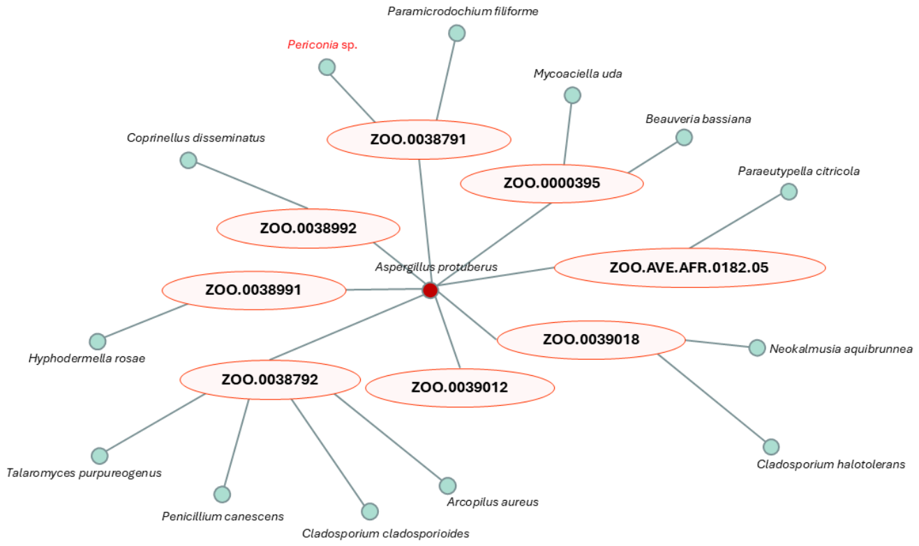
The re-isolation of the same species can occur from different samples. Therefore, fungal diversity was analysed according to species presence–absence in each of the eight sampled specimens. A simple co-occurrence network between species in the samples was constructed and visualized in the Cytoscape software (v.3.10.3, http://www.cytoscape.org (accessed on 5 September 2025)) [42].
Following this survey on culturable fungal diversity, isolates that could not be identified at the species level due to low match similarity or inconclusive results, as well as unique morphological characteristics, were recognized as potential new species and maintained in axenic cultures until further morphological and molecular studies were conducted.

2.3. Morphological Characterization

Two isolates, which were identical but did not yield a conclusive match in GenBank and were therefore considered potential representatives of novel species, were selected for detailed morphological characterization. The isolates were cultivated on Potato Dextrose Agar (PDA; Difco, Sparks, MD, USA), Malt Extract Agar (MEA; Difco, Sparks, MD, USA), and Oatmeal Agar (OA) for three weeks at 25 °C in darkness. PDA was used to assess general colony growth and morphology, MEA to further evaluate colony characteristics and pigmentation, and OA to promote sporulation and facilitate the observation of reproductive structures and other diagnostic morphological features. Oatmeal Agar was prepared by boiling 30 g of oatmeal and 15 g of agar in 1 L of distilled water prior to sterilization. Inoculation was performed on 9 cm Petri dishes using a three-point pattern. After incubation, colony diameter (longest axis), degree of sporulation, colony colour on both obverse and reverse sides, texture, form, and the release of soluble pigments into the medium were documented through direct observation. Colour descriptions follow the ISCC-NBS Colour System [43]. Macro photographs were taken with a Sony α6100 camera equipped with a Sony f/3.5-5.6 18-135 mm lens.
Micromorphological features were examined directly on growing colonies or using the slide culture technique, prepared from 3- to 5-week-old colonies on Synthetic Nutrient-Poor Agar (SNA) and OA [44]. Observations were made with a Leica DM750 light microscope (Leica, Wetzlar, Germany) equipped with a Leica ICC50W digital camera (Leica, Wetzlar, Germany). Size data were recorded based on at least 30 measurements per structure, and mean dimensions were calculated. Photo plates were assembled in Microsoft PowerPoint 2016.
A holotype (dried specimen) and additional cultures were deposited in the Micoteca da Universidade do Minho (MUM), Braga, Portugal. The new descriptions and nomenclature were registered in MycoBank [40,41].

2.4. Molecular Characterization and Phylogenetic Analyses

Following the initial molecular screening and subsequent morphological characterization, a detailed molecular analysis was performed for the two isolates considered potential representatives of novel species. Genomic DNA was extracted from 3-week-old pure cultures following the same procedure described in Section 2.2. The extracted DNA was used to amplify the ITS, LSU, SSU, RPB2, and TEF1α regions using the primer pairs and amplification conditions described above. These loci were selected based on phylogenetic studies of Periconia, including those published by Phookamsak et al. [45], Liao et al. [46], Su et al. [47], Yang et al. [48], Tian et al. [49], Chen et al. [50], and Pommer et al. [51], in which these markers were shown to be informative for species-level identification and delimitation within the genus. PCR reactions were performed in 50 µL volumes containing 25 µL NZYTaq Green Master Mix (NZYTech, Lisboa, Portugal), 2 µL of each primer (10 mM), 19 µL ultra-pure water, and 2 µL template DNA. Reactions were carried out in an ABI GeneAmp 9700 PCR System (Applied Biosystems, USA). Amplification success was verified by agarose gel electrophoresis (1.2%) stained with GreenSafe Premium (NZYTech, Lisboa, Portugal) and visualized using a Bio-Rad Gel Doc XR imaging system (Bio-Rad, Hercules, CA, USA). Amplicons were purified and sequenced on an ABI 3730xl DNA Analyzer (96 capillary system) at STABVIDA, Portugal [52].
DNA sequences were quality-checked using Chromas v.2.6.6 (Technelysium, Southport, QLD, Australia). Forward and reverse reads were aligned, assembled into consensus sequences, examined for ambiguous bases, and trimmed at both ends using BioEdit v.7.2.5 [53]. The resulting consensus sequences were deposited in GenBank, with accession numbers listed in Supplementary Table S2. Sequence similarity searches were performed against the NCBI nucleotide database using BLASTN v.2.6 [39] under the MEGABLAST option. Based on preliminary BLAST results, six datasets were constructed to assess the phylogenetic placement of the isolates. These included individual alignments for ITS, LSU, SSU, RPB2, and TEF1α, as well as a concatenated dataset incorporating all five loci. The resulting multilocus dataset was used to further assess the phylogenetic placement of the isolates, providing molecular support for the preliminary identification and complementing the morphological characterization.
Sequences of each region were individually aligned using the online version of MAFFT v.7 [54], and alignments manually adjusted and concatenated using MEGA software v.11.0.11 [55]. Prior to the phylogenetic analysis, the model of nucleotide substitution was estimated under the Akaike Information Criterion (AIC) using ModelFinder [56] on the W-IQ-TREE v.2.4.0 webserver [57]. The best-fit models for each partition and statistics are provided in Supplementary Table S3. Maximum likelihood (ML) analyses were performed on the same web server, with branch support assessed using 1000 replicates of ultrafast bootstrap analysis [58], the Shimodaira-Hasegawa-like approximate likelihood ratio test (SH-aLRT) [59], and the approximate Bayes test (aBayes) [60].
All alignment matrices and tree files were deposited in Figshare under DOI 10.6084/m9.figshare.30566054. Phylogenetic trees were visualized and edited using FigTree v.1.2.2, MEGA v.11.0.11, and Microsoft PowerPoint 2016.

2.5. Genealogical Concordance Phylogenetic Species Recognition (GCPSR)

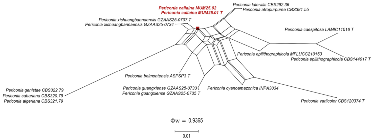
GCPSR analyses based on the pairwise homoplasy index (PHI; Φw) were performed in SplitsTree4 [61] to assess recombination among phylogenetically closely related taxa and to provide additional support for species delimitation [62]. The analysis included the newly generated strains of Periconia callaina examined in this study together with the strains of the most closely related species [63]. A concatenated dataset comprising ITS, LSU, SSU, RPB2, and TEF1α sequences was used for the PHI test and subsequent split network analyses. Relationships among the newly generated strains and their closest relatives were visualized using split networks based on the LogDet transformation and NeighborNet algorithm. PHI values equal to or greater than 0.05 (Φw ≥ 0.05) indicated no significant evidence of recombination within the dataset, supporting genetic differentiation among the analysed taxa. In contrast, PHI values below 0.05 (Φw < 0.05) indicated significant recombination, suggesting genetic exchange among the analysed strains.

3. Results

3.1. Culturable Fungi Diversity

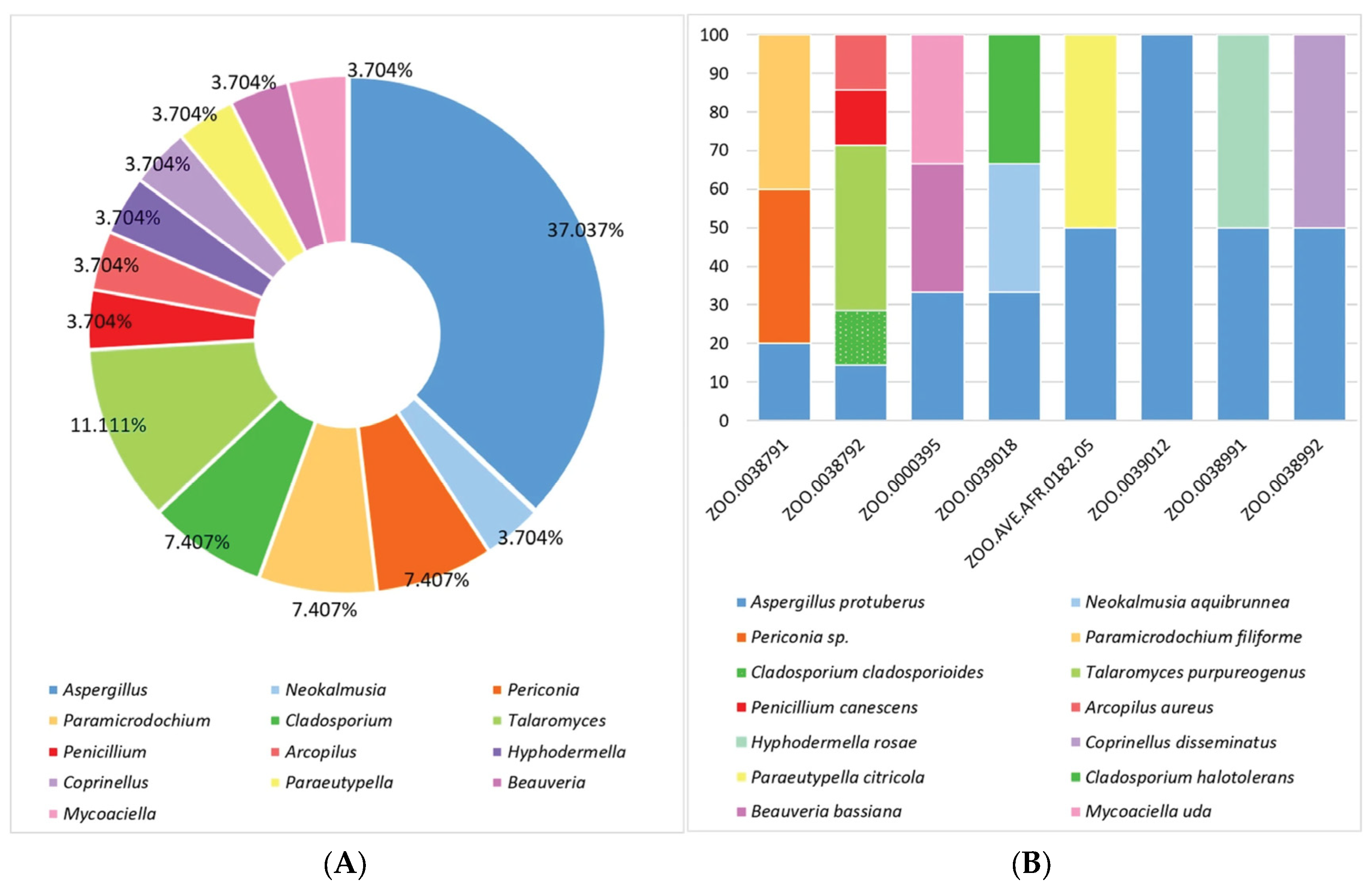
A total of 27 fungal isolates were obtained across all eight samples. Seven isolates were recovered from ZOO.0038792, five from ZOO.0038791, three from ZOO.0000395, ZOO.0039018, and ZOO.0039012, and two each from ZOO.AVE.AFR.0182.05, ZOO.0038991, and ZOO.0038992. Molecular analysis indicated that, of these 27 isolates, 25 belonged to 13 different previously described species. The majority (approximately 37%) were identified as Aspergillus protuberus Munt.-Cvetk. (Figure 2), with no other Aspergillus species detected. Talaromyces was the second most represented genus (11.11%), with T. purpureogenus (Stoll) Samson, Yilmaz, Houbraken, Spierenburg, Seifert, Peterson, Varga & Frisvad. The genera Periconia, Paramicrodochium and Cladosporium each accounted for 7.41% of the isolates, with Paramicrodochium being represented by the recently described P. filiforme D.S. Paiva [52], Cladosporium by C. cladosporioides (Fresen.) G.A. de Vries and C. halotolerans Zalar, de Hoog & Gunde-Cim., the only genus with more than one species identified, and finally Periconia with one unidentifiable species. The remaining taxa were Arcopilus aureus (Chivers) X.Wei Wang & Samson, Beauveria bassiana (Bals.-Criv.) Vuill., Coprinellus disseminatus (Pers.) J.E. Lange, Hyphodermella rosae (Bres.) Nakasone, Mycoaciella uda (Fr.) C.L. Zhao, Neokalmusia aquibrunnea J. Yang, Jian K. Liu & K.D. Hyde, Paraeutypella citricola (Speg.) L.S. Dissan., Wijayaw., J.C. Kang & K.D. Hyde, and Penicillium canescens Sopp, where each represented 3.70% of the total isolates.
Aspergillus protuberus was the only species present across all samples, whereas the remaining fourteen species were exclusively associated with a single sample. Specimen ZOO.0038792 accommodated the highest diversity, with four other species in addition to A. protuberus (Arcopilus aureus, Cladosporium cladosporioides, Penicillium canescens and Talaromyces purpureogenus). Sample ZOO.0038791 displayed two additional species, Periconia sp. and Paramicrodochium filiforme, as did ZOO.0000395 (Beauveria bassiana and Mycoaciella uda) and ZOO.0039018 (Neokalmusia aquibrunnea and Cladosporium halotolerans). ZOO.AVE.AFR.0182.05, ZOO.0038991, and ZOO.0038992 were only found to contain one additional species each, namely Paraeutypella citricola, Hyphodermella rosae, and Coprinellus disseminatus, respectively. Finally, ZOO.0039012 yielded only Aspergillus protuberus (Figure 3). Overall, the isolated fungi were largely sample-specific, with limited overlap between specimens, suggesting a high degree of spatial heterogeneity and potentially localized sources or dispersal processes influencing their distribution.

3.2. Phylogenetic Analysis

Initial comparisons with sequences available in the NCBI database were conducted to determine the generic placement of the studied isolates. The ITS, LSU, SSU, and TEF1α loci consistently showed high sequence similarity (97–99%) with several Periconia species, including P. macrospinosa Lefebvre & Aar.G. Johnson (isolates S1c, 107, A2S4-D47, NWU47, among others), P. epilithographicola Coronado-Ruiz, Avendaño, Escudero-Leyva, Conejo-Barboza, P. Chaverri & Chavarría (MFLUCC 21-0153), P. lateralis Ellis & Everh. (CBS 292.36), P. genistae Lunghini (CBS 322.79), P. algeriana Lunghini (CBS 321.72), P. byssoides Pers. (isolates C457, 202007001, w282, C445B, among others), P. pseudobyssoides Markovsk. & A. Kačergius (KUNCC 23-13932, KUMCC 20-0263), P. wurfbainiae C.F. Liao, Doilom & K.D. Hyde (ZHKUCC 23-0999, ZHKUCC 23-1000), and P. circinata (L. Mangin) Sacc. & D. Sacc. (CBS 263.37), as well as several unidentified Periconia species and other Pleosporales isolates. In contrast, the RPB2 locus exhibited only distant matches (≤88% similarity), primarily with P. byssoides (MFLUCC 18-1555, MFLUCC 18-1553, among others) and P. cynodontis Z.Hua Lu, P.W. Su & Maharachch. (isolate Lu4). These results strongly suggested an affiliation with Periconia genus; however, definitive taxonomic placement remained unresolved.
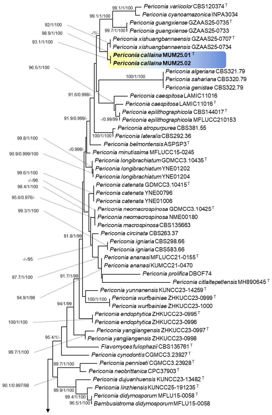
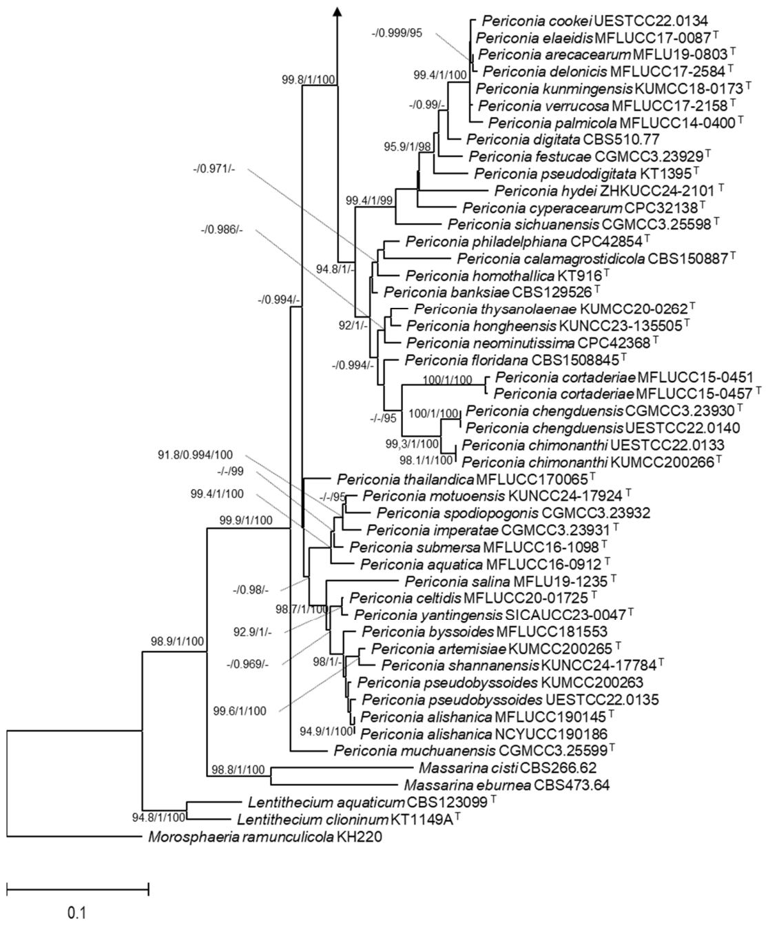
Phylogenetic relationships were inferred using a concatenated dataset of five loci, comprising 3452 aligned characters (471 for ITS, 747 for LSU, 1022 for SSU, 514 for RPB2, and 698 for TEF1α, including alignment gaps). The optimal nucleotide substitution models for each of the loci are listed in Supplementary Table S3. The resulting tree (Figure 4) was consistent with existing knowledge, with overall topology aligning with recent taxonomic studies regarding this genus [45,46,47,48,49,50,51]. The phylogenetic analysis demonstrated that the studied isolates resolved in a distinct, strongly supported monophyletic branch (93.1% SH-aLRT, 1 aBayes, 100% bootstrap support), sister to P. xishuangbannaensis X.G. Tian & D.F. Bao. Both species occupied a strongly supported cluster (98.9% SH-aLRT, 1 aBayes, 100% bootstrap support) closely related to the cluster encompassing P. variicolor S.A. Cantrell, Hanlin & E. Silva, P. cyanoamazonica I.C. Bandeira, and P. guangxiense X.G. Tian & D.F. Bao. These taxa collectively formed an independent clade (87.9% SH-aLRT, 1 aBayes, 90% bootstrap support), distinct from other Periconia species. Additionally, independent phylogenetic analyses of ITS, LSU, SSU, RPB2, and TEF1α loci supported these findings, further confirming the novelty of the studied isolates (Supplementary Figures S1–S5). Based on this evidence, we propose the establishment of Periconia callaina sp. nov. to accommodate this newly identified species.
The PHI test based on the concatenated ITS, LSU, SSU, RPB2, and TEF1α dataset showed no significant evidence of recombination between the Periconia callaina isolates and representative strains of phylogenetically closely related species (Φw = 0.9365), further supporting their genetic differentiation from the closely related taxa and supporting their recognition as a distinct species within Periconia (Figure 5).

3.3. Taxonomy

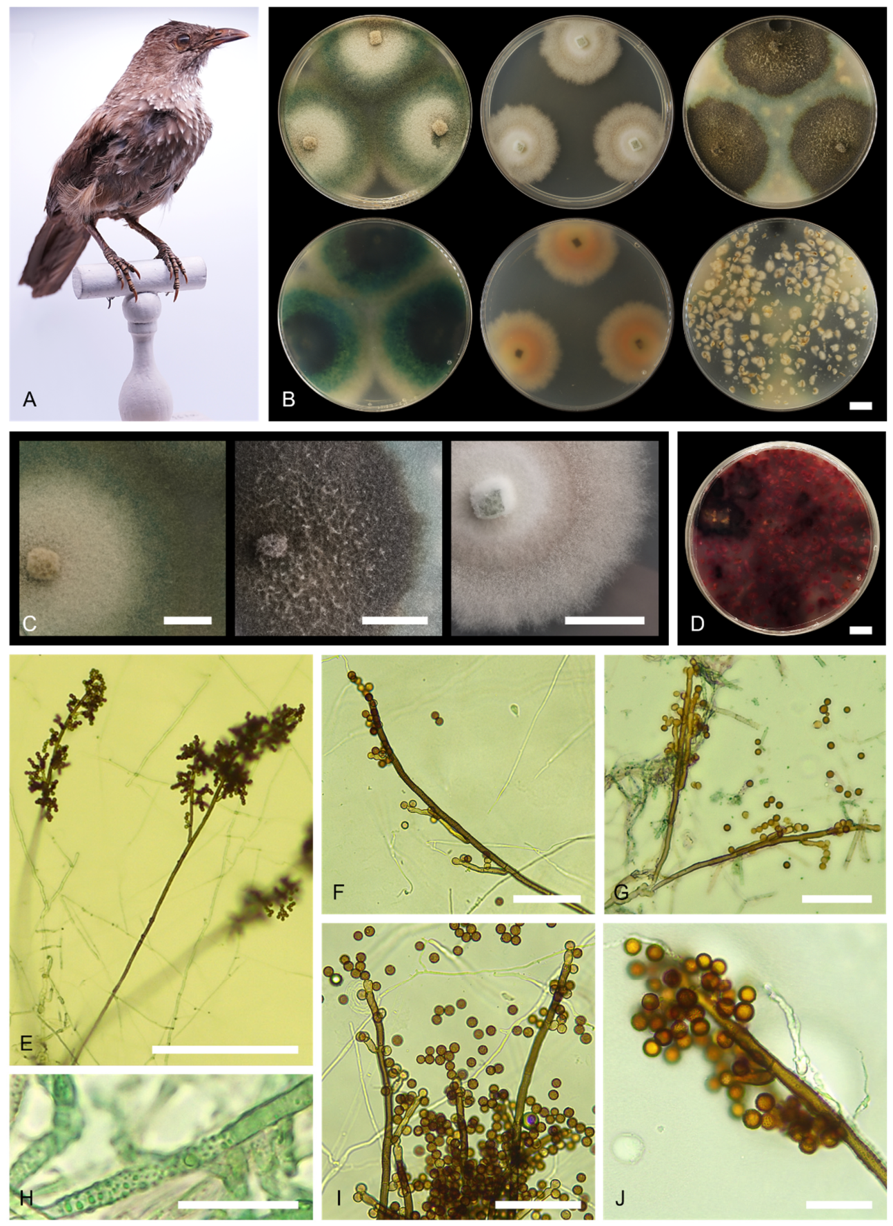
Periconia callaina L. Fernandes & D.S. Paiva, sp. nov., Figure 6.
MycoBank MB858472
Etymology:Callaina” from Latin, meaning greenish-blue or turquoise, referring to the characteristic coloration of the colonies in PDA.
Classification: division Ascomycota, class Dothideomycetes, subclass Pleosporomycetidae, order Pleosporales, family Periconiaceae, genus Periconia.
Typus: Portugal, Coimbra, isolated from a taxidermized bird (Turdoides jardineii tamalakanei, specimen no. ZOO.0038791) at the Science Museum of the University of Coimbra, 2 April 2024, L. Fernandes (holotype MUM-H 25.01, dried specimen); ex-type culture MUM 25.01.
Description: Spore masses on SNA after 4 wk scattered, appearing as black spots on colonies. Hyphae hyaline when young turning moderate yellowish green (ISCC-NBS No. 136) when ageing, septate, verrucose, 2–4 µm wide (mean = 3 µm, n = 30). Conidiophores arising from substrate mycelium, dark olive brown (ISCC-NBS No. 96), 295–645 µm long (mean = 430, n = 30), 3–5 µm wide (mean = 3.7 µm, n = 30), macronematous, mononematous, straight or slightly flexuous, branched, solitary or gregarious, septate, verrucose, thick-walled. Conidiogenous cells polyblastic, light olive brown (ISCC-NBS No. 94), terminal, integrated, oblong, with rounded apex, verrucose. Conidia globose, 4.5–7 µm (mean = 5.8 µm, n = 30), dark olive brown (ISCC-NBS No. 96), aseptate, verrucose, thick-walled, solitary or catenate. Sexual morph unknown.
Culture diameter, 3 weeks (in mm): PDA 60–65; MEA 35–40; OA 55–60.
Culture characteristics: PDA 25 °C, 3 wk: Colonies circular, dense, lightly raised at centre, with concentric zonation in the middle region, margins filamentous, slightly fluffy; mycelium white (ISCC-NBS No. 263) at centre, transitioning into strong bluish green (ISCC-NBS No. 160) to moderate yellow green (ISCC-NBS No. 120) towards the margin, texture cottony, smooth; exudate absent; soluble pigment absent; reverse colour very dark bluish green (ISCC-NBS No. 166) at centre fading into strong bluish green (ISCC-NBS No. 160) with a white margin (ISCC-NBS No. 263). MEA 25 °C, 3 wk: Colonies circular, dense, flat; margins narrow, filamentous; mycelium white (ISCC-NBS No. 263) at centre, pale greenish yellow (ISCC-NBS No. 104) from the middle region to the margin; texture cottony, smooth; with slight soluble pigment present, vivid red (ISCC-NBS No. 11); exudate absent; reverse colour pale yellow (ISCC-NBS No. 89). OA 25 °C, 3 wk: Colonies circular, moderately deep, flat, margins filamentous, slightly fluffy; mycelium sparce, white (ISCC-NBS No. 263); texture cottony to floccose; sporulation dense, conidia en masse dark greyish olive (ISCC-NBS No. 111); exudate absent; soluble pigment absent; reverse colour dark greyish olive (ISCC-NBS No. 111).
Additional specimens examined: Portugal, Coimbra, isolated from a taxidermized bird (Turdoides jardineii tamalakanei, specimen no. ZOO.0038791) at the Science Museum of the University of Coimbra, 2 April 2024, L. Fernandes, MUM 25.02.
DNA barcodes: MUM 25.01 ITS: PV341358; LSU: PV387312; SSU: PV387571; RPB2: PV423526; TEF1α: PV423528. MUM 25.02 ITS: PV341359; LSU: PV387313; SSU: PV387572; RPB2: PV423527; TEF1α: PV423529.
Notes: Periconia callaina exhibits the characteristic morphology of Periconia species, forming macronematous, mononematous conidiophores with a spherical conidial head, polyblastic conidiogenous cells, and catenate, globose, brown, aseptate, verruculose conidia. Phylogenetic analyses based on concatenated ITS, LSU, SSU, RPB2, and TEF1α sequences strongly support its placement within Periconia, positioning P. callaina on an independent branch, sister to P. xishuangbannaensis, and closely related to the clade containing P. variicolor, P. guangxiense and P. cyanoamazonica, the latter two also being recent additions to the genus (Figure 4). Morphologically, P. callaina is distinguished by its characteristic blue-green coloration, which is also evident at the micromorphological level, particularly in its hyphae, that appear to contain similarly coloured vesicle-like structures. A comparable pigmentation has been reported in P. cyanoamazonica [64], although the overall micromorphology differs. Like P. caespitosa Cantillo, Gusmão & Madrid, P. variicolor, and P. guangxiense it produces a deep red pigment, but in P. callaina, this feature is observed only in older colonies [49,65,66]. Additionally, its conidiophores exceed 600 µm in length, whereas P. xishuangbannaensis reach an average of 243 µm, P. epilithographicola and P. variicolor approximately 270 µm, and P. caespitosa up to 500 µm [46,49,65,66,67]. In contrast, P. callaina tends to produce slightly smaller conidia, measuring up to 7 µm, while the conidia of these related species range from 9 to 10 µm, with the exception of P. guangxiense and P. xishuangbannaensis, which show partially overlapping size ranges, 6–7.5 μm and 4.5–7 μm respectively. Another distinguishing feature of P. callaina is its branched conidiophores, a trait it shares with P. epilithographicola [46], P. guangxiense and P. xishuangbannaensis [49]. Notably, P. callaina was isolated from a taxidermized animal, marking the first known occurrence of this genus in such a context. Periconia species are widely distributed, predominantly in terrestrial habitats, with most being saprophytes or endophytes, while a few are plant pathogens. The species most similar in terms of environmental context is P. epilithographicola, which was isolated from a university repository of drawings and lithographs, an environment significantly affected by fungal biodeterioration [67].

4. Discussion

4.1. Culturable Fungi Diversity

In the present study, taxidermized birds from the MCUC were found to harbour fungi previously reported from similar cultural heritage assets. However, assessing fungal contamination in historical taxidermized specimens presents several challenges, particularly in terms of sampling representativeness. The non-invasive approach adopted in this study was necessary to minimise disturbance to the specimens, but sampling was necessarily restricted to accessible areas showing visible fungal growth. Given the heterogeneous composition of taxidermized specimens and the potentially uneven distribution of fungal propagules across their surfaces, the isolates recovered may not represent the entire fungal community associated with each specimen. In addition, visible fungal growth may account for only part of the fungal propagules present, while fungi occurring in less accessible or apparently unaffected areas may remain undetected. Although multiple fungal taxa were recovered from individual specimens, the limited number of specimens examined for each bird species, together with the influence of shared museum environmental conditions, prevents any conclusions regarding species-specific fungal associations, which remain an interesting avenue for future research.
Furthermore, the presence of fungi on a specimen does not necessarily imply established colonization or active biodeterioration. Museum specimens are continuously exposed to airborne fungal propagules, making it difficult to distinguish transient environmental contamination from fungi that have become established on the specimen. The culture-dependent approach used in this study may also favour organisms able to grow under the selected culture conditions, potentially overlooking fungi with different nutritional or physiological requirements. Therefore, the fungal diversity recovered in this study should be regarded as the cultivable fraction associated with the visibly affected areas sampled, rather than as a complete representation of the mycobiota of the preserved specimens. Despite these limitations, the recovery of fungi previously associated with cultural heritage materials highlights the value of targeted, non-invasive sampling as a first approach for assessing fungal occurrence in historical museum collections.
Aspergillus protuberus was the only species present in all samples, and this may suggest that it was the main fungus responsible for the outbreak observed in the collection. The predominance of the Aspergillus genus in this museum’s environment had already been observed in our previous work concerning a different collection, in a different room [24]. However, no overlap of species was observed between both works, and while fewer species were observed in this current assessment, a wider range of genera (13) was detected when compared to our work in the Anthropology archive, where only 6 genera were identified.
Already associated with organic acid production, calcium carbonate dissolution and mineralization, and the production of ligninolytic, fibrinolytic, keratinolytic, estereolytic, lipolytic, proteolytic and cellulolytic activity [68,69], the Aspergillus genus is of particular concern in the context of biodeterioration. Aspergillus protuberus has been previously found to be contaminating cultural heritage artifacts such as silk textiles [70], books [71], limestone monuments [72], and paintings [73]. It has well-reported cellulolytic and proteolytic abilities [68] and produces extracellular pigments [74].
Also commonly reported in museum environments and cultural heritage settings, the Talaromyces, Cladosporium and Penicillium genera were also identified in this study, with the isolated species, T. purporeogenus, C. cladosporioides, C. halotolerans and P. canescens, having previous associations with cultural heritage [8,15,17,72,75,76,77,78,79,80,81,82,83,84,85,86,87,88]. These species have also been described as possessing deteriorative potential including, cellulolytic, estereolytic, proteolytic, and ligninolytic activity, alongside organic acid and pigment production [68,89,90,91,92]. Of particular concern is C. cladosporioides known keratinolytic ability [68], considering the context of this work and the taxidermized birds sampled. Keratinases, including those produced by C. cladosporioides, have the ability to degrade feathers [93,94]. As such, the presence of this fungus in this collection represents a significant potential risk to its integrity.
The discovery of Beauveria bassiana is also of particular concern. Although it is primarily known as an entomopathogen, this species has been previously isolated from wall paintings in China [95], and has known ligninolytic, fibrinolytic, proteolytic, cellulolytic, lipolytic, chitinolytic, cellulolytic and, concerningly, keratinolytic capabilities [68,96]. While it has also been used as conservation agent for metal artifacts due to its ability to transform corrosion products into metal-oxalate complexes [97,98,99], the above-described abilities pose a severe threat not only to the integrity of the collection from which this species was isolated, but also to others across the museum. Its entomogenous nature can also provide an insight into the potential processes through which it entered and/or propagated inside the museum, as it also struggles with occasional insect outbreaks.
Of the identified fungal isolates, and to the best of our knowledge, four ascomycetes were here associated with museum environments for the first time, namely Arcopilus aureus, Neokalmusia aquibrunnea, Paraeutypella citricola and Paramicrodochium filiforme. Arcopilus aureus is an endophytic fungus and a known pigment producer [100]. Neokalmusia aquibrunnea is a saprobic fungus described by Yang and colleagues [101] from decaying, submerged bamboo culms, later isolated from decaying branches of chinese windmill palm, Trachycarpus fortunei [102]. This present work marks only the third report of detection of this species. Paraeutypella citricola has been previously isolated from dead branches and woody litter [103,104,105]. A recent addition to the Sordariomycetes incertae sedis class, Paramicrodochium filiforme was first described by Paiva and colleagues from a deteriorated limestone monument [52]. To the best of our knowledge, this is only the second time this species has been isolated.
Three basidiomycetes, Coprinellus disseminatus, Hyphodermella rosae, and Mycoaciella uda were found in this study. Typically associated with the degradation of wooden artifacts [8,106], basidiomycetes too are common contaminants of cultural heritage. In fact, of the three Basidiomycota species identified, only M. uda has no previous reports of contamination of cultural heritage. Both C. disseminatus and H. rosae have been associated with the deterioration of paper and parchment documents [18,107,108], with C. disseminatus also being isolated from wooden iconostasis in a cave church in Serbia [109]. Both species have been reported to be ligninolytic [108], with C. disseminatus also being a known cellulase, proteinase, and lipase producer [107].
The distribution of the recovered fungi among the sampled specimens further highlights the heterogeneity of the fungal community associated with the taxidermized birds. Specimen ZOO.0038792 harboured the highest fungal diversity, with Aspergillus protuberus, Arcopilus aureus, Cladosporium cladosporioides, Penicillium canescens and Talaromyces purpureogenus recovered, whereas other specimens harboured fewer taxa, with ZOO.0039012 yielding only A. protuberus. The remaining specimens showed distinct combinations of fungal taxa, with limited overlap between samples. This largely specimen-specific distribution suggests considerable spatial heterogeneity in fungal occurrence and may reflect localized environmental conditions, deposition, or dispersal processes within the collection. While fungal growth was mainly observed in the feathers, the multi-substrate nature of the sampled specimens, alongside the potential for the recovered isolates to be present merely as propagules limits the ability to establish clear relationships between individual fungal taxa and the substrate from which they were recovered. Nevertheless, the presence of taxa with known biodeteriorative potential, particularly those capable of keratinolytic activity, on individual specimens highlights the importance of considering specimen-specific fungal occurrence when assessing potential risks to taxidermized collections.
Taking all this into consideration, the presence of these fungi constitutes a severe threat not only to this collection, but also to all other collections of the MCUC. Also important to note is the potential health risk caused by the presence of these contaminants, with genera such Aspergillus and Penicillium, here identified, being known for their ability to cause respiratory tract infections and other clinical manifestations [23,110,111].
Additionally, this study constitutes, to the best of our knowledge, the first record of A. aureus, M. uda, N. aquibrunnea, P. citricola, and P. filiforme being isolated from museum artifacts, providing further insight into their adaptability and potential substrates. Finally, several identified species have never been evaluated for their biodeteriorative potential, with further studies being essential for a better understanding of their impact on cultural heritage.

4.2. Description of a New Periconia Species

In the present study, the taxonomic status of two unidentified Periconia isolates obtained from a taxidermized bird specimen in the MCUC collection was investigated using an integrative approach. The combined morphological, phylogenetic, and GCPSR analyses supported their recognition as a distinct species, which is here introduced as Periconia callaina.
The phylogenetic analysis placed the two isolates in a distinct and well-supported lineage within Periconia, closely related to P. xishuangbannaensis. The strong support for the nodes defining the P. callaina lineage, together with the morphological differences observed between P. callaina and its closest relatives, provides robust evidence for its recognition as a separate species. Species delimitation was further supported by the GCPSR analysis, which showed no significant evidence of recombination between the newly proposed species and closely related taxa, providing independent support for the inferred species boundaries. Despite this well-supported placement, the broader relationships within Periconia should be interpreted with some caution. The genus has a complex taxonomic history and has repeatedly been shown to be polyphyletic, while relationships among several species remain unresolved or weakly supported [47]. Recent studies have also emphasized that improved taxon sampling and the inclusion of additional protein-coding loci are needed to obtain a clearer understanding of species relationships and phylogenetic boundaries within the genus [49].
First introduced in 1791 by H. J. Tode, the genus Periconia is a polyphyletic one, and is classified as a member of the Periconiaceae and separated from Massarineae as a sister taxon, with distinct phylogeny [112]. Typically known for their asexual morphs, Periconia species are characterised by macronematous, mononematous conidiophores, and globose to ellipsoidal conidia, while the sexual morph is characterised by immersed to erumpent, scattered or aggregated, subglobose to globose ascomata, hyaline, 8-spored, bitunicate, cylindrical, septate, guttulate, ascospores with an entire sheath [113]. While Index Fungorum (www.indexfungorum.org) notes the existence of 182 currently accepted species of this genus, most of these do not have phylogenetic marker data available and as such were unable to be included in our analysis beyond morphology. However, P. callaina presents very distinct morphological features, particularly its characteristic blue-green coloration, a trait only displayed by P. cyanoamazonica, a species that our phylogenetic analysis places in a separate clade to that of P. callaina. Future revisions may be necessary as molecular data becomes available for these formerly described species or for additional newly identified species. Nevertheless, with the most current information, P. callaina stands as a new addition to this ever-growing genus.
Species from this genus have been reported as saprobes and endophytes, as well as both plant and human pathogens [46,47] with reports of Periconia species causing mycotic keratitis [114]. Two species of the Periconia genus have also been described as cultural heritage contaminants, namely P. epilithographicola, which was discovered and described as a contaminant of a 19th century artwork with cellulolytic capabilities [67], and P. macrospinosa, found in a biofilm on the interior of a gothic church in Southern Italy [115].
The environmental context from which P. callaina was isolated is also of relevance. A taxidermized bird stored in a museum represents an ecological niche new to the genus as a whole, thus providing new insights into the adaptability of Periconia and the substrates it can be found in. Furthermore, the biodeteriorative potential of this new species remains a mystery, and a further evaluation of these characteristics is key in understanding its potential role as a cultural heritage contaminant, even though it has already shown itself as a strong pigment producer.
Finally, the Periconia genus has been previously reported as a strong producer of bioactive secondary metabolites, with activity spectrums as wide as antimicrobial, anti-human immunodeficiency virus (HIV), and anti-inflammatory, among others [112]. As such, a new addition to this genus represents an opportunity for new, naturally occurring secondary compounds to be explored for their biotechnological potential.

5. Conclusions

In the present work, the fungal contamination observed in a collection of taxidermized birds from the Science Museum of the University of Coimbra was studied. This analysis led to the detection of 27 isolates, belonging to 13 different genera and 14 different species. Most of these have been previously associated with cultural heritage contamination and deterioration, with several isolates possessing capabilities that could compromise not only the sampled collection, but most of the museum’s artifacts. Of particular concern was the identification of C. cladosporioides and B. bassiana, for their keratinolytic potential, given the nature of the sampled pieces.
Among the 27 isolates, two were unable to be identified at the species level but were shown to belong to the Periconia genus. Further phylogenetic analysis demonstrated that these isolates belonged to a new species, P. callaina, here described. This work provides valuable molecular and morphological data that enhance our understanding of fungi in this ever-growing genus. Additional studies of the physiological and biodeteriorative potential of this new taxon are needed to fully understand its role as a contaminant of cultural heritage. Understanding the communities that inhabit and are involved in the deterioration of cultural heritage is key to the development and implementation of safeguarding measures that ensure the preservation of our cultural heritage for future generations.

Supplementary Materials

The following supporting information can be downloaded at: https://www.mdpi.com/article/10.3390/microorganisms14091894/s1, Figure S1: Maximum likelihood (ML) tree based on sequence data from ITS showing the placement of the new species, Periconia callaina, described in this study. The scale bar indicates the number of substitutions per site. Nodes are labelled with SH-aLRT/aBayes/ultrafast bootstrap support values ≥ 70%/0.70/70%, with type strains designated by superscript T. The novel species is highlighted in bold font. The tree is rooted with Lentithecium; Figure S2: Maximum likelihood (ML) tree based on sequence data from LSU showing the placement of the new species, Periconia callaina, described in this study. The scale bar indicates the number of substitutions per site. Nodes are labelled with SH-aLRT/aBayes/ultrafast bootstrap support values ≥ 70%/0.70/70%, with type strains designated by superscript T. The novel species is highlighted in bold font. The tree is rooted with Morosphaeria ramunculicola (KH220); Figure S3: Maximum likelihood (ML) tree based on sequence data from SSU showing the placement of the new species, Periconia callaina, described in this study. The scale bar indicates the number of substitutions per site. Nodes are labelled with SH-aLRT/aBayes/ultrafast bootstrap support values ≥ 70%/0.70/70%, with type strains designated by superscript T. The novel species is highlighted in bold font. The tree is rooted with Morosphaeria ramunculicola (KH220); Figure S4: Maximum likelihood (ML) tree based on sequence data from RPB2 showing the placement of the new species, Periconia callaina, described in this study. The scale bar indicates the number of substitutions per site. Nodes are labelled with SH-aLRT/aBayes/ultrafast bootstrap support values ≥ 70%/0.70/70%, with type strains designated by superscript T. The novel species is highlighted in bold font. The tree is rooted with Massarina cisti (CBS266.62); Figure S5: Maximum likelihood (ML) tree based on sequence data from TEF1α showing the placement of the new species, Periconia callaina, described in this study. The scale bar indicates the number of substitutions per site. Nodes are labelled with SH-aLRT/aBayes/ultrafast bootstrap support values ≥ 70%/0.70/70%, with type strains designated by superscript T. The novel species is highlighted in bold font. The tree is rooted with Morosphaeria ramunculicola (KH220); Table S1: Identification of all isolates retrieved in this study; Table S2: Strain collection references and GenBank accession numbers for the novel species outlined in this study, along with additional strains included in phylogenetic analyses, with Massarina cisti as the outgroup; Table S3: The best–fit substitution models for each partition determined by ModelFinder based on the Akaike Information Criterion.

Author Contributions

Conceptualization—D.S.P. and L.F.; Methodology—L.F., D.S.P. and E.P.; Software—D.S.P. and L.F.; Validation—L.F., D.S.P. and A.P.; Formal analysis—L.F. and D.S.P.; Investigation—L.F. and D.S.P.; Resources—C.C., A.P. and N.M.; Data Curation—L.F. and D.S.P.; Writing, Original Draft Preparation—L.F. and D.S.P.; Writing, Review and Editing—L.F., D.S.P., C.C., A.P. and N.M.; Visualization—L.F. and D.S.P.; Supervision—C.C., A.P. and N.M.; Project Administration—N.M.; Funding Acquisition—N.M. and A.P. All authors have read and agreed to the published version of the manuscript.

Funding

This research was funded by FCT—Fundação para a Ciência e Tecnologia, I.P., through the following projects: UIDB/04004/2025 (Centre for Functional Ecology), LA/P/0092/2020—DOI: 10.54499/LA/P/0092/2020 (Associate Laboratory TERRA), UIDB/04539/2020—DOI: 10.54499/UIDB/04539/2020, UIDP/04539/2020—DOI: 10.54499/UIDP/04539/2020, LA/P/0058/2020—DOI: 10.54499/UIDP/04539/2020 (CIBB Strategic Projects), ANOXYOILS project: 2022.07967.PTDC. Luís Fernandes is supported by a PhD research grant with reference PRT/BD/155044/2023. Diana Paiva is supported by a PhD research grant with reference UI/BD/150843/2021 awarded by the Centre for Functional Ecology—Science for People & the Planet (CFE) and co–funded by Fundação para a Ciência e Tecnologia, I.P. (FCT) through national funding by the Ministério da Ciência, Tecnologia e Ensino Superior (MCTES) from Fundo Social Europeu (FSE). Nuno Mesquita is funded through national funds (OE) and the Foundation for Science and Technology (FCT, Portugal), in the scope of Decree-Law 57/2016. The authors also thank the funding of PRR—Recovery and Resilience Plan—and the NextGeneration EU European Funds.

Institutional Review Board Statement

Not applicable.

Informed Consent Statement

Not applicable.

Data Availability Statement

All relevant data supporting the findings of this study are included in the article and its Supplementary Information File. The nucleotide sequences have been deposited in the GenBank database under the following accession numbers: PV341358-PV341359, PV484568-PV484589 (ITS); PV387312-PV387313, PV484675-PV484686 (LSU); PV387571-PV387572, PV484690-PV484694 (SSU); PX434372-PX434378; PX438799-PX438800 (BTUB); PV423528-PV423529, PX434388 (TEF1α); PX434379-PX434386 (CAM); and PV423526-PV423527, PX434387 (RPB2). The isolates studied in this work are preserved in the Micoteca da Universidade do Minho (MUM), Braga, Portugal, under the references MUM 25.01 and MUM 25.02. Additionally, alignment matrices and phylogenetic tree files are accessible on Figshare with the DOI identifier: 10.6084/m9.figshare.30566054.

Acknowledgments

The authors would like to thank the Science Museum of the University of Coimbra, who kindly received us and allowed the collection of samples used in this research as well for providing valuable information about the artifacts.

Conflicts of Interest

The authors declare no conflicts of interest.

References

  1. O’Connell, A.F.; Gilbert, A.T.; Hatfield, J.S. Contribution of Natural History Collection Data to Biodiversity Assessment in National Parks. Conserv. Biol. 2004, 18, 1254–1261. [Google Scholar] [CrossRef] [Scilit]
  2. Lo Brutto, S.; Badalucco, A.; Iacovera, R.; Cilli, E.; Sarà, M. Checklist of the Mammal Collection Preserved at the University of Palermo under the Framework of the National Biodiversity Future Center. Diversity 2023, 15, 518. [Google Scholar] [CrossRef] [Scilit]
  3. Ferrari, G.; Esselens, L.; Hart, M.L.; Janssens, S.; Kidner, C.; Mascarello, M.; Peñalba, J.V.; Pezzini, F.; von Rintelen, T.; Sonet, G.; et al. Developing the Protocol Infrastructure for DNA Sequencing Natural History Collections. Biodivers. Data J. 2023, 11, e102317. [Google Scholar] [CrossRef] [Scilit]
  4. Fong, J.J.; Blom, M.P.K.; Aowphol, A.; McGuire, J.A.; Sutcharit, C.; Soltis, P.S. Editorial: Recent Advances in Museomics: Revolutionizing Biodiversity Research. Front. Ecol. Evol. 2023, 11, 1188172. [Google Scholar] [CrossRef] [Scilit]
  5. Kakakhel, M.A.; Wu, F.; Gu, J.D.; Feng, H.; Shah, K.; Wang, W. Controlling Biodeterioration of Cultural Heritage Objects with Biocides: A Review. Int. Biodeterior. Biodegrad. 2019, 143, 104721. [Google Scholar] [CrossRef] [Scilit]
  6. Koestler, R.J. Biodeterioration in Museum Collections. In Met Objectives; The Sherman Fairchild Center for Objects Conservation: New York, NY, USA, 2002. [Google Scholar]
  7. Gadd, G.M.; Fomina, M.; Pinzari, F. Fungal Biodeterioration and Preservation of Cultural Heritage, Artwork, and Historical Artifacts: Extremophily and Adaptation. Microbiol. Mol. Biol. Rev. 2024, 88, e0020022. [Google Scholar] [CrossRef] [Scilit]
  8. Sterflinger, K. Fungi: Their Role in Deterioration of Cultural Heritage. Fungal Biol. Rev. 2010, 24, 47–55. [Google Scholar] [CrossRef] [Scilit]
  9. Zucconi, L.; Canini, F.; Isola, D.; Caneva, G. Fungi Affecting Wall Paintings of Historical Value: A Worldwide Meta-Analysis of Their Detected Diversity. Appl. Sci. 2022, 12, 2988. [Google Scholar] [CrossRef] [Scilit]
  10. De Leo, F.; Marchetta, A.; Urzì, C. Black Fungi on Stone-Built Heritage: Current Knowledge and Future Outlook. Appl. Sci. 2022, 12, 3969. [Google Scholar] [CrossRef] [Scilit]
  11. Sterflinger, K.; Pinzari, F. The Revenge of Time: Fungal Deterioration of Cultural Heritage with Particular Reference to Books, Paper and Parchment. Environ. Microbiol. 2012, 14, 559–566. [Google Scholar] [CrossRef] [Scilit]
  12. Vivar, I.; Borrego, S.; Ellis, G.; Moreno, D.A.; García, A.M. Fungal Biodeterioration of Color Cinematographic Films of the Cultural Heritage of Cuba. Int. Biodeterior. Biodegrad. 2013, 84, 372–380. [Google Scholar] [CrossRef] [Scilit]
  13. Gutarowska, B.; Celikkol-Aydin, S.; Bonifay, V.; Otlewska, A.; Aydin, E.; Oldham, A.L.; Brauer, J.I.; Duncan, K.E.; Adamiak, J.; Sunner, J.A.; et al. Metabolomic and High-Throughput Sequencing Analysis-Modern Approach for the Assessment of Biodeterioration of Materials from Historic Buildings. Front. Microbiol. 2015, 6, 979. [Google Scholar] [CrossRef] [Scilit]
  14. Isola, D.; Bartoli, F.; Meloni, P.; Caneva, G.; Zucconi, L. Black Fungi and Stone Heritage Conservation: Ecological and Metabolic Assays for Evaluating Colonization Potential and Responses to Traditional Biocides. Appl. Sci. 2022, 12, 2038. [Google Scholar] [CrossRef] [Scilit]
  15. Paiva, D.S.; Fernandes, L.; Trovão, J.; Mesquita, N.; Tiago, I.; Portugal, A. Uncovering the Fungal Diversity Colonizing Limestone Walls of a Forgotten Monument in the Central Region of Portugal by High-Throughput Sequencing and Culture-Based Methods. Appl. Sci. 2022, 12, 10650. [Google Scholar] [CrossRef] [Scilit]
  16. Paiva, D.S.; Trovão, J.; Fernandes, L.; Mesquita, N.; Tiago, I.; Portugal, A. Expanding the Microcolonial Black Fungi Aeminiaceae Family: Saxispiralis lemnorum Gen. et sp. Nov. (Mycosphaerellales), Isolated from Deteriorated Limestone in the Lemos Pantheon, Portugal. J. Fungi 2023, 9, 916. [Google Scholar] [CrossRef] [Scilit]
  17. Trovão, J.; Portugal, A.; Soares, F.; Paiva, D.S.; Mesquita, N.; Coelho, C.; Pinheiro, A.C.; Catarino, L.; Gil, F.; Tiago, I. Fungal Diversity and Distribution across Distinct Biodeterioration Phenomena in Limestone Walls of the Old Cathedral of Coimbra, UNESCO World Heritage Site. Int. Biodeterior. Biodegrad. 2019, 142, 91–102. [Google Scholar] [CrossRef] [Scilit]
  18. de Carvalho, H.P.; Mesquita, N.; Trovão, J.; da Silva, J.P.; Rosa, B.; Martins, R.; Bandeira, A.M.L.; Portugal, A. Diversity of Fungal Species in Ancient Parchments Collections of the Archive of the University of Coimbra. Int. Biodeterior. Biodegrad. 2016, 108, 57–66. [Google Scholar] [CrossRef] [Scilit]
  19. Rakotonirainy, M.S.; Lavédrine, B. Screening for Antifungal Activity of Essential Oils and Related Compounds to Control the Biocontamination in Libraries and Archives Storage Areas. Int. Biodeterior. Biodegrad. 2005, 55, 141–147. [Google Scholar] [CrossRef] [Scilit]
  20. Stupar, M.; Grbić, M.L.; Džamić, A.; Unković, N.; Ristić, M.; Jelikić, A.; Vukojević, J. Antifungal Activity of Selected Essential Oils and Biocide Benzalkonium Chloride against the Fungi Isolated from Cultural Heritage Objects. S. Afr. J. Bot. 2014, 93, 118–124. [Google Scholar] [CrossRef] [Scilit]
  21. Palla, F.; Bruno, M.; Mercurio, F.; Tantillo, A.; Rotolo, V. Essential Oils as Natural Biocides in Conservation of Cultural Heritage. Molecules 2020, 25, 730. [Google Scholar] [CrossRef] [Scilit]
  22. Sapaat, A.; Sabran, S.F.; Mohamed, M. Occurrence of Pest, the Management of Zoological Museum Specimens Collection and Climate Change; Atlantis Press: Dordrecht, The Netherlands, 2022. [Google Scholar]
  23. Di Carlo, E.; Chisesi, R.; Barresi, G.; Barbaro, S.; Lombardo, G.; Rotolo, V.; Sebastianelli, M.; Travagliato, G.; Palla, F. Fungi and Bacteria in Indoor Cultural Heritage Environments: Microbial-Related Risks for Artworks and Human Health. Environ. Ecol. Res. 2016, 4, 257–264. [Google Scholar] [CrossRef] [Scilit]
  24. Fernandes, L.; Paiva, D.S.; Pereira, E.; Rufino, A.C.; Landim, E.; Marques, M.P.; Cabral, C.; Portugal, A.; Mesquita, N. Evaluating the Antifungal Activity of Volatilized Essential Oils on Fungi Contaminating Artifacts from a Museum Collection. Appl. Sci. 2025, 15, 2378. [Google Scholar] [CrossRef] [Scilit]
  25. Gardes, M.; Bruns, T.D. ITS Primers with Enhanced Specificity for Basidiomycetes—Application to the Identification of Mycorrhizae and Rusts. Mol. Ecol. 1993, 2, 113–118. [Google Scholar] [CrossRef] [Scilit] [PubMed]
  26. White, T.J.; Bruns, T.; Lee, S.; Taylor, J. Amplification and Direct Sequencing of Fungal Ribosomal RNA Genes for Phylogenetics. In PCR Protocols: A Guide to Methods and Applications; Academic Press: New York, NY, USA, 1990; Volume 18, pp. 315–322. [Google Scholar]
  27. Vilgalys, R.; Hester, M. Rapid Genetic Identification and Mapping of Enzymatically Amplified Ribosomal DNA from Several Cryptococcus Species. J. Bacteriol. 1990, 172, 4238–4246. [Google Scholar] [CrossRef] [Scilit]
  28. Rehner, S.A.; Samuels, G.J. Molecular Systematics of the Hypocreales: A Teleomorph Gene Phylogeny and the Status of Their Anamorphs. Can. J. Bot. 1995, 73, 816–823. [Google Scholar] [CrossRef] [Scilit]
  29. Crous, P.W.; Summerell, B.A.; Carnegie, A.J.; Wingfield, M.J.; Hunter, G.C.; Burgess, T.I.; Andjic, V.; Barber, P.A.; Groenewald, J.Z. Unravelling Mycosphaerella: Do You Believe in Genera? Persoonia-Mol. Phylogeny Evol. Fungi 2009, 23, 99–118. [Google Scholar] [CrossRef] [Scilit]
  30. Liu, Y.J.; Whelen, S.; Hall, B.D. Phylogenetic Relationships among Ascomycetes: Evidence from an RNA Polymerse II Subunit. Mol. Biol. Evol. 1999, 16, 1799–1808. [Google Scholar] [CrossRef] [Scilit]
  31. Sung, G.-H.; Sung, J.-M.; Hywel-Jones, N.L.; Spatafora, J.W. A Multi-Gene Phylogeny of Clavicipitaceae (Ascomycota, Fungi): Identification of Localized Incongruence Using a Combinational Bootstrap Approach. Mol. Phylogenetics Evol. 2007, 44, 1204–1223. [Google Scholar] [CrossRef] [Scilit]
  32. Glass, N.L.; Donaldson, G.C. Development of Primer Sets Designed for Use with the PCR to Amplify Conserved Genes from Filamentous Ascomycetes. Appl. Environ. Microbiol. 1995, 61, 1323–1330. [Google Scholar] [CrossRef] [Scilit]
  33. Hong, S.-B.; Cho, H.-S.; Shin, H.-D.; Frisvad, J.C.; Samson, R.A. Novel Neosartorya Species Isolated from Soil in Korea. Int. J. Syst. Evol. Microbiol. 2006, 56, 477–486. [Google Scholar] [CrossRef] [Scilit]
  34. Stielow, J.B.; Levesque, C.A.; Seifert, K.A.; Meyer, W.; Irinyi, L.; Smits, D.; Renfurm, R.; Verkley, G.J.M.; Groenewald, M.; Chaduli, D. One Fungus, Which Genes? Development and Assessment of Universal Primers for Potential Secondary Fungal DNA Barcodes. Persoonia-Mol. Phylogeny Evol. Fungi 2015, 35, 242–263. [Google Scholar] [CrossRef] [Scilit]
  35. Crous, P.W.; Braun, U.; Hunter, G.C.; Wingfield, M.J.; Verkley, G.J.M.; Shin, H.-D.; Nakashima, C.; Groenewald, J.Z. Phylogenetic Lineages in Pseudocercospora. Stud. Mycol. 2013, 75, 37–114. [Google Scholar] [CrossRef] [Scilit]
  36. Hunter, G.C.; Wingfield, B.D.; Crous, P.W.; Wingfield, M.J. A Multi-Gene Phylogeny for Species of Mycosphaerella Occurring on Eucalyptus Leaves. Stud. Mycol. 2006, 55, 147–161. [Google Scholar] [CrossRef] [Scilit]
  37. Houbraken, J.; Samson, R.A. Phylogeny of Penicillium and the Segregation of Trichocomaceae into Three Families. Stud. Mycol. 2011, 70, 1–51. [Google Scholar] [CrossRef] [Scilit]
  38. Lee, S.; Groenewald, J.Z.; Crous, P.W. Phylogenetic Reassessment of the Coelomycete Genus Harknessia and Its Teleomorph Wuestneia (Diaporthales), and the Introduction of Apoharknessia Gen. Nov. Stud. Mycol. 2004, 50, 235–252. [Google Scholar]
  39. Altschul, S. Gapped BLAST and PSI-BLAST: A New Generation of Protein Database Search Programs. Nucleic Acids Res. 1997, 25, 3389–3402. [Google Scholar] [CrossRef] [Scilit]
  40. Robert, V.; Stegehuis, G.; Stalpers, J. The MycoBank Engine and Related Databases. 2005. Available online: www.mycobank.org (accessed on 10 February 2025).
  41. Crous, P.W.; Gams, W.; Stalpers, J.A.; Robert, V.; Stegehuis, G. MycoBank: An Online Initiative to Launch Mycology into the 21st Century. Stud. Mycol. 2004, 50, 19–22. [Google Scholar]
  42. Shannon, P.; Markiel, A.; Ozier, O.; Baliga, N.S.; Wang, J.T.; Ramage, D.; Amin, N.; Schwikowski, B.; Ideker, T. Cytoscape: A Software Environment for Integrated Models of Biomolecular Interaction Networks. Genome Res. 2003, 13, 2498–2504. [Google Scholar] [CrossRef] [Scilit] [PubMed]
  43. Centore, P. SRGB Centroids for the ISCC-NBS Colour System. Munsell Colour Sci. Paint 2016, 21, 1–21. [Google Scholar]
  44. Nirenberg, H. Untersuchungen Liber Die Morphologishe und Biologishe Differenzierung in Der Fusarium-Section Liseola. In Mitteilungen aus der Biologishen Bunderanstalt fur Land und Forstwirt-Schaft. Berlin. Dahlem; Parey Buchverlag: Berlin, Germany, 1976. [Google Scholar]
  45. Phookamsak, R.; Hongsanan, S.; Bhat, D.J.; Wanasinghe, D.N.; Promputtha, I.; Suwannarach, N.; Kumla, J.; Xie, N.; Dawoud, T.M.; Mortimer, P.E.; et al. Exploring Ascomycete Diversity in Yunnan II: Introducing Three Novel Species in the Suborder Massarineae (Dothideomycetes, Pleosporales) from Fern and Grasses. MycoKeys 2024, 104, 9–50. [Google Scholar] [CrossRef] [Scilit]
  46. Liao, C.; Hyde, K.D.; Thilini Chethana, K.W.; Dong, W.; Yang, Y.; Doilom, M. Three New Periconia Species Isolated from Wurfbainia villosa in Guangdong, China: A Discussion on the Doubtful Taxa Clustering in This Genus. Diversity 2024, 16, 141. [Google Scholar] [CrossRef] [Scilit]
  47. Su, P.; Lu, Z.; Tian, W.; Chen, Y.; Maharachchikumbura, S.S.N. Six Additions to the Genus Periconia (Dothideomycetes: Periconiaceae) from Graminaceous Plants in China. J. Fungi 2023, 9, 300. [Google Scholar] [CrossRef] [Scilit]
  48. Yang, E.F.; Phookamsak, R.; Jiang, H.B.; Tibpromma, S.; Bhat, D.J.; Karunarathna, S.C.; Dai, D.Q.; Xu, J.C.; Promputtha, I. Taxonomic Reappraisal of Periconiaceae with the Description of Three New Periconia Species from China. J. Fungi 2022, 8, 243. [Google Scholar] [CrossRef] [Scilit]
  49. Tian, X.-G.; Han, J.-J.; Hongsanan, S.; Lu, Y.-Z.; Karunarathna, S.C.; Bao, D.-F.; Tibpromma, S. Two New Periconia Species (Pleosporales, Ascomycota) in China. MycoKeys 2025, 125, 307–324. [Google Scholar] [CrossRef] [Scilit]
  50. Chen, K.; Mao, L.; Li, K.; Zhang, C. Exploring the Dominant Endophytic Pleosporalean Fungi in Poaceae Plants: Taxonomic Novelties within the Suborder Massarineae. Mycology 2025, 17, 481–508. [Google Scholar] [CrossRef] [Scilit]
  51. Pommer, V.; Barbosa, R.d.N.; Geremia, F.; Alves, I.D.; Camargo, M.d.S.; Rocha, E.M.; Sbaraini, N.; Staats, C.; de Souza-Motta, C.M.; Schrank, A.; et al. Fungi from Brazilian Oceanic Islands: Identification and Genome Characterization of Periconia belmontensis Sp. Nov. Isolated from São Pedro and São Paulo Archipelago. Fungal Genet. Biol. 2026, 182, 104055. [Google Scholar] [CrossRef] [Scilit]
  52. Paiva, D.S.; Fernandes, L.; Pereira, E.; Mesquita, N.; Tiago, I.; Trovão, J.; Portugal, A. Unearthing Limestone Fungal Diversity: Description of Seven Novel Species from Portugal. Fungal Syst. Evol. 2025, 15, 47–78. [Google Scholar] [CrossRef] [Scilit]
  53. Hall, T.A. BioEdit: A User-Friendly Biological Sequence Alignment Editor and Analysis Program for Windows 95/98/NT. Nucleic Acids Symp. Ser. 1999, 41, 95–98. [Google Scholar]
  54. Katoh, K.; Standley, D.M. MAFFT Multiple Sequence Alignment Software Version 7: Improvements in Performance and Usability. Mol. Biol. Evol. 2013, 30, 772–780. [Google Scholar] [CrossRef] [Scilit]
  55. Tamura, K.; Stecher, G.; Kumar, S. MEGA11: Molecular Evolutionary Genetics Analysis Version 11. Mol. Biol. Evol. 2021, 38, 3022–3027. [Google Scholar] [CrossRef] [Scilit] [PubMed]
  56. Kalyaanamoorthy, S.; Minh, B.Q.; Wong, T.K.F.; von Haeseler, A.; Jermiin, L.S. ModelFinder: Fast Model Selection for Accurate Phylogenetic Estimates. Nat. Methods 2017, 14, 587–589. [Google Scholar] [CrossRef] [Scilit] [PubMed]
  57. Trifinopoulos, J.; Nguyen, L.-T.; von Haeseler, A.; Minh, B.Q. W-IQ-TREE: A Fast Online Phylogenetic Tool for Maximum Likelihood Analysis. Nucleic Acids Res. 2016, 44, W232–W235. [Google Scholar] [CrossRef] [Scilit] [PubMed]
  58. Edler, D.; Klein, J.; Antonelli, A.; Silvestro, D. RaxmlGUI 2.0: A Graphical Interface and Toolkit for Phylogenetic Analyses Using RAxML. Methods Ecol. Evol. 2021, 12, 373–377. [Google Scholar] [CrossRef] [Scilit]
  59. Guindon, S.; Dufayard, J.-F.; Lefort, V.; Anisimova, M.; Hordijk, W.; Gascuel, O. New Algorithms and Methods to Estimate Maximum-Likelihood Phylogenies: Assessing the Performance of PhyML 3.0. Syst. Biol. 2010, 59, 307–321. [Google Scholar] [CrossRef] [Scilit]
  60. Anisimova, M.; Gil, M.; Dufayard, J.-F.; Dessimoz, C.; Gascuel, O. Survey of Branch Support Methods Demonstrates Accuracy, Power, and Robustness of Fast Likelihood-Based Approximation Schemes. Syst. Biol. 2011, 60, 685–699. [Google Scholar] [CrossRef] [Scilit]
  61. Huson, D.H.; Bryant, D. Application of Phylogenetic Networks in Evolutionary Studies. Mol. Biol. Evol. 2006, 23, 254–267. [Google Scholar] [CrossRef] [Scilit] [PubMed]
  62. Taylor, J.W.; Jacobson, D.J.; Kroken, S.; Kasuga, T.; Geiser, D.M.; Hibbett, D.S.; Fisher, M.C. Phylogenetic Species Recognition and Species Concepts in Fungi. Fungal Genet. Biol. 2000, 31, 21–32. [Google Scholar] [CrossRef] [Scilit]
  63. Bruen, T.C.; Philippe, H.; Bryant, D. A Simple and Robust Statistical Test for Detecting the Presence of Recombination. Genetics 2006, 172, 2665–2681. [Google Scholar] [CrossRef] [Scilit]
  64. Bandeira, I.C. Genomic Mining and Chemical Characterization of Antifungal Compounds in a New Amazonian Periconia Species; Universidade Federal do Amazonas: Manaus, Brazil, 2025. [Google Scholar]
  65. Crous, P.W.; Luangsa-Ard, J.J.; Wingfield, M.J.; Carnegie, A.J.; Hernández-Restrepo, M.; Lombard, L.; Roux, J.; Barreto, R.W.; Baseia, I.G.; Cano-Lira, J.F.; et al. Fungal Planet Description Sheets: 785–867. Persoonia—Mol. Phylogeny Evol. Fungi 2018, 41, 238–417. [Google Scholar] [CrossRef] [Scilit]
  66. Cantrell, S.A.; Hanlin, R.T.; Emiliano, A. Periconia variicolor Sp. Nov., a New Species from Puerto Rico. Mycologia 2007, 99, 482–487. [Google Scholar] [CrossRef]
  67. Coronado-Ruiz, C.; Avendaño, R.; Escudero-Leyva, E.; Conejo-Barboza, G.; Chaverri, P.; Chavarría, M. Two New Cellulolytic Fungal Species Isolated from a 19th-Century Art Collection. Sci. Rep. 2018, 8, 7492. [Google Scholar] [CrossRef] [Scilit]
  68. Trovão, J.; Portugal, A. Current Knowledge on the Fungal Degradation Abilities Profiled through Biodeteriorative Plate Essays. Appl. Sci. 2021, 11, 4196. [Google Scholar] [CrossRef] [Scilit]
  69. Joseph, E. Microorganisms in the Deterioration and Preservation of Cultural Heritage; Springer Nature: London, UK, 2021. [Google Scholar]
  70. Kavkler, K.; Gunde-Cimerman, N.; Zalar, P.; Demšar, A. Fungal Contamination of Textile Objects Preserved in Slovene Museums and Religious Institutions. Int. Biodeterior. Biodegrad. 2015, 97, 51–59. [Google Scholar] [CrossRef] [Scilit]
  71. Micheluz, A.; Manente, S.; Tigini, V.; Prigione, V.; Pinzari, F.; Ravagnan, G.; Varese, G.C. The Extreme Environment of a Library: Xerophilic Fungi Inhabiting Indoor Niches. Int. Biodeterior. Biodegrad. 2015, 99, 1–7. [Google Scholar] [CrossRef] [Scilit]
  72. Paiva, D.S.; Fernandes, L.; Pereira, E.; Trovão, J.; Mesquita, N.; Tiago, I.; Portugal, A. Exploring Differences in Culturable Fungal Diversity Using Standard Freezing Incubation—A Case Study in the Limestones of Lemos Pantheon (Portugal). J. Fungi 2023, 9, 501. [Google Scholar] [CrossRef] [Scilit]
  73. Zhgun, A.A.; Potapov, M.P.; Avdanina, D.A.; Karpova, N.V.; Yaderets, V.V.; Dzhavakhiya, V.V.; Kardonsky, D.A. Biotransformation of Androstenedione by Filamentous Fungi Isolated from Cultural Heritage Sites in the State Tretyakov Gallery. Biology 2022, 11, 883. [Google Scholar] [CrossRef] [Scilit]
  74. Géry, A.; Rioult, J.-P.; Heutte, N.; Séguin, V.; Bonhomme, J.; Garon, D. First Characterization and Description of Aspergillus Series Versicolores in French Bioaerosols. J. Fungi 2021, 7, 676. [Google Scholar] [CrossRef] [Scilit]
  75. Mesquita, N.; Soares, F.; de Carvalho, H.P.; Trovão, J.; Pinheiro, A.C.; Tiago, I.; Portugal, A. Air and Wall Mycobiota Interactions—A Case Study in the Old Cathedral of Coimbra. In Viruses, Bacteria and Fungi in the Built Environment; Elsevier: Amsterdam, The Netherlands, 2022; pp. 101–125. [Google Scholar]
  76. Verma, R.K.; Jain, N. Anti-Fungal Potency of Essential Oils with Coating for Surface Treatment of Heritage Solani Aqueduct, Roorkee, India. Indian J. Nat. Prod. Resour. 2023, 14, 219–228. [Google Scholar] [CrossRef] [Scilit]
  77. Wijayanti, C.; Kuswytasari, N.D.; Shovitri, M.; Danilyan, E.; Zulaika, E. Fungal Diversity in the Temple with an Internal Transcribed Spacer (ITS) Molecular and Morphological Approach. Biodiversitas 2024, 25, 3151. [Google Scholar] [CrossRef] [Scilit]
  78. Geweely, N.S.; Hassaneen, H.M.; Ali, R.A.; Soliman, M.M.; Abdelhamid, I.A. New Inhibitory Effect by Green Synthesized Chalcone Derivatives on the Fungal Deterioration of Archaeological Egyptian Mummy, Egypt. Polycycl. Aromat. Compd. 2024, 44, 3133–3145. [Google Scholar] [CrossRef] [Scilit]
  79. Trepova, E.; Khazova, S.; Popikhina, E.; Velikova, T.; Manoyan, M.; Ivanova, O.; Panin, A. Micromycetes of Damaged Documents and Air Environment of Book Depositories as Potential Cellulase Producers. Int. Trans. J. Eng. Manag. Appl. Sci. Technol. 2023, 14, 1–14. [Google Scholar]
  80. Alexandra, Š.; Elena, P.; Sanja, N. Microfungal Colonisation in Some Bratislava-Old Town Historical Objects (Slovak Republic). A Preliminary Study. Biologia 2025, 80, 1625–1634. [Google Scholar] [CrossRef] [Scilit]
  81. Mesquita, N.; Portugal, A.; Videira, S.; Rodríguez-Echeverría, S.; Bandeira, A.M.L.; Santos, M.J.A.; Freitas, H. Fungal Diversity in Ancient Documents. A Case Study on the Archive of the University of Coimbra. Int. Biodeterior. Biodegrad. 2009, 63, 626–629. [Google Scholar] [CrossRef] [Scilit]
  82. Boniek, D.; de Abreu, C.S.; dos Santos, A.F.B.; de Resende Stoianoff, M.A. Evaluation of Microbiological Air Parameters and the Fungal Community Involved in the Potential Risks of Biodeterioration in a Cultural Heritage of Humanity, Ouro Preto, Brazil. Folia Microbiol. 2021, 66, 797–807. [Google Scholar] [CrossRef] [Scilit]
  83. Štafura, A.; Nagy, Š.; Bučková, M.; Puškárová, A.; Kraková, L.; Čulík, M.; Beronská, N.; Nagy, Š.; Pangallo, D. The Influence of Microfilamentous Fungi on Wooden Organ Pipes: One Year Investigation. Int. Biodeterior. Biodegrad. 2017, 121, 139–147. [Google Scholar] [CrossRef] [Scilit]
  84. Mabrouk, N.; Rashad, Y.; Elmitwalli, H.; Shabana, Y.; Sreenivasaprasad, P.; Elsayed, Y. Assessment of Some Green Fungicides Against Fungi Isolated from Different Heritage Sites and Museums in Egypt. Sci. Cult. 2023, 9, 101. [Google Scholar]
  85. Wang, Y.; Wang, C.; Yang, X.; Ma, K.; Guo, P.; Sun, Q.; Jia, S.; Pan, J. Analysis and Control of Fungal Deterioration on the Surface of Pottery Figurines Unearthed from the Tombs of the Western Han Dynasty. Front. Microbiol. 2022, 13, 956774. [Google Scholar] [CrossRef] [Scilit]
  86. Cardoso, R.N.; Do nascimento Correa, F.; Chaves, A.L.O.; Lutterbach, M.T.S.; de Mello Ferreira, A.C.; de Souza, M.V.; da Silva, L.P.; da Costa, A.C.A. 137Cs Gamma Radiation Effect on Fungal Strains on an Artwork by Candido Portinari. Int. J. Conserv. Sci. 2024, 15, 939–954. [Google Scholar] [CrossRef] [Scilit]
  87. El-Sayed, M.H.; AlHarbi, M.; Elsehemy, I.A.; Haggag, W.M.; Refaat, B.M.; Ali, S.M.; Elkelish, A. Natural Inhibitory Treatment of Fungi-Induced Deterioration of Carbonate and Cellulosic Ancient Monuments: Isolation, Identification and Simulation of Biogenic Deterioration. J. Microbiol. Biotechnol. 2024, 34, 2049–2069. [Google Scholar] [CrossRef] [Scilit]
  88. Bogomolova, E.V.; Ivanova, A.M.; Kirtsideli, I.Y.; Mel’nik, V.A.; Sokolenko, D. V Micromycetes Complexes on Photographs from Old Collections (1839–1912). Top. Photogr. Preserv. 2007, 12, 55–63. [Google Scholar]
  89. Wang, Y.; Wang, C.; Hou, L.; Yang, X.; Li, C.; Cui, S.; Ma, C.; Wang, L.; Zhang, L.; Liu, Y.; et al. Microbial Diversity and Biodegradation Mechanism of Microorganisms in the Dingtao M2 Tomb. Int. J. Mol. Sci. 2024, 25, 12270. [Google Scholar] [CrossRef] [Scilit]
  90. Di Napoli, M.; Silvestri, B.; Castagliuolo, G.; Carpentieri, A.; Luciani, G.; Di Maro, A.; Sorbo, S.; Pezzella, A.; Zanfardino, A.; Varcamonti, M. High Density Polyethylene (HDPE) Biodegradation by the Fungus Cladosporium halotolerans. FEMS Microbiol. Ecol. 2023, 99, fiac148. [Google Scholar] [CrossRef] [Scilit]
  91. Zhang, K.; Hu, J.; Yang, S.; Xu, W.; Wang, Z.; Zhuang, P.; Grossart, H.-P.; Luo, Z. Biodegradation of Polyester Polyurethane by the Marine Fungus Cladosporium halotolerans 6UPA1. J. Hazard. Mater. 2022, 437, 129406. [Google Scholar] [CrossRef] [Scilit]
  92. Savković, Ž.; Stupar, M.; Unković, N.; Ivanović, Ž.; Blagojević, J.; Vukojević, J.; Ljaljević Grbić, M. In Vitro Biodegradation Potential of Airborne Aspergilli and Penicillia. Sci. Nat. 2019, 106, 8. [Google Scholar] [CrossRef] [Scilit]
  93. Patience, N.; Abigail, O.; Ponchang, W.; Deborah, A. Keratinolytic Activity of Cladosporium and Trichoderma Species Isolated from Barbers’ Landfill. Int. J. Biosci. (IJB) 2015, 6, 104–115. [Google Scholar] [CrossRef] [Scilit]
  94. Gupta, S.; Singh, R. Hydrolyzing Proficiency of Keratinases in Feather Degradation. Indian J. Microbiol. 2014, 54, 466–470. [Google Scholar] [CrossRef] [Scilit]
  95. He, D.; Wu, F.; Ma, W.; Gu, J.-D.; Xu, R.; Hu, J.; Yue, Y.; Ma, Q.; Wang, W.; Li, S.-W. Assessment of Cleaning Techniques and Its Effectiveness for Controlling Biodeterioration Fungi on Wall Paintings of Maijishan Grottoes. Int. Biodeterior. Biodegrad. 2022, 171, 105406. [Google Scholar] [CrossRef] [Scilit]
  96. Vara, S.; Karnena, M.K.; Dash, S.; Sanjana, R. Entomogenous Fungi and the Conservation of the Cultural Heritage. In Microbial Biotechnology Approaches to Monuments of Cultural Heritage; Springer: Singapore, 2020; pp. 41–69. [Google Scholar]
  97. Joseph, E.; Cario, S.; Simon, A.; Wörle, M.; Mazzeo, R.; Junier, P.; Job, D. Protection of Metal Artifacts with the Formation of Metal–Oxalates Complexes by Beauveria Bassiana. Front. Microbiol. 2012, 2, 270. [Google Scholar] [CrossRef] [Scilit]
  98. Joseph, E.; Junier, P. Metabolic Processes Applied to Endangered Metal and Wood Heritage Objects: Call a Microbial Plumber! New Biotechnol. 2020, 56, 21–26. [Google Scholar] [CrossRef] [Scilit]
  99. Comensoli, L.; Bindschedler, S.; Junier, P.; Joseph, E. Iron and Fungal Physiology. Adv. Appl. Microbiol. 2017, 98, 31–60. [Google Scholar] [CrossRef] [Scilit]
  100. De Faria Silva, L.A.; Ferreira Alves, M.; Florêncio Filho, D.; Aparecida Takahashi, J.; Soares Santos, L.; De Carvalho, S.A. Pigment Produced from Arcopilus Aureus Isolated from Grapevines: Promising Natural Yellow Colorants for the Food Industry. Food Chem. 2022, 389, 132967. [Google Scholar] [CrossRef] [Scilit]
  101. Yang, J.; Liu, L.-L.; Jones, E.B.G.; Hyde, K.D.; Liu, Z.-Y.; Bao, D.-F.; Liu, N.-G.; Li, W.-L.; Shen, H.-W.; Yu, X.-D.; et al. Freshwater Fungi from Karst Landscapes in China and Thailand. Fungal Divers. 2023, 119, 1–212. [Google Scholar] [CrossRef] [Scilit]
  102. Li, W.; Liang, R.; Dissanayake, A.; Liu, J. Mycosphere Notes 413–448: Dothideomycetes Associated with Woody Oil Plants in China. Mycosphere 2023, 14, 1436–1529. [Google Scholar] [CrossRef] [Scilit]
  103. Dissanayake, L.; Wijayawardene, N.; Dayarathne, M.; Samarakoon, M.; Dai, D.-Q.; Hyde, K.; Kang, J.-C. Paraeutypella guizhouensis Gen. et Sp. Nov. and Diatrypella longiasca Sp. Nov. (Diatrypaceae) from China. Biodivers. Data J. 2021, 9, e63864. [Google Scholar] [CrossRef] [Scilit]
  104. Afshari, N.; Karimi, O.; Gomes de Farias, A.R.; Suwannarach, N.; Bhunjun, C.S.; Zeng, X.-Y.; Lumyong, S. Additions to Diatrypaceae (Xylariales): Novel Taxa and New Host Associations. J. Fungi 2023, 9, 1151. [Google Scholar] [CrossRef] [Scilit]
  105. Yang, E.-F.; Karunarathna, S.C.; Dai, D.-Q.; Stephenson, S.L.; Elgorban, A.M.; Al-Rejaie, S.; Xiong, Y.-R.; Promputtha, I.; Samarakoon, M.C.; Tibpromma, S. Taxonomy and Phylogeny of Fungi Associated with Mangifera Indica from Yunnan, China. J. Fungi 2022, 8, 1249. [Google Scholar] [CrossRef] [Scilit]
  106. Schmidt, O. Indoor Wood-Decay Basidiomycetes: Damage, Causal Fungi, Physiology, Identification and Characterization, Prevention and Control. Mycol. Prog. 2007, 6, 261–279. [Google Scholar] [CrossRef] [Scilit]
  107. Pavlović, J.; Sclocchi, M.C.; Planý, M.; Ruggiero, D.; Puškárová, A.; Bučková, M.; Šoltys, K.; Colaizzi, P.; Riccardi, M.L.; Pangallo, D.; et al. The Microbiome of Candle Beeswax Drops on Ancient Manuscripts. Int. Biodeterior. Biodegrad. 2022, 174, 105482. [Google Scholar] [CrossRef] [Scilit]
  108. Paolino, B.; Sorrentino, M.C.; Pacifico, S.; Garrigos, M.C.; Riccardi, M.G.; Paradiso, R.; Lahoz, E.; Borriello, G. A Preliminary Study on the Efficacy of Essential Oils Against Trichoderma Longibrachiatum Isolated from an Archival Document in Italy. Heritage 2025, 8, 187. [Google Scholar] [CrossRef] [Scilit]
  109. Ljaljević Grbić, M.; Dimkić, I.; Savković, Ž.; Stupar, M.; Knežević, A.; Jelikić, A.; Unković, N. Mycobiome Diversity of the Cave Church of Sts. Peter and Paul in Serbia—Risk Assessment Implication for the Conservation of Rare Cavern Habitat Housing a Peculiar Fresco Painting. J. Fungi 2022, 8, 1263. [Google Scholar] [CrossRef] [Scilit]
  110. Jia, J.; Chen, M.; Mo, X.; Liu, J.; Yan, F.; Li, Z.; Xie, S.; Chen, D. The First Case Report of Kerion-Type Scalp Mycosis Caused by Aspergillus Protuberus. BMC Infect. Dis. 2019, 19, 506. [Google Scholar] [CrossRef] [Scilit]
  111. Borsa, B.A.; Özgün, G.; Houbraken, J.; Ökmen, F. The First Case of Persistent Vaginitis Due to Aspergillus Protuberus in an Immunocompetent Patient. Mikrobiyoloji Bul. 2015, 49, 130–134. [Google Scholar] [CrossRef] [Scilit]
  112. Azhari, A.; Supratman, U. The Chemistry and Pharmacology of Fungal Genus Periconia: A Review. Sci. Pharm. 2021, 89, 34. [Google Scholar] [CrossRef] [Scilit]
  113. He, S.-C.; Hyde, K.D.; Jayawardena, R.S.; Thiyagaraja, V.; Wanasinghe, D.N.; Zhao, Y.-W.; Wang, Z.-Y.; Cai, T.; Yang, Y.-Y.; Al-Otibi, F.; et al. Taxonomic Contributions to Pleosporales and Kirschsteiniotheliales from the Xizang Autonomous Region, China. Mycology 2025, 17, 2493072. [Google Scholar] [CrossRef] [Scilit]
  114. Gunasekaran, R.; Janakiraman, D.; Rajapandian, S.G.K.; Appavu, S.P.; Namperumalsamy Venkatesh, P.; Prajna, L. Periconia Species—An Unusual Fungal Pathogen Causing Mycotic Keratitis. Indian J. Med. Microbiol. 2021, 39, 36–40. [Google Scholar] [CrossRef] [Scilit]
  115. Mascaro, M.E.; Pellegrino, G.; Palermo, A.M. Analysis of Biodeteriogens on Architectural Heritage. An Approach of Applied Botany on a Gothic Building in Southern Italy. Sustainability 2021, 14, 34. [Google Scholar] [CrossRef] [Scilit]
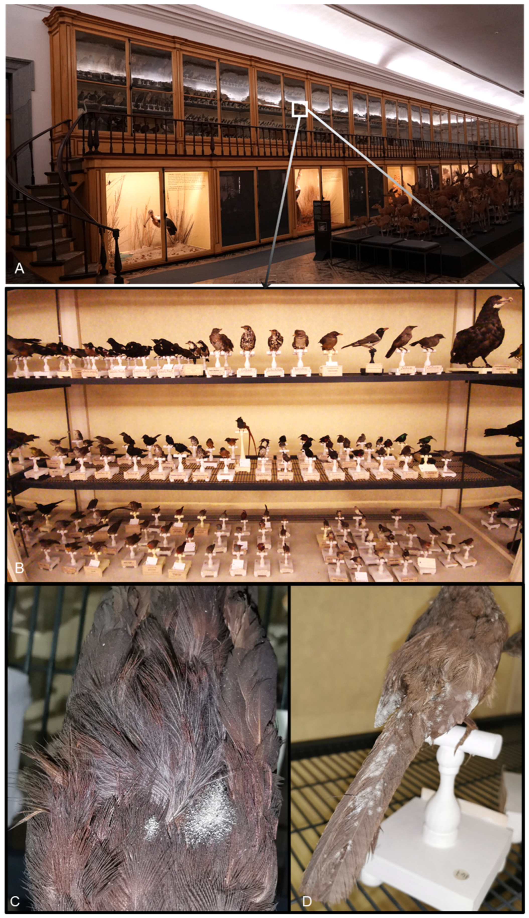
Figure 1. Science Museum of the University of Coimbra. (A). Overview of the exhibition room displaying taxidermized birds from the zoological collection. (B). Close-up of the taxidermized bird collection. (C,D). Close-up of two of the sampled specimens showing visible fungal growth.
Microorganisms 14 01894 g001
Figure 2. Taxa identified through culture-dependent methods. (A) Relative abundance of genera across all samples; (B) species identification and relative abundance per sample.
Microorganisms 14 01894 g002
Figure 3. Network analysis showing the co-occurrence patterns of the cultivable species among samples. ZOO.0038791, ZOO.0038792: Turdoides jardineii tamalakanei; ZOO.0000395: Nectarinia bocagii; ZOO.0039018, ZOO.0039012, ZOO.AVE.AFR.0182.05: Chalcomitra senegalensis saturatior; and ZOO.0038991, ZOO.0038992: Melaenornis pammelaina pammelaina.
Microorganisms 14 01894 g003
Figure 4. Maximum likelihood (ML) tree based on concatenated sequence data from ITS, LSU, SSU, RPB2 and TEF1α showing the placement of the new species, Periconia callaina, described in this study. The scale bar indicates the number of substitutions per site. Nodes are labelled with SH-aLRT/aBayes/ultrafast bootstrap support values ≥ 90%/0.95/95%, with type strains designated by superscript T. The novel species is highlighted in bold font. The tree is rooted with Morosphaeria ramunculicola (KH220).
Microorganisms 14 01894 g004aMicroorganisms 14 01894 g004b
Figure 5. Split network from the PHI test based on combined ITS, LSU, SSU, RPB2, and TEF1α sequences of Periconia callaina (in red) and related taxa, inferred using LogDet transformation and the NeighborNet algorithm. Φw > 0.05 indicates no significant recombination.
Microorganisms 14 01894 g005
Figure 6. Periconia callaina (MUM 25.01, ex-type). (A). Turdoides jardineii tamalakanei, specimen no. ZOO.0038791, from which this fungal species was isolated. (B). Colony morphology on PDA, MEA, and OA, respectively. (C). Colony details on PDA, OA, and MEA, respectively. (D). Red pigment in older colony on OA. (EG,I,J). Conidiophores and conidia. (H). Detail of verrucose hyphae. Scale bars: (BD) = 1 cm; (E) = 200 µm; (F,G,I) = 50 µm; (H,J) = 20 µm.
Microorganisms 14 01894 g006
Disclaimer/Publisher’s Note: The statements, opinions and data contained in all publications are solely those of the individual author(s) and contributor(s) and not of MDPI and/or the editor(s). MDPI and/or the editor(s) disclaim responsibility for any injury to people or property resulting from any ideas, methods, instructions or products referred to in the content.