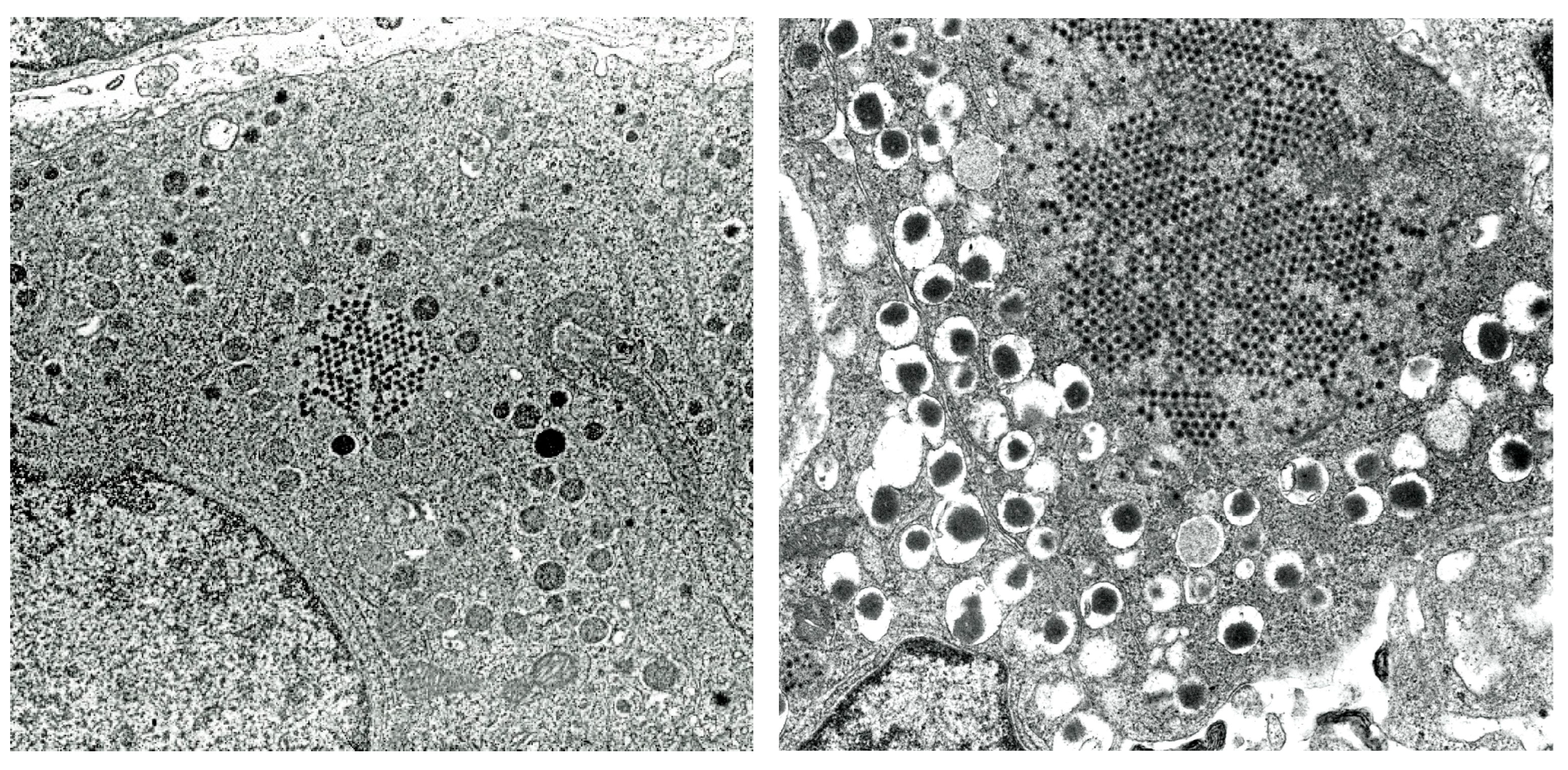
Hyperglycemia in Cats Infected by SARS-CoV-2: Pancreatic Alterations and Potential Antiviral Therapeutics





Abstract
1. Introduction
2. Virus-Induced Hyperglycemia in Humans and Animals
3. SARS-CoV-2 Infection in Cats and Hyperglycemia
3.1. Natural Infection of SARS-CoV-2 in Cats
3.2. Experimental SARS-CoV-2 Infection and Feline Diabetes Induction, Viral Replication and Tissue Distribution
3.3. Metabolic Alterations and Pancreatic Pathology
3.4. Alpha-Cell Tropism and Viral Receptors
3.5. Comparative Feline Models of Infection with Variants of SARS-CoV-2: Metabolic Shift and Cardiorespiratory Reprogramming
3.6. Transmission Dynamics and Tissue Tropism
3.7. Summary of Divergent Evidence in Feline Pancreatic Pathology
4. Main Antiviral Agents for Treating SARS-CoV-2 and Other Coronavirus Infections
5. Discussion
5.1. The Bidirectional Relationship Between Coronaviruses and Glycemic Control
5.2. Pancreatic ACE2 Expression and Comparative Viral Tropisms
5.3. Methodological Challenges and Discrepancies in Autopsy Data
5.4. A Framework for Investigating Coronavirus-Induced Hyperglycemia
5.5. Environmental Triggers of Endocrine Failure
6. Conclusions
Author Contributions
Funding
Institutional Review Board Statement
Informed Consent Statement
Data Availability Statement
Conflicts of Interest
Abbreviations
| ACE2 | Angiotensin-converting enzyme 2 |
| AMPK | AMP-activated protein kinase |
| 3CLpro | 3C-like protease |
| CYP3A4 | Cytochrome P450 3A4 |
| DKA | Diabetic ketoacidosis |
| EMCV | Encephalomyocarditis virus |
| FECV | Feline enteric coronavirus |
| FCoV | Feline coronavirus |
| FIP | Feline infectious peritonitis |
| FIPV | Feline infectious peritonitis virus |
| IC50 | Half-maximal inhibitory concentration |
| IHC | Immunohistochemical |
| Mpro | Main protease enzyme |
| NP | Nucleocapsid protein |
| RAAS | Renin–angiotensin–aldosterone system |
| RdRp | RNA-dependent RNA-polymerase |
| SP | Spike protein |
| T1DM | Type 1 diabetes mellitus |
| TR-FRET | Time-resolved fluorescence resonance energy transfer |
References
- Dahl-Jørgensen, K. Virus as the cause of type 1 diabetes. Trends Mol. Med. 2025, 30, 1020–1027. [Google Scholar] [CrossRef] [Scilit] [PubMed]
- Yoon, J.W.; Austin, M.; Onodera, T.; Notkins, A.L. Virus-induced diabetes mellitus, isolation of a virus from the pancreas of a child with diabetic ketoacidosis. N. Engl. J. Med. 1979, 300, 1173–1179. [Google Scholar] [PubMed]
- Champsaur, H.; Dussaix, E.; Samolyk, D.; Fabre, N.; Back, C.; Assan, R. Diabetes and coxsackie B5 infection. Lancet 1980, 1, 251. [Google Scholar] [CrossRef] [Scilit] [PubMed]
- Szopa, T.M.; Titchener, P.A.; Portwood, N.D.; Taylor, K.W. Diabetes due to viruses—Some recent developments. Diabetologia 1993, 36, 687–695. [Google Scholar] [CrossRef] [Scilit] [PubMed]
- Pedini, B.; Avellini, G.; Morettini, B.; Comodo, N. Diabete mellito post-aftoso nei bovini. Att. Soc. Ital. Sci. Vet. 1962, 16, 443–450. [Google Scholar]
- Craighead, J.E.; McLar, M.F. Diabetes mellitus: Induction in mice by encephalomyocarditis virus. Science 1968, 162, 913–914. [Google Scholar] [CrossRef] [Scilit] [PubMed]
- Horwitz, M.S.; Bladley, L.M.; Hanbertson, J.; Krahl, T.; Lee, J.; Sarvetnick, N. Diabetes induced by Coxsackie virus: Initiation by bystander damage and not by molecular mimicry. Nat. Med. 1998, 4, 781–785. [Google Scholar] [CrossRef] [Scilit] [PubMed]
- Onodera, T.; Jenson, A.B.; Yoon, J.W.; Notkins, A.L. Virus-induced diabetes mellitus: Reovirus infection of pancreatic β cells in mice. Science 1978, 201, 529–531. [Google Scholar] [CrossRef] [Scilit] [PubMed]
- Hayashi, K.; Fujisaki, Y. Abnormal glucose metabolism in murine cytomegalovirus (MCMV) infected mice. Microbiol. Immunol. 1985, 29, 429–439. [Google Scholar] [CrossRef] [Scilit] [PubMed]
- Oldstone, M.B.A.; NerenBerg, M.; Southern, P.; Price, J.; Lewicki, H. Virus infection triggers insulin-dependent diabetes mellitus in transgenic model: Role of anti-viral cytotoxic lymphocytes. Cell 1991, 65, 319–331. [Google Scholar] [PubMed]
- Yoon, J.W.; London, W.T.; Curfman, B.L.; Brown, R.L.; Notkins, A.L. Coxsackie virus B4 produces transient diabetes in non-human primates. Diabetes 1986, 35, 712–716. [Google Scholar] [CrossRef] [Scilit]
- Woo, P.C.Y.; De Groot, R.J.; Haagmans, B.; Lau, S.K.P.; Neuman, B.W.; Perlman, S.; Sola, I.; Van Der Hoek, L.; Wong, A.C.P.; Yeh, S.H. ICTV Virus taxonomy profile: Coronaviridae 2023. J. Gen. Virol. 2023, 104, 001843. [Google Scholar] [CrossRef] [Scilit] [PubMed]
- Gao, Y.-Y.; Wang, Q.; Liang, X.-Y.; Zhang, S.; Bao, D.; Zhao, H.; Li, S.-B.; Wang, K.; Hu, G.-X.; Gao, F.-S. An updated review of feline coronavirus: Mind the two biotypes. Virus Res. 2023, 326, 199059. [Google Scholar] [CrossRef] [Scilit] [PubMed]
- Kiss, I.; Kecskeméti, S.; Tanyi, J.; Klingeborn, B.; Belák, S. Preliminary studies on feline Coronavirus distribution in naturally and experimentally infected cats. Res. Vet. Sci. 2000, 68, 237–242. [Google Scholar] [CrossRef] [Scilit] [PubMed]
- Wills, S.E.; Beaufrère, H.H.; Brisson, B.A.; Fraser, R.S.; Smith, D.A. Pancreatitis and systemic coronavirus Infection in a Ferret (Mustela putorius furo). Comp. Med. 2018, 68, 208–211. [Google Scholar] [CrossRef] [Scilit] [PubMed]
- Park, E.S.; Kuroda, Y.; Uda, A.; Kaku, Y.; Okutani, A.; Hatta, A.; Takemoto, K.; Ishijima, K.; Inoue, Y.; Harada, M.; et al. The comparison pathogenicity among SARS-CoV-2 variants in domestic cats. Sci. Rep. 2024, 14, 21825. [Google Scholar] [CrossRef] [Scilit] [PubMed]
- Steenblock, C.; Richter, S.; Berger, I.; Barovic, M.; Schmid, J.; Schubert, U.; Jarzebska, N.; von Mässenhausen, A.; Linkermann, A.; Schürmann, A.; et al. Viral infiltration of pancreatic islets in patients with COVID-19. Nat. Commun. 2021, 12, 3534. [Google Scholar] [CrossRef] [Scilit] [PubMed]
- Dietter, D.; Bjork, J.; Palmkvist, E.; Carlsson, A. Trends in type 1 diabetes incidence between 2007 and 2023 and their association with SARS-CoV-2 infection in a population-based match cohort study among individuals under 30 years old in Sweden. Diabetologia 2025, 68, 2732–2742. [Google Scholar] [CrossRef] [Scilit] [PubMed]
- Zhang, Y.; Gao, J.; Huang, K.; Zhao, Y.; Hui, X.; Wang, T.; Hu, C.; Sun, X.; Yang, Y.; Wu, C.; et al. SARS-CoV-2 infection causes hyperglycemia in cats. J. Infect. Dis. 2022, 226, 1568–1576. [Google Scholar] [CrossRef] [Scilit] [PubMed]
- Rochowski, M.T.; Jayathilake, K.; Balcerak, J.M.; Selvan, M.T.; Gunasekara, S.; Miller, C.; Rudd, J.M.; Lacombe, V.A. Impact of delta SARS-CoV-2 infection on glucose metabolism: Insights on host metabolism and virus crosstalk in a feline model. Viruses 2024, 16, 295. [Google Scholar] [CrossRef] [Scilit] [PubMed]
- Debuysschere, C.; Nekoha, M.P.; Alidjinou, E.K.; Hober, D. The relationship between SARS-CoV-2 and type 1 diabetes mellitus. Nat. Rev. Endocrinol. 2024, 10, 588–599. [Google Scholar] [CrossRef] [Scilit] [PubMed]
- Nishimura, A.; Matsumura, K.; Kikuno, S.; Nagasawa, K.; Okubo, M.; Mori, Y.; Kobayashi, T. Slowly progressive type 1 diabetes mellitus: Current knowledge and future perspectives. Diabetes Metab. Syndr. Obes. 2019, 12, 2461–2477. [Google Scholar] [CrossRef] [Scilit] [PubMed]
- Toniolo, A.; Onodera, T.; Yoon, J.W.; Notkins, A.L. Induction of diabetes by cumulative environmental insults from virus and chemicals. Nature 1980, 288, 383–385. [Google Scholar] [CrossRef] [Scilit] [PubMed]
- van der Hide, V.; McArdle, S.; Nelson, M.S.; Cerosaletti, K.; Gnjatic, S.; Mikulski, Z.; Posgai, A.L.; Kusmartseva, I.; Atkinson, M.A.; Homann, D. Integrated histopathology of the human pancreas throughout stages of type 1 diabetes progression. Nat. Commun. 2026. Online ahead of print. [Google Scholar] [CrossRef] [Scilit] [PubMed]
- Vazeou, G.A.; Kordonouri, O.; Witsch, M.; Hermann, J.M.; Forsander, G.; de Beaufort, C.; Veeze, H.J.; Maffeis, C.; Cherubini, V.; Cinek, O.; et al. Seasonality at the clinical onset of type 1 diabetes- lessons from the SWEET database. Pediatr. Diabetes 2016, 17, 32–37. [Google Scholar] [CrossRef] [Scilit] [PubMed]
- Hanafusa, T.; Imagawa, A. Fluminant type 1 diabetes: A novel clinical entity requiring special attentions by all medical practitioners. Nat. Clin. Pract. Endoclinol. Metab. 2007, 3, 36–45. [Google Scholar] [CrossRef] [Scilit] [PubMed]
- Salvatoni, A.; Baj, A.; Bianchi, G.; Federico, G.; Colombo, M.; Toniolo, A. Intrafamilial spread of enterovirus infections at the clinical onset of type 1 diabetes. Pediatr. Diabetes 2013, 14, 407–416. [Google Scholar] [CrossRef] [Scilit] [PubMed]
- Bopegamage, S.; Precechteva, J.; Marosova, L.; Stipalova, D.; Sojka, M.; Borsanyiova, M.; Gomolcak, P.; Berkakova, K.; Galama, J.M.D. Outcome of challenge with Coxsackievirus B4 in young mice after maternal infection with the same virus during gestation. FEMS Immunol. Med. Microbiol. 2012, 64, 184–190. [Google Scholar] [CrossRef] [Scilit] [PubMed]
- Onodera, T.; Toniolo, A.; Ray, U.R.; Jenson, A.B.; Knazek, R.A.; Notkins, A.L. Virus-induced diabetes mellitus. XX. Polyendocrinopathy and autoimmunity. J. Exp. Med. 1981, 153, 1457–1473. [Google Scholar] [CrossRef] [Scilit] [PubMed]
- Srinivasappa, J.; Garzelli, C.; Onodera, T.; Ray, U.R.; Notkins, A.L. Virus-induced thyroiditis. Endocrinology 1988, 122, 563–566. [Google Scholar] [CrossRef] [Scilit] [PubMed]
- Kobayashi, T.; Tanaka, S.; Aida, K. Unique pathological changes in the pancreas of fulminant type 1 diabetes. Diabetol. Int. 2020, 11, 323–328. [Google Scholar] [CrossRef] [Scilit] [PubMed]
- Pan, Y.; Zhong, X.; Qiu, W.; Zhao, F.; Yao, J. New-Onset Fulminant Type 1 Diabetes After SARS-CoV-2 Infection. Diabetes Care 2023, 46, e140–e142. [Google Scholar] [CrossRef] [Scilit] [PubMed]
- Pagani, G.; Lai, A.; Berga, A.; Rizzo, A.; Stranieri, A.; Giordano, A.; Paltrinieri, S.; Lelli, D.; Decaro, N.; Rusconi, S.; et al. Human-to-cat SARS-CoV-2 transmission; Case report and full-genome sequencing from an infected pet and its owner in Northern Italy. Pathogens 2021, 10, 252. [Google Scholar] [CrossRef] [Scilit] [PubMed]
- USDA APHIS. SARS-CoV-2 in Animals. Available online: https://www.aphis.usda.gov/sars-cov-2 (accessed on 9 May 2026).
- Homepage-FEDIAF. Available online: https://fediaf.org/ (accessed on 6 August 2021).
- Kraft, E.; Denolly, S.; Boson, B.; Angelloz-Pessey, S.; Levaltier, S.; Nesi, N.; Corber, S.; Leterrier, B.; Fritz, M.; Leroy, E.M.; et al. Report of one-year prospective surveillance of SARS-CoV-2 in dog and cats in France with various exposure risks: Confirmation of a low prevalence of shedding, detection and complete sequencing of an alpha variant in a cat. Viruses 2021, 13, 1759. [Google Scholar] [CrossRef] [Scilit] [PubMed]
- Tracking SARS-CoV-2 Variants. Available online: https://www.who.int/activities/tracking-SARS-CoV-2-variants (accessed on 9 May 2026).
- Hamer, S.A.; Chai, R.R.; Zecca, I.B.; Auchland, L.D.; Boundy, C.M.; Davila, E.; Busselman, R.E.; Tang, W.; Pauvolid-Correa, A.; Kilan, M.L.; et al. SARS-CoV-2B.1.1.7 variant of concern detected in a pet dog and cat after exposure to a person with COVID-19, USA. Transbound. Emerg. Dis. 2022, 69, 1656–1658. [Google Scholar] [CrossRef] [Scilit] [PubMed]
- Zoccola, F.; Beltramo, C.; Magris, G.; Peletto, S.; Acutis, P.; Bozzetta, E.; Radovic, S.; Zappulla, F.; Porzio, A.M.; Gennero, M.S.; et al. First detection of an Italian human-to-cat SARS-CoV-2 alpha variant-linage B.1.1.7. One Health 2021, 13, 100295. [Google Scholar] [CrossRef] [Scilit] [PubMed]
- Barroso-Arevalo, S.; Rivera, B.; Dominguez, L.; Sanchez-Vizcaino, J. First detection of SARS-CoV-2B.1.1.7 variant of concern in an asymptomatic dog in Spain. Viruses 2021, 13, 1379. [Google Scholar] [CrossRef] [Scilit] [PubMed]
- Shi, J.; Wen, Z.; Zhong, G.; Yang, H.; Wang, C.; Huang, B.; Liu, R.; He, X.; Shuai, L.; Sun, Z.; et al. Susceptibility of ferrets, cats, dogs and other domesticated animals to SARS-coronavirus-2. Science 2020, 368, 1016–1020. [Google Scholar] [CrossRef] [Scilit] [PubMed]
- Yamada, K.; Kuroda, Y.; Yamamoto, T.; Nishio, H.; Yamada, C.; Kobayashi, M.; Morishima, Y.; Maeda, K. A domestic cat with respiratory symptoms caused by severe acute respiratory syndrome coronavirus 2 infection. J. Jpn. Vet. Med. Assoc. 2022, 75, e62–e68. [Google Scholar] [CrossRef] [Scilit]
- Tang, X.; Uhl, S.; Zhang, T.; Xue, D.; Li, B.; Vandana, J.J.; Acklin, J.A.; Bonnycastle, L.L.; Narisu, N.; Erdos, M.R.; et al. SARS-CoV-2 infection induces beta cell transdifferentiation. Cell Metab. 2021, 33, 1577–1591.e7. [Google Scholar] [CrossRef] [Scilit] [PubMed]
- Watts, B.; Altilio, I.; Alcazar, O.; Alvarez-Cubela, S.; Berggren, P.-O.; Abdulreda, M.H.; Pastori, R.L.; Dominguez-Bendala, J. Β-cell neogenesis from the pancreatic ductal epithelium revealed dynamically by long-term intravital imaging. Diabetes 2025, 74, 2016–2022. [Google Scholar] [CrossRef] [Scilit] [PubMed]
- Beeck, A.; Eizirik, D.L. Viral infections in type 1 diabetes mellitus—Why the β cells? Nat. Rev. Endocrinol. 2016, 12, 263–273. [Google Scholar] [CrossRef] [Scilit] [PubMed]
- Kim, H.S.; Lee, M.K. Β-cell regeneration through the transdifferentiation of pancreatic cells: Pancreatic progenitor cells in the pancreas. J. Diabetes Investig. 2016, 7, 286–296. [Google Scholar] [CrossRef] [Scilit] [PubMed]
- Kerper, N.; Ashe, S.; Hebrok, M. Pancreatic β-cell development and regeneration. Cold Spring Harb. Perspect. Biol. 2022, 14, a040741. [Google Scholar] [CrossRef] [Scilit] [PubMed]
- Onodera, T.; Hayashi, T. Reoviruses. Diabetes and Viruses; Hyoty, H., Toniolo, A., Zuckerman, A.J., Eds.; Springer Science + Business Media: New York, NY, USA, 2013; pp. 71–81. [Google Scholar]
- Papadimitriou, J.M.; Walters, M.N. Studies on the exocrine pancreas. II. Ultrastructural investigation of reovirus pancreatitis. Am. J. Pathol. 1967, 51, 387–403. [Google Scholar] [PubMed]
- Nooruzzaman, M.; Diel, D.G. Induction dynamics, pathogenesis and immunity to SARS-CoV-2 in naturally susceptible animal species. J. Immunol. 2023, 211, 1195–1201. [Google Scholar] [CrossRef] [Scilit] [PubMed]
- Brüssow, H. Antivirals Targeting Coronavirus RNA-Dependent RNA Polymerase and Main Protease: From Mechanisms of Action to Outcomes in COVID-19 Clinical Trials. Microb. Biotechnol. 2026, 19, e70342. [Google Scholar] [CrossRef] [Scilit] [PubMed]
- Tasker, S.; Spiri, A.M.; Hartmann, K.; Addie, D.D.; Belák, S.; Bergmann, M.; Egberink, H.; Frymus, T.; Hofmann-Lehmann, R.; Marsilio, F.; et al. Update on Treatment of Feline Infectious Peritonitis: European Advisory Board on Cat Diseases (ABCD) Guidelines. Viruses 2026, 18, 452. [Google Scholar] [CrossRef] [Scilit] [PubMed]
- Khalil, A. Antiviral Nucleoside Mimics: The Development and Future Perspective of Molnupiravir. Future Med. Chem. 2026, 18, 1895–1906. [Google Scholar] [CrossRef] [Scilit] [PubMed]
- Loos, N.H.C.; Beijnen, J.H.; Schinkel, A.H. The Inhibitory and Inducing Effects of Ritonavir on Hepatic and Intestinal CYP3A and Other Drug-Handling Proteins. Biomed. Pharmacother. 2023, 162, 114636. [Google Scholar] [CrossRef] [Scilit] [PubMed]
- Syed, Y.Y. Ensitrelvir Fumaric Acid: First Approval. Drugs 2024, 84, 721–728. [Google Scholar] [CrossRef] [Scilit] [PubMed]
- Mik, V.; Benz, L.S.; Voller, J.; Dunzendorfer-Matt, T.; Weiss, M.S.; Kryštof, V. A Patent Review of Mpro Protease Inhibitors for the Treatment of COVID-19 Infections (2020–Present). Expert Opin. Ther. Pat. 2026, 36, 91–109. [Google Scholar] [CrossRef] [Scilit] [PubMed]
- Wang, W.; Zeng, P.; Liu, T.; Zhou, X.; Lin, C.; Guo, L.; Wang, Q.; Li, J. Structural Basis of Main Proteases of Coronavirus Bound to Bofutrelvir. J. Mol. Biol. 2024, 436, 168784. [Google Scholar] [CrossRef] [Scilit] [PubMed]
- Ye, Z.; Dai, W.; Zhang, S.; Xiang, Y.; Wang, J.; Zhang, Y.; Cao, W.; Neyts, J.; Li, Z.; Feng, F.; et al. A Clinical SARS-CoV-2 Mpro Inhibitor Blocks Replication of Multiple Enteroviruses and Confers Oral in Vivo Protection in Animal Models. PLoS Pathog. 2026, 22, e1014051. [Google Scholar] [CrossRef] [Scilit] [PubMed]
- Liang, J.; Xu, W.; Pan, X.; Han, S.; Zhang, L.; Wen, H.; Ding, M.; Zhang, W.; Peng, D. Advances Research in Porcine Enteric Coronavirus Therapies and Antiviral Drugs. Vet. Q. 2024, 44, 1–49. [Google Scholar] [CrossRef] [Scilit] [PubMed]
- Wang, Q.; Wu, J.; Wang, H.; Gao, Y.; Liu, Q.; Mu, A.; Ji, W.; Yan, L.; Zhu, Y.; Zhu, C.; et al. Structural basis for RNA replication by the SARS-CoV-2 polymerase. Cell 2020, 182, 417–428.e13. [Google Scholar] [CrossRef] [Scilit] [PubMed]
- Chen, G.; Yuan, J.; Wei, Y.; Wu, Y.; Zhang, Q.; Li, X.; Fu, L.; Gao, Y.; Zheng, Y.; Sun, H.; et al. Chinese Expert Consensus on the combined use of antiviral drugs for novel coronavirus infection. Biosci. Trends 2025, 19, 484–494. [Google Scholar] [CrossRef] [Scilit] [PubMed]
- Lu, J.; Bai, Y.; Wang, Y.; Yang, L.; Jin, Y.; Dong, J. Effect of GS-441524 in combination with the 3C-like protease inhibitor GC376 on treatment of naturally transmitted feline infectious peritonitis. Front. Vet. Sci. 2022, 9, 1002488. [Google Scholar] [CrossRef] [Scilit] [PubMed]
- Uema, M.; Hyuga, M.; Yonemitsu, K.; Hyuga, S.; Amakura, Y.; Uchiyama, N.; Mizoguchi, K.; Odaguchi, H.; Goda, Y. Antiviral effect of ephedrine alkaloids-free ephedra herb extract against SARS-CoV-2 in vitro. Microorganisms 2023, 11, 534. [Google Scholar] [CrossRef] [Scilit] [PubMed]
- Metwally, A.A.; Mehta, P.; Johnson, B.S.; Nagarjuna, A.; Snyder, M.P. COVID-19-induced new-onset diabetes: Trends and technologies. Diabetes 2021, 70, 2733–2744. [Google Scholar] [CrossRef] [Scilit] [PubMed]
- Gagliardi, S.; Hotchin, T.H.; Tibebe, H.; Hillmer, G.; Marquez, D.; Izumi, C.; Chang, J.; Diggs, A.; Ezaki, J.; Suzuki, Y.; et al. The renin-angiotensin system modulates SARS-CoV-2 entry via ACE2 receptor. Viruses 2025, 17, 1014. [Google Scholar] [CrossRef] [Scilit] [PubMed]
- Yang, J.K.; Lin, S.S.; Ji, X.L.; Guo, L.M. Binding of SARS coronavirus to its receptor damages islets and causes acute diabetes. Acta Diabetol. 2010, 47, 193–199. [Google Scholar] [CrossRef] [Scilit] [PubMed]
- Yang, L.; Han, Y.; Nilsson-Payant, B.E.; Gupta, V.; Wang, P.; Duan, X.; Tang, X.; Zhu, Z.; Jaffre, F.; Zhang, T.; et al. A human pluripotent stem cell-based platform to study SARS-CoV-2 tropism and model virus infection in human cell and organoids. Cell Stem Cell 2020, 27, 125–136. [Google Scholar] [CrossRef] [Scilit] [PubMed]
- Rodriguez-Calvo, T. Enterovirus infection and type 1 diabetes: Unraveling the crime scene. Clin. Exp. Immunol. 2018, 195, 15–24. [Google Scholar] [CrossRef] [Scilit] [PubMed]
- Krogvold, L.; Genoni, A.; Puggioni, A.; Campani, D.; Richardson, S.J.; Flaxman, C.S.; Edwin, B.; Buanes, T.; Dahl-Jorgensen, K.; Toniolo, A. Live enteroviruses, but not other viruses, detected in human pancreas at the onset of type 1 diabetes in the DiViD study. Diabetologia 2022, 65, 2108–2120. [Google Scholar] [CrossRef] [Scilit] [PubMed]
- Federico, G.; Genoni, A.; Puggioni, A.; Saba, A.; Gallo, D.; Randazzo, E.; Salvatoni, A.; Toniolo, A. Vitamin D status, enterovirus infection, and type 1 diabetes in Italian children/adolescents. Pediatr. Diabetes 2018, 19, 923–929. [Google Scholar] [CrossRef] [Scilit] [PubMed]
- Carre, A.; Vecchio, F.; Flodstrom-Tullberg, M.; You, S.; Mallone, R. Coxsackievirus and type 1 diabetes: Diabetogenic mechanisms and implication for prevention. Endocr. Rev. 2023, 44, 737–751. [Google Scholar] [CrossRef] [Scilit] [PubMed]
- Rogers, M.A.M. Decline in type 1 diabetes in young children after rotavirus vaccination: Data from 8 countries. AJPM Focus 2025, 4, 100409. [Google Scholar] [CrossRef] [Scilit] [PubMed]
- Wu, C.T.; Lidsky, P.V.; Xiao, Y.; Lee, I.T.; Cheng, R.; Nakayama, T.; Jiang, S.; Demeter, J.; Bevacqua, R.J.; Chang, C.A.; et al. SARS-CoV-2 infects human pancreatic β cells and elicits β cell impairment. Cell Metab. 2021, 33, 1565–1576. [Google Scholar] [CrossRef] [Scilit] [PubMed]
- Kusmartseva, J.; Wu, W.; Syed, F.; Van Der Heide, V.; Jorgensen, M.; Joseph, P.; Tang, X.; Candelario-Jalil, E.; Yang, C.; Nick, H.; et al. Expression of SARS-CoV-2 entry factors in the pancreas of normal organ donors and individuals with COVID-19. Cell Metab. 2020, 32, 1041–1051. [Google Scholar] [CrossRef] [Scilit] [PubMed]
- Qadir, M.M.F.; Bhondeley, M.; Beatty, W.; Gaupp, D.D.; Doyle-Meyers, L.A.; Fischer, T.; Bandyopadhyay, I.; Blair, R.V.; Bohm, R.; Rappaport, J.; et al. SARS-CoV-2 infection of the pancreas promotes thrombofibrosis and is associated with new-onset diabetes. JCI Insight 2021, 6, e151551. [Google Scholar] [CrossRef] [Scilit] [PubMed]
- Muller, J.A.; Gros, R.; Conzelmann, C.; Kruger, J.; Merle, U.; Steinhart, J.; Weil, T.; Koepke, L.; Buzzo, C.P.; Read, C.; et al. SARS-CoV-2 infects and replicates in cells of the human endocrine and exocrine pancreas. Nat. Metab. 2021, 3, 149–165. [Google Scholar] [CrossRef] [Scilit] [PubMed]
- Craig, M.E.; Nair, S.; Stein, H.; Rawlinson, W.D. Viruses and type1 diabetes: A new look at an old story. Pediatr. Diabetes 2013, 14, 149–158. [Google Scholar] [CrossRef] [Scilit] [PubMed]
- Rodriguez-Calvo, T.; Sabouri, S.; Anquetil, F.; von Herrath, M.G. The viral paradigm in type 1 diabetes: Who are the main suspects? Autoimm. Rev. 2016, 15, 964–969. [Google Scholar] [CrossRef] [Scilit] [PubMed]


| Animal Host | Virus | Viral Family/Genus | Primary Mechanism/Pathogenesis | Metabolic & Clinical Outcome | Key References |
|---|---|---|---|---|---|
| Bovines (Cattle) | Foot-and-Mouth Disease Virus (FMDV) | Picornaviridae, Aphtovirus | Lytic infection of pancreatic islet beta-cells leading to loss of insulin synthesis. | Transient or permanent hyperglycemia, chronic insulin deficiency. | Pedini 1962 [5] |
| Mice | Encephalomyocarditis Virus, Variant D (EMC-D) | Picornaviridae, Cardiovirus | Cytolytic destruction of pancreatic beta-cells without autoimmune T-cell involvement. | Type 1-like insulin-dependent diabetes mellitus (rapid onset within 3–5 days). | Craighead 1968 [6] |
| Mice | Coxsackievirus B | Picornaviridae, Enterovirus | Beta-cell cytolysis and with virus-induced autoimmunity (bystander activation and release of sequestered autoantigens). | Acute or chronic hyperglycemia, autoimmune Type 1-like diabetes susceptible strains (e.g., SJL/J, CD-1). | Horwitz 1998 [7] |
| Mice | Reovirus type 1 and 3 | Reoviridae, Orthoreovirus | Infection of alpha, beta, delta pancreatic islet cells with autoantibodies against islet cells and other endocrine organs. | Moderate hyperglycemia and polyendocrine autoimmunity. | Onodera 1978 [8] |
| Mice | Murine Cytomegalovirus (MCMV) | Herpesviridae, Betaherpesvirinae | Infection of pancreatic acinar and stromal cells with focal pancreatitis, immune infiltration, cytokine-mediated beta-cell dysfunction. | Non immune-mediated glucose intolerance. | Hayashi 1985 [9] |
| Mice | Lymphocytic Choriomeningitis Virus (LCMV) | Arenaviridae, Mammarenavirus | Virus-triggered autoimmune beta-cell destruction mediated by CD8+ T lymphocytes targeting viral antigens in islet cells. | Rapid-onset autoimmune type 1-like diabetes with hyperglycemia and insulitis. | Oldstone 1991 [10] |
| Monkeys (Rhesus, Cynomolgus) | Coxsackievirus B4 | Picornaviridae, Enterovirus | Viral insulitis, focal necrosis of islet beta-cells, inflammatory depression of insulin secretion. | Impaired glucose tolerance, transient or overt hyperglycemia. | Yoon 1986 [11] |
| Study | Primary Pancreatic/Metabolic Findings | Islet Architecture Status |
|---|---|---|
| Zhang et al. [19] | Marked hyperglycemia; strong viral NP expression localized to islet alpha-cells. | Documented IHC alterations. |
| Rochowski et al. [20] | Significantly decreased serum insulin; cardiac/pulmonary metabolic reprogramming. | Preserved beta-cell numbers; minimal islet inflammation (1/24 cats). |
| Nooruzzaman & Diel [50] | Robust respiratory replication and shedding; clear systemic immune response. | No primary endocrine or pancreatic deficits reported. |
| Antiviral Drug | Primary Target | Mechanism of Action | Pharmacology | References |
|---|---|---|---|---|
| Remdesivir | Inhibitor of viral RdRp | Prodrug of GS-441524 (adenosine analog). Converted into active nucleoside triphosphate, it competes with natural ATP for incorporation into the nascent RNA strand, causing premature and delayed chain termination. | Broad-spectrum efficacy due to highly conserved RdRp target across Coronaviridae. | Brüssow 2026 [51] |
| Tasker 2026 [52] | ||||
| Molnupiravir | Inhibitor of viral RdRp | Metabolized into beta-D-hydroxycytidine triphosphate, incorporated into viral RNA. During replication cycles, the viral polymerase misinterprets the embedded modified nucleotides. This results in accumulating transition mutations that make the viral genome non-functional. | Estimated 2–10 times higher potency than remdesivir in certain in vitro models; requires monitoring for mutagenic risks. | Khalil 2026 [53] |
| Ritonavir | Inhibitor of main viral protease | Targets the viral 3C-like protease (3CLpro)/aspartic protease. However, as monotherapy, its direct benefit against Coronaviruses is negligible. | Primary function: by inhibiting host CYP3A4 enzymes extends the half-life of co-administered drugs. | Loos 2023 [54] |
| Ensitrelvir | Inhibitor of main viral protease | Non-covalent, non-peptide inhibitor that binds to 3CLpro, halting viral polyprotein processing and protease-derived cellular damage. | High affinity for wild-type protease with an IC50 = 0.049 mM, comparable to nirmatrelvir. | Syed 2024 [55] |
| Mik 2026 [56] | ||||
| Bofutrelvir | Inhibitor of main viral protease | Inhibitor of the main protease 3CLpro, blocking polyprotein cleavage and reducing viral yield and RNA copy numbers. | IC50 = 0.053 μM, TR-FRET assays. Scientific profiles emerging in 2025–2026. | Wang 2024 [57] |
| Ye 2026 [58] |
| Antiviral Drug | Primary Mechanism | United States (FDA) | Europe (EMA) | Japan (PMDA) |
|---|---|---|---|---|
| Remdesivir | RdRp Inhibitor (chain termination) | Fully Approved | Fully Approved | Fully Approved |
| Nirmatrelvir/Ritonavir | 3CLpro inhibitor plus CYP3A4 booster | Fully Approved | Fully Approved | Fully Approved |
| Molnupiravir | RdRp Inhibitor (lethal mutagenesis) | Emergency Use Authorization | Not Approved (refused by EMA in 2023) | Fully Approved |
| Ensitrelvir | 3CLpro Inhibitor (non-covalent) | Not Approved (Phase 3 Trials) | Not Approved (Phase 3 Trials) | Fully Approved (Conditional/Emergency) |
Disclaimer/Publisher’s Note: The statements, opinions and data contained in all publications are solely those of the individual author(s) and contributor(s) and not of MDPI and/or the editor(s). MDPI and/or the editor(s) disclaim responsibility for any injury to people or property resulting from any ideas, methods, instructions or products referred to in the content. |
© 2026 by the authors. Licensee MDPI, Basel, Switzerland. This article is an open access article distributed under the terms and conditions of the Creative Commons Attribution (CC BY) license.
Share and Cite
Onodera, T.; Seo, S.; Sakudo, A.; Toniolo, A. Hyperglycemia in Cats Infected by SARS-CoV-2: Pancreatic Alterations and Potential Antiviral Therapeutics. Microorganisms 2026, 14, 1884. https://doi.org/10.3390/microorganisms14091884
Onodera T, Seo S, Sakudo A, Toniolo A. Hyperglycemia in Cats Infected by SARS-CoV-2: Pancreatic Alterations and Potential Antiviral Therapeutics. Microorganisms. 2026; 14(9):1884. https://doi.org/10.3390/microorganisms14091884
Chicago/Turabian StyleOnodera, Takashi, Sungwook Seo, Akikazu Sakudo, and Antonio Toniolo. 2026. "Hyperglycemia in Cats Infected by SARS-CoV-2: Pancreatic Alterations and Potential Antiviral Therapeutics" Microorganisms 14, no. 9: 1884. https://doi.org/10.3390/microorganisms14091884
APA StyleOnodera, T., Seo, S., Sakudo, A., & Toniolo, A. (2026). Hyperglycemia in Cats Infected by SARS-CoV-2: Pancreatic Alterations and Potential Antiviral Therapeutics. Microorganisms, 14(9), 1884. https://doi.org/10.3390/microorganisms14091884