Article

Verrucones A–H, Non-Acetate Starter Aromatic Polyketides Discovered by Heterologous Expression of a Type II PKS Gene Cluster


Helmholtz International Lab for Anti-Infectives, Shandong University-Helmholtz Institute of Biotechnology, State Key Laboratory of Microbial Technology, Shandong University, Qingdao 266237, China
*
Authors to whom correspondence should be addressed.
These authors contributed equally to this work.
Microorganisms 2026, 14(8), 1669; https://doi.org/10.3390/microorganisms14081669
Submission received: 29 June 2026 / Revised: 22 July 2026 / Accepted: 27 July 2026 / Published: 30 July 2026
(This article belongs to the Special Issue Exploration of Marine Microbial Resources)

Abstract

Genome mining of the marine-derived Streptomyces sp. S42 uncovered a type II polyketide synthetase (T2 PKS) biosynthetic gene cluster (BGC) harboring a gene for 3-ketoacyl-ACP synthase III (KAS III), a hallmark of non-acetate starter unit incorporation, suggesting that the BGC may produce previously unidentified aromatic polyketides. Heterologous expression and promoter engineering of this prioritized BGC in host Streptomyces albus J1074 activated the biosynthetic pathway, leading to the isolation of eight new polycyclic aromatic derivatives, verrucones A–H (18). Comprehensive structural elucidation via NMR and HRESIMS revealed that these compounds feature either a 2-methylbutyryl or an isobutyryl starter unit and can be classified into three distinct skeletal types. Based on these findings and bioinformatic analysis, a plausible biosynthetic pathway for 18 involving divergent spontaneous cyclization from a common nascent polyketide intermediate was proposed. Among the isolated compounds, 15 exhibited inhibitory activity against several protein tyrosine phosphatases (PTPs) with IC50 values ranging from 1.84 μM to 24.82 μM. This study presents a successful case study demonstrating that combining KAS III-targeted genome mining with heterologous expression is a viable approach for discovering non-acetate-primed aromatic polyketides.

1. Introduction

Aromatic polyketides from actinobacteria represent a class of natural products with potential for clinical application due to their potent biological activities. Representative aromatic polyketide drugs include doxorubicin and tetracycline, which are approved anticancer and antibacterial drugs, respectively [1,2]. In addition, aromatic polyketides exhibit many other biological activities, such as antiviral, antioxidant, and enzyme inhibition activities, making them invaluable leads in drug discovery and development [3]. Among these diverse bioactivities, the inhibitory effects against protein tyrosine phosphatases (PTPs) have garnered increasing attention [4,5]. Protein tyrosine phosphatases jointly regulate tyrosine phosphorylation in cellular processes. Modulating PTP activity holds promise for the treatment of various human diseases, such as type II diabetes, obesity, cancer, and inflammatory diseases [6,7,8,9]. Therefore, discovery of new aromatic polyketides for PTP inhibitor screening is of significant value for the treatment of related diseases.
Bacterial aromatic polyketides are generally biosynthesized by type II polyketide synthase (T2 PKS) systems [3,10]. A defining feature of these systems is the so-called “minimal PKS”, which catalyzes iterative decarboxylative condensations between an acyl-CoA starter unit and malonyl-CoA extender units, thereby generating a growing polyketide chain. Subsequently, ketoreductases (KRs) together with aromatases (AROs) and cyclases (CYCs) coordinate the regioselective folding and cyclization of the linear intermediate. In this process, the C-9 KR, along with specific ARO and CYC, is essential for proper aromatization; otherwise, the highly reactive nascent polyketide chain will undergo intramolecular spontaneous aberrant cyclization, forming shunt products [11]. Finally, the normal cyclized product then serves as a common precursor, which undergoes a series of tailoring reactions such as redox reactions, group transfers, and rearrangements to yield the mature polycyclic aromatic products [12].
Based on the biosynthetic logic outlined above, the structural diversity of aromatic polyketides is largely governed by variations in starter units, cyclization patterns, and tailoring modifications. While the vast majority of aromatic polyketides are initiated with acetyl-CoA, a growing number of biosynthetic pathways have been found to employ nonacetate primers, including benzoate, propionate, butyrate, and even amino acids derived units [13,14,15]. Unlike canonical acetyl-CoA primed formation, the biosynthesis and incorporation of such atypical starters often involve the additional 3-ketoacyl-ACP synthase (KAS III) that selectively recognizes and loads the alternative starter units into the minimal PKS for elongation. Consequently, KAS III-encoding genes within a biosynthetic gene cluster (BGC) can serve as a valuable genomic marker for the targeted discovery of non-acetate primed aromatic polyketides.
With the expanding availability of genomic data, increasing evidence indicates that actinobacteria genomes harbor an abundance of T2 PKS BGCs, many of which remain silent under standard laboratory cultivation conditions [16]. To unlock this hidden reservoir, genome mining combined with heterologous expression has emerged as one of the most effective and widely adopted strategies [17,18]. Bioinformatics platform antiSMASH [19] assists in the prediction of T2 PKS BGCs from genomic sequences, serving as the basis for subsequent prioritization and dereplication, which facilitated the targeted selection of promising gene clusters. Subsequent heterologous expression of entire BGCs in well-characterized heterologous hosts effectively bypasses the regulatory constraints of the native producer and often leads to the activation of silent biosynthetic pathways [20,21]. This approach has proven remarkably successful by our research along with others, leading to the discovery of numerous novel aromatic polyketides from previously uncharacterized clusters and substantially expanding the chemical space of bacterial type II PKS products [21,22,23,24,25].
During our ongoing genome mining efforts to discover new bioactive aromatic polyketides from actinobacteria [22,26], the marine-derived Streptomyces sp. S42 attracted our attention. Bioinformatic analysis revealed that its genome contains a T2 PKS BGC, designated ver, which harbors a KAS III encoding gene, suggesting a non-acetate starter unit incorporation in its corresponding products. Compared with homologous gene clusters [27,28], the ver cluster stands out by possessing several oxidoreductases with lower similarity alongside the conserved core genes, suggesting a potential for producing new metabolites. Heterologous expression of this BGC in the host Streptomyces albus J1074 successfully activated the biosynthetic pathway, leading to the identification of eight new aromatic polyketides, verrucones A–H (18). These compounds not only feature unique 2-methylbutanyl or isobutyryl moieties as their starter units but also display diverse cyclization patterns, giving rise to three distinct skeletal frameworks. Details of the structure elucidation, biological evaluation, and plausible biosynthetic pathways of these new compounds are reported herein.

2. Materials and Methods

2.1. General Experimental Procedures

UV spectra and UHPLC-HRESIMS data were acquired using a Thermo Scientific™ Dionex™ Ultimate 3000 system (Thermo Fisher Scientific, Waltham, MA, USA) coupled with a Bruker microTOF Q III mass spectrometer (Impact HD) (Bruker Corporation, Rheinstetten, Germany) equipped with a standard ESI ion source in positive electrospray ionization mode. A Thermo Scientific™ Acclaim™ C18 column (2.1 × 100 mm, 2.2 μm) (Thermo Fisher Scientific, Waltham, MA, USA) was used. The elution program was set as follows: solvent A, H2O with 0.1% formic acid (FA); solvent B, 0.1% FA in acetonitrile (ACN), 0–3 min 5% B, 3–18 min, 5–95% B, 18–22 min, 95% B, and 23–25 min, 5% B, with flow rate of 0.3 mL/min. Column chromatography (CC) was performed using silica gel (200–300 mesh, Qingdao Marine Chemical Factory, Qingdao, China). Semipreparative HPLC was conducted on either a Shimadzu Essentia LC-20AT (SHIMADZU Corporation, Kyoto, Japan) or an Agilent 1260 Infinity II LC system (Agilent Technologies Inc., Santa Clara, CA, USA). Separations utilized reverse-phase C18 columns: Agilent ZORBAX SB-C18 (9.4 × 250 mm, 5 μm) (Agilent Technologies Inc., Santa Clara, CA, USA) or YMC-pack ODS-A (10 × 250 mm, 5 μm) (YMC CO., Ltd., Kyoto, Japan), with a flow rate of 2 mL/min. NMR spectra (1H, 13C, and 2D NMR data) were recorded on a Bruker Avance NEO 600 MHz spectrometer (Bruker Corporation, Rheinstetten, Germany). Chemical shifts (δ) are reported in ppm relative to the residual solvent signals: DMSO-d6 (δH/C 2.50/39.52). Structural assignments were supported by analysis of 2D NMR data (gCOSY, gHSQC, and gHMBC).

2.2. Strain Isolation, Identification, and Genome Sequencing

The S42 strain was isolated from a sea mud sample collected from the coastal area of Aoshanwan, Qingdao, Shandong Province, China (36°20′ N, 120°43′ E) in September 2018. 16S rRNA gene sequence analysis identified its closest phylogenetic relative as Streptomyces verrucosisporus, sharing 98.71% sequence identity. The S42 strain grown on salinated ISP2 plates was inoculated into 50 mL of salinated YEME medium (Table S1) in 250 mL flasks. The culture was incubated at 30 °C at 200 rpm for 36 h for genomic DNA extraction according to a previously described method [29]. Whole-genome sequencing was conducted by Novogene using the PacBio platform. The biosynthetic gene clusters within the S42 genome were analyzed by antiSMASH (https://antismash.secondarymetabolites.org) (accessed on 20 November 2018) [19]. Protein similarity searches were performed using the Basic Local Alignment Search Tool (https://blast.ncbi.nlm.nih.gov/Blast.cgi) (accessed on 25 November 2018) [30] against the NCBI database.

2.3. Direct Cloning and Heterologous Expression of Ver Gene Cluster

The pBeloBAC11 vector was used to directly clone the 60 kb ver gene cluster by means of the ExoCET method [31]. The prepared genomic DNA (50 μg) above was digested with NdeI-HindIII, extracted with phenol/chloroform/isoamyl alcohol (25:24:1, pH 8.0), and precipitated with ethanol. The DNA was dissolved in ddH2O. The pBeloBAC11 vector carrying homology arms of ver genes was amplified with PCR using the 2× ApexHF FS PCR Master Mix (Accurate Biology, Qingdao, China). The digested genomic DNA (10 μg) and amplified pBeloBAC11 (1 μg) were assembled in vitro and then electroporated for homologous recombination. The recombinant plasmid pBAC-ver was identified with AscI and KpnI restriction analysis (Figure S2b). Thereafter, it was modified via linear plus circular homologous recombination (LCHR) to insert the oriT-attP-phiC31 cassette from pR6K-oriT-phiC31 amplified by PCR (Table S2), forming pBAC-ver-apra-phiC31 for targeting in a heterologous host’s genome (Figure S2a). The correct plasmid pBAC-ver-apra-phiC31 was introduced into S. albus J1074, termed J1074-ver, via conjugation from E. coli ET12567/pUZ8002 according to the literature procedure [32].

2.4. Insertion of the Strong Constitutive Promoter kasOp*

The ermE*p-hyg-kasOp* cassette harboring 40 bp homology arms was amplified from the p15A-ermE*p-hyg-kasOp* plasmid using the 2× ApexHF FS PCR Master Mix (AG). The amplified cassette was subsequently inserted upstream of the verA3 gene by LCHR, resulting in the construction of pBAC-ver-kasOp* (Figure S3).

2.5. Inactivation of “Core PKS” Genes

The core PKS genes verA1A3 were deleted utilizing the CcdB counterselection strategy [33], thereby avoiding the introduction of a new resistance gene. The DNA fragment amp-ccdB was amplified by PCR using the plasmid p15A-amp-ccdB as a template with primers that contained 40 bp homology arms and the PacI restriction site (TTAATTAA). The amplified fragment was inserted into pBAC-ver by LCHR to replace verA1A3, and positive clones were selected on ampicillin-containing LB plates (100 μg·mL−1) and verified by restriction digestion. The resulting plasmid was then digested with PacI to remove the amp-ccdB cassette, and the product was ligated in vitro with T4 DNA ligase before being electroporated into E. coli GB2005. Recombinants were selected on antibiotic-free LB plates, and the resulting plasmid was extracted and confirmed by restriction digestion (Figure S3C), yielding the correct gene knockout plasmid designated pBAC-ver-ΔPKS.

2.6. Fermentation, Extraction, and Isolation of Compounds 18

For product detection, the mutant strains were grown on MS agar plates (containing 2% mannitol, 2% soya flour, and 2% agar). Three separate fermentations started from different single colonies were performed using YM medium in 250 mL flasks. After incubation at 30 °C and 200 rpm for 5 days, 2% (v/v) XAD-16 resin was added, and the mixture was further incubated for 3 days. Metabolites were then extracted from the resin with methanol and analyzed by LC-MS. For analytical measurements, three independently prepared sample sets derived from separate biological replicates were analyzed.
For large-scale fermentation, the J1074-ver-kasOp* strain was grown on MS agar plates. The colonies from this plate were inoculated into 50 mL of YM medium in 250 mL flasks. The culture was incubated at 30 °C and 200 rpm for 36 h to serve as the seed culture. Subsequently, 2% (v/v) of the seed culture was transferred into 50 mL of YM fermentation medium in 250 mL flasks. Following incubation at 30 °C and 200 rpm for 5 days, 2% (v/v) XAD-16 resin was added, and the mixture was incubated for another 3 days. Metabolites were then extracted from the XAD-16 resin using methanol and analyzed by LC-MS. The crude extracts obtained from 13.5 L fermentation were fractionated using normal-phase silica gel column chromatography and eluted with a stepwise gradient of dichloromethane (CH2Cl2) and methanol (MeOH) (100:0, 50:1, 40:1, 30:1, 20:1, 15:1, 10:1, 5:1, 2:1, v/v), yielding 9 fractions (Fr.1–Fr.9). Fr.4 was purified by semipreparative HPLC (ODS; 5 μm, 250 × 9.4 mm; ACN–H2O, 42:58, v/v; 2 mL/min) to afford compound 4 (8.7 mg). Fr.5 was purified under the same column conditions with ACN–H2O (33:67, v/v) to yield compound 2 (10.0 mg). Fr.6 was eluted by semipreparative HPLC with ACN–H2O (34:66, v/v) to yield compound 5 (2.1 mg). Fr.7 was separated into seven subfractions (Fr.7.1–Fr.7.7) by MPLC with a step gradient elution with ACN–H2O (15–100% in 40 min). Fr.7.4 was further purified by using semipreparative HPLC with 28% ACN–H2O to obtain 1 (5.2 mg) and 3 (3.5 mg). Fr.7.5 was purified on semipreparative HPLC (ODS; 5 μm, 250 × 10 mm) with 35% ACN–H2O to give 6 (5.0 mg), 7 (3.0 mg), and 8 (5.0 mg).

2.7. Bioactivity Assays

Cell Counting Kit (CCK-8) was used to evaluate the antitumor activity [34]. Compounds were dissolved in the cell-grade DMSO after accurately weighing to generate the 10 mM drug solution. The drug solution was further diluted to a tenfold detection concentration in cell culture medium. Six cell lines, including human breast cancer cell line (MCF-7), human liver cancer cell line (HepG2), human colorectal adenocarcinoma cell line (HT29), human promyelocytic leukemia cell line (HL60), Human acute T-cell leukemia cell line (Jurkat), and Human embryonic kidney cell line (293T) were used.
Four protein tyrosine phosphatases (PTPs), PTP1B, SHP1, SHP2, and TCPTP, were selected for inhibitory activity screening according to the previous report with slight adjustments [35]. The PTPs were first pre-incubated with the test compounds at 30 °C for 15 min. Subsequently, a reaction mixture (50 mM HEPES, 100 mM NaCl, 1 mM EDTA, 1 mM dithiothreitol) containing the enzyme–compound complex was incubated in 96-well plates at 30 °C for 15 min. The enzymatic reaction was initiated by adding 2 mM p-nitrophenyl phosphate (pNPP), followed by a further 15–30 min incubation at 37 °C, and terminated with 50 μL of 3 M NaOH. Sodium orthovanadate (Na3VO4) and DMSO were used as positive and negative controls, respectively. Since pNPP is dephosphorylated by PTPs to yield p-nitrophenol, the inhibitory activity could be assessed by measuring the absorbance at 405 nm, which reflects the amount of product formed. Compounds exhibiting an inhibition rate greater than 50% at 50 μg/mL were selected for further IC50 determination. Three independent biological replicates were conducted. And within each biological replicate, all samples were run in four technical wells to account for intra-assay variability. The percentage inhibition was calculated as [1 − (sample − blank)/(control − blank)] × 100%. The IC50 values were determined by fitting the dose–response curves using non-linear regression analysis with GraphPad Prism 6.0 and are expressed as mean ± standard deviation (SD) derived from three biological replicates.

3. Results

3.1. Identification of KAS III Containing Type II PKS BGC Ver

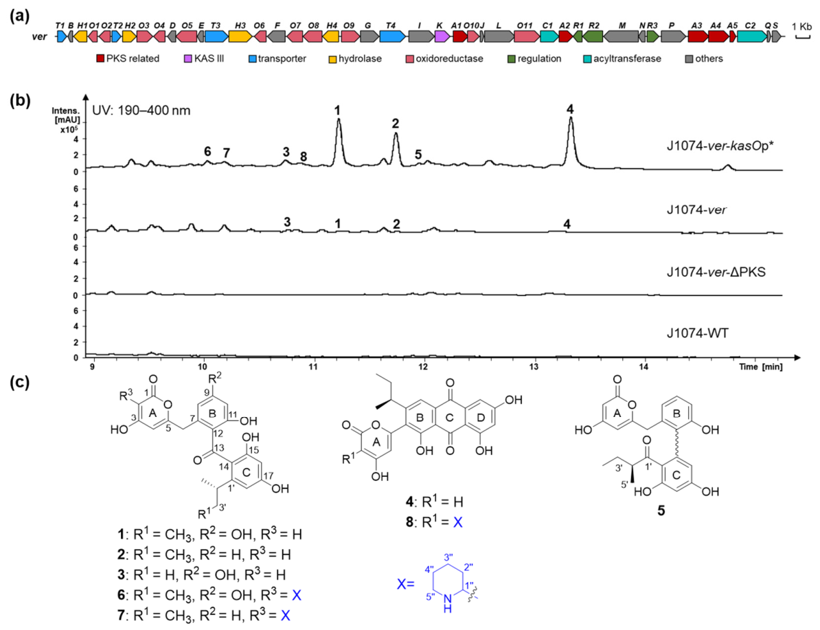
The presence of KAS III in type II polyketide gene clusters broadens the structural diversity of the resulting polyketides by enabling the incorporation of non-acetate starter units, which leads to variations in the aromatic initiation moieties [36]. Genomic analysis of our stored strains identified a type II PKS BGC ver (GenBase accession number: C_AA474359.1) harboring KAS III in Streptomyces sp. S42. AntiSMASH analysis indicates that only two known KAS III-containing type II PKS clusters txn [27] and lld [28] show 55% and 54% similarity to ver, respectively. Based on comparison with these characterized BGCs, the ver gene cluster was proposed to span approximately 48.9 kb, containing 43 open reading frames (Figure 1a). The PKS backbone-related genes of the ver cluster comprise “minimal PKS” VerA3A5 and aromatase/cyclase VerA1 but lack a ketoreductase. Additionally, the ver gene cluster also contains several other genes, including putative oxidoreductases and regulatory factors. (Figure S1, Table S4), which show low sequence homology to those in the other two BGCs, suggesting its potential to generate novel non-acetate primed products.

3.2. Identification of Verrucones by Heterologous Expression

To activate the ver gene cluster and obtain its biosynthetic products, a genomic fragment encompassing the entire cluster was cloned into the pBeloBAC11 vector in E. coli using the ExoCET method [31] (Figure S2) and then heterologously expressed in S. albus J1074. Fermentation of the engineered strain J1074-ver and HPLC analysis of the crude extract revealed several new peaks, indicating the activation of target products, though production was initially low (Figure 1b). To further improve the yield of the products, a constitutive strong promoter kasOp* was inserted upstream of the core gene verA3 (Figure S3). HPLC analysis indicated that the yield of the target compounds was significantly increased (Figure 1b). Deletion of the “minimal PKS” genes verA3A5 completely abolished all observed peaks, confirming that the ver gene cluster is responsible for the biosynthesis of these compounds.
To obtain enough amounts of the corresponding products for structural characterization, a scale-up fermentation (13.5 L) of the engineered strain J1074-ver-kasOp* was performed. The crude extract was then fractionated by silica gel column chromatography and further purified by semipreparative HPLC, which led to the isolation of eight compounds, named verrucones A–H (18) (Figure 1c).
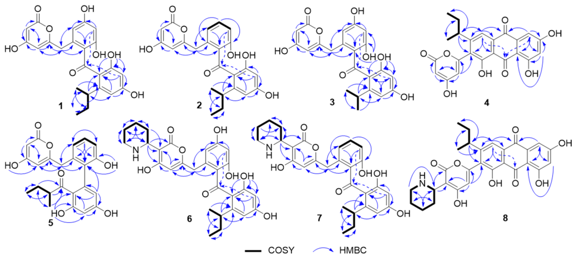
Verrucones A–C (13) were isolated as brown solids, with the molecular formulas C23H22O8, C23H22O7, and C22H20O8, respectively, as deduced from their HRESIMS ions at m/z 427.1387, 411.1438, and 413.1231 [M + H]+. The 1H, 13C, DEPT, and HSQC NMR data of 1 (Table 1, Figures S6–S9) revealed the presence of one ketone carbonyl (δC 199.8), eleven non-protonated carbons with seven oxygenated aromatic carbons (δC 170.3, 164.9, 163.7, 161.0 (2C), 159.9, 157.0), six sp2 methines (δH/C 6.21/101.7, 6.15/104.3, 6.14/110.0, 6.09/100.2, 5.66/100.8, 5.19/88.4), one sp3 methine (δH/C 2.48/36.6), two methylenes (δH/C 3.57/36.8; 1.43,1.33/30.4), and two methyl groups (δH/C 0.97/21.4, 0.66/12.2). Four structural fragments, including a pyrone moiety (ring A), two tetrasubstituted benzene rings (rings B and C), and a butyl side chain, were readily established by analysis of HMBC and COSY correlations (Figure 2, Figures S10 and S11). Key HMBC correlations from H-6 to C-4, C-5, C-7, C-8, and C-12 confirmed the connection of rings A and B through C-6 (Figure 2 and Figure S11). The weak long-range correlations from H-10 and H-16 to C-13 supported the location of C-13 between rings B and C. Similarly, the attachment of the sec-butyl side chain at C-1′ was determined by the key HMBC correlations from H-2′ and H-5′ to C-1′. The 1H and 13C NMR data of 2 and 3 were highly similar to those of 1 (Table 1, Figures S12, S13, S18 and S19), with 2 lacking one hydroxy group and 3 missing one methylene moiety. Further analysis of successive COSY correlations from H-8 (δH 6.71)/H-9 (δH 7.19)/H10 (δH 6.74) in 2 (Figure 2 and Figure S16) and H-3′ (δH 1.01)/H-2′ (δH 2.76)/H-4′ (δH 1.01) in 3 (Figure 2 and Figure S21) indicates that the tetrasubstituted ring B in 1 becomes a trisubstituted benzene ring in 2, and the sec-butyl side chain is replaced by isopropyl in 3, respectively. The detailed planar structures of 23 were further confirmed by the COSY and HMBC correlations (Figure 2). According to bioinformatic analysis and the established biosynthesis of TXN4a from L-isoleucine (confirmed by 13C-labeled precursor feeding experiments [27]), we propose that the sec-butyl is derived from L-isoleucine, indicating that the absolute configuration at C-2′ is S. A literature search using SciFinder revealed that compounds 13 differ from the known compound SEK15 in that they bear unique 2-methylbutyryl or iso-butyryl side chains [37,38].
Verrucone D (4) was isolated as a yellow solid. Its molecular formula was determined to be C23H18O8 based on the HRESIMS ion at m/z 423.1060 [M + H]+. The one-dimensional (1D) NMR data of 4 (Table 2, Figures S23 and S24) showed that it shares the same pyrone ring (ring A) and sec-butyl group as 1. The remaining 1H and 13C NMR signals exhibited characteristic resonances consistent with an anthraquinone skeleton, including two carbonyl carbons at δC 189.0 and 181.1, twelve aromatic carbons in the range of δC 108.1–166.4, two meta-coupled aromatic protons at δH 7.13 (d, J = 2.0 Hz) and 6.58 (d, J = 2.0 Hz), and one singlet aromatic proton at δH 7.41. The linkage of the above three units, as well as the complete structure of 4, was further established by 2D NMR correlations (Figure 2 and Figures S26–S28).
Verrucone E (5) was isolated as a yellow solid, sharing the same molecular formula C23H22O7 with 2, as evidenced by the HRESIMS ion at m/z 411.1428 [M + H]+. Although compounds 5a and 5b exhibited different HPLC retention times, they interconvert rapidly during the separation process. Therefore, the NMR spectrum of the mixture was acquired to elucidate their structures. The 1D NMR data of 5a and 5b (Table 2, Figures S29 and S30) were similar to those of 2, especially the chemical shifts in C-1–C-12, suggesting that rings A and B remain intact. Key HMBC cross-peak from H-14 to C-12 indicates the direct connection between rings B and C, while correlations from 17-OH and H-2′/H-3′/H-5′ to C-1′ confirmed the attachment of the 2-methylbutyryl group at C-18 (Figure 2 and Figure S33). Thus, compound 5 was determined to have a biphenyl skeleton; however, the absolute configuration of its axial chirality has not yet been established because of the rapid interconversion of 5a and 5b.
Verrucone F–H (68) were obtained as brown solids, and their molecular formulas were assigned to be C28H32NO8, C28H32NO7, C28H28NO8 from HRESIMS peaks at 510.2109, 494.2164, 506.1797 [M + H]+, respectively. The 1D NMR data of 6 were similar to those of 1 (Table 1 and Table 3, Figures S34 and S35), except for five additional carbon signals in the range of δC 22.1–53.7 (C-1″–C-5″), indicating the presence of a piperidine moiety. This assignment was further supported by successive COSY correlations from H-1″ to H-5″ (Figure 2 and Figure S38). HMBC correlations from H-1″ to C-1 and C-2 established that the piperidine ring is located at C-2 (Figure 2 and Figure S39). Similarly, compounds 78 were determined to be piperidine-substituted derivatives of 23, respectively, as deduced from their molecular formulas and further confirmed by 2D NMR (Figure 2, Figures S43–S45 and S49–S51).

3.3. Proposed Biosynthetic Pathway of Verrucones

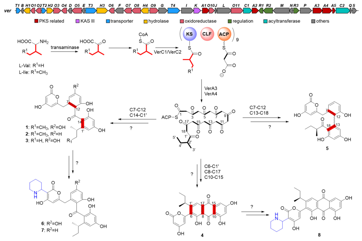
Based on bioinformatic analysis of the ver gene cluster and the structural characterization of verrucones A–H (18), a plausible biosynthetic pathway for these compounds was proposed (Figure 3). Following deamination and decarboxylation, L-isoleucine or valine is proposed to be converted by a CoA transferase into its CoA ester, generating the corresponding acyl-CoA precursor [27]. The ATs encoded by verC1 and verC2 then are thought to specifically recognize the 2-methylbutyryl-CoA and isobutyryl-CoA as starter units and transfer them to the ACP encoded by verA5. Subsequently, the KSα and KSβ encoded by verA3 and verA4 could catalyze the condensation of the initiation unit and nine molecules of malonyl-CoA, thereby forming the nascent polyketide chain. This reactive intermediate may then undergo divergent spontaneous cyclization pathways, ultimately giving rise to three distinct skeletal frameworks, as observed in verrucones A–H (18). Among them, the cyclization patterns of C7-C12 and C14-C1′ form 13, a combination of C6-C1′, C8-C17, and C10-C15 cyclization generates 4, whereas C7-C12 and C13-C18 cyclization yield 5. Subsequently, the hydroxyl group at C-9 of 1 is removed either enzymatically by a dehydroxylase or spontaneously, resulting in 2. Additionally, compounds 1, 2, and 4 were converted to their piperidine derivatives 68, respectively. The detailed mechanism of this transformation, as well as the complete biosynthetic pathway, remains to be established by further studies.

3.4. Bioactivities of Verrucones

In the bioassay, only compound 3 exhibited weak cytotoxicity against the human breast cancer cell line MCF-7, with an IC50 of 16.39 μM, but showed no cytotoxicity against the normal human kidney cell line 293T (Table S5). Compounds 15 displayed inhibitory activities against PTP1B and TCPTP, with IC50 values ranges of 1.84–12.38 μM and 1.84–10.24 μM, respectively. Compounds 1, 2, and 4 also inhibited SHP1, whereas compounds 1 and 4 were active against SHP2 (Table 4). Compounds 68 were inactive against all tested PTPs.

4. Discussion and Conclusions

KAS III enzymes are normally involved in the initiation of type II PKSs assembly and exhibit broad substrate flexibility, allowing them to utilize diverse non-acetate starter units [13,36]. Through genome mining of the marine-derived Streptomyces sp. S42, we identified a T2 PKS BGC (ver) carrying KAS III-encoding genes. The presence of the KASIII enzyme in the ver cluster suggests that its products may have non-acetate start units. Analysis of ver revealed that it contains two genes verC1 and verC2 encoding two separate ATs. AntiSMASH analysis predicts that these two ATs recognize 2-methylbutyryl-CoA and isobutyryl-CoA, respectively. These prediction results are consistent with the two non-acetate starting units in the structure of the isolated compounds, verrucones A–H (18). We hypothesize that the 2-methylbutyryl and isobutyryl groups originate from the deamination and decarboxylation of L-isoleucine and L-valine, respectively. The involvement of KASIII in the initiation of type II PKSs increases the structural complexity and diversity of aromatic polyketide products.
The correct cyclization of T2 PKS products is dependent on the action of a C-9 KR in conjunction with specific AROs and CYCs [11]. In the absence of these auxiliary proteins, the highly reactive nascent polyketide chain fails to undergo regioselective aromatization and instead proceeds via spontaneous, non-enzymatic cyclization [38]. Consistent with this paradigm, the ver BGC does not encode a C-9 KR, and its products exhibit three distinct skeletal types, possibly arising from spontaneous cyclization. From the perspective of type II PKS evolution, the absence of KR effectively reverts the system to a more primitive evolutionary state, where the minimal PKS merely drives carbon chain elongation, while all subsequent cyclization cascades are spontaneous [39]. This also demonstrates the flexibility of PKS as a source of structurally varied natural products.
In comparison with the homologous gene clusters txn [27] and lld [28], the ver cluster exhibits conserved minimal PKS core genes. However, it differs by harboring several oxidoreductase genes (verO4, verO8, verO10, and verO11) while being devoid of glycosyltransferase genes. This compositional feature implies that the ver biosynthetic pathway may produce non-glycosylated products, and the presence of oxidoreductases further hints at potential oxidative modifications. Consequently, ver potentially generating products with unique starter units and diverse cyclization patterns that are structurally different from previously characterized natural products. As a result, the compounds we isolated display variability in post-PKS modifications, notably at C-9 hydroxylation and the introduction of a piperidine unit. The piperidine moiety in natural products generally originates from the incorporation of L-pipecolic acid, which is an intermediate in L-lysine metabolism [40]. Since the ver cluster does not contain a homologous gene encoding lysine cyclodeaminase, we speculate that the gene responsible for catalyzing the formation of piperidine may be present in the host genome.
Verrucones A–E (15) exhibited differential inhibitory potencies against PTP targets. Specifically, a comparison of compounds 1 and 2 revealed that the absence of the hydroxyl group at C-9 position attenuates the inhibitory activity. Similarly, comparison of 1 and 3 indicated that the 2-methylbutyryl group influences activity, with compound 3 being less potent than 1. Notably, compound 4 was the most potent among the tested compounds, suggesting that the anthraquinone structure may contribute favorably to the PTP inhibitory activity, consistent with a previous report [5]. In contrast, compounds 68 were inactive, implying that C-2 piperidine substitution significantly diminishes PTPs’ inhibitory potency. Although compound 4 displayed promising activity, its selectivity remained insufficient. Future structural modifications potentially via synthetic biology approaches could be employed to improve its selectivity profile. Collectively, these findings provide a basis for the rational design of selective PTP inhibitors.
In summary, genome mining of Streptomyces sp. S42 led to the discovery of a type II PKS BGC, designated ver, which encodes a KAS III homolog. Heterologous expression and promoter engineering successfully activated this BGC, affording eight non-acetate primed aromatic polyketides, verrucones A–H (18). These compounds feature three distinct skeletal types: isolated benzene ring, biphenyl, and anthraquinone. The successful activation of ver BGC underscores the utility of heterologous expression as a general strategy for targeted mining of chemically novel compounds from sequenced genomes. Among the isolates, compound 4 exhibited potent inhibitory activity against multiple PTP targets, providing a valuable lead for further drug discovery efforts targeting metabolic disorders such as diabetes and obesity. Our work not only enriches the structural diversity of aromatic polyketides through the incorporation of unusual starter units but also establishes a foundation for future pathway engineering to generate designer analogs with enhanced bioactivities.

Supplementary Materials

The following supporting information can be downloaded at: https://www.mdpi.com/article/10.3390/microorganisms14081669/s1. Table S1: The media used in this study; Table S2: Strains and plasmids used in this study; Table S3: Primers used in this study; Table S4: Deduced functions of ORFs in the ver biosynthetic gene cluster; Table S5: Cytotoxicity activity of verrucone C (3); Figure S1: Comparison of ver with homologous gene clusters; Figure S2: Construction of expression vector of the ver gene cluster; Figure S3: Construction of plasmid for the insertion of kasOp* and the inactivation of verA3A5; Figure S4: The HRMS data for verrucone A–H (18); Figure S5: The UV absorbance spectra of verrucones A–H (18); Figures S6–S51: NMR spectra of verrucones A–H (18).

Author Contributions

Conceptualization, Y.Z., X.B. and H.Z.; formal analysis, X.H., M.Y. and H.Z.; investigation, X.H., M.Y., P.Y., Q.S., Y.L. and H.Z.; writing—original draft preparation, X.H. and H.Z.; writing—review and editing, X.H., X.B. and H.Z.; supervision, Y.Z., X.B. and H.Z.; funding acquisition, Y.Z., X.B. and H.Z. All authors have read and agreed to the published version of the manuscript.

Funding

This research was funded by the National Key R&D Program of China (2023YFC3402000, 2019YFA0905700), the Shandong Provincial Natural Science Foundation (ZR2023ZD29) and Key R&D Program of Shandong Province (2025CXPT191), the Young Talent Development Program of SKLMT (M2025YA03), Intramural Joint Program Fund of State Key Laboratory of Microbial Technology (SKLMTIJP-2024-04), SKLMT Frontiers and Challenges Project (SKLMTFCP-2023-05), and the 111 Project (B16030).

Institutional Review Board Statement

Not applicable.

Informed Consent Statement

Not applicable.

Data Availability Statement

The data presented in this study are available in the article and Supplementary Materials.

Acknowledgments

We would like to thank Zhifeng Li, Jingyao Qu, Guannan Lin, Jing Zhu, Xiangmei Ren, and Haiyan Sui from the State Key Laboratory of Microbial Technology of Shandong University for their help and guidance in HRESIMS, HPLC, and NMR measurements.

Conflicts of Interest

The authors declare no conflicts of interest.

References

  1. Hulst, M.B.; Grocholski, T.; Neefjes, J.J.C.; van Wezel, G.P.; Metsä-Ketelä, M. Anthracyclines: Biosynthesis, engineering and clinical applications. Nat. Prod. Rep. 2022, 39, 814–841. [Google Scholar] [CrossRef] [PubMed]
  2. Wolff, J.T.; Tsai, S.-C. Type II polyketide synthases: Impact on human health, current knowledge, and future directions. J. Biol. Chem. 2025, 301, 110749. [Google Scholar] [CrossRef] [PubMed]
  3. Hertweck, C.; Luzhetskyy, A.; Rebets, Y.; Bechthold, A. Type II polyketide synthases: Gaining a deeper insight into enzymatic teamwork. Nat. Prod. Rep. 2007, 24, 162–190. [Google Scholar] [CrossRef] [PubMed]
  4. Baumgartner, C.K.; Ebrahimi-Nik, H.; Iracheta-Vellve, A.; Hamel, K.M.; Olander, K.E.; Davis, T.G.R.; McGuire, K.A.; Halvorsen, G.T.; Avila, O.I.; Patel, C.H.; et al. The PTPN2/PTPN1 inhibitor ABBV-CLS-484 unleashes potent anti-tumour immunity. Nature 2023, 622, 850–862. [Google Scholar] [CrossRef] [PubMed]
  5. Zhang, Z.; Shang, Z.P.; Jiang, Y.; Qu, Z.X.; Yang, R.Y.; Zhang, J.; Lin, Y.X.; Zhao, F. Selective Inhibition of PTP1B by New Anthraquinone Glycosides from Knoxia valerianoides. J. Nat. Prod. 2022, 85, 2836–2844. [Google Scholar] [CrossRef] [PubMed]
  6. Chan, G.; Kalaitzidis, D.; Neel, B.G. The tyrosine phosphatase Shp2 (PTPN11) in cancer. Cancer Metastasis Rev. 2008, 27, 179–192. [Google Scholar] [CrossRef] [PubMed]
  7. Watson, H.A.; Wehenkel, S.; Matthews, J.; Ager, A. SHP-1: The next checkpoint target for cancer immunotherapy? Biochem. Soc. Trans. 2016, 44, 356–362. [Google Scholar] [CrossRef] [PubMed]
  8. Feng, G.-S. Shp2 as a therapeutic target for leptin resistance and obesity. Expert Opin. Ther. Targets 2006, 10, 135–142. [Google Scholar] [CrossRef] [PubMed]
  9. Liao, H.; Pei, D. Cell-permeable bicyclic peptidyl inhibitors against T-cell protein tyrosine phosphatase from a combinatorial library. Org. Biomol. Chem. 2017, 15, 9595–9598. [Google Scholar] [CrossRef] [PubMed]
  10. Das, A.; Khosla, C. Biosynthesis of Aromatic Polyketides in Bacteria. Acc. Chem. Res. 2009, 42, 631–639. [Google Scholar] [CrossRef] [PubMed]
  11. Zhou, H.; Li, Y.; Tang, Y. Cyclization of aromatic polyketides from bacteria and fungi. Nat. Prod. Rep. 2010, 27, 839–868. [Google Scholar] [CrossRef] [PubMed]
  12. Olano, C.; Méndez, C.; Salas, J.A. Post-PKS tailoring steps in natural product-producing actinomycetes from the perspective of combinatorial biosynthesis. Nat. Prod. Rep. 2010, 27, 571–616. [Google Scholar] [CrossRef] [PubMed]
  13. Tan, W.F.; Zhang, J.X.; Sun, J.L.; Zhu, A.; Shi, J.; Jiao, R.H.; Guo, Z.K. Discovery of antibacterial tetracycline-type natural products misiomycins E–H through KASIII genome mining and regulator overexpression. Org. Lett. 2026, 28, 7196–7201. [Google Scholar] [CrossRef] [PubMed]
  14. Zhao, L.Y.; Shi, J.; Xu, Z.Y.; Sun, J.L.; Yan, Z.Y.; Tong, Z.W.; Tan, R.X.; Jiao, R.H.; Ge, H.M. Hybrid type I and II polyketide synthases yield distinct aromatic polyketides. J. Am. Chem. Soc. 2024, 146, 29462–29468. [Google Scholar] [CrossRef] [PubMed]
  15. Schwarzer, P.; Tsypik, O.; Zuo, C.; Alali, A.; Wunsch-Palasis, J.; Heitzler, T.; Derochefort, J.; Bernhardt, M.; Yan, X.; Paululat, T.; et al. Early steps in the biosynthetic pathway of rishirilide B. Molecules 2020, 25, 1955. [Google Scholar] [CrossRef] [PubMed]
  16. Chen, S.; Zhang, C.; Zhang, L. Investigation of the Molecular Landscape of Bacterial Aromatic Polyketides by Global Analysis of Type II Polyketide Synthases. Angew. Chem. Int. Ed. 2022, 61, e202202286. [Google Scholar] [CrossRef]
  17. Huo, L.; Hug, J.J.; Fu, C.; Bian, X.; Zhang, Y.; Müller, R. Heterologous expression of bacterial natural product biosynthetic pathways. Nat. Prod. Rep. 2019, 36, 1412–1436. [Google Scholar] [CrossRef] [PubMed]
  18. Zhao, S.; Feng, R.; Gu, Y.; Han, L.; Cong, X.; Liu, Y.; Liu, S.; Shen, Q.; Huo, L.; Yan, F. Heterologous expression facilitates the discovery and characterization of marine microbial natural products. Eng. Microbiol. 2024, 4, 100137. [Google Scholar] [PubMed]
  19. Blin, K.; Shaw, S.; Vader, L.; Szenei, J.; Reitz, Z.L.; Augustijn, H.E.; Cediel-Becerra, J.D.D.; de Crécy-Lagard, V.; Koetsier, R.A.; Williams, S.E.; et al. antiSMASH 8.0: Extended gene cluster detection capabilities and analyses of chemistry, enzymology, and regulation. Nucleic Acids Res. 2025, 53, W32–W38. [Google Scholar] [CrossRef] [PubMed]
  20. Baral, B.; Akhgari, A.; Metsä-Ketelä, M. Activation of microbial secondary metabolic pathways: Avenues and challenges. Synth. Syst. Biotechnol. 2018, 3, 163–178, Erratum in Synth. Syst. Biotechnol. 2020, 5, 328. [Google Scholar] [CrossRef] [PubMed]
  21. Lasch, C.; Myronovskyi, M.; Luzhetskyy, A. Streptomyces as a versatile host platform for heterologous production of microbial natural products. Nat. Prod. Rep. 2026, 43, 371–390. [Google Scholar] [CrossRef] [PubMed]
  22. Hao, X.; Wang, Z.; Shi, X.; Liu, Y.; Yan, P.; Yang, Q.; Ren, X.; Li, X.; Sui, H.; Li, F.; et al. Genome mining and targeted discovery of twistane-containing angucyclinones from marine Streptomyces phaeochromogenes XSA2. Org. Lett. 2025, 27, 2987–2991. [Google Scholar] [CrossRef] [PubMed]
  23. Yang, C.; Huang, C.; Zhang, W.; Zhu, Y.; Zhang, C. Heterologous expression of fluostatin gene cluster leads to a bioactive heterodimer. Org. Lett. 2015, 17, 5324–5327. [Google Scholar] [CrossRef] [PubMed]
  24. Huang, X.; Xu, X.; Zhou, L.; Li, J.; Ma, C.; Wang, W.; Che, Q.; Li, D.; Zhu, T.; Zhang, G. (±)-Feionemycin A and Chromonemycins A–D, Rearranged Aromatic Polyketides Uncovered by Type II Polyketide Gene Cluster Expression. J. Nat. Prod. 2025, 88, 768–776. [Google Scholar] [CrossRef] [PubMed]
  25. Li, Y.; Xu, Z.; Chen, P.; Zuo, C.; Chen, L.; Yan, W.; Jiao, R.; Ye, Y. Genome Mining and Heterologous Expression Guided the Discovery of Antimicrobial Naphthocyclinones from Streptomyces eurocidicus CGMCC 4.1086. J. Agric. Food Chem. 2023, 71, 2914–2923. [Google Scholar] [CrossRef] [PubMed]
  26. Shen, Q.; Dai, G.; Li, A.; Liu, Y.; Zhong, G.; Li, X.; Ren, X.; Sui, H.; Fu, J.; Jiao, N.; et al. Genome-guided discovery of highly oxygenated aromatic polyketides, saccharothrixins D–M, from the rare marine actinomycete Saccharothrix sp. D09. J. Nat. Prod. 2021, 84, 2875–2884. [Google Scholar] [CrossRef] [PubMed]
  27. Zhang, M.; Hou, X.-F.; Qi, L.-H.; Yin, Y.; Li, Q.; Pan, H.-X.; Chen, X.-Y.; Tang, G.-L. Biosynthesis of trioxacarcin revealing a different starter unit and complex tailoring steps for type II polyketide synthase. Chem. Sci. 2015, 6, 3440–3447. [Google Scholar] [CrossRef] [PubMed]
  28. Dong, L.; Shen, Y.; Hou, X.-F.; Li, W.-J.; Tang, G.-L. Discovery of druggability-improved analogues by investigation of the LL-D49194α1 biosynthetic pathway. Org. Lett. 2019, 21, 2322–2325. [Google Scholar] [CrossRef] [PubMed]
  29. Wang, H.; Li, Z.; Jia, R.; Hou, Y.; Yin, J.; Bian, X.; Li, A.; Müller, R.; Stewart, A.F.; Fu, J.; et al. RecET direct cloning and redαβ recombineering of biosynthetic gene clusters, large operons or single genes for heterologous expression. Nat. Protoc. 2016, 11, 1175–1190. [Google Scholar] [CrossRef] [PubMed]
  30. AltschuP, S.F.; Gish, W.; Miller, W.; Myers, E.W.; Lipman, D.J. Basic local alignment search tool. J. Mol. Biol. 1990, 215, 403–410. [Google Scholar] [CrossRef]
  31. Wang, H.; Li, Z.; Jia, R.; Yin, J.; Li, A.; Xia, L.; Yin, Y.; Müller, R.; Fu, J.; Stewart, A.F.; et al. ExoCET: Exonuclease in vitro assembly combined with RecET recombination for highly efficient direct DNA cloning from complex genomes. Nucleic Acids Res. 2018, 46, e28. [Google Scholar] [PubMed]
  32. Keiser, T.; Bibb, M.J.; Buttner, M.J.; Charter, K.F.; Hopwood, D.A. Practical Streptomyces Genetics; The John Innes Foundation: Norwich, UK, 2000. [Google Scholar]
  33. Wang, H.; Bian, X.; Xia, L.; Ding, X.; Müller, R.; Zhang, Y.; Fu, J.; Stewart, A.F. Improved seamless mutagenesis by recombineering using ccdB for counterselection. Nucleic Acids Res. 2014, 42, e37. [Google Scholar] [CrossRef] [PubMed]
  34. Tominaga, H.; Ishiyama, M.; Ohseto, F.; Sasamoto, K.; Hamamoto, T.; Suzuki, K.; Watanabe, M. A water-soluble tetrazolium salt useful for colorimetric cell viability assay. Anal. Commun. 1999, 36, 47–50. [Google Scholar] [CrossRef]
  35. Shen, Q.; Zhou, H.; Dai, G.; Zhong, G.; Huo, L.; Li, A.; Liu, Y.; Yang, M.; Ravichandran, V.; Zheng, Z.; et al. Characterization of a cryptic NRPS gene cluster in Bacillus velezensis FZB42 reveals a discrete oxidase involved in multithiazole biosynthesis. ACS Catal. 2022, 12, 3371–3381. [Google Scholar] [CrossRef]
  36. Nofiani, R.; Philmus, B.; Nindita, Y.; Mahmud, T. 3-ketoacyl-ACP synthase (KAS) III homologues and their roles in natural product biosynthesis. MedChemComm 2019, 10, 1517–1530. [Google Scholar] [CrossRef] [PubMed]
  37. Chemical Abstracts Service (CAS). SciFinder-n. Available online: https://scifinder-n.cas.org (accessed on 20 June 2026).
  38. Shen, Y.; Yoon, P.; Yu, T.W.; Floss, H.G.; Hopwood, D.; Moore, B.S. Ectopic expression of the minimal whiE polyketide synthase generates a library of aromatic polyketides of diverse sizes and shapes. Proc. Natl. Acad. Sci. USA 1999, 96, 3622–3627. [Google Scholar] [CrossRef] [PubMed]
  39. Hertweck, C. The biosynthetic logic of polyketide diversity. Angew. Chem. Int. Ed. Engl. 2009, 48, 4688–4716. [Google Scholar] [CrossRef] [PubMed]
  40. Xiong, X.; Zhu, X.; Yu, D.; Huang, Q.; Wu, X.; Tan, G.; Sun, P. Piperidine polyketide alkaloids of bacterial origin: Occurrence, bioactivity, and biosynthesis. Eur. J. Med. Chem. 2025, 289, 117498. [Google Scholar] [CrossRef] [PubMed]
Figure 1. (a) Gene organization of the ver gene cluster. (b) Comparative metabolite analysis of the J1074-ver strain and the J1074-ver-kasOp* cultivated in YM medium. (c) Structures of 18.
Microorganisms 14 01669 g001
Figure 2. Key COSY and HMBC correlations of 18.
Microorganisms 14 01669 g002
Figure 3. Proposed biosynthetic pathway of verrucones A–H (18).
Microorganisms 14 01669 g003
Table 1. The 1H (600 MHz) and 13C NMR (150 MHz) data of 13 in DMSO-d6.
No.123
δC, TypeδH, Mult. (J in Hz)δC, TypeδH, Mult. (J in Hz)δC, Type δH, Mult. (J in Hz)
1163.7, C 163.7, C 163.9, C
288.4, CH5.19, d (1.9)88.4, CH5.20, d (2.0)88.5, CH5.12, s
3170.3, C 170.4, C 170.6, C
4100.8, CH5.66, d (1.9)100.8, CH5.67, d (2.0)100.9, CH5.62, s
5164.9, C 165.0, C 165.1, C
636.8, CH23.57, s36.6, CH23.64, s36.9, CH23.56, s
7137.9, C 134.0, C 138.3, C
8110.0, CH6.14, d (2.2)120.9, CH6.71, d (7.6)110.3, CH6.14, d (2.3)
9161.0, C 130.2, CH7.19, t (7.9)161.3, C
10101.7, CH 6.21, d (2.2)114.8, CH6.74, d (8.2)101.8, CH6.20, d (2.3)
11161.0, C 155.1, C 161.5, C
12119.4, C 130.8, C 119.2, C
13199.8, C 199.7, C 200.1, C
14120.8, C 119.3, C 120.7, C
15157.0, C 159.7, C 156.6, C
16100.2, CH6.09, d (2.0)100.1, CH6.06, d (2.2)100.2, CH6.08, d (2.1)
17159.9, C 160.1, C 159.9, C
18104.3, CH6.15, d (2.0)105.2, CH6.19, d (2.2)104.2, CH6.20, d (2.1)
1′148.6, C 151.4, C 149.5, C
2′36.6, CH2.48, m36.0, CH2.72, m29.9, CH2.76, m
3′30.4, CH21.43, m30.6, CH21.47, m24.0, CH31.01, d (6.7)
1.33, m 1.33, m
4′12.2, CH30.66, t (7.3)12.2, CH30.68, t (7.4)24.0, CH31.01, d (6.7)
5′21.4, CH30.97, d (6.7)21.5, CH30.98, d (6.7)
3-OH 11.13, s 9.83, brs
9-OH 10.13. s
11-OH 11.61, s 11.6, s
15-OH 9.90, s 10.48, s
17-OH 9.61, s 9.70. s
Table 2. The 1H (600 MHz) and 13C NMR (150 MHz) data of 45 in DMSO-d6.
No.45a5b
δC, TypeδH, Mult. (J in Hz)δC, TypeδH, Mult. (J in Hz)δC, Type δH, Mult. (J in Hz)
1163.8, C 163.7, C 163.7, C
289.4, CH5.32, d (1.7)88.4, CH5.16, d (1.9)88.4, CH5.16, d (1.9)
3170.9, C 170.2, C 170.2, C
4105.8, CH6.21, d (1.7)100.6, CH5.55, d (1.9)100.6, CH5.54, d (1.9)
5155.9, C 164.9, C 164.8, C
6127.3, C 37.1, CH23.53, d (9.9)37.1, CH23.40, d (7.7)
3.50, d (9.9) 3.38, d (7.7)
7159.5, C 135.1, C 135.1, C
8113.7, C 120.1, CH6.69, d (7.3)120.0, CH6.68, d (7.3)
9189.0, C 128.5, CH7.13, t (7.9)128.4, CH7.12, t (7.9)
10108.8, C 114.0, CH6.79, d (8.0)113.9, CH6.78, d (8.0)
11164.6, C 145.5, C 145.4, C
12108.1, CH6.58, d (2.0)128.1, C 128.0, C
13166.4, C 137.9, C 137.7, C
14109.4, CH7.13, d (2.0)110.1, CH5.89, d (2.3)110.0, CH5.88, d (2.3)
15135.0, C 159.8, C 159.7, C
16181.1, C 101.8, CH6.29, d (2.3)101.8, CH6.28, d (2.3)
17133.8, C 158.8, C 158.7, C
18116.0, CH7.64, s118.8, C 118.6, C
1′155.5, C 209.4, C 209.2, C
2′38.0, CH2.69, m45.7, CH2.55, m45.9, CH2.56, m
3′29.6, CH21.62, m25.7, CH21.49, m25.4, CH21.31, m
1.08, m 0.99, m
4′11.9, CH30.74, t (7.3)11.4, CH30.64, t (7.4)11.2, CH30.45, t (7.4)
5′21.2, CH31.21, d (6.8)15.5, CH30.63, d (6.8)15.9, CH30.82, d (6.8)
3-OH 11.59, brs 11.59, brs
11-OH 9.38, s 9.36, s
15-OH 9.86, brs 9.86, brs
17-OH 10.70, s 10.65, s
Table 3. The 1H (600 MHz) and 13C NMR (150 MHz) data of 68 in DMSO-d6.
No.678
δC, TypeδH, Mult. (J in Hz)δC, TypeδH, Mult. (J in Hz)δC, Type δH, Mult. (J in Hz)
1164.3, C 164.3, C 164.4, C
292.8, C 92.7, C 93.6, C
3177.6, C 177.7, C 176.9, C
4108.9, CH5.23, s108.6, CH5.21, s112.2, CH5.65, s
5160.1, C 160.2, C 152.9, C
636.6, CH23.31, s36.4, CH23.41, s128.9, C
7139.7, C 135.1, C 159.5, C
8109.5, CH6.07, d (2.2)120.1, CH6.66, d (7.9)113.9, C
9161.8, C 129.8, CH7.15, t (7.9)187.2, C
10101.2, CH6.15, d (1.8)114.3, CH6.70, d (7.9)107.2, C
11161.6, C 154.9, C 164.4, C
12118.6, C 130.8, C 108.0, CH6.38, s
13200.0, C 199.7, C 164.9, C
14120.9, C 118.6, C 111.7, CH7.02, s
15157.1, C 160.7, C 134.9, C
16100.3, CH6.11, d (2.0)100.2, CH6.05, d (2.0)181.8, C
17160.1, C 161.8, C 133.4, C
18104.4, CH6.16, d (2.0)105.5, CH6.17, d (2.0)115.7, CH7.59, s
1′148.5, C 151.7, C 154.5, C
2′36.6, CH2.50, m a35.8, CH2.72, m a37.7, CH2.78, m a
3′30.4, CH21.43, m30.7, CH21.44, m a29.6, CH21.61, m a
1.34, m 1.30, m
4′12.2, CH30.67, t (7.2)12.2, CH30.64, t (7.3)12.1, CH30.73, t (7.3)
5′21.5, CH30.98, d (6.7)21.6, CH30.95, d (6.6)21.5, CH31.19, d (6.8)
1″53.7, CH3.98, dt (2.8, 12.0)53.7, CH3.98, dt (2.3, 11.9)53.7, CH4.10, d (11.3)
2″27.5, CH21.93, m27.5, CH21.93, m27.5, CH22.05, m
1.58, m 1.58, m 1.71, m a
3″22.5, CH21.72, m22.5, CH21.73, m22.5, CH21.78, m
1.45, m a 1.44, m a 1.50, m
4″22.1, CH21.68, m22.1, CH21.68, m22.2, CH21.71, m a
1.52, m 1.53, m 1.61, m a
5″44.2, CH23.23, m44.1, CH23.24, m44.2, CH23.27, d (11.6)
2.74, m 2.72, m a 2.78, m a
a: overlapped.
Table 4. PTP inhibitory activities of 18.
CompoundIC50 (μM)
PTP1BSHP1SHP2TCPTP
13.65 ± 0.286.79 ± 0.3915.22 ± 0.862.34 ± 0.37
25.35 ± 0.3324.82 ± 0.44>506.08 ± 0.45
38.23 ± 0.39/>507.51 ± 0.58
41.84 ± 0.261.84 ± 0.3815.37 ± 1.011.84 ± 0.33
512.38 ± 1.09//10.24 ± 1.37
6>50//>50
7>50//>50
8>50//>50
Na3VO4 a1.02 ± 0.303.40 ± 0.634.82 ± 0.842.71 ± 0.48
a Na3VO4 was used as a positive control. /: Not detected.
Disclaimer/Publisher’s Note: The statements, opinions and data contained in all publications are solely those of the individual author(s) and contributor(s) and not of MDPI and/or the editor(s). MDPI and/or the editor(s) disclaim responsibility for any injury to people or property resulting from any ideas, methods, instructions or products referred to in the content.