Article

Mycoplasma tracheobuteonis sp. nov., a Novel Respiratory Mycoplasma Species from the Common Buzzard (Buteo buteo)

1
Research Institute of Wildlife Ecology, Department of Interdisciplinary Sciences, University of Veterinary Medicine, 1160 Vienna, Austria
2
Unit Pathology, Department of Biological Sciences and Pathobiology, University of Veterinary Medicine, 1210 Vienna, Austria
3
Unit Infectiology and Virology, Department of Biological Sciences and Pathobiology, University of Veterinary Medicine, 1210 Vienna, Austria
4
Unit Microbiology, Department of Biological Sciences and Pathobiology, University of Veterinary Medicine, 1210 Vienna, Austria
5
Clinic for Birds and Reptiles, Faculty of Veterinary Medicine, University of Leipzig, 04103 Leipzig, Germany
6
Instituto Universitario de Sanidad Animal y Seguridad Alimentaria, University of Las Palmas de Gran Canaria, 35413 Arucas, Spain
*
Authors to whom correspondence should be addressed.
Microorganisms 2026, 14(6), 1224; https://doi.org/10.3390/microorganisms14061224
Submission received: 26 April 2026 / Revised: 22 May 2026 / Accepted: 25 May 2026 / Published: 29 May 2026
(This article belongs to the Section Veterinary Microbiology)

Abstract

Mycoplasmas are frequently recovered from the upper respiratory tract of birds of prey, yet many isolates remain taxonomically unresolved. In the present study, a collection of ten previously unclassified Mycoplasma strains, predominantly isolated from the trachea of the common buzzard (Buteo buteo), was subjected to comprehensive phenotypic and genomic characterization. All strains grew well in modified Hayflick’s medium and formed colonies with the characteristic fried-egg appearance. None of the strains produced acid from glucose or hydrolyzed arginine or urea. Phylogenetic analyses based on 16S rRNA gene, 16S–23S intergenic spacer, and partial rpoB gene sequences placed the strains within the Mycoplasma synoviae cluster, in close proximity to five recently described Mycoplasma species associated with raptors such as eagles and kites. Matrix-assisted laser desorption ionization–time of flight (MALDI-ToF) mass spectrometry enabled the clear discrimination of the investigated strains from closely related taxa. Whole-genome comparisons, together with phylogenomic analyses, supported the assignment of these strains to a novel species within the genus Mycoplasma. The name Mycoplasma tracheobuteonis sp. nov. is proposed, corresponding to its preference for colonizing the upper respiratory tract of the common buzzard, with strain 48589BT (=DSM 115882T = NCTC 14927T) designated as the type strain.

1. Introduction

Members of the class Mollicutes are small prokaryotes characterized by the absence of a cell wall, a minute cellular and genomic size, and a low genomic G+C content. The genus Mycoplasma (commonly referred to as mycoplasmas) represents the most diverse lineage within the class Mollicutes and currently comprises more than 130 recognized species. They colonize a wide range of hosts, including humans and other mammals as well as birds, reptiles, fish, and mollusks. Mycoplasmas generally exhibit pronounced host and tissue specificity, predominantly inhabiting the respiratory and urogenital mucosa, but also ocular, mammary and joint tissues, as well as the blood. Several species are considered commensals, whereas others are well recognized as opportunistic or primary pathogens [1,2].
In birds, mycoplasmas have been studied predominantly in domestic poultry and waterfowl, whereas their diversity in birds of prey remains less well characterized [3,4]. Predatory birds are considered frequent carriers, particularly within Falconiformes [5,6,7], but comparatively little is known about Mycoplasma species associated with the family Accipitridae (order Accipitriformes), which includes eagles, hawks, buzzards, kites, harriers, and Old World vultures.
Only a limited number of species are regarded as primarily associated with accipitrid hosts, including Mycoplasma (M.) buteonis, M. gypis, and M. neophronis, reported mainly from common buzzards, Eurasian griffon vultures, and Canarian Egyptian vultures, respectively [8,9]. A non-cultivable organism provisionally designated “Mycoplasma vulturii” has also been detected in Oriental white-backed vultures [10]. Additional mycoplasmas recovered from raptors include M. falconis, M. hafezii, and M. seminis, primarily in falcons but occasionally in Accipitridae [5,6,7,11,12], and M. corogypsi in black vultures [13,14,15]. Molecular investigations have further indicated the presence of additional undescribed Mycoplasma species in predatory birds, highlighting the incomplete understanding of their diversity and host associations [16,17,18,19].
Recently, we described five novel Mycoplasma species—M. aquilae, M. paraquilae, M. milvi, M. haliaeeti, and M. razini—isolated from the respiratory tract of eagles and kites, thereby expanding the recognized diversity of accipitrid-associated mycoplasmas [20]. The organism described in the present study is phylogenetically closely related to this group, sharing 98.75–99.10% 16S rRNA gene sequence similarity with these taxa, but it can be clearly differentiated by MALDI-ToF mass spectrometry [21]. Moreover, it was recovered predominantly from the trachea of the common buzzard (Buteo buteo), suggesting a distinct taxonomic entity with apparent host association.
In the present study, eight isolates obtained from the respiratory tract of the common buzzard and two from additional raptors were subjected to comprehensive phenotypic and genetic characterization in accordance with guidelines and recommendations for the description of new species in the class Mollicutes [22], supplemented by whole-genome-based analyses. Comparative analyses, including MALDI-ToF mass spectrometry, phylogenetic reconstruction, and genomic relatedness indices, demonstrated that these strains represent a coherent and previously undescribed taxon within the genus Mycoplasma. Based on the data presented, the name Mycoplasma tracheobuteonis sp. nov. is proposed for this organism, given that it has been predominantly isolated from the trachea of the common buzzard.

2. Materials and Methods

2.1. Primary Isolation, Cultural and Morphological Characterization

Samples from Accipitridae were taken during necropsies at the Research Institute of Wildlife Ecology, University of Veterinary Medicine, Vienna, Austria as well as during field investigations of raptors conducted in Germany and Spain. Most birds were healthy individuals that died from non-infectious causes (e.g., trauma, poisoning, or emaciation), and no relevant gross or histologic respiratory lesions were identified. Tracheal or choanal swabs were placed into Amies transport medium and stored at 4 °C until processing at the Microbiology diagnostic unit at the University of Veterinary Medicine Vienna, Austria. For mycoplasma isolation, swabs were suspended in 1 mL of 2SP medium (containing 0.2 mol/L sucrose in 0.02 mol/L phosphate buffer (pH 7.0 ± 0.2) composed of mono- and dibasic phosphate salts and supplemented with 10% fetal calf serum) and mixed by vortexing for 30 s. Aliquots of 100 µL were inoculated onto modified Hayflick’s agar [23] and incubated at 37 °C in an atmosphere containing 5% CO2 for a maximum of 7 days. Individual colonies displaying mycoplasma-like morphology were subsequently transferred into 5 mL modified Hayflick’s broth [23] and cultivated at 37 °C under ambient atmospheric conditions for 3 to 4 days. Colony-derived cultures from 2016 and earlier were identified using 16S rRNA gene sequencing followed by phylogenetic analysis (see below). From 2017 on, colony-derived cultures were identified by MALDI-ToF mass spectrometry as described previously [21]. Unclassified Mycoplasma isolates were maintained at −80 °C until additional investigations were performed. For the present study, ten representative strains were selected from a group (n = 36) of unknown but closely related Mycoplasma isolates, which were predominantly obtained from the common buzzard and exhibited close phylogenetic relationships to the species of the recently described M. aquilae species complex and M. razini [20]. Selection was based on host species and country and year of isolation, with a focus on epidemiological unrelatedness (Table 1).
The ten selected strains underwent comprehensive taxonomic analysis, beginning with triple-filter cloning [24] to ensure pure cultures, and cultivation in/on modified Hayflick’s medium [23] at different temperatures (4 °C, 15 °C, room temperature, 28 °C, and 42 °C) and conditions (aerobic and anaerobic) to determine culture characteristics and colony morphology. Furthermore, cultures were filtrated through membrane filters with pore sizes of 450 and 220 nm. Microcinematography was performed to assess gliding motility as described previously [25]. In addition, the cell morphology of the proposed type strain was determined by transmission electron microscopy as described before [9].

2.2. Phylogenetic Analyses

DNA was extracted from pellets obtained from 9 mL broth cultures following centrifugation at 20,000× g for 10 min using the DNeasy Blood & Tissue Kit (Qiagen, Hilden, Germany). Nearly full-length 16S rRNA gene sequences of all ten isolates were amplified with primers 27f and 1492r [26]. To further determine the relatedness of the novel species to established Mycoplasma species, the 16S–23S intergenic spacer region (ISR) and a partial fragment of the rpoB gene were investigated according to previously published protocols [20,27]. Amplicons were purified using the Exo-CIPTM Rapid PCR Cleanup Kit (New England Biolabs, Ipswich, MA, USA) and subjected to bidirectional sequencing at LGC Genomics (Berlin, Germany). Sequence similarities for the 16S rRNA gene, ISR, and partial rpoB gene were determined using EzBioCloud [28] and BLASTn searches against GenBank databases [29], respectively. Multiple sequence alignments of 16S rRNA and partial rpoB gene sequences were generated with ClustalW [30]. Phylogenetic analyses including closely related taxa were carried out in MEGA11 using the maximum likelihood method based on Tamura-Nei substitution model with 1000 bootstrap replicates [31]. Final tree visualization and annotation were performed using iTOL v6 [32].

2.3. Standard Phenotypic Analyses and MALDI-ToF Mass Spectrometry

The investigated strains were tested for glucose utilization, arginine and urea hydrolysis [33], hemolysis [34], and production of ‘film and spots’ [35]. MALDI-ToF mass spectrometric analyses of broth cultures derived from the ten investigated strains as well as from phylogenetically related Mycoplasma species were carried out as previously reported [21]. Reference spectra, also known as main spectrum profiles (MSPs), were generated from the acquired spectra. Based on an arbitrary distance matrix, a score-oriented dendrogram was calculated using the correlation distance measure in combination with the average linkage algorithm implemented in Bruker Daltonics software package (MBT Compass HT version 2025) (Bremen, Germany).

2.4. Genome Sequencing, Genome Coherence, and Phylogenomic Analyses

For whole-genome sequencing, all ten strains were cultivated in 20 mL modified Hayflick’s medium at 37 °C in ambient atmospheric conditions for 4 days. Cells were subsequently harvested by centrifugation at 20,000× g for 10 min, and genomic DNA was purified using the DNeasy Blood and Tissue Kit (Qiagen, Hilden, Germany). DNA concentration and integrity were evaluated by fluorometric quantification using QubitTM 4 fluorometer (Thermo Scientific, Waltham, MA, USA) and by capillary electrophoresis employing the Tape Station 4150 system (Agilent, Santa Clara, CA, USA). Sequencing of all strains was conducted on an Illumina MiniSeq platform (2 × 150 bp; Illumina, San Diego, CA, USA). Adaptor sequences and low-quality reads were removed with the BBDuk plugin implemented in Geneious Prime® 2025.02 (Biomatters Ltd., Auckland, New Zealand). Filtered reads were assembled de novo using SPAdes 4.0.0. [36] with default parameters. The proposed type strain 48589BT was additionally subjected to long-read sequencing on a MinION device (Oxford Nanopore Technologies, Oxford, UK). Libraries were prepared using the Ligation Sequencing Kit V14 (SQK-LSK114), and the sequencing run was performed on a FLO-MIN114 flow cell. Base calling was carried out with MinKNOW version 25.05.12. Generated FASTQ files were processed in Geneious Prime 2025.02, and reads shorter than 1000 bp were excluded. Hybrid genome assembly combining Illumina short reads and Nanopore long reads was performed using Unicycler version 0.5.1 with default settings [37]. The circularized complete genome of strain 48589BT as well as draft genome assemblies of the remaining nine strains were annotated using the NCBI Prokaryotic Genome Annotation Pipeline (PGAP).
Genomic relatedness was assessed by calculating average nucleotide identity values based on BLAST (ANIb) and MUMmer (ANIm) algorithms as well as tetranucleotide signature correlation (TETRA) coefficients using the JSpeciesWS online software package (version 5.0.3) [38]. Digital DNA-DNA hybridization (dDDH) estimates were additionally obtained with the Genome-to-Genome Distance Calculator (GGDC, version 3.0), applying the recommended formula, formula 2, based on identities/high-scoring segment pair (HSP) length [39]. For phylogenomic analysis, a genome-based phylogenetic tree including the investigated strains and closely related Mycoplasma species was generated using the Codon Tree approach implemented at the Bacterial and Viral Bioinformatics Resource Center (BV-BRC). This method evaluates single-copy global protein families (PGFams) and reconstructs phylogenies using Randomized Axelerated Maximum Likelihood (RAxML, version 8.2.11) combined with fast bootstrap analysis [40]. Final visualization and annotation of the phylogenomic tree were carried out with iTOL v6 [32].
For comparative genomics, the genomes generated from the ten strains were compared by analyzing whole-genome alignments using progressive Mauve [41] implemented in Geneious Prime® (Biomatters Ltd., Auckland, New Zealand). Functional genomic analyses were performed by comparing selected genomic regions and predicted protein sequences against the NCBI non-redundant protein database using BLASTp. Matches showing ≥60% query coverage and ≥40% amino acid identity were regarded as significant.

3. Results and Discussion

3.1. Cultural and Morphological Traits

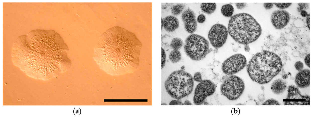
The Mycoplasma strains were grown on modified Hayflick’s agar incubated at 37 °C in an atmosphere containing 5% CO2 for up to 10 days. Typical umbonate fried-egg colonies became visible after approximately 3 days of incubation. The colonies showed irregular margins and a radially granulated surface structure (Figure 1a). The colony diameters ranged from 200 to 800 μm. The strains did not exhibit satellite colony formation, which is indicative of gliding motility [22], a result corroborated by a microcinematographic examination. The strains showed rapid growth at 37 °C, whereas only limited growth was observed at room temperature (approximately 20–22 °C), 28 °C, and 42 °C. No colony formation occurred at 4 °C or 15 °C. Growth under anaerobic conditions at 37 °C was likewise observed using the GasPak™ system (BD Diagnostics, Franklin Lakes, NJ, USA). The isolates were capable of passing through membrane filters with pore sizes of 450 nm and 220 nm. Filtration through the 220 nm membrane resulted in an approximately 1 log10 cfu/mL reduction in viable colony counts. Transmission electron microscopy of the proposed type strain 48589BT revealed pleomorphic cells lacking a cell wall and enclosed by a single membrane with a fuzz-like coating. The cells contained a finely granular cytoplasm with variably electron-dense regions, possibly representing ribosomes and sections of the circular chromosome (Figure 1b) [42]. The cultural and morphological characteristics of strain 48589BT were indistinguishable from those previously described for other Mycoplasma species isolated from buzzards, including M. buteonis and members of the M. aquilae complex [8,20].

3.2. Phylogenetic Positioning

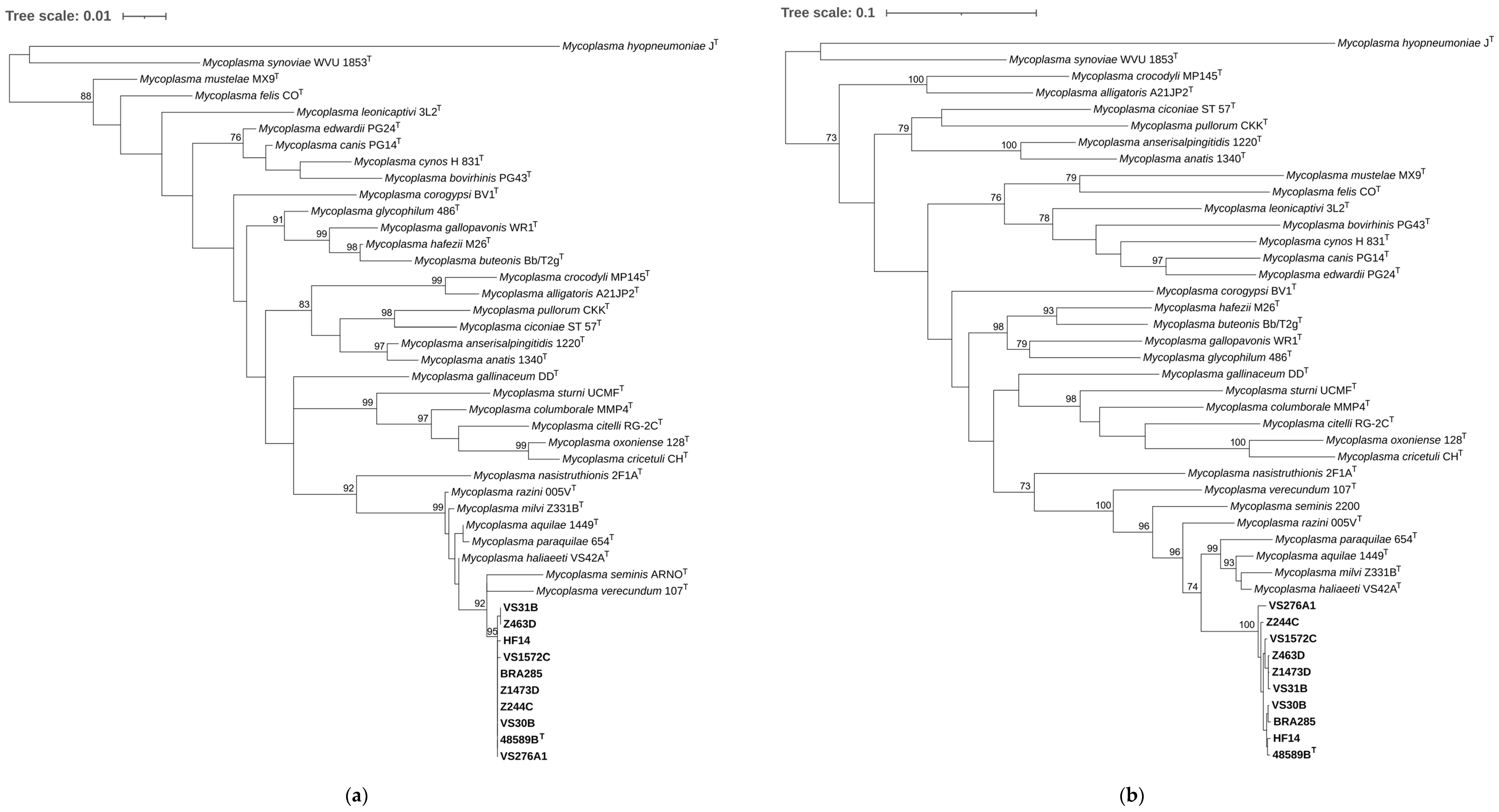
The comparative analysis of the 16S rRNA gene sequences assigned all ten isolates to the genus Mycoplasma. The nearly complete 16S rRNA gene sequence of strain 48589BT (1437 nt) showed the highest similarity to accipitrid-associated M. haliaeeti VS42AT (99.10%), M. aquilae 1449T (98.96%), M. razini 005VT, M. paraquilae 654T (both 98.82%), and M. milvi Z331BT (98.75%), as well as to M. verecundum 107T (98.66%) and M. seminis ARNOT (98.47%). The 16S rRNA gene sequences of 48589BT, BRA285, VS30B, VS276A1, Z244C, and Z1473D were identical and differed only slightly from those of HF14, VS31B, VS1572C, and Z463D (one base difference). Despite these high 16S rRNA gene sequence similarity values with the most closely related species, the resulting phylogenetic tree reveals a clear separation of 48589BT (and related strains) from their closest relatives, a finding supported by a high bootstrap value. In summary, the phylogenetic analysis of 16S rRNA gene sequences shows that the ten strains investigated form a monophyletic cluster, which, together with M. verecundum 107T, M. seminis ARNOT, the five accipitrid-associated Mycoplasma species (M. aquilae, M. paraquilae, M. haliaeeti, M. milvi, and M. razini), and M. nasistruthionis 2F1AT, constitutes a clade that is phylogenetically positioned within the M. synoviae cluster of the Hominis group of genus Mycoplasma (Figure 2a).
All ISR sequences obtained from the ten investigated strains had a length of 262 nt and were identical, with the exception of strain BRA285, which exhibited a single nucleotide polymorphism. The ISR sequence of 48589BT displayed the highest similarity to that of M. aquilae 1449T (97.34%), M. paraquilae 654T (96.96%), M. razini 005VT (96.63%), M. seminis ARNOT (93.89%), M. haliaeeti VS42AT (93.41%), M. milvi Z331BT (92.34%), and M. verecundum 107T (91.67%), taking into account the full coverage of the ISR sequences compared.
All investigated strains possessed partial rpoB gene sequences of 1698 nt, with the closest sequence match identified for M. haliaeeti VS42AT (93.29%), M. milvi Z331BT (92.71%), M. paraquilae 654T (92.65%), M. aquilae 1449T (92.53%), M. razini 005VT (92.05%), M. seminis 2200 (90.41%), and M. verecundum 107T (88.46%). The partial rpoB sequences showed a high degree of similarity among the investigated strains, with pairwise sequence identities ranging from 98.88 to 99.88%. Despite exceeding the generally recommended threshold of 90–91% for species differentiation in mycoplasmas based on rpoB [43], these values were still below the thresholds recently proposed for distinguishing species within the M. aquilae complex, which comprises M. aquilae, M. paraquilae, M. haliaeeti, and M. milvi [20]. As observed for 16S rRNA gene sequences, the phylogenetic tree based on rpoB sequences illustrates a clear separation of the 48589BT strain cluster from strains of related species (Figure 2b), which further supports the conclusion that the investigated strains represent a novel species within the M. synoviae cluster.

3.3. Phenotypic Traits and MALDI-ToF Mass Spectrometry Results

The examined strains neither metabolized glucose nor hydrolyzed arginine or urea. No hemolysis was observed on Columbia agar containing 5% sheep blood, and ‘film and spots’ production was absent in colonies grown on modified Hayflick’s agar for up to 10 days. Altogether, no distinguishing phenotypic characteristics were observed that differentiate the tested strains from their relatives.
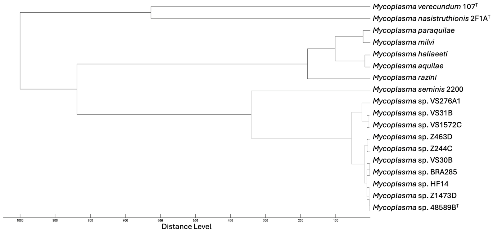
In MALDI-ToF mass spectrometric analysis, however, the ten strains formed a homogeneous cluster that was clearly distinct from the spectra generated for the phylogenetically closely related species M. seminis, M. nasistruthionis, M. verecundum, and the accipitrid-associated species M. aquilae, M. paraquilae, M. haliaeeti, M. milvi, and M. razini (Figure 3).

3.4. Genomic Traits and Coherence and Phylogenomics

The hybrid assembly combining Illumina short-read and Nanopore long-read data obtained from the proposed type strain 48589BT yielded a circular chromosome with a sequencing coverage of 850.0×. The genome comprised 897,976 bp and exhibited a G+C content of 29.2%. Genome annotation of strain 48589BT (CM135954) identified 687 predicted coding sequences (CDSs), of which 174 (25.3%) were classified as hypothetical proteins. The genome harbored three complete copies each of the 5S, 16S, and 23S rRNA genes. Whereas the 16S and 23S rRNA genes were organized into three operons, the 5S rRNA genes were located separately at more distant genomic positions. Comparison of the three 16S rRNA gene copies demonstrated only minor inter-operon sequence polymorphisms (a total of four base differences), similar to the minor differences observed when comparing the Sanger consensus sequences of the ten strains studied. Additional genomic characteristics of 48589BT and of the remaining strains are summarized in Table 2.
The average nucleotide identity analyses performed between 48589BT and closely related Mycoplasma species yielded ANIb values ranging from 76.45 to 80.58% (Table S1) and ANIm values between 85.56 and 86.65% (Table S2). These values were clearly below the proposed species boundary threshold of 95–96% [44]. In contrast, a comparison within the 48589BT strain’s cluster resulted in markedly higher ANIb values of 96.87–98.25% (Table S3) and ANIm values of 97.81–98.46% (Table S4), supporting the assignment of the ten investigated strains to a single species. Similarly, tetranucleotide signature correlation analysis produced TETRA coefficients ranging from 0.894 to 0.924 in a pairwise comparison between strain 48589BT and related Mycoplasma taxa (Table S5). These coefficients remained well below the ≥0.989 threshold proposed for genomes belonging to the same species [44]. By comparison, genomes within the 48589BT strain group exhibited TETRA values consistent with species-level relatedness (Table S6). The results of ANIb, ANIm, and TETRA analyses were further corroborated by low digital DNA-DNA hybridization estimates obtained for strain 48589BT and its closest relatives, including M. haliaeeti VS42AT (24.5%), M. milvi Z331BT (24.5%), M. aquilae 1449T (24.4%), M. razini 005VT (23.9%), and M. paraquilae 654T (23.7%). Collectively, these findings strongly support the classification of strain 48589BT as representing a novel species within the genus Mycoplasma.
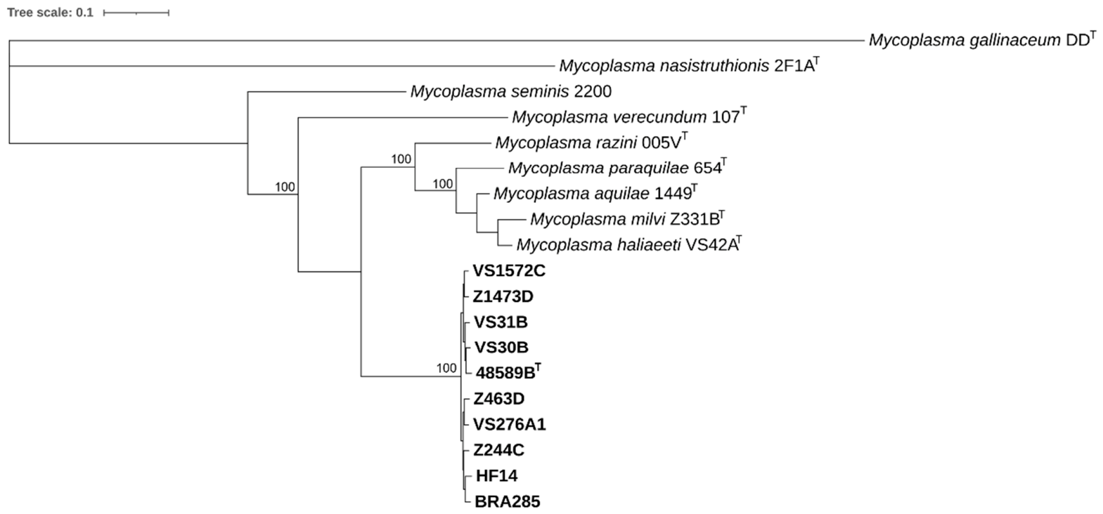
Phylogenomic reconstruction based on 273 single-copy coding genes, corresponding to an alignment of 101,344 amino acids and 304,032 nucleotides, yielded results consistent with those obtained from ANI/dDDH analyses and from phylogenetic studies of 16S rRNA genes and partial rpoB genes. The phylogenomic data therefore provide additional evidence that the investigated strains represent a separate species at the genomic level (Figure 4).

3.5. Functional and Comparative Genomics

Genome annotation indicated that the metabolic pathways of 48589BT are highly similar to those described recently for related accipitrid-associated mycoplasmas [20]. In brief, the genome of 48589BT lacks key enzymes of the upper Embden–Meyerhof–Parnas pathway and the oxidative pentose phosphate pathway, as well as a complete phosphoenolpyruvate phosphotransferase system. Conversely, genes for glycerol-3-phosphate metabolism and the lower EMP pathway were present, indicating glycerol-derived substrates as the primary entry point into central carbon metabolism. Downstream pathways for pyruvate oxidation and acetyl-CoA-to-acetate conversion appeared incomplete, suggesting restricted energy conservation from pyruvate.
Comparative genomic analysis identified multiple mobile genetic elements within the genomes analyzed in this study, highlighting their potential contribution to genomic plasticity. Insertion sequence (IS) family transposases were abundant in the circularized genome of 48589BT, whereas only a small number were detected in the draft genomes of the remaining strains (Table 2). This discrepancy is most likely attributable to differences in genome assembly quality, as repetitive elements such as IS sequences are known to be underrepresented or fragmented in draft assemblies derived from short-read sequencing data.
Additional mobile genetic elements included a ~14 kb putative prophage identified in strains 48589BT, Z1473D, Z244C (two copies), VS30B, VS276A1, and BRA285. This element displayed a gene content and overall organization reminiscent of the M. arthritidis MAV1 prophage group [45], although the encoded proteins shared relatively low amino acid identity (<40%) with MAV1 phage proteins. In contrast, a higher degree of similarity (58–87% amino acid identity) was observed with a related prophage identified in M. paraquilae 654T (Figure S1). A second, larger prophage of approximately 33 kb was identified in strains VS31B, Z1473D, and HF14. Its gene content and structural organization resembled those of M. agalactiae MAgV1 and M. molare MAgV1-like prophages. Both gene synteny and protein sequences showed the highest similarity to an MAgV1-like prophage present in M. milvi Z331BT, with amino acid identities ranging from 63 to 86% among conserved core phage proteins (Figure S1).
In addition, mycoplasma integrative and conjugative elements (MICEs) were identified as single-copy insertions in the genomes of VS30B, Z244C, and Z1473D (Table 2) and were collectively designated ICETbVS30B. This element spanned approximately 29 kb and comprised 27 co-orientated genes, including a conserved set of backbone genes characteristic of MICEs [46]. A comparative analysis based on the amino acid identity of these backbone coding sequences indicated that ICETbVS30B is related to MICEs described in accipitrid-associated Mycoplasma species, including M. aquilae, M. paraquilae, and M. haliaeeti [20]. The highest similarities (49–82% amino acid identity) were observed with backbone CDSs of the ICEPaq654 identified in M. paraquilae 654T (Figure S2).
In silico screening for defense mechanisms against invading foreign DNA identified only restriction-modification systems (type I and II) in the genomes analyzed, whereas other systems such as CRISPR/Cas or toxin–antitoxin modules were not detected. The genomes also encoded several factors potentially associated with virulence and pathogenicity. These included proteins commonly described as cytadhesins in other Mycoplasma species (e.g., P60, P68, P80, OppA, and variable surface lipoproteins), as well as a pair of the Mycoplasma Ig binding (MIB)/Mycoplasma Ig protease (MIP) immune evasion system [47]. In addition, multiple orphan MIP family Ig-specific serine endopeptidases were identified. Together, these features indicate adaptation to a host-associated lifestyle and suggest that the investigated strains may harbor determinants that could contribute to opportunistic behavior.

4. Conclusions

Collectively, the phenotypic, phylogenetic, and genomic data presented here provide compelling evidence that the investigated strains constitute a novel species of the genus Mycoplasma. However, the results indicate that within this cluster of closely related Mycoplasma species, cultural characteristics, phenotypic profiles, and 16S rRNA gene sequences possess limited discriminatory power and should therefore be interpreted alongside MALDI-ToF mass spectrometry and genome-level analyses, which provide decisive evidence supporting the novelty of the species described here. The authors propose the name Mycoplasma tracheobuteonis sp. nov. The epithet tracheobuteonis was chosen to reflect the predominant isolation of these strains from the trachea of common buzzards (Table 3).

Supplementary Materials

The following supporting information can be downloaded at: https://www.mdpi.com/article/10.3390/microorganisms14061224/s1. Table S1: Average nucleotide identities based on BLAST (ANIb, in %) between the genome of 48589BT and those of closely related Mycoplasma species. Values in brackets represent aligned nucleotides in %. Values for ‘48589BT versus strain of related species’ genome pairs are highlighted in blue. Table S2: Average nucleotide identities based on MUMmer (ANIm, in %) between the genome of 48589BT and those of closely related Mycoplasma species. Values in brackets represent aligned nucleotides in %. Values for ‘48589BT versus strain of related species’ genome pairs are highlighted in blue. Table S3: Average nucleotide identities based on BLAST (ANIb, in %) between genomes of 48589BT and its related strains, and of closely related M. razini 005VT. Values in brackets represent aligned nucleotides in %. ANIb values above the proposed species delineation threshold of 95–96% are highlighted in bold. Table S4: Average nucleotide identities based on MUMmer (ANIm, in %) between genomes of 48589BT and its related strains, and of closely related M. razini 005VT. Values in brackets represent aligned nucleotides in %. ANIb values above the proposed species delineation threshold of 95–96% are highlighted in bold. Table S5: Tetranucleotide signature correlation (TETRA) coefficients between the genome of 48589BT and those of closely related Mycoplasma species. TETRA coefficients for ‘48589BT versus strain of related species’ genome pairs are highlighted in bold. Table S6: Tetranucleotide signature correlation (TETRA) coefficients between genomes of 48589BT and its related strains, and of closely related M. razini 005VT. TETRA coefficients above cutoff (≥0.999) or in range (≥0.989) are highlighted in bold and in green or blue, respectively. Figure S1: (a) A prophage identified in M. tracheobuteonis sp. nov. resembling φMAV1 of M. arthritidis, sharing only <40% aa identity to MAV1 phage proteins but showing higher similarity with a related prophage identified in M. paraquilae 654T. No homologues were identified for non-labelled proteins. GenBank locus tag numbers of the first and the last prophage proteins are indicated below the MAV1-like prophage of 48589BT. HtpN—HtpB, structural proteins; RepB, replicative DNA helicase, A and P, replication initiators; MarMP, putative C5 methylase; MarRP, transcriptional regulator; Int, integrase, Exis, excisionase, Vir, protein to exclude superinfecting phage; Imm, phage repressor; (b) A large prophage identified in M. tracheobuteonis sp. nov. reminiscent of M. agalactiae MAgV1-like prophages identified in M. molare H542T and M. milvi Z331BT. GenBank locus tag numbers of the first and the last prophage proteins are indicated below the MAgV1-like prophage of VS31B. H, helicase; Pol, DNA polymerase; D, DNA primase; X, Xer recombinase; C, prohead protease; P, portal protein; T, terminase. Figure S2: Mycoplasma Integrative and Conjugative Element (MICE) backbone and structural organization of a MICE identified in the genome of VS30B (ICETbVS30B) related to ICEPaq654 identified in M. paraquilae 654T. MICE backbone CDS conserved across MICEs (labeled by *) or that may be absent or truncated in MICEs are represented by color filled arrows. GenBank locus tag numbers of the first and the last MICE genes are indicated below ICETbVS30B. SSBP: single-stranded DNA binding protein, P: pilin-like protein, ParA: ParA family protein, REase: restriction endonuclease, MTase: methyltransferase, IR: inverted repeats.

Author Contributions

Conceptualization, J.S. and A.S.R.; methodology, J.S.; software, J.S.; validation, J.S.; formal analysis, S.K. and J.S.; investigation, S.K., N.D. and J.S.; resources, S.K., A.K.-H., I.L., V.S., A.S.R. and J.S.; data curation, J.S.; writing—original draft preparation, S.K. and J.S.; writing—review and editing, S.K., A.K.-H., N.D., A.A., I.L., V.S., A.S.R. and J.S.; visualization, S.K. and J.S.; supervision, A.K.-H. and J.S.; project administration, J.S.; funding acquisition, J.S. All authors have read and agreed to the published version of the manuscript.

Funding

Open Access Funding by the University of Veterinary Medicine Vienna.

Institutional Review Board Statement

Ethical review and approval were waived for this study due to the exclusive use of deceased animals sampled during routine post-mortem procedures.

Informed Consent Statement

Not applicable.

Data Availability Statement

Genome assemblies and marker gene sequences generated in this study have been deposited in the NCBI GenBank database. The corresponding accession numbers are provided in the main text, tables, and Supplementary Materials.

Acknowledgments

The authors are grateful to Martina Zimmermann for her excellent technical assistance.

Conflicts of Interest

The authors declare no conflicts of interest.

References

  1. Razin, S.; Yogev, D.; Naot, Y. Molecular Biology and Pathogenicity of Mycoplasmas. Microbiol. Mol. Biol. Rev. 1998, 62, 1094–1156. [Google Scholar] [CrossRef] [PubMed]
  2. Balish, M.F.; Chopra-Dewasthaly, R.; Pereyre, S.; Ramírez, A.S.; Viver, T.; Spergser, J. Mycoplasma. In Bergey’s Manual of Systematics of Archaea and Bacteria; John Wiley & Sons, Inc.: Hoboken, NJ, USA, 2024; pp. 1–101. [Google Scholar] [CrossRef]
  3. Bradbury, J.M. Poultry Mycoplasmas: Sophisticated Pathogens in Simple Guise. Br. Poult. Sci. 2005, 46, 125–136. [Google Scholar] [CrossRef]
  4. Grózner, D.; Sulyok, K.M.; Kreizinger, Z.; Rónai, Z.; Jánosi, S.; Turcsányi, I.; Károlyi, H.F.; Kovács, Á.B.; Kiss, M.J.; Volokhov, D.; et al. Detection of Mycoplasma anatis, M. anseris, M. cloacale and Mycoplasma sp. 1220 in Waterfowl Using Species-Specific PCR Assays. PLoS ONE 2019, 14, e0219071. [Google Scholar] [CrossRef]
  5. Lierz, M.; Hagen, N.; Lueschow, D.; Hafez, H.M. Species-Specific Polymerase Chain Reactions for the Detection of Mycoplasma buteonis, Mycoplasma falconis, Mycoplasma gypis, and Mycoplasma corogypsi in Captive Birds of Prey. Avian Dis. 2008, 52, 94–99. [Google Scholar] [CrossRef] [PubMed]
  6. Lierz, M.; Hagen, N.; Hernadez-Divers, S.J.; Hafez, H.M. Occurrence of Mycoplasmas in Free-Ranging Birds of Prey in Germany. J. Wildl. Dis. 2008, 44, 845–850. [Google Scholar] [CrossRef]
  7. Lierz, M.; Obon, E.; Schink, B.; Carbonell, F.; Hafez, H.M. The Role of Mycoplasmas in a Conservation Project of the Lesser Kestrel (Falco naumanni). Avian Dis. 2008, 52, 641–645. [Google Scholar] [CrossRef]
  8. Poveda, J.; Giebel, J.; Flossdorf, J.; Meier, J.; Kirchhoff, H. Mycoplasma buteonis sp. nov., Mycoplasma falconis sp. nov., and Mycoplasma gypis sp. Nov., Three Species from Birds of Prey. Int. J. Syst. Bacteriol. 1994, 44, 94–98. [Google Scholar] [CrossRef]
  9. Suárez-Pérez, A.; Ramírez, A.S.; Rosales, R.S.; Calabuig, P.; Poveda, C.; Rosselló-Móra, R.; Nicholas, R.A.J.; Poveda, J.B. Mycoplasma neophronis sp. nov., Isolated from the Upper Respiratory Tract of Canarian Egyptian Vultures (Neophron percnopterus majorensis). Int. J. Syst. Evol. Microbiol. 2012, 62, 1321–1325. [Google Scholar] [CrossRef] [PubMed]
  10. Oaks, J.L.; Donahoe, S.L.; Rurangirwa, F.R.; Rideout, B.A.; Gilbert, M.; Virani, M.Z. Identification of a Novel Mycoplasma Species from an Oriental White-Backed Vulture (Gyps bengalensis). J. Clin. Microbiol. 2004, 42, 5909–5912. [Google Scholar] [CrossRef]
  11. Ziegler, L.; Möller Palau-Ribes, F.; Enderlein, D.; Herbst, W.; Schmidt, L.; Lierz, M. Mycoplasma hafezii Sp. Nov., Isolated from the Trachea of a Peregrine Falcon (Falco peregrinus). Int. J. Syst. Evol. Microbiol. 2019, 69, 773–777. [Google Scholar] [CrossRef]
  12. Fischer, L.; Möller Palau-Ribes, F.; Enderlein, D.; Fischer, D.; Herbst, W.; Baudler, L.; Hafez, H.M.; Lierz, M. Description, Occurrence and Significance of Mycoplasma seminis sp. Nov. Isolated from Semen of a Gyrfalcon (Falco rusticolus). Vet. Microbiol. 2020, 247, 108789, Corrigendum in Vet. Microbiol. 2020, 249, 108800. https://doi.org/10.1016/j.vetmic.2020.108800. [Google Scholar] [CrossRef]
  13. Panangala, V.S.; Stringfellow, J.S.; Dybvig, K.; Woodard, A.; Sun, F.; Rose, D.L.; Gresham, M.M. Mycoplasma corogypsi sp. Nov., a New Species from the Footpad Abscess of a Black Vulture, Coragyps Atratus. Int. J. Syst. Bacteriol. 1993, 43, 585–590. [Google Scholar] [CrossRef][Green Version]
  14. Ruder, M.G.; Feldman, S.H.; Wünschmann, A.; McRuer, D.L. Association of Mycoplasma Corogypsi and Polyarthritis in a Black Vulture (Coragyps atratus) in Virginia. J. Wildl. Dis. 2009, 45, 808–816. [Google Scholar] [CrossRef] [PubMed]
  15. Van Wettere, A.J.; Ley, D.H.; Scott, D.E.; Buckanoff, H.D.; Degernes, L.A. Mycoplasma Corogypsi Associated Polyarthritis and Tenosynovitis in Black Vultures (Coragyps atratus). Vet. Pathol. 2013, 50, 291–298. [Google Scholar] [CrossRef]
  16. Loria, G.R.; Ferrantelli, E.; Giardina, G.; Vecchi, L.L.; Sparacino, L.; Oliveri, F.; McAuliffe, L.; Nicholas, R.A.J. Isolation and Characterization of Unusual Mycoplasma spp. from Captive Eurasian Griffon (Gyps fulvus) in Sicily. J. Wildl. Dis. 2008, 44, 159–163. [Google Scholar] [CrossRef]
  17. Lecis, R.; Chessa, B.; Cacciotto, C.; Addis, M.F.; Coradduzza, E.; Berlinguer, F.; Muzzeddu, M.; Lierz, M.; Carcangiu, L.; Pittau, M.; et al. Identification and Characterization of Novel Mycoplasma spp. Belonging to the Hominis Group from Griffon Vultures. Res. Vet. Sci. 2010, 89, 58–64. [Google Scholar] [CrossRef] [PubMed]
  18. Lecis, R.; Secci, F.; Mandas, L.; Muzzeddu, M.; Pittau, M.; Alberti, A. Molecular Identification and Sequence Characterization of Mycoplasmas in Free-Living Birds of Prey. J. Zoo Wildl. Med. 2016, 47, 917–922. [Google Scholar] [CrossRef]
  19. Sawicka-Durkalec, A.; Kursa, O.; Bednarz, Ł.; Tomczyk, G. Occurrence of Mycoplasma spp. in Wild Birds: Phylogenetic Analysis and Potential Factors Affecting Distribution. Sci. Rep. 2021, 11, 17065. [Google Scholar] [CrossRef] [PubMed]
  20. Spergser, J.; Kugler, S.; Kübber-Heiss, A.; Höfle, U.; Dinhopl, N.; Szostak, M.P.; Loncaric, I.; Viver, T.; Ramírez, A.S. Mycoplasma aquilae sp. nov., Mycoplasma paraquilae sp. nov., Mycoplasma haliaeeti sp. nov., Mycoplasma milvi sp. nov., and Mycoplasma razini sp. nov., Isolated from Predatory Birds of the Accipitridae Family. Syst. Appl. Microbiol. 2025, 48, 126663. [Google Scholar] [CrossRef]
  21. Spergser, J.; Hess, C.; Loncaric, I.; Ramírez, A.S. Matrix-Assisted Laser Desorption Ionization–Time of Flight Mass Spectrometry Is a Superior Diagnostic Tool for the Identification and Differentiation of Mycoplasmas Isolated from Animals. J. Clin. Microbiol. 2019, 57, 10.1128. [Google Scholar] [CrossRef]
  22. Brown, D.R.; Whitcomb, R.F.; Bradbury, J.M. Revised Minimal Standards for Description of New Species of the Class Mollicutes (Division Tenericutes). Int. J. Syst. Bacteriol. 2007, 57, 2703–2719. [Google Scholar] [CrossRef]
  23. Tully, J.G. Culture Medium Formulation for Primary Isolation and Maintenance of Mollicutes. In Molecular and Diagnostic Procedures in Mycoplasmology; Razin, S., Tully, J.G., Eds.; Academic Press: San Diego, CA, USA, 1995; pp. 33–39. ISBN 978-0-12-583805-4. [Google Scholar]
  24. Tully, J.G. Cloning and Filtration Techniques for Mycoplasmas. In Methods in Mycoplasmology; Razin, S., Tully, J.G., Eds.; Academic Press: New York, NY, USA, 1983; pp. 173–177. [Google Scholar]
  25. Indikova, I.; Vronka, M.; Szostak, M.P. First Identification of Proteins Involved in Motility of Mycoplasma gallisepticum. Vet. Res. 2014, 45, 99. [Google Scholar] [CrossRef][Green Version]
  26. Lane, D.J. 16S/23S rRNA Sequencing. In Nucleic Acid Techniques in Bacterial Systematics; Stackebrandt, E., Goodfellow, M., Eds.; Wiley: Chichester, UK, 1991; pp. 115–175. [Google Scholar]
  27. Harasawa, R.; Pitcher, D.G.; Ramírez, A.S.; Bradbury, J.M. A Putative Transposase Gene in the 16S–23S rRNA Intergenic Spacer Region of Mycoplasma Imitans. Microbiology 2004, 150, 1023–1029. [Google Scholar] [CrossRef]
  28. Yoon, S.-H.; Ha, S.-M.; Kwon, S.; Lim, J.; Kim, Y.; Seo, H.; Chun, J. Introducing EzBioCloud: A Taxonomically United Database of 16S rRNA Gene Sequences and Whole-Genome Assemblies. Int. J. Syst. Evol. Microbiol. 2017, 67, 1613–1617. [Google Scholar] [CrossRef]
  29. Altschul, S.F.; Gish, W.; Miller, W.; Myers, E.W.; Lipman, D.J. Basic Local Alignment Search Tool. J. Mol. Biol. 1990, 215, 403–410. [Google Scholar] [CrossRef]
  30. Thompson, J.D.; Gibson, T.J.; Plewniak, F.; Jeanmougin, F.; Higgins, D.G. The CLUSTAL_X Windows Interface: Flexible Strategies for Multiple Sequence Alignment Aided by Quality Analysis Tools. Nucleic Acids Res. 1997, 25, 4876–4882. [Google Scholar] [CrossRef] [PubMed]
  31. Tamura, K.; Stecher, G.; Kumar, S. MEGA11: Molecular Evolutionary Genetics Analysis Version 11. Mol. Biol. Evol. 2021, 38, 3022–3027. [Google Scholar] [CrossRef] [PubMed]
  32. Letunic, I.; Bork, P. Interactive Tree of Life (iTOL) v6: Recent Updates to the Phylogenetic Tree Display and Annotation Tool. Nucleic Acids Res. 2024, 52, W78–W82. [Google Scholar] [CrossRef]
  33. Aluotto, B.B.; Wittler, R.G.; Williams, C.O.; Faber, J.E. Standardized Bacteriologic Techniques for the Characterization of Mycoplasma Species. Int. J. Syst. Evol. Microbiol. 1970, 20, 35–58. [Google Scholar] [CrossRef]
  34. Gardella, R.S.; Del Giudice, R.A. Haemagglutination, Haemadsorption and Haemolysis. In Methods in Mycoplasmology; Razin, S., Tully, J.G., Eds.; Academic Press: New York, NY, USA, 1983; pp. 379–384. [Google Scholar]
  35. Freundt, E.A. Film and Spot Production. In Methods in Mycoplasmology; Razin, S., Tully, J.G., Eds.; Academic Press: New York, NY, USA, 1983; pp. 373–374. [Google Scholar]
  36. Bankevich, A.; Nurk, S.; Antipov, D.; Gurevich, A.A.; Dvorkin, M.; Kulikov, A.S.; Lesin, V.M.; Nikolenko, S.I.; Pham, S.; Prjibelski, A.D.; et al. SPAdes: A New Genome Assembly Algorithm and Its Applications to Single-Cell Sequencing. J. Comput. Biol. 2012, 19, 455–477. [Google Scholar] [CrossRef] [PubMed]
  37. Wick, R.R.; Judd, L.M.; Gorrie, C.L.; Holt, K.E. Unicycler: Resolving Bacterial Genome Assemblies from Short and Long Sequencing Reads. PLoS Comput. Biol. 2017, 13, e1005595. [Google Scholar] [CrossRef]
  38. Richter, M.; Rosselló-Móra, R.; Oliver Glöckner, F.; Peplies, J. JSpeciesWS: A Web Server for Prokaryotic Species Circumscription Based on Pairwise Genome Comparison. Bioinformatics 2016, 32, 929–931. [Google Scholar] [CrossRef]
  39. Meier-Kolthoff, J.P.; Carbasse, J.S.; Peinado-Olarte, R.L.; Göker, M. TYGS and LPSN: A Database Tandem for Fast and Reliable Genome-Based Classification and Nomenclature of Prokaryotes. Nucleic Acids Res. 2022, 50, D801–D807. [Google Scholar] [CrossRef] [PubMed]
  40. Stamatakis, A. RAxML Version 8: A Tool for Phylogenetic Analysis and Post-Analysis of Large Phylogenies. Bioinformatics 2014, 30, 1312–1313. [Google Scholar] [CrossRef] [PubMed]
  41. Darling, A.C.E.; Mau, B.; Blattner, F.R.; Perna, N.T. Mauve: Multiple Alignment of Conserved Genomic Sequence With Rearrangements. Genome Res. 2004, 14, 1394–1403. [Google Scholar] [CrossRef] [PubMed]
  42. Razin, S.; Hayflick, L. Highlights of Mycoplasma Research—An Historical Perspective. Biologicals 2010, 38, 183–190. [Google Scholar] [CrossRef]
  43. Volokhov, D.V.; Simonyan, V.; Davidson, M.K.; Chizhikov, V.E. RNA Polymerase Beta Subunit (rpoB) Gene and the 16S–23S rRNA Intergenic Transcribed Spacer Region (ITS) as Complementary Molecular Markers in Addition to the 16S rRNA Gene for Phylogenetic Analysis and Identification of the Species of the Family Mycoplasmataceae. Mol. Phylogenet. Evol. 2012, 62, 515–528. [Google Scholar] [CrossRef]
  44. Richter, M.; Rosselló-Móra, R. Shifting the Genomic Gold Standard for the Prokaryotic Species Definition. Proc. Natl. Acad. Sci. USA 2009, 106, 19126–19131. [Google Scholar] [CrossRef]
  45. Citti, C.; Baranowski, E.; Dordet-Frisoni, E.; Faucher, M.; Nouvel, L.-X. Genomic Islands in Mycoplasmas. Genes 2020, 11, 836. [Google Scholar] [CrossRef]
  46. Citti, C.; Dordet-Frisoni, E.; Nouvel, L.X.; Kuo, C.; Baranowski, E. Horizontal Gene Transfers in Mycoplasmas (Mollicutes). Curr. Issues Mol. Biol. 2018, 29, 3–22. [Google Scholar] [CrossRef]
  47. Nottelet, P.; Bataille, L.; Gourgues, G.; Anger, R.; Lartigue, C.; Sirand-Pugnet, P.; Marza, E.; Fronzes, R.; Arfi, Y. The Mycoplasma Surface Proteins MIB and MIP Promote the Dissociation of the Antibody-Antigen Interaction. Sci. Adv. 2021, 7, eabf2403. [Google Scholar] [CrossRef]
Figure 1. (a) Fried-egg colonies of 48589BT on modified Hayflick’s Agar. Bar, 500 μm; (b) Spherical- to oval-shaped cells with a discrete fuzz-like coating; transmission electron micrograph of 48589BT. Bar, 500 nm.
Microorganisms 14 01224 g001
Figure 2. Maximum likelihood trees, demonstrating the phylogenetic relatedness of the 10 strains studied to related species of the M. synoviae cluster based on (a) 16S rRNA genes and (b) partial rpoB gene sequences. M. hyopneumoniae JT (M. neurolyticum cluster) was used as out-group organism. Numbers at nodes represent bootstrap confidence values (1000 replications). Only values ≥ 70% are shown. Tree scale, number of nucleotide substitutions per site.
Microorganisms 14 01224 g002
Figure 3. MALDI-ToF mass spectrometry score-oriented dendrogram based on distances between spectra from 48589BT and related isolates and their closest relatives.
Microorganisms 14 01224 g003
Figure 4. Phylogenomic tree derived from genome comparison of the ten strains investigated and their closest relatives within the M. synoviae cluster. M. gallinaceum DDT was used as out-group organism. Numbers at nodes represent bootstrap confidence values (100 rounds). Tree scale, number of substitutions per site.
Microorganisms 14 01224 g004
Table 1. Strain metadata and corresponding accession numbers (acc. no.) for 16S rRNA gene, 16S–23S intergenic spacer region (ISR), and partial rpoB gene sequences.
Strain
Designation
Host SpeciesIsolation SiteYear of
Isolation
Country of IsolationAcc. No. 16S rRNAAcc. No. ISRAcc. No. rpoB
48589BTButeo buteochoana2009GermanyPZ033355PZ036093PZ044315
Z463DButeo buteotrachea2019AustriaPZ033361PZ036099PZ044321
Z1473DButeo buteotrachea2019AustriaPZ033362PZ036100PZ044322
VS1572CButeo buteotrachea2019AustriaPZ033359PZ036097PZ044319
Z244CButeo buteotrachea2020AustriaPZ033360PZ036098PZ044320
VS30BButeo buteotrachea2021AustriaPZ033356PZ036094PZ044316
VS31BButeo buteotrachea2021AustriaPZ033357PZ036095PZ044317
VS276A1Buteo buteotrachea2022AustriaPZ033358PZ036096PZ044318
HF14Aquila fasciatachoana2005SpainPZ033364PZ036102PZ044324
BRA285Haliaeetus albicillachoana2009GermanyPZ033363PZ036101PZ044323
Table 2. Genomic characteristics of the proposed type strain 48589BT (finished genome) and its related strains Z463D, Z1473D, VS1572C, Z244C, VS30B, VS31B, VS276A1, HF14, and BRA285 (draft genomes).
48589BTZ463DZ1473DVS1572CZ244CVS30BVS31BVS276A1HF14BRA285
Accession numberCM135954JBTKTC010000000JBTKTB010000000JBTKTE010000000JBTKTD010000000JBTKTH010000000JBTKTG010000000JBTKTF010000000JBTKTI010000000JBTKTJ01
0000000
Genome size (bp)897,976814,744909,243852,966879,432872,886873,148839,335847,403845,754
Genome coverage850×186×203×222×197×185×200×215×210×220×
Contig #1314549504434583739
Contig N50 (kB)-46.435.242.233.235.859.135.842.232.2
G+C (%)29.229.129.129.029.029.029.229.129.329.1
Genes
(total)
744646747682712700696689691669
CDSs
(total)
701609711648676664660651653633
CDSs (with protein)687607709646674662658647649628
Genes (RNA)43373634363636383836
tRNAs31313131313131313131
ncRNAs3332333333
Pseudo-genes14222222445
Transposases32 (+5 pseudo) a4 b5 c9 d4 e6 f7 g8 h6 i6 j
MICEs--ICETbVS30B-ICETbVS30BICETbVS30B----
ProphagesMAV1-like-MAV1-like MAgV1-like-MAV1-likeMAV1-likeMAgV1-likeMAV1-likeMAgV1-likeMAV1-like
a Five IS1634-, 11 IS256-(five of them pseudogenes), 15 IS30-, and one IS3-family transposases; five unclassified transposases. b One IS30-family transposase; three unclassified transposases. c One IS1634- and two IS30-family transposases; two unclassified transposases. d One IS1634- and six IS30-family transposases; two unclassified transposases. e One IS256- and one IS3-family transposases; two unclassified transposases. f Three IS30-family transposases; three unclassified transposases. g One IS1634-, three IS30- and one IS3-family transposases; two unclassified transposases. h One IS1634-, one IS256-, three IS30- and one IS3-family transposases; two unclassified transposases. i Two IS1634- and two IS30-family transposases; two unclassified transposases. j One IS1634-, one IS256-, three IS30-, and one IS3-family transposases.
Table 3. Description of Mycoplasma tracheobuteonis sp. nov.
Genus nameMycoplasma
Species nameMycoplasma tracheobuteonis
Species epithettracheobuteonis
Species statussp. nov.
Species etymologytra.che.o.bu.te.o’nis. Gr. fem. n. tracheia, windpipe; N.L. gen. n. buteonis of Buteo, a bird genus; N.L. gen. n. tracheobuteonis, of the trachea of a buzzard (Buteo), referring to the site of isolation and principal host
Description of the new taxon and diagnostic traitsCells are wall-less and predominantly spherical to oval in morphology. Typical fried-egg colonies develop within 3 days of incubation on modified Hayflick’s agar at 37 °C in an atmosphere containing 5% CO2. Growth occurs between 20 °C and 42 °C, with optimal growth at 37 °C. Cells are non-motile. Neither arginine nor urea is hydrolyzed, and acid production from glucose is absent.
MALDI-ToF mass spectrometry profiles are distinct from those of closely related Mycoplasma species. Although 16S rRNA gene and 16S–23S intergenic spacer region sequences show high similarity to related taxa, they allow differentiation from neighboring species. Partial rpoB gene sequences are unique and enable clear discrimination from closely related species. Genome similarity indices together with phylogenomic analyses further support the distinct species status of the investigated strains.
Country of originGermany
Region of originSaxony
Date of isolation30 September 2009
Source of isolationChoana of a common buzzard (Buteo buteo)
Sampling date16 September 2009
Latitude51°19′06.4′’ N
Longitude12°23′28.2′’ E
Altitude (meters above sea level)115
16S rRNA gene accession numberPZ033355
Genome accession numberCM135954
Genome statusComplete
Genome size (bp)897,976
G + C (%)29.2
Number of strains in study10
Source of isolation of non-type strainsZ463D—trachea of a common buzzard (Buteo buteo), Austria (2019); Z1473D—trachea of a common buzzard (Buteo buteo), Austria (2019); VS1572C—trachea of a common buzzard (Buteo buteo), Austria (2019); Z244C—trachea of a common buzzard (Buteo buteo), Austria (2020); VS30B—trachea of a common buzzard (Buteo buteo), Austria (2021); VS31B—trachea of a common buzzard (Buteo buteo), Austria (2021); VS276A1—trachea of a common buzzard (Buteo buteo), Austria (2022); HF14—choana of a Bonelli’s eagle (Aquila fasciata), Spain (2005); BRA285—choana of a white-tailed eagle (Haliaeetus albicilla), Germany (2009)
Designation of the type strain48589BT
Strain collection numbersDSM 115882T, NCTC 14927T
Disclaimer/Publisher’s Note: The statements, opinions and data contained in all publications are solely those of the individual author(s) and contributor(s) and not of MDPI and/or the editor(s). MDPI and/or the editor(s) disclaim responsibility for any injury to people or property resulting from any ideas, methods, instructions or products referred to in the content.