Clinical Features of Adult Patients with Isolated Pulmonary Valve Endocarditis: A Systematic Review

 ,
,  , ,
, , 
 ,
,
Abstract
1. Introduction
2. Materials and Methods
Systematic Review
3. Results
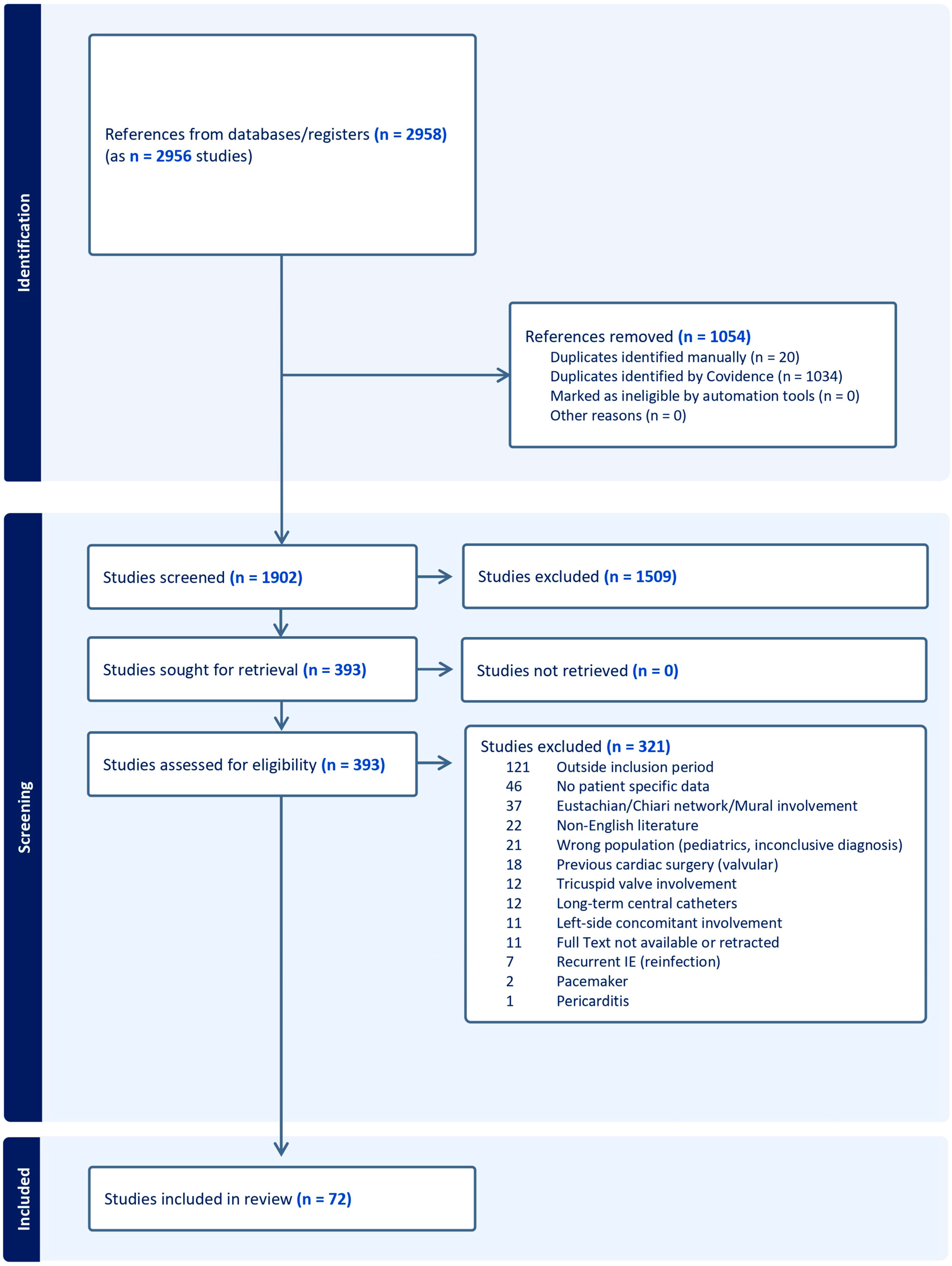
3.1. Systematic Review
3.2. Baseline Demographics
3.3. Clinical and Echocardiographic Characteristics
3.4. Treatment and Outcomes
3.5. Global Description of Fatal Cases
3.6. Comparison of Cases According to Risk Factor
4. Discussion
5. Conclusions
Supplementary Materials
Author Contributions
Funding
Institutional Review Board Statement
Informed Consent Statement
Data Availability Statement
Acknowledgments
Conflicts of Interest
References
- Habib, G.; Erba, P.A.; Iung, B.; Donal, E.; Cosyns, B.; Laroche, C.; Popescu, B.A.; Prendergast, B.; Tornos, P.; Sadeghpour, A.; et al. Clinical presentation, aetiology and outcome of infective endocarditis. Results of the ESC-EORP EURO-ENDO (European infective endocarditis) registry: A prospective cohort study. Eur. Heart J. 2019, 40, 3222–3232, Corrigendum to Eur. Heart J. 2020, 41, 2091. https://doi.org/10.1093/eurheartj/ehz803. [Google Scholar] [CrossRef]
- Miranda, W.R.; Connolly, H.M.; Desimone, D.C.; Phillips, S.D.; Wilson, W.R.; Sohail, M.R.; Steckelberg, J.M.; Baddour, L.M. Infective Endocarditis Involving the Pulmonary Valve. Am. J. Cardiol. 2015, 116, 1928–1931. [Google Scholar] [CrossRef] [PubMed]
- Pericàs, J.M.; Llopis, J.; Athan, E.; Hernández-Meneses, M.; Hannan, M.M.; Murdoch, D.R.; Kanafani, Z.; Freiberger, T.; Strahilevitz, J.; Fernández-Hidalgo, N.; et al. Prospective Cohort Study of Infective Endocarditis in People Who Inject Drugs. J. Am. Coll. Cardiol. 2021, 77, 544–555. [Google Scholar] [CrossRef]
- Tsao, M.M.; Katz, D. Central venous catheter-induced endocarditis: Human correlate of the animal experimental model of endocarditis. Rev. Infect. Dis. 1984, 6, 783–790. [Google Scholar] [CrossRef]
- Sharma, S.; Malavia, G.A. Pulmonary valve infective endocarditis: A case series. Ann. Pediatr. Cardiol. 2021, 14, 496–500. [Google Scholar] [CrossRef]
- Hicklin, H.E.; Huang, G.; Davis, K.A.; Barnes, E.W.; Peacock, J.E. Pulmonic valve infective endocarditis: Transesophageal echocardiography is critical to diagnosis. Infect. Dis. 2020, 52, 746–748. [Google Scholar] [CrossRef] [PubMed]
- Kulahcioglu, S.; Sari, M.; Yilmaz, C.; Kizmaz, Y.U.; Adademir, T.; Ceyran, H. Unexpected isolated native pulmonary valve endocarditis; really benign as thought? J. Card. Surg. 2022, 37, 2120–2123. [Google Scholar] [CrossRef] [PubMed]
- Yuan, S.M. Right-sided infective endocarditis: Recent epidemiologic changes. Int. J. Clin. Exp. Med. 2014, 7, 199–218. [Google Scholar]
- Li, J.S.; Sexton, D.J.; Mick, N.; Nettles, R.; Fowler, V.G., Jr.; Ryan, T.; Bashore, T.; Corey, G.R. Proposed modifications to the Duke criteria for the diagnosis of infective endocarditis. Clin. Infect. Dis. 2000, 30, 633–638. [Google Scholar] [CrossRef]
- Fowler, V.G.; Durack, D.T.; Selton-Suty, C.; Athan, E.; Bayer, A.S.; Chamis, A.L.; Dahl, A.; DiBernardo, L.; Durante-Mangoni, E.; Duval, X.; et al. The 2023 Duke-International Society for Cardiovascular Infectious Diseases Criteria for Infective Endocarditis: Updating the Modified Duke Criteria. Clin. Infect. Dis. 2023, 77, 518–526, Correction in Clin. Infect. 2023, 77, 1222. https://doi.org/10.1093/cid/ciad510. [Google Scholar] [CrossRef]
- Page, M.J.; McKenzie, J.E.; Bossuyt, P.M.; Boutron, I.; Hoffmann, T.C.; Mulrow, C.D.; Shamseer, L.; Tetzlaff, J.M.; Akl, E.A.; Brennan, S.E.; et al. The PRISMA 2020 statement: An updated guideline for reporting systematic reviews. BMJ 2021, 372, n71. [Google Scholar] [CrossRef]
- Zhang, M.-X.; Zhang, W.-M.; Yu, C.; Zhao, B.-W.; Chen, R.; Pan, M.; Wang, B. Isolated Pulmonary Valve Endocarditis with Rapid Progression: A Case Report and Literature Review. J. Cardiothorac. Surg. 2021, 16, 16. [Google Scholar] [CrossRef]
- Zhang, H. A Rare Case of Pulmonary Valve Infective Endocarditis. Am. J. Respir. Crit. Care Med. 2023, 207, A6701. [Google Scholar] [CrossRef]
- Xiong, J.; Embry, E.; Prasad, S.; Abdelghani, L. Isolated Pulmonic Valve Endocarditis With Septic Embolism. Am. J. Respir. Crit. Care Med. 2025, 211, A1629. [Google Scholar] [CrossRef]
- Whitehead, T.; Moore, A.; Agarwal, A.; Appleby, J. Isolated pulmonary valve endocarditis in a patient without structural heart disease or a history of intravenous drug use. BMJ Case Rep. 2023, 16, e250098. [Google Scholar] [CrossRef] [PubMed]
- Velez, K.; Obrusnik, I.; Steuerwald, M.; Owlia, M. Isolated pulmonic valve endocarditis: An unusual case in a young woman with ventricular septal defect. J. Am. Coll. Cardiol. 2025, 85, 3850. [Google Scholar] [CrossRef]
- Vâta, A.; Loghin, I.I.; Miftode, R.S.; Crisu, D.; Luca, C.M.; Vasilescu, A.M.; Onofrei, I.M.; Parânga, T.; Miftode, I.L. Isolated Pulmonary Valve Endocarditis Caused by Enterococcus Faecalis-Facing the Unpredictable. Antibiotics 2025, 14, 10. [Google Scholar] [CrossRef]
- Valsky, S.; Mutschlechner, D.; Wiedemann, D.; Gremmel, T. Isolated Pulmonary Valve Endocarditis. Wien. Klin. Wochenschr. 2024, 136, 700–702. [Google Scholar] [CrossRef] [PubMed]
- Tominaga, Y.; Ueno, T.; Taira, M.; Miyagawa, S.; Sawa, Y. Infective Endocarditis in an Adult with Unrepaired Corrected Transposition. SAGE Open Med. Case Rep. 2022, 10, 2050313X221085101. [Google Scholar] [CrossRef]
- Toader, D.; Craciun Mirescu, A.; Cocora, M.; Munteanu, O.; Mustafa, E.R.; Florescu, C.; Berceanu, M.; Popescu, M.; Cojocaru, A.; Oprian, A. Pulmonary Valve Endocarditis Complicated with Fistula between Right Ventricle Outflow Tract and Aorta in a Patient with Patent Ductus Arteriosus. Eur. Heart J. Cardiovasc. Imaging 2020, 21, i388. [Google Scholar] [CrossRef]
- Stefaniak, S.; Bujak, R.; Puslecki, M.; Araszkiewicz, A.; Katarzynski, S.; Blazejewski, J.; Dobosiewicz, M.; Grzesk, G.; Barancewicz, W.; Jemielity, M. Pulmonary Valve Infective Endocarditis (PVIE) Fully Removed by the Percutaneous Approach with Angiovac: First Report on 6-Month Follow-Up. Kardiol. Pol. 2024, 82, 1161–1162. [Google Scholar] [CrossRef] [PubMed]
- Srdanovic, I.; Stefanovic, M.; Miljkovic, T.; Bjelic, S.; Trajkovic, M.; Pantic, T.; Velicki, L.; Milovancev, A. Pulmonary Valve Endocarditis during and beyond Euro ENDO Registry: A Single Center Case Series. Medicina 2023, 59, 1213. [Google Scholar] [CrossRef]
- Smits, M.W.L.; Tukkie, R.; Meregalli, P.G.; Robbers-Visser, D.; Bot, P.T.G. Pulmonary Valve Infective Endocarditis with Atrial Septal Defect and Pulmonary Valve Disease-Too Coincidental to Be True? Neth. Heart J. 2020, 28, 555–556. [Google Scholar] [CrossRef]
- Shah, I.; Sheikh, A.B.; Sagheer, S.; Javed, N.; Shekhar, R. Pneumococcal Endocarditis Involving the Pulmonic Valve in a Patient with HIV. Bayl. Univ. Med. Cent. Proc. 2021, 34, 627–628. [Google Scholar] [CrossRef]
- Rao, S.; Negi, P.C. Isolated Pulmonary Valve Endocarditis with Pulmonary Annular Abscess in a Patient of Noonan Syndrome. Natl. Med. J. India 2022, 35, 281–283. [Google Scholar] [CrossRef]
- Raja Shariff, R.E.F.; Kasim, S.S.; Zainal Abidin, H.A. A Rare Case of Pulmonary Valve Infective Endocarditis in a Patient with Ventricular Septal Defect. Proc. Singap. Healthc. 2020, 29, 206–208. [Google Scholar] [CrossRef]
- Platz, M.R.; Stobe, S.; Baum, P.; Metze, M. Case Report: Isolated Pulmonary Valve Endocarditis in a 39-Year-Old Patient with Intravenous Drug Abuse. Eur. Heart J. Case Rep. 2020, 4, 1–6. [Google Scholar] [CrossRef]
- Placido, R.; Almeida, A.G.; Pinto, F.J. Multisite Embolic Events Secondary to Pulmonary Valve Endocarditis. Eur. Heart J. Cardiovasc. Imaging 2020, 21, 1432. [Google Scholar] [CrossRef]
- Paudel, S.; Gopal, S.; Shrestha, D.; Ahmady, A.; Kiani, R.; Abugrin, M.; Panchal, A.; Qamar, I.; Chotiphansiri, K. VALVE OF SECRETS: A RARE CASE OF ENDOCARDITIS. J. Am. Coll. Cardiol. 2025, 85, 4359. [Google Scholar] [CrossRef]
- Patrassi, L.; Marinaccio, L.; Bracco, A.; Marchese, D. Pulmonary embolism from right-sided valve endocarditis revealing rectal neoplas. A “lucky” case. Eur. Heart J. Suppl. 2022, 24, C182–C183. [Google Scholar] [CrossRef]
- Patel, V.; Cheekati, M.; Naqvi, S.H.; Ding, B.; White, C.J.; Hakim, S.; Forrestal, B.; Magovern, C. Pulmonic valve infective endocarditis following cystoscopy: A rare case with complex cardiac sequelae. J. Gen. Intern. Med. 2024, 39, S362–S363. [Google Scholar] [CrossRef]
- Patel, S.; Prajapati, V.; Patel, P. Culture Negative Infective Endocarditis Secondary to Septic Emboli of Lung. Am. J. Respir. Crit. Care Med. 2024, 209, A2414. [Google Scholar] [CrossRef]
- Patel, N.; Patel, A.; Colna, M.; Elmezayen, R.; Donovan, B.; Forrest, T. A Culture Negative—Isolated Pulmonary Valve Endocarditis With 5 Cm Vegetation. South. Med. J. 2022, 115, 45–46. [Google Scholar]
- Parekh, S.; Loyola, G.; Caldas, R.A.; Choudhry, H.; Griffith, E.M. Pulmonic Valve Endocarditis and Septic Pulmonary Embolism Caused by Staphylococcus Haemolyticus: A Case Study. Am. J. Case Rep. 2025, 26, e948820. [Google Scholar] [CrossRef] [PubMed]
- Parato, V.; Di Geso, L.; Pelliccioni, S.; Parato, A.; Di Eusanio, M. Isolated Pulmonic Valve Endocarditis. J. Cardiovasc. Echogr. 2022, 32, 119–122. [Google Scholar] [CrossRef]
- Nour El Houda, L.; El Ouafi, N.; Ismaili, N. Pulmonary Valve Endocarditis in the Setting of a Patent Ductus Arteriosus: A Rare Clinical Case. Radiol. Case Rep. 2025, 20, 4545–4550. [Google Scholar] [CrossRef]
- Nguyen, N.; Singh, A.; Alexander, J. Interesting case of pulmonic valve endocarditis with subtle transesophageal echocardiogram findings. Chest 2021, 160, A115. [Google Scholar] [CrossRef]
- Navarrete, D.; Hannibal, D.; Patil, S.M.; Patel, T.P.; Roland, W. Postpartum-Isolated Native Pulmonic Valve Methicillin-Resistant Staphylococcus Aureus Infective Endocarditis Complicated by Pelvic Abscess, Clavicle Osteomyelitis, and Polyarticular Septic Arthritis. Case Rep. Infect. Dis. 2020, 2020, 8850340. [Google Scholar] [CrossRef]
- Nahhal, S.B.; Sarkis, P.; El Zakhem, A.E.; Arnaout, M.S.; Bizri, A.R. Streptococcus Oralis Pulmonic Valve Endocarditis: A Case Report and Review of the Literature. J. Med. Case Rep. 2023, 17, 120. [Google Scholar] [CrossRef]
- Munawar, F.; Ahmed, I. Right-Sided Infective Endocarditis with Ventricular Septal Defect. Pak. J. Med. Sci. 2024, 40, 1587–1590. [Google Scholar] [CrossRef]
- Monk, M.; Shakir, Z.; Rhodes, A.; Llop, A.; Torres, J.; Sarva, S. Isolated Pulmonic Valve Endocarditis With Septic Pulmonary Embolism. Am. J. Respir. Crit. Care Med. 2023, 207, A1771. [Google Scholar] [CrossRef]
- Ricci, M.; Testa, S.; Di Fulvio, M.; Ricci, F.; Renda, G.; Gallina, S. Isolated Pulmonary Valve Infective Endocarditis. J. Cardiovasc. Echogr. 2024, 34, S76. [Google Scholar]
- Lopez-Mora, G.R.; Vesga-Reyes, C.E.; Reyes-Cardona, M.J.; Zambrano-Franco, J.A.; Sanchez-Blanco, J.; Olaya, P.; Giraldo-Sandoval, M.; Calderon-Miranda, C.A. Isolated Pulmonary Valve Endocarditis Treated with Percutaneous Aspiration: A Case Report and Literature Overview. BMC Cardiovasc. Disord. 2025, 25, 686. [Google Scholar] [CrossRef]
- Lim, W.J.; Kaisbain, N.; Kim, H.S. Septic Pulmonary Emboli in Pulmonary Valve Endocarditis with Concurrent Ventricular Septal Defect and Coronary Artery Disease: A Case Report. Eur. Heart J. Case Rep. 2022, 6, ytac162. [Google Scholar] [CrossRef]
- Kisling, A.; Kunce, N.E.; Conte, L.; Rao, N.; Harrell, T. Isolated pulmonary valve infective endocarditis in isolated congenital supravalvular pulmonary stenosis. J. Am. Coll. Cardiol. 2024, 83, 4543. [Google Scholar] [CrossRef]
- Khosravi, A.; Rostami, Z.; Javanbakht, M.; Jafari, N.J.; Ghahroudi, M.S.; Kalantar-Motamed, M.H.; Jafari, R.; Einollahi, B. Pulmonary Endarteritis and Endocarditis Complicated with Septic Embolism: A Case Report and Review of the Literature. BMC Infect. Dis. 2020, 20, 212. [Google Scholar] [CrossRef]
- Alan, J.; Alvin, G.; Tanuka, D.; Alyson, O.; Yair, L. A peculiar case of pulmonic valve endocarditis. J. Am. Coll. Cardiol. 2020, 75, 2959. [Google Scholar] [CrossRef]
- Iturriagagoitia, A.; Mistrulli, R.; Gharehdaghi, S.; Shumkova, M.; Spapen, J.; Van Praet, F.; Penicka, M. Pulmonary Valve Endocarditis: Always Look on the (B)Right Side! CASE 2024, 8, 390–394. [Google Scholar] [CrossRef]
- Ignatius, R.; Stephen, T.; Varun, C. Pulmonary Valve Infective Endocarditis in an Adult Patient with Atrial Septal Defect Presenting with Multiple Pulmonary Emboli—A Case Report. J. Cardiol. Cases 2023, 28, 109–112. [Google Scholar] [CrossRef]
- Huynh, K.; Kandah, F.; Suma, V.S. The valve less traveled: A case of isolated pulmonic valve endocarditis. J. Am. Coll. Cardiol. 2025, 85, 3187. [Google Scholar] [CrossRef]
- Hussein, H.; Youssef, A.; Mahgoub, A.; Gamal, N.; Farrag, A.; Romeih, S. An Alternative Surgical Approach for Isolated Pulmonary Valve Infective Endocarditis Secondary to Restrictive Ventricular Septal Defect: A Case Report. Cardiothorac. Surg. 2024, 32, 4. [Google Scholar] [CrossRef]
- Hemli, J.M.; Arnone, P.G.; Guirola, A.J.; Seetharam, K.; Tseng, J.C.; Scheinerman, S.J.; Brinster, D.R. Homograft Replacement of the Pulmonic Root for IVDU-Associated Endocarditis. Innov. Technol. Tech. Cardiothorac. Vasc. Surg. 2020, 15, 44S–45S. [Google Scholar] [CrossRef]
- Haydon, S.; Kim, M.; Cooper, T.; George, V.; Maser, H.; Lafreniere, K.; Hempel, K.; Aboubakr, S.E. Isolated native pulmonic valve endocarditis with septic sequalae without intravenous drug use. Chest 2024, 166, A564–A565. [Google Scholar] [CrossRef]
- Hajsadeghi, S.; Kalantari, S.; Mirshafiee, S. Uncommon Site of Brucella Endocarditis in a Double Discordant Heart. Eur. Heart J. Case Rep. 2024, 8, ytae562. [Google Scholar] [CrossRef]
- Goldstein, J.; Wieschhaus, K.; Poellinger, N.; Gussak, G.; Malas, K.; Kashlan, B.; Diaz, G.; Paterick, T.; Malas, W.; Dajani, K. Native Pulmonary Valve Infective Endocarditis in the Absence of Typical Risk Factors. Circulation 2023, 148, A16833. [Google Scholar] [CrossRef]
- Gizaw, A.W.; Tadesse, A.; Alemu, H.; Worku, A.; Chanie, S.D.; Muluken, G. Isolated Pulmonic Valve Endocarditis: A Rare Clinical Entity. Clin. Med. Insights Case Rep. 2024, 17, 11795476241277328. [Google Scholar] [CrossRef]
- Ghanshani, S.; Loomis, K.; Lu, A. Vindictive vegetation: A rare case of isolated pulmonic valve endocarditis. Chest 2020, 158, A250. [Google Scholar] [CrossRef]
- Garatti, A.; Daprati, A.; Ronco, L.L.; Sassi, C.G.; De Vincentiis, C. Native Pulmonary Valve Massive Endocarditis in a Drug-Addicted Patients with COVID-19 Pneumonia: A Case Report. Egypt. Heart J. 2023, 75, 96. [Google Scholar] [CrossRef]
- Galuszka, O.M.; Kobza, R.; Stampfli, S.F. Isolated Native Pulmonary Valve Endocarditis. Cardiovasc. Med. 2023, 26, 34. [Google Scholar] [CrossRef]
- Funabashi, S.; Kohno, T.; Mohri, T.; Takeuchi, S.; Minamishima, T.; Yokoyama, K. Evaluation of Pulmonary Valve Endocarditis by Electrocardiography-Gated Cardiac Computed Tomography. Circ. Rep. 2023, 5, 99–100. [Google Scholar] [CrossRef] [PubMed]
- Fernandez Valledor, A.; Cepas Guillen, P.; Izquierdo, M.; Vidal, P.; Pereda, D.; Prat, S.; Vidal, B. Isolated Pulmonary Endocarditis. Eur. Heart J. Cardiovasc. Imaging 2020, 21, i271. [Google Scholar] [CrossRef]
- Fernandes, P.; Maia Oliveira, J.; Rocha, A.R.; Carvalho, S.; Vaz, J. Isolated Pulmonary Valve Infective Endocarditis With Persistent Staphylococcus Aureus Bacteremia and Rapid Clearance With Ertapenem Plus Cefazolin. Cureus 2025, 17, e77335. [Google Scholar] [CrossRef]
- Felix, M.; Diaz Lizarraga, A.; Diaz Ortiz, M.; Macedo de Freitas, P.A.; Treadwell, T. A Challenging Case of Isolated Pulmonic Valve Endocarditis With Septic Embolic and High-Grade Bacteremia. Cureus 2024, 16, e72007. [Google Scholar] [CrossRef]
- Eugenio, T.; Alessandro, L.; Elisabetta, G.; Gianluca, S.; Maurizio, S.; Federico, F. Enterococcus Faecalis Isolated Pulmonary Valve Endocarditis in an Elderly Patient. J. Cardiovasc. Echogr. 2024, 34, S91–S92. [Google Scholar]
- Doyle, J.; Bachar, R.; Paul, S.; Ishaq, M.; Sheffer, D.W. Unmasking a menace: Methamphetamine, MRSA and an elusive abscess leading to pulmonic valve endocarditis. J. Am. Coll. Cardiol. 2024, 83, 4418. [Google Scholar] [CrossRef]
- Darwish, I.; de Varennes, B.; Fiset, P.O.; Camilleri-Broet, S.; Lash, V.; Cantarovich, M.; Lee, T.C.; Costiniuk, C.T. A Renal Transplant Recipient with a Pulmonic Valve Mass. Cardiovasc. Pathol. 2025, 78, 107745. [Google Scholar] [CrossRef]
- Ciampi, C.; Ossola, P.; Massaro, S.; Tavasci, E.; Lobiati, E.; Riva, B.; Bianchi, A.; Lenatti, L.; Triggiani, M.; Farina, A. Post-endocarditis mobile thrombus in pulmonary valve: A unique case report of acute pulmonary embolism. Eur. Heart J. Suppl. 2024, 26, ii34–ii35. [Google Scholar] [CrossRef]
- Chung, H.; Bashir, H.; Patel, A.; Alirhayim, Z.; Garcia, S. A clean sweep: A case of AngioVac in pulmonary valve endocarditis. J. Am. Coll. Cardiol. 2024, 83, 4258. [Google Scholar] [CrossRef]
- Casey, B., 3rd; Hua, D.A.; Jyothidasan, A.; Bhandari, M. A Rare Case of Blood Culture-Negative Isolated Pulmonic Valve Endocarditis Causing Empyema. Cureus 2022, 14, e29137. [Google Scholar] [CrossRef]
- Biesboer, E.A.; Ayala, G.D.; Cummings, A.C.; Sutter, H.A.; Iqbal, Z.; Pagel, P.S.; Almassi, G.H. Isolated Enterococcus Faecalis Pulmonary Valve Endocarditis without Precipitating Risk Factors: A Case Report Describing Delayed Need for Surgery Three Years after Antimicrobial Therapy. Int. J. Surg. Case Rep. 2021, 87, 106426. [Google Scholar] [CrossRef]
- Berrajaa, M.; Jaouhari, H.; El Houari, A.; Beladel, W.; Elbaz, K.A.; El Minaoui, M. Pulmonary Valve Endocarditis With Mycotic Pulmonary Aneurysm Revealing a Previously Undiagnosed Perimembranous Ventricular Septal Defect in an Adult. Cureus 2025, 17, e92008. [Google Scholar] [CrossRef] [PubMed]
- Beam, K.; Alegria, J.; Elliott, M. Utilizing cardiac MRI to evaluate acute, severe pulmonic insufficiency in pulmonic valve infective endocarditis. J. Am. Coll. Cardiol. 2021, 77, 2845. [Google Scholar] [CrossRef]
- Barrios, E.L.; Drabick, Z.; Rodriguez, J.; Fahy, B.G.; Cochran, A.L.; Driscoll, I.R.; Munden, A.M. Precision Medicine Approach Using Triple Combination Antifungal Therapy for Fusarium Brain Abscesses and Endocarditis in an Adult Burn Patient. Mil. Med. 2024, 190, e869–e872. [Google Scholar] [CrossRef]
- Appiah-Kubi, E.; Maharjan, S.; Kaura, A.; Saleh, S.; Amoateng, R.; Tieliwaerdi, X.; Owusu-Antwi, P. Isolated pulmonary endocarditis in 47 year old male with atypical presentation and no predisposing factors. J. Am. Coll. Cardiol. 2024, 83, 3945. [Google Scholar] [CrossRef]
- Antoun, M.; Deel, J.; Halpin, D.; Al-Akhrass, F. Bacterial Endocarditis of Anatomically Normal Native Pulmonic Valve with No Predisposing Risk Factors: Case Report and Review. Case Rep. Infect. Dis. 2020, 2020, 1453126. [Google Scholar] [CrossRef]
- Ang, J.; Mohammed, A.; Thomas, B.; Moloi, S.; Raman, J.; Moosavi, V. Isolated Pulmonary Valve Infective Endocarditis: A Case Report With Diagnostic and Management Considerations. Heart Lung Circ. 2025, 34, S186. [Google Scholar] [CrossRef]
- Ali, A.M.; Waseem, G.R.; Arif, S. Rare Case Report of Infective Endocarditis Due to Kocuria Kristinae in a Patient with Ventricular Septal Defect. Access Microbiol. 2020, 2, acmi000076. [Google Scholar] [CrossRef]
- Al-Kourainy, N.K.; Yousif, P.; Ismail, R.; Afonso, L.C. Isolated native pulmonary valve endocarditis due to streptococcus pneumoniae with HIV infection: A rare clinical phenomenon. J. Am. Coll. Cardiol. 2020, 75, 2943. [Google Scholar] [CrossRef]
- Akkawi, A.R.; Mahdi, A.; Eid, F. Pneumonia, Arthritis, and Liver Injury: A Cardiac Diagnostic Conundrum. Cureus 2023, 15, e39505. [Google Scholar] [CrossRef]
- Mohamed, S.; Patel, A.J.; Mazhar, K.; Osman, A.; Balacumaraswami, L.; Ridley, P. Native Pulmonary Valve Endocarditis Requiring Pulmonary Valve Replacement in Adulthood: A Case Series. J. Surg. Case Rep. 2022, 2022, rjac137. [Google Scholar] [CrossRef] [PubMed]
- JBI Critical Appraisal Tools|JBI. Available online: https://jbi.global/critical-appraisal-tools (accessed on 16 December 2025).
- Frontera, J.A.; Gradon, J.D. Right-side endocarditis in injection drug users: Review of proposed mechanisms of pathogenesis. Clin. Infect. Dis. 2000, 30, 374–379. [Google Scholar] [CrossRef]
- Andaleeb Chowdhury, M.; Moukarbel, G.V. Isolated Pulmonary Valve Endocarditis. Cardiology 2016, 133, 79–82. [Google Scholar] [CrossRef]
- Miró, J.M.; Del Río, A.; Mestres, C.A. Infective endocarditis in intravenous drug abusers and HIV-1 infected patients. Infect. Dis. Clin. N. Am. 2002, 16, 273–295. [Google Scholar] [CrossRef]
- Schroeder, R.A. Pulmonic valve endocarditis in a normal heart. J. Am. Soc. Echocardiogr. 2005, 18, 197–198. [Google Scholar] [CrossRef] [PubMed]
- Cabezón, G.; López, J.; Vilacosta, I.; Habib, G.; Miró, J.M.; Olmos, C.; Sarriá, C.; Hernandez-Meneses, M.; González-Juanatey, C.; González-Juanatey, J.R.; et al. The three-noes right-sided infective endocarditis: An unrecognized type of right-sided endocarditis. Medicine 2023, 102, E34322. [Google Scholar] [CrossRef] [PubMed]
- Olmos, C.; Vilacosta, I.; Fernández-Pérez, C.; Bernal, J.L.; Ferrera, C.; García-Arribas, D.; Pérez-García, C.N.; San Román, J.A.; Maroto, L.; Macaya, C.; et al. The Evolving Nature of Infective Endocarditis in Spain: A Population-Based Study (2003 to 2014). J. Am. Coll. Cardiol. 2017, 70, 2795–2804. [Google Scholar] [CrossRef] [PubMed]
- Østergaard, L.; Bruun, N.E.; Voldstedlund, M.; Arpi, M.; Andersen, C.Ø.; Schønheyder, H.C.; Lemming, L.; Rosenvinge, F.; Valeur, N.; Søgaard, P.; et al. Prevalence of infective endocarditis in patients with positive blood cultures: A Danish nationwide study. Eur. Heart J. 2019, 40, 3237–3244. [Google Scholar] [CrossRef]
- Datar, Y.; Yin, K.; Wang, Y.; Lawrence, K.W.; Awtry, E.H.; Cervantes-Arslanian, A.M.; Kimmel, S.D.; Fagan, M.A.; Weinstein, Z.M.; Karlson, K.J.; et al. Surgical outcomes of pulmonary valve infective endocarditis: A US population-based analysis. Int. J. Cardiol. 2022, 361, 50–54. [Google Scholar] [CrossRef]


| Study_ID | Design | N | Age | Sex | CHD | IDU | Etiology | Sepsis | TTE | TEE | Lung Involvement—Description |
|---|---|---|---|---|---|---|---|---|---|---|---|
| Zhang 2021 [12] | CR | 1 | 28 | Male | No | No | MSSA | Yes | Two masses (14 × 13 mm and 11 × 16 mm), very mobile and attached to the PV | Multiple patchy and large lesions with cavities in both lungs | |
| Zhang 2023 [13] | CR | 1 | 43 | Female | PDA | No | Negative BC | No | Vegetation (8 × 8 mm) on the PV | Not available | |
| Xiong 2025 [14] | CR | 1 | 27 | Male | No | Yes | MSSA | Yes | A large PV vegetation (32 mm). There was no PV regurgitation and the right ventricular systolic function was normal | Septic embolism within the main PA as well as multiple bilateral upper-lobe peripheral soft tissue densities likely representing focal septic emboli | |
| Whitehead 2023 [15] | CR | 1 | Early 70s | Male | No | No | E. faecalis | No | Large vegetation on his native PV with severe PR | Confirmatory | Chest CT during admission showed a 3 cm cavitating mass |
| Velez 2025 [16] | CR | 1 | 22 | Female | VSD | No | S. gordonii | No | No obvious vegetations | Revealed a 4 mm vegetation attached to the arterial side of the PV with trace PR. All other heart valves were unaffected | Not available |
| Vâta 2025 [17] | CR | 1 | 69 | Male | No | No | E. faecalis | Yes | Two PV vegetations: 21 mm on the posterior cusp and 17 mm on the anterior cusp. Moderate-to-severe PR | Confirmatory | No |
| Valsky 2024 [18] | CR | 1 | 49 | Male | No | No | S. agalactiae, S. haemolyticus and S. pettenkoferi | No | Isolated PV vegetation of 40 mm | Confirmatory | Multiple peripheral pulmonary emboli |
| Tominaga 2022 [19] | CR | 1 | 28 | Male | CCTGA, VSD, and PS | No | A. defectiva | Yes | Mobile vegetation of 26 mm at the PV | Multiple septic pulmonary emboli | |
| Toader 2020 [20] | CR | 1 | 36 | Male | PDA with bidirectional shunt | No | Negative BC | Yes | Large vegetations located on PV and PR and dilation of the PV. Fistula between RVOT and aorta | Confirmatory | Pulmonary embolism |
| Stefaniak 2024 [21] | CR | 1 | 45 | Male | No | No | H. parainfluenzae | Yes | Mass of 6 × 10 mm in the RVOT with a connection to the PV. Moderate PR | Mass increased to 8 × 12 mm | Not available |
| Srdanovic 2023 [22] | CR | 1 | 36 | Female | Congenital PA stenosis | No | Staphylococcus spp. and Corynebacterium spp. | No | Multiple floating vegetations at the RVOT, PV, and the walls of the PA | Multiple septic pulmonary emboli | |
| Smits 2020 [23] | CR | 1 | 68 | Male | Small ASD type 2 with left-to-right shunt as well as a moderate PS | No | E. faecalis | Not available | Mobile structure on the PV with an increased transpulmonary valve gradient | Confirmatory. Severe PR | Not available |
| Shah 2021 [24] | CR | 1 | 57 | Male | No | Yes | S. pneumoniae | Yes | PV vegetation and severe global hypokinesis. No vegetation was noted on other valves | Not available | |
| Rao 2022 [25] | CR | 1 | 50 | Male | Noonan syndrome with dysplastic PV and severe PS | No | S. haemolyticus | Yes | Mobile vegetation on the PV with moderate PR. Pulmonary abscess of 3 mm extending into the RVOT | On chest X-ray, there was a patch in the left lower lobe | |
| RajaShariff 2020 [26] | CR | 1 | 23 | Male | 0.8 cm perimembranous, restrictive VSD | No | MSSA | Not available | No visible vegetations or masses were demonstrated | Abnormal PV morphology, with evidence of anterior cusp prolapse. Multiple hyperechoic structures in the entirety of the anterior cusp, with the largest measuring 5 × 6 mm in size. Severe PR | Not available |
| Platz 2020 [27] | CR | 1 | 39 | Male | No | Yes | S. dysgalactiae | Yes | No signs of endocarditis | A round and mobile vegetation of 14 × 14 mm in size on PV, mid- to high-grade PR | Bilateral diffuse pulmonary infiltrates |
| Placido 2020 [28] | CR | 1 | 45 | Female | Morphologic LV-type univentricular heart with both atrioventricular valve openings. The great arteries were transposed, with the PA arising from the morphologic LV and the aorta arising from a rudimentary chamber (RV). The LV ejected into the outflow tract through an interventricular septum defect | No | K. pneumoniae | Not available | A large mobile vegetation was seen in the PV | Chest CT showed septic pulmonary emboli | |
| Paudel 2025 [29] | CR | 1 | 68 | Male | No | No | B. quintana (serology) | No | Thickening of the PV | 19 mm vegetation on PV and moderate PR | Not available |
| Patrassi 2022 [30] | CR | 1 | 64 | Male | No | No | S. gallolyticus | No | Lone PV endocarditis with large mobile vegetations swinging between RVOT and PA | Massive pulmonary embolism | |
| Patel 2024 [31] | CR | 1 | UK | Not available | No | No | E. faecalis | Yes | PV mass, right ventricular dilation, and elevated PA systolic pressure | Confirmed a 24 × 14 mm PV vegetation, severe PR, and severe RA enlargement leading to acute right heart failure | Repeat chest CT revealed septic pulmonary emboli |
| Patel 2024 [32] | CR | 1 | 32 | Female | No | Yes | Negative BC | Yes | PV vegetations | Multiple cavitary lesions throughout the lungs and dense consolidation in the right midlung field | |
| Patel 2022 [33] | CR | 1 | 37 | Male | No | Yes | MSSA | No | Mobile density on PV measuring 18 × 8 mm | Persistent vegetation with exponential growth (50 mm) | Numerous pulmonary cavitary lesions |
| Parekh 2025 [34] | CR | 1 | 31 | Male | No | No | S. haemolyticus | No | 9 mm PV vegetation | 18 mm vegetation on the PV associated with a flail leaflet, with an otherwise preserved ejection fraction. PFO | Right heart strain, ground-glass opacifications in the right lower lobe of the lung |
| Parato 2022 [35] | CR | 1 | 33 | Male | No | Yes | MSSA | No | Two giant, elongated, and highly mobile PV vegetations, with the largest one measuring 50 × 10 mm, with torrential PR | Excluded additional complications or other valve involvement | Chest X-ray demonstrated multiple-site basal pneumonia and Chest CT revealed multiple septic emboli as cavitary and precavitary lesions |
| NourElHouda 2025 [36] | CR | 1 | 43 | Female | Hemodynamically significant PDA | No | S. sanguinis and S. mitis | No | A mobile, hyperechogenic vegetation measuring 35 mm was visualized on the PV, located on the PA side. No significant PR | Pulmonary septic emboli | |
| Nguyen 2021 [37] | CR | 1 | 85 | Female | No | No | MSSA | Not available | Negative for valvular vegetations | Revealed 7 × 9 mm oscillating mass at the left cusp of the PV | Not available |
| Navarrete 2020 [38] | CR | 1 | 35 | Female | No | Yes | MRSA | Yes | Extensive mobile 37 × 5 mm vegetation on the PV with moderate to severe PR | Multiple small pulmonary emboli | |
| Nahhal 2023 [39] | CR | 1 | 81 | Male | No | No | S. oralis | Not available | PR, which was considered more severe compared with an old TTE. PV vegetation measuring 14 × 9 mm | Chest CT showed bilateral scattered lung nodules | |
| Munawar 2024 [40] | CR | 1 | 37 | Male | Small perimembranous VSD with a left-to-right shunt | No | Negative BC | Yes | Vegetations in the RVOT and on the PV | Multifocal areas of consolidation in the lungs, particularly on the left side | |
| Monk 2023 [41] | CR | 1 | 27 | Male | No | Yes | MSSA | Yes | PV regurgitation, with high suspicion for valvular vegetation | PV endocarditis, with a 14 × 10 mm vegetation on the PV, and moderate PR | Embolus in the right lower lobe PA with septic bilateral pulmonary emboli |
| Ricci 2024 [42] | CR | 1 | 76 | Male | No | No | S. gallolyticus | Not available | Large vegetation on the PV (13 × 9 mm) with moderate PR | Not available | |
| Lopez-Mora 2025 [43] | CR | 1 | 34 | Male | Restrictive perimembranous VSD, with a left-to-right shunt | No | MRSA | Yes | Two vegetations were visualized on the PV, measuring 20 × 8 mm and 17 × 9 mm. Trivial to mild PR | Multiple peribronchovascular consolidations. A focal consolidation with a reverse halo sign was noted in the posterior basal segment of the right lower lobe. Additionally, multiple randomly distributed bilateral pulmonary nodules | |
| Lim 2022 [44] | CR | 1 | 40 | Male | Perimembranous VSD | No | S. gordonii | No | Highly mobile mass at the PV measuring 23 × 12 mm with free-flow PR | Embolization involving bilateral posterobasal segments of the lungs, which caused septic atelectasis | |
| Kulahcioglu 2022 [7] | CR | 1 | 23 | Male | No | No | MRSA | Yes | Flail PV with 37 and 28 mm mobile multiple vegetations. Severe acute PR | Multiple pneumonic infiltrates were observed in the chest X-ray | |
| Kisling 2024 [45] | CR | 1 | 52 | Male | Congenital supravalvular PS | No | S. agalactiae | Yes | PV regurgitation and moderate supravalvular PS without evidence of IE | PV thickening, a 21 × 7 mm vegetation, and prolapse of the left and anterior PV cusps causing severe PR | No septic emboli |
| Khosravi 2020 [46] | CR | 1 | 47 | Male | Severe PS | No | Streptococcus spp. | No | No valve vegetations | Large mobile mass (15 × 18 mm) on arterial side of PV and another very large mobile mass (25 × 9 mm) was attached to the luminal of main PA | Large cavitary lesion at right upper lobe in favor of septic emboli |
| Alan 2020 [47] | CR | 1 | 65 | Male | No | No | A. urinae | No | 3 cm PV vegetation. Absence of significant PR. | Pulmonary emboli | |
| Iturriagagoitia 2024 [48] | CR | 1 | 64 | Male | No | No | E. faecalis | Yes | Large and mobile masses attached to the PV (9 × 19 mm) prolapsing into the RVOT. Massive PR | Confirmed these finding | Chest CT showed a large consolidation with air bronchogram in the right upper lobe as well as bilateral patchy subpleural consolidations in both lower lobes and bilateral pleural fluid. Pulmonary emboli not present |
| Ignatius 2023 [49] | CR | 1 | 51 | Male | Wide ostium secundum ASD measuring 17 mm with a large left-to-right shunt | No | MSSA | Yes | Large vegetation measuring 27 × 9 mm on the PV with trivial PR | Chest X-ray was significant for bilateral patchy homogenous opacities with bilateral pleural effusion. Angio CT with filling defects in the segmental branch of the lobar branches of right pulmonary artery and peripheral multifocal scattered areas of consolidation with surrounding ground glass opacities suggestive of pulmonary infarcts from showering of emboli | |
| Huynh 2025 [50] | CR | 1 | 31 | Male | No | No | S. haemolyticus | Yes | A 9 mm echogenic mass on the PV, PR, and dilated RV | A 18 mm vegetation with flail pulmonic leaflets and severe PR without abnormalities of the other valves | Right lower lobe segmental pulmonary embolism, pulmonary infarction |
| Hussein 2024 [51] | CR | 1 | 30 | Female | Restrictive perimembranous VSD | No | K. pneumoniae | Yes | Perimembranous VSD measuring 7 mm with left to right shunt. The PV showed 2 large highly mobile masses (28 × 12 mm and 24 × 9 mm). Severe PR | Bilateral consolidation and cavitations suggestive of septic pulmonary emboli | |
| Hemli 2020 [52] | CR | 1 | 27 | Male | No | Yes | MSSA | Not available | Large mobile mass on the PV associated with severe PR. Pulmonic root abscess | Multiple septic pulmonary emboli | |
| Haydon 2024 [53] | CR | 1 | 51 | Male | No | No | MSSA | Yes | Unremarkable | Multiple PV vegetations (largest 8 mm) and moderate PR | Chest CT with contrast displayed an acute right lower lobe pulmonary embolism and multiple scattered cavitary lesions, with the largest measuring 60 mm in the left upper lobe |
| Hajsadeghi 2024 [54] | CR | 1 | 42 | Male | CCTGA with PV stenosis | No | Brucella spp (serology) | No | A 19 × 17 mm mobile mass on the atrial side of the PV, which was connected to the root of the main PA | Not available | |
| Goldstein 2023 [55] | CR | 1 | 53 | Male | No | No | MRSA | Yes | Vegetation on PV measuring 10 × 3 mm | Vegetation on PV measuring 21 × 12 mm with severe PR | Not available |
| Gizaw 2024 [56] | CR | 1 | 52 | Male | No | No | Negative BC | Yes | There was 10 × 12 mm sized vegetation on PV. Thickened and scalloped PV suggestive of PS, and moderate PR | Not available | |
| Ghanshani 2020 [57] | CR | 1 | 28 | Male | No | Yes | S. viridans | Yes | A 18 × 12 mm vegetation on the PV complicated by leaflet destruction and severe PR | Chest CT demonstrated acute right heart strain, bilateral pulmonary emboli, and regions of lung infarct | |
| Garatti 2023 [58] | CR | 1 | 50 | Male | No | Yes | MSSA | Not available | Huge mobile mass (24 × 17 mm) adherent to the PV | Chest CT scan showed bilateral interstitial pneumonia with ground-glass opacities, and superimposed right lower lobe pneumonia, probably related to septic embolization | |
| Galuszka 2023 [59] | CR | 1 | 74 | Male | No | No | MSSA | Yes | Signs of right ventricular overload. No signs of endocarditis were detected | Masses on all cusps of the PV. A large (19 × 12 mm), mobile, ribbon-shaped vegetation was protruding into the RVOT | Multiple small consolidations, suggesting septic emboli |
| Funabashi 2023 [60] | CR | 1 | 58 | Male | No | No | MSSA | Yes | Small verrucous finding on the right ventricular side of the PV | The finding was not clear on TEE. An electrocardiography-gated cardiac CT showed the verrucae on the PV | No |
| FernandezValledor 2020 [61] | CR | 1 | 35 | Male | No | Yes | MSSA | Yes | A big vegetation (40 × 10 mm) on the PV that caused moderate PR | Chest X-ray showed a necrotizing bilateral pneumonia that was confirmed with the presence of cavitated images in the Chest CT | |
| Fernandes 2025 [62] | CR | 1 | 59 | Male | No | No | MSSA | Yes | No evidence of valvular vegetation | Isolated PV vegetation measuring 20 × 18 mm causing severe PR and RV dilation | Bilateral areas of consolidation and cavitation in the right upper lobe |
| Felix 2024 [63] | CR | 1 | 31 | Female | No | Yes | MSSA | Not available | Isolated large irregularly shaped (30 × 15 mm) mass on the PV | Chest CT showed moderate-to-large embolic load bilaterally with scattered peripheral nodular densities and consolidations | |
| Eugenio 2024 [64] | CR | 1 | 85 | Male | No | No | E. faecalis | No | A large mass on the PV inducing mild stenosis and PR | Bilateral multifocal pneumonia due to septic embolization | |
| Doyle 2024 [65] | CR | 1 | 66 | Male | No | Yes | MRSA | Yes | Inconclusive | 17 × 8 mm echodense mass attached to the PV’s anterior leaflet | Not available |
| Darwish 2025 [66] | CR | 1 | 43 | Female | Secundum ASD | No | A. fumigatus | No | 40 × 16 mm multi-lobulated, multi-cystic mass on the PV, causing RVO obstruction | Chest CT demonstrated multiple bilateral pulmonary nodules | |
| Ciampi 2024 [67] | CR | 1 | 66 | Male | No | No | Negative BC | no | Two iso-anechoic masses adherent to the PV, predominantly on the anterior and left cusps | Right segmental pulmonary embolism and associated pulmonary infarction | |
| Chung 2024 [68] | CR | 1 | 46 | Male | No | Yes | Not available | Not available | Large vegetation on the PV with severe PR | Not available | |
| Casey 2022 [69] | CR | 1 | 73 | Male | No | No | Negative BC (S. constellatus in pleural fluid) | Yes | PV vegetation measuring 13 × 9 mm, a small pericardial effusion, and mild PR without any involvement of other heart valves | Large left thoracic fluid collection (20 × 13 × 10 cm) as well as a large abnormality, which was concerning for a pulmonary embolism arising from the PV. Septic pulmonary microembolization | |
| Biesboer 2021 [70] | CR | 1 | Elderly | Not available | No | No | E. faecalis | No | A large vegetation on the right PV leaflet | Confirmatory | No emboli |
| Berrajaa 2025 [71] | CR | 1 | 33 | Male | Perimembranous VSD | No | S. constellatus | Yes | Left-to-right shunt (a gradient of 52 mmHg), as well as a PV bearing multiple vegetations, the largest measuring 12 × 7 mm, leading to severe PR | Confirmatory | Mycotic aneurysm of the superior segmental artery of the right upper lobe |
| Beam 2021 [72] | CR | 1 | 34 | Male | VSD | No | Negative BC | Not available | A small mobile echodensity just inferior to the PV annulus with mild right ventricle dilation and mildly reduced systolic function | Multifocal, subsegmental pulmonary emboli without right heart enlargement | |
| Barrios 2024 [73] | CR | 1 | 29 | Male | No | No | F. solani | No | New PV vegetations | Not available | |
| Appiah-Kubi 2024 [74] | CR | 1 | 47 | Male | No | No | MRSA | No | Reduced ejection fraction but no valvular issues | A large (36 × 14 mm), mobile echodensity at the PV’s ventricular side | Not available |
| Antoun 2020 [75] | CR | 1 | 56 | Male | No | No | MRSA | No | Large mass compatible with vegetation was seen over the PV measured ~19 × 9 mm | Patchy lung infiltration in both sides compatible with pneumonia. Increase in size of the existing nodules and cavitations, as well as multiple new nodules | |
| Ang 2025 [76] | CR | 1 | 39 | Male | No | No | MSSA | No | No evidence of PV endocarditis | Mobile vegetation on the ventricular aspect of the PV without PR | Areas of internal cavitation, suspicious of septic emboli |
| Ali 2020 [77] | CR | 1 | Young | Female | Perimembranous VSD | No | K. kristinae | No | A small 7 mm vegetation attached to PV | Confirmed findings | Not available |
| Al-Kourainy 2020 [78] | CR | 1 | 50 | Male | No | No | S. pneumoniae | Yes | A large, highly mobile vegetation on the PV with moderate PR, RV hypokinesis, and dilatation | Chest CT showed septic pulmonary emboli | |
| Akkawi 2023 [79] | CR | 1 | 40 | Male | No | Yes | MSSA | Yes | A 32 mm PV vegetation with severe PR | Initially inconclusive | Multifocal pneumonia |
| Mohamed 2022 [80] | CS | 1 * | 65 | Male | No | No | MSSA | Yes | Severe PR secondary to large vegetations and a dilated, severely impaired RV | Several small lung abscesses consistent with a diagnosis of septic pulmonary emboli | |
| Sharma 2021 [5] | CS | 4 ** | 18 | Male | VSD and PS | No | Negative BC | Not available | Mobile mass on PV, protruding in PA (flail PV), PR | Not available | |
| Sharma 2021 [5] | CS | 22 | Male | Tetralogy of Fallot | No | Negative BC | Not available | Mobile mass on PV, mild PR | Not available | ||
| Hicklin 2020 [6] | CS | 7 *** | 32 | Male | No | Yes | MSSA—A. odontolyticus | Not available | Positive findings (not described) | Not performed | Not available |
| Hicklin 2020 [6] | CS | 36 | Male | No | Yes | MRSA—K. pneumoniae | Not available | No findings | Positive findings (not described) | Not available | |
| Hicklin 2020 [6] | CS | 27 | Female | No | Yes | MRSA | Not available | No findings | Positive findings (not described) | Not available | |
| Hicklin 2020 [6] | CS | 46 | Female | No | Yes | MRSA | Not available | No findings | Positive findings (not described) | Not available | |
| Hicklin 2020 [6] | CS | 44 | Male | No | Yes | Negative BC | Not available | No findings | Positive findings (not described) | Not available | |
| Hicklin 2020 [6] | CS | 24 | Female | No | Yes | MRSA | Not available | No findings | Positive findings (not described) | Not available | |
| Hicklin 2020 [6] | CS | 57 | Male | No | No | S. anginosus | Not available | Positive findings (not described) | Positive findings (not described) | Not available | |
| Study_ID | Procedure | Description | In-Hospital Mortality | Follow-Up | Maximum Follow-Up (Months) |
|---|---|---|---|---|---|
| Zhang 2021 [12] | Cardiac Surgery | PV replacement with Saint Jude bioprosthetic valve (23 mm), vegetation removal, and right pulmonary thromboendarterectomy | No | No | |
| Zhang 2023 [13] | No | No | Yes | 12 | |
| Xiong 2025 [14] | No | No | No | ||
| Whitehead 2023 [15] | Cardiac Surgery | Pulmonary homograft | No | No | |
| Velez 2025 [16] | No | No | No | ||
| Vâta 2025 [17] | Cardiac Surgery | PV replacement using a Magna Ease (25 mm) | No | Yes | 12 |
| Valsky 2024 [18] | Cardiac Surgery | PV replacement with a cryopreserved homograft (29 mm) | No | No | |
| Tominaga 2022 [19] | Cardiac Surgery | PV replacement using a 19 mm Epic valve (Abbott, CA, USA) | No | Yes | 12 |
| Toader 2020 [20] | Cardiac Surgery | Debridement, vegetation excision with PV replacement, relief of RVOT, fistula closure with pericardial patch, and ligature of PDA | No | No | |
| Stefaniak 2024 [21] | PMA | AngiovacTM procedure. Vegetation suction under the PV | No | Yes | 6 |
| Srdanovic 2023 [20] | Cardiac Surgery | Homograft implantation | No | Yes | 60 |
| Smits 2020 [23] | No | No | No | ||
| Shah 2021 [22] | No | No | No | ||
| Rao 2022 [25] | Cardiac Surgery | PV replacement with a 21 mm Hancock II porcine heart valve with excision of the aneurysmal anterior wall of the PA with pericardial patch plasty | No | No | |
| Raja Shariff 2020 [26] | Cardiac Surgery | VSD closure and PV replacement in another institution | No | No | |
| Platz 2020 [27] | No | No | No | ||
| Placido 2020 [28] | Cardiac Surgery | Vegetation removal and PV replacement by a bioprosthesis | Yes | No | |
| Paudel 2025 [29] | Cardiac Surgery | Not described | No | No | |
| Patrassi 2022 [30] | Cardiac Surgery | A Magna Ease bioprosthetic valve was implanted | No | No | |
| Patel 2024 [31] | Cardiac Surgery | Urgent bioprosthetic PV replacement, pericardial patch augmentation, and coronary artery bypass grafting | No | No | |
| Patel 2024 [32] | No | No | No | ||
| Patel 2022 [33] | Cardiac Surgery | Vegetectomy | No | No | |
| Parekh 2025 [34] | Cardiac Surgery | PV replacement using a bioprosthetic valve, pericardial patch augmentation of the PA, and primary closure of his PFO | No | No | |
| Parato 2022 [35] | Cardiac Surgery | Implantation of a biological valved conduit model NR-2000C (Shelhigh, Inc., Milburn, NJ, USA) | No | Yes | 3 |
| NourElHouda 2025 [36] | Cardiac Surgery | Closure of the PDA and replacement of the dilated ascending aorta (elective; 3 months later). No residual vegetations were found intraoperatively | No | Yes | 3 |
| Nguyen 2021 [37] | No | No | No | ||
| Navarrete 2020 [38] | Cardiac Surgery | Bioprosthetic PV replacement and PA reconstruction with autologous pericardial patch | No | No | |
| Nahhal 2023 [39] | No | No | No | ||
| Munawar 2024 [40] | No | No | Yes | 1 | |
| Monk 2023 [41] | No | No | No | ||
| Ricci 2024 [42] | No | No | No | ||
| Lopez-Mora 2025 [43] | PMA | Aspiration of the vegetations with a large-caliber suction equipment (16 Fr Lightning Flash™ (Penumbra, Inc., Alameda, CA, USA) aspiration system) | Yes | No | |
| Lim 2022 [44] | Cardiac Surgery | Pericardial bioprosthetic PV replacement and VSD closure | No | Yes | 12 |
| Kulahcioglu 2022 [7] | Cardiac Surgery | Bioprosthesis PV replacement and surgical expansion of the main PA and RVOT by using a pericardial patch | Yes | No | |
| Kisling 2024 [45] | Cardiac Surgery | PV/RVOT pericardial patch repair with implantation of a 29 mm Edwards Magna Ease valve | No | Yes | Not available |
| Khosravi 2020 [46] | Cardiac Surgery | PV was replaced with a 23 mm On-X valve. RVOT repair | No | Yes | 3 |
| Alan 2020 [47] | Cardiac Surgery | Bioprosthetic PV replacement | No | No | |
| Iturriagagoitia 2024 [48] | Cardiac Surgery | After resection of all the cusps with preservation of the annulus, a PV replacement was performed using a bioprosthesis | No | No | |
| Ignatius 2023 [49] | Cardiac Surgery | ASD closure with PV vegetectomy and valve repair | No | Yes | 6 |
| Huynh 2025 [50] | Cardiac Surgery | PV replacement | No | No | |
| Hussein 2024 [51] | Cardiac Surgery | Complete excision of the PV and infected tissue at the RVOT. Reconstruction of the pulmonary root using a 27 mm Freestyle valved conduit | No | Yes | 12 |
| Hemli 2020 [52] | Cardiac Surgery | Aggressive debridement. An aortic homograft was used to reconstruct the RVOT, the PV, and the proximal main PA | No | No | |
| Haydon 2024 [53] | No | No | No | ||
| Hajsadeghi 2024 [54] | No | No | No | ||
| Goldstein 2023 [55] | Cardiac Surgery | Debridement of PV and RV. PV replacement | No | No | |
| Gizaw 2024 [56] | No | No | Yes | 3 | |
| Ghanshani 2020 [57] | Cardiac Surgery | PV replacement | No | Yes | 12 |
| Garatti 2023 [58] | Cardiac Surgery | A 25 mm Edwards Perimount bioprosthesis (Edwards Lifesciences, Irvine, CA, USA) was implanted, enlarging the RVOT with a heterologous pericardial patch. Finally, a TV De-Vega annuloplasty was performed without a prosthetic ring | No | No | |
| Galuszka 2023 [59] | No | No | No | ||
| Funabashi 2023 [60] | No | No | No | ||
| FernandezValledor 2020 [61] | Cardiac Surgery | Vegetectomy; the posterior valve needed to be repaired | No | No | |
| Fernandes 2025 [62] | Cardiac Surgery | Bioprosthetic PV replacement | No | No | |
| Felix 2024 [63] | No | No | No | ||
| Eugenio 2024 [64] | Cardiac Surgery | PV replacement with a biological prosthesis | No | No | |
| Doyle 2024 [65] | No | No | No | ||
| Darwish 2025 [66] | Cardiac Surgery | PV along with vegetations in the RVOT were removed. Replacement with a bioprosthetic valve. Secundum ASD was also repaired | No | Yes | 12 |
| Ciampi 2024 [67] | Cardiac Surgery | Replacement of PV with a bioprosthesis and removal of the two neoformations | No | No | |
| Chung 2024 [68] | PMA | Transcatheter vacuum-assisted mass extraction using the AngioVacTM system | No | No | |
| Casey 2022 [69] | No | No | No | ||
| Biesboer 2021 [70] | Cardiac Surgery | PV excised and replaced with a 27 mm porcine bioprosthesis (elective; 3 years after the infection) | No | Yes | 36 |
| Berrajaa 2025 [71] | No | No | No | ||
| Beam 2021 [72] | Cardiac Surgery | PV replacement and closure of the VSD | No | No | |
| Barrios 2024 [73] | No | No | Yes | 7 | |
| Appiah-Kubi 2024 [74] | PMA | Attempts were futile due to the vegetation’s small size | No | No | |
| Antoun 2020 [75] | Cardiac Surgery | PV replacement using a LivaNova extra-large sutureless valve | No | No | |
| Ang 2025 [76] | Cardiac Surgery | PV vegetectomy | No | No | |
| Ali 2020 [77] | No | No | No | ||
| Al-Kourainy 2020 [78] | No | Yes | No | ||
| Akkawi 2023 [79] | Cardiac Surgery | PV replacement | No | No | |
| Mohamed 2022 [80] | Cardiac Surgery | Native PV was excised, and tissue thoroughly debrided. A bioprosthetic valve was implanted | No | No | |
| Sharma 2021 [5] | No | Yes | No | ||
| Sharma 2021 [5] | Cardiac Surgery | VSD and RVOT patch repair | No | Yes | 24 |
| Hicklin 2020 [6] | No | No | No | ||
| Hicklin 2020 [6] | No | No | No | ||
| Hicklin 2020 [6] | No | No | No | ||
| Hicklin 2020 [6] | No | No | No | ||
| Hicklin 2020 [6] | No | No | No | ||
| Hicklin 2020 [6] | No | No | No | ||
| Hicklin 2020 [6] | No | Yes | No |
| CHD N = 24 | IDU N = 22 | No-CHD/IDU N = 33 | |
|---|---|---|---|
| Age (years)/Median (IQR) | 37 (30–45) | 35 (28–45) | 57.5 (47–68) a |
| Male Sex | 16 (66.7%) | 16 (72.7%) | 30 (90.9%) b |
| Etiology | |||
| MSSA | 2 (8.3%) | 10 (45.5%) c | 8 (24.2%) |
| MRSA | 1 (4.2%) | 6 (27.3%) d | 4 (12.1%) |
| Streptococci | 5 (20.8%) | 3 (13.6%) | 6 (18.2%) |
| E. faecalis | 1 (4.2%) | 0 | 6 (18.2%) e |
| Sepsis at presentation | 9 (37.5%) | 10 (45.5%) | 16 (48.5%) |
| Lung involvement | 15 (62.5%) | 13 (59.1%) | 21 (63.6%) |
| Procedures | |||
| Cardiac surgery | 15 (62.5%) | 8 (36.4%) | 20 (60.6%) |
| PMA | 1 (4.2%) | 1 (4.5%) | 2 (6.1%) |
| In-hospital mortality | 3 (12.5%) | 0 | 3 (9.1%) |
Disclaimer/Publisher’s Note: The statements, opinions and data contained in all publications are solely those of the individual author(s) and contributor(s) and not of MDPI and/or the editor(s). MDPI and/or the editor(s) disclaim responsibility for any injury to people or property resulting from any ideas, methods, instructions or products referred to in the content. |
© 2026 by the authors. Licensee MDPI, Basel, Switzerland. This article is an open access article distributed under the terms and conditions of the Creative Commons Attribution (CC BY) license.
Share and Cite
Cuervo, G.; Chesdachai, S.; Hindy, J.-R.; Gerberi, D.; DeSimone, C.V.; Deshmukh, A.J.; Miró, J.M.; El Sabbagh, A.; DeSimone, D.C.; Baddour, L.M. Clinical Features of Adult Patients with Isolated Pulmonary Valve Endocarditis: A Systematic Review. Microorganisms 2026, 14, 1208. https://doi.org/10.3390/microorganisms14061208
Cuervo G, Chesdachai S, Hindy J-R, Gerberi D, DeSimone CV, Deshmukh AJ, Miró JM, El Sabbagh A, DeSimone DC, Baddour LM. Clinical Features of Adult Patients with Isolated Pulmonary Valve Endocarditis: A Systematic Review. Microorganisms. 2026; 14(6):1208. https://doi.org/10.3390/microorganisms14061208
Chicago/Turabian StyleCuervo, Guillermo, Supavit Chesdachai, Joya-Rita Hindy, Danielle Gerberi, Christopher V. DeSimone, Abhishek J. Deshmukh, José M. Miró, Abdallah El Sabbagh, Daniel C. DeSimone, and Larry M. Baddour. 2026. "Clinical Features of Adult Patients with Isolated Pulmonary Valve Endocarditis: A Systematic Review" Microorganisms 14, no. 6: 1208. https://doi.org/10.3390/microorganisms14061208
APA StyleCuervo, G., Chesdachai, S., Hindy, J.-R., Gerberi, D., DeSimone, C. V., Deshmukh, A. J., Miró, J. M., El Sabbagh, A., DeSimone, D. C., & Baddour, L. M. (2026). Clinical Features of Adult Patients with Isolated Pulmonary Valve Endocarditis: A Systematic Review. Microorganisms, 14(6), 1208. https://doi.org/10.3390/microorganisms14061208