Review

Engineering the Gut Microbiome: Emerging Genome-Editing Strategies and Therapeutic Applications

1
College of Animal Science and Technology, Guangxi University, Nanning 530004, China
2
Guangxi Academy of Marine Sciences, Guangxi Academy of Sciences, Nanning 530007, China
3
Guangxi Key Laboratory of Animal Reproduction, Breeding and Disease Control, Nanning 530004, China
4
Guangxi Zhuang Autonomous Region Engineering Research Center of Veterinary Biologics, Nanning 530004, China
*
Authors to whom correspondence should be addressed.
Microorganisms 2026, 14(6), 1174; https://doi.org/10.3390/microorganisms14061174
Submission received: 9 April 2026 / Revised: 17 May 2026 / Accepted: 19 May 2026 / Published: 22 May 2026
(This article belongs to the Special Issue Microbial Gene Editing Technology)

Abstract

The gut microbiome, often termed the human “second genome”, profoundly influences host physiology through metabolic interactions, immune modulation, and gut–brain axis signaling. Dysbiosis is implicated in the pathogenesis of obesity, inflammatory bowel disease (IBD), malignancies, and neuropsychiatric disorders. However, traditional gut microbiota interventions, such as probiotic supplementation and fecal microbiota transplantation (FMT), still exhibit significant limitations in precision therapeutics. Probiotic intervention fails to achieve precise regulation at the strain or genetic level, and although FMT demonstrates definitive efficacy against recurrent Clostridioides difficile infection (rCDI), its therapeutic outcomes and safety profiles show marked interindividual variability in ulcerative colitis (UC), metabolic syndrome, and other diseases, with insufficient treatment specificity to meet the practical demands of clinical precision intervention. Recent advancements in genome editing technologies, particularly Clustered Regularly Interspaced Short Palindromic Repeats (CRISPR)–CRISPR-associated (Cas) proteins systems and base editors, have enabled targeted functional manipulation of specific gut commensals and optimization of community architectures. These engineered strategies, combined with sophisticated delivery systems, demonstrate substantial potential in disease treatment, diagnostic monitoring, and immune modulation. This review systematically examines core editing methodologies, innovative delivery platforms, and targeted design strategies, elucidating their applications in metabolic disorders, IBD, cancer immunotherapy, and neuropsychiatric conditions. We critically analyze current technical bottlenecks and biosafety concerns while prospecting future directions, including in situ editing, artificial intelligence (AI)-driven design, and personalized engineering. Collectively, these insights aim to facilitate the clinical translation of gut microbiome engineering from bench to bedside.

1. Introduction

1.1. Physiological Roles of the Intestinal Microbiota

The gut microbiota constitutes a complex microbial ecosystem colonizing the host gastrointestinal tract, with bacteria as the predominant taxa alongside archaea, fungi, and bacteriophage-dominated viral communities [1,2]. At the phylum level, Firmicutes and Bacteroidetes represent the core dominant groups; however, their relative abundances exhibit substantial interindividual variability, ranging from 11% to 95% for Firmicutes and 0.6% to 86.6% for Bacteroidetes. Because phyla such as Actinobacteria, Proteobacteria (formally reclassified as Pseudomonadota), and Verrucomicrobia also contribute substantially to the overall community abundance, the combined relative abundance of the two dominant phyla is often below 90% [3]. Furthermore, the Firmicutes/Bacteroidetes ratio demonstrates highly dynamic characteristics modulated by dietary patterns, geographic location, and sequencing methodologies [3,4]. The gut microbiota establishes intimate symbiotic relationships with the host, maintaining physiological homeostasis through three principal pathways: metabolic interactions, immune modulation, and neural regulation.
In terms of metabolic interactions, the gut microbiota ferments dietary fibers to generate short-chain fatty acids (SCFAs), primarily acetate, propionate, and butyrate, which are produced in the human colon at an approximate concentration ratio of 60:20:20 (mM/kg) [5,6]. This ratio is profoundly modulated by dietary substrate composition and structure: fermentable polysaccharides serve as preferred carbon sources that promote the proliferation and metabolic activity of butyrate-producing bacteria (e.g., Faecalibacterium prausnitzii and Roseburia spp.) via glycolytic pathways; conversely, excessive protein intake shifts microbial metabolism toward proteolysis, generating branched-chain fatty acids, ammonia, and hydrogen sulfide (H2S), which competitively suppress butyrate synthesis [7]. Among these metabolites, butyrate serves as the primary energy source for colonic epithelial cells (colonocytes) and exerts pleiotropic effects through two distinct yet complementary mechanisms: potent inhibition of histone deacetylase (HDAC) activity to regulate gene transcription, and differential activation of G protein-coupled receptors. Among SCFA receptors, G protein-coupled receptor 43 (GPR43) can be activated by acetate, propionate, and butyrate, whereas G protein-coupled receptor 41 (GPR41) primarily responds to propionate and butyrate, exhibiting extremely low affinity for acetate [6]. Of particular importance within the SCFA family, butyrate is the principal endogenous ligand for GPR109A, a receptor that displays significantly higher affinity for butyrate than for acetate and propionate [6,8]. The signaling network jointly mediated by HDAC inhibition and GPR109A activation positions butyrate as a critical regulator in maintaining intestinal immune tolerance and barrier homeostasis [8]. In contrast, acetate primarily signals via GPR43, while propionate and butyrate act through both GPR41 and GPR43, collectively modulating host glucose and lipid metabolism and insulin sensitivity [6,9]. Furthermore, upon activation of GPR41/GPR43 on intestinal epithelial cells, SCFAs trigger the extracellular signal-regulated kinase 1/2 (ERK1/2) and p38 mitogen-activated protein kinase (MAPK) signaling cascades, rapidly inducing the production of chemokines and cytokines in cultured cells and mouse models [10]. These processes are essential for leukocyte recruitment and effector T cell activation in the intestine, thereby mediating protective immune responses and tissue inflammation in mice [10]. Additionally, the gut microbiota participates in the biosynthesis of essential micronutrients, including vitamin K and B-complex vitamins [11,12], and directly mediates the metabolism and biotransformation of xenobiotic compounds [13,14].
In terms of immune modulation, the gut microbiota engages in dynamic interplay with the intestinal mucosal immune system to sustain immune homeostasis [15]. Exopolysaccharides (EPS) secreted by specific Bifidobacterium strains modulate dendritic cells via the Toll-like receptor 2 (TLR2) signaling pathway, promoting regulatory T cell (Treg) differentiation and thereby suppressing the release of pro-inflammatory cytokines such as interleukin-6 (IL-6) and tumor necrosis factor-α (TNF-α) [16]. However, this equilibrium is inherently fragile: dysbiosis, exemplified by the overexpansion of Pseudomonadota, compromises intestinal barrier integrity, enabling bacterial products (e.g., lipopolysaccharide [LPS]) to translocate into the bloodstream and activate pattern recognition receptors, including Toll-like receptor 4 (TLR4), which precipitates local and systemic immune dysregulation [17,18].
Neurally, the gut microbiota regulates central nervous system (CNS) function along the microbiota–gut–brain (MGB) axis. SCFAs can cross the blood–brain barrier (BBB) via monocarboxylate transporters, yet their physiological circulating concentrations are low, and direct BBB permeation remains controversial. Thus, peripheral GPR41/FFAR3 and GPR43/FFAR2 signaling, immune modulation, and vagal afferent pathways are regarded as the major routes for microbiota–CNS crosstalk [19,20,21]. Tryptophan metabolites (kynurenine and indole derivatives) further modulate neuroimmunoendocrine signaling through the aryl hydrocarbon receptor [20].
The vagus nerve constitutes the core anatomical pathway for bidirectional gut–brain communication [22,23]. Vagal afferents convey intestinal interoceptive and microbial signals to the brainstem nucleus tractus solitarius, whereas parasympathetic efferents control gastrointestinal motility and the cholinergic anti-inflammatory reflex [23]. Microbial metabolites detected by vagal terminals remodel central autonomic and mood-related circuits, while vagal efferent activity restrains peripheral inflammation by inhibiting macrophage pro-inflammatory cytokines such as TNF [23]. For instance, Lactobacillus rhamnosus JB-1 modulates brain gamma-aminobutyric acid (GABA) receptor expression and mitigates anxiety-like behaviors in a vagus-dependent fashion, and these effects are eliminated by subdiaphragmatic vagotomy [24].
Gut commensals also produce and regulate major neurotransmitters, including GABA, dopamine, 5-hydroxytryptamine (5-HT), and acetylcholine [24,25,26]. Under physiological conditions, GABA penetrates the BBB via specific transporters, whereas serotonin and dopamine show poor BBB permeability [24,25]. Accordingly, microbiota-derived neurotransmitters exert neuromodulatory effects mainly through peripheral pathways, including enteric nervous system regulation, vagal afferent signaling, and host neurotransmitter metabolism, whereas their direct central actions remain incompletely clarified [24,25,26,27,28,29].
However, dysbiosis has been increasingly implicated in causal relationships with various disease states. In metabolic disorders, obesity is associated with altered gut microbiota composition; notably, the directionality of change in the Firmicutes/Bacteroidetes ratio remains a subject of considerable debate, with studies reporting inconsistent findings regarding its increase or decrease in obese individuals [3,30,31]. Although fecal SCFA levels are frequently elevated in obese individuals, the abundance of specific butyrate-producing taxa, such as Faecalibacterium prausnitzii, may be diminished, suggesting that shifts in microbial metabolic function do not necessarily parallel taxonomic alterations [3,30]. In IBD, encompassing UC and Crohn’s disease (CD), a marked depletion of beneficial commensals, particularly Faecalibacterium prausnitzii, has been consistently observed and is associated with disease activity and postoperative recurrence [32,33,34]. In the oncological context, the gut microbiota modulates cancer pathogenesis and therapeutic responses through regulation of the tumor microenvironment; for instance, Fusobacterium nucleatum is enriched in colorectal cancer tissues, where it promotes tumor progression, metastasis, and induces chemoresistance through multiple molecular pathways, including TLR4/NF-κB activation, autophagy induction, and immunosuppressive microenvironment construction [35,36,37,38]. Regarding neuropsychiatric conditions, individuals with autism spectrum disorder (ASD) exhibit reduced gut microbiota diversity and dysregulated neurotransmitter metabolism, characterized by altered GABA/glutamate ratios and impaired serotonergic signaling, which may exacerbate social deficits via the MGB axis [39,40,41].

1.2. Limitations of Conventional Microbiome Intervention Strategies

Conventional microbiome modulation strategies harbor inherent limitations in precision and colonization stability, rendering them inadequate for personalized therapeutic applications. Current probiotic formulations predominantly derive from Lactobacillus and Bifidobacterium genera and aim to improve microbial community structure through exogenous supplementation of beneficial strains. However, most strains exhibit transient colonization patterns, with intestinal residence times varying considerably based on strain-specific traits and host background, typically resulting in clearance within days to weeks following discontinuation of administration [42,43,44,45]. Furthermore, the lack of target specificity in strain-mediated biological effects contributes to substantial interindividual heterogeneity in therapeutic outcomes [43,46,47]. Humans display marked heterogeneity in genetic background, gut microbiome configuration, and physiological responses, leading to highly variable and often conflicting clinical effects of the same probiotic intervention across individuals [47]. This individual-level variability is further compounded by the strain-specific and dose-dependent nature of probiotic mechanisms, coupled with the absence of precision-tailored selection criteria for matching specific strains to host contexts [46]. Consequently, the clinical efficacy of probiotics remains inconsistent across trials, with high levels of methodological and biological heterogeneity substantially reducing the certainty of evidence [48]. Collectively, these limitations underscore the urgent need to transition from empirical, “one-size-fits-all” probiotic supplementation toward precision-tailored microbiome interventions that account for individualized host–microbe contexts [47].
Prebiotics, though capable of promoting the proliferation of specific beneficial taxa such as Bifidobacterium [49,50], frequently induce gastrointestinal intolerance symptoms, including bloating, flatulence, and abdominal discomfort [49,51,52]. In patients with severe baseline dysbiosis, therapeutic efficacy is substantially constrained by the paucity of functional recipient strains capable of metabolic cross-feeding, since the production of beneficial metabolites such as SCFAs relies on cooperative interactions between primary and secondary fermenters within a sufficiently diverse microbial community [53,54]. By contrast, FMT achieves clinical remission rates of 85–95% in rCDI by reconstructing intestinal microecology and restoring colonization resistance [54,55,56]. Nevertheless, this intervention confronts multiple unresolved challenges: donor stool may harbor pathogenic microorganisms, including viruses, parasites, and antibiotic resistance genes, that evade routine screening, thereby posing significant biosafety risks, particularly in immunocompromised recipients [56,57]; substantial inter-donor heterogeneity in microbiota composition and functional capacity contributes to highly variable therapeutic responses [54,55,58]; and the persistent absence of standardized protocols for sample collection, processing, preservation, and administration compromises treatment reproducibility and scalability [54,55,56].
Furthermore, broad-spectrum antibiotics, while eliminating pathogenic bacteria, indiscriminately disrupt the architecture of the gut commensal microbiota, reduce microbial diversity, weaken host colonization resistance, and consequently increase the risk of colonization by drug-resistant pathogens and secondary infections [59,60]. Such intervention is essentially a nonspecific perturbation of the gut microecology rather than precise targeted therapy. Narrow-spectrum antimicrobial agents, although improving selectivity to some extent, remain confined within the traditional framework of passively imposing pharmacological selective pressure. For instance, fidaxomicin demonstrates cure rates for Clostridioides difficile infection comparable to those of vancomycin, with even lower recurrence rates [61], and the bacteriocin thuricin CD exhibits potent antimicrobial activity against C. difficile while minimally disturbing beneficial gut commensals [62]. However, direct comparison in the context of the gut microbiota environment indicates that only thuricin CD possesses truly narrow-spectrum antimicrobial properties [63]. Thus, even the optimal narrow-spectrum agents can only act passively upon the existing microbiota without actively reprogramming microbial functions. Moreover, traditional approaches such as probiotic supplementation, prebiotic modulation, and FMT all represent macroscopic-level microbiota interventions. Constrained by issues including transient colonization, functional dependence, and safety concerns, these methods cannot precisely rewrite the functional genes of specific bacterial species. This critical gap underscores the urgent imperative for developing next-generation technologies through programmable, sequence-specific intervention modalities, decoupling pathogen clearance from commensal protection, thereby completing the strategic transition from passive selection to active editing. Genome editing technologies serve as the core pathway to achieve this paradigm shift.

1.3. Potential of Genome Editing Technologies

The advent of genome editing technologies has overcome the constraints inherent in conventional intervention strategies, providing unprecedented opportunities for precision engineering of the gut microbiome. Through the design of specific guide RNAs (gRNAs) or editing templates, these technologies enable targeted modification of functional genes in specific intestinal bacteria, including virulence factor ablation [64,65], enhancement of beneficial metabolite synthesis [66,67], or selective modulation of community structure via pathogen elimination or targeted knockdown of dominant taxa [68,69], thereby effecting a strategic transition from broad-spectrum intervention to targeted therapeutics. The BACTRINS system illustrates this capability, achieving in situ knockout of the Stx2B gene in Shiga toxin-producing Escherichia coli through conjugative delivery of CRISPR-associated transposase (CAST) [65]. This approach precisely inactivates the toxin gene without killing the pathogen or disrupting resident commensal communities, conceptually converting the toxigenic strain into a non-pathogenic colonizer. In a murine gut colonization model, BACTRINS demonstrated efficient conjugative delivery (reaching near 100% in the asymptomatic model) and high-efficiency genomic integration (80–90%) of the therapeutic payload into target Stx loci, with accelerated pathogen clearance observed in the asymptomatic infection model; however, statistically significant survival improvement was not achieved in the severe symptomatic model, where rapid disease kinetics and incomplete target coverage limited therapeutic efficacy, underscoring its current proof-of-concept status [65]. Concurrently, genome editing technologies are driving a paradigm shift in microbiome research from descriptive correlation analyses to causal mechanistic investigations. Unlike metagenomic sequencing-based association studies, genetic manipulation coupled with phenotypic validation enables direct establishment of causal roles for specific bacterial taxa or functional genes. For instance, the ex vivo genetically engineered butyrate-producing Bacillus subtilis strain BsS-RS06551, when administered to mice, significantly attenuated high-fat-diet-induced weight gain and improved insulin resistance, providing actionable molecular targets for precision treatment of metabolic disorders [67].
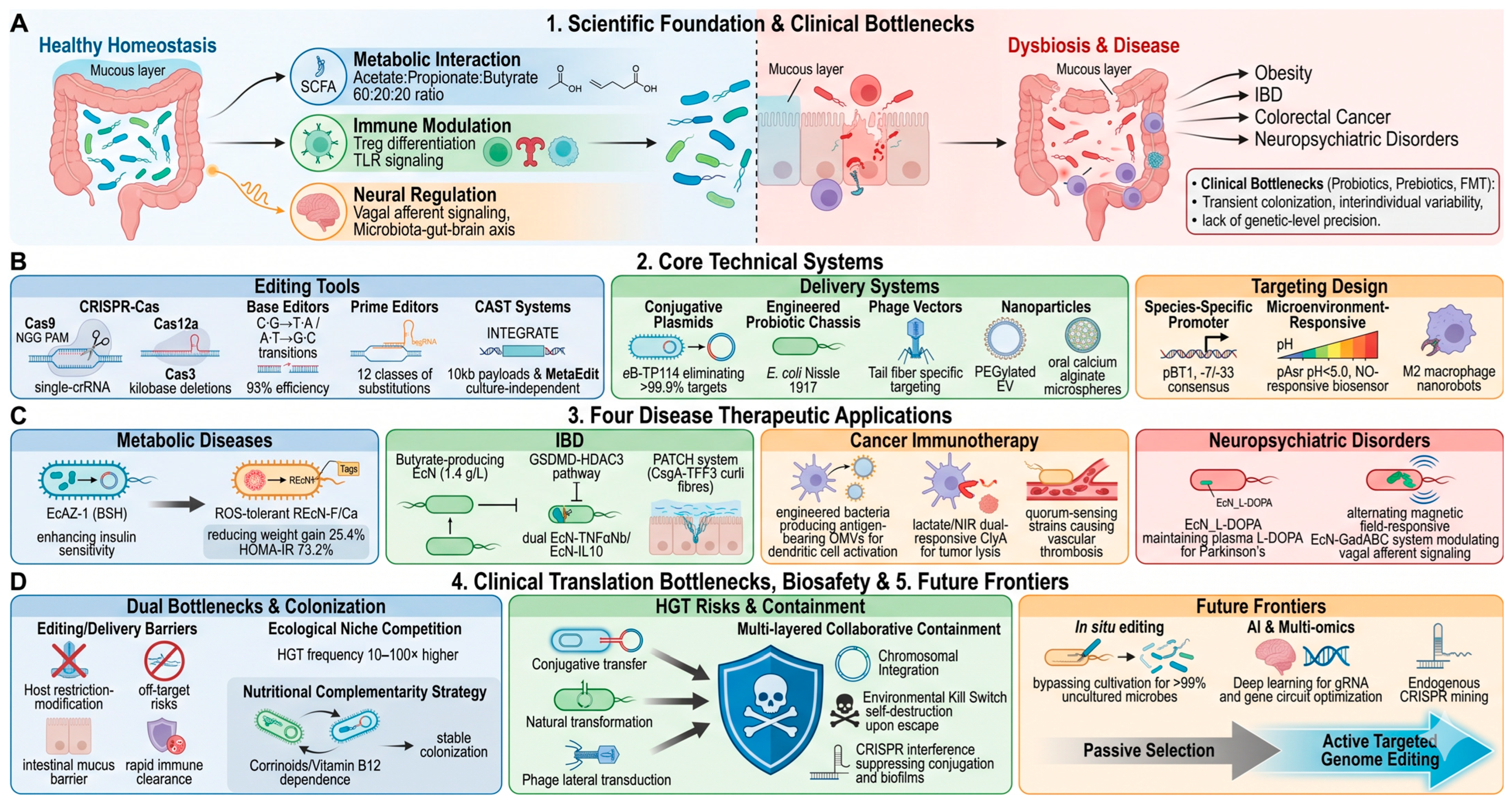
In summary, as illustrated in Figure 1, gut microbiome engineering, through the integration of genome editing technologies with advanced delivery systems, is transitioning from proof-of-concept validation toward clinical translation. This emerging discipline represents a pivotal direction in next-generation precision medicine, with substantial promise for applications in disease therapeutics, diagnostic monitoring, and health management.

2. Core Technologies of Gut Microbiome Genome Editing

The implementation of gut microbiome genome editing relies on the coordinated optimization of editing tools, delivery systems, and targeting design. Among these components, editing tools determine the efficiency and type of genetic modification. Delivery systems ensure the accurate access of these tools to targeted intestinal bacteria. Targeting design further enables the spatial and temporal control of the entire editing process. This section systematically reviews the research advances and application characteristics of these three categories of core technologies.

2.1. Development and Optimization of Editing Tools

Gut microorganisms exhibit substantial heterogeneity in oxygen tolerance, cell wall architecture, and division kinetics, placing stringent demands on the applicability and transformation efficiency of genome editing tools. The current technological repertoire for intestinal microbiota engineering encompasses Clustered Regularly Interspaced Short Palindromic Repeats (CRISPR)–CRISPR-associated (Cas) proteins systems, base and prime editors, and eliminate phage-derived recombination-transposition platforms (Table 1). Among these, CRISPR-Cas systems have emerged as the predominant technical framework by virtue of their programmable nuclease activity.

2.1.1. CRISPR-Cas Systems: Precise Targeted Cleavage and Adaptation to Gut Bacteria

The CRISPR-Cas system serves as an adaptive immune mechanism in prokaryotes, achieving precise cleavage of target DNA through the coordinated action of Cas effector proteins and gRNAs. The structural features of distinct Cas proteins determine their applicability in gut microbial genome editing [70,71]. Cas9 recognizes NGG-type protospacer adjacent motif (PAM) sequences and achieves editing efficiencies up to 100% in facultative anaerobic model organisms such as E. coli [72]. However, its application in obligate anaerobic gut commensals is limited by restriction–modification systems that constrain transformation efficiency [73], low host homologous recombination capacity leading to impaired repair of DNA double-strand breaks [74], and additional challenges, including PAM constraints [70], off-target effects, and toxicity from high-level expression [75], with efficiencies frequently falling below 30% in Bacteroides and Clostridium species [74]. Cas12a recognizes TTTV (V = A, C, or G) PAM sequences and requires only a single CRISPR RNA (crRNA) for target recognition [71,76,77], thereby simplifying multiplex editing design. Catalytically inactive dCpf1 combined with CRISPRi strategies enables efficient transcriptional repression in multiple Clostridium species to regulate short-chain fatty acid and bile acid metabolism [74], though this system similarly faces limitations of temperature sensitivity, PAM restrictions, and efficiency variations among different homologs [70,76]. Cas3 achieves large-scale deletions spanning tens of kilobases through unidirectional processive degradation, with efficiencies reaching 94% to 100% in excising antibiotic resistance gene clusters; yet, it presents risks of undefined deletion boundaries and potential cell lethality when targeting essential genes [78,79]. For editing complex gut microbial communities, the combined application of ET-seq and DART systems enables species- and locus-specific editing independent of culture-based methods, and has been successfully demonstrated in infant gut microbiota [80,81]. Overall, the success of gut microbial editing is constrained by multiple factors, namely restriction–modification systems [74,82], inefficient DNA repair pathways [82], species-specific differences in genetic element compatibility [74,83], and interspecies variability in editing efficiency [84]. Future efforts toward homology-independent CRISPR-RNA-guided integrase systems [71,82], ET-seq-based rapid identification of editable community members [80], machine-learning-driven directed evolution of Cas proteins [82], and the application of engineered functional strains for disease diagnosis and targeted therapeutics [85] represent critical avenues for overcoming existing bottlenecks.

2.1.2. Base Editing and Prime Editing: Precision Modification Without Double-Strand Breaks

Slow-growing nonmodel gut commensals, such as Faecalibacterium prausnitzii and Akkermansia muciniphila, possess weak DNA repair capabilities and often cannot survive double-strand breaks introduced by CRISPR-Cas systems, as these bacteria lack robust nonhomologous end joining repair pathways [86,87]. Base editing and prime editing circumvent this problem by completely avoiding double-strand breaks; they achieve precise modifications through single-strand nicks alone, substantially improving cell survival [86,88]. Base editors fuse catalytically inactive Cas9 nickase with deaminases to accomplish transition point mutations, including cytosine-to-thymine and adenine-to-guanine conversions; this approach offers dual value for knocking out virulence factors and optimizing metabolic functions [86,89]. In Bifidobacterium, the cBEST system achieves editing efficiencies of merely 16.7% to 75% in wild-type strains, with transformants remaining scarce; only after knocking out the host restriction–modification system does efficiency reach 100% [90]. This finding reveals restriction–modification barriers as the critical bottleneck for expanding base editing into nonmodel gut bacteria. Regarding fidelity, Brödel et al. successfully performed adenine base editing in the mouse gut using engineered phage particles; next-generation sequencing showed on-target editing efficiencies of approximately 98%, and no off-target mutations were detected at candidate sites with seven base mismatches [91]. Base editing has attained 93% editing efficiency in the mouse gut with stable maintenance for 42 days [91]. However, its mutational scope remains limited to transitions only.
Prime editing systems fuse Cas9 nickase with reverse transcriptase; through the 3′ extension of prime editing guide RNA, all twelve types of base substitutions and small insertions or deletions become possible. No exogenous DNA donor is required, fundamentally eliminating the risk of horizontal gene transfer (HGT) [87,92]. However, prime editing efficiency remains severely constrained in nonmodel gut bacteria. In wild-type Escherichia coli, initial efficiency falls below 0.1%, far below the 45% to 90% observed in Mycobacterium smegmatis [87]. Genetic screens identified SbcB, XseA, and ExoX as critical host limiting factors; these three DNA exonucleases act in the 3′→5′ direction, and their combined knockout boosts efficiency approximately 100-fold [87]. Whole-genome sequencing further demonstrates that prime editing achieves extremely high fidelity in E. coli chromosomal engineering, with only a single off-target nucleotide substitution detected [92]. Nevertheless, applying prime editing to obligate anaerobic commensals such as Faecalibacterium prausnitzii presents major challenges, as optimizing prime editing guide RNA (pegRNA) design and achieving anaerobic adaptation remain unresolved [87]. Extending prime editing to such organisms requires drawing on the cBEST strategy; host restriction–modification systems must be engineered in parallel with exonuclease repair pathways [87,90]. Overall, base editing achieves efficient in vivo gut editing but remains limited to transition mutations; prime editing offers a broader mutation spectrum and better suits environments sensitive to HGT risks, but it suffers from low efficiency and requires host engineering [87,92]. Future work must integrate both approaches, systematically characterizing and removing species-specific barriers, including restriction–modification systems, exonuclease activity, and anaerobic adaptation, to extend these technologies from model gut bacteria to the entire gut microbiome [87,90].

2.1.3. Phage-Derived Recombination and Transposon Systems: Synergistic Enhancement of Editing Efficiency

For bacteria with limited homologous recombination efficiency (e.g., Pseudomonas spp.), standalone CRISPR-Cas systems often fail to achieve optimal editing outcomes. Synergistic application of phage-derived recombination systems and transposon technologies offers an effective solution to this challenge. The λ-Red recombination system, derived from bacteriophage λ, facilitates homologous recombination repair through the action of recombination proteins. When coupled with CRISPR-Cas9, this system enhances editing efficiency in E. coli to over 95% for gene recoding events [93,94]. Conversely, transposon-encoded CRISPR-Cas systems enable RNA-guided, programmable chromosomal integration of exogenous genes without reliance on specific attachment sites. Combined with CRISPR technology, this approach achieves efficient, site-specific insertions across diverse bacteria [95,96]. For instance, the single-plasmid INTEGRATE system consolidates multiple functional components into a unified vector backbone, enabling marker-free DNA integration with efficiencies approaching 100% and supporting large-fragment insertions up to 10 kb [95]. Through multiplexed CRISPR arrays, this platform facilitates parallel insertions at three genomic loci simultaneously within a single cell. Integration with the Cre-LoxP recombination system further permits precise excision and reversible regulation of inserted sequences. Moreover, INTEGRATE demonstrates broad host applicability, functioning efficiently not only in various Gram-negative bacteria, including E. coli, Klebsiella oxytoca, and Pseudomonas putida, but also enabling species-specific, precision-targeted integration within complex gut microbiota environments [95]. The MetaEdit platform developed by Gelsinger et al. achieves culture-independent, precise, durable, and large-scale in vivo genome editing of specific target strains within complex native gut microbial communities. MetaEdit enables high-efficiency, high-specificity genomic integration in murine and human intestinal strains, successfully inserting metabolic pathways up to 7.5 kb into target sites. By engineering native Bacteroides with exogenous inulin utilization pathways, this platform achieves reversible and dynamic regulation of edited strain abundance through dietary intervention. Furthermore, it accomplished the inaugural genetic modification of segmented filamentous bacteria (SFB), which remain unculturable in isolation, directly within the host [97].
Table 1. Application Comparison of CRISPR-Cas Systems in Intestinal Bacteria.
Tool Name Cas9 (SpCas9) Cas12a (Cpf1) Cas3
(Type I CRISPR)
Base Editing (CBE/ABE) Prime Editing (PE) CAST (CRISPR-Associated Transposase)
PAM Recognition5′-NGG-3′5′-TTTN-3′ (i.e., 5′-TTN-3′): FnCas12a, a naturally relaxed PAMType I-C: 5′-TTC-3′;
Type I-B: 5′-CCW-3′;
Type I-E: 5′-AAG-3′
(PAM sequence varies by Type I subtype; Cascade recognizes PAM via Cas8 subunit)
5′-NGG-3′
nCas9: D10A (CBE)/H840A (ABE)
5′-NGG-3′ (SpCas9 H840A nickase)Type I-F: 5′-CC-3′ (canonical); 5′-CN-3′ (permissive);
Type V-K: 5′-TTN-3′/5′-TTTV-3′ (V = A/C/G) (lower fidelity)
Representative Target Gut StrainsE. coli K-12, EcN; Lactobacillus: L. reuteri, L. casei, L. acidophilus, L. gasseri, L. paracasei; Clostridium: C. tyrobutyricumBacteroidota: B. thetaiotaomicron, B. fragilis, B. ovatus, B. uniformis; P. vulgatus; Firmicutes: C. sporogenesE. coli: K-12, E. coli MG1655,
EcN;
Clostridioides difficile
B. thetaiotaomicron VPI 5482, B. fragilis NCTC 9343, B. longum NCIMB 8809, B. adolescentis DSM 20083, B. pseudolongum; E. coli MG1655, E. coli UTI89EcN; E. coli MG1655E. coli MG1655, K. oxytoca, P. vulgata, B. thetaiotaomicron, B. uniformis, B. fragilis, B. stercoris, SFB (C. savagella)
Core AdvantagesVersatile (knockout, insertion, replacement); Compatible with most common gut bacteria; Near 100% efficiency in optimized HDR in E. coli; Clear PAM requirement, predictable targeting; Mature sgRNA design toolsBroad targeting: 5′-TTTV/TTN-3 PAM covers AT-rich regions; crRNA-only, no tracrRNA required; Autonomous crRNA processing; aTc-inducible markerless editing; High specificity, minimal off-target effectsLarge fragment deletion (7–424 kb) at near-100% efficiency;
Low off-target risk via PAM + crRNA dual recognition;
Lower cytotoxicity than Cas9 (Type I-E in EcN)
No DSB, high cell viability;
No HDR template required;
Enables point mutations/premature stop codons;
Multiplexed editing (≤4 genes) (pnCasBS-CBE);
RM knockout dramatically improves efficiency(cBEST);
Compatible with engineered phages for in vivo delivery(ABE8e/evoAPOBEC1-CBE)
No DSB, no donor template;
12 base conversions, small indels
(≤97 bp del, ≤33 bp ins,
efficiency drops sharply >10 bp);
Low off-target (~15 vs.
iSTOP ~71.5, XSTART ~52.3);
ARG-free platform;
Barcoding enabled
Large fragment insertion (>10 kb), no DSB;
Site-specific integration (~48–50 bp from target); Low toxicity, multiplexed (3 loci), marker-free
- ~100% on-target specificity (Tn-seq verified);
gRNA-programmable across multiple species
Key LimitationsLimited efficiency in obligate anaerobes (RM system barrier, low HDR); complete vector typically >10 kb (promoter, selection marker, gRNA cassette); Cas9 toxicity in some Clostridium species; NGG PAM restricts target sites; ~26% unintended mutation frequency after dsDNA break repair in E. coliLow HR repair efficiency in Gram-positive bacteria; Reduced FnCas12a activity in eukaryotic hosts; DSB-induced lethality in NHEJ-deficient bacteria; CRISPRi-dCpf1 vectors >10 kb reduce conjugation efficiencyStochastic deletion boundaries;
Complex multi-protein deliveryReduced efficiency near essential genes (2–25%);
Insertion/replacement requires PEHR co-delivery
Only specific base conversions (C: G→T: A or A: T→G: C);
Cannot insert large fragments;
Editing window restricted (P4–P8/9);
PAM (NGG) dependency;
BE/sgRNA expression requires strain-specific optimization;
RM barriers limit transformation efficiency
Complex pegRNA
design (PBS +
RT optimization);
Efficiency drops
with larger edits
(>10 bp);
Low in WT
(<0.1–15%)
without exonuc.
Inhibition;
48 h induction
for optimal eff;
Obligate anaerobe
optimization
required
Requires TnsABC + TniQ-Cascade components;
Narrow host range for some variants; Self-targeting inactivation, target immunity;
Fitness cost from constitutive CAST expression;
Type V-K: lower fidelity (~20–40% on-target)
Typical ScenariosRoutine gene insertion/replacement; E. coli high-throughput knockout screening; probiotic metabolic engineering; chassis strain development; edited strain barcode labelingGene deletion and insertion of intestinal Bacteroidota; Multiplex metabolic gene transcriptional suppression of intestinal Clostridia; In vivo regulation of gut microbiota; Manipulation of metabolic gene clustersARG cluster elimination (in vivo 80–97% blocking);
Large virulence island removal (up to 424 kb);
Pathogen-specific killing (C. difficile)
Genome minimization and plasmid curing
Metabolic enzyme optimization;
Virulence gene inactivation;
Antibiotic resistance gene (bla, aph) inactivation;
Probiotic metabolic engineering;
Gut in situ precision editing;
Microbiome-targeted therapeutics
Point mutations;
Small indels;
Codon modificaTion; Engineered
Probiotics;
Strain barcoding;
ARG-free safe engineering
In situ gene therapy (intestinal microbiome);
Stable implantation of large functional genes;
Elimination of antibiotic resistance gene clusters;
Metabolic pathway engineering (e.g., 7.5-kb PUL);
Unculturable microbe editing (e.g., SFB);
Diet-responsive strain control (e.g., inulin-Bt)
Editing Efficiency Range (%), [Representative Strain, Condition]In vitro
E. coli K-12/MG1655: ~65–82%;
Lactobacillus reuteri: 90–100% via ssDNA recombineering;
Lactobacillus casei: 25–62% using nCas9 nickase;
Lactobacillus acidophilus, L. gasseri, L. paracasei: 35–100% via nCas9;
Clostridium tyrobutyricum: 25% after removing RM system and endogenous plasmids;
Bacteroides thetaiotaomicron: ~40%, lower than FnCas12a
In vitro
Bacteroides thetaiotaomicron: gene deletion ~60%, GFP insertion >80%, 48 kb deletion ~6%, 5/10 kb deletion ~100%; Prevotella vulgatus: editing efficiency up to 100%; Bacteroides fragilis, B. ovatus, B. uniformis: editing efficiency >60%
In vitro
E. coli MG1655:
Editing efficiency for lacZ: 51–90%; for near-essential gene hemB: 2–25%;
In vivo
EcN (zebrafish gut):
CRISPR-Cas3 inhibited the transfer of three antibiotic resistance genes (mcr-1, NDM-1, tet(X)) with efficiencies ranging from 80% to 97%.
In vitro
pnCasBS-CBE: Bacteroides thetaiotaomicron: BT0793 100%, BT2804 80%, BT3788-1 70%, BT2158-1 80%, BT2802 66.7%, Non-model gut species: 66–100%;
cBEST: Wild-type Bifidobacterium longum: cBEST2 ~27%, cBEST3 ~17%, cBEST4 ~75%; RM triple mutant: SpeE 100%, MetK 100%, bsh 100%; Bifidobacterium adolescentis: (Sau3AI) 100%;
ABE8e: E. coli MG1655 > 99%;
evoAPOBEC1-CBE: E. coli MG1655 > 99%, E. coli UTI89: clbH ~92%, clbJ ~83%, cnf1 ~53%
In vitro
EcN (48 h):
substitution: 66.7%, insertion: 52.0%, deletion: 25.0%;
Wild-type E. coli MG1655: deletion: ~26%, insertion: ~12.2%, substitution: ~6.8%;
E. coli MG1655 ΔsbcBΔxseAΔexoX:
up to ~40% editing efficiency at xylB locus
In vitro
E. coli MG1655, BW25113: ~100%;
P. vulgatus: >50–70% (pME0008);
Multiplex: 3 loci simultaneously
In vivo
(murine gut):
P. vulgatus: ~5% (day 3);
B. uniformis: ~2%; B. thetaiotaomicron: ~1.5% peak, 99.8% specificity;
SFB (C. savagella): ~2.3% (day 28), 100% specificity
References[70,71,72,73,74,75,98,99,100,101,102][70,71,74,76,102,103,104,105][78,79,106,107,108][86,88,89,90,91][87,92,109][93,95,96,97]

2.2. Innovation of Delivery Systems

The extreme physicochemical conditions of the intestinal tract, encompassing gastric acid (pH 1.5–3.5), the detergent activity of bile acids, and continuous peristaltic motility, coupled with biofilm barriers and deep mucosal colonization characteristics of target organisms, collectively constitute formidable delivery barriers that compromise the structural integrity and functional activity of gene editing tools prior to reaching their intended targets. Contemporary strategies address these challenges through three primary vector systems (Table 2): engineered bacterial chassis that leverage the intrinsic colonization capacity of commensal organisms to achieve sustained in situ release; bacteriophage-based vectors that exploit strain-specific recognition mechanisms for precise targeting; and nanoparticle platforms that utilize advanced materials engineering to enhance environmental resilience and mucosal penetration capabilities.

2.2.1. Phage Vectors: Precise Delivery with Host Specificity

As obligate bacterial viruses, bacteriophages represent ideal targeting delivery vehicles by virtue of their stringent host specificity; however, their application has been historically hampered by narrow host ranges and constrained genomic cargo capacities. The host range of a phage is primarily determined by tail fiber proteins located at the distal end of the phage tail, which mediate the initial reversible and specific recognition of susceptible bacteria through interaction with surface receptors such as lipopolysaccharide (LPS), outer membrane porins (e.g., OmpC), teichoic acids, and flagella [110]. At the molecular level, the receptor-binding domain at the tip of tail fibers engages in weak, dynamic interactions with bacterial surface receptors, enabling a “touch and search” mechanism that allows phages to randomly walk across the bacterial surface to locate optimal infection sites prior to irreversible attachment and genome injection [110]. Diversity-generating retroelements (DGRs) can further modify tail fiber proteins to alter host tropism, enabling individual temperate phages to infect a broad range of bacterial species, thus enhancing their adaptability within complex microbial communities [111]. To circumvent these limitations, researchers have employed directed evolution strategies involving mutational screening of tail fiber tip domains to successfully expand receptor recognition spectra [112]. Regarding cargo optimization, vectors constructed by inserting CRISPR-based systems into non-essential regions of λ phage have demonstrated versatile editing capabilities: dCas9-mediated transcriptional repression systems enable targeted gene silencing in E. coli while preserving cellular viability [102,113], whereas CRISPR-Cas3-equipped λ phage achieves sequence-specific antibacterial killing of pathogenic E. coli strains with enhanced efficiency [107]. More recently, engineered λ phage delivering DART (CRISPR-associated transposase) systems have accomplished kilobase-scale gene insertions and targeted disruptions with editing efficiencies exceeding 50% in monocultures and mixed bacterial communities [114], and phage-delivered cytosine base editors (CBEs) have enabled precise in situ point mutations within synthetic microbial communities [115]. The cardinal advantages of such platforms lie in their high targeting specificity and minimal off-target risk, with therapeutic potential having been extensively validated in animal models, exemplified by the engineered phage cocktail SNIPR001 featuring tail fiber engineering and a CRISPR-Cas payload, which has demonstrated efficient reduction of E. coli burden in mice [116]. Notably, studies utilizing M13 filamentous phage to deliver CRISPR-Cas9 systems have achieved strain-specific clearance and efficient genomic deletion of target E. coli strains within the murine gut, substantiating the efficacy of this strategy for precision editing of the intestinal microbiota in vivo [117]. It must be emphasized, however, that all successful in vivo demonstrations of phage-mediated CRISPR delivery reported to date have been limited to E. coli, a facultative anaerobe that serves as a convenient model organism. The extension of these strategies to obligate anaerobes, which comprise the majority of the human intestinal microbiota, remains a critical unmet challenge, as the technical barriers associated with phage propagation, infection dynamics, and CRISPR expression under strict anaerobic conditions have yet to be systematically overcome.

2.2.2. Nanoparticle Vectors: Efficient Delivery Across the Mucus Layer

The intestinal mucus layer ranges from tens to hundreds of micrometers in thickness and consists of a dense mucin network, which effectively prevents exogenous gene editing cargoes from reaching targets within the intestinal lumen. Nanoparticles with diameters of 100 to 200 nm, by virtue of their appropriate size and surface modifiability, are regarded as ideal carriers for mucus penetration and subsequent microbiome editing. Current design strategies must address three hierarchical challenges: mucus penetration, targeted recognition, and intracellular delivery of CRISPR-Cas into target cells. Surface polyethylene glycol (PEG)ylation can minimize nonspecific mucin adsorption and enhance mucosal penetrability, as demonstrated by Lei et al., who showed that precisely PEG1500 shielded selenium nanovaccines not only achieve excellent mucus penetration but also efficiently activate mucosa-associated lymphoid tissues to induce local immune responses [118]. Peptide functionalized delivery systems can protect macromolecular cargoes from destruction under harsh intestinal conditions. Jing et al. employed HIV-derived TAT-cell-penetrating peptides to functionalize milk-derived extracellular vesicles, achieving 95.7% efficient GFP loading with 73.7% remaining intact after simulated gastrointestinal fluid treatment [119]. In the context of CRISPR-Cas nanoparticle delivery, Lin et al. constructed an oral micro- to nanogenome editing system (RNP NCs@dOMV@CAM) that employs calcium alginate microspheres as a gastric protective layer and utilizes probiotic-derived, lipopolysaccharide-depleted outer membrane vesicles (dOMVs) to traverse the colonic mucosal barrier and target inflammatory macrophages, achieving conditional Cas9 activation and TNF-α gene editing under inflammatory conditions and demonstrating the feasibility of oral nanoparticle delivery across intestinal barriers [120]. Kim et al. identified two candidate carriers, LNP 496 and LNP 470, containing the cationic lipid DOTAP through high-throughput screening of 511 lipid nanoparticle (LNP) formulations, which could deliver CRISPR-Cas13a plasmids into Escherichia coli with the assistance of polymyxin B. In a bacterial infection model, the LNP-Cas13a-gRNA treatment group improved mouse survival from 10% to 70% [121]. However, the study by Kim et al. employed intraperitoneal injection and has not yet achieved oral intestinal targeted delivery, relying on membrane disruptors to overcome the outer membrane barrier of Gram-negative bacteria. Meanwhile, although the system by Lin et al. successfully achieved oral intestinal delivery and host targeted gene editing, its target was inflammatory macrophages rather than intestinal commensal bacteria themselves. To date, no nanoparticle-mediated CRISPR-Cas system has successfully achieved targeted in vivo gene editing of intestinal commensal bacteria, particularly the dominant functionally core obligate anaerobes. Future research must urgently develop synthetic nanocarriers that simultaneously possess mucus penetration, obligate anaerobe targeting recognition, and autonomous bacterial cell wall crossing capabilities in order to bridge the gap from proof-of-concept to precise in vivo application of nanoparticle-mediated intestinal microbiome editing.

2.2.3. Engineered Bacterial Vectors: Sustained Delivery via Live Colonization

Engineered bacterial chassis employ probiotics as programmable delivery platforms, harboring gene editing tools that enable sustained expression and in situ delivery following intestinal colonization. The core advantage of this strategy resides in the dual characteristics of intrinsic probiotic colonization capacity and synthetic biology regulation. Probiotic chassis inherently possess gastrointestinal colonization capabilities and establish immune tolerance with the host, while synthetic gene circuits can be designed to respond to specific intestinal microenvironmental cues such as localized hypoxia or particular metabolites, thereby achieving spatiotemporal control over editing tool expression and delivery [122,123]. Selection of an appropriate chassis requires careful consideration of colonization properties and genetic tractability. The non-pathogenic intestinal commensal Escherichia coli Nissle 1917 (EcN) achieves robust colonization in the murine gut [122,123], and engineered derivatives cured of cryptic plasmids via CRISPR-Cas9 editing exhibit plasmid stability over approximately 90 generations of in vitro cultivation [122]. Coupled with its well-established genetic toolbox, these attributes have established EcN as one of the most widely employed chassis organisms to date [123]. In contrast, Bifidobacterium animalis subsp. lactis BB-12 exhibits transient colonization in humans [124], yet displays potent metabolic activity during its intestinal residence. Through production of acetate and lactate, it facilitates cross-feeding interactions that enhance short-chain fatty acid synthesis by other community members, thereby significantly modulating microbiota architecture [124]. Regarding delivery strategies, engineered live bacteria has demonstrated substantial in vivo application potential. Although engineered EcN carrying a laboratory-evolved conjugative plasmid eB-TP114 achieved >99.9% elimination of targeted antibiotic-resistant E. coli in the mouse gut, this study relied on streptomycin pretreatment, used artificially introduced strains as targets, and the CRISPR-Cas9 system functioned as bacterial killing rather than precise genome editing [125]; by contrast, conjugative delivery of CRISPR-associated transposase systems via an engineered E. coli donor has achieved precise editing of Bacteroides thetaiotaomicron within the endogenous gut microbiota with >99% targeting specificity [97]. Furthermore, EcN engineered with a type I-E CRISPR-Cas3 system successfully blocked the horizontal transfer of multiple antibiotic resistance genes in the zebrafish gut [108]. Circuit design constitutes the cornerstone of precision control. The strictly anaerobic colonic environment activates anaerobic promoters such as nirB and fdhF to drive heterologous protein expression, whereas their activity is markedly reduced in the moderately hypoxic small intestinal milieu, potentially enabling spatial restriction of transgene expression to the colon [126,127]. Engineered FNR-dependent promoters have achieved more refined regulation, with anaerobic-activated variants demonstrating 24- to 138-fold enhanced activity under anoxic versus aerobic conditions and anaerobic-repressed variants dropping to 8–17% of baseline activity. These systems respond rapidly to oxygen depletion (within 30 min) and have been successfully implemented in proof-of-concept metabolic engineering applications, achieving pyruvate titers of 5.76 g/L at 55.7% yield [128], providing a theoretical foundation for precision intestinal therapeutics based on high-dynamic-range responses. Notably, oxygen sensing alone cannot fully discriminate between anatomically adjacent intestinal regions. Future iterations may incorporate multi-input logic gates combining bile-acid-responsive or pH-sensitive elements to further enhance targeting fidelity [126]. A critical consideration for clinical translation remains the immunogenicity of exogenous editing proteins such as Cas nucleases. This challenge can be mitigated through DNA-free editing systems involving ribonucleoprotein (RNP) delivery or inducible bacterial lysis strategies that minimize prolonged exposure to foreign antigens [126]. Careful chassis selection combined with circuit optimization will be essential for advancing engineered bacterial vectors from preclinical proof-of-concept to therapeutic reality.
Table 2. Comparison of Three Classes of Delivery Systems.
Delivery System Type Bacteriophage Vectors Nanoparticle Vectors (e.g., LNP, PBAE) Engineered Bacterial Vectors (e.g., EcN)
Host SpecificityHigh (vast majority infect specific bacterial species/strains; some engineered phages exhibit broad host range)No intrinsic host specificity; cell-type-dependent delivery efficiency;
targeted delivery achievable through surface modification
(targeting specific cells/tissues)
Variable (no strict host species limitations; colonization efficiency is microbiome-dependent)
Genetic Loading CapacityHighly variable by type: T4 natural genome ~171 kb; T7 packaging limit <2.2 kb; λ genome ~48.5 kb, max effective payload insertion ~5 kb (without replacement), up to ~10.8 kb with replacement of non-essential regions (1–8.5 kb); M13 genome ~6.4 kb with expandable capsidRelatively high (>95% encapsulation); challenging to achieve efficient
encapsulation of large-size plasmids
Relatively high (plasmid or chromosomal integration; large DNA payload capacity)
BiocompatibilityHigh safety (well tolerated via oral delivery; no systemic exposure; does not disturb gut microbiota; validated in murine and minipig models)Relatively high (toxicity can be reduced through material modification)Extremely high (probiotic chassis; capable of intestinal colonization)
Core AdvantagesPrecise bacterial targeting with minimal off-target effects; host specificity can be tuned via tail fiber engineeringPenetrate mucus layers after surface modification; broad applicability
(non-viral, no host range limitation)
Capable of in vivo colonization for sustained delivery; can be designed with environmentally responsive genetic circuits
Primary ChallengesNarrow host range (majority are narrow-host); prone to developing phage resistance; requires coating for oral deliveryUnmodified LNPs easily adsorbed and trapped by mucus; extremely low intestinal
absorption efficiency (unmodified <5%; with bile acid modification: up to ~47%);
reduced efficiency for large-size plasmids (9–19 kb); insufficient batch-to-batch
consistency in manufacturing; uncontrollable size, surface charge and pDNA loading
Chassis colonization capacity affected by intestinal microbial competition; genetic stability requires optimization
References[107,110,112,113,114,115,116][129,130,131,132,133][108,122,123,125,126,127]

2.3. Targeting and Controllability Design

To achieve precision spatiotemporal control over gut microbiome editing, current strategies employ a multi-layered safeguard system built upon two synergistic dimensions: species-specific recognition and environmentally responsive triggering [134]. At the species-specific level, researchers exploit the molecular incompatibility of target-exclusive promoters to strictly confine editing machinery activation within the intended taxon. For instance, the 16S rRNA promoter pBT1 derived from Bacteroides thetaiotaomicron leverages its characteristic −7 and −33 consensus sequences to exclude heterologous transcriptional machinery from E. coli and other non-target species, thereby restricting editing activity exclusively to the desired lineage [135,136]. Building on this principle, Gelsinger et al. employed the P1 promoter to drive guide RNA expression and, via a conjugative CAST delivery system, achieved efficient metagenome editing of B. thetaiotaomicron and related Bacteroidaceae commensals in the murine gut. This system not only demonstrated greater than 99% specificity in targeted tagging but also enabled integration of metabolic pathway payloads up to 7.5 kb in length. Through inulin utilization capability at the PUL locus, edited strains gained a selective fitness advantage upon dietary inulin supplementation, achieving 30- to 40-fold population enrichment [97]. In parallel, Huang et al. coupled the P1 promoter with CRISPRi technology, utilizing an inducible P1 promoter to regulate single guide RNA (sgRNA) expression and thereby realizing species-specific asymmetric adaptive regulation in a dual-species consortium [137]. To further mitigate off-target risks computationally, tools including CRISPRdirect, Cas-OFFinder, and CRISOT enable whole-genome prediction of potential off-target sites during the guide RNA design phase [138,139,140]; among these, CRISOT performs whole-genome off-target prediction based on molecular dynamics simulations of RNA–DNA interaction fingerprints. More recently, CCLMoff, an RNA language model-based approach integrating training data from thirteen off-target detection technologies, has demonstrated AUROC values of 0.985 in cross-dataset validation, exhibiting exceptional generalization capacity and predictive accuracy while effectively overcoming the limitations of earlier tools in predicting unseen sequences [141]. Complementing these in silico approaches, Kulcsar et al. established Cas9 fidelity cleavage rules and, based on these, constructed the CRISPRecise toolkit encompassing 19 high-fidelity SpCas9 variants. When paired with GUIDE-seq genome-wide off-target detection for target-matching screening, this platform drives off-target editing to undetectable levels, effectively resolving the longstanding trade-off between efficiency and specificity [142].
On the environmental response front, site-specific strategies harness the intestinal pH gradient to achieve segment-specific regulation. The acid-responsive promoter pAsr, for example, is strongly induced in E. coli under external acidic conditions (pH < 5.0), with peak activity at approximately pH 4.0 to 4.5, providing a theoretical molecular basis for segment-specific transcriptional control in the gut, though direct translation to in vivo intestinal editing remains experimentally unvalidated [143,144]. A more mature approach employs pH-sensitive materials and chemical engineering to co-encapsulate engineered bacteria; Jiang and colleagues used polyelectrolyte complex coatings to modify EcN, enhancing engineered bacterial survival in the gastrointestinal tract by 40- to 74-fold, while simultaneously leveraging genetic editing to overexpress the siderophore microcin, which targets and eliminates pathogenic bacteria through a “Trojan horse” mechanism; this synergistic strategy achieved both anti-inflammatory efficacy and microbiome modulation in a model of UC, offering a new paradigm for stable delivery and precise release of live bacterial editing tools [145]. Pathology-responsive strategies further endow editing systems with intelligent intervention capability, enabling them to sense disease-specific signals and generate therapeutic outputs at lesion sites. Engineered EcN carrying a nitric-oxide-responsive genetic circuit, for instance, can detect elevated NO concentrations at colonic inflammatory sites and trigger local expression of anti-TNF-alpha nanobodies to neutralize pro-inflammatory cytokines [146]. Complementing this strategy, Luo et al. developed M2 macrophage membrane-coated nanorobots encapsulated in alginate microspheres that, leveraging membrane surface receptors for specific recognition of inflammatory macrophages and intestinal epithelial cells, accumulate in inflamed tissue and neutralize inflammatory factors through receptor–ligand interactions while simultaneously driving M1-to-M2 macrophage repolarization, thereby exerting potent anti-colitis therapeutic effects [147]. Collectively, these hierarchical designs spanning molecular recognition, material responsiveness, and cell-mimetic delivery constitute a paradigm shift from passive intervention to active adaptation in precision microbiome editing.

3. Therapeutic Applications and Case Studies

Gut microbiome engineering offers a novel therapeutic paradigm for intractable conditions, including metabolic disorders, IBD, cancer immunotherapy, and neuropsychiatric diseases, through precision manipulation of microbial genetic circuits or metabolic networks. While current investigations remain predominantly at the stage of ex vivo proof-of-concept validation and preclinical animal modeling, with limited accumulation of systematic clinical efficacy data, synthetic biology design strategies, encompassing the construction of environmentally responsive genetic circuits, targeted biosynthesis of therapeutic molecules, and modulation of microbe–host interactions, have demonstrated theoretical potential to remodel gut microecological architecture and achieve durable precision interventions. The following sections delineate disease-specific microbiome editing strategies and their applications.

3.1. Metabolic Diseases: Modulation of Energy Homeostasis and Glycolipid Metabolism

One of the core pathological mechanisms underlying metabolic disorders, including obesity, type 2 diabetes (T2D), and non-alcoholic fatty liver disease (NAFLD), involves the structural imbalance of gut microbiota metabolic function. This dysregulation is characterized by decreased abundance of SCFA-producing taxa and increased lipopolysaccharide (LPS) translocation. Targeting these pathological foundations, microbiome engineering is opening new avenues for the treatment of metabolic diseases through the directed modification of strain metabolic capabilities or stress resistance.
In the field of microbiome engineering, proof-of-concept studies that target metabolic diseases through directed modification of bacterial metabolism or stress resistance have achieved major breakthroughs. These investigations span not only obesity and T2D but also complex multi-organ metabolic disorders of the gut–liver–brain axis. Russell et al. isolated a native Escherichia coli strain, EcAZ-1, from feces of conventionally raised mice and engineered it to express functional genes including bile salt hydrolase (BSH), creating an optimized chassis for live bacterial therapeutics. This engineered strain achieved permanent engraftment in the host gut without antibiotic pretreatment, with persistence reaching statistical significance at p < 0.0001. In the Ob/Ob genetic model of diabetes, a single administration enhanced host insulin sensitivity at p < 0.05 and lowered postprandial blood glucose over 15 weeks, thereby reversing diabetic pathophysiology in a sustained manner [148]. In parallel, Mao et al. subjected probiotic E. coli Nissle 1917 to directed evolution under iterative hydrogen peroxide stress and isolated the engineered strain REcN with enhanced tolerance to reactive oxygen species. Surface functionalization with fructooligosaccharide–calcium carbonate composites yielded REcN-F/Ca. In high-fat-diet-induced obese male C57BL/6J mice, this engineered probiotic activated PPAR signaling pathways, enriched butyrate-producing taxa such as Lachnospiraceae and Blautia, and restored intestinal short-chain fatty acid levels at p < 0.0001. These effects translated into a 25.4% reduction in weight gain, markedly improved glucose homeostasis, and a 73.2% decrease in the homeostatic model assessment for insulin resistance (HOMA-IR), all at p < 0.0001 [149]. Furthermore, Oh et al. constructed an engineered Lactobacillus reuteri strain that stably secretes recombinant interleukin-22. In a diet-induced obesity mouse model, eight weeks of treatment significantly reduced both liver weight and hepatic triglyceride levels at p < 0.001, exceeding the baseline improvements seen with wild-type probiotic therapy and providing proof-of-concept for live bacterial therapeutics in non-alcoholic fatty liver disease [150]. Extending this paradigm to multi-metabolite disorders of the gut–liver–brain axis, Aggarwal et al. engineered two Lactobacillus plantarum strains, Lp-NH3 and Lp-Q, to simultaneously achieve ammonia consumption, branched-chain amino acid (BCAA) synthesis, and L-glutamine metabolism. Lp-NH3 couples ammonia assimilation with BCAA biosynthesis, while Lp-Q enhances glutamine utilization to block ammonia regeneration. In two preclinical hepatic encephalopathy models (hyperammonemia and bile duct ligation models), the Lp-NH3+Q cocktail reduced serum and brain ammonia concentrations by up to 10-fold, restored BCAA and glutamine balance, and significantly improved anxiety-like behavior and cognitive function. Brain transcriptomic analysis further revealed restoration of neuronal signaling and attenuation of neuroinflammation. This engineered cocktail outperformed rifaximin in therapeutic efficacy while preserving gut microbiota diversity; the strains were completely cleared within 72 h after dosing cessation with a favorable safety profile [151]. Collectively, these studies demonstrate that microbiome engineering offers broad therapeutic potential against metabolic diseases through precision functional modification of bacterial strains. The approaches span directed insertion of functional genes into native chassis strains, adaptive evolution coupled with surface engineering to enhance stress resistance, engineered secretion and delivery of therapeutic proteins, and multi-metabolite pathway rewiring for simultaneous modulation of interconnected metabolites across the gut–liver–brain axis.

3.2. IBD: Mucosal Barrier Restoration and Inflammation Attenuation

The core pathological mechanisms of IBD involve disruption of the intestinal mucosal barrier and immune-mediated chronic inflammation, with gut microbiota dysbiosis serving as a critical driver characterized by the overexpansion of adherent-invasive E. coli (AIEC) and depletion of butyrate-producing commensals. Microbiome engineering achieves precise intervention through a dual strategy encompassing pathogen virulence inactivation and enhancement of local anti-inflammatory capacity [152]. The former employs CRISPR-Cas systems to neutralize AIEC virulence genes in situ or inhibit their colonization, while the latter utilizes engineered bacteria to locally secrete anti-inflammatory molecules such as IL-10 and anti-TNF-α nanobodies to remodel the immune microenvironment [152,153]. Engineered bacteria-mediated local delivery of anti-inflammatory metabolites provides sustained support for mucosal repair. Gong et al. introduced a heterologous butyrate biosynthesis pathway into EcN, achieving in situ butyrate delivery at 1.4 g/L in artificial intestinal environments; this strain significantly ameliorated dextran sulfate sodium (DSS)-induced chronic colitis, restored mucosal barrier integrity, and predominantly depended on GSDMD–HDAC3 pathway inhibition, wherein butyrate functions as an HDAC3 inhibitor to suppress gasdermin D-mediated pyroptosis [154]. Additionally, EcN has been engineered to synthesize the ketone body (R)-3-hydroxybutyrate (3HB), enabling sustainable production within the intestinal lumen and alleviating DSS-induced colitis [155].
Significant progress has been achieved in engineering probiotic bacteria for targeted anti-inflammatory therapy. A dual-engineered EcN system comprising EcN-TNFαNb and EcN-IL10, respectively expressing anti-TNF-α nanobodies and IL-10, significantly ameliorated DSS-induced colitis in mice while reducing inflammatory cell infiltration and pro-inflammatory cytokine levels [153]. Hua et al. constructed EcNΔlpp::A5-aTN displaying ANXA5 to target inflammatory sites and secrete anti-TNF-α nanobodies, outperforming wild-type EcN and infliximab in DSS-induced UC by suppressing reactive oxygen species (ROS)/JNK/p38/Caspase-mediated intestinal epithelial cell apoptosis [141]. The PATCH system employed engineered EcN to secrete curli fibers displaying trefoil factors (TFFs), where CsgA-TFF3 fusion proteins demonstrated protective effects by lowering IL-6, IL-17A, and TNF-α levels, enhancing mucosal barrier function, and reducing Th17 responses [156]. Furthermore, smart responsive engineered probiotics represent a new frontier, as Weibel et al. engineered EcN with an NO biosensor (NorR-pNorVβ) that senses the inflammatory biomarker NO and utilizes the type I hemolysin A secretion system to secrete anti-TNFα nanobodies with binding affinity comparable to adalimumab [146].
Beyond anti-inflammatory molecule delivery, advanced engineering strategies have explored antioxidant protection and targeted pathogen elimination. Cao et al. developed artificial enzyme-armed Bifidobacterium longum (BL@B-SA50) using single-atom catalyst Fe SA to mimic SOD and CAT antioxidant enzymes, effectively scavenging ROS and demonstrating therapeutic potential in mouse UC and CD models as well as beagle dog colitis [157]. Guo et al. engineered Lactobacillus casei with embedded selenium nanodots (Se-fLac), where the periplasmic film enhanced gastric acid resistance and mucosal adhesion while selenium nanodots cleared ROS, alleviating inflammation in multiple mouse models and non-human primates [158]. Jiang et al. applied a polyelectrolyte complex coating to EcN that improved gastric survival by 40-fold and small intestine survival by 74-fold, while the EcN::mcmA strain overexpressed the siderophore microcin MccM, exploiting a “Trojan horse” mechanism to target and destroy pathogenic bacteria in DSS-induced UC models [145]. Collectively, these CRISPR-Cas9-mediated gene editing advances, encompassing targeted anti-inflammatory delivery, ROS clearance, barrier restoration, and microbiota homeostasis, herald a new era of engineered probiotics for IBD therapy, with future intelligent systems capable of real-time inflammation monitoring and dynamic therapeutic release promising to further enhance clinical precision.

3.3. Cancer Immunotherapy: Remodeling the Tumor Microenvironment and Potentiating Anti-Tumor Immunity

The gut microbiota profoundly modulates tumor immunotherapy responses through the microbiota–metabolite–immune axis; its metabolites not only regulate dendritic cell cross-presentation and subsequent CD8+ T-cell-mediated anti-tumor effector functions but also exert direct anti-tumor effects. For instance, Yue et al. engineered Escherichia coli to produce antigen-bearing OMVs in situ within the intestine; these vesicles traverse the intestinal epithelial barrier and are taken up by lamina propria dendritic cells to mediate antigen presentation, eliciting robust anti-tumor immunity against melanoma and colorectal cancer [159]. Building upon this, they further developed an oral probiotic vaccine platform integrating dual-antigen ferritin nanoparticle scaffolds with a controllable bacterial lysis system; via RGD peptide-mediated M-cell-targeted delivery, this platform promotes mucosal dendritic cell antigen uptake and presentation, achieving durable protection in melanoma subcutaneous tumor and lung metastasis models [160].
At the spatiotemporal precision level, Chen et al. developed a lactate/NIR-responsive system that enables engineered bacteria to express and accumulate cytolysin A (ClyA) cytolysin within tumors and achieve precision lysis via near-infrared-triggered temperature-controlled circuits [161]. Zou et al. employed a lactate-sensing circuit coupled with the essential gene asd to confine EcN to tumor sites; quorum sensing systems then drive on-demand alpha-hemolysin release to kill tumor cells, while coagulase-mediated vascular thrombosis cuts off tumor blood supply and establishes a physical barrier to contain bacterial spread [162]. Complementarily, Xu et al. reconstructed the arginine–nitric oxide (NO) metabolic circuit to sustain NO synthesis, normalizing tumor vasculature, alleviating hypoxia, enhancing immune cell infiltration, and reversing T cell exhaustion [163]. Collectively, these strategies achieve spatially selective activation by exploiting tumor microenvironmental hallmarks, circumventing the off-target toxicities associated with systemic drug administration, and providing safe and feasible new paradigms for engineered bacteria-mediated precision tumor therapy.

3.4. Neuropsychiatric Disorders: Modulation of the MGB Axis

The pathogenesis and progression of neuropsychiatric disorders are intimately associated with dysfunction of the MGB axis. Lee et al. systematically elucidated the mechanisms by which gut microbiota modulate central nervous system function through multiple pathways encompassing neurotransmitter precursor metabolism, vagal afferent signaling, and microbial metabolite-mediated immune modulation, thereby establishing a theoretical foundation for microbiome engineering in neuropsychiatric therapeutics [164]. In the dopaminergic domain, Padhi et al. constructed a genetically engineered EcN system (EcN_L-DOPA) wherein the hpaBC operon was chromosomally integrated under the control of a programmable L-rhamnose-inducible promoter, enabling continuous, titratable biosynthesis of L-DOPA from dietary L-tyrosine; oral administration of EcN_L-DOPA combined with a peripheral decarboxylase inhibitor maintained stable therapeutic plasma L-DOPA concentrations, increased striatal dopamine levels, and notably alleviated depressive-like behaviors in murine and canine models without the pulsatile fluctuations associated with conventional oral L-DOPA therapy [165]. In the GABAergic domain, Wang et al. developed an alternating magnetic field (AMF)-responsive engineered probiotic system (EcN-GadABC@Fe3O4-NE) by transforming EcN with a temperature-sensitive plasmid expressing gadA, gadB, and gadC under the control of a thermal-responsive promoter; Fe3O4 nanoparticles coated on the bacterial membrane converted AMF stimuli into thermal signals, achieving non-invasive, spatiotemporally precise control of GABA release in the gastrointestinal tract, and oral administration of this system significantly attenuated anxiety-like behaviors in restraint-stressed mice through synergistic inactivation of the nucleus of the solitary tract and locus coeruleus via gastrointestinal vagal afferent signaling while concurrently restoring gut microbiota homeostasis [166]. Lebovich et al. further leveraged synthetic biology tools to implement hierarchical negative feedback gene circuits for precise dynamic regulation of GABA biosynthesis, constructing intelligent engineered probiotics capable of autonomously sensing GABA concentrations and regulating glutamate decarboxylase expression, thereby providing a programmable solution for maintaining gut–central GABA signaling homeostasis [167]. Needham et al. revealed from a microbial metabolite perspective that 4-ethylphenol (4EP) produced by bioengineered gut bacteria is sulfated by the host into 4-ethylphenyl sulfate (4EPS). This metabolite enters the brain and alters region-specific activity and functional connectivity across limbic circuits by impairing oligodendrocyte maturation and reducing axonal myelination, which induces anxiety-like behaviors in mice. Pharmacological promotion of oligodendrocyte differentiation reversed these myelination defects and prevented the behavioral abnormalities, providing causal mechanistic evidence for intervening in neuropsychiatric disorders through targeted modulation of specific microbial metabolites [168]. Collectively, these strategies spanning continuous dopamine precursor delivery, remotely controlled GABA release, autonomous feedback-regulated neurotransmitter synthesis, microbial metabolite-directed brain function modulation, and precision treatment systems ranging from microbiota transplantation to engineered commensal microorganisms demonstrate substantial potential for intervening in neuropsychiatric disorders through precise, multimodal manipulation of gut–brain axis metabolic signals by engineered probiotics, while underscoring the need for further translational studies to validate their clinical efficacy and long-term safety.

4. Challenges and Solutions

The clinical translation of gut microbiome engineering faces dual bottlenecks in editing efficiency and delivery precision. At the gene editing tool level, CRISPR-Cas systems are constrained by the restriction–modification systems of bacterial hosts [90]; the risk of HGT [169] and off-target effects [170] further limit their application in live bacteria editing. At the delivery system level, the dense mucin network of the intestinal mucus layer can trap phage vectors and other carriers, obstructing effective contact with target bacteria. Immunoglobulin-like domains on the phage capsid surface bind to mucin glycans, enriching phages in mucus by 4.4-fold while reducing their diffusion rate by 8-fold [171]. More critically, immune clearance constitutes the core obstacle to delivery: systemically administered phages face complement activation, opsonization, and reticuloendothelial system phagocytosis and can be cleared within minutes. PEGylation shields capsid antigenic epitopes through a hydrophilic steric barrier, significantly reducing anti-phage IgG and increasing the area under the curve by up to 54-fold [172].
Beyond delivery efficiency, the colonization stability of engineered bacteria following genetic manipulation represents another critical limiting factor. Due to ecological niche competition and the abundance of mobile genetic elements in the gut, engineered bacteria face the risk of clearance by wild-type microbiota, with HGT frequencies 10 to 100 times higher than in other environments [169]. In response, synthetic biology has provided innovative solutions through the establishment of nutritional complementarity. Degnan et al. revealed that over 80% of gut bacteria depend on exogenous corrinoids (represented by vitamin B12, yet fewer than 25% possess the capacity for biosynthesis. This metabolic supply-demand imbalance constitutes a natural cross-feeding network, providing an ecological foundation for engineered bacteria to achieve long-term colonization through metabolic reciprocity [173].
In addition to these technical and ecological challenges, clinical translation must also carefully address HGT as a biosafety risk. Engineered genetic elements can spread through three pathways: plasmid conjugation, mediated by pili for direct transfer between bacteria, occurs at frequencies 10 to 100 times higher in the gut than in other environments [169]; natural transformation occurs through uptake of free DNA from the environment; and phage transduction is mediated by lysogenic-to-lytic switching of temperate phages [169]. Phage transduction encompasses multiple mechanisms: specialized transduction arises from imprecise prophage excision (approximately 1/104 lambda lambda phage particles) [174]; generalized transduction results from terminase misrecognition of host DNA (approximately 1.8% of P22 phage particles and 4.5% of P1 phage particles contain host DNA) [174]; and lateral transduction sequentially packages up to 7 headfuls (>300 kb) of host DNA at frequencies 2 to 3 orders of magnitude higher than generalized transduction [175,176]. Metagenomic evidence further demonstrates that 8.6% of mouse gut bacterial contigs carry transduction signals [174]. To address this risk, a multi-layered collaborative containment system has been established: chromosomal integration technology can reduce plasmid-mediated HGT frequency from 1.2 × 10⁻⁵ to less than <1 × 10⁻⁷ [169] events per cell [169]; an environmentally responsive kill switch achieves 99.9% mortality of engineered bacteria within 6 h at 39 °C, enabling self-destruction upon environmental escape [169]; and CRISPRi targeting rbsB gene silencing reduces conjugation transfer rates by 88.7% and biofilm formation by 68.2% [177]. The coordinated deployment of these strategies provides systematic safeguards for the safe clinical translation of gut microbiome engineering.

5. Conclusions and Future Perspectives

Gut microbiome engineering is undergoing a paradigm shift from proof-of-concept to clinical approval in specific indications, such as recurrent Clostridioides difficile infection [178], while continuing to advance across a broader disease spectrum. This synergistic approach overcomes the coarse intervention model of traditional microbial therapeutics, demonstrating the potential for precise reprogramming of gut microbiome structure and function in model bacterial strains and showing differentiated therapeutic advantages in metabolic diseases, IBD, and tumor immunomodulation alongside preliminary exploratory efficacy signals in neuropsychiatric conditions. The frontier of technological innovation centers on spatially specific editing and rational design powered by artificial intelligence. Engineered probiotics encapsulated in protective hydrogels and nanocarriers equipped with CRISPR in early exploratory stages serve as the technical foundation, complemented by large scale mining and utilization of endogenous CRISPR systems, progressively enabling in situ editing of model strains independent of ex vivo cultivation while expanding editing targets toward uncultured microorganisms that constitute the majority of species diversity. Meanwhile, the integration of multiomics data and deep learning is reshaping the design paradigm for editing strategies, leveraging neural network models to infer regulatory relationships in microbe–host–metabolic interactions and systematically optimizing guide RNA sequences, delivery vehicle formulation and component design, and gene circuit parameters in combination with traditional optimization algorithms, collaboratively enhancing editing efficiency and spatial specificity. Nevertheless, this pathway remains subject to multiple practical constraints. The high dimensional heterogeneity and batch effects of microbiome data introduce systematic bias into model training; the black box nature of deep learning undermines the interpretability of editing decisions; and the geographic and ethnic biases inherent in existing reference databases limit the cross-population generalizability of models. The scarcity of high-quality annotated data, the high costs of iterative experimental validation and computational prediction loops, and biosafety and ecological ethics considerations collectively constitute barriers that AI-driven strategies must overcome in transitioning from algorithmic optimization to clinical deployment. Clinical translation will exhibit dual characteristics of stratified medicine and multimodal synergy. Precision stratification based on patient microbiome heterogeneity with research stage AUC values of 0.89 to 0.95, alongside dynamic monitoring technologies currently in early-stage validation, is poised to drive therapeutic strategies from standardized regimens toward individualized adaptation, gradually realizing microbiome-guided precision intervention. Gut microbiome engineering will also form synergistic networks with immune checkpoint inhibitors, small molecule drugs, and nutritional interventions, breaking through the efficacy bottlenecks of monotherapy by modulating tumor microenvironment immune infiltration, optimizing host-drug-metabolism kinetics, or enhancing intestinal barrier function. As regulatory science systems undergo progressive refinement and manufacturing standards for live biotherapeutic products (LBPs) are progressively established, particularly regarding quality control frameworks, long-term safety evaluation guidelines under development, and the ongoing exploration and progressive maturation of ecological risk assessment frameworks, gut microbiome engineering is expected to complete the transition from experimental therapy to routine clinical practice in specific indication areas and progressively expand toward a broader disease spectrum, emerging as a microbial targeted therapeutic modality with substantial potential within precision medicine systems.

Author Contributions

Writing-original draft preparation, L.W.; writing-review and editing, J.Z. and Z.S.; visualization, Z.L.; investigation, L.Y. and M.L.; supervision, H.C.; project administration, Y.Y.; funding acquisition, Y.Y. All authors have read and agreed to the published version of the manuscript.

Funding

This research was funded by National Natural Science Foundation of China (No. 32460015, to Huahai Chen); Guangxi Special Project for Supporting the Cultivation of Maritime Economic Talents; and “Strengthening Foundations” Action Plan of Guangxi Science and Technology Plan Project (No. 2025LT0103004, to Yeshi Yin).

Institutional Review Board Statement

Not applicable.

Informed Consent Statement

Not applicable.

Data Availability Statement

No new data were created or analyzed in this study. Data sharing is not applicable to this article.

Acknowledgments

During the preparation of this manuscript, the authors used Gemini-Nano Banana, Nano Banana v2 Pro for the purposes of generating Figure 1. The authors have reviewed and edited the output and take full responsibility for the content of this publication.

Conflicts of Interest

The authors declare no conflicts of interest.

Abbreviations

The following abbreviations are used in this manuscript:
IBDInflammatory bowel disease
FMTFecal microbiota transplantation
EcNEscherichia coli Nissle 1917
HOMA-IRHomeostatic model assessment for insulin resistance
SCFAShort-chain fatty acid
GABAGamma-aminobutyric acid
gRNAGuide RNAs
LPSLipopolysaccharide
BSHBile salt hydrolase
3HB(R)-3-hydroxybutyrate
DSSDextran sulfate sodium
HDACHistone deacetylase
EPSExopolysaccharides
MGB Microbiota–gut–brain
TregRegulatory T cell
HGTHorizontal gene transfer
CRISPRiCRISPR interference
LBPLive Biotherapeutic Product
UCUlcerative colitis
CDCrohn’s disease
AIECAdherent-invasive Escherichia coli
ASDAutism spectrum disorder
PAMProtospacer adjacent motif
pegRNAPrime editing guide RNA
SFBSegmented filamentous bacteria
PEGPolyethylene glycol
RNPRibonucleoprotein
CASTCRISPR-associated transposase
T2DType 2 diabetes
NAFLDNon-alcoholic fatty liver disease
H2SHydrogen sulfide
CNSCentral nervous system
BBBBlood-brain barrier
ERK1/2Extracellular signal-regulated kinase 1/2
MAPKMitogen-activated protein kinase
DGRDiversity-generating retroelement
dOMVDepleted outer membrane vesicle
LNPLipid nanoparticle
AMFAlternating magnetic field
4EP4-ethylphenol
4EPS4-ethylphenyl sulfate
rCDIRecurrent Clostridioides difficile infection
ROSReactive oxygen species
TNF-αTumor necrosis factor-α
NF-κBNuclear factor kappa-B
NONitric oxide
BCAABranched-chain amino acid
OMVOuter membrane vesicle
dCas9Catalytically dead Cas9
sgRNASingle guide RNA
GPR41G protein-coupled receptor 41
IL-6Interleukin-6
TLR2Toll-like receptor 2
TLR4Toll-like receptor 4
GPR43G protein-coupled receptor 43
CBECytosine base editor
ClyACytolysin A
5-HT5-hydroxytryptamine
AIArtificial intelligence
CRISPRClustered Regularly Interspaced Short Palindromic Repeats
CasCRISPR-associated
FFARFree fatty acid receptor

References

  1. Krishnamurthy, H.K.; Pereira, M.; Bosco, J.; George, J.; Jayaraman, V.; Krishna, K.; Wang, T.; Bei, K.; Rajasekaran, J.J. Gut commensals and their metabolites in health and disease. Front. Microbiol. 2023, 14, 1244293. [Google Scholar] [CrossRef]
  2. Garcia-Bonete, M.J.; Rajan, A.; Suriano, F.; Layunta, E. The Underrated Gut Microbiota Helminths, Bacteriophages, Fungi, and Archaea. Life 2023, 13, 1765. [Google Scholar] [CrossRef]
  3. Magne, F.; Gotteland, M.; Gauthier, L.; Zazueta, A.; Pesoa, S.; Navarrete, P.; Balamurugan, R. The Firmicutes/Bacteroidetes Ratio: A Relevant Marker of Gut Dysbiosis in Obese Patients? Nutrients 2020, 12, 1474. [Google Scholar] [CrossRef]
  4. Thursby, E.; Juge, N. Introduction to the human gut microbiota. Biochem. J. 2017, 474, 1823–1836. [Google Scholar] [CrossRef]
  5. Morrison, D.J.; Preston, T. Formation of short chain fatty acids by the gut microbiota and their impact on human metabolism. Gut Microbes 2016, 7, 189–200. [Google Scholar] [CrossRef] [PubMed]
  6. Koh, A.; De Vadder, F.; Kovatcheva-Datchary, P.; Bäckhed, F. From Dietary Fiber to Host Physiology: Short-Chain Fatty Acids as Key Bacterial Metabolites. Cell 2016, 165, 1332–1345. [Google Scholar] [CrossRef]
  7. Yuan, M.; Gao, K.; Peng, K.; Bi, S.; Cui, X.; Liu, Y. A Review of Nutritional Regulation of Intestinal Butyrate Synthesis: Interactions Between Dietary Polysaccharides and Proteins. Foods 2025, 14, 3649. [Google Scholar] [CrossRef]
  8. Martin-Gallausiaux, C.; Marinelli, L.; Blottière, H.M.; Larraufie, P.; Lapaque, N. SCFA: Mechanisms and functional importance in the gut. Proc. Nutr. Soc. 2021, 80, 37–49. [Google Scholar] [CrossRef] [PubMed]
  9. Kimura, I.; Ozawa, K.; Inoue, D.; Imamura, T.; Kimura, K.; Maeda, T.; Terasawa, K.; Kashihara, D.; Hirano, K.; Tani, T.; et al. The gut microbiota suppresses insulin-mediated fat accumulation via the short-chain fatty acid receptor GPR43. Nat. Commun. 2013, 4, 1829. [Google Scholar] [CrossRef] [PubMed]
  10. Shrivastav, K.; Pandey, M.; Gor, H.; Nema, V. Gut virome plays an extended role with bacteriome in neurological health and disease. J. Neurol. Sci. 2026, 481, 125754. [Google Scholar] [CrossRef]
  11. Dai, L.; Mafra, D.; Shiels, P.G.; Hackeng, T.M.; Stenvinkel, P.; Schurgers, L.J. Vitamin K and Hallmarks of Ageing: Focus on Diet and Gut Microbiome. Nutrients 2023, 15, 2727. [Google Scholar] [CrossRef]
  12. Tarracchini, C.; Bottacini, F.; Mancabelli, L.; Lugli, G.A.; Turroni, F.; van Sinderen, D.; Ventura, M.; Milani, C. Approaches to dissect the vitamin biosynthetic network of the gut microbiota. Microbiome Res. Rep. 2025, 4, 37. [Google Scholar] [CrossRef]
  13. Koppel, N.; Maini Rekdal, V.; Balskus, E.P. Chemical transformation of xenobiotics by the human gut microbiota. Science 2017, 356, eaag2770. [Google Scholar] [CrossRef]
  14. Gruszecka-Kosowska, A.; Ampatzoglou, A.; Aguilera-Gómez, M. Microbiota analysis for risk assessment of xenobiotics: Cumulative xenobiotic exposure and impact on human gut microbiota under One Health approach. EFSA J. 2022, 20, e200916. [Google Scholar] [CrossRef]
  15. Kaur, K. Application and Challenges of Using Probiotic Lactobacillus and Bifidobacterium to Enhance Overall Health and Manage Diseases. Diseases 2025, 13, 345. [Google Scholar] [CrossRef] [PubMed]
  16. Gavzy, S.J.; Kensiski, A.; Lee, Z.L.; Mongodin, E.F.; Ma, B.; Bromberg, J.S. Bifidobacterium mechanisms of immune modulation and tolerance. Gut Microbes 2023, 15, 2291164. [Google Scholar] [CrossRef] [PubMed]
  17. Wong, P.Y.; Yip, C.; Lemberg, D.A.; Day, A.S.; Leach, S.T. Evolution of a Pathogenic Microbiome. J. Clin. Med. 2023, 12, 7184. [Google Scholar] [CrossRef]
  18. Lynch, L.E.; Lahowetz, R.; Maresso, C.; Terwilliger, A.; Pizzini, J.; Hebib, V.M.; Britton, R.A.; Maresso, A.W.; Preidis, G.A. Present and future of microbiome-targeting therapeutics. J. Clin. Investig. 2025, 135, e184323. [Google Scholar] [CrossRef] [PubMed]
  19. Xiao, W.; Su, J.; Gao, X.; Yang, H.; Weng, R.; Ni, W.; Gu, Y. The microbiota-gut-brain axis participates in chronic cerebral hypoperfusion by disrupting the metabolism of short-chain fatty acids. Microbiome 2022, 10, 62, Correction in Microbiome 2024, 12, 100. [Google Scholar] [CrossRef]
  20. Panther, E.J.; Dodd, W.; Clark, A.; Lucke-Wold, B. Gastrointestinal Microbiome and Neurologic Injury. Biomedicines 2022, 10, 500. [Google Scholar] [CrossRef]
  21. Guo, C.; Huo, Y.-J.; Li, Y.; Han, Y.; Zhou, D. Gut-brain axis: Focus on gut metabolites short-chain fatty acids. World J. Clin. Cases 2022, 10, 1754–1763. [Google Scholar] [CrossRef]
  22. Yang, Y.; Eguchi, A.; Mori, C.; Hashimoto, K. Depression-like phenotypes in mice following common bile duct ligation: Insights into the gut–liver–brain axis via the vagus nerve. Neurobiol. Dis. 2024, 192, 106433. [Google Scholar] [CrossRef]
  23. Han, Y.; Wang, B.; Gao, H.; He, C.; Hua, R.; Liang, C.; Zhang, S.; Wang, Y.; Xin, S.; Xu, J. Vagus Nerve and Underlying Impact on the Gut Microbiota-Brain Axis in Behavior and Neurodegenerative Diseases. J. Inflamm. Res. 2022, 15, 6213–6230. [Google Scholar] [CrossRef]
  24. Strandwitz, P. Neurotransmitter modulation by the gut microbiota. Brain Res. 2018, 1693, 128–133. [Google Scholar] [CrossRef]
  25. Socała, K.; Doboszewska, U.; Szopa, A.; Serefko, A.; Włodarczyk, M.; Zielińska, A.; Poleszak, E.; Fichna, J.; Wlaź, P. The role of microbiota-gut-brain axis in neuropsychiatric and neurological disorders. Pharmacol. Res. 2021, 172, 105840. [Google Scholar] [CrossRef]
  26. Loh, J.S.; Mak, W.Q.; Tan, L.K.S.; Ng, C.X.; Chan, H.H.; Yeow, S.H.; Foo, J.B.; Ong, Y.S.; How, C.W.; Khaw, K.Y. Microbiota–gut–brain axis and its therapeutic applications in neurodegenerative diseases. Signal Transduct. Target. Ther. 2024, 9, 37. [Google Scholar] [CrossRef]
  27. Kwak, M.; Kim, S.H.; Kim, H.H.; Tanpure, R.; Kim, J.I.; Jeon, B.-H.; Park, H.-K. Psychobiotics and fecal microbial transplantation for autism and attention-deficit/hyperactivity disorder: Microbiome modulation and therapeutic mechanisms. Front. Cell. Infect. Microbiol. 2023, 13, 1238005. [Google Scholar] [CrossRef]
  28. Gao, J.; Zhao, L.; Cheng, Y.; Lei, W.; Wang, Y.; Liu, X.; Zheng, N.; Shao, L.; Chen, X.; Sun, Y.; et al. Probiotics for the treatment of depression and its comorbidities: A systemic review. Front. Cell. Infect. Microbiol. 2023, 13, 1167116. [Google Scholar] [CrossRef]
  29. Marzouk, N.H.; Rashwan, H.H.; El-Hadidi, M.; Ramadan, R.; Mysara, M. Proinflammatory and GABA eating bacteria in Parkinson’s disease gut microbiome from a meta-analysis perspective. npj Park. Dis. 2025, 11, 145. [Google Scholar] [CrossRef]
  30. Stojanov, S.; Berlec, A.; Štrukelj, B. The Influence of Probiotics on the Firmicutes/Bacteroidetes Ratio in the Treatment of Obesity and Inflammatory Bowel Disease. Microorganisms 2020, 8, 1715. [Google Scholar] [CrossRef]
  31. Liu, B.-N.; Liu, X.-T.; Liang, Z.-H.; Wang, J.-H. Gut microbiota in obesity. World J. Gastroenterol. 2021, 27, 3837–3850. [Google Scholar] [CrossRef]
  32. Cao, Y.; Shen, J.; Ran, Z.H. Association Between Faecalibacterium prausnitzii Reduction and Inflammatory Bowel Disease: A Meta-Analysis and Systematic Review of the Literature. Gastroenterol. Res. Pract. 2014, 2014, 872725. [Google Scholar] [CrossRef]
  33. Sokol, H.; Pigneur, B.; Watterlot, L.; Lakhdari, O.; Bermúdez-Humarán, L.G.; Gratadoux, J.-J.; Blugeon, S.; Bridonneau, C.; Furet, J.-P.; Corthier, G.; et al. Faecalibacterium prausnitzii is an anti-inflammatory commensal bacterium identified by gut microbiota analysis of Crohn disease patients. Proc. Natl. Acad. Sci. USA 2008, 105, 16731–16736. [Google Scholar] [CrossRef] [PubMed]
  34. Lopez-Siles, M.; Duncan, S.H.; Garcia-Gil, L.J.; Martinez-Medina, M. Faecalibacterium prausnitzii: From microbiology to diagnostics and prognostics. ISME J. 2017, 11, 841–852. [Google Scholar] [CrossRef]
  35. Dadgar-Zankbar, L.; Elahi, Z.; Shariati, A.; Khaledi, A.; Razavi, S.; Khoshbayan, A. Exploring the role of Fusobacterium nucleatum in colorectal cancer: Implications for tumor proliferation and chemoresistance. Cell Commun. Signal. 2024, 22, 547. [Google Scholar] [CrossRef] [PubMed]
  36. Ou, S.; Wang, H.; Tao, Y.; Luo, K.; Ye, J.; Ran, S.; Guan, Z.; Wang, Y.; Hu, H.; Huang, R. Fusobacterium nucleatum and colorectal cancer: From phenomenon to mechanism. Front. Cell. Infect. Microbiol. 2022, 12, 1020583. [Google Scholar] [CrossRef]
  37. Wang, S.; Liu, Y.; Li, J.; Zhao, L.; Yan, W.; Lin, B.; Guo, X.; Wei, Y. Fusobacterium nucleatum Acts as a Pro-carcinogenic Bacterium in Colorectal Cancer: From Association to Causality. Front. Cell Dev. Biol. 2021, 9, 710165. [Google Scholar] [CrossRef]
  38. Risoen, K.R.; Shaw, C.A.; Chien, J.; Weimer, B.C. Fusobacterium nucleatum and Its Impact on Colorectal Cancer Chemoresistance: A Meta-Analysis of In Vitro Co-Culture Infections. Cancers 2025, 17, 3247. [Google Scholar] [CrossRef] [PubMed]
  39. Guevara-Ramírez, P.; Tamayo-Trujillo, R.; Ruiz-Pozo, V.A.; Cadena-Ullauri, S.; Paz-Cruz, E.; Zambrano, A.K. Mechanistic Links Between Gut Dysbiosis, Insulin Resistance, and Autism Spectrum Disorder. Int. J. Mol. Sci. 2025, 26, 6537. [Google Scholar] [CrossRef]
  40. Wang, D.; Jiang, Y.; Jiang, J.; Pan, Y.; Yang, Y.; Fang, X.; Liang, L.; Li, H.; Dong, Z.; Fan, S.; et al. Gut microbial GABA imbalance emerges as a metabolic signature in mild autism spectrum disorder linked to overrepresented Escherichia. Cell Rep. Med. 2025, 6, 101919. [Google Scholar] [CrossRef]
  41. Li, N.; Chen, H.; Cheng, Y.; Xu, F.; Ruan, G.; Ying, S.; Tang, W.; Chen, L.; Chen, M.; Lv, L.; et al. Fecal Microbiota Transplantation Relieves Gastrointestinal and Autism Symptoms by Improving the Gut Microbiota in an Open-Label Study. Front. Cell. Infect. Microbiol. 2021, 11, 759435, Erratum in Front. Cell. Infect. Microbiol. 2021, 11, 801376. [Google Scholar]
  42. Kim, Y.S.; Choi, S.C. The Long and Winding Road to the Best Clinical Research and Personalized Therapy with Probiotics: Do Not Forget Host Physiology. J. Neurogastroenterol. Motil. 2023, 29, 129–131. [Google Scholar] [CrossRef]
  43. Zmora, N.; Zilberman-Schapira, G.; Suez, J.; Mor, U.; Dori-Bachash, M.; Bashiardes, S.; Kotler, E.; Zur, M.; Regev-Lehavi, D.; Brik, R.B.-Z.; et al. Personalized Gut Mucosal Colonization Resistance to Empiric Probiotics Is Associated with Unique Host and Microbiome Features. Cell 2018, 174, 1388–1405.e21. [Google Scholar] [CrossRef]
  44. Maldonado-Gómez, M.X.; Martínez, I.; Bottacini, F.; O’Callaghan, A.; Ventura, M.; van Sinderen, D.; Hillmann, B.; Vangay, P.; Knights, D.; Hutkins, R.W.; et al. Stable Engraftment of Bifidobacterium longum AH1206 in the Human Gut Depends on Individualized Features of the Resident Microbiome. Cell Host Microbe 2016, 20, 515–526. [Google Scholar] [CrossRef]
  45. Roselli, M.; Natella, F.; Zinno, P.; Guantario, B.; Canali, R.; Schifano, E.; De Angelis, M.; Nikoloudaki, O.; Gobbetti, M.; Perozzi, G.; et al. Colonization Ability and Impact on Human Gut Microbiota of Foodborne Microbes from Traditional or Probiotic-Added Fermented Foods: A Systematic Review. Front. Nutr. 2021, 8, 689084. [Google Scholar] [CrossRef] [PubMed]
  46. Yang, J.; Qin, S.; Zhang, H. Precise strategies for selecting probiotic bacteria in treatment of intestinal bacterial dysfunctional diseases. Front. Immunol. 2022, 13, 1034727. [Google Scholar] [CrossRef]
  47. Suez, J.; Zmora, N.; Segal, E.; Elinav, E. The pros, cons, and many unknowns of probiotics. Nat. Med. 2019, 25, 716–729. [Google Scholar] [CrossRef]
  48. Minaz, A.; Alam, R.; Jiwani, U.; Vadsaria, K.; Khan, A.; Ishaq, A.; Sultan, S.; Mohsin, M.; Sharif, A.; Nisar, Y.B.; et al. Efficacy of probiotics for treatment of acute or persistent diarrhoea in children from birth till 10 years: Systematic review and meta-analysis. J. Glob. Health 2024, 14, 04236. [Google Scholar] [CrossRef]
  49. Devrim-Lanpir, A.; Hill, L.; Knechtle, B. Efficacy of Popular Diets Applied by Endurance Athletes on Sports Performance: Beneficial or Detrimental? A Narrative Review. Nutrients 2021, 13, 491. [Google Scholar] [CrossRef]
  50. Saxami, G.; Kerezoudi, E.; Eliopoulos, C.; Arapoglou, D.; Kyriacou, A. The Gut–Organ Axis within the Human Body: Gut Dysbiosis and the Role of Prebiotics. Life 2023, 13, 2023. [Google Scholar] [CrossRef]
  51. Xi, Z.; Fenglin, X.; Yun, Z.; Chunrong, L. Efficacy of probiotics in the treatment of allergic diseases: A meta-analysis. Front. Nutr. 2025, 12, 1502390. [Google Scholar] [CrossRef]
  52. Vulevic, J.; Tzortzis, G.; Juric, A.; Gibson, G.R. Effect of a prebiotic galactooligosaccharide mixture (B-GOS®) on gastrointestinal symptoms in adults selected from a general population who suffer with bloating, abdominal pain, or flatulence. Neurogastroenterol. Motil. 2018, 30, e13440. [Google Scholar] [CrossRef]
  53. Pan, B.; Pan, Y.; Huang, Y.-S.; Yi, M.; Hu, Y.; Lian, X.; Shi, H.-Z.; Wang, M.; Xiang, G.; Yang, W.-Y.; et al. Efficacy and safety of gut microbiome-targeted treatment in patients with depression: A systematic review and meta-analysis. BMC Psychiatry 2025, 25, 64. [Google Scholar] [CrossRef] [PubMed]
  54. Gulliver, E.L.; Young, R.B.; Chonwerawong, M.; D’Adamo, G.L.; Thomason, T.; Widdop, J.T.; Rutten, E.L.; Rossetto Marcelino, V.; Bryant, R.V.; Costello, S.P.; et al. Review article: The future of microbiome-based therapeutics. Aliment. Pharmacol. Ther. 2022, 56, 192–208. [Google Scholar] [CrossRef]
  55. Nigam, M.; Panwar, A.S.; Singh, R.K. Orchestrating the fecal microbiota transplantation: Current technological advancements and potential biomedical application. Front. Med. Technol. 2022, 4, 961569. [Google Scholar] [CrossRef]
  56. Yadav, D.; Khanna, S. Safety of fecal microbiota transplantation for Clostridioides difficile infection focusing on pathobionts and SARS-CoV-2. Ther. Adv. Gastroenterol. 2021, 14, 17562848211009694. [Google Scholar] [CrossRef]
  57. Leung, V.; Vincent, C.; Edens, T.J.; Miller, M.; Manges, A.R. Antimicrobial Resistance Gene Acquisition and Depletion Following Fecal Microbiota Transplantation for Recurrent Clostridium difficile Infection. Clin. Infect. Dis. 2018, 66, 456–457. [Google Scholar] [CrossRef] [PubMed]
  58. Khanna, S.; Vazquez-Baeza, Y.; González, A.; Weiss, S.; Schmidt, B.; Muñiz-Pedrogo, D.A.; Rainey, J.F.; Kammer, P.; Nelson, H.; Sadowsky, M.; et al. Changes in microbial ecology after fecal microbiota transplantation for recurrent C. difficile infection affected by underlying inflammatory bowel disease. Microbiome 2017, 5, 55. [Google Scholar] [CrossRef]
  59. Dethlefsen, L.; Huse, S.; Sogin, M.L.; Relman, D.A. The Pervasive Effects of an Antibiotic on the Human Gut Microbiota, as Revealed by Deep 16S rRNA Sequencing. PLoS Biol. 2008, 6, e280. [Google Scholar] [CrossRef]
  60. Seekatz, A.M.; Safdar, N.; Khanna, S. The role of the gut microbiome in colonization resistance and recurrent Clostridioides difficile infection. Ther. Adv. Gastroenterol. 2022, 15, 17562848221134396. [Google Scholar] [CrossRef]
  61. Louie, T.J.; Miller, M.A.; Mullane, K.M.; Weiss, K.; Lentnek, A.; Golan, Y.; Gorbach, S.; Sears, P.; Shue, Y.-K. Fidaxomicin versus Vancomycin for Clostridium difficile Infection. N. Engl. J. Med. 2011, 364, 422–431. [Google Scholar] [CrossRef] [PubMed]
  62. Rea, M.C.; Sit, C.S.; Clayton, E.; O’Connor, P.M.; Whittal, R.M.; Zheng, J.; Vederas, J.C.; Ross, R.P.; Hill, C. Thuricin CD, a posttranslationally modified bacteriocin with a narrow spectrum of activity against Clostridium difficile. Proc. Natl. Acad. Sci. USA 2010, 107, 9352–9357. [Google Scholar] [CrossRef]
  63. Walsh, L.; Lavelle, A.; O’Connor, P.; Hill, C.; Ross, R.P. Comparison of fidaxomicin, thuricin CD, vancomycin and nisin highlights the narrow spectrum nature of thuricin CD. Gut Microbes 2024, 16, 2342583. [Google Scholar] [CrossRef] [PubMed]
  64. Kundar, R.; Gokarn, K. CRISPR-Cas System: A Tool to Eliminate Drug-Resistant Gram-Negative Bacteria. Pharmaceuticals 2022, 15, 1498. [Google Scholar] [CrossRef] [PubMed]
  65. Ronda, C.; Perdue, T.; Schwanz, L.; Rivera Gelsinger, D.; Brockmann, L.; Kaufman, A.; Huang, Y.; Sternberg, S.H.; Wang, H.H. Precise virulence inactivation using a CRISPR-associated transposase for combating Enterobacteriaceae gut pathogens. Nat. Biomed. Eng. 2025, 9, 2017–2027. [Google Scholar] [CrossRef]
  66. Rahmati, R.; Zarimeidani, F.; Ghanbari Boroujeni, M.R.; Sadighbathi, S.; Kashaniasl, Z.; Saleh, M.; Alipourfard, I. CRISPR-Assisted Probiotic and In Situ Engineering of Gut Microbiota: A Prospect to Modification of Metabolic Disorders. Probiotics Antimicrob. Proteins 2026, 18, 1570–1586. [Google Scholar] [CrossRef]
  67. Wang, L.; Cheng, X.; Bai, L.; Gao, M.; Kang, G.; Cao, X.; Huang, H. Positive Interventional Effect of Engineered Butyrate-Producing Bacteria on Metabolic Disorders and Intestinal Flora Disruption in Obese Mice. Microbiol. Spectr. 2022, 10, e01147-21. [Google Scholar] [CrossRef]
  68. Gomaa, A.A.; Klumpe, H.E.; Luo, M.L.; Selle, K.; Barrangou, R.; Beisel, C.L. Programmable Removal of Bacterial Strains by Use of Genome-Targeting CRISPR-Cas Systems. mBio 2014, 5, e00928-13. [Google Scholar] [CrossRef]
  69. Beller, Z.W.; Wesener, D.A.; Seebeck, T.R.; Guruge, J.L.; Byrne, A.E.; Henrissat, S.; Terrapon, N.; Henrissat, B.; Rodionov, D.A.; Osterman, A.L.; et al. Inducible CRISPR-targeted “knockdown” of human gut Bacteroides in gnotobiotic mice discloses glycan utilization strategies. Proc. Natl. Acad. Sci. USA 2023, 120, e2311422120. [Google Scholar] [CrossRef]
  70. Wang, J.; Zhang, C.; Feng, B. The rapidly advancing Class 2 CRISPR-Cas technologies: A customizable toolbox for molecular manipulations. J. Cell. Mol. Med. 2020, 24, 3256–3270. [Google Scholar] [CrossRef]
  71. Wei, J.; Li, Y. CRISPR-based gene editing technology and its application in microbial engineering. Eng. Microbiol. 2023, 3, 100101. [Google Scholar] [CrossRef]
  72. Zerbini, F.; Zanella, I.; Fraccascia, D.; König, E.; Irene, C.; Frattini, L.F.; Tomasi, M.; Fantappiè, L.; Ganfini, L.; Caproni, E.; et al. Large scale validation of an efficient CRISPR/Cas-based multi gene editing protocol in Escherichia coli. Microb. Cell Factories 2017, 16, 68. [Google Scholar] [CrossRef] [PubMed]
  73. Zhang, J.; Hong, W.; Guo, L.; Wang, Y.; Wang, Y. Enhancing plasmid transformation efficiency and enabling CRISPR-Cas9/Cpf1-based genome editing in Clostridium tyrobutyricum. Biotechnol. Bioeng. 2020, 117, 2911–2917. [Google Scholar] [CrossRef] [PubMed]
  74. Jin, W.-B.; Li, T.-T.; Huo, D.; Qu, S.; Li, X.V.; Arifuzzaman, M.; Lima, S.F.; Shi, H.-Q.; Wang, A.; Putzel, G.G.; et al. Genetic manipulation of gut microbes enables single-gene interrogation in a complex microbiome. Cell 2022, 185, 547–562.e22. [Google Scholar] [CrossRef] [PubMed]
  75. Dong, H.; Cui, Y.; Zhang, D. CRISPR/Cas Technologies and Their Applications in Escherichia coli. Front. Bioeng. Biotechnol. 2021, 9, 762676. [Google Scholar] [CrossRef]
  76. Zetsche, B.; Abudayyeh, O.O.; Gootenberg, J.S.; Scott, D.A.; Zhang, F. A Survey of Genome Editing Activity for 16 Cas12a Orthologs. Keio J. Med. 2020, 69, 59–65. [Google Scholar] [CrossRef]
  77. Kim, H.; Kim, S.-T.; Ryu, J.; Kang, B.-C.; Kim, J.-S.; Kim, S.-G. CRISPR/Cpf1-mediated DNA-free plant genome editing. Nat. Commun. 2017, 8, 14406. [Google Scholar] [CrossRef]
  78. Csörgő, B.; León, L.M.; Chau-Ly, I.J.; Vasquez-Rifo, A.; Berry, J.D.; Mahendra, C.; Crawford, E.D.; Lewis, J.D.; Bondy-Denomy, J. A compact Cascade–Cas3 system for targeted genome engineering. Nat. Methods 2020, 17, 1183–1190. [Google Scholar] [CrossRef]
  79. Zheng, W.; Xia, Y.; Wang, X.; Gao, S.; Zhou, D.; Ravichandran, V.; Jiang, C.; Tu, Q.; Yin, Y.; Zhang, Y.; et al. Precise genome engineering in Pseudomonas using phage-encoded homologous recombination and the Cascade–Cas3 system. Nat. Protoc. 2023, 18, 2642–2670. [Google Scholar] [CrossRef]
  80. Rubin, B.E.; Diamond, S.; Cress, B.F.; Crits-Christoph, A.; Lou, Y.C.; Borges, A.L.; Shivram, H.; He, C.; Xu, M.; Zhou, Z.; et al. Species- and site-specific genome editing in complex bacterial communities. Nat. Microbiol. 2021, 7, 34–47. [Google Scholar] [CrossRef]
  81. Blasche, S.; Mozzachiodi, S.; Roux, I.; Patil, K.R. Non-model microbes and microbial communities: Opportunities for biotechnology. Curr. Opin. Biotechnol. 2026, 99, 103474. [Google Scholar] [CrossRef]
  82. Sarsaiya, S.; Jain, A.; Chen, J.; Gong, Q. Unlocking non-model organisms with CRISPR-Cas: A roadmap for sustainable biotechnology. Biotechnol. Adv. 2026, 90, 108890. [Google Scholar] [CrossRef] [PubMed]
  83. Hwang, S.; Joung, C.; Kim, W.; Palsson, B.; Cho, B.-K. Recent advances in non-model bacterial chassis construction. Curr. Opin. Syst. Biol. 2023, 36, 100471. [Google Scholar] [CrossRef]
  84. González-Delgado, A.; Bonillo-Lopez, L.; Johnson, M.S.; Knödlseder, N.; Ko, C.-C.; Lekbach, Y.; Oh, J.-H.; Selvakumar, H.; Wold, M.C.; Yu, Z.; et al. Genome editing of phylogenetically distinct bacteria using cross-species retron-mediated recombineering. Nat. Biotechnol. 2026. [Google Scholar] [CrossRef] [PubMed]
  85. Nam, J.; Lee, Y.; Lee, S.; Choi, H.; Lee, S.Y.; Yang, D. Synthetic biology strategies for engineering probiotics and commensal bacteria for diagnostics and therapeutics. Biotechnol. Adv. 2026, 87, 108782. [Google Scholar] [CrossRef]
  86. Saber Sichani, A.; Ranjbar, M.; Baneshi, M.; Torabi Zadeh, F.; Fallahi, J. A Review on Advanced CRISPR-Based Genome-Editing Tools: Base Editing and Prime Editing. Mol. Biotechnol. 2023, 65, 849–860. [Google Scholar] [CrossRef]
  87. Zhang, H.; Ma, J.; Wu, Z.; Chen, X.; Qian, Y.; Chen, W.; Wang, Z.; Zhang, Y.; Zhu, H.; Huang, X.; et al. BacPE: A versatile prime-editing platform in bacteria by inhibiting DNA exonucleases. Nat. Commun. 2024, 15, 825. [Google Scholar] [CrossRef] [PubMed]
  88. Anzalone, A.V.; Koblan, L.W.; Liu, D.R. Genome editing with CRISPR–Cas nucleases, base editors, transposases and prime editors. Nat. Biotechnol. 2020, 38, 824–844. [Google Scholar] [CrossRef]
  89. Liang, J.; Tan, Y. Highly efficient CRISPR-mediated base editing for the gut Bacteroides spp. with pnCasBS-CBE. Biotechnol. J. 2023, 18, 2200504. [Google Scholar] [CrossRef]
  90. Lin, H.-C.; Hsiao, W.-C.; Hsu, Y.-C.; Lin, M.-C.; Hsu, C.-C.; Zhang, M.M. Highly efficient CRISPR-Cas9 base editing in Bifidobacterium with bypass of restriction modification systems. Appl. Environ. Microbiol. 2025, 91, e01985-24. [Google Scholar] [CrossRef]
  91. Brödel, A.K.; Charpenay, L.H.; Galtier, M.; Fuche, F.J.; Terrasse, R.; Poquet, C.; Havránek, J.; Pignotti, S.; Krawczyk, A.; Arraou, M.; et al. In situ targeted base editing of bacteria in the mouse gut. Nature 2024, 632, 877–884. [Google Scholar] [CrossRef]
  92. Tong, Y.; Jørgensen, T.S.; Whitford, C.M.; Weber, T.; Lee, S.Y. A versatile genetic engineering toolkit for E. coli based on CRISPR-prime editing. Nat. Commun. 2021, 12, 5206. [Google Scholar] [CrossRef]
  93. Liu, Z.; Dong, H.; Cui, Y.; Cong, L.; Zhang, D. Application of different types of CRISPR/Cas-based systems in bacteria. Microb. Cell Factories 2020, 19, 172. [Google Scholar] [CrossRef] [PubMed]
  94. Ronda, C.; Pedersen, L.E.; Sommer, M.O.A.; Nielsen, A.T. CRMAGE: CRISPR Optimized MAGE Recombineering. Sci. Rep. 2016, 6, 19452. [Google Scholar] [CrossRef]
  95. Vo, P.L.H.; Ronda, C.; Klompe, S.E.; Chen, E.E.; Acree, C.; Wang, H.H.; Sternberg, S.H. CRISPR RNA-guided integrases for high-efficiency, multiplexed bacterial genome engineering. Nat. Biotechnol. 2021, 39, 480–489. [Google Scholar] [CrossRef]
  96. Klompe, S.E.; Vo, P.L.H.; Halpin-Healy, T.S.; Sternberg, S.H. Transposon-encoded CRISPR–Cas systems direct RNA-guided DNA integration. Nature 2019, 571, 219–225. [Google Scholar] [CrossRef] [PubMed]
  97. Gelsinger, D.R.; Ronda, C.; Ma, J.; Kar, O.B.; Edwards, M.; Huang, Y.; Mavros, C.F.; Sun, Y.; Perdue, T.; Vo, P.L.; et al. Metagenomic editing of commensal bacteria in vivo using CRISPR-associated transposases. Science 2025, 390, eadx7604. [Google Scholar] [CrossRef]
  98. Jiang, W.; Bikard, D.; Cox, D.; Zhang, F.; Marraffini, L.A. RNA-guided editing of bacterial genomes using CRISPR-Cas systems. Nat. Biotechnol. 2013, 31, 233–239. [Google Scholar] [CrossRef]
  99. Nahar, S.; Bano, R.; Arafat, S.; Siddiqui, K.; Hossain, S.; Sumaiya, J.; Haque, M.; Zulfiqar, R. Quantitative Analysis of CRISPR-Cas9 Efficiency in Targeted Gene Disruption Across Multiple Bacterial Species. Yaoxue Xuebao 2025, 60, 771–778. [Google Scholar]
  100. Oh, J.-H.; van Pijkeren, J.-P. CRISPR–Cas9-assisted recombineering in Lactobacillus reuteri. Nucleic Acids Res. 2014, 42, e131. [Google Scholar] [CrossRef] [PubMed]
  101. Wang, Y.; Zhang, Z.-T.; Seo, S.-O.; Choi, K.; Lu, T.; Jin, Y.-S.; Blaschek, H.P. Markerless chromosomal gene deletion in Clostridium beijerinckii using CRISPR/Cas9 system. J. Biotechnol. 2015, 200, 1–5. [Google Scholar] [CrossRef]
  102. Zheng, L.; Tan, Y.; Hu, Y.; Shen, J.; Qu, Z.; Chen, X.; Ho, C.L.; Leung, E.L.-H.; Zhao, W.; Dai, L. CRISPR/Cas-Based Genome Editing for Human Gut Commensal Bacteroides Species. ACS Synth. Biol. 2022, 11, 464–472. [Google Scholar] [CrossRef] [PubMed]
  103. Krumbach, K.; Sonntag, C.K.; Eggeling, L.; Marienhagen, J. CRISPR/Cas12a Mediated Genome Editing to Introduce Amino Acid Substitutions into the Mechanosensitive Channel MscCG of Corynebacterium glutamicum. ACS Synth. Biol. 2019, 8, 2726–2734. [Google Scholar] [CrossRef]
  104. Zetsche, B.; Gootenberg, J.S.; Abudayyeh, O.O.; Slaymaker, I.M.; Makarova, K.S.; Essletzbichler, P.; Volz, S.E.; Joung, J.; van der Oost, J.; Regev, A.; et al. Cpf1 Is a Single RNA-Guided Endonuclease of a Class 2 CRISPR-Cas System. Cell 2015, 163, 759–771. [Google Scholar] [CrossRef]
  105. Kim, D.; Kim, J.; Hur, J.K.; Been, K.W.; Yoon, S.; Kim, J.-S. Genome-wide analysis reveals specificities of Cpf1 endonucleases in human cells. Nat. Biotechnol. 2016, 34, 863–868, Erratum in Nat. Biotechnol. 2016, 34, 888. [Google Scholar] [CrossRef]
  106. Selle, K.; Fletcher, J.R.; Tuson, H.; Schmitt, D.S.; McMillan, L.; Vridhambal, G.S.; Rivera, A.J.; Montgomery, S.A.; Fortier, L.-C.; Barrangou, R.; et al. In Vivo Targeting of Clostridioides difficile Using Phage-Delivered CRISPR-Cas3 Antimicrobials. mBio 2020, 11, e00019-20. [Google Scholar] [CrossRef]
  107. Jin, M.; Chen, J.; Zhao, X.; Hu, G.; Wang, H.; Liu, Z.; Chen, W.-H. An Engineered λ Phage Enables Enhanced and Strain-Specific Killing of Enterohemorrhagic Escherichia coli. Cell Rep. 2021, 37, 109930. [Google Scholar] [CrossRef]
  108. Fang, M.; Zhang, R.; Wang, C.; Liu, Z.; Fei, M.; Tang, B.; Yang, H.; Sun, D. Engineering probiotic Escherichia coli Nissle 1917 to block transfer of multiple antibiotic resistance genes by exploiting a type I CRISPR-Cas system. Appl. Environ. Microbiol. 2024, 90, e00811-24. [Google Scholar] [CrossRef] [PubMed]
  109. Chen, P.-R.; Wei, Y.; Li, X.; Yu, H.-Y.; Wang, S.-G.; Yuan, X.-Z.; Xia, P.-F. Precision engineering of the probiotic Escherichia coli Nissle 1917 with prime editing. Appl. Environ. Microbiol. 2025, 91, e0003125. [Google Scholar] [CrossRef] [PubMed]
  110. Mourosi, J.T.; Awe, A.; Guo, W.; Batra, H.; Ganesh, H.; Wu, X.; Zhu, J. Understanding Bacteriophage Tail Fiber Interaction with Host Surface Receptor: The Key “Blueprint” for Reprogramming Phage Host Range. Int. J. Mol. Sci. 2022, 23, 12146. [Google Scholar] [CrossRef]
  111. Shrivastav, K.; Nasser, H.; Ikeda, T.; Nema, V. Possible Crosstalk and Alterations in Gut Bacteriome and Virome in HIV-1 Infection and the Associated Comorbidities Related to Metabolic Disorder. Viruses 2025, 17, 990. [Google Scholar] [CrossRef]
  112. Yehl, K.; Lemire, S.; Yang, A.C.; Ando, H.; Mimee, M.; Torres, M.D.T.; de la Fuente-Nunez, C.; Lu, T.K. Engineering Phage Host-Range and Suppressing Bacterial Resistance Through Phage Tail Fiber Mutagenesis. Cell 2019, 179, 459–469.e9. [Google Scholar] [CrossRef]
  113. Hsu, B.B.; Plant, I.N.; Lyon, L.; Anastassacos, F.M.; Way, J.C.; Silver, P.A. In situ reprogramming of gut bacteria by oral delivery. Nat. Commun. 2020, 11, 5030. [Google Scholar] [CrossRef]
  114. Roberts, A.; Adler, B.A.; Cress, B.F.; Doudna, J.A.; Barrangou, R. Phage-based delivery of CRISPR-associated transposases for targeted bacterial editing. Proc. Natl. Acad. Sci. USA 2025, 122, e2504853122. [Google Scholar] [CrossRef]
  115. Nethery, M.A.; Hidalgo-Cantabrana, C.; Roberts, A.; Barrangou, R. CRISPR-based engineering of phages for in situ bacterial base editing. Proc. Natl. Acad. Sci. USA 2022, 119, e2206744119. [Google Scholar] [CrossRef]
  116. Gencay, Y.E.; Jasinskytė, D.; Robert, C.; Semsey, S.; Martínez, V.; Petersen, A.Ø.; Brunner, K.; de Santiago Torio, A.; Salazar, A.; Turcu, I.C.; et al. Engineered phage with antibacterial CRISPR–Cas selectively reduce E. coli burden in mice. Nat. Biotechnol. 2024, 42, 265–274. [Google Scholar] [CrossRef]
  117. Lam, K.N.; Spanogiannopoulos, P.; Soto-Perez, P.; Alexander, M.; Nalley, M.J.; Bisanz, J.E.; Nayak, R.R.; Weakley, A.M.; Yu, F.B.; Turnbaugh, P.J. Phage-delivered CRISPR-Cas9 for strain-specific depletion and genomic deletions in the gut microbiome. Cell Rep. 2021, 37, 109930. [Google Scholar] [CrossRef] [PubMed]
  118. Lei, S.; Dai, G.; Deng, Y.; Zhu, A.; Wang, Y.; Jiang, D.; Zhang, J.; Wang, D.; Li, C.; Xu, L.; et al. Mucin-defined repulsion domain inspiring precise PEGylation shielding empowers selenium nanovaccines as universal mucosal ones against infections. Chem 2026, 12, 102776. [Google Scholar] [CrossRef]
  119. Jing, R.; Zhang, L.; Li, R.; Yang, Z.; Song, J.; Wang, Q.; Cao, N.; Han, G.; Yin, H. Milk-derived extracellular vesicles functionalized with anti-tumour necrosis factor-α nanobody and anti-microbial peptide alleviate ulcerative colitis in mice. J. Extracell. Vesicles 2024, 13, e12462. [Google Scholar] [CrossRef] [PubMed]
  120. Lin, S.; Han, S.; Wang, X.; Wang, X.; Shi, X.; He, Z.; Sun, M.; Sun, J. Oral Microto-Nano Genome-Editing System Enabling Targeted Delivery and Conditional Activation of CRISPR-Cas9 for Gene Therapy of Inflammatory Bowel Disease. ACS Nano 2024, 18, 25657–25670. [Google Scholar] [CrossRef] [PubMed]
  121. Kim, B.; Seo, H.W.; Lee, K.; Yong, D.; Park, Y.K.; Lee, Y.; Lee, S.; Kim, D.; Kim, D.; Ryu, C. Lipid Nanoparticle-Mediated CRISPR-Cas13a Delivery for the Control of Bacterial Infection. Adv. Healthc. Mater. 2025, 14, 2403281. [Google Scholar] [CrossRef] [PubMed]
  122. Zainuddin, H.S.; Bai, Y.; Mansell, T.J. CRISPR-based curing and analysis of metabolic burden of cryptic plasmids in Escherichia coli Nissle 1917. Eng. Life Sci. 2019, 19, 478–485. [Google Scholar] [CrossRef]
  123. Sadhu, S.; Paul, T.; Yadav, N. Therapeutic engineering of the gut microbiome using synthetic biology and metabolic tools: A comprehensive review with E. coli Nissle 1917 as a model case study. Arch. Microbiol. 2025, 207, 213. [Google Scholar] [CrossRef]
  124. Collins, F.W.J.; Vera-Jiménez, N.I.; Wellejus, A. Understanding the probiotic health benefits of Bifidobacterium animalis subsp. lactis, BB-12TM. Front. Microbiol. 2025, 16, 1605044. [Google Scholar] [CrossRef]
  125. Neil, K.; Allard, N.; Roy, P.; Grenier, F.; Menendez, A.; Burrus, V.; Rodrigue, S. High-efficiency delivery of CRISPR-Cas9 by engineered probiotics enables precise microbiome editing. Mol. Syst. Biol. 2021, 17, e10335. [Google Scholar] [CrossRef] [PubMed]
  126. Claesen, J.; Fischbach, M.A. Synthetic Microbes as Drug Delivery Systems. ACS Synth. Biol. 2015, 4, 358–364. [Google Scholar] [CrossRef] [PubMed]
  127. Anderson, J.C.; Clarke, E.J.; Arkin, A.P.; Voigt, C.A. Environmentally Controlled Invasion of Cancer Cells by Engineered Bacteria. J. Mol. Biol. 2006, 355, 619–627. [Google Scholar] [CrossRef]
  128. Yang, S.; Tong, W.-Y.; Guo, C.-H.; Shi, N.; Liu, X.-Y.; Kang, M. Novel FNR-Dependent Oxygen-Responsive Promoters in Escherichia coli: Design, Characterization, and Metabolic Engineering Applications. ACS Synth. Biol. 2025, 14, 2832–2844. [Google Scholar] [CrossRef]
  129. He, Z.; Hu, Y.; Nie, T.; Tang, H.; Zhu, J.; Chen, K.; Liu, L.; Leong, K.W.; Chen, Y.; Mao, H.-Q. Size-controlled lipid nanoparticle production using turbulent mixing to enhance oral DNA delivery. Acta Biomater. 2018, 81, 195–207. [Google Scholar] [CrossRef]
  130. Kim, K.S.; Suzuki, K.; Cho, H.; Youn, Y.S.; Bae, Y.H. Oral Nanoparticles Exhibit Specific High-Efficiency Intestinal Uptake and Lymphatic Transport. ACS Nano 2018, 12, 8893–8900. [Google Scholar] [CrossRef]
  131. Liu, B.; Sajiki, Y.; Littlefield, N.; Hu, Y.; Stuart, W.D.; Sridharan, A.; Cui, X.; Siefert, M.E.; Araki, K.; Ziady, A.G.; et al. PBAE-PEG-based lipid nanoparticles for lung cell-specific gene delivery. Mol. Ther. 2025, 33, 1154–1165. [Google Scholar] [CrossRef] [PubMed]
  132. Cai, J.; Chen, S.; Liu, Z.; Li, H.; Wang, P.; Yang, F.; Li, Y.; Chen, K.; Sun, M.; Qiu, M. RNA technology and nanocarriers empowering in vivo chimeric antigen receptor therapy. Immunology 2024, 173, 634–653. [Google Scholar] [CrossRef]
  133. López-Royo, T.; Sebastián, V.; Moreno-Martínez, L.; Uson, L.; Yus, C.; Alejo, T.; Zaragoza, P.; Osta, R.; Arruebo, M.; Manzano, R. Encapsulation of Large-Size Plasmids in PLGA Nanoparticles for Gene Editing: Comparison of Three Different Synthesis Methods. Nanomaterials 2021, 11, 2723. [Google Scholar] [CrossRef]
  134. Lai, Y.; Hayashi, N.; Lu, T.K. Engineering the human gut commensal Bacteroides thetaiotaomicron with synthetic biology. Curr. Opin. Chem. Biol. 2022, 70, 102178. [Google Scholar] [CrossRef]
  135. Mastropaolo, M.D.; Thorson, M.L.; Stevens, A.M. Comparison of Bacteroides thetaiotaomicron and Escherichia coli 16S rRNA gene expression signals. Microbiology 2009, 155, 2683–2693. [Google Scholar] [CrossRef]
  136. Lim, B.; Zimmermann, M.; Barry, N.A.; Goodman, A.L. Engineered Regulatory Systems Modulate Gene Expression of Human Commensals in the Gut. Cell 2017, 169, 547–558.e15. [Google Scholar] [CrossRef] [PubMed]
  137. Huang, B.D.; Groseclose, T.M.; Wilson, C.J. Transcriptional programming in a Bacteroides consortium. Nat. Commun. 2022, 13, 3901. [Google Scholar] [CrossRef]
  138. Naito, Y.; Hino, K.; Bono, H.; Ui-Tei, K. CRISPRdirect: Software for designing CRISPR/Cas guide RNA with reduced off-target sites. Bioinformatics 2015, 31, 1120–1123. [Google Scholar] [CrossRef]
  139. Bae, S.; Park, J.; Kim, J.-S. Cas-OFFinder: A fast and versatile algorithm that searches for potential off-target sites of Cas9 RNA-guided endonucleases. Bioinformatics 2014, 30, 1473–1475. [Google Scholar] [CrossRef] [PubMed]
  140. Chen, Q.; Chuai, G.; Zhang, H.; Tang, J.; Duan, L.; Guan, H.; Li, W.; Li, W.; Wen, J.; Zuo, E.; et al. Genome-wide CRISPR off-target prediction and optimization using RNA-DNA interaction fingerprints. Nat. Commun. 2023, 14, 7521. [Google Scholar] [CrossRef]
  141. Du, W.; Zhao, L.; Diao, K.; Zheng, Y.; Yang, Q.; Zhu, Z.; Zhu, X.; Tang, D. A versatile CRISPR/Cas9 system off-target prediction tool using language model. Commun. Biol. 2025, 8, 882. [Google Scholar] [CrossRef]
  142. Kulcsár, P.I.; Tálas, A.; Ligeti, Z.; Tóth, E.; Rakvács, Z.; Bartos, Z.; Krausz, S.L.; Welker, Á.; Végi, V.L.; Huszár, K.; et al. A cleavage rule for selection of increased-fidelity SpCas9 variants with high efficiency and no detectable off-targets. Nat. Commun. 2023, 14, 5746. [Google Scholar] [CrossRef]
  143. Li, C.; Gao, X.; Peng, X.; Li, J.; Bai, W.; Zhong, J.; He, M.; Xu, K.; Wang, Y.; Li, C. Intelligent microbial cell factory with genetic pH shooting (GPS) for cell self-responsive base/acid regulation. Microb. Cell Factories 2020, 19, 202. [Google Scholar] [CrossRef]
  144. Šeputienė, V.; Motiejūnas, D.; Sužiedėlis, K.; Tomenius, H.; Normark, S.; Melefors, Ö.; Sužiedėlienė, E. Molecular Characterization of the Acid-Inducible asr Gene of Escherichia coli and Its Role in Acid Stress Response. J. Bacteriol. 2003, 185, 2475–2484. [Google Scholar] [CrossRef]
  145. Jiang, J.; Ma, Y.; Zhou, L.; Han, W.; Liang, Y.; Dong, J.; Ding, Y.; Li, W.; Lei, Q.; Li, J.; et al. Synergistic Genetic and Chemical Engineering of Probiotics for Enhanced Intestinal Microbiota Regulation and Ulcerative Colitis Treatment. Adv. Mater. 2025, 37, 2417050. [Google Scholar] [CrossRef]
  146. Weibel, N.; Curcio, M.; Schreiber, A.; Arriaga, G.; Mausy, M.; Mehdy, J.; Brüllmann, L.; Meyer, A.; Roth, L.; Flury, T.; et al. Engineering a Novel Probiotic Toolkit in Escherichia coli Nissle 1917 for Sensing and Mitigating Gut Inflammatory Diseases. ACS Synth. Biol. 2024, 13, 2376–2390. [Google Scholar] [CrossRef]
  147. Luo, R.; Liu, J.; Cheng, Q.; Shionoya, M.; Gao, C.; Wang, R. Oral microsphere formulation of M2 macrophage-mimetic Janus nanomotor for targeted therapy of ulcerative colitis. Sci. Adv. 2024, 10, eado6798. [Google Scholar] [CrossRef]
  148. Russell, B.J.; Brown, S.D.; Siguenza, N.; Mai, I.; Saran, A.R.; Lingaraju, A.; Maissy, E.S.; Dantas Machado, A.C.; Pinto, A.F.M.; Sanchez, C.; et al. Intestinal transgene delivery with native E. coli chassis allows persistent physiological changes. Cell 2022, 185, 3263–3277.e15. [Google Scholar] [CrossRef]
  149. Mao, C.; Jin, W.; Dou, L.; Guo, T.; Huang, J.; Wang, Y.; Liu, X.; Wu, S.; Qiao, W.; Xiang, Y.; et al. Bioengineered ROS-tolerant probiotic reshapes gut microbiota-host axis to ameliorate type 2 diabetes in male mice. Nat. Commun. 2026, 17, 3339. [Google Scholar] [CrossRef]
  150. Oh, J.-H.; Schueler, K.L.; Stapleton, D.S.; Alexander, L.M.; Yen, C.-L.E.; Keller, M.P.; Attie, A.D.; Van Pijkeren, J.-P. Secretion of Recombinant Interleukin-22 by Engineered Lactobacillus reuteri Reduces Fatty Liver Disease in a Mouse Model of Diet-Induced Obesity. mSphere 2020, 5, e00183-20. [Google Scholar] [CrossRef]
  151. Aggarwal, N.; Shen, H.; Lee, L.T.; Zhou, L.; Zhu, M.T.; Koh, X.Q.; Ng, A.X.Y.; Li, M.; Jumat, N.H.B.; Cheah, W.Y.; et al. Engineered commensals for metabolic modulation of the gut-liver-brain axis. Cell 2026, S0092867426003843. [Google Scholar] [CrossRef]
  152. Duan, S.; Wang, Y.; Zhan, S.; Ye, Z.; Luo, T.; Zhou, Y.; Xu, H.; Huang, H. Engineered probiotics: A new era in treating inflammatory bowel disease. J. Transl. Med. 2025, 23, 1223. [Google Scholar] [CrossRef]
  153. Wu, Y.-Q.; Zou, Z.-P.; Zhou, Y.; Ye, B.-C. Dual engineered bacteria improve inflammatory bowel disease in mice. Appl. Microbiol. Biotechnol. 2024, 108, 333. [Google Scholar] [CrossRef]
  154. Gong, X.; Geng, H.; Yang, Y.; Zhang, S.; He, Z.; Fan, Y.; Yin, F.; Zhang, Z.; Chen, G.-Q. Metabolic engineering of commensal bacteria for gut butyrate delivery and dissection of host-microbe interaction. Metab. Eng. 2023, 80, 94–106. [Google Scholar] [CrossRef]
  155. Yan, X.; Liu, X.-Y.; Zhang, D.; Zhang, Y.-D.; Li, Z.-H.; Liu, X.; Wu, F.; Chen, G.-Q. Construction of a sustainable 3-hydroxybutyrate-producing probiotic Escherichia coli for treatment of colitis. Cell. Mol. Immunol. 2021, 18, 2344–2357. [Google Scholar] [CrossRef]
  156. Praveschotinunt, P.; Duraj-Thatte, A.M.; Gelfat, I.; Bahl, F.; Chou, D.B.; Joshi, N.S. Engineered E. coli Nissle 1917 for the delivery of matrix-tethered therapeutic domains to the gut. Nat. Commun. 2019, 10, 5580. [Google Scholar] [CrossRef] [PubMed]
  157. Cao, F.; Jin, L.; Gao, Y.; Ding, Y.; Wen, H.; Qian, Z.; Zhang, C.; Hong, L.; Yang, H.; Zhang, J.; et al. Artificial-enzymes-armed Bifidobacterium longum probiotics for alleviating intestinal inflammation and microbiota dysbiosis. Nat. Nanotechnol. 2023, 18, 617–627. [Google Scholar] [CrossRef] [PubMed]
  158. Guo, P.; Wang, W.; Xiang, Q.; Pan, C.; Qiu, Y.; Li, T.; Wang, D.; Ouyang, J.; Jia, R.; Shi, M.; et al. Engineered probiotic ameliorates ulcerative colitis by restoring gut microbiota and redox homeostasis. Cell Host Microbe 2024, 32, 1502–1518.e9. [Google Scholar] [CrossRef]
  159. Yue, Y.; Xu, J.; Li, Y.; Cheng, K.; Feng, Q.; Ma, X.; Ma, N.; Zhang, T.; Wang, X.; Zhao, X.; et al. Antigen-bearing outer membrane vesicles as tumour vaccines produced in situ by ingested genetically engineered bacteria. Nat. Biomed. Eng. 2022, 6, 898–909. [Google Scholar] [CrossRef]
  160. Yue, Y.; Xin, Q.; Zhu, Y.; Zhu, D.; Zhang, B.; Yan, X.; Jiang, B. Probiotic-based oral vaccine mucosal delivery system enabling genetically encoded dual-antigen arrays. Nat. Commun. 2025, 16, 11540. [Google Scholar] [CrossRef] [PubMed]
  161. Chen, H.; Shi, Q.; Wu, J.; Zhou, T.; Feng, J.; Zhou, X.; Wang, M.; Fan, Y.; Cai, S.; Xu, H.; et al. Dual-responsive engineered bacteria with spatiotemporal controlled release of Cytolysin-A for oral management of colorectal cancer. J. Control. Release 2025, 387, 114265. [Google Scholar] [CrossRef]
  162. Zou, Z.-P.; Wang, X.-G.; Shi, X.; Sun, S.-T.; Mi, J.; Zhang, X.-P.; Yin, B.-C.; Zhou, Y.; Ye, B.-C. Self-Adjusting Engineered Probiotic for Targeted Tumor Colonization and Local Therapeutics Delivery. Adv. Sci. 2025, 12, e06486. [Google Scholar] [CrossRef]
  163. Xu, S.; Zhang, T.; Song, Y.; Wang, M.; Wu, R.; Li, M.; Wang, H.; Xie, X.; Chen, Q.; Ma, X.; et al. Sustained nitric oxide production by engineered E. coli remodels the tumor microenvironment and potentiates immunotherapy. Nat. Biotechnol. 2026. [Google Scholar] [CrossRef] [PubMed]
  164. Lee, S.-H.; Han, C.; Shin, C. IUPHAR review: Microbiota-gut-brain axis and its role in neuropsychiatric disorders. Pharmacol. Res. 2025, 216, 107749. [Google Scholar] [CrossRef] [PubMed]
  165. Padhi, P.; Abdalla, A.; Schneider, B.; Backes, N.; Otto, A.A.; Scheibe, I.J.; Thomas, J.P.; Khadse, G.; Samidurai, M.; Jochmans, A.K.; et al. Bioengineered gut bacterium synthesizing levodopa alleviates motor deficits in models of Parkinson’s disease. Cell Host Microbe 2025, 33, 1837–1854.e13. [Google Scholar] [CrossRef]
  166. Wang, X.; Zhang, Y.; Chang, Y.; Zhu, Y.; Xiang, G.; Chen, K.; Feng, C.; Hang, Z.; Li, X.; Zhang, D. Alternating magnetic field-responsive engineered probiotics for anxiety therapy via gut-brain axis modulation. J. Nanobiotechnol. 2025, 23, 463. [Google Scholar] [CrossRef]
  167. Lebovich, M.; Lora, M.A.; Gracia-David, J.; Andrews, L.B. Genetic Circuits for Feedback Control of Gamma-Aminobutyric Acid Biosynthesis in Probiotic Escherichia coli Nissle 1917. Metabolites 2024, 14, 44. [Google Scholar] [CrossRef] [PubMed]
  168. Needham, B.D.; Funabashi, M.; Adame, M.D.; Wang, Z.; Boktor, J.C.; Haney, J.; Wu, W.-L.; Rabut, C.; Ladinsky, M.S.; Hwang, S.-J.; et al. A gut-derived metabolite alters brain activity and anxiety behaviour in mice. Nature 2022, 602, 647–653. [Google Scholar] [CrossRef]
  169. Wang, X.; Cheng, Y.; Huang, J.; Xu, F.; Jiang, J.; Nalinratana, N.; Jin, L.; Xue, Y. Engineered probiotics for inflammatory bowel disease therapy: Mechanisms, delivery strategies, and precision medicine. Front. Microbiol. 2026, 16, 1696524. [Google Scholar] [CrossRef]
  170. Rostain, W.; Grebert, T.; Vyhovskyi, D.; Pizarro, P.T.; Tshinsele-Van Bellingen, G.; Cui, L.; Bikard, D. Cas9 off-target binding to the promoter of bacterial genes leads to silencing and toxicity. Nucleic Acids Res. 2023, 51, 3485–3496. [Google Scholar] [CrossRef]
  171. Barr, J.J.; Auro, R.; Furlan, M.; Whiteson, K.L.; Erb, M.L.; Pogliano, J.; Stotland, A.; Wolkowicz, R.; Cutting, A.S.; Doran, K.S.; et al. Bacteriophage adhering to mucus provide a non–host-derived immunity. Proc. Natl. Acad. Sci. USA 2013, 110, 10771–10776. [Google Scholar] [CrossRef]
  172. Shamsuzzaman, M.; Choi, Y.-J.; Kim, S.; Jeong, J.Y.; Hong, C.A.; Kim, J. PEGylation Overcomes Pharmacological Barriers to Improve Systemic Pharmacokinetics and Therapeutic Efficacy of Phages Against MDR Escherichia coli. J. Microbiol. Biotechnol. 2025, 35, e2509050. [Google Scholar] [CrossRef]
  173. Degnan, P.H.; Taga, M.E.; Goodman, A.L. Vitamin B12 as a Modulator of Gut Microbial Ecology. Cell Metab. 2014, 20, 769–778. [Google Scholar] [CrossRef] [PubMed]
  174. Kleiner, M.; Bushnell, B.; Sanderson, K.E.; Hooper, L.V.; Duerkop, B.A. Transductomics: Sequencing-based detection and analysis of transduced DNA in pure cultures and microbial communities. Microbiome 2020, 8, 158. [Google Scholar] [CrossRef]
  175. Chen, J.; Quiles-Puchalt, N.; Chiang, Y.N.; Bacigalupe, R.; Fillol-Salom, A.; Chee, M.S.J.; Fitzgerald, J.R.; Penadés, J.R. Genome hypermobility by lateral transduction. Science 2018, 362, 207–212. [Google Scholar] [CrossRef]
  176. Fillol-Salom, A.; Bacigalupe, R.; Humphrey, S.; Chiang, Y.N.; Chen, J.; Penadés, J.R. Lateral transduction is inherent to the life cycle of the archetypical Salmonella phage P22. Nat. Commun. 2021, 12, 6510. [Google Scholar] [CrossRef] [PubMed]
  177. Xiao, Y.; Zhang, Y.; Xie, F.; Olsen, R.H.; Shi, L.; Li, L. Inhibition of Plasmid Conjugation in Escherichia coli by Targeting rbsB Gene Using CRISPRi System. Int. J. Mol. Sci. 2023, 24, 10585. [Google Scholar] [CrossRef] [PubMed]
  178. Nogueira, L.M.; Meurer, E.C.; Pileggi, M. Modulation of Clostridioides difficile virulence by metabolites derived from probiotic consortia and genetically edited strains. Biotechnol. Adv. 2026, 88, 108818. [Google Scholar] [CrossRef]
Figure 1. Schematic paradigm of gut microbiome genome editing for targeted disease therapy. (A) Scientific foundation and clinical bottlenecks: the intestinal microbiome maintains homeostasis via metabolic interactions (SCFA acetate:propionate:butyrate at a 60:20:20 ratio), immune modulation (Treg differentiation, TLR signaling), and neural regulation (vagal afferent signaling, microbiota–gut–brain axis), whereas dysbiosis drives metabolic disorders, IBD, colorectal cancer, and neuropsychiatric disorders; conventional interventions (probiotics, prebiotics, FMT) face limitations of transient colonization, interindividual variability, and lack of genetic-level precision. (B) Core technical systems: editing tools encompass CRISPR-Cas (Cas9, Cas12a, Cas3 for kilobase deletions), base editors (C·G → T·A and A·T → G·C transitions, 93% efficiency), prime editors (12 classes of substitutions, 10 kb payloads, MetaEdit, culture-independent), and CAST transposases (INTEGRATE); delivery systems include conjugative plasmids (eB-TP114 eliminating > 99.9% targets), engineered probiotic chassis (E. coli Nissle 1917), phage vectors with tail-fiber-specific targeting, and PEGylated oral calcium alginate microspheres; targeting design employs species-specific promoters (e.g., pBT1, −7/−33 consensus) and microenvironment-responsive circuits (pAsr pH ≤ 5.0, NO-responsive biosensors, M2 macrophage nanorobots). (C) Four disease therapeutic applications: for metabolic diseases, EcAZ-1 bile salt hydrolase (BSH) enhances insulin sensitivity and reactive oxygen species (ROS)-tolerant REcN-F/Ca reduces weight gain by 25.4% and homeostatic model assessment for insulin resistance (HOMA-IR) by 73.2%; for IBD, butyrate-producing Escherichia coli Nissle 1917 (EcN) (1.4 g/L), GSDMD–HDAC3 pathway modulation, the PATCH system with CsAgA–TFF3 curli fibres, and dual EcN-TNFαNb/EcN-IL10 systems are applied; for cancer immunotherapy, engineered bacteria produce antigen-bearing outer membrane vesicles (OMVs) to activate dendritic cells, lactate/NIR dual-responsive cytolysin A (ClyA) mediates tumor lysis, and quorum-sensing strains induce vascular thrombosis; for neuropsychiatric disorders, EcN L-DOPA maintains plasma L-DOPA levels in Parkinson’s disease, and an alternating magnetic field-responsive EcN-GadABC system modulates vagal afferent signaling via the microbiota-gut-brain (MGB) axis. (D) Clinical translation bottlenecks, biosafety, and future frontiers: editing and delivery barriers include host restriction–modification systems, off-target risks, intestinal mucus and immune clearance, and ecological niche competition with elevated HGT frequency; nutritional complementarity strategies (corrinoids/vitamin B12 dependence) enable stable colonization; multi-layered collaborative containment addresses HGT risks through chromosomal integration, environmental kill switches for self-destruction upon escape, and CRISPR interference (CRISPRi) suppressing conjugation and biofilms; future directions emphasize in situ editing to bypass cultivation for > 99% uncultured microbes, AI-driven multi-omics design via deep learning for gRNA and gene circuit optimization, endogenous CRISPR mining, and individualized precision medicine, transitioning from passive selection to active targeted genome editing. Created with Google Gemini (2026). Available at: https://gemini.google.com/app/dcbd3fd8b258f59c?hl=zh (accessed on 16 May 2026).
Microorganisms 14 01174 g001
Disclaimer/Publisher’s Note: The statements, opinions and data contained in all publications are solely those of the individual author(s) and contributor(s) and not of MDPI and/or the editor(s). MDPI and/or the editor(s) disclaim responsibility for any injury to people or property resulting from any ideas, methods, instructions or products referred to in the content.