Article

Virulence Traits and Drug Resistance of STEC Isolated from Layer Poultry and Rattus Species

1
Centre for Applied Food Safety and Biotechnology, Department of Life Sciences, Central University of Technology, 1 Park Road, Bloemfontein 9300, South Africa
2
Unit for Environmental Sciences and Management, North-West University, Potchefstroom 2520, South Africa
3
College of Agriculture & Environmental Sciences, University of South Africa, Private Bag X6, Roodepoort 1710, South Africa
*
Author to whom correspondence should be addressed.
Microorganisms 2026, 14(5), 977; https://doi.org/10.3390/microorganisms14050977

Submission received: 9 March 2026 / Revised: 15 April 2026 / Accepted: 21 April 2026 / Published: 27 April 2026

Abstract

Shiga toxin-producing Escherichia coli (STEC) has emerged globally as a critical enteric foodborne zoonotic pathogen with significant public health implications. This study aimed to isolate and characterize STEC strains from Rattus spp. and layer chickens, specifically evaluating their antimicrobial resistance (AMR) profiles and the prevalence of extended-spectrum β-lactamase (ESBL)-producing isolates. A total of 274 fecal samples were collected from Rattus spp. (n = 154) and layer chickens (n = 120). Isolates were characterized using standard microbiological techniques, PCR amplification of specific genes (including uidA and stx), and antimicrobial susceptibility testing via the disk diffusion method. Results: Of the 248 presumptive E. coli isolates, 237 (95.5%) were confirmed via uidA gene amplification. Fifty-eight isolates were confirmed as STEC, including key O-serogroups (O103, O111, O26, and O157). Resistance was most prevalent against colistin (39.6%) and streptomycin (20.6%), with 8.6% of isolates exhibiting multidrug resistance (MDR). Additionally, 19 isolates showed ESBL-producing phenotypes, and resistance genes for colistin, phenicols, aminoglycosides, and carbapenems were detected. The presence of STEC and MDR strains in both rodents and poultry highlights a high pathogenic potential and a serious zoonotic risk to public health, necessitating enhanced surveillance.

1. Introduction

Escherichia coli is a Gram-negative bacterium belonging to the Enterobacteriaceae family, which is commonly found in the gastrointestinal tracts of diverse animal species, including humans, where it typically resides as a commensal organism. Escherichia coli exhibits versatility as an opportunistic pathogen, encompassing multiple pathotypes including enterotoxigenic E. coli (ETEC), enteropathogenic E. coli (EPEC), extraintestinal pathogenic E. coli (ExPEC), and Shiga toxin-producing E. coli (STEC) strains, each capable of causing distinct diseases in humans [1,2,3]. Animals, particularly livestock and wildlife, frequently carry and serve as reservoirs for STEC strains, harboring virulent factors that can be transmitted to humans through direct contact or contaminated food and water [4,5,6]. Poultry is recognized as a significant reservoir for this E. coli pathotypes, which then increases the likelihood of zoonotic transmission, posing a considerable risk to human health through potential contamination of food products and environmental exposure [7].
Besides E. coli being detected in various studies, rodents have been conclusively identified as carriers of E. coli, highlighting their potential role in the transmission and dissemination of this bacterium [8,9,10]. Rodents serve as significant reservoirs for zoonotic pathogens, posing a substantial public health risk through the maintenance and transmission of infectious diseases [11,12,13].
Shiga toxin-producing Escherichia coli (STEC) is a subset of the highly pathogenic enterohemorrhagic E. coli (EHEC), a well-known classification of E. coli linked to severe foodborne diseases [5,14]. STEC strains are known for producing one or both types of Shiga toxins—Stx1 and Stx2 [15,16]—which bind specifically to the receptor globotriaosylceramide (Gb3), triggering a cascade of cellular events that disrupt protein synthesis, ultimately leading to the death of epithelial and endothelial cells [17]. STEC has been detected in rodents across various countries, including Argentina [17,18], Canada [15], and Brazil [19]. Research identifies rodents as key reservoirs of AMR E. coli, with resistance profiles distinct from livestock. This unique environmental signature underscores the role of both urban and rural rodents in the global maintenance of antimicrobial resistance [9,12,15].
Judicious antimicrobial selection informed by susceptibility testing is essential to optimize treatment success and curb the global trajectory of AMR. The rise of multidrug-resistant (MDR) E. coli is a major public health threat. Of particular concern is the increasing resistance to fluoroquinolones, beta-lactams, and last-resort antibiotics such as aminoglycosides, carbapenems, and colistin [20,21,22]. Among Enterobacterales, the most clinically significant antibiotic resistance genes include Beta-lactamase genes (bla genes), colistin resistance genes, tetracycline resistance genes, and aminoglycoside resistance genes [23,24].
A global escalation of ESBL-producing pathogens has been documented, exhibiting heterogeneous distribution patterns across nations [25]. The detection of ESBL-producing E. coli in humans, animals, and environmental sources constitutes a significant public health risk [26]. Empirical treatment strategies for ESBL-producing Enterobacteriaceae infections are facing significant obstacles due to rising antimicrobial resistance [26,27].
In South Africa, previous research has identified STEC across various animal reservoirs, including raw beef [28] and small ruminants such as sheep and goats [29,30]. The epidemiology and prevalence of STEC strains in rodents and layer chickens remain largely unknown. Applying a “One Health” perspective, this study uniquely evaluates the co-occurrence of STEC in both Rattus spp. and layer chickens within the same farm environments. We aim to characterize these STEC strains and compare their ESBL-producing phenotypes, AMR patterns, and underlying resistance genotypes to identify potential cross-species transmission risks.

2. Materials and Methods

2.1. Ethics Approval

Prior to the study’s commencement, the research proposal was approved by the North-West University Research Ethics Regulatory Committee (NWU-RERC), in accordance with the guidelines of the Animal Research Ethics Committee (NWU-00274-18A5). All methods in this study were approved by the scientific committee of the Unit for Environmental Sciences and Management of NWU. All experimental procedures were conducted by qualified personnel in strict adherence to the university’s ethical standards and regulations.

2.2. Sampling

Poultry farms in the Mafikeng area were identified using North West Department of Agriculture records, with representative sites randomly selected across four geographical quadrants. Following informed consent from farm owners, a stratified random sampling approach was implemented for layer chickens. This was supplemented by convenience sampling of Rattus spp., based on their observed presence and activity within the farm environments. In six commercial farms, a total of 274 fecal samples were collected from chickens (n = 120) and rats (n = 154). A total of 154 rats were collected from six commercial layer farms, and the target number of rodents (rats) was 150–200. Surfaces were cleaned with 70% ethanol before sampling. Rat cecum samples were harvested using a surgical blade and forceps. Chicken fecal samples were collected weekly from 3 floors per farm for representative distribution. Samples were packed in sterile bags, transported in an icebox, and processed immediately. Unanalyzed samples were refrigerated at −20 °C.

2.3. Rodent Identification

A total of 154 rats were captured using Sherman rat traps that were baited with a mixture of peanut butter and cheese. These rats were collected from chicken farms and, upon arrival at the lab, they were euthanized with 200 mg/kg of sodium pentobarbital [31]. The QIAamp DNA Blood and Tissue Kit (Qiagen, Hilden, Germany) was used to obtain DNA from skeletal muscle. Briefly, 20–24 mg tissue was lysed with proteinase K, incubated at 56 °C, and purified using spin columns. DNA was eluted in 200 μL Buffer AE and stored at −70 °C. The purity of DNA was determined spectrophotometrically from the ratio of absorbance at 260 and 280 nm (A260/A280). A ratio of between 1.7 and 2 indicates excellent DNA.

2.4. Molecular Identification of Rats

Cytochrome oxidase 1 (CO1) genes were targeted for species identification. The final reaction mixture for both gene reactions was 25 μL and consisted of two μL of template DNA, 8.5 μL double distilled water, 2X Dream Taq Green PCR Master Mix (2X Dream Taq Green buffer, 4 mM MgCl2, 0.4 mM of each dNTP and 1 unit/μL of thermo stable Taq polymerase (Thermo Scientific, Johannesburg, South Africa), the primer mix contained 1 μM of each oligonucleotide primer. Positive PCR products were sent to Inqaba Biotechnical Industries (Pty) Ltd. in Pretoria, South Africa, for sequencing. Negative control was included by replacing the test DNA with 5 µL of sterile nuclease-free water. The cycling conditions and primer information are detailed in Supplementary Table S1.

2.5. Isolation of Escherichia coli

Fecal samples were then amassed from the caecum for further analysis. Furthermore, 120-layer chicken fecal samples were also collected within layer chickens’ houses of the same farms. A volume of 45 mL of peptone enrichment broth was added to the five grams of fecal samples and then incubated at 37 °C for 24 h. The enriched broth was then inoculated onto MacConkey agar (MAC) using the spread plate method. Generated colonies were examined for characteristic features. The E. coli colonies on MAC typically appeared as non-mucoid, pink or red colonies. For identity confirmation, about 2–3 presumptive pink to red (lactose-fermenting) colonies from each incubated plate were subcultured onto MAC. Pure isolates were preserved in 20% glycerol (Merck, Johannesburg, South Africa) and stored at −80 °C for further analysis.

2.6. Genomic DNA Extraction of E. coli

Genomic DNA was extracted from the isolates using the Fungal/Bacterial Soil Microbe DNA Mini Prep kit (Zymo-Research, Tustin, CA, USA), following the manufacturer’s protocol. The extracted DNA was eluted in 100 µL of DNA elution buffer into a clean 1.5 mL microcentrifuge tube and stored at −20 °C for subsequent molecular confirmation of E. coli, as well as detection of virulence and antibiotic-resistant genes.

2.7. Identification of Escherichia coli Using uidA PCR Assay

The PCR assay using the primers uidA-F (AAA ACG GCA AGA AAA AGC AG) and uidA-R (ACG CGT GGT TAC AGT CTT GCG) was utilized to amplify the E. coli uidA housekeeping gene, as described by Ramatla et al. [32]. The PCR assay was conducted on a ProFlex PCR System (Applied Biosystems, Waltham, MA, USA) with a total reaction mixture of 25 μL containing 12.5 μL AmpliTaq Gold DNA Polymerase Master Mix (Applied Biosystems, Carlsbad, CA, USA), 2 μL of DNA template, 1 μL each of 10 μM forward and reverse primer, and 8.5 μL of nuclease-free PCR water. PCR cycling conditions included: 95 °C for 5 min for initial denaturation, and then 40 cycles of 95 °C for 40 s of denaturation, 60 °C for 30 s of annealing, and 72 °C for 40 s of elongation, and 72 °C for 7 min of final extension. Negative control was included using nuclease-free water as a DNA-free template. For standardization, 100 bp and 1 kb DNA molecular weight marker (PROMEGA, Madison, WI, USA) were used to determine PCR amplicon size. A total of 5 µL of the PCR amplicons were electrophoresed on a 1.5% agarose gel for 45 min at 80 V and stained with ethidium bromide for visualization under UV light.

2.8. Identification of E. coli Using the 16S rRNA Gene

The E. coli isolate that tested positive for the uidA PCR assay underwent additional screening via 16S rRNA PCR, utilizing the primer pair 27F (AGA GTT TGA TCM TGG CTC AG) and 1492R (GGT TAC CTT GTT ACG ACT T) [33]. For PCR assays targeting 16S rRNA gene, a 25 µL reaction mixture was prepared, comprising 12.5 µL of 2X AmpliTaq Gold DNA Polymerase, 10 µM of each primer, 2 µL of template DNA, and 8.5 µL of nuclease-free water. Optimized PCR conditions included an initial 4 min denaturation at 96 °C, followed by 30 cycles of denaturation (94 °C, 30 s), annealing (57 °C, 30 s), and extension (72 °C, 1 min), and a final 10 min extension at 72 °C. The resulting PCR products were purified using ExoSAP-IT, sequenced using the BigDye Terminator v3.1 kit (ThermoScientific, Waltham, MA, USA), and analyzed on the SeqStudio genetic analyzer at North-West University, Potchefstroom, South Africa.

2.9. Detection of Serogroups and Virulence Genes Using PCR

In this study, we utilized previously published primers to detect virulent genes (eaeA, stx1 and stx2) using PCR. Individual PCR reactions for each virulence gene were set up in a 25 µL volume, comprising 12.5 µL of AmpliTaq Gold 360 PCR Master Mix (Applied Biosystems, Carlsbad, CA, USA). This master mix contained AmpliTaq Gold DNA Polymerase (0.05 units/µL), Gold buffer (930 mM Tris/HCl, pH 8.05), 100 mM KCl, 400 mM of each dNTP, and 5 mM MgCl2. The reaction mixture was supplemented with 2.5 mM of each primer, 2 µL of template DNA, and ddH2O to reach the final volume. PCR cycling conditions included: 95 °C for 5 min for initial denaturation, and then 40 cycles of 95 °C for 40 s of denaturation, 55–50 °C for 30 s of annealing, and 72 °C for 40 s of elongation, and 72 °C for 7 min of final extension (Supplementary Table S1). Negative control was included by replacing the test DNA with 5 µL of sterile nuclease-free water. The cycling conditions and primer information are detailed in Supplementary Table S1.

2.10. Antimicrobial Susceptibility Testing

Pure E. coli isolates were subjected to antibiotic susceptibility testing utilizing the Kirby–Bauer disc diffusion method on Müller–Hinton (MH) agar plates at a depth of 4–6 mm, as per standard protocol. Pure cultures of isolates from layers and Rattus spp. were used to determine antibiotic resistance patterns. The antibiotic discs used in this study included aminoglycosides (streptomycin, 10 μg; gentamicin, 10 μg), phenicol (chloramphenicol, 30 μg), fluoroquinolones (ciprofloxacin, 5 μg), nalidixic acid (30 μg), and carbapenems (meropenem, 10 μg; imipenem, 10 μg). These discs were used to test for antibiotic resistance in Enterobacteriaceae isolates, following the Clinical and Laboratory Standards Institute (CLSI) antimicrobial susceptibility testing standards [34]. Colistin susceptibility was determined using the broth microdilution method, as recommended by the Clinical and Laboratory Standards Institute [35]. Antibiotic discs were obtained from ThermoFisher Scientific (Thermo Fisher, Johannesburg, South Africa). Escherichia coli ATCC 25922 served as a quality control reference strain for antimicrobial susceptibility testing. Isolates were classified as multidrug-resistant (MDR) if they exhibited resistance to three or more classes of antibiotics.

2.11. Screening of ESBL-Producing E. coli

Phenotypic confirmation of ESBL-producing E. coli was performed using CHROMagar ESBL (CHROMagar, Saint-Denis, France). This medium differentiates ESBL-producing E. coil, which appears as red colonies. The selection of these isolates was based on their ability to produce extended-spectrum β-lactamases (ESBL). Reference strains used in this study included E. coli ATCC 10536, a pathogenic E. coli strain, and ESBL-producing E. coli ATCC 35218 (Microbiologics, St Cloud, MN, USA) [36].

2.12. Molecular Detection of Resistance Genes

The E. coli isolates were screened for the presence of β-lactamase resistance encoding genes, including blaSHV, blaOXA, blaCARB, blaTEM, blaCTX-M, and specific blaCTX-M groups (blaCTX-M-1, blaCTX-M-2, blaCTX-M-8, blaCTX-M-9, blaCTX-M-15, and blaCTX-M-25). The genomic DNA extracted from E. coli isolates was screened for the presence of colistin genes (mcr-1, mcr-2, mcr-3, mcr-4 and mcr-5), carbapenems (blaKPC and blaVIM), phenicol (catI, catII, catIII, catIV and floR), aminoglycoside [strA, strB, aadA, aac(6′)-Ib, armA, rmtB and aadE], and fluoroquinolones (qnrA, qnrD, qnrS and aac(6′)-Ib-cr). PCR assays were performed using previously described primers (Supplementary Table S2). The PCR reaction mixture consisted of 2 μL template DNA, 8.5 μL nuclease-free water, 1 μL each of the oligonucleotide primers, and 12.5 μL 2X DreamTaq Green Master Mix (Thermo Fisher Scientific, Johannesburg, South Africa), containing 0.4 mM dNTPs, 4 mM MgCl2, and a loading buffer. The amplification program included an initial denaturation step at 94 °C for 5 min, followed by 35 cycles of amplification, comprising 1 min denaturation at 94 °C, 1 min (annealing temperature range from 46 to 66 °C) and 1 min primer extension at 72 °C. A final 10 min extension step at 72 °C completed the program.

2.13. Data Analysis

Statistical analysis was performed using Microsoft Excel 2016 (Microsoft Corporation, Redmond, WA, USA) and IBM SPSS Statistics version 26 (IBM Corporation, Armonk, NY, USA). The sequenced 16S rRNA gene of the representative isolates were aligned with BLASTn (version: 2.17.0 http://www.ncbi.nlm.nih.gov/BLAST/, accessed on 13 September 2025) for identity match on the database of National Center for Biotechnology Information database (NCBI). The ChipPlot (https://www.chiplot.online/#, accessed on 13 September 2025) was utilized to generate heatmap plots of the virulence and antibiotic resistance profiles of the E. coli bacteria isolated in this study. Furthermore, to compare whether the results were statistically significant or not based on the phenotypic and genotypic AMR and virulence genes, Fisher’s test was conducted. A p-value of less than 0.05 was regarded as significant.

3. Results

3.1. Rattus spp. Identification

Identification of rodent species was performed based on the analysis of the cytochrome oxidase 1 (CO1) gene sequence, obtained as described in the materials and methods section. Out of the 154 rodents screened, species identification via CO1 sequencing confirmed 64.3% (n = 99) as Rattus rattus and 35.7% (n = 55) as Rattus tanezumi through comparison with NCBI GenBank records. The obtained CO1 sequences were deposited in GenBank with accession numbers MK645246-MK645295.

3.2. Identification of E. coli Strains from Rattus spp. and Layer Chickens’ Feces

A total of 248 presumptive E. coli isolates were subjected to further analysis. A total of 237 E. coli isolates were confirmed with the PCR amplification of the uidA gene, a housekeeping gene characteristic of E. coli, with 94 isolates from layer chickens and 143 from Rattus spp. detected as E. coli. The E. coli isolates were found in the six farms screened: Farm E had the highest number with 66 (27.8%), followed by Farm A with 52 (21.9%), Farm D with 41 (17.2%), Farm C with 23 (9.7%), and lastly Farm B with 20 (8.4%) (Table 1).
The 16S rRNA from two isolates from rodents and another two from chickens were sequenced to corroborate that they were E. coli isolates. The BLASTn results confirmed the identity of the E. coli isolates, showing high nucleotide similarities (98.2–99.5%) to reference E. coli strains from the GenBank database. The obtained sequences were deposited in GenBank under accession numbers PV951861 and PV951862 (Rattus spp.), and PV952222 and PV952223 (Layer chickens).

3.3. Detection of STEC Strains and Serogroup Assignation

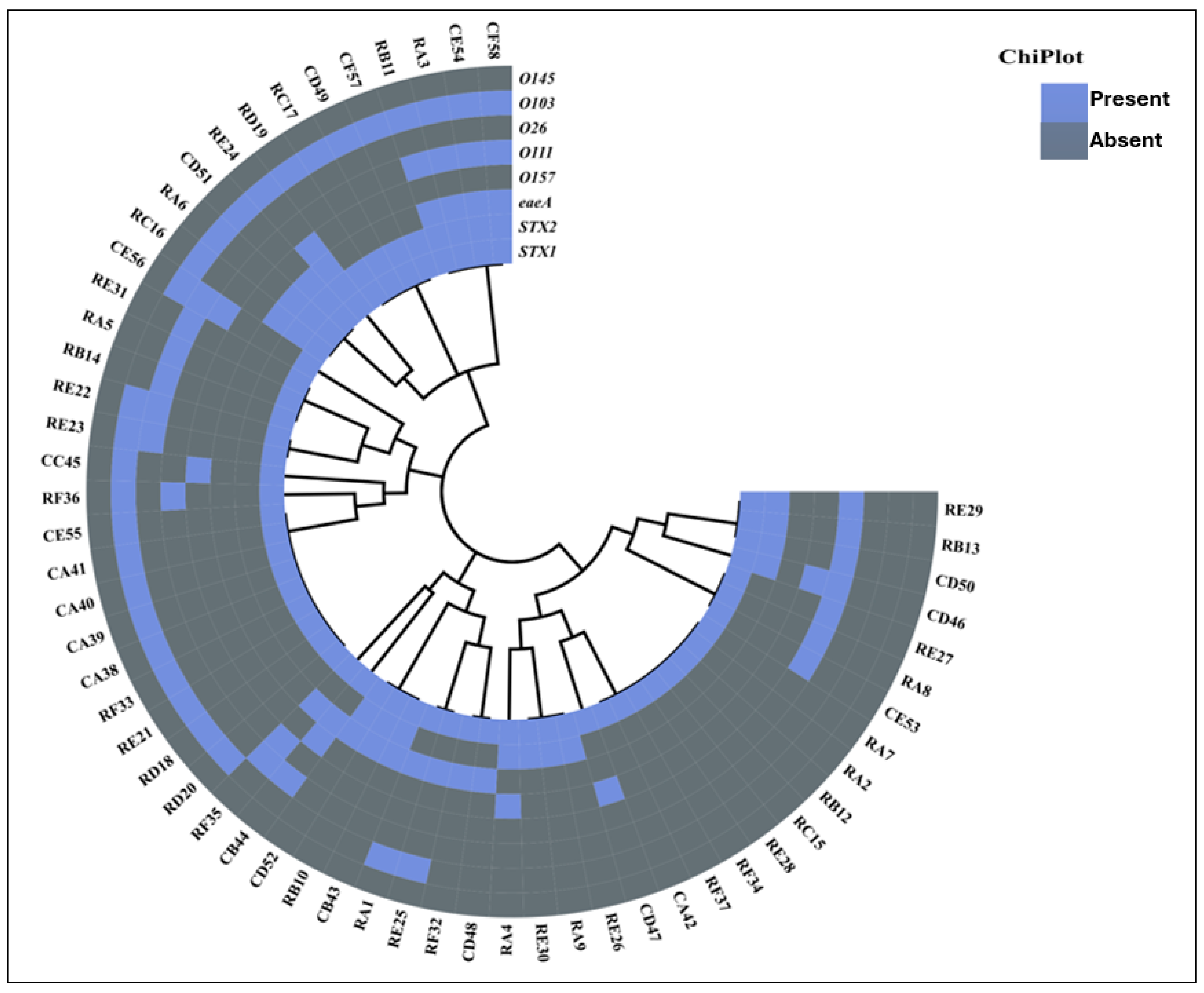
STEC strains were identified via PCR amplification of specific virulence markers. Among the confirmed E. coli isolates, the stx1 gene was the most prevalent at 24.4%. Within these positive isolates, 9.7% harbored the stx2 gene, while the eaeA gene was detected in 17.2%. A total of 17.2% isolates carried a mix of all three genes; namely, stx1, stx2, and eaeA. Furthermore, 20.6% of isolates carried both stx1 and stx2 genes, while 10.3% isolates harbored a mix of both stx1 and eaeA genes (Figure 1). Molecular characterization confirmed 58 E. coli isolates as STEC.
The 58 confirmed STEC isolates were further subjected to O-serogrouping. The O103 O-serogroup was the most detected, representing 48.2% of all STEC isolates. This was followed by O111 with 14 (24.1%), O26 with 13.8%, then O157 with 10.3%. Notably, the O145 O-serogroup was not detected in this study (Figure 1).

3.4. Phenotypic Antibiotic Resistance and ESBL-Producing STEC Isolates

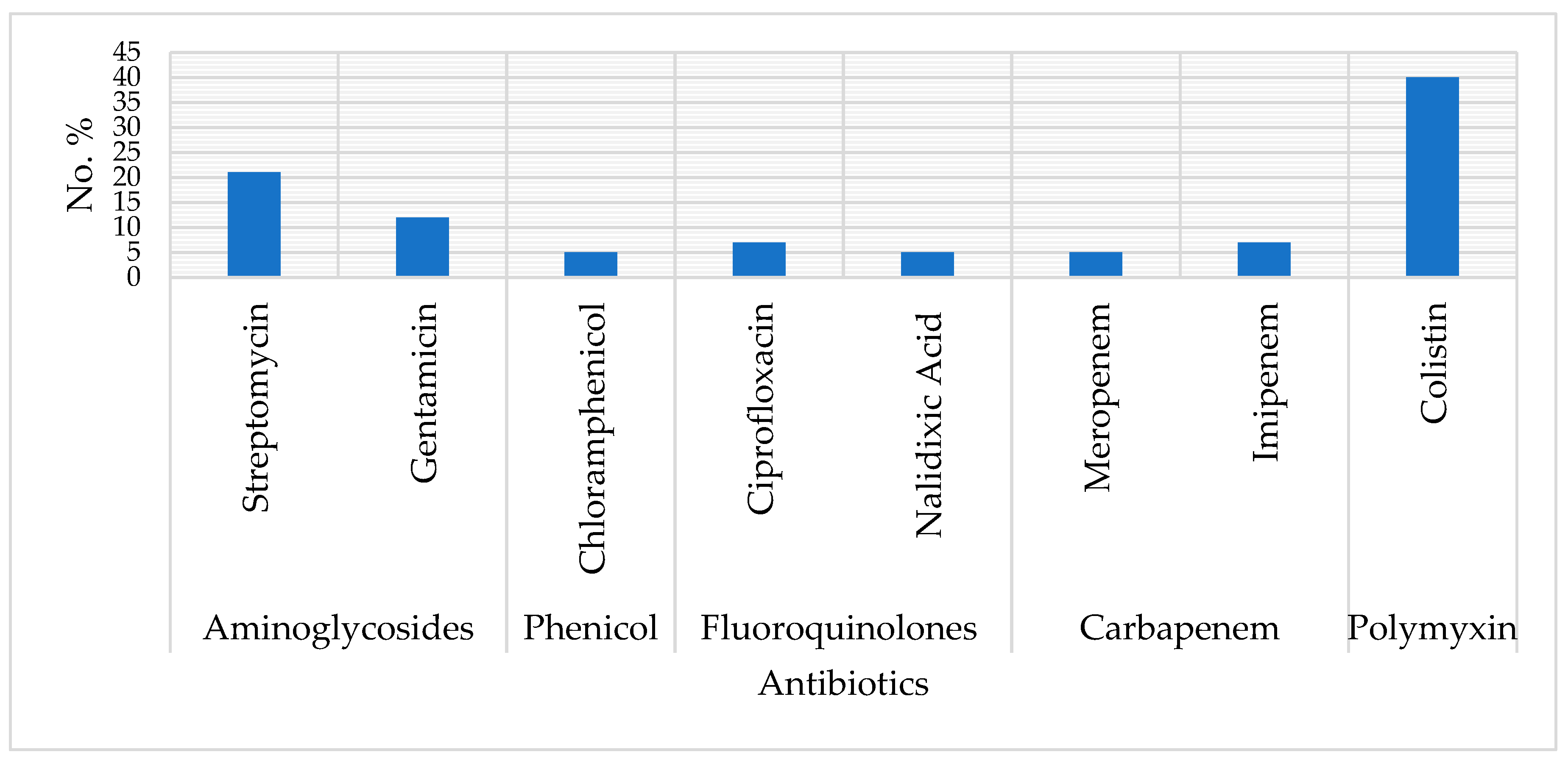
The STEC isolates exhibited resistance to the tested antibiotics depending on the isolate: 39.6% were colistin resistant (MIC ≥ 4 µg/mL), 20.6% showed resistance to streptomycin, 12.0% to gentamicin, 6.8% to ciprofloxacin, 6.8% to imipenem and 5.1% to chloramphenicol, nalidixic acid or meropenem (Figure 2). A total of 8.6% of the isolates were multidrug resistant, showing resistance to three or more antimicrobial classes.

3.5. Antimicrobial Resistance Genes for STEC E. coli Isolates

The distribution of extended-spectrum β-lactamase genes among the isolates was as follows: blaCTX-M (32.8%), ampC (31.0%), blaCTX-M-1 (10.3%), blaCTX-M-15 (5.2%) and blaCTX-M-8 (3.4%). Multiple β-lactamase genes co-existed in 8.6% isolates. The most prevalent combination consisted of two genes, specifically blaCTX-M and blaCTX-M-1, which occurred in four isolates. Furthermore, one notable isolate was found to co-harbor both blaCTX-M and blaCTX-M-8.
Among the screened carbapenem resistance genes, the blaKPC resistance gene was found more frequently, in 17.2% of the isolates, while blaVIM appeared in 5.1% of them.
The prevalence of mobile colistin genes among the isolates was as follows: mcr-1 (69%), mcr-2 (31%), and mcr-4 (40%). Three isolates carried the mcr-1, mcr-2, and mcr-4 genes together, while others had different combinations of these genes. This included three isolates with mcr-1 and mcr-2, six isolates with mcr-1 and mcr-4. The mcr-3 and mcr-5 were not detected in this study.
For aminoglycoside, 12.0% harbored strA, 6.8% carried aadA, and only 5.1% carried aadE. Furthermore, chloramphenicol resistance genes were detected, with catI present in 10.3% and catIII present in 5.1% of the isolates. The catII, catIV, strB, armA, rmtB and aac(6′)-Ib genes were not detected.

3.6. The Phenotype and Genotype ESBL-Producing STEC Isolates

Considering only the 19 isolates found to be positive ESBL producers through the CHROMEagar tests, the following blaCTX genes were detected: blaCTX-M (47.4%), blaCTX-M-1 (21.1%), blaCTX-M-8 (10.5%), and blaCTX-M-15 (10.5%) (Table 1).

3.7. Coexistence of Phenotypic and Genotypic Antibiotic Resistance

The resistance gene profiling of the E. coli isolates revealed several important findings. The catI gene, which was detected in four chloramphenicol-resistant isolates, was associated with resistance to chloramphenicol. Carbapenem resistance was associated with the blaKPC gene, identified in three isolates resistant to meropenem and four isolates resistant to imipenem. This included two isolates resistant to both meropenem and imipenem. Additionally, colistin resistance was connected to various mcr genes. Three isolates had combinations of mcr-1 and mcr-2, six isolates with mcr-1 and mcr-4, four isolates with only mcr-1, and one isolate with mcr-4.

3.8. Statistical Analysis Results

Chi-square tests showed no significant associations (p > 0.05) between AMR genotypes/phenotypes, stx genes, ESBL genes, or specific serogroups (eaeA, O111, O26, O103, and O145). However, strong correlations were found between catI and blaKPC genes and phenotypic chloramphenicol resistance (i.e., p < 0.015 and p < 0.029 in rats, respectively), and between aadA and streptomycin resistance (p < 0.029).

4. Discussion

This study reveals an alarmingly high prevalence of E. coli in layer chickens and rodents, accompanied by widespread antimicrobial resistance and arrays of antibiotic-resistant genes, highlighting a pressing public health concern. Notably, chickens and Rattus spp. act as natural reservoirs, harboring E. coli in their gastrointestinal tracts [32,37]. To detect and confirm the presence of E. coli, we utilized PCR assay, a widely adopted method for identifying pathogens in food, animal, clinical, and environmental samples. Specifically, the uidA gene served as a confirmatory marker, enabling identification of the E. coli isolates. The findings of this study revealed that 237 isolates were identified as E. coli.
Among the 237 E. coli isolates identified, 58 (24.4%) were classified as Shiga toxin-producing E. coli (STEC). The prevalence of virulence genes revealed that stx1 was detected in 100% of STEC strains, while stx2 was present in 39.6%. Additionally, the eaeA gene associated with enteropathogenic E. coli (EPEC), was found in 29.3% of the isolates. This is contrary to previous studies in South Africa, where stx2 was most prevalent in sheep and goats [36]. In contrast, a study in Ghana reported distinct findings, where none of the E. coli isolates harbored stx1, stx2, or eaeA genes [38]. These variations likely reflect differences in sample sources, sampling methodologies, geographic locations, and host populations, highlighting the complexity and diversity of E. coli strains. E. coli strains possessing the stx1 gene are linked to the risk of inducing severe diarrhea, especially in immunocompromised individuals [39]. This highlights the necessity for further characterization of stx1 subtypes in livestock, including their geographic distribution, and implications for both animal and human health. Furthermore, investigating stx1 subtypes in chickens and rodents is crucial for understanding their genetic diversity and pathogenic potential. The detection of EPEC in chicken highlights its zoonotic potential, suggesting a potential risk of gastrointestinal illnesses in humans who handle or consume contaminated poultry products [2].
All STEC isolates from chickens and rodents in this study were successfully serotyped using PCR. Notably, none of the 58 stx-positive samples belonged to the O145 serogroup. Only 2.5% of the STEC isolates harbored O157. The relatively low prevalence of O157 STEC observed in this study indicates a reduced risk of O157 infection transmission to humans from these animals. In the present study, we detected only three non-O157 STEC serogroups (O111, O26, and O103) in chickens and rodents’ feces. Notably, STEC O103 strains were identified in a significant proportion of samples in this study, which have previously been implicated in numerous foodborne illness outbreaks in England and Wales [40].
Antimicrobials play a crucial role in managing microbial infections in both veterinary and human medicine. Additionally, they are often used as growth promoters in livestock production [32,41]. The rise of antimicrobial resistance is a global concern, prompting the WHO to prioritize ESBL-producing Enterobacteriaceae as a critical threat requiring new treatments [42,43]. Among Enterobacterales, the most critical antibiotic resistance genes include Beta-lactamase genes (bla genes), colistin resistance genes (mcr genes), tetracycline resistance genes (tet genes), and aminoglycoside resistance genes, which confer resistance to various clinically important antibiotics [44,45]. Antibiotic susceptibility testing revealed that 39.6% STEC isolates were resistant to colistin, which was significantly lower (81.5%) in Chile from cattle and swine [46].
In South Africa, antibiotics such as ampicillin, quinolones, streptomycin, and florfenicol have been widely used in animal husbandry for decades to prevent and treat animal diseases, as well as to promote growth [47]. This extensive use of antibiotics may contribute to the high resistance rates (ranging from 5.1% to 39.6%) observed among STEC isolates to these drugs, along with the notable multidrug resistance (MDR) rate of 8.6% identified in this study. Antibiotic susceptibility testing showed that 39.6% of STEC isolates were resistant to colistin, which was significantly lower (81.5%) in Chile [46].
The persistence of colistin-resistant genes in STEC represents a serious threat to global food safety and public health, with poultry farming being a key area of concern. In this study, 39.6% of STEC isolates exhibited resistance to colistin. The findings are slightly lower than those reported in a Chilean study, which found that 81.5% of STEC isolates derived from cattle and swine exhibited resistance to colistin [46]. In this study, three colistin resistance genes were detected: mcr-1 (69%), mcr-2 (31%), and mcr-4 (40%). Similar findings have been documented in Europe; Garcia et al. [48] identified mcr-1 and mcr-4 in Spain, while Ewers et al. [49] reported mcr-1 and mcr-2 in German swine.
The high prevalence of colistin resistance (39.6%) observed in this study suggests a well-established environmental resistome maintained by both layer chickens and commensal rodents. Historically, the widespread use of colistin in poultry for growth promotion and group therapy created a powerful selection pressure that favored the persistence of resistant strains [50]. The detection of plasmid-mediated genes is particularly significant, as these mobile genetic elements allow resistance to persist and spread horizontally even in the absence of active antibiotic use [51]. The transmission dynamics between Rattus spp. and poultry are likely driven by a fecal–oral cycle within the farm’s shared agricultural niche. Rodents act as biological bridges, contaminating feed and water systems with infected feces while moving between manure pits and clean production areas [52]. This cycle of cross-species transmission underscores the critical role of rodents as overlooked reservoirs that maintain last resort antibiotic resistance in livestock environments, posing a significant challenge to farm biosecurity [53].
The colistin resistance was linked to various mcr genes, including mcr-1, mcr-2, and mcr-4, often found in combination among the isolates. The diverse genetic mechanisms of IncI and IncHI2/HI2A plasmids carrying mcr-1, such as the ISApl1 insertion element, facilitate the high rate of horizontal transfer of mcr-1, enabling its rapid spread [54]. Polymyxins like colistin are classified as critically important antimicrobials. However, their effectiveness is declining due to overuse and misuse in livestock production, compromising their ability to treat life-threatening infections [54,55]. Resistance rates to other antibiotics were also observed, including Streptomycin (20.6%), Gentamicin (12.0%), Ciprofloxacin (6.8%), and Imipenem (6.8%), which aligns with findings from previous studies [2,54,56].
Beta-lactamase (bla) genes are among the most prevalent antibiotic resistance determinants in Enterobacteriaceae and other bacterial species, conferring resistance to beta-lactam antibiotics [57,58]. Specifically, the blaTEM genes are known to mediate resistance to β-lactam antibiotics such as ampicillin [59]. Our study revealed a notably high prevalence (32.7%) of ESBL-producing STEC in broilers and rodents, consistent with findings from similar studies conducted in chicken production facilities in Bangladesh, India and Ghana [2,60,61]. In this study, none of the ESBL-producing STEC isolates carried blaTEM genes. However, four blaCTX-M genes were detected: blaCTX-M (32.8%), blaCTX-M-1 (10.3%), blaCTX-M-8 (3.4%), and blaCTX-M-15 (5.2%). Notably, co-existence of these genes was observed in 8.6% of the STEC isolates.
The co-existence of these genes was found in 8.6% STEC isolates. These results are consistent with previous studies from Saudi Arabia (73.9%), Germany (69.9%), and Nigeria (44.7%), which also reported high occurrences of blaCTX-M [62,63,64]. Among the blaCTX-M groups, blaCTX-M-1 exhibited the highest detection rate, at 21.1%. Our findings are supported by previous studies, including a German investigation that reported a 6.9% prevalence of blaCTX-M-1 in E. coli isolates from humans, cattle, and poultry in Pakistan [65], and a Saudi Arabian study that found a higher prevalence of 54.3% in fecal specimens from animals [53]. Globally, blaCTX-M-15 and blaCTX-M-14 are the most commonly reported blaCTX-M enzymes, widespread across human, animal, and environmental niches [66,67]. Studies have revealed a link between the dissemination of blaCTX-M-producing enzymes and E. coli strains belonging to the ST131 clonal group. Notably, ST131 isolates have been found to harbor multiple blaCTX-M types, including the predominant blaCTX-M-15 [26]. ESBL-producing Enterobacteriaceae infections are becoming increasingly hard to treat empirically [27]. Notably, ESBL-producing genes were detected in STEC isolates from both layer chickens and rodents, with a significantly higher prevalence of STEC isolates found in rodents compared to layers.
Among the carbapenem-resistance genes identified, blaKPC emerged as the most prevalent, detected in 17.2% of isolates, followed by blaVIM, found in 5.1%. Carbapenems, often considered the last line of defense against ESBL-producing Enterobacteriaceae are increasingly compromised by rising resistance, narrowing treatment options and complicating infection control efforts in healthcare settings worldwide [68]. An additional noteworthy finding was the detection of aminoglycoside-resistance genes, which exhibited varying frequencies. Notably, genes such as strA, aadA, and aadE were detected, further illustrating the multifaceted nature of antimicrobial resistance in these isolates.
A concerning finding of our study is the high detection frequency of mcr genes in STEC isolates, predominantly from rodents. This raises alarming concerns, as rodents can thrive in various environments, including human surroundings, facilitating the spread and maintenance of resistance genes in uncontrollable settings. Colistin and carbapenems are critical antibiotics of last resort, used to combat MDR bacterial infections in humans [69,70]. Notably, this study marks the first detection of mcr-1, mcr-2, and mcr-4 genes in STEC isolates from rodents and chickens in South Africa. Colistin usage has surged dramatically in recent years, particularly in countries where it has proven effective in treating antibiotic-resistant infections [36]. These findings highlight the need for enhanced surveillance, stricter antimicrobial stewardship, and targeted interventions to mitigate the risk of zoonotic transmission and preserve the efficacy of critical antibiotics.
The lack of a significant association between virulence determinants and antimicrobial resistance observed in this study [48,49] is a noteworthy finding that suggests these traits may be evolving independently within the STEC population. This lack of correlation has been previously documented in other European porcine studies. For instance, García et al. [48] found that while mcr genes were highly prevalent in Spanish farms, their distribution among different pathotypes did not show a linear relationship with specific virulence profiles, even within dominant clonal groups like ST10. Similarly, Ewers et al. [49] reported that while mcr-1 and mcr-2 were identified in various porcine E. coli lineages in Germany, these resistance genes were often carried on highly mobile IncX4 or IncI2 plasmids, which can move between strains regardless of their underlying virulome.
This finding is particularly alarming, as rodents are highly adaptable and capable of thriving in wide range of environments, including those closely associated with human activity. Consequently, they can serve as reservoirs and vectors for the dissemination of resistance genes, making control efforts increasingly challenging. The dynamic interactions between the environment, rodents, poultry, and humans create a complex network through which antibiotic-resistant bacteria and genes can circulate. This is further exacerbated by the use of antimicrobials in poultry farming, which contributes to the selection and persistence of resistant strains [70,71].

5. Conclusions

This study demonstrates a concerning convergence of virulence and AMR in Shiga toxin-producing Escherichia coli (STEC) isolated from layer chickens and Rattus spp., posing a significant threat to public health and food safety. Our findings identify rodents as critical reservoirs and vectors that facilitate the transmission of resistant STEC within poultry facilities and throughout the food chain, highlighting a major biosecurity gap. To mitigate this risk, it is imperative to implement integrated pest management strategies that prioritize rodent exclusion from feed and water systems, alongside intensified surveillance for last-resort resistance genes like mcr. Furthermore, the high prevalence of colistin resistance underscores the urgent need for stricter antimicrobial stewardship and the transition toward antibiotic alternatives in livestock production. Future research utilizing Whole Genome Sequencing (WGS) is essential to map the precise evolutionary pathways of these resistant clones and to evaluate the spill over risk to farm workers. Furthermore, to guarantee that emerging antibiotics, including newer agents like omadacycline, are explored in the veterinary setting, comprehensive pharmacovigilance studies assessing their real-world safety profiles are necessary [72]. Simultaneously, the investigation of non-conventional antimicrobial strategies, like food-derived bioactive peptides (FDBPs), presents a promising potential owing to their multitarget activity, favorable safety profiles, and capacity to penetrate biological barriers. Exploration of innovative drug delivery platforms, particularly those involving metal-ion mediation, could improve the stability, bioavailability, and targeted action of these biomolecules [73].

Supplementary Materials

The following supporting information can be downloaded at: https://www.mdpi.com/article/10.3390/microorganisms14050977/s1, Table S1: Primer pairs used in this study to determine the E. coli pathotypes and O-serogroups; Table S2: List of antibiotic resistance genes primers used in this study. References [14,70,74,75,76,77,78,79,80,81,82,83,84] are cited in the Supplementary Materials.

Author Contributions

Methodology, T.R. and N.G.K.; Validation, T.R.; Formal analysis, K.E.L.; Resources, J.N. and O.T.; Data curation, M.T.; Writing—original draft, T.R.; Writing—review & editing, J.N., K.E.L., M.T., O.T. and N.G.K.; Supervision, J.N., K.E.L., O.T. and N.G.K.; Project administration, T.R. All authors have read and agreed to the published version of the manuscript.

Funding

This research was funded by NRF Incentive grant for rated researchers (GUN: 118949) made available to Oriel Thekisoe.

Institutional Review Board Statement

Prior to the study’s commencement, the research proposal was approved by the North-West University Research Ethics Regulatory Committee (NWU-RERC), in accordance with the guidelines of the Animal Research Ethics Committee (NWU-00274-18A5, Approval Date: 12 April 2018). All methods in this study were approved by the scientific committee of the Unit for Environmental Sciences and Management of NWU. All experimental procedures were conducted by qualified personnel in strict adherence to the university’s ethical standards and regulations.

Informed Consent Statement

Not applicable.

Data Availability Statement

The data and materials supporting this study are available from the corresponding author upon reasonable request. The genomic sequences of the two strains analyzed have been deposited in the National Center for Biotechnology Information (NCBI) GenBank nucleotide sequence database, accessible through the National Library of Medicine, and assigned the accession number https://www.ncbi.nlm.nih.gov/nuccore/PV951861, https://www.ncbi.nlm.nih.gov/nuccore/PV951862, https://www.ncbi.nlm.nih.gov/nuccore/PV952222, https://www.ncbi.nlm.nih.gov/nuccore/PV952223 (all accessed on 20 April 2026).

Conflicts of Interest

The authors declare no conflicts of interest.

References

  1. Martínez-Álvarez, S.; Sanz, S.; Olarte, C.; Hidalgo-Sanz, R.; Carvalho, I.; Fernández-Fernández, R.; Campaña-Burguet, A.; Latorre-Fernández, J.; Zarazaga, M.; Torres, C. Antimicrobial resistance in Escherichia coli from the broiler farm environment, with detection of SHV-12-producing isolates. Antibiotics 2022, 11, 444. [Google Scholar] [CrossRef]
  2. Ali, M.; Tipu, J.H.; Islam, O.; Raquib, A.; Hossain, F.M.A.; Al Mamun, M.; Rahman, M.M.; Kamal, A.H.M.; Noor, M. Molecular characterization of multidrug-resistant and extended-spectrum beta-lactamase (ESBL)-producing Escherichia coli isolated from Sonali chicken meat in Bangladesh. Sci. Rep. 2025, 15, 25350. [Google Scholar] [CrossRef] [PubMed]
  3. Wang, L.; Lan, X.; Bai, Y.; Huang, Y.; Jin, W.; Zhang, M.; Zheng, X.; Zhang, Y.; Du, X.; Liu, Y.; et al. Co-resistance and plasmid-mediated co-dissemination of florfenicol and azithromycin resistance in non-O157 Shiga toxin-producing Escherichia coli from cattle in Xinjiang, China. BMC Microbiol. 2025, 25, 465. [Google Scholar] [CrossRef]
  4. Bagel, A.; Sergentet, D. Shiga toxin-producing Escherichia coli and milk fat globules. Microorganisms 2022, 10, 496. [Google Scholar] [CrossRef]
  5. Khasapane, N.G.; de Smidt, O.; Lekota, K.E.; Nkhebenyane, J.; Thekisoe, O.; Ramatla, T. Antimicrobial Resistance and Virulence Determinants of Escherichia coli Isolates from Raw Milk of Dairy Cows with Subclinical Mastitis. Animals 2025, 15, 1980. [Google Scholar] [CrossRef]
  6. Olaitan, J.O.; Dieseru, M.A.; Oluwajide, O.O.; Titilawo, M.A.; Akinde, S.B.; Daramola, O.B. Surveillance and potential health risks of antibiotic-resistant shiga toxigenic Escherichia coli isolated from beef sold in Osogbo, Nigeria. Discov. Bact. 2025, 2, 6. [Google Scholar] [CrossRef]
  7. Dutta, T.K.; Roychoudhury, P.; Bandyopadhyay, S.; Wani, S.A.; Hussain, I. Detection & characterization of Shiga toxin producing Escherichia coli (STEC) & enteropathogenic Escherichia coli (EPEC) in poultry birds with diarrhoea. Indian J. Med. Res. 2011, 133, 541–545. [Google Scholar] [PubMed]
  8. Guenther, S.; Bethe, A.; Fruth, A.; Semmler, T.; Ulrich, R.G.; Wieler, L.H.; Ewers, C. Frequent combination of antimicrobial multiresistance and extraintestinal pathogenicity in Escherichia coli isolates from urban rats (Rattus norvegicus) in Berlin, Germany. PLoS ONE 2012, 7, e50331. [Google Scholar] [CrossRef]
  9. Le Huy, H.; Koizumi, N.; Nuradji, H.; Noor, S.M.; Dharmayanti, N.I.; Haga, T.; Hirayama, K.; Miura, K. Antimicrobial resistance in Escherichia coli isolated from brown rats and house shrews in markets, Bogor, Indonesia. J. Vet. Med. Sci. 2021, 83, 531–534. [Google Scholar] [CrossRef] [PubMed]
  10. Le Huy, H.; Koizumi, N.; Ung, T.T.H.; Le, T.T.; Nguyen, H.L.K.; Hoang, P.V.M.; Nguyen, C.N.; Khong, T.M.; Hasebe, F.; Haga, T.; et al. Antibiotic-resistant Escherichia coli isolated from urban rodents in Hanoi, Vietnam. J. Vet. Med. Sci. 2020, 82, 653–660. [Google Scholar] [CrossRef]
  11. Guenther, S.; Wuttke, J.; Bethe, A.; Vojtěch, J.; Schaufler, K.; Semmler, T.; Ulrich, R.G.; Wieler, L.H.; Ewers, C. Is fecal carriage of extended-spectrum-β-lactamase-producing Escherichia coli in urban rats a risk for public health? Antimicrob. Agents Chemother. 2013, 57, 2424–2425. [Google Scholar] [CrossRef] [PubMed]
  12. Himsworth, C.G.; Zabek, E.; Desruisseau, A.; Parmley, E.J.; Reid-Smith, R.; Jardine, C.M.; Tang, P.; Patrick, D.M. Prevalence and characteristics of Escherichia coli and Salmonella spp. in the feces of wild urban Norway and black rats (Rattus norvegicus and Rattus rattus) from an inner-city neighborhood of Vancouver, Canada. J. Wildl. Dis. 2015, 51, 589–600. [Google Scholar] [CrossRef]
  13. Ramatla, T.; Taioe, M.O.; Thekisoe, O.M.; Syakalima, M. Confirmation of antimicrobial resistance by using resistance genes of isolated Salmonella spp. in chicken houses of North West, South Africa. World J. Vet. 2019, 9, 158–165. [Google Scholar] [CrossRef]
  14. Ranjbar, R.; Masoudimanesh, M.; Dehkordi, F.S.; Jonaidi-Jafari, N.; Rahimi, E. Shiga (Vero)-toxin producing Escherichia coli isolated from the hospital foods; virulence factors, o-serogroups and antimicrobial resistance properties. Antimicrob. Resist. Infect. Control 2017, 6, 4. [Google Scholar] [CrossRef]
  15. Stratakos, A.C.; Sima, F.; Ward, P.; Linton, M.; Kelly, C.; Pinkerton, L.; Stef, L.; Pet, I.; Corcionivoschi, N. The in vitro effect of carvacrol, a food additive, on the pathogenicity of O157 and non-O157 shiga-toxin producing Escherichia coli. Food Control 2018, 84, 290–296. [Google Scholar] [CrossRef]
  16. Do, K.H.; Seo, K. Animal models of Shiga toxin-producing Escherichia coli infection. J. Appl. Anim. Res. 2024, 52, 2300625. [Google Scholar] [CrossRef]
  17. Blanco Crivelli, X.; Bonino, M.P.; Von Wernich Castillo, P.; Navarro, A.; Degregorio, O.; Bentancor, A. Detection and characterization of enteropathogenic and Shiga toxin-producing Escherichia coli strains in Rattus spp. from Buenos Aires. Front. Microbiol. 2018, 9, 199. [Google Scholar] [CrossRef]
  18. Blanco Crivelli, X.; Rumi, M.V.; Carfagnini, J.C.; Degregorio, O.; Bentancor, A. Synanthropic rodents as possible reservoirs of shigatoxigenic Escherichia coli strains. Front. Cell Infect. Microbiol. 2012, 2, 134. [Google Scholar] [CrossRef]
  19. Sobrinho, C.P.; Godoi, J.L.; Souza, F.N.; Zeppelini, C.G.; Santo, V.E.; Santiago, D.C.; Alves, R.S.; Khalil, H.; Pereira, T.C.; Pinna, M.H.; et al. Prevalence of Diarrheagenic Escherichia coli (DEC) and Salmonella spp. with zoonotic potential in urban rats in Salvador, Brazil. Epidemiol. Infect. 2021, 149, e128. [Google Scholar] [CrossRef] [PubMed]
  20. Paitan, Y. Current Trends in Antimicrobial Resistance of Escherichia coli. In Escherichia coli, a Versatile Pathogen; Frankel, G., Ron, E., Eds.; Current Topics in Microbiology and Immunology; Springer: Berlin/Heidelberg, Germany, 2018; Volume 416. [Google Scholar] [CrossRef]
  21. Islam, M.S.; Hossain, M.J.; Sobur, M.A.; Punom, S.A.; Rahman, A.T.; Rahman, M.T. A systematic review on the occurrence of antimicrobial-resistant Escherichia coli in poultry and poultry environments in Bangladesh between 2010 and 2021. Biomed. Res. Int. 2023, 2023, 2425564. [Google Scholar] [CrossRef]
  22. Mkopi, J.; Mushi, J.; Mzula, M. Occurrence and antimicrobial resistance pattern of Escherichia coli and Salmonella species isolated from domestic and peridomestic rodents. Ger. J. Microbiol. 2024, 4, 15–28. [Google Scholar] [CrossRef]
  23. Mmatli, M.; Mbelle, N.M.; Fourie, B.; Osei Sekyere, J. Enterobacterales use capsules, transporters, mobile genetic elements, and other evolutionary adaptations to survive antibiotics exposure in the absence of resistance genes. Virulence 2025, 16, 2514092. [Google Scholar] [CrossRef]
  24. Kovarova, A.; Prole, G.; Farrell, M.L.; Maguire, M.; Murphy, L.C.; Chueiri, A.; O’Connor, L.; Miliotis, G.; Morris, D.; Burke, L.P. Antimicrobial resistant Enterobacterales of clinical importance in mute swans. Sci. Total Environ. 2025, 961, 178400. [Google Scholar] [CrossRef]
  25. Chinemerem Nwobodo, D.; Ugwu, M.C.; Oliseloke Anie, C.; Al-Ouqaili, M.T.; Chinedu Ikem, J.; Victor Chigozie, U.; Saki, M. Antibiotic resistance: The challenges and some emerging strategies for tackling a global menace. J. Clin. Lab. Anal. 2022, 36, e24655. [Google Scholar] [CrossRef] [PubMed]
  26. Ramatla, T.; Mafokwane, T.; Lekota, K.; Monyama, M.; Khasapane, G.; Serage, N.; Nkhebenyane, J.; Bezuidenhout, C.; Thekisoe, O. “One Health” perspective on prevalence of co-existing extended-spectrum β-lactamase (ESBL)-producing Escherichia coli and Klebsiella pneumoniae: A comprehensive systematic review and meta-analysis. Ann. Clin. Microbiol. Antimicrob. 2023, 22, 88. [Google Scholar] [CrossRef] [PubMed]
  27. Stone, T.J.; DeWitt, M.; Johnson, J.W.; Beardsley, J.R.; Munawar, I.; Palavecino, E.; Luther, V.P.; Ohl, C.A.; Williamson, J.C. Analysis of infections among patients with historical culture positive for extended-spectrum beta-lactamase (ESBL)–producing Escherichia coli or Klebsiella pneumoniae: Is ESBL-targeted therapy always needed? Antimicrob. Steward. Healthc. Epidemiol. 2023, 3, e47. [Google Scholar] [CrossRef] [PubMed]
  28. Onyeka, L.O.; Adesiyun, A.A.; Keddy, K.H.; Manqele, A.; Madoroba, E.; Thompson, P.N. Prevalence, risk factors and molecular characteristics of Shiga toxin-producing Escherichia coli in beef abattoirs in Gauteng, South Africa. Food Control 2021, 123, 107746. [Google Scholar] [CrossRef]
  29. Malahlela, M.N.; Cenci-Goga, B.T.; Marufu, M.C.; Fonkui, T.Y.; Grispoldi, L.; Etter, E.; Kalake, A.; Karama, M. Occurrence, serotypes and virulence characteristics of Shiga-toxin-producing Escherichia coli isolates from goats on communal rangeland in South Africa. Toxins 2022, 14, 353. [Google Scholar] [CrossRef]
  30. Howard, J.; Thekisoe, O.; Ramatla, T.; Lekota, K.E. Serogroups, antibiotic resistance profiles and virulence factors of non-O157 Shiga-toxin producing Escherichia coli from ovine and caprine. Sci. Rep. 2026. [Google Scholar] [CrossRef]
  31. Mhlanga, T.A.; Chitanga, S.; Matthee, S.; Malatji, M.P.; Mukaratirwa, S. Prevalence and genetic diversity of Bartonella spp. in wild small mammals from South Africa. Appl. Environ. Microbiol. 2024, 90, e0084224. [Google Scholar] [CrossRef]
  32. Ramatla, T.; Motlhaping, T.; Ndlovu, N.; Mileng, K.; Howard, J.; Khasapane, G.; Ramaili, T.; Mokgokong, P.; Nkhebenyane, J.; Ndou, R.; et al. Molecular Detection of Shiga Toxin–Producing Escherichia coli O177 Isolates, Their Antibiotic Resistance, and Virulence Profiles from Broiler Chickens. Int. J. Microbiol. 2024, 2024, 9778058. [Google Scholar] [CrossRef] [PubMed]
  33. Mlangeni, L.N.; Ramatla, T.; Price, C.; Thekisoe, O.; Weldon, C. Uncovering hidden threats: Prevalence, antibiotic resistance and virulence gene profiles of Escherichia coli strains isolated from Testudines and their aquatic habitats. Mol. Biol. Rep. 2025, 52, 778. [Google Scholar] [CrossRef] [PubMed]
  34. Clinical and Laboratory Standards Institute (CLSI). Performance Standards for Antimicrobial Susceptibility Testing, 33rd ed.; CLSI Supplement M100; Clinical and Laboratory Standards Institute: Wayne, PA, USA, 2023. [Google Scholar]
  35. Clinical and Laboratory Standards Institute (CLSI). Methods for Dilution Antimicrobial Susceptibility Tests for Bacteria that Grow Aerobically, 14th ed.; Approved Standard M7-A6; Clinical and Laboratory Standards Institute: Wayne, PA, USA, 2016. [Google Scholar]
  36. Ramatla, T.; Tutubala, M.; Motlhaping, T.; de Wet, L.; Mokgokong, P.; Thekisoe, O.; Lekota, K. Molecular detection of Shiga toxin and extended-spectrum beta-lactamase (ESBL)-producing Escherichia coli isolates from sheep and goats. Mol. Biol. Rep. 2024, 51, 57. [Google Scholar] [CrossRef] [PubMed]
  37. Bélanger, L.; Garenaux, A.; Harel, J.; Boulianne, M.; Nadeau, E.; Dozois, C.M. Escherichia coli from animal reservoirs as a potential source of human extraintestinal pathogenic E. coli. FEMS Immunol. Med. Microbiol. 2011, 62, 1–10. [Google Scholar] [CrossRef]
  38. Dela, H.; Egyir, B.; Majekodunmi, A.O.; Behene, E.; Yeboah, C.; Ackah, D.; Bongo, R.N.; Bonfoh, B.; Zinsstag, J.; Bimi, L.; et al. Diarrhoeagenic E. coli occurrence and antimicrobial resistance of Extended Spectrum Beta-Lactamases isolated from diarrhoea patients attending health facilities in Accra, Ghana. PLoS ONE 2022, 17, e0268991. [Google Scholar] [CrossRef]
  39. Thevenot, J.; Cordonnier, C.; Rougeron, A.; Le Goff, O.; Nguyen, H.T.; Denis, S.; Alric, M.; Livrelli, V.; Blanquet-Diot, S. Enterohemorrhagic Escherichia coli infection has donor-dependent effect on human gut microbiota and may be antagonized by probiotic yeast during interaction with Peyer’s patches. Appl. Microbiol. Biotechnol. 2015, 99, 9097–9110. [Google Scholar] [CrossRef]
  40. Chen, Y.; Liu, P.; Li, H.; Huang, W.; Yang, C.; Kang, M.; Jiang, X.; Shan, B.; He, H.; Hu, F.; et al. An outbreak of Shiga toxin-producing Escherichia coli serotype O103: H2 associated with unpasteurized soft cheese, England and Wales, 2022. Epidemiol. Infect. 2025, 152, e172. [Google Scholar] [CrossRef]
  41. Gwenzi, W.; Chaukura, N.; Muisa-Zikali, N.; Teta, C.; Musvuugwa, T.; Rzymski, P.; Abia, A.L.K. Insects, rodents, and pets as reservoirs, vectors, and sentinels of antimicrobial resistance. Antibiotics 2021, 10, 68. [Google Scholar] [CrossRef]
  42. Moniruzzaman, M.; Hussain, M.T.; Ali, S.; Hossain, M.; Hossain, M.S.; Alam, M.A.; Galib, F.C.; Islam, M.T.; Paul, P.; Islam, M.S.; et al. Multidrug-resistant Escherichia coli isolated from patients and surrounding hospital environments in Bangladesh: A molecular approach for the determination of pathogenicity and resistance. Heliyon 2023, 9, e22109. [Google Scholar] [CrossRef]
  43. Shrivastava, S.R.; Shrivastava, P.S.; Ramasamy, J. World health organization releases global priority list of antibiotic-resistant bacteria to guide research, discovery, and development of new antibiotics. J. Med. Soc. 2018, 32, 76–77. [Google Scholar] [CrossRef]
  44. Hasan, N.; Kannan, E.P.; Hakami, O.; Alamri, A.A.; Gopal, J.; Muthu, M. Reviewing the Phenomenon of Antimicrobial Resistance in Hospital and Municipal Wastewaters: The Crisis, the Challenges and Mitigation Methods. Appl. Sci. 2024, 14, 8358. [Google Scholar] [CrossRef]
  45. Kiskó, G.; Bajramović, B.; Elzhraa, F.; Erdei-Tombor, P.; Dobó, V.; Mohácsi-Farkas, C.; Taczman-Brückner, A.; Belák, Á. The Invisible Threat of Antibiotic Resistance in Food. Antibiotics 2025, 14, 250. [Google Scholar] [CrossRef] [PubMed]
  46. Galarce, N.; Sánchez, F.; Fuenzalida, V.; Ramos, R.; Escobar, B.; Lapierre, L.; Paredes-Osses, E.; Arriagada, G.; Alegría-Morán, R.; Lincopán, N.; et al. Phenotypic and genotypic antimicrobial resistance in non-O157 Shiga toxin-producing Escherichia coli isolated from cattle and swine in Chile. Front. Vet. Sci. 2020, 7, 367. [Google Scholar] [CrossRef]
  47. Eagar, H.; Swan, G.; Van Vuuren, M. A survey of antimicrobial usage in animals in South Africa with specific reference to food animals. J. S. Afr. Vet. Assoc. 2012, 83, 16. [Google Scholar] [CrossRef]
  48. García, V.; García-Meniño, I.; Mora, A.; Flament-Simon, S.C.; Díaz-Jiménez, D.; Blanco, J.E.; Alonso, M.P.; Blanco, J. Co-occurrence of mcr-1, mcr-4 and mcr-5 genes in multidrug-resistant ST10 Enterotoxigenic and Shiga toxin-producing Escherichia coli in Spain (2006–2017). Int. J. Antimicrob. Agents 2018, 52, 104–108. [Google Scholar] [CrossRef] [PubMed]
  49. Ewers, C.; Göpel, L.; Prenger-Berninghoff, E.; Semmler, T.; Kerner, K.; Bauerfeind, R. Occurrence of mcr-1 and mcr-2 colistin resistance genes in porcine Escherichia coli isolates (2010–2020) and genomic characterization of mcr-2-positive E. coli. Front. Microbiol. 2022, 13, 1076315. [Google Scholar] [CrossRef]
  50. Apostolakos, I.; Piccirillo, A. A review on the current situation and challenges of colistin resistance in poultry production. Avian Pathol. 2018, 47, 546–558. [Google Scholar] [CrossRef]
  51. Anyanwu, M.U.; Jarrar, B.M.; Shoyinka, S.V.; Ngwu, G.I. Mobile Colistin Resistance (mcr) Gene-Containing Organisms in the Poultry Sector. Antibiotics 2023, 12, 1117. [Google Scholar] [CrossRef]
  52. Umali, D.V.; Lapuz, R.R.S.P.; Suzuki, T.; Shirota, K.; Katoh, H. Transmission and shedding patterns of Salmonella in naturally infected captive wild roof rats (Rattus rattus) from a Salmonella-contaminated layer farm. Avian Dis. 2012, 56, 288–294. [Google Scholar] [CrossRef]
  53. Velkers, F.C.; Blokhuis, S.J.; Veldhuis Kroeze, E.J.; Burt, S.A. The role of rodents in avian influenza outbreaks in poultry farms: A review. Vet. Q. 2017, 37, 182–194. [Google Scholar] [CrossRef]
  54. Tang, B.; Wang, J.; Zheng, X.; Chang, J.; Ma, J.; Wang, J.; Ji, X.; Yang, H.; Ding, B. Antimicrobial resistance surveillance of Escherichia coli from chickens in the Qinghai Plateau of China. Front. Microbiol. 2022, 13, 885132. [Google Scholar] [CrossRef]
  55. Di, K.N.; Pham, D.T.; Tee, T.S.; Binh, Q.A.; Nguyen, T.C. Antibiotic usage and resistance in animal production in Vietnam: A review of existing literature. Trop. Anim. Health Prod. 2021, 53, 340. [Google Scholar] [CrossRef]
  56. Duc, H.M.; Ha, C.T.; Hoa, T.T.; Hung, L.V.; Thang, N.V.; Son, H.M. Prevalence, molecular characterization, and antimicrobial resistance profiles of Shiga toxin-producing Escherichia coli isolated from raw beef, pork, and chicken meat in Vietnam. Foods 2024, 13, 2059. [Google Scholar] [CrossRef] [PubMed]
  57. Santos, A.L.; Dos Santos, A.P.; Ito, C.R.; Queiroz, P.H.; de Almeida, J.A.; de Carvalho Júnior, M.A.; de Oliveira, C.Z.; Avelino, M.A.; Wastowski, I.J.; Gomes, G.P.; et al. Profile of enterobacteria resistant to beta-lactams. Antibiotics 2020, 9, 410. [Google Scholar] [CrossRef] [PubMed]
  58. Ibrahim, M.E.; Algak, T.B.; Abbas, M.; Elamin, B.K. Emergence of blaTEM, blaCTX-M, blaSHV and blaOXA genes in multidrug-resistant Enterobacteriaceae and Acinetobacter baumannii in Saudi Arabia. Exp. Ther. Med. 2021, 22, 1450. [Google Scholar] [CrossRef]
  59. Delmani, F.; Jaran, A.S.; Tarazi, Y.A.; Masaadeh, H.; Zaki, O.; Irbid, T. Characterization of ampicillin resistant gene (blaTEM-1) isolated from E. coli in Northern Jordan. Asian J. Biomed. Pharm. Sci. 2017, 7, 11–15. [Google Scholar]
  60. Habib, M.; Banerjee, J.; Batabyal, S.; Nanda, P.K.; Das, A.K.; De, U.K.; Samanta, I.; Dandapat, P.; Bandyopadhyay, S. Molecular Epidemiology of Multi-Drug Resistant E. coli Isolated from Poultry Birds in Six Agroclimatic Zones of West Bengal, India: A Cross-Sectional Study. Indian J. Microbiol. 2024, 65, 1850–1864. [Google Scholar] [CrossRef]
  61. Ayim-Akonor, M.; Ohene Larbi, R.; Owusu-Ntumy, D.D.; Sasu, B.K.; Ohene Asa, H.E.; Odoom, T. Antimicrobial Susceptibility Profile and Extended-Spectrum Beta-Lactamase Phenotype of E. coli Isolated from Poultry. Int. J. Microbiol. 2025, 2025, 9468425. [Google Scholar] [CrossRef]
  62. Ejaz, H.; Younas, S.; Abosalif, K.O.; Junaid, K.; Alzahrani, B.; Alsrhani, A.; Abdalla, E.A.; Ullah, M.I.; Qamar, M.U.; Hamam, S.S. Molecular analysis of blaSHV, blaTEM, and blaCTX-M in extended-spectrum β-lactamase producing Enterobacteriaceae recovered from fecal specimens of animals. PLoS ONE 2021, 16, e0245126. [Google Scholar] [CrossRef]
  63. Michael, G.B.; Kaspar, H.; Siqueira, A.K.; de Freitas Costa, E.; Corbellini, L.G.; Kadlec, K.; Schwarz, S. Extended-spectrum β-lactamase (ESBL)-producing Escherichia coli isolates collected from diseased food-producing animals in the GERM-Vet monitoring program 2008–2014. Vet. Microbiol. 2017, 200, 142–150. [Google Scholar] [CrossRef] [PubMed]
  64. Olowe, O.A.; Adewumi, O.; Odewale, G.; Ojurongbe, O.; Adefioye, O.J. Phenotypic and molecular characterisation of extended-spectrum beta-lactamase producing Escherichia coli obtained from animal fecal samples in Ado Ekiti, Nigeria. J. Environ. Public Health 2015, 2015, 497980. [Google Scholar] [CrossRef] [PubMed]
  65. Umair, M.; Mohsin, M.; Ali, Q.; Qamar, M.U.; Raza, S.; Ali, A.; Guenther, S.; Schierack, P. Prevalence and Genetic Relatedness of Extended Spectrum-ß-Lactamase-Producing Escherichia coli Among Humans, Cattle, and Poultry in Pakistan. Microb. Drug Resist. 2019, 25, 1374–1381. [Google Scholar] [CrossRef] [PubMed]
  66. Cantón, R.; González-Alba, J.M.; Galán, J.C. CTX-M enzymes: Origin and diffusion. Front. Microbiol. 2012, 3, 110. [Google Scholar] [CrossRef]
  67. Belmahdi, M.; Chenouf, N.S.; Ait Belkacem, A.; Martinez-Alvarez, S.; Pino-Hurtado, M.S.; Benkhechiba, Z.; Lahrech, S.; Hakem, A.; Torres, C. Extended spectrum β-lactamase-producing Escherichia coli from poultry and wild birds (Sparrow) in Djelfa (Algeria), with frequent detection of CTX-M-14 in Sparrow. Antibiotics 2022, 11, 1814. [Google Scholar] [CrossRef]
  68. Acharya, J.; Jha, R.; Bhatta, R.R.; Shrestha, L.; Sharma, B.K.; Chapagain, S.; Gompo, T.R.; Rijal, N.; Jha, P.; Baines, S.L.; et al. Extended spectrum beta-lactamase producing Escherichia coli and antimicrobial resistance gene sharing at the interface of human, poultry and environment: Results of ESBL tricycle surveillance in Kathmandu, Nepal. One Health Outlook 2025, 7, 25. [Google Scholar] [CrossRef]
  69. Mthembu, T.P.; Zishiri, O.T.; El Zowalaty, M.E. Genomic characterization of antimicrobial resistance in food chain and livestock-associated Salmonella species. Animals 2021, 11, 872. [Google Scholar] [CrossRef]
  70. Ramatla, T.; Mileng, K.; Ndou, R.; Mphuti, N.; Syakalima, M.; Lekota, K.E.; Thekisoe, O.M. Molecular detection of integrons, colistin and β-lactamase resistant genes in Salmonella enterica serovars Enteritidis and Typhimurium isolated from chickens and rats inhabiting poultry farms. Microorganisms 2022, 10, 313. [Google Scholar] [CrossRef]
  71. Sonola, V.S.; Katakweba, A.S.; Misinzo, G.; Matee, M.I. Occurrence of multi-drug-resistant Escherichia coli in chickens, humans, rodents and household soil in Karatu, Northern Tanzania. Antibiotics 2021, 11, 1137. [Google Scholar] [CrossRef] [PubMed]
  72. Liu, B.; Du, H.; Zhang, J.; Jiang, J.; Zhang, X.; He, F.; Niu, B. Developing a new sepsis screening tool based on lymphocyte count, international normalized ratio and procalcitonin (LIP score). Sci. Rep. 2022, 12, 20002. [Google Scholar] [CrossRef]
  73. Zhao, Y.-L.; Qu, Q.; Wang, Y.-M.; Zhang, Y.-T.; Qu, J. A disproportionality analysis of adverse events associated with omadacycline based on the FDA adverse event reporting system database. J Antimicrob. Chemother. 2026, 81, dkag006. [Google Scholar] [CrossRef]
  74. Gundran, R.S.; Cardenio, P.A.; Villanueva, M.A.; Sison, F.B.; Benigno, C.C.; Kreausukon, K.; Pichpol, D.; Punyapornwithaya, V. Prevalence and distribution of blaCTX-M, blaSHV, blaTEM genes in extended-spectrum β-lactamase-producing E. coli isolates from broiler farms in the Philippines. BMC Vet. Res. 2019, 15, 1–8. [Google Scholar] [CrossRef] [PubMed]
  75. Hong, B.; Ba, Y.; Niu, L.; Lou, F.; Zhang, Z.; Liu, H.; Pan, Y.; Zhao, Y. A comprehensive research on antibiotic resistance genes in microbiota of aquatic animals. Front. Microbiol. 2018, 9, 1617. [Google Scholar] [CrossRef]
  76. Jiang, L.; Hu, X.; Xu, T.; Zhang, H.; Sheng, D.; Yin, D. Prevalence of antibiotic resistance genes and their relationship with antibiotics in the Huangpu River and the drinking water sources, Shanghai, China. Sci. Total Environ. 2013, 458, 267–272. [Google Scholar] [CrossRef] [PubMed]
  77. Liu, G.; Ding, L.; Han, B.; Piepers, S.; Naqvi, S.A.; Barkema, H.W.; Ali, T.; De Vliegher, S.; Xu, S.; Gao, J. Characteristics of Escherichia coli isolated from bovine mastitis exposed to subminimum inhibitory concentrations of cefalotin or ceftazidime. BioMed Res. Int. 2018, 2018, 4301628. [Google Scholar] [CrossRef] [PubMed]
  78. Omolajaiye, S.A.; Afolabi, K.O.; Iweriebor, B.C. Pathotyping and antibiotic resistance profiling of Escherichia coli isolates from children with acute diarrhea in amatole district municipality of Eastern Cape, South Africa. Biomed Res. Int. 2020, 18, 4250165. [Google Scholar] [CrossRef]
  79. Ramatla, T.; Mphuthi, N.; Gofaone, K.; Taioe, M.O.; Thekisoe, O.M.; Syakalima, M. Identification of rodent species that infest poultry houses in Mafikeng, North West Province, South Africa. Int. J. Zool. 2019, 2019, 1280578. [Google Scholar] [CrossRef]
  80. Remya, P.; Shanthi, M.; Sekar, U. Prevalence of blaKPC and its occurrence with other beta-lactamases in Klebsiella pneumoniae. J. Lab. Physicians 2018, 10, 387–391. [Google Scholar] [CrossRef]
  81. Sheikh, F.A.; Rostami, S.; Jolodar, A.; Tabatabaiefar, M.A.; Khorvash, F.; Saki, A.; Shoja, S.; Sheikhi, R. Detection of metallo-beta lactamases among carbapenem-resistant Pseudomonas aeruginosa. Jundishapur J. Microbiol. 2014, 7, e12289. [Google Scholar]
  82. Sun, W.; Qian, X.; Gu, J.; Wang, X.J.; Duan, M.L. Mechanism and effect of temperature on variations in antibiotic resistance genes during anaerobic digestion of dairy manure. Sci. Rep. 2016, 6, 30237. [Google Scholar] [CrossRef]
  83. Wolde, A.; Deneke, Y.; Sisay, T.; Mathewos, M. Molecular Characterization and Antimicrobial Resistance of Pathogenic Escherichia coli Strains in Children from Wolaita Sodo, Southern Ethiopia. J. Trop. Med. 2022, 2022, 9166209. [Google Scholar] [CrossRef]
  84. Xu, J.; Xu, Y.; Wang, H.; Guo, C.; Qiu, H.; He, Y.; Zhang, Y.; Li, X.; Meng, W. Occurrence of antibiotics and antibiotic resistance genes in a sewage treatment plant and its effluent-receiving river. Chemosphere 2015, 119, 1379–1385. [Google Scholar] [CrossRef] [PubMed]
Figure 1. Heatmap showing O-serogroup and STEC genes detected in 58 STEC isolates from the feces of Rattus spp. and layer chickens. Light blue and dark blue indicate absence and presence of O-serogroup and STEC genes, respectively. Isolate names: R = Rattus spp., C = chickens. https://www.chiplot.online/# (accessed on 26 November 2025).
Microorganisms 14 00977 g001
Figure 2. The antibiotic resistance profiles from the STEC isolated from Layer Chickens and Rattus species.
Microorganisms 14 00977 g002
Table 1. The β-lactamase-encoding genes patterns of the ESBL-producing E. coli isolates isolated from fecal samples of layer chickens and Rattus spp.
IsolatesESBLblaCTX-MblaCTX-M-1blaCTX-M-8blaCTX-M-15
1RA1+++
2RA3+
3RA4+
4RA6+++
5RA9++
6RB12+
7RB13+++
8RB14+
9RC17+
10RE21+
11RE27+
12RE28++
13CA40+
14CB43++
15CD48++
16CD49+++
17CD50++
18CE56+++
19CF58++
+ = positive; − = negative.
Disclaimer/Publisher’s Note: The statements, opinions and data contained in all publications are solely those of the individual author(s) and contributor(s) and not of MDPI and/or the editor(s). MDPI and/or the editor(s) disclaim responsibility for any injury to people or property resulting from any ideas, methods, instructions or products referred to in the content.