


Abstract
Inflammatory bowel disease (IBD) is a chronic intestinal disorder with recurrent inflammation for which effective therapeutic options remain limited. Probiotics from the Bifidobacterium genus have potential beneficial effects on the prevention of IBD by improving intestinal barrier integrity and modulating immune responses. However, whether these effects are mediated by the regulation of gut metabolism remains largely unclear. This study was designed to explore the protective effect of an infant-derived Bifidobacterium animalis subsp. lactis 832 (B. lactis 832) on dextran sulfate sodium (DSS)-induced colitis in mice and its underlying mechanism. B. lactis 832 treatment significantly alleviated colitis severity (p < 0.05), as evidenced by reduced weight loss, disease activity index (DAI), and colonic injury, accompanied by significantly decreased pro-inflammatory cytokine expression and increased Il10 expression (p < 0.05). It also improved intestinal barrier integrity and modulated gut microbiota composition by reducing potentially pathogenic bacteria while enriching beneficial taxa. Surprisingly, metabolomic analysis revealed that B. lactis 832 intervention enhanced intestinal phospholipid metabolism, particularly increasing phosphatidylethanolamine (PE) and phosphatidylcholine (PC) levels. Notably, PE or PC supplementation recapitulated the protective effects against DSS-induced colitis (p < 0.05). These findings suggest that B. lactis 832 alleviates colitis through microbiota-associated metabolic regulation, highlighting a key role for phospholipid metabolism in mediating probiotic effects.
1. Introduction
Inflammatory bowel disease (IBD) is characterized by chronic and relapsing intestinal inflammation that includes ulcerative colitis (UC) and Crohn’s disease (CD) [1]. The clinical manifestations of IBD patients are diarrhea, blood in the stool, weight loss, and diffuse inflammation of the colonic mucosa [2]. IBD has become a prominent global healthcare problem, impacting more than 4 million individuals worldwide [3]. Although the pathogenesis of IBD is poorly understood, multiple lines of evidence suggest that the disease is caused by genetic susceptibility, intestinal barrier dysfunction, dysbiosis of the gut microbiota, and immune-mediated inflammation [4,5]. Recent studies have emphasized the crucial role of gut microbiota dysbiosis in the pathogenesis of IBD [6,7]. For instance, a reduced richness of microbial diversity, characterized by a decrease in Firmicutes and an overgrowth of Proteobacteria, is a typical hallmark of dysbiosis [8]. There is currently no cure for IBD and, despite available treatments, nearly half of patients develop refractory disease requiring ongoing management [9]. Given the complexity and appearing complications associated with immunosuppressive drugs (e.g., sulfasalazine and mesalazine) [10], it is crucial to develop novel therapeutic strategies and alternative treatments for IBD.
Considering the flexible manipulation of gut microbiota, potential remodeling by supplementing probiotics or providing specific substrates (such as food-derived nutrients) may provide a new method for the prevention of IBD [11]. Previous studies have shown that probiotic and prebiotic therapies are potentially natural and effective interventions for the treatment of IBD with fewer side effects than conventional therapies [12]. Among various probiotics, the Bifidobacterium genus exhibits multiple probiotic functions beneficial to gastrointestinal health, including repairing the intestinal barrier, modulating the intestinal microbiota, regulating the host immune system, and facilitating host nutrient absorption [13,14,15]. Bifidobacterium animalis subsp. lactis is one of the most extensively studied subspecies belonging to the genus Bifidobacterium. In vitro, strains of this subspecies present good probiotic properties, including high tolerance to acid and bile salts, strong adhesion ability, and potent anti-pathogenic activity [16,17,18]. In addition, some studies have confirmed that the B. lactis species had beneficial effects in alleviating colitis. For example, B. lactis XLTG11 has been reported to alleviate DSS-induced colitis by inhibiting the TLR4/MYD88/NF-κB signaling pathway [19]. Moreover, B. lactis A6 effectively alleviated DSS-induced colitis by preserving intestinal barrier integrity, reducing oxidative stress, and suppressing inflammatory responses [20]. However, the mechanisms underlying these protective effects are not fully understood, particularly whether they are mediated by microbiota-associated metabolic regulation.
Gut microbiota can influence host physiology through the production of bioactive metabolites, which play essential roles in immune regulation, signaling, and the maintenance of intestinal homeostasis [21,22]. Among these metabolites, phospholipids are critical components of intestinal mucosa with diverse biological functions, including supporting enterocyte proliferation, regulating lipid metabolism, and maintaining mucus secretion [23,24,25]. Phosphatidylcholine (PC) and phosphatidylethanolamine (PE) constitute the major phospholipid species in the intestinal mucosa, representing over 45% and around 25% of total phospholipids, respectively [26,27]. Dysregulation of phospholipid composition has been linked to the development of IBD. Reduced PC levels in patients with ulcerative colitis impair the hydrophobic and protective properties of the mucus layer [28,29]. In addition, PE has been reported to regulate mucus secretion by goblet cells and promote intestinal development [30]. These findings suggest that phospholipid metabolism may represent a key link between gut microbiota and intestinal barrier function in IBD.
In this study, we investigated whether an infant-derived Bifidobacterium animalis subsp. lactis strain (B. lactis 832) alleviated DSS-induced colitis in a mouse model by modulating microbiota-associated metabolic pathways. Special attention was directed toward uncovering critical metabolic pathways that connected microbial shifts with host protective effects. Furthermore, we aimed to characterize the interplay between microbial alterations and metabolic reprogramming to better understand the mechanistic basis of the protective effects of B. lactis 832.
2. Materials and Methods
2.1. Bacterial Strains
Bifidobacterium animalis subsp. lactis 832 (B. lactis 832), a strain isolated from infant feces in this study, was grown anaerobically at 37 °C in De Man, Rogosa and Sharpe medium (MRS) for 24–48 h. This strain was identified as Bifidobacterium animalis subsp. lactis based on 16S rRNA gene sequencing and phylogenetic analysis (GenBank accession no.: PZ135826; Figure S1 and Table S1). Fresh bacterial suspensions were prepared daily by centrifugation at 5500× g for 5 min at 4 °C, followed by two washes with phosphate-buffered saline (PBS, pH 7.4), and resuspension to approximately 5.0 × 10⁹ CFU/mL before administration.
2.2. Acid and Bile Tolerance Assay In Vitro
The survival of B. lactis 832 in a low pH environment and bile salt conditions was performed by evaluating viable colony counts as previously reported [18]. B. lactis 832 was grown overnight in MRS at 37 °C. For the acid tolerance assay, 1% (v/v) of the overnight bacterial culture was inoculated into the corresponding broth previously adjusted to pH 2.5 and 6.2 (control) with 1 N HCl. The samples from each tube of specified pH were taken at 0 and 4 h, and serially diluted. For the bile salt tolerance assay, 1% (v/v) of the overnight bacterial culture was inoculated into the corresponding broth supplemented with 0% (control) and 0.3% (w/v) oxgall bile salt (Sigma-Aldrich, St. Louis, MO, USA). The samples from each bile salt concentration group were taken at 0 and 24 h, and serially diluted. Cell counts for both assays were performed in triplicate by the standard plate count technique. The inoculated agar plates were incubated at the optimal temperature for 72 h, and the number of viable cells was calculated and expressed as the log10 value of CFU/mL. The survival rate of bacteria was calculated using the following equation [18].
Survival rate (%) = (log10 CFU/mL at a certain time point/log10 CFU/mL at 0 h) × 100
2.3. Caco-2 Cell Adhesion and Anti-Inflammatory Assay In Vitro
The methods for adherence and anti-inflammatory assays have been described previously [18,31]. For the adherence assay, Caco-2 cells were seeded into 6-well plates with Dulbecco’s Modified Eagle Medium (DMEM) containing 10% FBS at 37 °C with 5% CO2 and incubated until a complete monolayer was obtained. After washing with sterile PBS, bacteria were added at an MOI of 100 and co-incubated for 2 h. Nonadherent bacteria were removed by washing three times with PBS. The Caco-2 cells were then lysed with 0.5% Triton X-100 for 5 min. The lysates were diluted and plated on MRS agar and incubated anaerobically at 37 °C for 24–48 h. Adhesion efficiency was determined by counting the number of bacterial colonies. For the anti-inflammatory assay, after removing the culture medium, cells in 6-well plates were treated with B. lactis 832 at a concentration of 1 × 10⁸ CFU/mL for 2 h. Subsequently, all wells except the negative control were stimulated with LPS at a final concentration of 100 ng/mL. After an additional 24 h of incubation, total RNA was extracted, and the mRNA expression levels of TNF-α, IL-1β, IL-6, and IL-10 were determined by qRT-PCR.
2.4. Animal and DSS-Induced Colitis Model
Eight-week-old female C57BL/6J mice were obtained from Vital River Laboratory Animal Technology (Beijing, China). Animals were maintained under specific pathogen-free (SPF) conditions in the animal facility of Nankai University, with unrestricted access to standard chow and water, and kept on a 12 h light/dark cycle. All experimental procedures were approved by the Institutional Animal Care and Use Committee of Nankai University (Tianjin, China). All animal experiments were conducted in accordance with the guidelines of the Institutional Animal Care and Use Committee (IACUC) and were approved by the Animal Ethics Committee of Nankai University (Tianjin, China; Approval No. 2026-SYDWLL-000159).
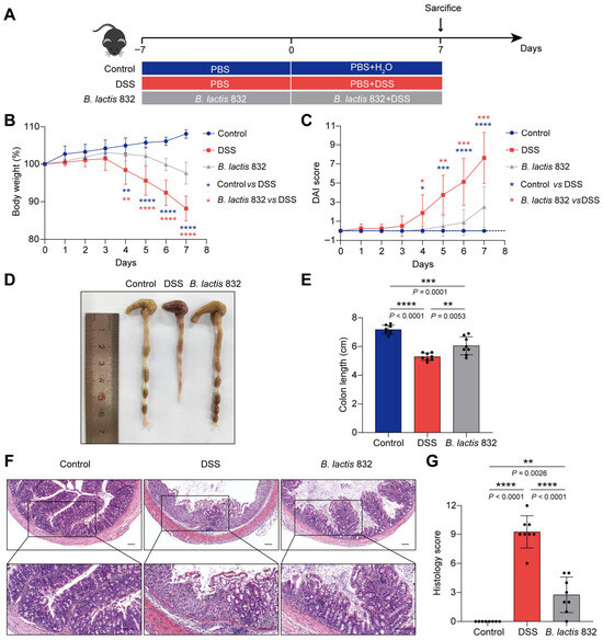
The entire experiment lasted for 14 days, and the experimental design is presented in Figure 1A and Figure S3A, according to previous studies [14,32]. Following a 7-day acclimation period, mice were randomly assigned to different treatment groups. In the probiotic intervention experiment, animals were divided into four groups (n = 8 per group): Control, Control + B. lactis 832, DSS, and B. lactis 832. The control group received sterile water and PBS, whereas the DSS group was administered 3% (w/v) dextran sulfate sodium (DSS; 36–50 kDa, MeilunBio, Dalian, China) together with PBS. The Control + B. lactis 832 and B. lactis 832 group received sterile water or 3% DSS in combination with a daily oral gavage of 0.2 mL bacterial suspension (1 × 10⁹ CFU/mL). For PE or PC supplementation experiments, mice were divided into four groups (n = 8 per group): Control, DSS, PE, and PC. Animals in the control group were given sterile water and PBS, while DSS-treated mice received 3% DSS. The PE and PC groups were administered DSS along with daily gavage of PE or PC at a dose of 50 mg/kg (0.2 mL per mouse). Gavage treatment was maintained throughout the 14-day experimental period, whereas DSS exposure was limited to the last 7 days. Throughout the experiment, clinical parameters including body weight, stool consistency, and rectal bleeding were recorded to evaluate disease progression. The disease activity index (DAI) was calculated based on these parameters. Refer to Table 1 below for detailed scores [33]. The mice were euthanized on day 7, and the entire colon was excised, measured, and observed for signs of mucosal ulcers. A segment of the distal colon measuring 1 cm was collected for histological, AB-PAS, and immunofluorescence staining, the remaining colon and stools were snap-frozen in liquid nitrogen and stored at −80 °C for molecular analysis. Heart, liver, spleen, lung, and kidney tissues were harvested from mice in the Control and Control + B. lactis 832 groups for histopathological evaluation to assess biosafety [34].
Figure 1.
Effects of B. lactis 832 on DSS-induced colitis in mice. (A) Schematic of B. lactis 832 supplementation in DSS-induced mouse model. (B) Changes in body weight. (C) DAI score recorded from day 0 to day 7. (D) Gross morphology of the colon. (E) Quantification of colon length (cm). (F) Histological appearance of colon sections following H&E staining; scale bar: 100 μm. (G) Scoring of histological alterations in colonic tissues. n = 8. Statistical significance was determined using one-way ANOVA followed by Tukey’s multiple comparison tests (* p < 0.05; ** p < 0.01; *** p < 0.001; **** p < 0.0001).
Table 1.
Disease activity index.
2.5. Quantitative Reverse Transcription Polymerase Chain Reaction (qRT-PCR)
Total RNA was isolated from colon or ileal tissues using TRIzol reagent (GeneStar, Beijing, China) in accordance with the manufacturer’s protocol. All procedures were carried out under RNase-free conditions. First-strand cDNA was synthesized from RNA templates using oligo(dT) primers and StarScript Ⅲ MasterMix (GeneStar, Beijing, China). Quantitative real-time PCR (qRT-PCR) was performed on an Applied Biosystems 7500 system using 2× RealStar Fast SYBR qPCR mix (GeneStar). Relative gene expression levels were calculated using the 2−ΔΔCt method with Gapdh serving as the internal reference gene [35]. The primer sequences used in this study are provided in Table 2.
Table 2.
The primers used for qRT-PCR.
2.6. Histological Assessment
Colon tissues were collected and rinsed with pre-chilled PBS to eliminate residual intestinal contents. Samples were subsequently fixed in 4% paraformaldehyde at room temperature for 72 h, followed by paraffin embedding. Paraffin blocks were sectioned using a Dakewe MT1 microtome (Dakewe Biotech, Shenzhen, China) and subjected to hematoxylin and eosin (H&E) as well as periodic acid–Schiff (AB-PAS) staining. Histological features were observed under a Leica DM2500 LED microscope (Leica, Wetzlar, Germany) at 100× magnification for pathological evaluation. Tissue damage was scored according to previously established criteria (Table 3) [36]. The number of goblet cells was quantified using ImageJ software (version 1.8.0.112; Media Cybernetics, Rockville, MD, USA).
Table 3.
Histological scores of colon damage.
2.7. Immunofluorescence Staining
Immunofluorescence staining was conducted following a standard procedure using primary antibodies against ZO-1 (1:500, #ab216880; Abcam, Cambridge, UK) and occludin (1:500, #FNab05957; FineTest, Wuhan, China). Tissue samples were incubated with the primary antibodies overnight at 4 °C, followed by washing with PBS. Subsequently, samples were incubated with DyLight 594-conjugated goat anti-rat IgG (H + L) secondary antibody (1:1000, E032440-01; EarthOx, San Francisco, CA, USA) for 1 h at 37 °C in the absence of light. After nuclear staining with DAPI, images were acquired using a laser scanning confocal microscope (ZEISS, Wetzlar, Germany). Quantification of ZO-1 and occludin fluorescence intensity was performed using ImageJ software (version 1.8.0.112, Media Cybernetics, Rockville, MD, USA).
2.8. Intestinal Permeability Assay
Intestinal barrier function was evaluated by assessing permeability to fluorescein isothiocyanate (FITC)-dextran following an established protocol. Briefly, mice were fasted for 4 h prior to the administration of 4 kDa FITC-dextran (Sigma-Aldrich, St. Louis, MO, USA) by oral gavage at 600 mg/kg body weight. After 3 h, blood samples were obtained via retro-orbital collection, and fluorescence intensity was determined at 525 nm using a microplate reader (BioTek, Winooski, VT, USA). Serum FITC-dextran levels were subsequently quantified based on a standard calibration curve.
2.9. 16S rRNA Sequencing Analysis
Fecal samples were collected from mice in different groups, immediately snap-frozen in liquid nitrogen, and submitted to Majorbio Bio-Pharm Technology Co., Ltd. (Shanghai, China) for 16S rRNA gene sequencing to analyze gut microbial composition. Microbial genomic DNA was isolated from feces using the HiPure Soil DNA Kit (cat. no. D3142-02B; Magen, Guangzhou, China). The V3–V4 hypervariable regions of the bacterial 16S rRNA gene were amplified using primer pairs 338F (5′-ACTCCTACGGGAGGCAGCAG-3′) and 806R (5′-GGACTACHVGGGTWTCTAAT-3′), followed by library preparation. Sequencing was carried out on Illumina MiSeq (PE300) or NovaSeq (PE250) platforms (Illumina, San Diego, CA, USA) according to standard procedures. Raw sequencing reads were first demultiplexed and subjected to quality control using fastp (v0.20.0), and paired-end sequences were subsequently merged with FLASH (v1.2.7). Operational taxonomic units (OTUs) were defined at a 97% sequence similarity threshold using UPARSE (v7.1), with chimeric sequences removed during processing. Taxonomic assignment of representative OTU sequences was performed using the RDP Classifier (v2.2) against the Silva 16S rRNA database (e.g., v138, https://www.arb-silva.de/) with a confidence cutoff of 0.7 [37,38]. All sequencing and bioinformatic analyses were performed on the Majorbio cloud platform (https://cloud.majorbio.com).
2.10. Metabolome Analysis
Fecal metabolomic profiling was performed using the Q300 Metabolite Assay Kit (Majorbio Bio-Pharm Technology Co., Ltd., Shanghai, China) with slight modifications to established protocols [1]. Briefly, approximately 50 mg of freeze-dried fecal material was weighed and homogenized in 400 μL methanol, followed by extraction with L-2-chlorophenylalanine (0.02 mg/mL) as an internal standard. The homogenates were centrifuged for 20 min, and 5 μL of the resulting supernatant was transferred into a 96-well plate for derivatization according to the manufacturer’s instructions. Metabolite quantification was carried out using an ultra-high-performance liquid chromatography system coupled to a Q Exactive HF-X mass spectrometer (Thermo Fisher Scientific, Waltham, MA, USA). Raw LC–MS data were processed using Progenesis QI software (version 3.0; Waters Corporation, Milford, CT, USA) for peak detection, alignment, and quantification. Differential metabolites were identified through univariate statistical analysis with a significance threshold of p < 0.05. For biomarker screening among the Control, DSS, and B. lactis 832 groups, metabolites meeting the criteria of |log2FC| ≥ 1 and p ≤ 0.05 in univariate analysis, along with a variable importance in projection (VIP) value > 1 from multivariate analysis, were considered significant. Data analysis was performed using the Majorbio cloud platform (https://cloud.majorbio.com).
2.11. Statistical Analysis
Data are expressed as the mean ± standard deviation (SD). Statistical analyses were performed by GraphPad Prism software (version 8.0.1; GraphPad Inc., San Diego, CA, USA). Differences between two groups were evaluated using an unpaired two-tailed Student’s t test, whereas comparisons among multiple groups were analyzed by one-way analysis of variance (ANOVA) followed by Tukey’s multiple comparison tests. A p value of less than 0.05 was considered indicative of statistical significance (* p < 0.05, ** p < 0.01, *** p < 0.001, **** p < 0.0001).
3. Results
3.1. In Vitro Characterization of B. lactis 832
Prior to in vivo experiments, we evaluated the probiotic properties of B. lactis 832. After treatment with simulated MRS (pH 2.5) for 4 h, the survival rate of B. lactis 832 was 78.99 ± 0.15% (Table S2). After exposure to 0.3% oxgall bile salt for 24 h, the survival rate remained 59.31 ± 0.12%, indicating good resistance to harsh gastrointestinal environments (Table S2). The adhesion rate of B. lactis 832 to Caco-2 cells was 40.92 ± 0.09%, demonstrating strong intestinal adhesion capacity (Table S2). Moreover, B. lactis 832 exerted prominent anti-inflammatory effects in vitro. Compared with the LPS-stimulated group, pre-treatment with B. lactis 832 significantly downregulated the mRNA expression of pro-inflammatory cytokines Tnfa (p < 0.0001), Il1b (p < 0.001) and Il6 (p < 0.001), while markedly increasing the expression of the anti-inflammatory cytokine Il10 (p < 0.01) (Figure S2). These in vitro probiotic properties confirm that B. lactis 832 is a reliable probiotic candidate for subsequent in vivo research.
3.2. B. lactis 832 Supplementation Alleviates DSS-Induced Colitis in Mice
Then, to investigate the prophylactic efficacy of B. lactis 832 against intestinal inflammation in vivo, we established a murine model of DSS-induced acute colitis (Figure 1A). Firstly, we administered 1 × 10⁹ CFU B. lactis 832 or PBS every day via oral gavage for two weeks and 3% DSS or H2O was added to the drinking water during the second week to induce colitis and the mice were sacrificed on day 7 after DSS treatment. Colitis severity was assessed daily using a composite DAI derived from body weight change, fecal bleeding, and stool consistency [2]. Continuous safety evaluation showed that single B. lactis 832 intervention caused no obvious physiological or histological abnormalities in healthy mice, with no significant differences in body weight, DAI score, colon length, or major organ histopathology between the normal control group and the B. lactis 832-alone treated group (p > 0.05) (Figure S3). The results showed that mice exposed to DSS exhibited greater weight loss and higher DAI scores than those in the control group (p < 0.05). While the B. lactis 832 group exhibited more modest weight loss and lower DAI scores than the DSS group (p < 0.05, Figure 1B,C). Moreover, colonic shortening may serve as an additional indicator for exacerbations of IBD [2]. On day 7, DSS-treated mice displayed a marked reduction in colon length relative to the control group (p < 0.0001), whereas B. lactis 832 administration partially restored colon length compared with the DSS group (p < 0.01, Figure 1D,E). Histological analysis showed an intact and well-organized mucosal structure without inflammatory cell infiltration in the control group. In contrast, DSS resulted in epithelial erosion, goblet cell loss, and areas of mucosal ulceration, together with increased leukocyte infiltration in the mucosa and submucosa, accompanied by higher histological scores of tissue damage and inflammation. However, the B. lactis 832 group exhibited less severe epithelial disruption, fewer damaged crypts, and reduced inflammatory cell infiltration resulting in a lower histological score compared to the DSS group (p < 0.001, Figure 1F,G). Taken together, these results demonstrated that B. lactis 832 supplementation reduced the severity of colitis symptoms in mice.
3.3. B. lactis 832 Supplementation Prevents DSS-Induced Colonic Inflammation
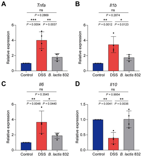
To assess the impact of B. lactis 832 on intestinal inflammation, mRNA levels of Tnfa, Il1b, Il6, and Il10 were quantified in colonic tissues following DSS exposure. DSS treatment was associated with elevated expression of pro-inflammatory cytokines (Tnfa, Il1b, and Il6) and reduced Il10 levels compared with the control group (p < 0.01, Figure 2A–D). In contrast, administration of B. lactis 832 suppressed the upregulation of these pro-inflammatory markers while restoring Il10 expression relative to the DSS group (p < 0.05). Collectively, these findings suggest that B. lactis 832 modulates cytokine profiles toward an anti-inflammatory state.
Figure 2.
Effect of the B. lactis 832 on mRNA expression levels of Tnfa, Il1b, Il6, and Il10 in DSS-induced colitis mice. (A–D) mRNA expression levels of Tnfa, Il1b, Il6, and Il10. n = 4. Statistical differences were evaluated by one-way ANOVA followed by Tukey’s multiple comparison tests (* p < 0.05; ** p < 0.01; *** p < 0.001).
3.4. B. lactis 832 Attenuates DSS-Induced Mucus Impairment and Goblet Cell Depletion
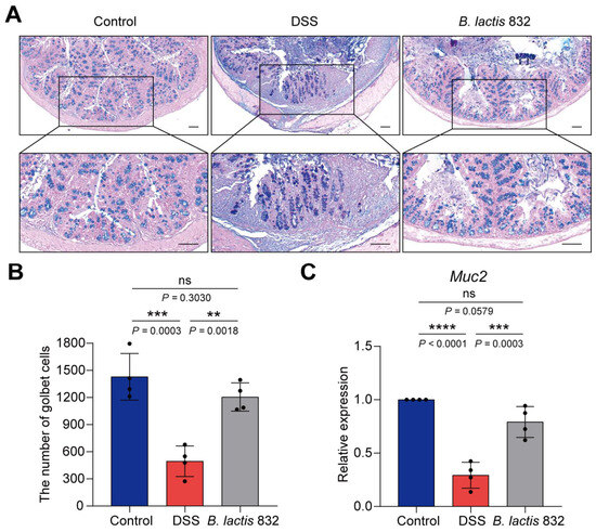
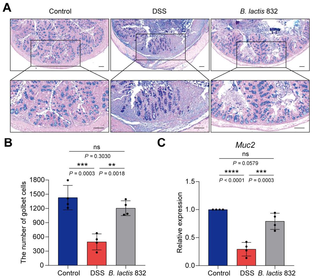
Both clinical and experimental studies have implicated intestinal mucosal barrier dysfunction as a key factor underlying the pathogenesis and progression of IBD [8]. To further verify the alleviating effect of B. lactis 832 on the DSS-induced intestinal barrier disruption, the colon tissues were collected and AB-PAS staining and the RT-qPCR experiment were performed to analyze intestinal goblet cells contents and mucin secretion levels after DSS treatment. AB-PAS images showed the mucins were abundant in goblet cells and were mainly distributed on the surface of colonic epithelial cells. In contrast, the number of goblet cells was obviously decreased due to the damage to the inner and outer mucus layers of the colonic epithelium in the DSS group (p < 0.001). However, administration with B. lactis 832 effectively improved the loss of mucus layer and goblet cell counts, which showed protective effects on mucosa (p < 0.01, Figure 3A,B). Additionally, DSS treatment markedly reduced Muc2 mRNA expression relative to control group (p < 0.0001), whereas B. lactis 832 supplementation restored its expression in colonic tissues (p < 0.001, Figure 3C). Above all, B. lactis 832 supplementation significantly improved mucus layer structure and increased goblet cell numbers in DSS-treated mice.
Figure 3.
B. lactis 832 ameliorated DSS-induced mucus disruption and goblet cell exhaustion in DSS-induced colitis mice. (A) Representative Alcian blue and PAS staining of colon tissues. Scale bar: 100 μm. (B) Quantitative analysis of goblet cell abundance in the colon. (C) Muc2 mRNA levels in colonic tissue. n = 4. Statistical significance was determined using one-way ANOVA followed by Tukey’s multiple comparison tests (** p < 0.01; *** p < 0.001; **** p < 0.0001).
3.5. B. lactis 832 Supplementation Mediates Gut Barrier Function by Enhancing TJs Expression
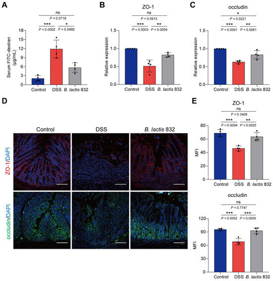
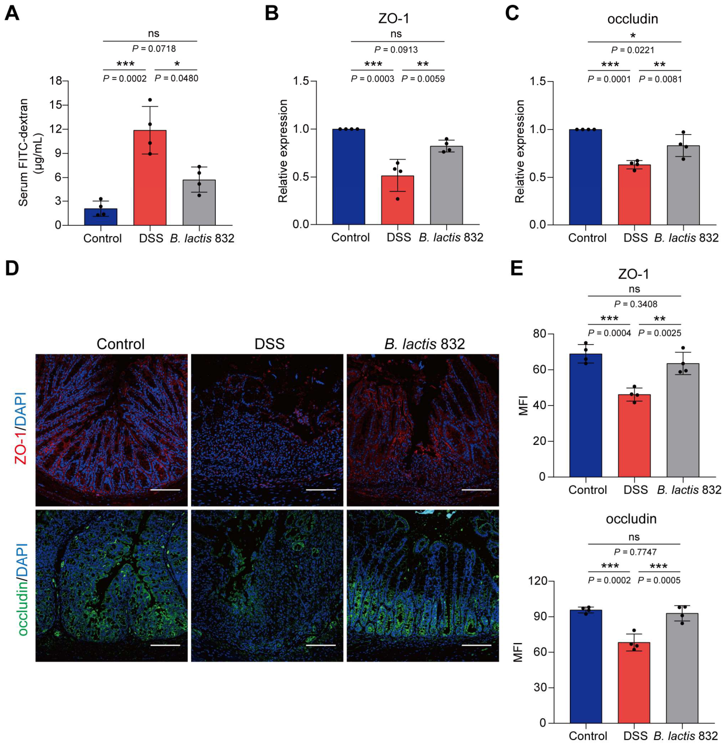
Gut barrier integrity was assessed by measuring intestinal permeability and the colonic expression of tight junction proteins, including ZO-1 and occludin, following DSS treatment. Firstly, intestinal permeability was assessed by measuring serum FITC-dextran levels 4 hours after administration for different groups on day 7 after DSS treatment. DSS treatment markedly increased serum FITC-dextran levels compared with control group (p < 0.001), while B. lactis 832 treatment attenuated this increase in serum FITC-dextran concentration (p < 0.05, Figure 4A). Consistently, mRNA levels of the tight junction proteins ZO-1 and occludin were reduced in DSS-treated mice (p < 0.001). In contrast, B. lactis 832 supplementation restored the expression of these genes relative to the DSS group (p < 0.01, Figure 4B,C). In addition, immunofluorescence staining confirmed that in situ protein expressions of ZO-1 and occludin in colon tissues were largely down-regulated after DSS treatment (p < 0.001), whereas B. lactis 832 treatment up-regulated their expression compared to the DSS group (p < 0.01, Figure 4D,E). Overall, B. lactis 832 supplementation significantly restored the DSS-induced reduction in tight junction protein expression at both mRNA and protein levels.
Figure 4.
B. lactis 832 enhances intestinal barrier function in DSS-induced colitis. (A) Serum FITC-dextran levels as an indicator of intestinal permeability. (B,C) mRNA levels of ZO-1 (B) and occludin (C) in colonic tissue. (D,E) Immunofluorescence staining of ZO-1 (red) and occludin (green) with nuclear counterstaining (blue) in the colon (D), along with corresponding quantification (E). n = 4. Statistical significance was determined using one-way ANOVA followed by Tukey’s multiple comparison tests (* p < 0.05; ** p < 0.01; *** p < 0.001).
3.6. B. lactis 832 Supplementation Improves the Intestinal Microbiota
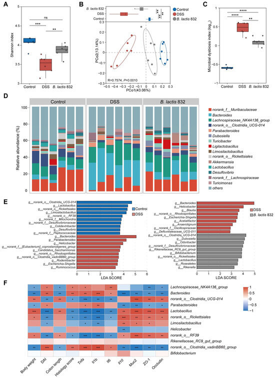
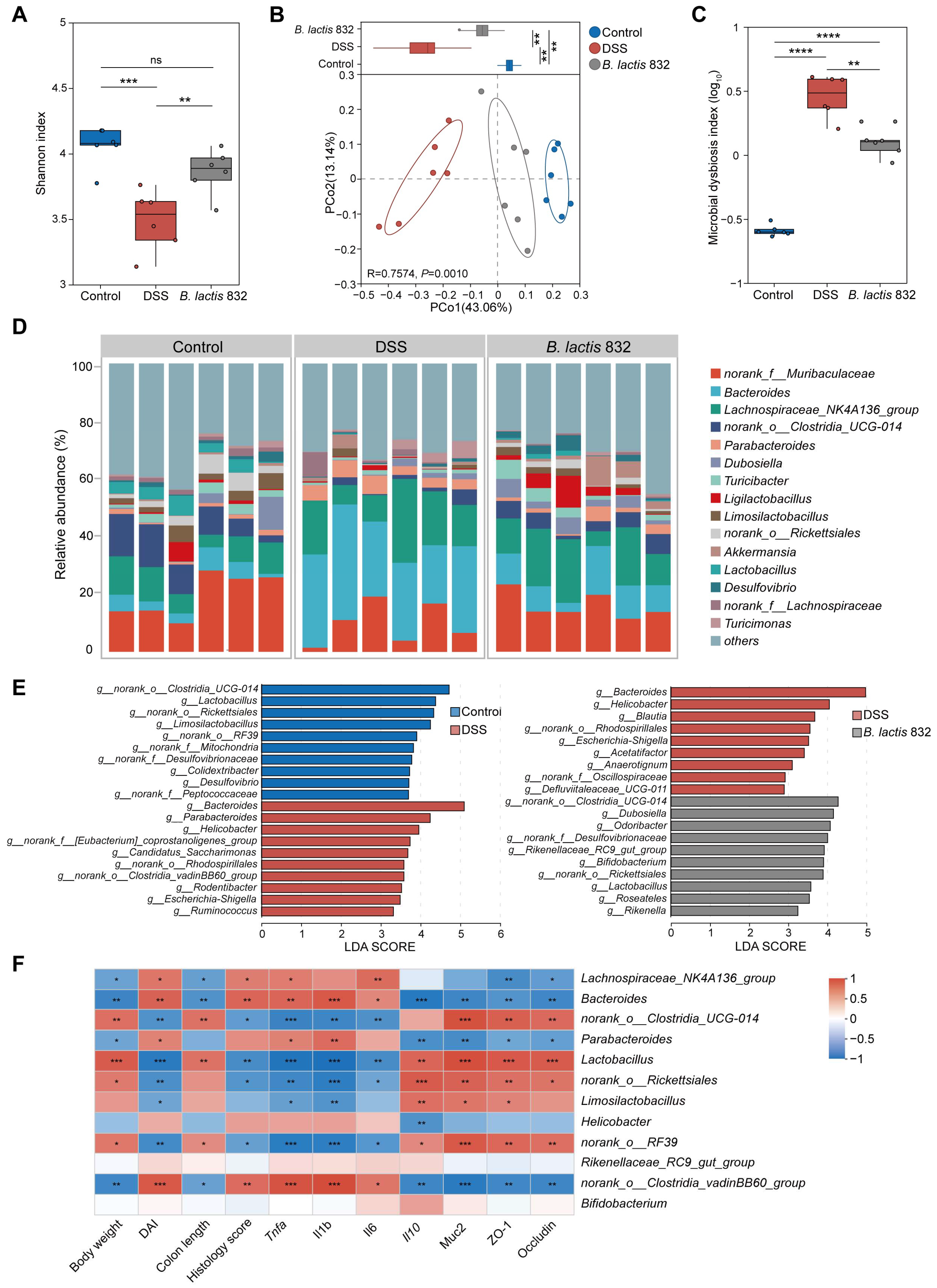
Since gut dysbiosis is a pathological determinant of IBD, we investigated the regulatory effects of B. lactis 832 on the composition of gut microbiota using the 16S rRNA sequencing assay of fecal bacteria after DSS induced colitis. Amplicon sequencing of the 16S rRNA V3–V4 gene region yielded a total 1,328,283 high-quality reads (median 71,548.5 [IQR 5860.25] reads per sample) (Table S3). Rarefaction curves confirmed that sequencing depth was sufficient for reliable analysis (Figure S3). We firstly observed that the alpha diversity based on species richness and evenness information in OTU, shown by Shannon and Simpson index, showed a significant difference in the DSS group relative to the controls (p < 0.01), while B. lactis 832 intervention induced a larger Shannon index and a smaller Simpson index in colitis mice consistent with the control group (p < 0.01, Figure 5A and Figure S4). Principal coordinate analysis (PCoA) and non-metric multidimensional scaling (NMDS) based on Bray–Curtis distances revealed distinct clustering of microbial communities along the PC2 axis among the control, DSS, and B. lactis 832 groups, with the B. lactis 832 group positioned closer to the control group (p = 0.001, Figure 5B and Figure S5). The microbial dysbiosis index (MDI) was elevated in both DSS and B. lactis 832 groups relative to controls (p < 0.0001) but was lower in the B. lactis 832 group than in the DSS group (p < 0.01, Figure 5C). Subsequently, we conducted the linear discriminant analysis effect size (LEfSe) to discover the signature microbiota genera by assessing compositional differences between DSS and control groups, as well as between the DSS and B. lactis 832 groups. At the genus level, the control group significantly enriched norank_o__Clostridia_UCG-014, Lactobacillus, and norank_o__Rickettsiales, while Bacteroides, Parabacteroides, and Helicobacter were uniquely enriched in the DSS group (p < 0.05). However, B. lactis 832 supplementation dramatically enriched norank_o__Clostridia_UCG-014, Dubosiella, Lactobacillus, and Bifidobacterium compared to the DSS group (p < 0.05, Figure 5D,E and Table S4). Furthermore, we found that the signature bacterial genera of the DSS group had a positive correlation with the inflammatory indicators, in which Bacteroides, Parabacteroides, and Helicobacter had a positive correlation with Tnfa, Il1b, and Il6 expression levels, DAI, and histology score, while these genera had a negative correlation with Il10, Muc2, ZO-1, and occludin expression levels, body weight and colon length (Figure 5F). Conversely, the signature bacterial genera of the control and B. lactis 832 groups, norank_o__Clostridia_UCG-014, Lactobacillus, and norank_o__Rickettsiales, had a positive correlation with Il10, Muc2, ZO-1, and occludin expression levels, body weight and colon length, while these genera had a negative correlation with Tnfa, Il1b, and Il6 expression levels, DAI, and histology score (Figure 5F). Taken together, B. lactis 832 treatment improved the diversity and composition of gut microbiota in DSS-induced colitis mice.
Figure 5.
B. lactis 832 reshapes fecal microbial communities in DSS-induced colitis. (A) Shannon index reflecting alpha diversity of the gut microbiota. (B) Bray–Curtis-based principal coordinate analysis (PCoA) with PC1 scores (upper panels). (C) Microbial dysbiosis index (MDI) across Control, DSS, and B. lactis 832 groups. (D) Relative abundance of the top 15 genera in each group. (E) Differential taxa identified by LEfSe analysis between Control vs. DSS and B. lactis 832 vs. DSS groups (LDA score ≥ 2). (F) Spearman correlation heatmap showing associations between bacterial taxa and colitis-related biochemical parameters. n = 4–6. Statistical significance was determined using one-way ANOVA followed by Tukey’s multiple comparison tests (* p < 0.05; ** p < 0.01; *** p < 0.001; **** p < 0.0001).
3.7. Prophylactic B. lactis 832 Elevates Intestinal PE and PC Levels in DSS-Induced Colitis Mice
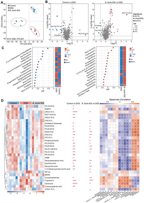
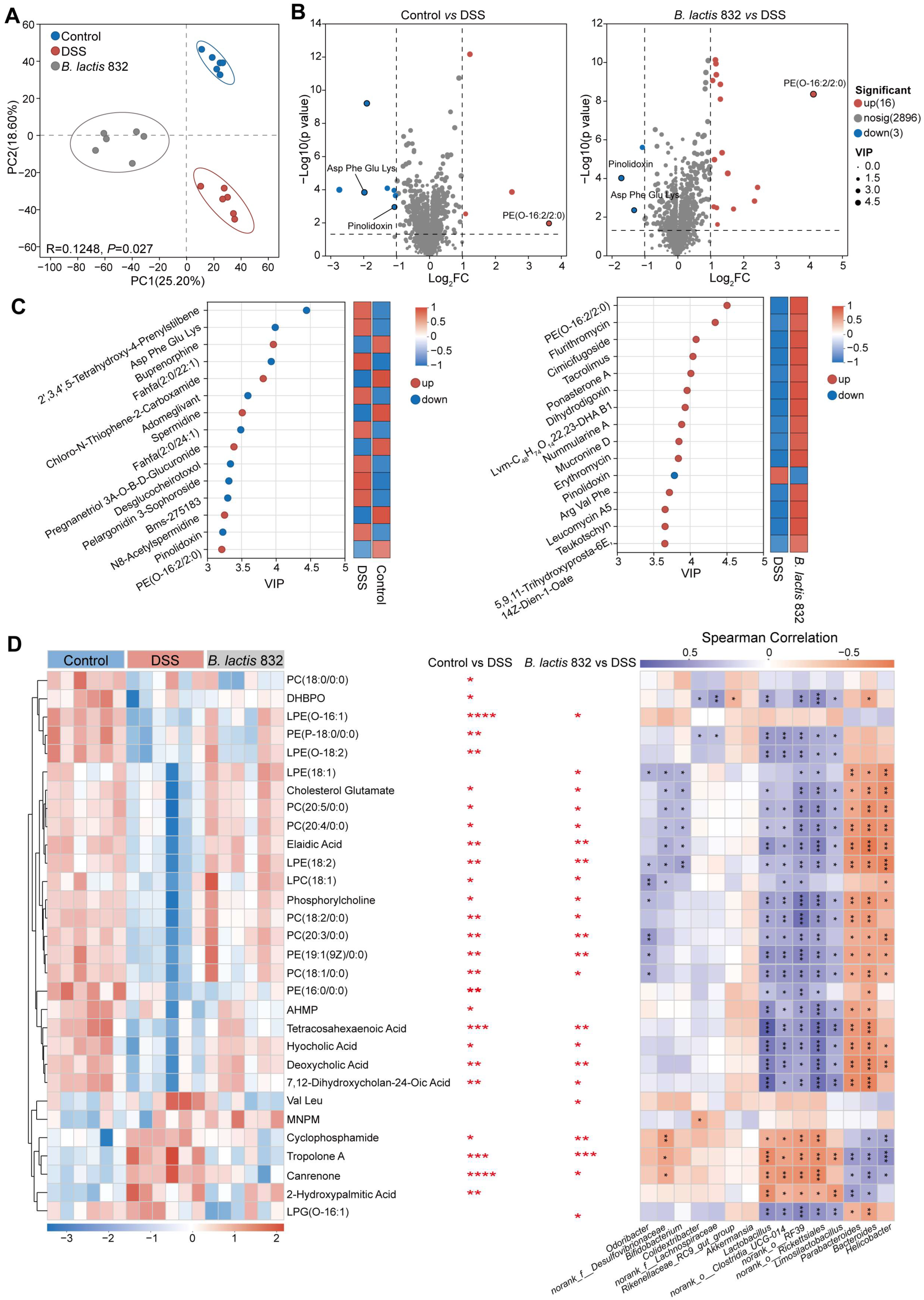
The biological effects of the gut microbiota after B. lactis 832 supplementation were further investigated through metabolomic analysis conducted on fecal samples after DSS induced colitis. The robust separation based on PCoA and PLS-DA analysis demonstrated the significant discrimination in metabolic profiles between the control, DSS and B. lactis 832 groups (p < 0.05, Figure 6A and Figure S6). Across the three groups, 1008 metabolites showed differential abundance. Using thresholds of |log2FC| ≥ 1 and p ≤ 0.05, 4 metabolites were upregulated and 7 were downregulated in the control group relative to the DSS group. In comparison, 16 metabolites were increased and 3 were decreased in the B. lactis 832 group versus the DSS group. Meanwhile, compared with the DSS group, PE (O-16:2/2:0) (phosphatidylethanolamine) was significantly up-regulated both in the control and B. lactis 832 groups, while Pinolidoxin and Asp Phe Glu Lys were significantly downregulated (Figure 6B). In addition, VIP calculated using OPLS-DA combined the differential accumulated metabolites analysis to identify the top 15 significant discriminant metabolites between the control and DSS groups, as well as between the DSS and B. lactis 832 groups. Compared with the DSS group, the VIP scores of up-regulated PE (O-16:2/2:0) were 3.2 and 4.5 in the control and B. lactis 832 groups, while the VIP scores of down-regulated Pinolidoxin were 3.2 and 3.8, respectively (Figure 6C). In addition, a heatmap was created to list and cluster the relative quantities of the top 30 fecal metabolites in the three groups. It was found that the relative abundance of phospholipid metabolic molecules, such as PC (18:0/0:0) (phosphatidylcholine), PE (P-18:0/0:0), and PE (16:0/0:0), were higher in the control group than in the DSS group (p < 0.05). Notably, B. lactis 832 supplementation, relative to DSS treatment, also significantly increased the relative abundance of molecules involved in phospholipid metabolism, such as (LPE (O-16:1) (lysophosphatidylethanolamine), LPC (18:1) (lysophosphatidylcholine), phosphorylcholine, PE (19:1(9Z)/0:0) and PC (18:1/0:0), as well as molecules related to secondary bile acid biosynthesis, including hyocholic acid and deoxycholic acid (p < 0.05, Figure 6D, Tables S5 and S6). To investigate the microbial factors responsible for these changes, a Spearman correlation analysis was conducted between the enriched metabolites and the bacterial taxa. Notably, B. lactis 832-enriched phospholipid metabolites, including PE and PC, were positively associated with beneficial bacteria such as Bifidobacterium, Limosilactobacillus, norank_o__Rickettsiales, norank_o__RF39, norank_o__Clostridia_UCG-014, and Lactobacillus (Figure 6D). Taken together, B. lactis 832 reshapes phospholipid metabolism in colitis mice, particularly increasing the intestinal PE and PC levels.
Figure 6.
B. lactis 832 supplementation altered the composition of fecal metabolites of colitis mice. (A) Principal component analysis (PCA) showing group-wise differences in fecal metabolite profiles among Control, DSS, and B. lactis 832 groups. (B) Volcano plots displaying metabolites with significant changes (|log2FC| ≥ 1, p ≤ 0.05) in Control vs DSS and B. lactis 832 vs DSS groups. (C) Variable importance in projection (VIP) scores derived from the OPLS-DA model indicating the contribution of key metabolites to group separation. (D) Hierarchical clustering heatmap illustrating metabolite abundance patterns across groups (left), alongside Spearman correlation analysis between metabolites and gut microbiota (right). Color gradients represent relative metabolite levels, while correlation strength is reflected by color intensity (purple, positive; orange, negative). The asterisks (*, **, ***, ****) indicate the significance of metabolite abundance differences in the Control vs DSS and B. lactis 832 vs DSS groups. Long metabolite names are abbreviated in this figure for readability (DHBPO: (3R,6′Z)-3,4-Dihydro-8-hydroxy-3-(6-pentadecenyl)-1H-2-benzopyran-1-one; AHMP: (3β,5β,6α)-17-(acetyloxy)-3-hydroxy-6-methylpregnan-20-one; MNPM: (4-Methyl-3-Nitrophenyl)-(4-Methylpiperidin-1-Yl)Methanone). n = 6. Statistical significance was assessed using an unpaired two-tailed Student’s t test (* p < 0.05; ** p < 0.01; *** p < 0.001; ****p < 0.0001).
3.8. Exogenous PE and PC Supplementation Alleviates DSS-Induced Colitis in Mice
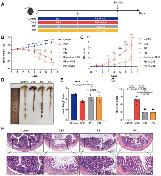
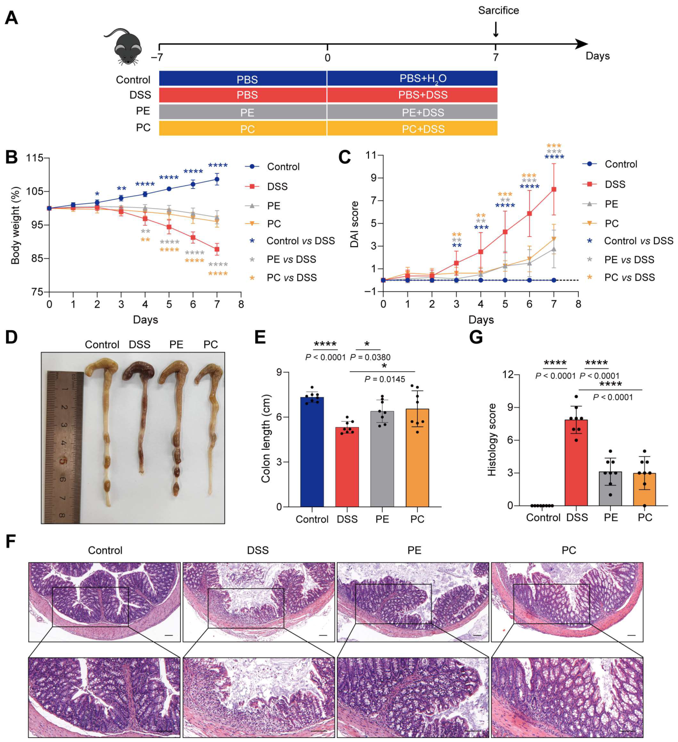
To further investigate whether the supplementation of B. lactis 832 improves colitis by promoting intestinal phospholipid metabolism, we directly supplemented PE or PC in the diet of mice to explore their effects on colitis (Figure 7A). Mice received daily oral gavage with PC or PE (50 mg/kg) or PBS for two weeks. Colitis was induced by administering 3% DSS in drinking water during the second week, while control mice received normal water. DSS treatment led to pronounced body weight loss and elevated DAI scores compared with the controls (p < 0.05). In contrast, mice supplemented with PC or PE exhibited attenuated weight loss and reduced DAI scores (p < 0.01, Figure 7B,C). Colon shortening was evident in DSS-treated mice (p < 0.0001), whereas PC or PE supplementation partially restored colon length (p < 0.05, Figure 7D,E). Histological analysis showed severe mucosal edema, epithelial disruption, crypt damage, and extensive inflammatory cell infiltration in the DSS group, resulting in higher histological scores (p < 0.0001). These pathological alterations were alleviated in mice receiving PC or PE, accompanied by lower histological scores (p < 0.0001, Figure 7F,G). Collectively, exogenous supplementation with PE or PC markedly relieved DSS-induced intestinal inflammation and pathological damage in colitis mice.
Figure 7.
PE or PC supplementation alleviates DSS-induced colitis in mice. (A) Schematic of PE or PC supplementation in DSS-induced mouse model. PC: Phosphatidylcholine; PE: Phosphatidylethanolamine. (B) Changes in body weight. (C) DAI score recorded from day 0 to day 7. (D) Gross morphology of the colon. (E) Quantification of colon length (cm). (F) Histological appearance of colon sections following H&E staining Scale bar: 100 μm. (G) Scoring of histological alterations in colonic tissues. n = 8. Statistical significance was determined using one-way ANOVA followed by Tukey’s multiple comparison tests (* p < 0.05; ** p < 0.01; *** p < 0.001; **** p < 0.0001).
4. Discussion
Within microbiota-oriented interventions, probiotics are increasingly recognized as a promising option for the prevention and treatment of IBD [39]. Bifidobacterium have been well documented to alleviate colitis by reshaping gut microbiota, reinforcing intestinal barrier integrity and modulating host immune homeostasis [13,14,15,20,40]. Similarly, B. lactis 832 exerted consistent beneficial effects in this study, including mitigating intestinal inflammation and improving mucosal barrier function, and shared common microbial regulation characteristics with Bifidobacterium longum subsp. infantis FJSYZ1M3, B. breve and B. lactis BL-99, such as reducing the abundance of Turicibacter and Bacteroides in colitis mice [14,32,41]. Nevertheless, distinct microbial regulation patterns existed among different B. lactis strains. B. lactis 832 specifically enriched norank_o__Clostridia_UCG-014 and Dubosiella, whereas B. lactis BL-99 increased the abundance of Adlercreutzia, and B. lactis XLTG11 markedly elevated Muribaculaceae levels in colitis mice [19,41]. Notably, most Bifidobacterium strains exert anti-colitis effects mainly by regulating short-chain fatty acid (SCFA) metabolism, whereas the core protective mechanism of B. lactis 832 is uniquely dependent on the remodeling of intestinal phospholipid metabolism, accompanied by significant increases in intestinal PE and PC contents [14,41,42]. These findings highlight the unique therapeutic potential of B. lactis 832 and its downstream phospholipid-dependent pathway, providing an innovative metabolic perspective for understanding probiotic function. Consistently, dietary supplementation with PE or PC recapitulated the protective effects, supporting a functional link between microbial modulation and host phospholipid metabolism [43,44]. Nevertheless, further clinical validation is still essential to verify its translational application in human IBD.
Mechanistically, the intestinal microbiome plays a fundamental role in the maintenance of gut homeostasis by regulating immune balance [45]. In our study, IBD-associated and pro-inflammatory taxa such as Bacteroides, Parabacteroides, and Helicobacter were enriched in the DSS group and positively correlated with the pathology of colitis and pro-inflammatory factors [46,47]. Notably, functional divergence exists among different species within the Bacteroides genus; not all members are pro-inflammatory, and certain strains such as Bacteroides vulgatus have been reported to alleviate colitis and maintain intestinal immune homeostasis [48]. Enterotoxigenic Bacteroides fragilis secretes enterotoxin (BFT, Bacteroides fragilis toxin) to elicit intestinal inflammation via E-cadherin cleavage/NF-κB signaling [49]. In addition, pathogenic Helicobacter spp., such as Helicobacter muridarum and Helicobacter hepaticus, have been proven to aggravate intestinal inflammation and elevate disease severity in multiple colitis models [6,50]. Moreover, IgA-coated Helicobacter and B. fragilis can penetrate into the mucous layer in gnotobiotic mice and exacerbate DSS-induced colitis [51]. Conversely, B. lactis 832 supplementation dramatically decreased the pro-inflammatory bacteria species, while effectively enriching beneficial commensals including norank_o__Clostridia_UCG-014, Dubosiella, Lactobacillus, and Bifidobacterium. B. lactis 832 may competitively exclude pathobionts by producing short-chain fatty acids (SCFAs) and lowering intestinal luminal pH, as well as support the metabolic cross-feeding of beneficial commensals, thereby stabilizing gut microbiota homeostasis under colitis conditions [42,52]. Notably, these enriched beneficial commensals negatively correlated with the pathology of colitis and pro-inflammatory factors in our study. Previous studies have shown that Clostridium sporogenes-derived metabolites, including IPA, SCFAs and BCFAs, increased the numbers of colonic tuft cells, the production of IL-22, and the polarization of Foxp3+ Tregs to alleviate colitis [53]. In addition, Dubosiella newyorkensis has a probiotic immunomodulatory effect on DSS-induced colitis, rebalancing Treg/Th17 responses and ameliorating mucosal barrier injury by producing SCFAs [54].
Emerging evidence suggests that gut microbiota can influence host physiology through the production and modulation of bioactive metabolites, which act as key mediators of microbe–host interactions [55,56]. Notably, phospholipid metabolism has been identified as a core pathway regulated by both gut commensals and pathogenic microbes, mainly through modulating phospholipase-mediated PC hydrolysis and plsC-dependent phospholipid synthesis [57,58,59]. On the one hand, B. lactis 832 may effectively inhibit excessive PC hydrolysis in the gut by reducing the relative abundance of Helicobacter. These pathobionts exhibit high ectophospholipase activity, including secreted phospholipase A2 (sPLA2), which efficiently degrades mucosal PC and disrupts the intestinal barrier [60,61]. On the other hand, Bifidobacterium and Lactobacillus species enriched by B. lactis 832 exhibit relatively low phospholipase activity, which may reduce the excessive hydrolysis of intestinal PC and PE [61,62,63]. Meanwhile, these beneficial commensals may possess a strong capacity to generate key precursors such as fatty acids, lysophospholipids, choline, and ethanolamine, thereby supporting host PC and PE synthesis and maintaining intestinal phospholipid homeostasis [64,65,66]. Correlation analysis further indicated that Lactobacillus was positively correlated with PC levels and could suppress plc-mediated PC hydrolysis, thereby promoting PC accumulation in the intestinal mucosa [59]. Notably, PC and PE are the predominant phospholipids in the intestine and are essential for mucosal integrity. Intestinal epithelial cells secrete mucus that forms a protective surface layer supported by a PC–PE bilayer structure [28,65]. Their depletion disrupts mucus integrity, exacerbates inflammation, and promotes disease progression in IBD [29]. Mechanistically, PE maintains lipid homeostasis and inhibits ferroptosis in a GPX4-independent manner, thereby protecting against colitis, while PC supports mucosal integrity when properly balanced [67]. Consistent with these roles, dietary PC alleviates DSS-induced colitis by improving barrier function, reducing inflammatory cytokines, and modulating gut microbiota composition [43,44]. This may also involve alterations in host metabolic pathways, including tryptophan and arginine metabolism [43]. Taken together, these findings suggest that PC and PE play essential and non-redundant roles in protecting against colitis. Restoration of phospholipid metabolism may represent a key mechanism underlying the anti-colitis effects of probiotics. The enrichment of beneficial bacteria, including members of Bifidobacterium and Lactobacillus, may contribute to this metabolic reprogramming and intestinal homeostasis. Collectively, these findings support a model in which B. lactis 832 alleviates colitis through a microbiota–phospholipid metabolism axis, thereby improving intestinal barrier function and modulating immune responses.
5. Conclusions
In conclusion, B. lactis 832 could effectively alleviate DSS-induced colitis symptoms in a murine model. The main mechanisms of B. lactis 832 that significantly alleviated DSS-induced colitis include inhibiting pro-inflammatory factors, maintaining intestinal barrier integrity, and improving the composition of gut microbiota and metabolomics. Multi-omics analysis further revealed that prophylactic B. lactis 832 enriched beneficial commensals norank_o__Clostridia_UCG-014, Dubosiella, Lactobacillus, and Bifidobacterium and enhanced intestinal levels of phospholipid metabolites, particularly phosphatidylethanolamine (PE) and phosphatidylcholine (PC). Furthermore, exogenous PE and PC administration recapitulated the protective effects of B. lactis 832 against colitis, supporting the critical role of phospholipid metabolism in mediating its probiotic function. A limitation of this study is the lack of direct microbial functional validation, which will be addressed in future metagenomic investigations to identify key strains and enzymes involved in phospholipid regulation. Nonetheless, our findings demonstrate that B. lactis 832 protects against colitis through a microbiota–phospholipid metabolism axis, supporting its potential as an innovative probiotic for IBD management. Future studies should include targeted gene knockout, mechanistic validation, and clinical trials to confirm its efficacy and safety. Collectively, this study provides preclinical evidence and a theoretical basis for developing microbiota-based strategies for IBD prevention and treatment.
Supplementary Materials
The following supporting information can be downloaded at: https://www.mdpi.com/article/10.3390/microorganisms14051090/s1, Figure S1: The phylogenetic tree was constructed by the DNA sequence of Bifidobacterium animalis 16S using MEGA 7 with the method of bootstrap consensus tree. The Sequence1, B. lactis 832, and other aligned species are marked with red and blue circles; Figure S2: Effect of the B. lactis 832 on mRNA expression levels of Tnfa, Il1b, Il6, and Il10 in LPS-stimulated Caco-2 cells. n = 4. Statistical differences were evaluated by one-way ANOVA followed by Tukey’s multiple comparison tests (* p < 0.05; ** p < 0.01; *** p < 0.001; **** p < 0.0001); Figure S3: Safety evaluation of B. lactis 832 supplementation in mice. (A) Schematic of B. lactis 832 supplementation in mouse model. (B) Changes in body weight. (C) DAI score recorded from day 0 to day 7. (D) Gross morphology of the colon. (E) Quantification of colon length (cm). (F) Histological appearance of colon sections following H&E staining. Scale bar: 100 μm. (G) Scoring of histological alterations in colonic tissues. (H) Representative images of major organs (heart, liver, spleen, lung, kidney) stained with H&E staining. Scale bar: 100 μm. n = 8. Statistical significance was determined using one-way ANOVA followed by Tukey’s multiple comparison tests (ns p > 0.05); Figure S4: Rarefaction curves of observed species for all samples; Figure S5: Alpha diversity of the gut microbiota analysis by the Simpson index (n = 6). Figure S6: Non-metric Multidimensional Scaling (NMDS) and scores of the PC1 axis (upper panels) of Bray–Curtis dissimilarity based on OTUs; Figure S7: Partial Least Squares Discriminant Analysis (PLS-DA) illustrating changes in the composition of the fecal metabolites among Control, DSS and B. lactis 832 groups; Table S1: Identification of Bifidobacterium animalis subsp. lactis strain 832 based on 16S rRNA gene sequence similarity. Table S2: Determination of key in vitro probiotic traits of B. lactis 832; Table S3: Statistics of Sample 16S rRNA Sequencing Information; Table S4: Gut microbiota composition among Control, DSS and B. lactis 832 groups at the genus level; Table S5: Differentially expressed metabolites between Control and DSS groups; Table S6: Differentially expressed metabolites between B. lactis 832 and DSS groups.
Author Contributions
Conceptualization, L.W., B.L. and X.C.; methodology, Q.W.; software, Q.W.; validation, Q.W., X.G., D.L., X.W., X.L., X.Z., Y.L., S.H. and L.F.; formal analysis, Q.W. and X.C.; investigation, Q.W.; resources, X.C.; data curation, Q.W.; writing—original draft preparation, L.W., B.L. and X.C.; writing—review and editing, all authors; visualization, Q.W.; supervision, Q.W. and X.C.; project administration, X.C.; funding acquisition, L.W. and L.F. All authors have read and agreed to the published version of the manuscript.
Funding
This work was funded by the Yunnan Provincial Science and Technology Project at Southwest United Graduate School, 202502AO070002, (to L.W.), National Natural Science Foundation of China Grant, 32130003, 31820103002, and 32370194 (to L.W.), 32070133, 32470111 (to L.F.), Shenzhen Science and Technology Program, JCYJ20210324135007019 (to L.F.), and Key Laboratory Major Project (Tianjin) (25ZXZSSS00710 to B.L.).
Institutional Review Board Statement
All animal experiments were performed in accordance with protocols approved by the Institutional Animal Care Committee of Nankai University (Tianjin, China; protocol code 2026-SYDWLL-000159).
Informed Consent Statement
Not applicable.
Data Availability Statement
16S rRNA sequencing data and analysis codes are available in the NCBI-SRA database (BioProject: PRJNA1227203). Metabolomics data have been deposited in the NGDC-OMIX database (BioProject: PRJCA050718).
Conflicts of Interest
The authors declare no conflicts of interest.
References
- Sugihara, K.; Kamada, N. Metabolic network of the gut microbiota in inflammatory bowel disease. Inflamm. Regen. 2024, 44, 11. [Google Scholar] [CrossRef] [PubMed]
- Wu, Y.; Jha, R.; Li, A.; Liu, H.; Zhang, Z.; Zhang, C.; Zhai, Q.; Zhang, J. Probiotics (Lactobacillus plantarum HNU082) Supplementation Relieves Ulcerative Colitis by Affecting Intestinal Barrier Functions, Immunity-Related Gene Expression, Gut Microbiota, and Metabolic Pathways in Mice. Microbiol. Spectr. 2022, 10, e0165122. [Google Scholar] [CrossRef]
- Fan, L.; Qi, Y.; Qu, S.; Chen, X.; Li, A.; Hendi, M.; Xu, C.; Wang, L.; Hou, T.; Si, J.; et al. B. adolescentis ameliorates chronic colitis by regulating Treg/Th2 response and gut microbiota remodeling. Gut Microbes 2021, 13, 1826746. [Google Scholar] [CrossRef]
- Caruso, R.; Lo, B.C.; Nunez, G. Host-microbiota interactions in inflammatory bowel disease. Nat. Rev. Immunol. 2020, 20, 411–426. [Google Scholar] [CrossRef]
- Axelrad, J.E.; Cadwell, K.H.; Colombel, J.F.; Shah, S.C. Systematic review: Gastrointestinal infection and incident inflammatory bowel disease. Aliment. Pharmacol. Ther. 2020, 51, 1222–1232. [Google Scholar] [CrossRef]
- Ni, J.; Wu, G.D.; Albenberg, L.; Tomov, V.T. Gut microbiota and IBD: Causation or correlation? Nat. Rev. Gastroenterol. Hepatol. 2017, 14, 573–584. [Google Scholar] [CrossRef] [PubMed]
- Haneishi, Y.; Furuya, Y.; Hasegawa, M.; Picarelli, A.; Rossi, M.; Miyamoto, J. Inflammatory Bowel Diseases and Gut Microbiota. Int. J. Mol. Sci. 2023, 24, 3817. [Google Scholar] [CrossRef] [PubMed]
- Shan, Y.; Lee, M.; Chang, E.B. The Gut Microbiome and Inflammatory Bowel Diseases. Annu. Rev. Med. 2022, 73, 455–468. [Google Scholar] [CrossRef]
- Viladomiu, M.; Metz, M.L.; Lima, S.F.; Jin, W.B.; Chou, L.; Guo, C.J.; Diehl, G.E.; Simpson, K.W.; Scherl, E.J.; Longman, R.S. Adherent-invasive E. coli metabolism of propanediol in Crohn’s disease regulates phagocytes to drive intestinal inflammation. Cell Host Microbe 2021, 29, 607–619.e608. [Google Scholar] [CrossRef]
- Abraham, C.; Cho, J.H. Inflammatory bowel disease. N. Engl. J. Med. 2009, 361, 2066–2078. [Google Scholar] [CrossRef]
- Li, Q.; Zhou, S.; Wang, Y.; Cong, J. Changes of intestinal microbiota and microbiota-based treatments in IBD. Arch. Microbiol. 2022, 204, 442. [Google Scholar] [CrossRef]
- Huang, Z.; Liu, B.; Xiao, L.; Liao, M.; Huang, L.; Zhao, X.; Ma, K.; Wang, R.; Ji, F.; Li, W.; et al. Effects of breast-fed infants-derived Limosilactobacillus reuteri and Bifidobacterium breve ameliorate DSS-induced colitis in mice. iScience 2024, 27, 110902. [Google Scholar] [CrossRef]
- Han, M.; Liang, J.; Hou, M.; Liu, Y.; Li, H.; Gao, Z. Bifidobacterium bifidum Ameliorates DSS-Induced Colitis in Mice by Regulating Microbial Metabolome and Targeting Gut Microbiota. Agric. Food Chem. 2024, 72, 13593–13609. [Google Scholar] [CrossRef]
- Niu, M.-M.; Guo, H.-X.; Cai, J.-W.; Meng, X.-C. Bifidobacterium breve Alleviates DSS-Induced Colitis in Mice by Maintaining the Mucosal and Epithelial Barriers and Modulating Gut Microbes. Nutrients 2022, 14, 3671. [Google Scholar] [CrossRef]
- Cui, Q.-y.; Tian, X.-y.; Liang, X.; Zhang, Z.; Wang, R.; Zhou, Y.; Yi, H.-x.; Gong, P.-m.; Lin, K.; Liu, T.-j.; et al. Bifidobacterium bifidum relieved DSS-induced colitis in mice potentially by activating the aryl hydrocarbon receptor. Food Funct. 2022, 13, 5115–5123. [Google Scholar] [CrossRef] [PubMed]
- Lv, H.; Tao, F.; Peng, L.; Chen, S.; Ren, Z.; Chen, J.; Yu, B.; Wei, H.; Wan, C. In Vitro Probiotic Properties of Bifidobacterium animalis subsp. lactis SF and Its Alleviating Effect on Non-Alcoholic Fatty Liver Disease. Nutrients 2023, 15, 1355. [Google Scholar] [CrossRef]
- Pápai, G.; Torres-Maravilla, E.; Chain, F.; Varga-Visi, É.; Antal, O.; Naár, Z.; Bermúdez-Humarán, L.G.; Langella, P.; Martín, R. The Administration Matrix Modifies the Beneficial Properties of a Probiotic Mix of Bifidobacterium animalis subsp. lactis BB-12 and Lactobacillus acidophilus LA-5. Probiotics Antimicrob. Proteins 2020, 13, 484–494. [Google Scholar] [CrossRef] [PubMed]
- Tsai, H.Y.; Wang, Y.C.; Liao, C.A.; Su, C.Y.; Huang, C.H.; Chiu, M.H.; Yeh, Y.T. Safety and the probiotic potential of Bifidobacterium animalis CP-9. J. Food Sci. 2022, 87, 2211–2228. [Google Scholar] [CrossRef] [PubMed]
- Wang, N.; Wang, S.; Xu, B.; Liu, F.; Huo, G.; Li, B. Alleviation Effects of Bifidobacterium animalis subsp. lactis XLTG11 on Dextran Sulfate Sodium-Induced Colitis in Mice. Microorganisms 2021, 9, 2093. [Google Scholar] [CrossRef]
- Wang, H.; Fan, C.; Zhao, Z.; Zhai, Z.; Hao, Y. Anti-inflammatory effect of Bifidobacterium animalis subsp. lactis A6 on DSS-induced colitis in mice. J. Appl. Microbiol. 2022, 133, 2063–2073. [Google Scholar] [CrossRef]
- Franzosa, E.A.; Sirota-Madi, A.; Avila-Pacheco, J.; Fornelos, N.; Haiser, H.J.; Reinker, S.; Vatanen, T.; Hall, A.B.; Mallick, H.; McIver, L.J.; et al. Gut microbiome structure and metabolic activity in inflammatory bowel disease. Nat. Microbiol. 2019, 4, 293–305. [Google Scholar] [CrossRef]
- Lavelle, A.; Sokol, H. Gut microbiota-derived metabolites as key actors in inflammatory bowel disease. Nat. Rev. Gastroenterol. Hepatol. 2020, 17, 223–237. [Google Scholar] [CrossRef]
- Ito, M.; Adachi-Akahane, S. Inter-organ Communication in the Regulation of Lipid Metabolism: Focusing on the Network Between the Liver, Intestine, and Heart. J. Pharmacol. Sci. 2013, 123, 312–317. [Google Scholar] [CrossRef]
- Morita, H.; Nakanishi, K.; Dohi, T.; Yasugi, E.; Oshima, M. Phospholipid turnover in the inflamed intestinal mucosa: Arachidonic acid-rich phosphatidyl/plasmenyl-ethanolamine in the mucosa in inflammatory bowel disease. J. Gastroenterol. 1999, 34, 46–53. [Google Scholar] [CrossRef]
- Patel, D.; Witt, S.N.; Waugh, M.G. Ethanolamine and Phosphatidylethanolamine: Partners in Health and Disease. Oxid. Med. Cell Longev. 2017, 2017, 4829180. [Google Scholar] [CrossRef] [PubMed]
- van der Veen, J.N.; Kennelly, J.P.; Wan, S.; Vance, J.E.; Vance, D.E.; Jacobs, R.L. The critical role of phosphatidylcholine and phosphatidylethanolamine metabolism in health and disease. Biochim. Biophys. Acta (BBA) Biomembr. 2017, 1859, 1558–1572. [Google Scholar] [CrossRef] [PubMed]
- Wang, B.; Tontonoz, P. Phospholipid Remodeling in Physiology and Disease. Annu. Rev. Physiol. 2019, 81, 165–188. [Google Scholar] [CrossRef]
- Gustafsson, J.K.; Johansson, M.E.V. The role of goblet cells and mucus in intestinal homeostasis. Nat. Rev. Gastroenterol. Hepatol. 2022, 19, 785–803. [Google Scholar] [CrossRef] [PubMed]
- Ehehalt, R.; Wagenblast, J.; Erben, G.; Lehmann, W.D.; Hinz, U.; Merle, U.; Stremmel, W. Phosphatidylcholine and lysophosphatidylcholine in intestinal mucus of ulcerative colitis patients. A quantitative approach by nanoElectrospray-tandem mass spectrometry. Scand. J. Gastroenterol. 2004, 39, 737–742. [Google Scholar] [CrossRef]
- Wang, N.; Wang, C.; Qi, M.; Lin, X.; Zha, A.; Tan, B.; Yin, Y.; Wang, J. Phosphatidylethanolamine Improves Postnatal Growth Retardation by Regulating Mucus Secretion of Intestinal Goblet Cells in Piglets. Animals 2024, 14, 1193. [Google Scholar] [CrossRef]
- You, Y.; Kim, T.-R.; Sohn, M.; Yoo, D.; Park, J. Protective Effects of a Bifidobacterium-Based Probiotic Mixture on Gut Inflammation and Barrier Function. Microbiol. Res. 2025, 16, 168. [Google Scholar] [CrossRef]
- Li, M.; Ding, J.; Stanton, C.; Ross, R.P.; Zhao, J.; Yang, B.; Chen, W. Bifidobacterium longum subsp. infantis FJSYZ1M3 ameliorates DSS-induced colitis by maintaining the intestinal barrier, regulating inflammatory cytokines, and modifying gut microbiota. Food Funct. 2023, 14, 354–368. [Google Scholar] [CrossRef]
- Wen, W.; Xu, Y.; Qian, W.; Huang, L.; Gong, J.; Li, Y.; Zhu, W.; Guo, Z. PUFAs add fuel to Crohn’s disease-associated AIEC-induced enteritis by exacerbating intestinal epithelial lipid peroxidation. Gut Microbes 2023, 15, 2265578. [Google Scholar] [CrossRef]
- Liu, Y.; Zheng, H.; Wang, J.; Xin, J.; Wu, R.; Zhang, J.; Zhong, Z.; Liu, H.; Huang, Y.; Fu, H.; et al. Whole genome analysis and in vivo safety assessment of probiotic candidate Lactobacillus acidophilus L177. BMC Microbiol. 2025, 25, 398. [Google Scholar] [CrossRef]
- Vlantis, K.; Polykratis, A.; Welz, P.-S.; van Loo, G.; Pasparakis, M.; Wullaert, A. TLR-independent anti-inflammatory function of intestinal epithelial TRAF6 signalling prevents DSS-induced colitis in mice. Gut 2016, 65, 935–943. [Google Scholar] [CrossRef] [PubMed]
- Carvalho, F.A.; Barnich, N.; Sivignon, A.; Darcha, C.; Chan, C.H.; Stanners, C.P.; Darfeuille-Michaud, A. Crohn’s disease adherent-invasive Escherichia coli colonize and induce strong gut inflammation in transgenic mice expressing human CEACAM. J. Exp. Med. 2009, 206, 2179–2189. [Google Scholar] [CrossRef]
- Edgar, R.C. UPARSE: Highly accurate OTU sequences from microbial amplicon reads. Nat. Methods 2013, 10, 996–998. [Google Scholar] [CrossRef]
- Chen, S.; Zhou, Y.; Chen, Y.; Gu, J. fastp: An ultra-fast all-in-one FASTQ preprocessor. Bioinformatics 2018, 34, i884–i890. [Google Scholar] [CrossRef]
- Wang, X.; Zhang, P.; Zhang, X. Probiotics Regulate Gut Microbiota: An Effective Method to Improve Immunity. Molecules 2021, 26, 6076. [Google Scholar] [CrossRef] [PubMed]
- Bocchio, F.; Mancabelli, L.; Milani, C.; Lugli, G.A.; Tarracchini, C.; Longhi, G.; De Conto, F.; Turroni, F.; Ventura, M. Compendium of Bifidobacterium-based probiotics: Characteristics and therapeutic impact on human diseases. Microbiome Res. Rep. 2025, 4, 2. [Google Scholar] [CrossRef]
- Nan, X.; Zhao, W.; Liu, W.-H.; Li, Y.; Li, N.; Hong, Y.; Cui, J.; Shang, X.; Feng, H.; Hung, W.-L.; et al. Bifidobacterium animalis subsp. lactis BL-99 ameliorates colitis-related lung injury in mice by modulating short-chain fatty acid production and inflammatory monocytes/macrophages. Food Funct. 2023, 14, 1099–1112. [Google Scholar] [CrossRef]
- Fukuda, S.; Toh, H.; Hase, K.; Oshima, K.; Nakanishi, Y.; Yoshimura, K.; Tobe, T.; Clarke, J.M.; Topping, D.L.; Suzuki, T.; et al. Bifidobacteria can protect from enteropathogenic infection through production of acetate. Nature 2011, 469, 543–547. [Google Scholar] [CrossRef]
- Li, Q.; Chen, G.; Zhu, D.; Zhang, W.; Qi, S.; Xue, X.; Wang, K.; Wu, L. Effects of dietary phosphatidylcholine and sphingomyelin on DSS-induced colitis by regulating metabolism and gut microbiota in mice. J. Nutr. Biochem. 2022, 105, 109004. [Google Scholar] [CrossRef] [PubMed]
- Wen, Y.; Tan, L.; Chen, S.; Wu, N.; Yao, Y.; Xu, L.; Xu, M.; Zhao, Y.; Tu, Y. Egg yolk phosphatidylcholine alleviates DSS-induced colitis in BALB/c mice. Food Funct. 2023, 14, 9309–9323. [Google Scholar] [CrossRef]
- Chen, Y.; Cui, W.; Li, X.; Yang, H. Interaction Between Commensal Bacteria, Immune Response and the Intestinal Barrier in Inflammatory Bowel Disease. Front. Immunol. 2021, 12, 761981. [Google Scholar] [CrossRef]
- Sechi, L.A.; Vrakas, S.; Mountzouris, K.C.; Michalopoulos, G.; Karamanolis, G.; Papatheodoridis, G.; Tzathas, C.; Gazouli, M. Intestinal Bacteria Composition and Translocation of Bacteria in Inflammatory Bowel Disease. PLoS ONE 2017, 12, e0170034. [Google Scholar] [CrossRef]
- Lopetuso, L.R.; Petito, V.; Graziani, C.; Schiavoni, E.; Paroni Sterbini, F.; Poscia, A.; Gaetani, E.; Franceschi, F.; Cammarota, G.; Sanguinetti, M.; et al. Gut Microbiota in Health, Diverticular Disease, Irritable Bowel Syndrome, and Inflammatory Bowel Diseases: Time for Microbial Marker of Gastrointestinal Disorders. Dig. Dis. 2018, 36, 56–65. [Google Scholar] [CrossRef]
- Liu, L.; Xu, M.; Lan, R.; Hu, D.; Li, X.; Qiao, L.; Zhang, S.; Lin, X.; Yang, J.; Ren, Z.; et al. Bacteroides vulgatus attenuates experimental mice colitis through modulating gut microbiota and immune responses. Front. Immunol. 2022, 13, 1036196. [Google Scholar] [CrossRef] [PubMed]
- Hwang, S.; Jo, M.; Hong, J.E.; Kim, W.S.; Kang, D.H.; Yoo, S.H.; Kang, K.; Rhee, K.J. Caffeic Acid Phenethyl Ester Administration Reduces Enterotoxigenic Bacteroides fragilis-Induced Colitis and Tumorigenesis. Toxins 2024, 16, 403. [Google Scholar] [CrossRef] [PubMed]
- Monceaux, C.P.; Testerman, T.L.; Boktor, M.; Jordan, P.; Adegboyega, P.; McGee, D.J.; Jennings, M.H.; Parker, C.P.; Gupta, S.; Yi, P.; et al. Helicobacter infection decreases basal colon inflammation, but increases disease activity in experimental IBD. Open J. Gastroenterol. 2013, 3, 177–189. [Google Scholar] [CrossRef][Green Version]
- Kayama, H.; Okumura, R.; Takeda, K. Interaction Between the Microbiota, Epithelia, and Immune Cells in the Intestine. Annu. Rev. Immunol. 2020, 38, 23–48. [Google Scholar] [CrossRef] [PubMed]
- Samara, J.; Moossavi, S.; Alshaikh, B.; Ortega, V.A.; Pettersen, V.K.; Ferdous, T.; Hoops, S.L.; Soraisham, A.; Vayalumkal, J.; Dersch-Mills, D.; et al. Supplementation with a probiotic mixture accelerates gut microbiome maturation and reduces intestinal inflammation in extremely preterm infants. Cell Host Microbe 2022, 30, 696–711.e695. [Google Scholar] [CrossRef]
- Krause, F.F.; Mangold, K.I.; Ruppert, A.-L.; Leister, H.; Hellhund-Zingel, A.; Lopez Krol, A.; Pesek, J.; Watzer, B.; Winterberg, S.; Raifer, H.; et al. Clostridium sporogenes-derived metabolites protect mice against colonic inflammation. Gut Microbes 2024, 16, 2412669. [Google Scholar] [CrossRef]
- Zhang, Y.; Tu, S.; Ji, X.; Wu, J.; Meng, J.; Gao, J.; Shao, X.; Shi, S.; Wang, G.; Qiu, J.; et al. Dubosiella newyorkensis modulates immune tolerance in colitis via the L-lysine-activated AhR-IDO1-Kyn pathway. Nat. Commun. 2024, 15, 1333. [Google Scholar] [CrossRef]
- Krautkramer, K.A.; Fan, J.; Backhed, F. Gut microbial metabolites as multi-kingdom intermediates. Nat. Rev. Microbiol. 2021, 19, 77–94. [Google Scholar] [CrossRef]
- Yang, W.; Cong, Y. Gut microbiota-derived metabolites in the regulation of host immune responses and immune-related inflammatory diseases. Cell Mol. Immunol. 2021, 18, 866–877. [Google Scholar] [CrossRef]
- Chittim, C.L.; Martínez del Campo, A.; Balskus, E.P. Gut bacterial phospholipase Ds support disease-associated metabolism by generating choline. Nat. Microbiol. 2018, 4, 155–163. [Google Scholar] [CrossRef] [PubMed]
- Jackson, D.R.; Cassilly, C.D.; Plichta, D.R.; Vlamakis, H.; Liu, H.; Melville, S.B.; Xavier, R.J.; Clardy, J. Plasmalogen Biosynthesis by Anaerobic Bacteria: Identification of a Two-Gene Operon Responsible for Plasmalogen Production in Clostridium perfringens. ACS Chem. Biol. 2020, 16, 6–13. [Google Scholar] [CrossRef] [PubMed]
- Jiang, X.W.; Zhang, L.; Liu, Z.C.; Zhou, T.; Li, W.Q.; Liu, W.D.; Zhang, L.F.; You, W.C.; Zhang, Y.; Pan, K.F. Integrative metabolomics and microbiomics analysis reveals distinctive microbiota-metabolites interactions in gastric carcinogenesis. Int. J. Cancer 2025, 156, 2389–2400. [Google Scholar] [CrossRef]
- Stremmel, W.; Staffer, S.; Stuhrmann, N.; Gan-Schreier, H.; Gauss, A.; Burger, N.; Hornuss, D. Phospholipase A2 of Microbiota as Pathogenetic Determinant to Induce Inflammatory States in Ulcerative Colitis: Therapeutic Implications of Phospholipase A2 Inhibitors. Inflamm. Intest. Dis. 2017, 2, 180–187. [Google Scholar] [CrossRef]
- Langton, S.R.; Cesareo, S.D. Helicobacter pylori associated phospholipase A2 activity: A factor in peptic ulcer production? J. Clin. Pathol. 1992, 45, 221–224. [Google Scholar] [CrossRef]
- Wang, H.; Zhang, L.; Shi, G. Secretory expression of a phospholipase A2 from Lactobacillus casei DSM20011 in Kluyveromyces lactis. J. Biosci. Bioeng. 2015, 120, 601–607. [Google Scholar] [CrossRef]
- Manasian, P.; Bustos, A.-S.; Pålsson, B.; Håkansson, A.; Peñarrieta, J.M.; Nilsson, L.; Linares-Pastén, J.A. First Evidence of Acyl-Hydrolase/Lipase Activity From Human Probiotic Bacteria: Lactobacillus rhamnosus GG and Bifidobacterium longum NCC 2705. Front. Microbiol. 2020, 11, 1534. [Google Scholar] [CrossRef]
- Kim, S.H.; Song, J.H.; Kim, J.; Kang, D.-K. Characterisation of a lysophospholipase from Lactobacillus mucosae. Biotechnol. Lett. 2020, 42, 1735–1741. [Google Scholar] [CrossRef] [PubMed]
- Boldyreva, L.V.; Morozova, M.V.; Saydakova, S.S.; Kozhevnikova, E.N. Fat of the Gut: Epithelial Phospholipids in Inflammatory Bowel Diseases. Int. J. Mol. Sci. 2021, 22, 11682. [Google Scholar] [CrossRef]
- Zhu, S.; Yu, Q.; Xue, Y.; Li, J.; Huang, Y.; Liu, W.; Wang, G.; Wang, L.; Zhai, Q.; Zhao, J.; et al. Bifidobacterium bifidum CCFM1163 alleviates cathartic colon by activating the BDNF-TrkB-PLC/IP3 pathway to reconstruct the intestinal nerve and barrier. Food Funct. 2025, 16, 2057–2072. [Google Scholar] [CrossRef] [PubMed]
- Huang, X.; Yang, X.; Zhang, M.; Li, T.; Zhu, K.; Dong, Y.; Lei, X.; Yu, Z.; Lv, C.; Huang, J. SELENOI Functions as a Key Modulator of Ferroptosis Pathway in Colitis and Colorectal Cancer. Adv. Sci. 2024, 11, e2404073. [Google Scholar] [CrossRef] [PubMed]

Disclaimer/Publisher’s Note: The statements, opinions and data contained in all publications are solely those of the individual author(s) and contributor(s) and not of MDPI and/or the editor(s). MDPI and/or the editor(s) disclaim responsibility for any injury to people or property resulting from any ideas, methods, instructions or products referred to in the content. |
© 2026 by the authors. Licensee MDPI, Basel, Switzerland. This article is an open access article distributed under the terms and conditions of the Creative Commons Attribution (CC BY) license.