Article

Analysis of Genomic and Characterization Features of Microbulbifer weihaiensis sp. nov., Isolated from Coastal Sediment

1
SDU-ANU Joint Science College, Shandong University, Weihai 264209, China
2
Marine College, Shandong University, Weihai 264209, China
*
Author to whom correspondence should be addressed.
Microorganisms 2025, 13(9), 2005; https://doi.org/10.3390/microorganisms13092005
Submission received: 23 July 2025 / Revised: 25 August 2025 / Accepted: 25 August 2025 / Published: 28 August 2025
(This article belongs to the Section Environmental Microbiology)

Abstract

A strictly aerobic, straight-rod, motile Gram-negative bacterium, SDUM041083T, was isolated from marine sediment in Xiaoshidao, Weihai, China, in the formation of yellowish-brown colonies. Its growing conditions are as follows: 20–40 °C, pH 5.5–9.5, and 0.5–11% (w/v) NaCl. Phylogenetic analysis of the 16S rRNA gene sequence showed that SDUM041083T was related to members of the genus Microbulbifer. Strain SDUM041083T showed the highest 16S rRNA gene sequence similarity (98.23%) with Microbulbifer okinawensis JCM 16147T. The primary cellular fatty acids of SDUM041083T were iso-C11:0 3-OH, iso-C11:0, and iso-C15:0. The respiratory quinone of SDUM041083T was Q-8, and the polar lipids were phosphatidylglycerol, phosphatidylethanolamine, and one aminolipid. The genomic DNA G+C content of SDUM041083T was 57.5 mol%. The phenotypic and genotypic characteristics of SDUM041083T indicate that the strain should be classified as a new species representing the genus Microbulbifer, with the name Microbulbifer weihaiensis sp. nov. being proposed. The type strain was SDUM041083T (=KCTC 8896T = MCCC 1H01537T). Comparative genomic analysis showed that the 32 Microbulbifer species shared 1446 core genes and differed mainly in terms of lipid metabolism, signal transduction and xenobiotic biodegradation and metabolism. Preliminary research showed that SDUM041083T has the potential to degrade chitin. Biogeographic distribution analysis showed that the marine environments constitute the main habitat of the genus Microbulbifer.

1. Introduction

Marine ecosystems harbor a vast diversity of microorganisms that play pivotal roles in global biogeochemical cycles, particularly in the degradation and recycling of complex organic matter. The genus Microbulbifer allocated to the class Gammaproteobacteria was first proposed by González et al. (1997) [1] as a strictly aerobic Gram-negative bacterium capable of utilizing a variety of hydrocarbons [1]. At the time of writing this paper, the genus already contains 36 species with validly published names at https://lpsn.dsmz.de/genus/microbulbifer (accessed on 25 March 2025). Other than Microbulbifer halophilus from the saline soil of the Ganjiahu Natural Reserve [2] and Microbulbifer rhizosphaerae from the rhizosphere of a halophytic plant [3], the majority of Microbulbifer members were isolated from diverse marine and saline environments, such as tidal flat [4], deep-sea sediment [5], intertidal sediment [6], estuarine sediment [7], marine solar saltern [8], the surfaces of algae [9], sponge [10] and mangrove sediment [11,12,13]. Members of the Microbulbifer genus are characterized as Gram-stain-negative, some with sliding properties and some with single or multiple flagella [5,14].
Their extensive distribution and adaptive capabilities enable them to degrade a wide array of recalcitrant polysaccharides, including agar, carrageenan, alginate, chitin, cellulose, xylan, and pectin [15]. More than half of the Microbulbifer species exhibit amylase activity, as exemplified by Microbulbifer aggregans JCM 31875T [7]. Additionally, several strains, including Microbulbifer agarilyticus JCM 14708T [5] and Microbulbifer thermotolerans JCM 14709T [5], possess the capability to degrade agar. Moreover, chitin degradation has been observed in strains such as Microbulbifer okinawensis JCM 16147T [11]. Additionally, numerous studies have demonstrated that certain strains within this genus, such as Microbulbifer elongatus [14] and Microbulbifer mangrovi [12], possess alginate degradation pathways. Notably, Microbulbifer mangrovi [12] is a bacterium with remarkable polysaccharide-degrading capabilities, capable of breaking down 11 types of polysaccharides, including chitin, agar, pectin, and carrageenan. This enzymatic characterization positions the strains of Microbulbifer as key players in marine carbon cycling, particularly in the breakdown of macroalgal biomass and other complex biopolymers that are inaccessible to many other microorganisms. Many polysaccharide enzymes (e.g., agarases, carrageenases, chitinases, cellulases) possess unique properties such as salt tolerance [16], thermal stability [17], and optimal alkaline pH [17], making them ideal candidates for industrial processes in biofuel production, food processing, textile manufacturing, and waste bioremediation. Additionally, several Microbulbifer strains demonstrate capabilities in the bioremediation of pollutants such as hydrocarbons [1], polyhydroxybutyrate (a biodegradable plastic) [18,19], and polyethylene [20], highlighting their applicability in environmental cleanup.
Moreover, the genus is a promising source of bioactive compounds; 32 different natural compounds have been isolated from this genus [15], including 4-hydroxybenzoate [21] and alkaloids [22]. The rich biosynthetic gene clusters in the genome will lead to the discovery of new natural chemical products in the future.
With the maturation of new bacterial identification techniques, an increasing number of strains from the genus Microbulbifer have been discovered. However, there are few studies analyzing the genomic comparisons [23] and geographical distributions of this genus. During research into the diversity of culturable marine bacteria found in Xiaoshidao, Weihai, China, a bacterial strain forming yellow-brown colonies, named SDUM041083T, was isolated, representing a potential new species of the genus Microbulbifer. The strain representing a new species of the genus Microbulbifer isolated from shoal sediments is characterized in this study.

2. Materials and Methods

2.1. Bacterial Isolation and Cultivation

Strain SDUM041083T was isolated from shoal sediments collected from Xiaoshi Island, Weihai, China (37.5° N, 122.1° E) in 2023. The samples were diluted with sterile seawater in five gradients using a standard dilution-plating technique [24], and 0.1 mL aliquots of each dilution were uniformly coated on marine agar 2216 (MA; Becton Dickinson, Franklin Lakes, NJ, USA) and incubated at a constant temperature of 28 °C in an incubator. Single colonies were separated from MA for identification, and finally, the new species SDUM041083T was isolated and stored in sterile 20% (v/v) glycerol supplemented with 3% (w/v) NaCl at −80 °C. The reference strain was identified by 16S rRNA gene sequence comparisons and phylogenetic trees as Microbulbifer okinawensis [11], which can grow on MA media.

2.2. 16S rRNA Gene Sequencing and Phylogenetic Analysis

The 16S rRNA gene of the strain was amplified by PCR using universal primers 27F and 1492R [25]. Pairwise comparisons of 16S rRNA gene sequences were carried out using the NCBI [National Centre for Biotechnology Information (nih.gov)] databases [26] and EzBioCloud (https://www.ezbiocloud.net/, accessed on 10 April 2025) [27]. Phylogenetic analyses were carried out using maximum parsimony (MP) [28], neighbor joining (NJ) [29], and maximum likelihood (ML) [30] methods, as well as the software package MEGA version 11.0 [31,32]. For the NJ algorithm, genetic distances were calculated using the Kimura two-parameter model [33]. The generated tree topologies were evaluated by bootstrap analysis based on 1000 replicates.

2.3. Whole-Genome Sequencing and Genomic and Phylogenomic Analyses

The genomic DNA of SDUM041083T was extracted and purified using the Bacterial Genomic DNA Mini Kit (Takara Bio, Kusatsu, Japan) according to the manufacturer’s instructions. The draft genome of strain SDUM041083T was sequenced using the NovaSeq 6000 platform (Illumina) by Beijing Novogene Bioinformatics Technology (Beijing, China). The processing steps were as described by Wang et al. [34]. Genomic data for the remaining strains in this research were obtained from the NCBI genome repository.
Genome annotation was carried out using the NCBI Prokaryotic Genome Annotation Pipeline (PGAP), which relies on initial gene prediction algorithms and homology-based approaches [35]. The secondary metabolite biosynthetic gene clusters within the SDUM287046T strain genome were examined using the online antiSMASH server (version 7.0; https://antismash.secondarymetabolites.org/, accessed on 3 April 2025) [36].
To define the exact taxonomic position, the genome-wide average nucleotide identity (ANI) and average amino acid identity (AAI) were estimated using the JSpeciesWS online service (https://jspecies.ribohost.com/jspeciesws/, accessed on 2 April 2025) [37] and the AAI calculator (http://enve-omics.ce.gatech.edu/aai/, accessed on 3 April 2025), respectively. Digital DNA-DNA hybridization (dDDH) of whole genome sequences between strains was carried out utilizing DSMZ’s online service (http://ggdc.dsmz.de, accessed on 3 April 2025) [38]. Pathway analysis was performed on the KEGG website (https://www.genome.jp/kegg/, accessed on 16 April 2025) [39]. The concatenated alignment sequences of 120 ubiquitous single-copy proteins were obtained by GTDB-Tk (version 2.4.0) [40], and the phylogenetic tree was reconstructed by using the JTT + CAT parameters in FastTree [41] and the LG + F + I + G4 model with 1000 bootstrap replicates in IQ-TREE [42].

2.4. Comparative Genomic Analysis and Biogeographic Distribution of the Genus Microbulbifer

KEGG’s BlastKOALA (https://www.kegg.jp/blastkoala/, accessed on 18 April 2025) [43] server was utilized to thoroughly examine the metabolic routes of all genomes. To estimate the genomic diversity and identify orthologous groups among the species of the genus Microbulbifer, pan-genome analysis was performed using the bacterial pan-genomic analysis (BPGA) tool, with default parameters (50% amino acid sequence identity) [44].
Given that previous studies on Microbulbifer reported superior activities for polysaccharide hydrolysis or degradation, the CAZy (carbohydrate-active enzymes) database (https://www.cazy.org/, accessed on 3 May 2025) [45] and the dbCAN2 web server with default parameters (https://bcb.unl.edu/dbCAN2, accessed on 5 May 2025) were used to investigate the polysaccharide hydrolysis or degradation activities of SDUM041083T.
The analysis pipeline Microbe Atlas Project (MAP, https://microbeatlas.org/, accessed on 22 April 2025) was used to assess the global distribution and habitat preferences of the genus Microbulbifer. In this study, a strict 96% sequence similarity threshold was adopted. Microbial community abundance was determined using MAPseq, a closed reference method for the analysis of ribosomal RNA sequences [46].

2.5. Morphology, Physiology, and Biochemistry

The morphology of the cell was observed by light microscopy (E600, Nikon, Tokyo, Japan) and field emission scanning electron microscopy (Nova NanoSEM 450, FEI, Portland, OR, USA). To facilitate subsequent experiments, the temperature, salinity, pH ranges, and optimum conditions of the strains were first tested. To determine its growth temperature range and optimal temperature, SDUM041083T was cultured on MA medium across 11 temperature gradients (4, 10, 20, 25, 28, 30, 33, 35, 37, 40, and 48 °C). Specifically, each MA plate was streak-inoculated via three-zone streaking with uniformly sized single colonies from pre-cultured SDUM041083T, with three replicate plates per temperature. Plates were observed every 12 h to record the emergence time of colonies in Zone 1 and Zone 3. For replicates with time differences ≤24 h, the mean value was used; differences > 24 h (not observed here) would have required re-testing. The optimal growth temperature was defined as the temperature with the shortest mean emergence time of Zone 3, while the growth temperature range included all temperatures where Zone 1 colonies appeared. To detect the growth salinity, MA was prepared with artificial seawater, and after removing the original NaCl, the salinity was divided into 11 gradients (0%, 1%, 2%, 3%, 4%, 5%, 6%, 7%, 8%, 9%, and 10%) by adding different concentrations of NaCl to then be placed in a constant temperature incubator at 33 °C simultaneously for cultivation, using the same observation method as the temperature. The experiment was repeated, and the salinity gradients were increased to 15 (0.5%, 1%, 1.5%, 2%, 2.5%, 3%, 3.5%, 4%, 5%, 7%, 10%, 11%, 12%, 13%, and 14%). Zhu et al.’s approach was used to identify the pH range in which it can grow and its optimum pH [25]. OD600 measurements were analyzed to determine the growth range by identifying pH values where statistically significant and sustained increases in biomass occurred. The optimal pH was determined by integrating both maximum biomass accumulation and growth rate metrics across the pH gradient. The Gram staining was tested using a Hopebio Gram-stain Kit (HB8278-2, Qingdao Hope Biotechnology, Qingdao, China). Nitrate reduction is determined as follows: Two MA mediums were prepared—one was the normal version, while the other MA medium was added with 0.1% (w/v) KNO3. The strain was cultured on MA with and without KNO3 and then incubated in an anaerobic bag for 15 days to test the growth in anaerobic conditions, while the other group was not placed in an anaerobic bag, being incubated normally in an incubator. SDUM041083T was found on an MA semisolid medium supplemented with 0.3% agar and observed for gliding movements, as described by Bowman et al. [47]. The degradation of Tween (including Tween 20, 40, 60, and 80), casein, carboxymethylcellulose, sodium alginate, and agar was tested on MA plates supplied with the corresponding substrates [48]. For the preparation of chitinase activity assay plates, colloidal chitin (0.8% w/v) was employed as the sole carbon source in a medium formulated with artificial seawater and ammonium sulfate. The pH of the medium was adjusted to 7.0 ± 0.2 using a phosphate buffer system (potassium hydrogen phosphate/potassium dihydrogen phosphate). Single colonies of strain SDUM041083T and reference strains were inoculated onto these plates, with three replicates per strain, and were incubated at their optimal growth temperature of 35 °C for 7 days; the hydrolysis zone-to-colony diameter ratio (H/C) was calculated for each strain, while Welch’s t-test was used to assess chitinolytic activity. The DNAase activity was tested using DNAase agar (HB4118, Hope Bio-Technology Co., Ltd., Qingdao, China) supplemented with 3% (w/v) NaCl. Oxidase activity and catalase activity were tested according to Wang et al. [34]. The disk diffusion method was used to investigate antibiotic sensitivity on MA as described by Wang et al. [49]. Additional physiological or biochemical characterization was carried out using API 20E, API ZYM, and API 50CHB test strips (all from BioMérieux, Shanghai, China) and GEN III microtiter plates (Biolog, Hayward, CA, USA) according to the manufacturer’s instructions.

2.6. Chemotaxonomy

For the analysis of cellular fatty acids, polar lipids, and isoprenoid quinones, both SDUM041083T and the reference bacterium were incubated in an MA medium at 33 °C for 48 h. The determination of fatty acids and the processing of the results were carried out in full accordance with the steps of Liang et al. [50]. Silica gel TLC plates (Kieselgel 60 F254; Merck, Rahway, NJ, USA) were used to analyze the quinone types, and the content of each quinone type was subsequently analyzed by HPLC as described previously [51]. Polar lipids were analyzed according to the method of Tindall et al. [52]. Molybdenum phosphate was employed to detect total lipids, whereas spray reagents were used to identify the various functional groups. For the detection of respiratory quinone, the organisms were collected to make lyophilized powder, and the assay was performed as described by Liu et al. [53].

3. Results and Discussion

3.1. Phenotypic Properties

The cells of strain SDUM041083T were Gram-negative and aerobic, measuring 4.780 ± 0.2 μm and 0.326 ± 0.1 μm in length and in width, respectively (Figure S1). The optimal growth temperature for strain SDUM041083T was 35 °C, with a growth temperature range of 20–40 °C. It was observed that a 3.5% NaCl concentration is the optimal salinity for the growth of SDUM041083T, similar to Microbulbifer okinawensis [11]. The bacterium could grow at salinities from 0.5 to 11%, although growth was very slow at 7–11%. It grows across a pH range of 5.5 to 9.5, with an optimal pH of 7.0. Both the range and optimal pH are comparable to those of Microbulbifer okinawensis [11] and Microbulbifer yueqingensis [54]. The bacterium is motile despite lacking flagella; some strains in this genus, such as Microbulbifer thermotolerans [5] and Microbulbifer elongatus [14], are also motile. According to the manufacturer’s instructions, Biolog GENIII microplate reagent strips were used to assess the substrate utilization of the isolated bacterium SDUM041083T. After five days, 58 substrates tested positive, including D-Maltose, D-Cellobiose, Sucrose, N-Acetyl-D-Glucosamine, N-Acetyl-β-D-Mannosamine, N-Acetyl-D-Galactosamine, Gelatin, L-Arginine, L-Histidine, Tween-40, α-D-Glucose, D-Mannose, D-Fructose, and D-Galactose, among others. The API ZYM test results indicated positive activity for lipase (C14) and N-acetyl-glucosaminase (Table 1).
Strain SDUM041083T exhibited a lack of alginate lyase, cellulase, agarase, and DNAase activities. It utilized Tween 20, 40, 60, and 80 and displayed both tyrosinase and catalase activities. In the chitinase activity assay, hydrolysis zones appeared around colonies after 7-day incubation at 35 °C (Figure S2). Strain SDUM041083T demonstrated a hydrolysis zone-to-colony diameter ratio (H/C) of 4.89 ± 0.30, with the hydrolysis zone and colony diameter being 1.03 ± 0.14 cm and 0.22 ± 0.04 cm, respectively. In contrast, the reference strain M. okinawensis JCM 16147T had a significantly lower H/C ratio of 2.84 ± 0.03, with a hydrolysis zone and colony diameter of 2.37 ± 0.07 cm and 0.83 ± 0.03 cm, respectively (Figure S2). Notably, the H/C ratio of SDUM041083T was significantly higher than that of M. okinawensis JCM 16147T (* p < 0.05). This implies that despite its constrained apparent hydrolytic coverage (smaller absolute zones), SDUM041083ᵀ shows superior chitinolytic efficacy per unit biomass.
In addition, SDUM041083T displayed resistance to lincomycin (2 μg), tetracycline (30 μg), vancomycin (30 μg), penicillin (10 μg), streptomycin (10 μg), ceftriaxone (30 μg) and kanamycin (30 μg).

3.2. Chemotaxonomic Characteristics

The main fatty acids of strain SDUM041083T were iso-C15:0 (24.33%), iso-C11:0 3-OH (10.78%), and iso-C11:0 (10.61%) (Table S1), which shared a major fatty acid iso-C15:0 with other Microbulbifer species, such as Microbulbifer okinawensis [11], Microbulbifer marinus [54], Microbulbifer taiwanensis [56], and Microbulbifer bruguierae [23], and were the same as those of Microbulbifer yueqingensis [54]. Among them, only C11:0 was not the main fatty acid of the reference bacterium (Table S1). The respiratory quinone of SDUM041083T was Q-8, which is consistent with the reference bacterium [11]. The polar lipids of strain SDUM041083T were phosphatidylglycerol (PG), phosphatidylethanolamine (PE), and Aminolipid (AL) (Figure S3), which were likewise identical to the reference bacteria in this experiment.

3.3. The 16S rRNA Gene Sequence and Phylogenetics

The 16S rRNA gene sequence of strain SDUM041083T (1415 bp) was aligned with the EzBioCloud database and NCBI. Strain SDUM041083T has the highest similarity to Microbulbifer okinawensis (98.23%). Phylogenetic analysis of 16S rRNA gene sequences revealed that SDUM041083T was grouped with members of the genus Microbulbifer and formed a coherent cluster, indicating that it might be considered a novel representative of the genus Microbulbifer (Figure 1). Comparable structural patterns of the SDUM041083T strain and its related species were similarly observed in the phylogenetic trees reassembled using the ML and ME algorithms.

3.4. Genomic Features and Phylogenomics

The draft genome sequence of strain SDUM041083T was assembled, showing that its DNA G+C content was 57.5 mol%, with a genome of 4,481,582 bp, generating 38 contigs and with a maximum length of 924,620 bp, an average length of 117,918.5 bp, an N50 value of 398,728 bp, and 3686 predicted gene sequences. Scaffolds of the draft genome sequences of strain SDUM041083T were deposited in GenBank under the accession code JBLWFL000000000. According to the results of PGAP, the genome of strain SDUM041083T contained 3760 genes, including 3696 protein-coding genes, 8 pseudogenes, and 56 RNA genes (3 rRNA, 49 tRNA, and 4 ncRNA).
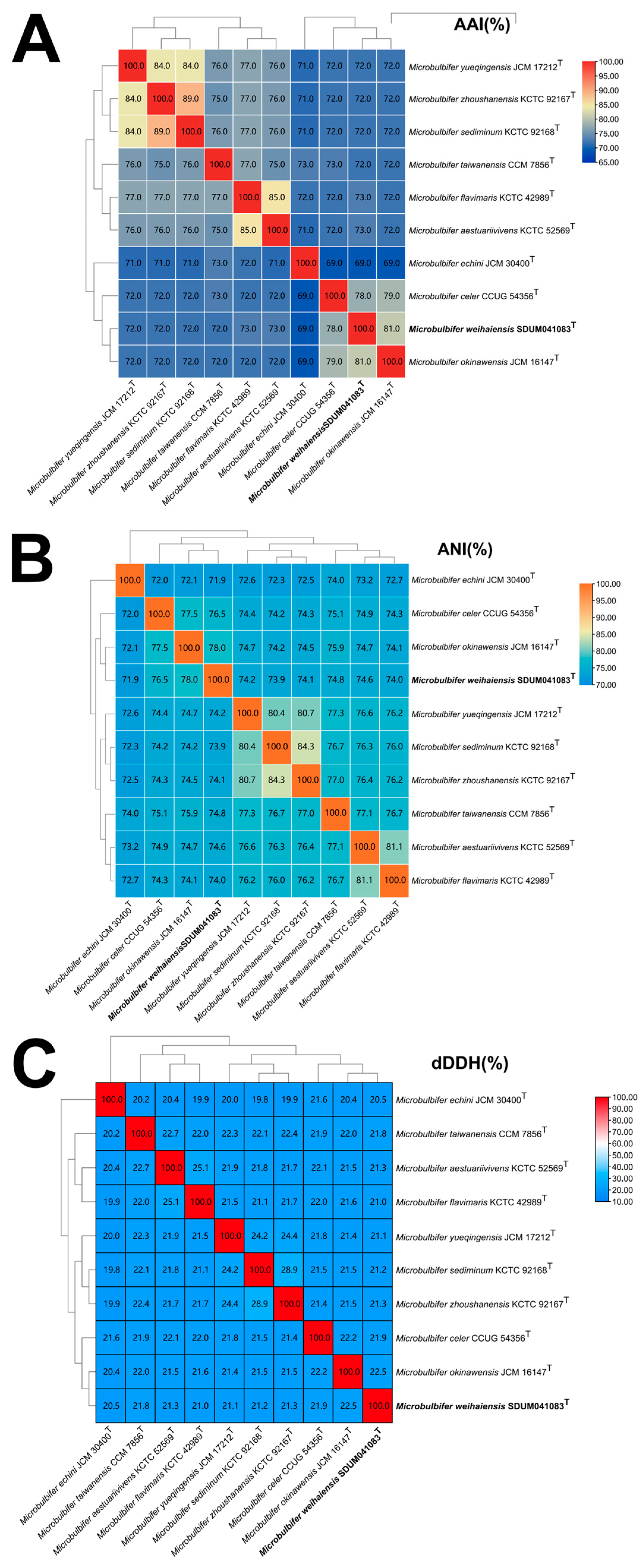
Based on the anti-SMASH results, eleven secondary metabolite biosynthesis gene clusters were predicted in the genome of the SDUM041083T strain, including gene clusters encoding non-ribosomal peptide synthetase-like fragments, mesophenol, Type I polyketide synthase, terpene-precursor, ectoine, non-ribosomal peptide synthetase, NRPS-independent IucA/IucC-like siderophores, terpene, aryl polyene, betalactone, and other unspecified ribosomally synthesized and post-translationally modified peptide products. The ANI value between strain SDUM041083T and Microbulbifer okinawensis was 78.01%, which is below the 95% interspecies threshold [57]; the AAI value was 73.73% and the dDDH value was 22.50%, also below the threshold [58,59]. The ANI, AAI, and dDDH values of SDUM041083T and other strains of the genus Microbulbifer were also lower than the threshold values (as shown in Figure 2), indicating that strain SDUM041083T is a novel species of the genus Microbulbifer. The protein phylogenetic tree, which depicted the evolutionary relationships between strain SDUM041083T and some related type strains, revealed that the strain was affiliated with the genus Microbulbifer (Figure 3), which was consistent with the result of the 16S rRNA gene phylogenetic analysis.

3.5. Comparative Genomic Analysis and Pan-Genome Analysis of the Genus Microbulbifer

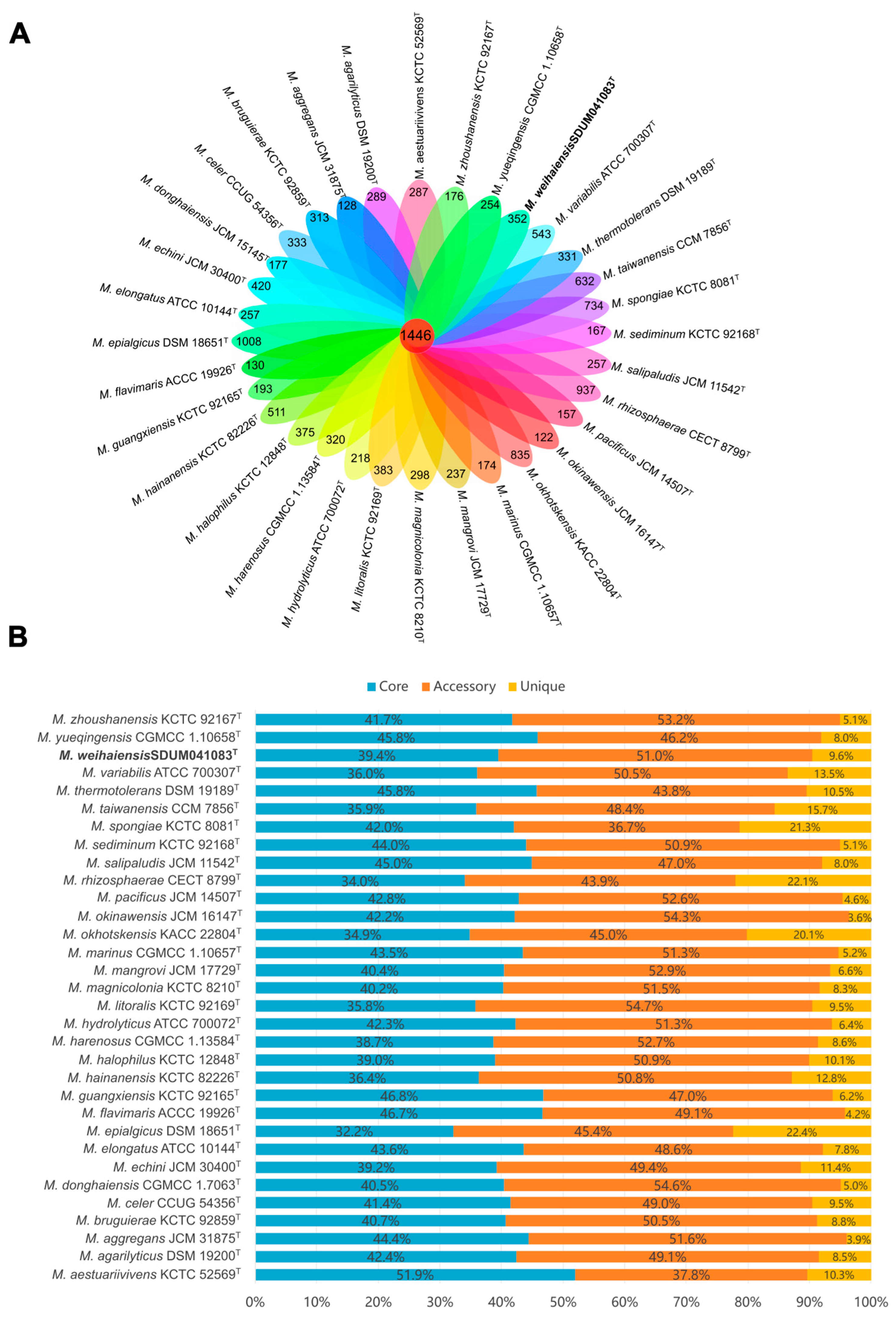
The genome sizes of the genus, including strain SDUM041083T, ranged from 3.4 Mbp (Microbulbifer aestuariivivens GHTF-23T [4]) to 5.7 Mbp (Microbulbifer epialgicus DSM 18651T [9]). The genomic DNA G+C content varied from 49.0 mol% (Microbulbifer variabilis ATCC 700307T [9] and Microbulbifer epialgicus DSM 18651T [9]) to 62.0 mol% (Microbulbifer yueqingensis JCM 17212T [54]). As illustrated in Figure 4, the pan-genome analysis based on orthologous groups of proteins revealed that 1446 core genes were shared by 32 Microbulbifer species, accounting for 32.2% to 51.9% of the genes in each genome (Figure 4A). The percentage of accessory genes and unique genes in each Microbulbifer genome ranged from 36.7% to 54.7% and from 3.6% to 22.4%, respectively (Figure 4B). The analysis of KEGG revealed that core genes predominantly contributed to essential metabolic pathways, including amino acid metabolism, cofactor and vitamin metabolism, nucleotide metabolism, replication and repair, and translation (Supplementary Figure S4). This result suggests a high degree of conservation in these cellular biological functions among the genus Microbulbifer. Additionally, accessory genes and unique genes were more distributed than core genes in carbohydrate metabolism, lipid metabolism, signal transduction, the metabolism of terpenoids and polyketides, and xenobiotic biodegradation and metabolism (Supplementary Figure S3), which indicated that these genes conferred metabolic diversity such as degrading a series of polysaccharides, producing a variety of natural products [15], degrading poly (3-hydroxybutyrate) (PHB) [18,19], and marine environment adaptability to the members of the genus Microbulbifer [60].
Notably, among the 32 analyzed strains, the genus Microbulbifer exhibits a broad genomic G+C content range (49.0–62.0 mol%) [9,54]. Such diversity may imply genomic variation; however, as mentioned above, all strains cluster into a well-supported clade in the protein phylogenetic tree (Figure 3), and the AAI, ANI, and dDDH values in Figure 2 all exceed the thresholds for defining a new genus, alongside partially conserved phenotypic traits (like aerobic metabolism and halotolerance). Additionally, pan-genome analysis further reveals 1446 core genes enriched in key pathways. For these reasons, it is tentatively maintained that they still belong to the single genus Microbulbifer. At the same time, it is preliminarily inferred that this G+C variation may stem from evolutionary processes, such as horizontal gene transfer (facilitated by the organic-rich habitats of this genus) and adaptive divergence into distinct ecological niches, while more accurate analysis will require further in-depth research in subsequent studies.
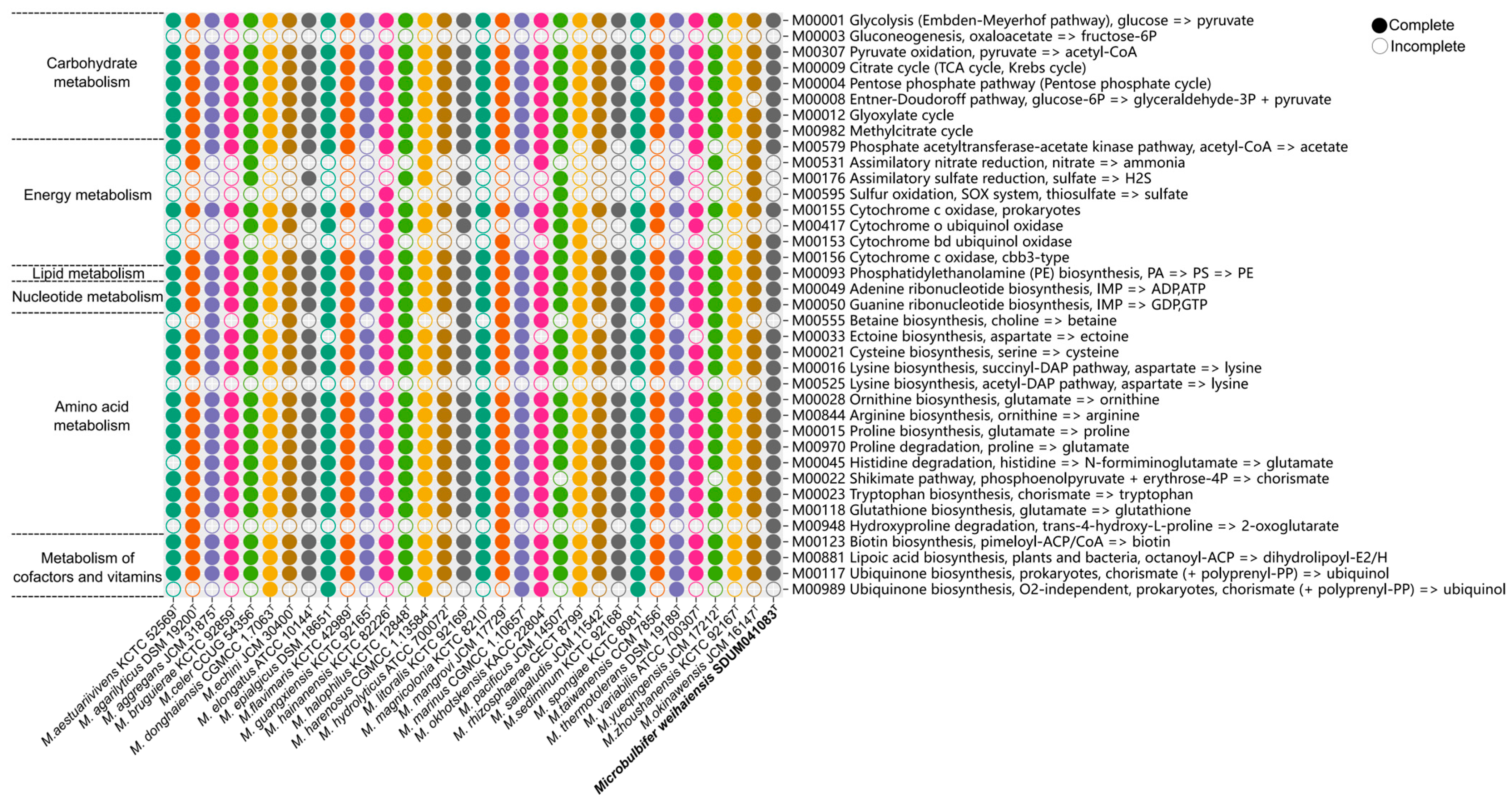
The metabolic pathways analyzed using KEGG’s BlastKOALA service of 32 Microbulbifer species (Figure 5) showed that the genus Microbulbifer demonstrated conservatism in carbohydrate metabolism, lipid metabolism, nucleotide metabolism, amino acid metabolism, and the metabolism of cofactors and vitamins, with some variations observed primarily in energy metabolism such as assimilatory nitrate reduction (M00531), assimilatory sulfate reduction (M00176), and sulfur oxidation (M00595). Specifically, in carbohydrate metabolism, all strains have complete key pathways like glycolysis (M00001), pyruvate oxidation (M00307), and TCA cycle (M00009). This conserved central metabolic network could efficiently process some monosaccharides derived from polysaccharide degradation into energy and biosynthetic precursors. Gluconeogenesis (M00003) was not complete in all 32 species of Microbulbifer. While central carbon metabolism exhibited broad conservation, Microbulbifer. spongiae KC 8081T uniquely lacked a complete pentose phosphate pathway (M00004), suggesting the species–specific modulation of carbon flux. Regarding amino acids, all strains within the Microbulbifer genus exhibited completeness in most amino acid biosynthesis pathways, including ornithine biosynthesis (M00028), proline biosynthesis (M00015), Arginine biosynthesis (M00844), and lysine biosynthesis pathway (M00527). Notably, SDUM041083T harbors a distinct lysine biosynthesis pathway (M00525), which is incomplete in other Microbulbifer strains. The lysine synthesis capacity of SDUM041083T may provide a survival advantage in marine sediment environments. Additionally, proline [61] and arginine [62] have been shown to promote bacterial growth under hypertonic conditions by enhancing osmotic tolerance through intracellular amino acid accumulation [63]. The Microbulbifer genus’s strains possess multiple complete amino acid biosynthesis pathways, which may help them endure the osmotic stress-induced cellular damage often encountered in marine environments [64]. Since most members of the genus were isolated from marine environments, other pathways associated with marine adaptation were also examined. To handle osmotic stress, bacteria accumulate compatible solutes, which are critical for maintaining cellular integrity and function under hyperosmotic conditions [63,65]. Betaine is a key compatible solute known for its protective role in bacterial osmoregulation. The current analysis revealed that 14 Microbulbifer strains lack the complete pathway for betaine biosynthesis (M00555). This absence suggests that these strains might employ alternative strategies in high-salinity marine habitats. Furthermore, bacteria depend on essential antioxidant enzymes, such as superoxide dismutase, catalase, glutathione, and cytochrome oxidase, to neutralize harmful free radicals [66]. All strains retained complete cytochrome c oxidase pathways (M00155 and M00156), which are vital for aerobic respiration under fluctuating oxygen levels, while pathways for cytochrome o oxidase (M00417) and cytochrome bd ubiquinol oxidase (M00153) showed strain-specific distribution, indicating niche-specific adaptations in electron transport (Figure 5). Conversely, the widespread presence of glutathione biosynthesis (M00118) across all 32 strains emphasizes its essential role in combating oxidative stress caused by marine conditions (Figure 5). Additionally, the phosphatidyl ethanolamine (PE) biosynthesis pathway (M00093) was annotated in all Microbulbifer strains, as is consistent with the phenotypic test of polar lipids (Table 1). Overall, the analysis of metabolic features and KEGG pathways within the genus Microbulbifer reflects their evolved adaptive diversification in response to diverse habitats.

3.6. Comparative Analysis of Carbohydrate-Active Enzymes of the Genus Microbulbifer

Building upon prior investigations demonstrating the polysaccharide utilization capabilities of Microbulbifer [5,11,12], an assessment of carbohydrate-active enzymes (CAZymes) across 32 strains was conducted (Figure 6A). Different from the research of Long et al. [23], this collection encompassed 31 validly published Microbulbifer type strains and the novel strain SDUM041083ᵀ. CAZyme abundance varied considerably (55–237 enzymes), with glycoside hydrolases (GHs) consistently dominating (e.g., >50% in M. mangrovi) (Figure 6A), aligning with the genus’s known degradative roles [5,11,12,23]. The proportional composition of these enzyme classes varied across individual strains. This enzymatic profile reflects the Microbulbifer genus’s adaptation to multiple ecological niches, such as gut microbiota [55] and aquatic environments [5,7,67], and underscores its evolutionary specialization in key metabolic roles like algal polysaccharide utilization [12] and chitin-degrading [11].
The novel strain SDUM041083T contains 115 annotated CAZymes, mainly including glycoside hydrolases (GHs, 42), glycosyltransferases (GTs, 33), carbohydrate esterases (CEs, 18), auxiliary activities (AAs, 10), and polysaccharide lyases (PLs, 3) (Figure 6B). The identified PLs (PL1_1, PL1_5, PL10_1) suggest pectin lyase activity, while families associated with alginate lyase (PL5/PL7) were absent, aligning with biochemical tests that showed no alginate degradation (Section 3.1). This finding provides genomic insight into the observed enzymatic deficiency. Genomic analysis revealed exceptional chitinolytic potential in SDUM041083ᵀ, featuring 8 GH18, 1 GH19_2, and 2 GH20 genes, representing the highest chitinase gene count among analyzed genomic chitinolytic Microbulbifer strains, a trait shared only with M. okhotskensis KACC 22804ᵀ [68]. The genomic prediction of robust chitinolytic potential was phenotypically validated through both hydrolysis zone formation (Section 3.1, Figure S2) and the detection of N-acetyl-glucosaminidase activity (GH20 function; Table 1) in biochemical assays. The plate-based assay unequivocally demonstrates high chitinolytic efficacy per unit biomass (H/C ratio), aligning with genomic predictions. Despite its extensive genetic capacity (eight GH18, one GH19_2, and two GH20 genes), the strain exhibits constrained extracellular hydrolytic performance as evidenced by smaller hydrolysis zones compared to M. okinawensis. This result may be partially attributed to resource allocation trade-offs favoring chitinase production (or activity) over rapid growth in monotrophic media [69], regulatory constraints requiring chitin oligomers for full induction [70], microbial synergistic degradation [70], substrate specificity of different chitinases [71], and limitations in culture conditions. Consequently, while genomic data confirm the presence of chitin-degrading machinery, its ecological role is likely contingent upon induction conditions specific to its habitat. Future transcriptomic profiling under chitin amendment and enzyme kinetic characterization will elucidate regulatory mechanisms.
Isolated from aquaculture sediment, strain SDUM041083ᵀ possesses genomic features indicating adaptive potential for chitin-rich environments, supported by the plate assay validation of chitin degradation. This suggests a plausible ecological role in marine niches such as aquaculture systems. However, direct ecological niche adaptation requires the isolation of site-specific metagenomic or transcriptomic validation.

3.7. Global Biogeographic Distribution of Microbulbifer Based on MAPseq Meta-Analysis

To comprehensively elucidate the global prevalence and habitat range of the genus Microbulbifer, an analysis of this genus’s distribution was performed using the MAP database online. An investigation was conducted on 12,374 samples spanning 2326 projects to pinpoint the characteristic sequence. Microbulbifer sequences were detected in 5980 aquatic (48.30%), 1097 soil (8.87%), 1078 animal (8.71%), and 109 plant samples (0.88%) (Figure 7A). Within aquatic environments, marine habitats (12.80%) and sediments (12.20%) showed the highest prevalence. Furthermore, a comparative analysis of database sequence reads against standard OTUs revealed that Microbulbifer is predominantly enriched in marine environments (19.50% of reads), with sediment environments representing the secondary niche (5.22%) (Figure 7B). This genus-level distribution aligns with the marine origins of most cultured Microbulbifer strains [23].
Critically, as emphasized above, the biogeographic distribution inferred from MAPseq (based on 16S rRNA gene profiling) reflects genus-level patterns and cannot attribute strain-specific functional traits, such as the chitinolytic proficiency of SDUM041083T. Microbulbifer’s broad environmental prevalence likely stems from metabolic conservation (e.g., osmotic stress amino acid synthesis; Section 3.5), CAZyme-mediated niche plasticity enabling polysaccharide versatility (Figure 6), and lineage-specific adaptations (e.g., betaine-independent osmoregulation) recurrent in coastal Microbulbifer strains, including SDUM041083T and 13 conspecific isolates (Section 3.5). While these traits facilitate habitat colonization, strain-level ecological roles require functional validation beyond 16S data.

3.8. Description of Microbulbifer weihaiensis sp. nov.

The cells are Gram-negative, motile, straight-rod shaped (4.780 ± 0.2 μm × 0.326 ± 0.1 μm), aerobic, oxidase-negative, and peroxidase-positive, and they form yellow circular colonies. Cells can grow on MA medium, and Na+ must be present for growth. Cell growth on MA can occur at temperatures ranging from 25 to 40 °C, with 35 °C being the optimal temperature for growth. Additionally, 2% (w/v) NaCl was the optimal condition for growth, and cells can grow on a medium containing 1–10% (w/v) NaCl, but growth was very slow at 7–10% salinity. The pH at which the cell could grow ranged from 5.5 to 9.5, with 7.0 being the optimal pH. The cells were able to hydrolyze Tween 20, 40, 60, and 80 and showed caseinase activity, but they did not respond to alginate, cellulose, agar, or DNA. Nitrate metabolism was positive. In the API ZYM system, alkaline phosphatase, esterase (C4), lipoid esterase (C8), lipase (C14), leucine arylamines, valine arylamines, cystine arylaminase, pancreatic coagulase, acid phosphatase, naphthol-AS-BI-phosphohydrolase, alpha-glucosidase, and N-acetyl-glucosaminidase were positive (intensities 3–5), while the rest were negative. In the API 20E system, the pyruvate, gelatinase, glucose, and arabinose test results were positive, but the remainder produced negative results. Q-8 was the only respiratory quinone. The main fatty acids (>10.0%) were iso-C15:0 (24.33%), iso-C11:0 3-OH (10.78%), and iso-C11:0 (10.61%). The major polar lipids were phosphatidylglycerol (PG), phosphatidylethanolamine (PE), and aminolipid (AL).
The type strain was SDUM041083T (= MCCC 1H01537T= KCTC 8896T), which was isolated from the shoal sediment of Xiaoshidao, Weihai, China. The DNA G+C content of this type strain was 57.5 mol%. The GenBank accession number for the 16S rRNA gene sequence of Microbulbifer weihaiensis SDUM041083T is PV225712. This Whole Genome Shotgun project has been deposited at DDBJ/ENA/GenBank under the accession JBLWFL000000000.

Supplementary Materials

The following supporting information can be downloaded at https://www.mdpi.com/article/10.3390/microorganisms13092005/s1, Figure S1: Scanning electron micrograph of cells of strain SDUM041083T. Bar, 2 μm; Figure S2: The chitin-degrading hydrolysis zone of strain SDUM041083T. The left half of the plate is SDUM041083T, and the right half is the reference strain M. okinawensis JCM 16147T; Figure S3: Two-dimensional TLC plate image of the total polar lipids of strain SDUM041083T (a) and M. okinawensis JCM 16147T.PE, phosphatidylethanolamine; AL, unidentified aminolipid; PG, phosphatidylglycerol; Figure S4: The distribution of core genes, accessory genes, and unique genes to different metabolic pathways in the genus Microbulbifer; Table S1: Cellular fatty acids composition (%) of the strain SDUM041083T and relative strains: 1, SDUM041083T; 2, M. okinawensis JCM 16147T; 3, M. taiwanensis CCM 7856T. Only those fatty acids accounting for 1% or more in one of the strains are given. tr, trace amount (<1.0%); −, not detected.

Author Contributions

Y.-X.Z. performed experimental operation, performed data processing and analysis, and finished the writing of the manuscript. A.-Q.L. offered experimental guidance and critical revision of the manuscript. All authors have read and agreed to the published version of the manuscript.

Funding

This research was funded by the General Program of China Postdoctoral Science Foundation, grant number 2015M582079.

Institutional Review Board Statement

Not applicable.

Informed Consent Statement

Not applicable.

Data Availability Statement

The GenBank accession number of Microbulbifer weihaiensis SDUM041083T for the 16S rRNA gene sequence is PV225712; for the whole-genome assembly, it is JBLWFL000000000. The BioProject accession number of Microbulbifer weihaiensis SDUM041083T is PRJNA1226674.

Acknowledgments

The scanning electron microscopy (model Nova NanoSEM450) was supported by the Physical-Chemical Materials Analytical and Testing Center of Shandong University at Weihai.

Conflicts of Interest

The authors declare there are no conflicts of interest.

References

  1. González, J.M.; Mayer, F.; Moran, M.A.; Hodson, R.E.; Whitman, W.B. Microbulbifer hydrolyticus gen. nov., sp. nov., and Marinobacterium georgiense gen. nov., sp. nov., two marine bacteria from a lignin-rich pulp mill waste enrichment community. Int. J. Syst. Bacteriol. 1997, 47, 369–376. [Google Scholar] [CrossRef]
  2. Tang, S.K.; Wang, Y.; Cai, M.; Lou, K.; Mao, P.H.; Jin, X.; Jiang, C.L.; Xu, L.H.; Li, W.J. Microbulbifer halophilus sp. nov., a moderately halophilic bacterium from north-west China. Int. J. Syst. Evol. Microbiol. 2008, 58, 2036–2040. [Google Scholar] [CrossRef]
  3. Camacho, M.; Del Carmen Montero-Calasanz, M.; Redondo-Gómez, S.; Rodríguez-Llorente, I.; Schumann, P.; Klenk, H.P. Microbulbifer rhizosphaerae sp. nov., isolated from the rhizosphere of the halophyte Arthrocnemum macrostachyum. Int. J. Syst. Evol. Microbiol. 2016, 66, 1844–1850. [Google Scholar] [CrossRef]
  4. Park, S.; Yoon, S.Y.; Ha, M.J.; Yoon, J.H. Microbulbifer aestuariivivens sp. nov., isolated from a tidal flat. Int. J. Syst. Evol. Microbiol. 2017, 67, 1436–1441. [Google Scholar] [CrossRef]
  5. Miyazaki, M.; Nogi, Y.; Ohta, Y.; Hatada, Y.; Fujiwara, Y.; Ito, S.; Horikoshi, K. Microbulbifer agarilyticus sp. nov. and Microbulbifer thermotolerans sp. nov., agar-degrading bacteria isolated from deep-sea sediment. Int. J. Syst. Evol. Microbiol. 2008, 58, 1128–1133. [Google Scholar] [CrossRef] [PubMed]
  6. Yoon, J.H.; Kim, I.G.; Oh, T.K.; Park, Y.H. Microbulbifer maritimus sp. nov., isolated from an intertidal sediment from the Yellow Sea, Korea. Int. J. Syst. Evol. Microbiol. 2004, 54, 1111–1116. [Google Scholar] [CrossRef] [PubMed]
  7. Moh, T.H.; Furusawa, G.; Amirul, A.A. Microbulbifer aggregans sp. nov., isolated from estuarine sediment from a mangrove forest. Int. J. Syst. Evol. Microbiol. 2017, 67, 4089–4094. [Google Scholar] [CrossRef]
  8. Yoon, J.H.; Jung, S.Y.; Kang, S.J.; Oh, T.K. Microbulbifer celer sp. nov., isolated from a marine solar saltern of the Yellow Sea in Korea. Int. J. Syst. Evol. Microbiol. 2007, 57, 2365–2369. [Google Scholar] [CrossRef]
  9. Nishijima, M.; Takadera, T.; Imamura, N.; Kasai, H.; An, K.D.; Adachi, K.; Nagao, T.; Sano, H.; Yamasato, K. Microbulbifer variabilis sp. nov. and Microbulbifer epialgicus sp. nov., isolated from Pacific marine algae, possess a rod-coccus cell cycle in association with the growth phase. Int. J. Syst. Evol. Microbiol. 2009, 59, 1696–1707. [Google Scholar] [CrossRef]
  10. Jeong, S.H.; Yang, S.H.; Jin, H.M.; Kim, J.M.; Kwon, K.K.; Jeon, C.O. Microbulbifer gwangyangensis sp. nov. and Microbulbifer pacificus sp. nov., isolated from marine environments. Int. J. Syst. Evol. Microbiol. 2013, 63, 1335–1341. [Google Scholar] [CrossRef]
  11. Baba, A.; Miyazaki, M.; Nagahama, T.; Nogi, Y. Microbulbifer chitinilyticus sp. nov. and Microbulbifer okinawensis sp. nov., chitin-degrading bacteria isolated from mangrove forests. Int. J. Syst. Evol. Microbiol. 2011, 61, 2215–2220. [Google Scholar] [CrossRef]
  12. Vashist, P.; Nogi, Y.; Ghadi, S.C.; Verma, P.; Shouche, Y.S. Microbulbifer mangrovi sp. nov., a polysaccharide-degrading bacterium isolated from an Indian mangrove. Int. J. Syst. Evol. Microbiol. 2013, 63, 2532–2537. [Google Scholar] [CrossRef]
  13. Cheng, Y.; Zhu, S.; Guo, C.; Xie, F.; Jung, D.; Li, S.; Zhang, W.; He, S. Microbulbifer hainanensis sp. nov., a moderately halophilic bacterium isolated from mangrove sediment. Antonie Leeuwenhoek 2021, 114, 1033–1042. [Google Scholar] [CrossRef]
  14. Yoon, J.H.; Kim, H.; Kang, K.H.; Oh, T.K.; Park, Y.H. Transfer of Pseudomonas elongata Humm 1946 to the genus Microbulbifer as Microbulbifer elongatus comb. Nov. Int. J. Syst. Evol. Microbiol. 2003, 53, 1357–1361. [Google Scholar] [CrossRef]
  15. Zhong, W.; Agarwal, V. Polymer degrading marine Microbulbifer bacteria: An un(der)utilized source of chemical and biocatalytic novelty. Beilstein J. Org. Chem. 2024, 20, 1635–1651. [Google Scholar] [CrossRef]
  16. Li, H.; Hu, Q.; Hong, X.; Jiang, Z.; Ni, H.; Li, Q.; Zhu, Y. Molecular cloning and characterization of a thermostable and halotolerant endo-β-1,4-glucanase from Microbulbifer sp. ALW1. 3 Biotech 2021, 11, 250. [Google Scholar] [CrossRef] [PubMed]
  17. Zhang, Q.; Gui, Y.; Zhao, L.; Zhang, A.; Fu, L.; Cao, Z.; Li, J. Identification and characterization of ι-carrageenase from macroalgae-associated bacterium Microbulbifer sp. YNDZ01. J. Sci. Food. Agric. 2023, 103, 6095–6104. [Google Scholar] [CrossRef] [PubMed]
  18. Park, S.L.; Cho, J.Y.; Kim, S.H.; Bhatia, S.K.; Gurav, R.; Park, S.H.; Park, K.; Yang, Y.H. Isolation of Microbulbifer sp. SOL66 with high polyhydroxyalkanoate-degrading activity from the marine environment. Polymers 2021, 13, 4257. [Google Scholar] [CrossRef] [PubMed]
  19. Park, S.L.; Cho, J.Y.; Kim, S.H.; Lee, H.J.; Kim, S.H.; Suh, M.J.; Ham, S.; Bhatia, S.K.; Gurav, R.; Park, S.H.; et al. Novel polyhydroxybutyrate-degrading activity of the Microbulbifer genus as confirmed by Microbulbifer sp. SOL03 from the marine environment. J. Microbiol. Biotechnol. 2022, 32, 27–36. [Google Scholar] [CrossRef]
  20. Li, Z.; Wei, R.; Gao, M.; Ren, Y.; Yu, B.; Nie, K.; Xu, H.; Liu, L. Biodegradation of low-density polyethylene by Microbulbifer hydrolyticus IRE-31. J. Environ. Manage. 2020, 263, 110402. [Google Scholar] [CrossRef]
  21. Peng, X.; Adachi, K.; Chen, C.; Kasai, H.; Kanoh, K.; Shizuri, Y.; Misawa, N. Discovery of a marine bacterium producing 4-hydroxybenzoate and its alkyl esters, parabens. Appl. Environ. Microbiol. 2006, 72, 5556–5561. [Google Scholar] [CrossRef]
  22. Karim, M.R.U.; Harunari, E.; Oku, N.; Akasaka, K.; Igarashi, Y. Bulbimidazoles A-C, antimicrobial and Cytotoxic alkanoyl imidazoles from a marine Gammaproteobacterium Microbulbifer Species. J. Nat. Prod. 2020, 83, 1295–1299. [Google Scholar] [CrossRef]
  23. Long, M.; Chen, D.K.; Fan, H.M.; Tang, S.S.; Gan, Z.; Xia, H.L.; Lu, Y.S. Microbulbifer bruguierae sp. nov., isolated from sediment of mangrove plant Bruguiera sexangula, and comparative genomic analyses of the genus Microbulbifer. Int. J. Syst. Evol. Microbiol. 2024, 74, 006209. [Google Scholar] [CrossRef] [PubMed]
  24. KleinJan, H.; Jeanthon, C.; Boyen, C.; Dittami, S.M. Exploring the cultivable Ectocarpus microbiome. Front. Microbiol. 2017, 8, 2456. [Google Scholar] [CrossRef] [PubMed]
  25. Zhu, K.L.; Wang, X.Q.; Zhang, T.S.; Shang, D.D.; Du, Z.J. Salibaculum halophilum gen. nov., sp. nov. and Salibaculum griseiflavum sp. nov., in the family Rhodobacteraceae. Int. J. Syst. Evol. Microbiol. 2021, 71, 004808. [Google Scholar] [CrossRef]
  26. Ciufo, S.; Kannan, S.; Sharma, S.; Badretdin, A.; Clark, K.; Turner, S.; Brover, S.; Schoch, C.L.; Kimchi, A.; DiCuccio, M. Using average nucleotide identity to improve taxonomic assignments in prokaryotic genomes at the NCBI. Int. J. Syst. Evol. Microbiol. 2018, 68, 2386–2392. [Google Scholar] [CrossRef]
  27. Yoon, S.H.; Ha, S.M.; Kwon, S.; Lim, J.; Kim, Y.; Seo, H.; Chun, J. Introducing EzBioCloud: A taxonomically united database of 16S rRNA gene sequences and whole-genome assemblies. Int. J. Syst. Evol. Microbiol. 2017, 67, 1613–1617. [Google Scholar] [CrossRef]
  28. Kannan, L.; Wheeler, W.C. Maximum parsimony on phylogenetic networks. Algorithms. Mol. Biol. 2012, 7, 9. [Google Scholar] [CrossRef]
  29. Saitou, N.; Nei, M. The neighbor-joining method: A new method for reconstructing phylogenetic trees. Mol. Biol. Evol. 1987, 4, 406–425. [Google Scholar] [CrossRef]
  30. Felsenstein, J. Evolutionary trees from DNA sequences: A maximum likelihood approach. J. Mol. Evol. 1981, 17, 368–376. [Google Scholar] [CrossRef] [PubMed]
  31. Kumar, S.; Stecher, G.; Tamura, K. MEGA7: Molecular Evolutionary Genetics Analysis Version 7.0 for bigger datasets. Mol. Biol. Evol. 2016, 33, 1870–1874. [Google Scholar] [CrossRef]
  32. Hall, B.G. Building phylogenetic trees from molecular data with MEGA. Mol. Biol. Evol. 2013, 30, 1229–1235. [Google Scholar] [CrossRef]
  33. Kimura, M. A simple method for estimating evolutionary rates of base substitutions through comparative studies of nucleotide sequences. J. Mol. Evol. 1980, 16, 111–120. [Google Scholar] [CrossRef]
  34. Wang, Y.W.; Zhang, J.; Wang, S.X.; Du, Z.J.; Mu, D.S. Aequorivita vitellina sp. nov. and Aequorivita xiaoshiensis sp. nov., isolated from marine sediment. Int. J. Syst. Evol. Microbiol. 2023, 73, 005801. [Google Scholar] [CrossRef]
  35. Tatusova, T.; DiCuccio, M.; Badretdin, A.; Chetvernin, V.; Nawrocki, E.P.; Zaslavsky, L.; Lomsadze, A.; Pruitt, K.D.; Borodovsky, M.; Ostell, J. NCBI prokaryotic genome annotation pipeline. Nucleic Acids Res. 2016, 44, 6614–6624. [Google Scholar] [CrossRef]
  36. Medema, M.H.; Blin, K.; Cimermancic, P.; De Jager, V.; Zakrzewski, P.; Fischbach, M.A.; Weber, T.; Takano, E.; Breitling, R. antiSMASH: Rapid identification, annotation and analysis of secondary metabolite biosynthesis gene clusters in bacterial and fungal genome sequences. Nucleic Acids Res. 2011, 39, W339–W346. [Google Scholar] [CrossRef] [PubMed]
  37. Richter, M.; Rosselló-Móra, R.; Glöckner, F.O.; Peplies, J. JSpeciesWS: A web server for prokaryotic species circumscription based on pairwise genome comparison. Bioinformatics 2016, 32, 929–931. [Google Scholar] [CrossRef]
  38. Meier-Kolthoff, J.P.; Carbasse, J.S.; Peinado-Olarte, R.L.; Göker, M. TYGS and LPSN: A database tandem for fast and reliable genome-based classification and nomenclature of prokaryotes. Nucleic Acid Res. 2022, 50, D801–D807. [Google Scholar] [CrossRef] [PubMed]
  39. Kanehisa, M.; Sato, Y.; Kawashima, M.; Furumichi, M.; Tanabe, M. KEGG as a reference resource for gene and protein annotation. Nucleic Acids Res. 2016, 44, 457–462. [Google Scholar] [CrossRef] [PubMed]
  40. Chaumeil, P.A.; Mussig, A.J.; Hugenholtz, P.; Parks, D.H. GTDB-Tk2: Memory friendly classification with the genome taxonomy database. Bioinformatics 2022, 38, 5315–5316. [Google Scholar] [CrossRef]
  41. Price, M.N.; Dehal, P.S.; Arkin, A.P. FastTree: Computing large minimum evolution trees with profiles instead of a distance matrix. Mol. Biol. Evol. 2009, 26, 1641–1650. [Google Scholar] [CrossRef]
  42. Nguyen, L.T.; Schmidt, H.A.; Von Haeseler, A.; Minh, B.Q. IQ-TREE: A fast and effective stochastic algorithm for estimating maximum-likelihood phylogenies. Mol. Biol. Evol. 2015, 32, 268–274. [Google Scholar] [CrossRef]
  43. Kanehisa, M.; Sato, Y.; Morishima, K. BlastKOALA and GhostKOALA: KEGG tools for functional characterization of genome and metagenome sequences. J. Mol. Biol. 2016, 428, 726–731. [Google Scholar] [CrossRef]
  44. Chaudhari, N.M.; Gupta, V.K.; Dutta, C. BPGA—An ultra-fast pan-genome analysis pipeline. Sci. Rep. 2016, 6, 24373. [Google Scholar] [CrossRef]
  45. Drula, E.; Garron, M.L.; Dogan, S.; Lombard, V.; Henrissat, B.; Terrapon, N. The Carbohydrate-Active Enzyme Database: Functions and literature. Nucleic Acids Res. 2022, 50, D571–D577. [Google Scholar] [CrossRef]
  46. Matias Rodrigues, J.F.; Schmidt, T.S.; Tackmann, J.; von Mering, C. MAPseq: Highly efficient k-mer search with confidence estimates, for rRNA sequence analysis. Bioinformatics 2017, 33, 3808–3810. [Google Scholar] [CrossRef] [PubMed]
  47. Bowman, J.P. Description of Cellulophaga algicola sp. nov., isolated from the surfaces of Antarctic algae, and reclassification of Cytophaga uliginosa (ZoBell and Upham 1944) Reichenbach 1989 as Cellulophaga uliginosa comb. nov. Int. J. Syst. Evol. Microbiol. 2000, 50, 1861–1868. [Google Scholar] [CrossRef] [PubMed]
  48. Du, Z.J.; Wang, Z.J.; Zhao, J.X.; Chen, G.J. Woeseia oceani gen. nov., sp. nov., a chemoheterotrophic member of the order Chromatiales, and proposal of Woeseiaceae fam. nov. Int. J. Syst. Evol. Microbiol. 2016, 66, 107–112. [Google Scholar] [CrossRef]
  49. Wang, N.N.; Li, C.M.; Li, Y.X.; Du, Z.J. Aquimarina celericrescens sp. nov., isolated from seawater. Int. J. Syst. Evol. Microbiol. 2018, 68, 1683–1688. [Google Scholar] [CrossRef]
  50. Liang, Q.Y.; Xu, Z.X.; Zhang, J.; Chen, G.J.; Du, Z.J. Salegentibacter sediminis sp. nov., a marine bacterium of the family Flavobacteriaceae isolated from coastal sediment. Int. J. Syst. Evol. Microbiol. 2018, 68, 2375–2380. [Google Scholar] [CrossRef] [PubMed]
  51. Hiraishi, A.; Ueda, Y.; Ishihara, J.; Mori, T. Comparative lipoquinone analysis of influent sewage and activated sludge by high-performance liquid chromatography and photodiode array detection. J. Gen. Appl. Microbiol. 1996, 42, 457–469. [Google Scholar] [CrossRef]
  52. Tindall, B.J. A comparative study of the lipid composition of Halobacterium saccharovorum from various sources. Syst. Appl. Microbiol. 1990, 13, 128–130. [Google Scholar] [CrossRef]
  53. Liu, J.C.; Ye, Y.Q.; Tan, X.Y.; Du, Z.J.; Ye, M.Q. Description of Aequorivita aurantiaca sp. nov. Isolated from coastal sediment, and comparative genomic analysis and biogeographic distribution of the genus Aequorivita. Microorganisms 2023, 11, 2518. [Google Scholar] [CrossRef]
  54. Zhang, D.S.; Huo, Y.Y.; Xu, X.W.; Wu, Y.H.; Wang, C.S.; Xu, X.F.; Wu, M. Microbulbifer marinus sp. nov. and Microbulbifer yueqingensis sp. nov., isolated from marine sediment. Int. J. Syst. Evol. Microbiol. 2012, 62, 505–510. [Google Scholar] [CrossRef] [PubMed]
  55. Lee, J.Y.; Kim, P.S.; Hyun, D.W.; Kim, H.S.; Shin, N.R.; Jung, M.J.; Yun, J.H.; Kim, M.S.; Whon, T.W.; Bae, J.W. Microbulbifer echini sp. nov., isolated from the gastrointestinal tract of a purple sea urchin, Heliocidaris crassispina. Int. J. Syst. Evol. Microbiol. 2017, 67, 998–1004. [Google Scholar] [CrossRef]
  56. Kämpfer, P.; Arun, A.B.; Young, C.C.; Rekha, P.D.; Martin, K.; Busse, H.J.; Chen, W.M. Microbulbifer taiwanensis sp. nov., isolated from coastal soil. Int. J. Syst. Evol. Microbiol. 2012, 62, 2485–2489. [Google Scholar] [CrossRef]
  57. Jain, C.; Rodriguez-R, L.M.; Phillippy, A.M.; Konstantinidis, K.T.; Aluru, S. High throughput ANI analysis of 90K prokaryotic genomes reveals clear species boundaries. Nat. Commun. 2018, 9, 5114. [Google Scholar] [CrossRef]
  58. Nicholson, A.C.; Gulvik, C.A.; Whitney, A.M.; Humrighouse, B.W.; Bell, M.E.; Holmes, B.; Steigerwalt, A.G.; Villarma, A.; Sheth, M.; Batra, D.; et al. Division of the genus Chryseobacterium: Observation of discontinuities in amino acid identity values, a possible consequence of major extinction events, guides transfer of nine species to the genus Epilithonimonas, eleven species to the genus Kaistella, and three species to the genus Halpernia gen. Nov., with description of Kaistella daneshvariae sp. nov. and Epilithonimonas vandammei sp. nov. derived from clinical specimens. Int. J. Syst. Evol. Microbiol. 2020, 70, 4432–4450. [Google Scholar] [CrossRef]
  59. Goris, J.; Konstantinidis, K.T.; Klappenbach, J.A.; Coenye, T.; Vandamme, P.; Tiedje, J.M. DNA-DNA hybridization values and their relationship to whole genome sequence similarities. Int. J. Syst. Evol. Microbiol. 2007, 57, 81–91. [Google Scholar] [CrossRef] [PubMed]
  60. Innamorati, K.A.; Earl, J.P.; Aggarwal, S.D.; Ehrlich, G.D.; Hiller, N.L. The bacterial guide to designing a diversified gene portfolio. In The Pangenome: Diversity, Dynamics and Evolution of Genomes.; Tettelin, H., Medini, D., Eds.; Springer: Cham, Switzerland, 2020; pp. 51–87. [Google Scholar]
  61. Hoffmann, T.; von Blohn, C.; Stanek, A.; Moses, S.; Barzantny, H.; Bremer, E. Synthesis, release, and recapture of compatible solute proline by osmotically stressed Bacillus subtilis cells. Appl. Environ. Microbiol. 2012, 78, 5753–5762. [Google Scholar] [CrossRef] [PubMed]
  62. Xu, S.; Zhou, J.W.; Liu, L.M.; Chen, J. Arginine: A novel compatible solute to protect Candida glabrata against hyperosmotic stress. Process Biochem. 2011, 46, 1230–1235. [Google Scholar] [CrossRef]
  63. Da, C.M.; Santos, H.; Galinski, E.A. An overview of the role and diversity of compatible solutes in bacteria and archaea. Adv. Biochem. Eng. Biotechnol. 1998, 61, 117–153. [Google Scholar]
  64. Roesser, M.; Muller, V. Osmoadaptation in bacteria and archaea: Common principles and differences. Environ. Microbiol. 2001, 3, 743–754. [Google Scholar] [CrossRef]
  65. Wood, J.M.; Bremer, E.; Csonka, L.N.; Kraemer, R.; Poolman, B.; van der Heide, T. Osmosensing and osmoregulatory compatible solute accumulation by bacteria. Comp. Biochem. Physiol. A Mol. Integr. Physiol. 2001, 130, 437–460. [Google Scholar] [CrossRef]
  66. Pomposiello, P.J.; Demple, B. Global adjustment of microbial physiology during free radical stress. Adv. Microb. Physiol. 2002, 46, 319–341. [Google Scholar]
  67. Xiong, Q.; Wang, D.; Dong, X.; Liu, D.; Liu, Y.; Li, P.; Wu, G.; Luo, Y.; Zhang, R.; Liu, S.; et al. Microbulbifer flavimaris sp. nov., a halophilic Gammaproteobacteria isolated from marine sediment of the Yellow Sea, China. Int. J. Syst. Evol. Microbiol. 2019, 69, 1135–1141. [Google Scholar] [CrossRef] [PubMed]
  68. Romanenko, L.; Kurilenko, V.; Otstavnykh, N.; Velansky, P.; Isaeva, M.; Mikhailov, V. Microbulbifer okhotskensis sp. nov., isolated from a deep bottom sediment of the Okhotsk Sea. Arch. Microbiol. 2022, 204, 548. [Google Scholar] [CrossRef] [PubMed]
  69. Ramin, K.I.; Allison, S.D. Bacterial tradeoffs in growth rate and extracellular enzymes. Front. Microbiol. 2019, 10, 2956. [Google Scholar] [CrossRef] [PubMed]
  70. Beier, S.; Bertilsson, S. Bacterial chitin degradation—Mechanisms and ecophysiological strategies. Front. Microbiol. 2013, 4, 149. [Google Scholar] [CrossRef]
  71. Liu, S.J.; Shao, S.J.; Li, L.L.; Cheng, Z.; Tian, L.; Gao, P.J.; Wang, L.S. Substrate-binding specificity of chitinase and chitosanase as revealed by active-site architecture analysis. Carbohydr. Res. 2015, 418, 50–56. [Google Scholar] [CrossRef]
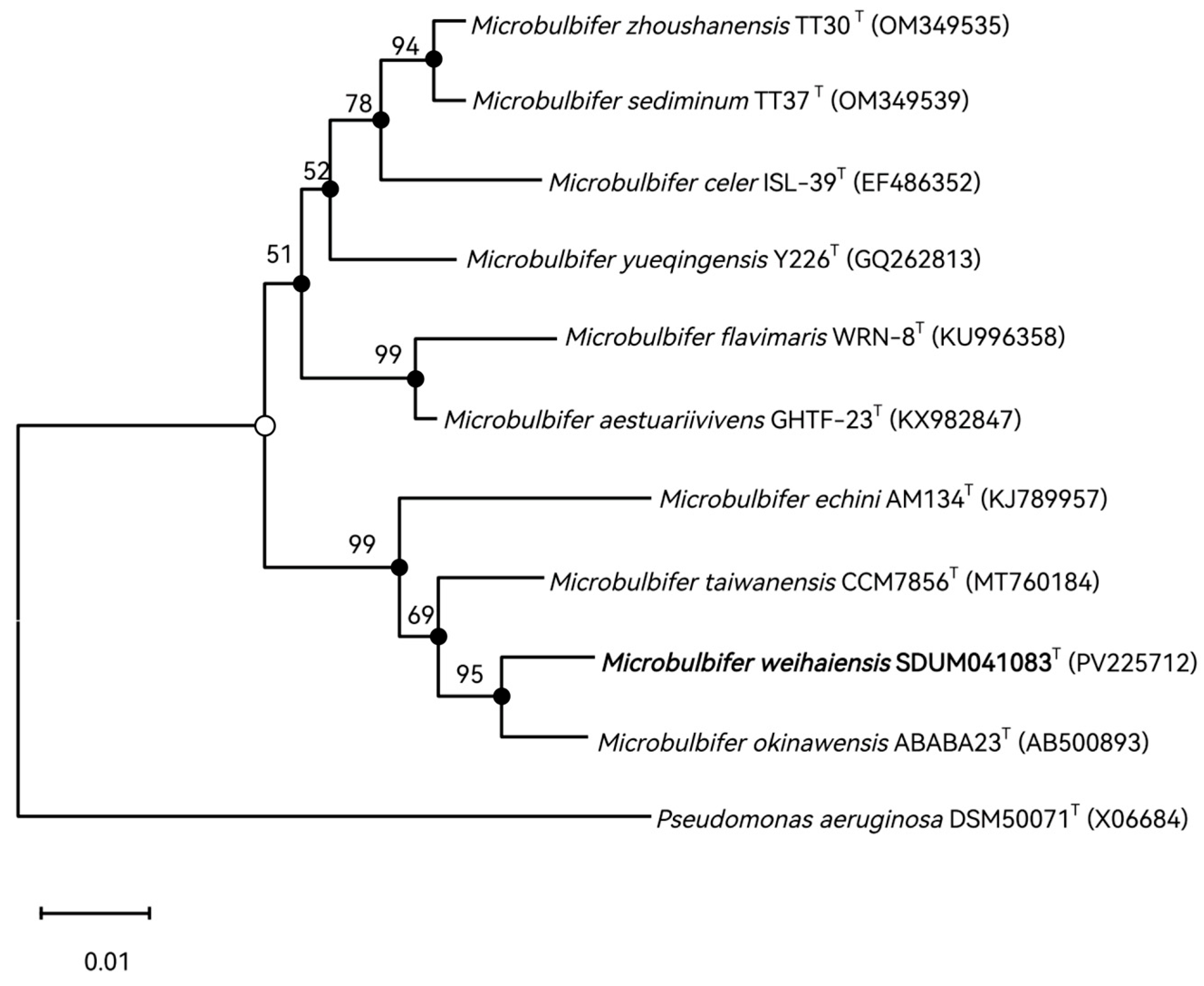
Figure 1. Phylogenetic tree reconstructed using MEGA based on 16S rRNA gene sequence data, showing the phylogenetic position of strain SDUM041083T among related taxa. GenBank accession numbers of 16S rRNA gene sequences are given in parentheses. Numbers on nodes represent bootstrap values based on 1000 replications. The strain characterized in this study is shown in bold type. Filled circles indicate that the corresponding nodes were also recovered in maximum-likelihood and maximum-evolution analyses. Pseudomonas aeruginosa DSM50071T was used as the outgroup. Open circles indicate that the corresponding nodes were also recovered in either the maximum-likelihood or the minimum-evolution analyses. Bar, 0.02 substitutions per nucleotide position.
Microorganisms 13 02005 g001
Figure 2. Genomic similarities of strain SDUM041083T to some members of the genus Microbulbifer. (A) The AAI values between species of the genus. AAI: average amino acid identity. (B) The ANI values between species of the genus. ANI: average nucleotide identity. (C) The dDDH values between species of the genus. dDDH: digital DNA-DNA hybridization.
Microorganisms 13 02005 g002
Figure 3. The FastTree is based on 120 ubiquitous single-copy proteins. Bootstrap values above 50% (1000 replicates) are shown at branch nodes. Filled circles indicate that the same topology is also obtained using the IQ-Tree algorithm. Pseudomonas aeruginosa ATCC 10145T was used as the outgroup. Bar: 0.20 substitutions per nucleotide position.
Microorganisms 13 02005 g003
Figure 4. Pan-genome analysis of some strains of the genus Microbulbifer. (A) Venn diagram displaying the number of core gene families and unique genes for each strain. (B) Percentage of core, accessory, and unique genes in each genome.
Microorganisms 13 02005 g004
Figure 5. The metabolic module integrity of the genus Microbulbifer. The solid circles and hollow circles indicate that the metabolic pathways were complete and incomplete, respectively.
Microorganisms 13 02005 g005
Figure 6. CAZyme family distributions in Microbulbifer strains. (A) Quantities and distributions of 6 CAZyme families (PL, GT, GH, CE, CBM, AA) across 32 Microbulbifer genomes. PL: Polysaccharide Lyases; GT: Glycosyl Transferases; GH: Glycoside Hydrolases; CE: Carbohydrate Esterases; CBM: Carbohydrate-Binding modules; AA: Auxiliary Activities. (B) 31 strains with chitin-related CAZymes, showing the presence of 4 chitin-degrading CAZyme families (GH18, GH19_1, GH19_2, GH20). These families are key for chitin degradation, reflecting the genus’s polysaccharide-processing potential.
Microorganisms 13 02005 g006
Figure 7. Biogeographic distribution analysis of the genus Microbulbifer. (A) Frequency of samples with representative OTU sequence, by habitat and sub-habitat. (B) Abundance of sequencing reads mapping to the representative OTU sequence, by habitat and sub-habitat.
Microorganisms 13 02005 g007
Table 1. Differential characteristics of SDUM041083T and closely related species. Strains: 1, SDUM041083T; 2, M. okinawensis JCM 16147T; 3, M. echini JCM 30400T; 4, M. yueqingensis JCM 17212T. +, Positive; −, negative; (+), weakly positive reaction. All strains are Gram-stain-negative, aerobic, and contain Q-8 as the major respiratory quinone. All of the data were from this study unless otherwise specified. Polar lipids, unidentified and unknown are not mentioned. Data from: a ([55]), b ([54]).
Characteristic123 a4 b
sampling environmentIntertidal
sediments
mud samples from mangrove foreststhe gastrointestinal tract of a purple sea urchinsediment
Motility/flagella+/−−/−−/−−/−
Temperature range (optimum) (°C)20–40 (35)10–45 (37)10–36 (30)15–45 (30–37)
NaCl range (optimum) (g/L)5–110 (35)5–150 (30)10–80 (20)0–100 (20–30)
pH range (optimum)5.5–9.5 (7.0)5.5–9.5 (7.0–7.5)6.2–9.0 (7.0)5.0–10.0 (7.0–8.0)
DNA G+C content (mol%)57.557.856.156.7
Polar lipidsPG, PE, ALPG, PEPE, PSPE, PG
Oxidase+++
Hydrolysis of:
  Tween-20++
  Tween-40+++
  Casein++++
  Starch++
  Gelatin+++
API ZYM tests:
  Cystine arylamines++
  chymotrypsin+
  N-acetyl-glucosaminidase++
  Trypsin+
  lipase (C14)(+)++
Disclaimer/Publisher’s Note: The statements, opinions and data contained in all publications are solely those of the individual author(s) and contributor(s) and not of MDPI and/or the editor(s). MDPI and/or the editor(s) disclaim responsibility for any injury to people or property resulting from any ideas, methods, instructions or products referred to in the content.