Article

Molecular Surveillance of ESBL and Carbapenemase Genes in Gram-Negative Bacterial Pathogens Isolated from Various Clinical Samples Collected from Northern Region of United Arab Emirates

by
Premalatha Ragupathi
1,†,
Vaneezeh Khamisani
1,†,
Aisha Fadila Sadiq
1,
Mariam Aliyu Mobiddo
1,
Nasir Parwaiz
2,
Sovan Bagchi
1 and
Nazeerullah Rahamathullah
1,3,*
1
Department of Biomedical Sciences, College of Medicine, Gulf Medical University, Ajman 4184, United Arab Emirates
2
Thumbay Laboratory, Thumbay University Hospital, Gulf Medical University, Ajman 4184, United Arab Emirates
3
Thumbay Research Institute for Precision Medicine, College of Medicine, Gulf Medical University, Ajman 4184, United Arab Emirates
*
Author to whom correspondence should be addressed.
These authors contributed equally to this work.
Microorganisms 2025, 13(8), 1880; https://doi.org/10.3390/microorganisms13081880
Submission received: 8 July 2025 / Revised: 3 August 2025 / Accepted: 7 August 2025 / Published: 12 August 2025
(This article belongs to the Special Issue Evolution and Spread of Drug-Resistant Pathogens)

Abstract

The aim of this study was to explore the prevalence of ESBL and carbapenemase genes in Gram-negative bacteria isolated from various clinical samples collected from northern regions of UAE. In total 3670 clinical samples were obtained from patients attending various hospitals and clinics in the northern regions of the UAE. All the samples underwent routine bacterial culture examination, and their antibiotic sensitivity patterns mainly on beta-lactam and carbapenem resistance in Gram-negative bacteria. Molecular detection of ESBL and carbapenemase genes (blaCTX-M, blaTEM, blaSHV, blaNDM, blaIMP, and blaOXA-48) was performed on them. A total of 249 MDR Gram-negative bacteria (E. coli, K. pneumoniae, P. aeruginosa, P. mirabilis and A. baumannii) were isolated. The genes blaCTX-M, blaTEM, and blaSHV were detected in all the MDR isolates. Among them, the blaCTX-M was predominant especially in E. coli. The blaNDM and blaIMP were detected in a few K. pneumoniae and A. baumannii. The genes combination blaCTX-M+TEM and blaCTX-M+SHV, blaCTX-M+SHV, blaTEM+SHV, and blaTEM+NDM were detected mostly in K. pneumoniae and E. coli, and few A. baumannii. The gene combination blaCTX-M+TEM+SHV and blaCTX-M+TEM+SHV+IMP were also detected in few E. coli, P. aeruginosa, and A. baumannii. The current findings highlight the importance of molecular detection of ESBL and carbapenemase genes to emphasize monitoring and controlling the development of MDR bacterial pathogens.

1. Introduction

Antimicrobial resistance (AMR) is a serious public health challenge worldwide and poses a significant threat to the resilience of the global healthcare system, animal production, and environmental health. It increases the mortality and morbidity rates and inflates the healthcare expenditure [1]. Globally, over 7 million people die every year due to multidrug-resistant (MDR) bacterial infections. It is estimated that 10 million people will die from antimicrobial-resistant infections by 2050 [2]. Overprescribing, improper use of broad-spectrum antibiotics, and not adhering to prescriptions are some of the leading causes of emergence of AMR bacteria [3,4]. Gram-negative bacteria are more prone to developing antibiotic resistance than Gram-positive bacteria and due to the enhanced use of beta-lactam antibiotics, bacterial pathogens have developed the ability to produce the enzyme beta-lactamase using plasmids which counteract the beta-lactam compound [5,6].
Rapid spread of antibiotic resistance genes among the Gram-negative bacteria facilitated by mobile genetic elements, such as plasmids and transposons, is responsible for resistance to a broad spectrum of antibiotics and plays a pivotal role in the dissemination of AMR. The spread of plasmid mediated beta-lactamase in Gram-negative bacterial pathogens is known as extended-spectrum beta-lactamase (ESBL). Molecular classification of the β-lactamases divides them into A, B, C, and D enzymes, with the genes blaTEM, blaSHV, and blaCTX-M grouped under class A [7]. Over 172 CTX-M, 223 TEM, and 193 SHV types have been identified and listed in public databases [8].
ESBL producers have emerged as a major source of resistance among Gram-negative bacteria, predominantly in Escherichia coli and Klebsiella pneumoniae, thereby becoming a global concern [8,9]. K. pneumoniae strains that produce ESBLs and carbapenemase have a worldwide distribution and cause multidrug-resistant (MDR) infections in many countries [10]. Carbapenems are one of the few drugs that are useful for the treatment of MDR Gram-negative bacterial infections. The emergence of carbapenem resistance in the family Enterobacteriaceae leads to serious public health concern due to the large spectrum of resistant genes and lack of therapeutic options [11,12]. Currently carbapenem resistance non-fermenting bacteria such as Acinetobacter baumannii and Pseudomonas aeruginosa and other Gram-negative bacteria have received more attention. A number of reports from worldwide sources have stated that carbapenem-resistant genes, such as classes A (e.g., KPC, GES, IMI, SME), B (e.g., IMP, VIM, NDM, GIM), and D (e.g., OXA-23, OXA- 48), are frequently detected in Enterobacteriaceae and non-fermentative bacteria, mainly A. baumannii, P. aeruginosa, and others [13].
Furthermore, ESBL-developing species show co-resistance to many other groups of antibiotics, limiting therapeutic options [14,15,16]. The rising prevalence of Gram-negative bacterial infections in the UAE constitutes a major public health threat, amplified by the increasing burden of MDR organisms in both community and healthcare environments. A national surveillance program, spanning 2010 to 2021 and involving over 660,000 clinical isolates across the country, identified E. coli (27.8%), K. pneumoniae (11.4%), and Pseudomonas aeruginosa (5.9%) as leading pathogens among priority AMR agents [17,18]. However, there is still a scarcity of data on the prevalence of ESBL and carbapenemase-producing genes in Gram-negative fermentative bacteria such as E. coli and K. pneumoniae, and non-fermentative bacteria including P. aeruginosa, Acinetobacter baumannii, and Proteus mirabilis isolated from various clinical samples in the UAE. Although some data have been published from this region and neighboring countries, a limited number of studies are available on the genes blaTEM, blaSHV, blaCTX-M, blaNDM, blaIMP, and blaOXA-48. However, no detailed studies on the prevalence and harboring of multiple genes in MDR bacteria have yet been performed in the UAE. Therefore, the current research focuses on the prevalence of class A ESBL (blaTEM, blaSHV, and blaCTX-M) and carbapenemase-encoding genes (blaNDM, blaIMP, blaOXA-48) in Gram-negative bacterial pathogens isolated from various clinical samples collected from the northern region of the UAE.

2. Materials and Methods

2.1. Ethical Approval

This study was approved by the Institutional Review Board (IRB) of Gulf Medical University, Ajman, UAE with an approval letter dated 14 October 2021, and reference number IRB/COM/STD/101/Oct.2021. The study design, data collection, study population, and protocol adhered to the World Medical Association (WMA) Declaration of Helsinki (DoH-Oct 2013) and to the 2021 Good Clinical Practice guidelines established by the National Drug Abuse Treatment Clinical Trials Network. In addition, ethics and participant information confidentiality were adhered to in the Declaration of Helsinki. Sample data were obtained from PubMed, Scopus, references from relevant articles, and other databases.

2.2. Study Design

A laboratory-based experimental study was conducted on various clinical samples such as urine, blood, pus, and sputum collected and received from various hospitals in the Northern Emirates of the UAE including provinces of Ajman, Sharjah, Umm Al Quwain, and Fujairah. The samples were sent to the Department of Microbiology at Thumbay Laboratory, Thumbay University Hospital (TUH), Ajman, UAE.

2.3. Data Collection and Clinical Sample Processing

In accordance with the IRB approval, the patients’ clinical samples and other details were collected from Thumbay Laboratory, TUH for the period from 14 October 2021 to 31 July 2022.

2.4. Study Populations

All the clinical samples were received and collected from patients attending the inpatient and outpatient departments of the internal medicine and urology departments at TUH, as well as other Thumbay hospitals and clinics in Ajman, Sharjah, and Fujairah, and other private hospitals and clinics in the northern region of the UAE, including Ajman, Sharjah, Fujairah, and Umm Al Quwain, for bacterial culture testing. The samples were directly collected and received from the patients residing in Ajman and Umm Al Quwain by the phlebotomy department and other patient samples were collected/received by the hospitals and clinics in concern situated in other emirates of UAE, and immediately transported using transport media to the phlebotomy department and then sent to the Microbiology department at Thumbay Laboratory, TUH, for culturing bacterial pathogens, staining, and antibiotic sensitivity testing of the isolated bacterial pathogens. The inclusion criteria included all clinical samples received for the purpose of culturing bacteria during the study period. Blood samples specifically designated for viral antigen detection, fungal culture, and other tests such as genetic or biochemical tests not involving bacterial culture tests were excluded.

2.5. Bacterial Isolation and Staining

The clinical samples were plated on MacConkey’s agar to isolate bacterial colonies belonging to the Enterobacteriaceae family including E. coli and K. pneumoniae (fermentative Gram-negative bacteria), CysteineLactose-Electrolyte-Deficient (CLED) agar for urinary tract pathogens, and Cetrimide Agar (MEDYSINAL FZCO, Dubai, United Arab Emirates) for Pseudomonas species. Blood agar (MEDYSINAL FZCO, Dubai, United Arab Emirates) was used for the isolation of Gram-positive bacteria and other non-fermentative Gram-negative bacteria. Gram staining of the clinical samples, except for the blood sample, was performed side by side while inoculating the clinical samples on the appropriate culture media plates. All culture plates were incubated overnight at 37 °C for 18–24 h. After isolating bacterial colonies from the different culture media, they were again Gram-stained to confirm whether they are Gram-negative or Gram-positive bacteria [19,20].

2.6. Bacterial Identification and Antibiotic Sensitivity Test

MicroScan WalkAway automated detection system was used to identify the isolated bacterial pathogens from the different agar medium and determine their MDR nature. The bacterial inoculum was prepared from single colonies of isolated bacteria using the Prompt™ Inoculation System-D (Beckman Coulter, Brea, CA, USA), which consists of a wand designed to hold a specific quantity of bacteria and a 30 mL Prompt™ Inoculation Water bottle (Beckman Coulter, Brea, CA, USA). The inoculum was thoroughly suspended, poured into a RENOK inoculator tray, and then transferred into the MicroScan Neg Breakpoint Combo 50 panel for Gram-negative organism plus ESβL Panels (B1027-101; Beckman Coulter) [21] using RENOK inoculator (Beckman Coulter) to obtain confirmation and susceptibility testing for EsβL and carbapenemase-producing organisms using Clinical and Laboratory Standards Institute (CLSI) recommended guidelines [22]. The plate was incubated at 16–18 °C in the MicroScan WalkAway automated system DxM 1096 (B1018-496; Beckman Coulter) [23]. Supplementary Tables S1 and S2 demonstrates various tests performed on the MicroScan Neg Breakpoint Combo 50 panel. The Beckman Coulter MicroScan LabPro software WalkAway 9631049 was used to evaluate the Gram-negative bacterial pathogens based on their biochemical utilization and antibiotic sensitivity pattern, especially ESBL and carbapenem-resistant strains [24]. The details of the MicroScan Neg Breakpoint Combo 50 panel for Gram-negative bacteria are provided in the Supplementary Tables S1 and S2.

2.7. Detection of ESBL and Carbapenem-Resistant Genes

After the identification of MDR bacteria, they were sub-cultured in sterile double-strength Muller Hinton broth at 37 °C for 24 h for bacterial DNA extraction using the G-spinTM for Genomic DNA extraction kit (iNtRON Bio, Seongnam-si, Gyeonggi-do, Republic of Korea) [25]. The DNA extracts that were positive for the 16SrRNA gene in PCR assays were processed for the presence of the class A ESBL genes blaSHV, blaCTX-M, blaTEM, and carbapenem-resistant genes blaNDM, blaIMP, and blaOXA-48 with consensus primers in a VeritiTM 96-well Thermal cycler (Applied Bio-systemsTM, Foster City, MA, USA). The PCR assays were performed with the Thermo Scientific PCR Master Mix kit (ThermoFisher SCIENTIFIC, Waltham, MA, USA). The PCR reaction mixture and volume were prepared as follows: 5.0 µL of bacterial DNA was taken in 6 different PCR tubes. 2.5 µL of the forward primer sequence of each resistant gene was added to their appropriately labeled tubes, and likewise, 2.5 µL of the reverse primer sequence of each resistant gene was added to their tubes. 12.5 µL of PCR master mix was added to all the tubes, and using molecular grade water (2.5 µL), the final reaction volume was made up to 25 µL (ThermoFisher SCIENTIFIC, MA, USA). The PCR amplification cycle program was set as follows: 1 cycle of initial denaturation at 95 °C for 5 min, 30 cycles of denaturation at 94 °C for 45 s, primer annealing at 52 °C for 40 s, and extension at 72 °C for 60 s. One cycle of final extension at 72 °C for 10 min, and the PCR product was held at 4 °C [26]. The resistant gene primers and their specifications are given in Table 1. Primers for all the ESBL and carbapenem-resistant genes were synthesized by e-oligos, Gene LinkTM, Hawthorne, NY, USA. The PCR products underwent agarose gel electrophoresis to separate the DNA fragments and visualize the target genes as shown in Figure 1.

2.8. Partial Genomic Sequencing of blaCTX-M

Partial gene sequencing of the most frequently isolated ESBL gene blaCTX-M, mostly from the pathogen E. coli, was performed by Sanger sequencing method and the sequenced product was compared with existing gene sequences in GenBank, NCBI. The cladogram of the phylogenetic tree relationship of the blaCTX-M was constructed using Mega 11 software. It was constructed using the Dice coefficient with a 1% tolerance limit and 1% optimization. Cluster relatedness of collected isolates with ≥70% similarity was considered to indicate an identical pattern type.

2.9. Statistical Analysis

Data were analyzed using the Statistical Package for the Social Sciences, version 28.0 for Windows (IBM Corp., Armonk, NY, USA). A chi-square test was conducted to determine the association between different clinical samples (urine, blood, pus, and sputum) and bacterial growth (presence or absence of bacterial growth). The results are presented in Table 2; the significance is indicated at p value ≤ 0.05.

3. Results

A total of 3670 samples were received, including pus (n = 469; 12.8%), blood (n = 879; 23.9%), sputum (n = 352; 9.6%), and urine (n = 1970; 53.7%), and processed for routine bacterial culture. The details are provided in Table 2 and Figure 2.

3.1. Isolation of Bacterial Pathogens

In 1098 (30%) clinical samples, bacterial pathogens were isolated and 2572 (70%) showed no growth. The highest positive bacterial growth was detected in the urine samples (n = 540; 49%), followed by pus (n = 253; 23%), blood (n = 119; 11%), and sputum (n = 186; 17%). Out of the 1098 positive samples, 833 (76%) were Gram-negative bacterial pathogens, and 265 (24%) were Gram-positive bacteria as shown in Table 2 and Figure 2. The results showed that the pus samples had the highest level of bacterial growth at 61%, followed by sputum samples with a bacterial growth rate of 53%. Urine samples showed a bacterial growth percentage of 27%, while blood samples had the lowest percentage of bacterial growth at 12.6%. The difference between the clinical samples is statistically significant, χ2(3, 3670) = 335, p < 0.001.

3.2. Bacterial Identification and Antibiotic Susceptibility Test

Among the Gram-negative bacterial isolates, 249 (30%) were ESBL producers (n = 209; 25%) and both ESBL and carbapenemase producers (n = 40; 5%). Among the 249 bacterial isolates, 157 (63%) were E. coli, 45 (18%) were K. pneumoniae, 20 (8%) were P. aeruginosa, 17 (7%) were Proteus mirabilis, and 10 (4%) were A. baumannii, as shown in Table 3; Figure 3.
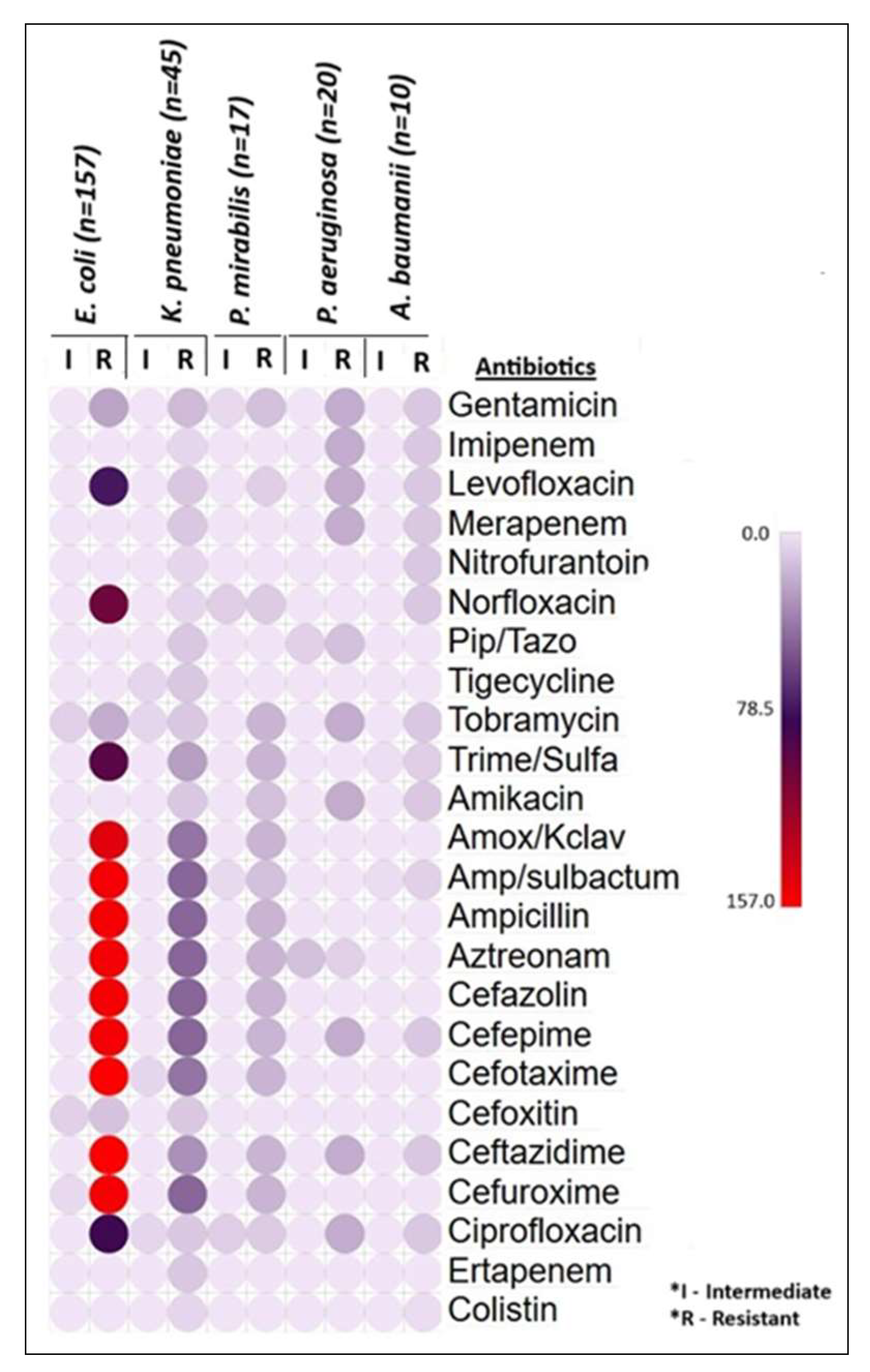
Among the ESBL-producing E. coli isolates (n = 157), 100% resistance was observed against cefotaxime and ceftazidime. High to moderate resistance rates were recorded for ampicillin/sulbactam, ampicillin, aztreonam, cefazolin, cefepime, cefuroxime (97.5%), amoxicillin/clavulanate (92.5%), norfloxacin (63.7%), trimethoprim/sulfamethoxazole (58.6%), ciprofloxacin (51.6%), and levofloxacin (46.5%). Lower resistance rates were noted for gentamicin (14.6%) and tobramycin (12.7%), while the lowest resistance was observed for cefoxitin (7.6%). Notably, all the E. coli isolates were 100% sensitive to other remaining antibiotics. In K. pneumoniae isolates (n = 45), 100% resistance was observed in ampicillin, ampicillin/sulbactam, aztreonam, cefazolin, cefepime, and cefuroxime. High resistance rates were also noted for amoxicillin/clavulanate (89%) and cefotaxime (89%). Moderate resistance levels were found for ceftazidime (66.7%), nitrofurantoin (66.7%), norfloxacin (55.6%), and trimethoprim/sulfamethoxazole (55.6%). Variable resistance were noted to multiple antibiotics and no complete susceptibility observed on the K. pneumoniae isolates. For ESBL-producing P. mirabilis isolates (n = 17), 100% resistance was observed to 10 antibiotics including beta-lactams and tobramycin. 76.5% of P. mirabilis isolates resistance to gentamicin and amikacin, 52.9% of isolates were resistant to norfloxacin, ciprofloxacin, and levofloxacin. The bacteria showed intermediate resistance to norfloxacin, ciprofloxacin, gentamicin, and ampicillin/sulbactam. They were 100% sensitive to the remaining antibiotics. P. aeruginosa (n = 20) showed 100% resistance to the drugs gentamicin, imipenem, levofloxacin, meropenem, tobramycin, amikacin, cefepime, ceftazidime, and ciprofloxacin. 65% and 35% of them were resistant to the drugs piperacillin/tazobactam and aztreonam, respectively. None of them were 100% sensitive to any antibiotics used in the test except colistin. Similarly, the bacteria A. baumannii (n = 10) showed 100% resistance to 11 different drugs. 70–80% of the strains were resistant to ampicillin/sulbactam and trimethoprim/sulfamethoxazole, and 30% of them were resistant to colistin. The details of the antibiotic sensitivity pattern of all the isolates are illustrated in Table 4 and Figure 4.

3.3. Unveiling the ESBL and Carbapenem-Resistant Genes in Gram-Negative Bacterial Pathogens

All the ESBL and carbapenem-resistant bacteria (n = 249) had positive results for all three ESBL genes and carbapenem-resistant genes blaNDM and blaIMP, either harboring them singly or in multiple copies. Among the 249 isolates, E. coli (n = 157; 63%) harbored all the ESBL genes, with blaCTX-M (n = 72; 46%) being the most identified, followed by blaTEM (n = 59; 37.6%) and blaSHV (n = 31; 20%). Of the 157 E. coli isolates, 20.33% (n = 32) co-harbored more than one gene, such as blaCTX-M+TEM or blaCTX-M+SHV. In these combinations, blaCTX-M+TEM (n = 15; 9.5%) and blaCTX-M+SHV (n = 15; 9.5%) were detected in 15 E. coli strains, while all three genes were identified together in 3.8% of the E. coli isolates. Similarly, blaCTX-M (n = 18; 40%) was the predominant gene in MDR K. pneumoniae (n = 45), followed by blaTEM (n = 14; 31%), blaSHV (n = 14; 31%), blaIMP (n = 6; 13.3%), and blaNDM (n = 1; 2.2%). Combinations of genes like blaCTX-M+TEM (n = 14; 31%), blaCTX-M+SHV (n = 10; 22%), blaTEM+SHV (n = 3; 6.6%), or blaTEM+NDM (n = 1; 2%) were detected in 62.22% of the K. pneumoniae isolates. In P. aeruginosa, blaCTX-M and blaTEM were detected in almost all isolates, with blaIMP found in 7 (35%) strains and blaSHV in 13 (65%) strains. Gene combinations like blaCTX-M+SHV (n = 1; 5%) and blaCTX-M+TEM+SHV+IMP (n = 1; 5%) were detected in 10% of the organisms. Similarly, blaCTX-M was detected in all 17 ESBL-positive P. mirabilis strains (100%), with blaTEM found in 8 strains (47.1%). Among the 17 P. mirabilis strains, 17.6% (n = 3) were positive for both genes (blaCTX-M+TEM). These resistant-gene-carrying bacterial strains were predominantly isolated from urine and pus samples, as detailed in Table 5 and Figure 5.
All the A. baumannii isolates (n = 10; 100%) showed positive for the genes blaCTX-M and blaTEM. Additionally, 20% (n = 2) of the strains individually carried the genes blaNDM and blaSHV, while blaIMP was detected in 10% of the A. baumannii isolates from the sputum samples. The gene combinations blaTEM+NDM, blaTEM+IMP, and blaCTX-M+TEM+SHV+IMP were each detected in 10% (n = 1) of the strains. None of the ESBL and carbapenem-producing strains tested positive for the gene blaOXA-48 and the above-mentioned details are given in Table 6 and Figure 6.

3.4. Genomic Sequencing and Phylogenetic Tree Relationships of the blaCTX-M

The partial genomic sequence of the gene blaCTX-M was compared with other existing blaCTX-M genes from various Gram-negative bacterial pathogens already deposited in the GenBank, NCBI. Our genomic sequence blaCTX-M showed 99.74% similarity with blaCTX-M genes from E. coli and K. pneumoniae. Using the 99.74% similarity index, a cladogram of the phylogenetic tree was constructed. The cladogram of the phylogenetic tree is illustrated in Figure 7.

4. Discussion

The present study collected 3670 various clinical samples, of which the urine samples were predominantly (51.2%) received and had shown the highest bacterial growth rate (49.1%). The bacterial growth rate was significant among the samples collected, and the overall positivity rate of the bacterial growth was 30%.
Over the recent decades, the spread of ESBL and carbapenemase-enzyme-producing Enterobacteriaceae and non-fermentative Gram-negative bacteria, especially P. aeruginosa, P. mirabilis, and A. baumannii, pose a significant threat to healthcare of Arabian Peninsula region [28]. These organisms exhibit resistance to multiple classes of antibiotics, including last-resort drugs like carbapenems, limiting treatment options and increasing morbidity, mortality, and healthcare costs [14,29]. Reduced treatment options, complex infections, and costly treatments are some of the major concerns for people infected with ESBL and carbapenemase-enzyme-producing organisms. A major driver of ESBL and carbapenemase-producing Gram-negative fermentative and non-fermentative bacteria is the horizontal transfer of mobile genetic elements carrying genes for ESBL and/or carbapenemase enzyme production [13].
In this study, 64.3% of the bacterial isolates were Gram-negative bacteria and the remaining 35.7% were Gram-positive bacteria. Among the Gram-negative bacteria, the E. coli (42.3%) was the most predominant isolate followed by K. pneumoniae (19.4%), P. aeruginosa (13.5%), A. baumannii (7.3%), and P. mirabilis (3.3%). In 2020, National AMR Surveillance system of the UAE [30] reported that the E. coli was the most commonly (80%) isolated Gram-negative bacteria, then K. pneumoniae (55–60%), P. aeruginosa (20%), A. baumannii (<10%), and P. mirabilis (<5%).
A comparison of the resistance rate of ESBL E. coli strains to previous studies across other Gulf Cooperation Council (GCC) countries revealed similar patterns. With 100% resistance to drugs such as cefotaxime and ceftazidime [27,31], there was a 46.5–97.5% (medium–high resistance) rate to 11 different beta-lactams and a low resistance rate to gentamycin and tobramycin [32,33]. Furthermore, our study found ESBL E. coli showing 100% sensitivity to eight antibiotics (Table 4) which is similar to studies from Qatar and Saudi Arabia [34,35]. The predominance of ESBL E. coli in urine samples (Table 5) highlights the widespread occurrence of urinary tract infections (UTIs). UTIs are common, but when caused by ESBL strains, they can lead to severe complications such as sepsis, prolonged hospital stays, and even increased mortality [36].
The ESBL K. pnuemoniae strains showed complete resistance to various antibiotics like ampicillin, aztreonam, cefazolin, cefepime, and cefuroxime which was consistent with findings from studies conducted in UAE [37,38] and other GCC countries [15,31,34,39]. The strain exhibited 89% resistance rate to cefotaxime and amoxicillin + clavulanate but in contrast, a lower resistance rate to the same drugs was reported in Kuwait [40], UAE [38], Qatar [34], and Saudi Arabia [32]. Interestingly, the rate of resistance to antibiotics such as tigecycline and meropenem in our study rose to 22% from a mere 8% reported in previous studies indicating the prevalence of carbapenem-resistant K. pnuemoniae [27,41]. Moreover, there has been a significant rise in the colistin resistance rate from 0 to over 20% over the last decade, posing a serious threat as colistin is currently the last-line treatment for carbapenem-resistant K. pneumoniae [28]. Antibiotic sensitivity pattern of ESBL P. mirabilis revealed high resistance to most beta-lactams as expected but least resistance to carbapenems with sensitivity to colistin. Similarly, a study in Saudi Arabia reported P. mirabilis being susceptible to carbapenems such as meropenem, imipenem, etc., but was reported as intrinsically resistant to colistin [42]. P. mirabilis is the second leading cause of UTIs, after E. coli, hence using carbapenems or piperacillin/tazobactam antibiotics can help manage ESBL P. mirabilis infections effectively.
The bacteria P. aeruginosa was 100% resistant to nine different drugs and 100% susceptible to colistin and medium–low resistance rate was noted in piperacillin/tazobactam (65%) and aztreonam (35%). In contrast to our study, the studies from Sudan [43] and Egypt [44] observed that the bacteria had low resistance rate and was highly susceptible to the above-mentioned drugs. In the case of A. baumannii, the bacteria were 100% resistant to 11 drugs (Table 4), 70–80% resistance rate was observed with ampicillin/sulbactam and trimethoprim/sulfamethoxazole and a 30% resistance to colistin. This was higher compared to studies from Oman [45] and Iran [46]. Resistance to colistin is rapidly emerging among the MDR A. baumannii and K. pneumoniae, which is becoming a global burden [47,48].
Molecular characterization of the resistant Gram-negative isolate was carried out to determine prevalence rate of class A ESBL genes (blaCTX-M, blaSHV, blaTEM) and carbapenem-resistant genes (blaIMP, blaNDM, bla0XA-48) within UAE. The most predominant ESBL gene identified was blaCTX-M with high prevalence in E. coli (45.9%), P. mirabilis (58.8%), K. pneumoniae (42.2%), A. baumannii (40%), and P. aeruginosa (35%) followed by blaTEM and blaSHV mainly in the E. coli (35.6% and 18.5%) and K. pneumoniae (16.4% and 26.7%). A similar pattern of gene prevalence was noted in other GCC countries [27,34,38,39]. In contrast, studies from Iraq [49], India [50], and Egypt [51] stated that the blaTEM was the most predominant gene in the Enterobacterales followed by blaCTX-M and blaSHV.
The carbapenemase-encoding gene blaIMP was detected in 13.3% of K. pneumoniae isolated from the urine samples and in 10% of A. baumannii from sputum and 10% of P. aeruginosa from both the samples. Likewise, 20% of A. baumannii and 2.2% of K. pneumoniae harbored the blaNDM. Almost 94% of them were positive to the blaIMP in Bharain [52] where 6% of the A. baumannii were positive to the blaNDM, whereas 8.7% of the K. pneumoniae were positive to the blaIMP in Saudi Arabia [41]. A global perspective on carbapenem-resistant A. baumannii (CRAB) revealed predominance of blaOXA-48 [53]. In neighboring countries like Saudi Arabia, roughly 71% of the carbapenem-resistance gene was OXA-48, mostly detected on K. pneumoniae CRE isolates [41]. In the North African country Morocco, 83% of carbapenemase-producing Enterobacterales carried OXA-48 [54]. However, a study from Qatar stated that 30% of the carbapenemase producers carried OXA-48 gene and NDM-1 as well [34]. But in contrast, our study did not find blaOXA-48 amongst any bacterial isolate. The emergence of carbapenem-resistant bacterial isolates poses a significant threat, as carbapenems are considered the last-resort antibiotics for treating infections caused by ESBL-producing pathogens.
Additionally, our study found co-existence of resistant genes in various combinations in bacterial pathogens. The highest co-existence patterns were blaCTX-M+TEM and blaCTX-M+SHV represented by E. coli (9.6% of each), K. pneumoniae (31.1% and 22.2%), and P. aeruginosa (15% of each). In 17.65% of the P. mirabilis, the co-existence gene was blaCTX-M+TEM. The lowest co-existence patterns were blaTEM+SHV and blaCTX-M+TEM+SHV represented by K. pneumoniae (6.67%) and E. coli (3.82%), respectively. Similarly, the highest co-existence of the gene blaCTX-M+TEM (21%) was found in the ESBL E. coli and K. pneumoniae from Qatar [34], Saudi Arabia [15,27], and followed by the least co-existence of the genes blaTEM+SHV, blaCTX-M+SHV, and blaCTX-M+TEM+SHV which were detected in the same studies; in contrast, Moglad et al. [55], Badger-Emeka et al. [29], and Sid-Ahmed et al. [56] found that the highest co-harbored genes were blaCTX-M+TEM+SHV (50% and 24.7%) and blaCTX-M+SHV (50% and 10.1%) found in the E. coli, K. pneumoniae, and other Enterobacterales in their studies. Likewise, the lowest co-expression of the genes blaTEM+NDM, blaTEM+IMP, and blaCTX-M+TEM+SHV+IMP were also detected in 2.22% K. pneumoniae, 5% P. aeruginosa, and 10% of each A. baumannii to each gene combination. In such a way, Elbadawi HS et al. found in their study that the lowest co-expression of the ESBL and carbapenemase-producing genes were noticed in the K. pneumoniae, A. baumannii, E. coli, and P. aeruginosa [57].
As with all other studies, this study also reported a few limitations as follows. The study samples were received from TUH, other Thumbay hospitals, and clinics located in and around the northern emirates of the UAE (Ajman, Sharjah, and Fujairah) as well as other hospitals and clinics from the same northern part of the UAE, including Umm Al Quwain. However, the study samples were not received and collected from other emirates including Dubai, Abu Dhabi, and Ras Al Khaimah. The carbapenemase-encoding gene blaOXA-48 was not detected in any of the ESBL and carbapenem-resistant Gram-negative bacterial pathogens, even in A. baumannii. In this study, genomic sequencing of the predominant gene blaCTX-M was performed by the Sanger method, but no other class A genes were sequenced. In addition, patient demographic details and their clinical correlations, including critical care unit patients, are not included in this study and statistical analysis correlating genotypic profiles with phenotypic resistance patterns was not conducted. These important limitations should be addressed in future studies.

5. Conclusions

The current findings confirm UAE to have a slightly high prevalence of the ESBL and carbapenem-resistant genes. According to the study the gene blaCTX-M, blaTEM and blaSHV were the most detected in the E. coli and K. pneumoniae especially isolated from the urine samples. The highest percentage of the bacteria K. pneumoniae harbored all the ESBL and carbapenemase-resistant genes, and the multiple two-gene combinations like blaCTX-M+TEM, blaCTX-M+SHV, blaTEM+SHV, and blaTEM+NDM as well. A. baumannii also harbored all the ESBL and carbapenem-resistant genes, and co-harbored the genes blaTEM+NDM, blaTEM+IMP, and blaCTX-M+TEM+SHV+IMP. The current findings underscore the critical importance of rational antibiotic therapy, supported by ongoing surveillance and robust infection control strategies, to effectively curb the spread of ESBL and carbapenem-resistant genes in Gram-negative bacteria. In the UAE, where blaCTX-M-mediated resistance is particularly dominant, empirical antibiotic therapy must be strategically revised based on local antimicrobial resistance patterns. A multifaceted approach combining data-driven antibiotic selection, strong antimicrobial stewardship, enhanced molecular surveillance of resistance genes, and enhanced infection prevention measures is essential to manage and contain the rising threat of multidrug-resistant ESBL-producing pathogens in the region.

Supplementary Materials

The following supporting information can be downloaded at: https://www.mdpi.com/article/10.3390/microorganisms13081880/s1. Table S1: Test wells on the MicroScan Gram-negative panels (Neg Breakpoint Combo 50); Table S2: Antibiotic sensitivity test wells on the MicroScan Gram-negative panels (Neg Breakpoint Combo 50).

Author Contributions

Conceptualization, N.R.; Methodology, N.R., P.R., V.K., A.F.S. and M.A.M.; Formal analysis, P.R., V.K., A.F.S. and M.A.M.; Investigation, N.R., P.R., V.K., A.F.S. and M.A.M.; Writing—original draft, P.R., V.K., A.F.S., M.A.M., N.R. and S.B.; Writing—review and editing, N.R., P.R. and S.B.; Project administration, N.R. and N.P.; Software, N.R., P.R. and V.K.; Supervision, N.R. and N.P.; Validation, N.R., P.R. and S.B. All authors have read and agreed to the published version of the manuscript.

Funding

This research received no external funding.

Institutional Review Board Statement

This study was approved by the Institutional Review Board (IRB) of Gulf Medical University, Ajman, UAE with an approval letter dated 14 October 2021, and reference number IRB/COM/STD/101/Oct.2021.

Informed Consent Statement

Not applicable.

Data Availability Statement

The raw data supporting the conclusions of this article will be made available by the authors on request.

Acknowledgments

We would like to acknowledge the efforts of Physicians, Microbiologists, and other relevant staffs of Thumbay Laboratory and Thumbay University Hospital, Ajman, UAE for collecting the Gram-negative bacterial pathogens and data and conducting this study. Also, we want to express our gratitude to the College of Medicine, GMU, for its constant support in conducting this research. Additionally, the authors would like to thank Macrogen, South Korea, for the sequencing service for the gene blaCTX-M by Sanger method.

Conflicts of Interest

The authors declare no conflicts of interest.

Abbreviations

The following abbreviations are used in this manuscript:
AMRAntimicrobial resistance
MDRMultidrug resistant
ESBLExtended Spectrum of Beta-lactamase
GMUGulf Medical University
TUHThumbay University Hospital
GCCGulf Cooperation Council
UAEUnited Arab Emirates

References

  1. Aljeldah, M.M. Antimicrobial Resistance and Its Spread Is a Global Threat. Antibiotics 2022, 11, 1082. [Google Scholar] [CrossRef]
  2. de Kraker, M.E.A.; Stewardson, A.J.; Harbarth, S. Will 10 Million People Die a Year due to Antimicrobial Resistance by 2050? PLoS Med. 2016, 13, e1002184. [Google Scholar] [CrossRef] [PubMed]
  3. Breijyeh, Z.; Jubeh, B.; Karaman, R. Resistance of Gram-Negative Bacteria to Current Antibacterial Agents and Approaches to Resolve It. Molecules 2020, 26, 1340. [Google Scholar] [CrossRef]
  4. Tooke, C.L.; Hinchliffe, P.; Bragginton, E.C.; Colenso, C.K.; Hirvonen, V.H.A.; Takebayashi, Y.; Spencer, J. β-Lactamases and β-Lactamase Inhibitors in the 21st Century. J. Mol. Biol. 2019, 431, 3472–3500. [Google Scholar] [CrossRef]
  5. Exner, M.; Bhattacharya, S.; Christiansen, B.; Gebel, J.; Goroncy-Bermes, P.; Hartemann, P.; Heeg, P.; Ilschner, C.; Kramer, A.; Larson, E.; et al. Antibiotic resistance: What is so special about multidrug-resistant Gram-negative bacteria? GMS Hyg. Infect. Control 2017, 12, 1–24. [Google Scholar]
  6. ElAila, N.A.; Al-Laham, N.A.; Ayesh, B.M. Prevalence of extended spectrum beta lactamase and molecular detection of blaTEM, blaSHV and blaCTX-M genotypes among Gram negative bacilli isolates from pediatric patient population in Gaza strip. BMC Infect. Dis. 2023, 23, 99. [Google Scholar]
  7. Sawa, T.; Kooguchi, K.; Moriyama, K. Molecular diversity of extended-spectrum β-lactamases and carbapenemases, and antimicrobial resistance. J. Intensive Care 2020, 8, 13. [Google Scholar] [CrossRef] [PubMed]
  8. Ramadan, A.A.; Abdelaziz, N.A.; Amin, M.A.; Aziz, R.K. Novel blaCTX-M variants and genotype-phenotype correlations among clinical isolates of extended spectrum beta lactamase-producing Escherichia coli. Sci. Rep. 2019, 9, 4224. [Google Scholar] [CrossRef]
  9. Chong, Y.; Shimoda, S.; Shimono, N. Current epidemiology, genetic evolution and clinical impact of extended-spectrum β-lactamase-producing Escherichia coli and Klebsiella pneumoniae. Infect Genet. Evol. 2018, 61, 185–188. [Google Scholar] [CrossRef]
  10. Mahony, M.; McMullan, B.; Brown, J.; Kennedy, S.E. Multidrug-resistant organisms in urinary tract infections in children. Pediatr. Nephrol. 2020, 35, 1563–1573. [Google Scholar] [CrossRef]
  11. Tsilipounidaki, K.; Athanasakopoulou, Z.; Müller, E.; Burgold-Voigt, S.; Florou, Z.; Braun, S.D.; Monecke, S.; Gatselis, N.K.; Zachou, K.; Stefos, A.; et al. Plethora of Resistance Genes in Carbapenem-Resistant Gram-Negative Bacteria in Greece: No End to a Continuous Genetic Evolution. Microorganisms 2022, 10, 159. [Google Scholar] [CrossRef]
  12. Cantón, R.; Akóva, M.; Carmeli, Y.; Giske, C.G.; Glupczynski, Y.; Gniadkowski, M.; Livermore, D.M.; Miriagou, V.; Naas, T.; Rossolini, G.M.; et al. Rapid evolution and spread of carbapenemases among Enterobacteriaceae in Europe. Clin. Microbiol. Infect. 2012, 18, 413–431. [Google Scholar] [CrossRef]
  13. Halat, D.H.; Moubareck, C.A. The Current Burden of Carbapenemases: Review of Significant Properties and Dissemination among Gram-Negative Bacteria. Antibiotics 2020, 9, 186. [Google Scholar] [CrossRef]
  14. Tufa, T.B.; Mackenzie, C.R.; Orth, H.M.; Wienemann, T.; Nordmann, T.; Abdissa, S.; Hurissa, Z.; Schönfeld, A.; Bosselmann, M.; Häussinger, D.; et al. Prevalence and characterization of antimicrobial resistance among gram-negative bacteria isolated from febrile hospitalized patients in central Ethiopia. Antimicrob. Resist. Infect. Control 2022, 11, 8. [Google Scholar] [CrossRef] [PubMed]
  15. Ibrahim, M.E.; Algak, T.B.; Abbas, M.; Elamin, B.K. Emergence of bla TEM, bla CTX-M, bla SHV and bla OXA genes in multidrug-resistant Enterobacteriaceae and Acinetobacter baumannii in Saudi Arabia. Exp. Ther. Med. 2021, 22, 1450. [Google Scholar] [CrossRef]
  16. Moghnieh, R.A.; Kanafani, Z.A.; Tabaja, H.Z.; Sharara, S.L.; Awad, L.S.; Kanj, S.S. Epidemiology of common resistant bacterial pathogens in the countries of the Arab League. Lancet Infect. Dis. 2018, 18, e379–e394. [Google Scholar] [CrossRef]
  17. Al-Musawi, T.; Al-Agha, R.; Al-Khiami, S.; Al-Shamari, H.; Baghdadi, M.; Bosaeed, M.; Hadi, H.A.; Mady, A.; Sabra, N. Bacteremia in the Gulf Cooperation Council Region: A Review of the Literature 2013–2023. Infect. Drug Resist. 2025, 18, 2329–2355. [Google Scholar] [CrossRef] [PubMed]
  18. Thomsen, J.; Najiba, M.; Abdulrazzaq, N.M.; AlRand, H. The UAE AMR Surveillance Consortium. Surveillance of antimicrobial resistance in the United Arab Emirates: The early implementation phase. Front. Public Health 2023, 11, 1247627. [Google Scholar] [CrossRef]
  19. Kebede, B.; Yihunie, W.; Abebe, D.; Tegegne, B.A.; Belayneh, A. Gram-negative bacteria isolates and their antibiotic-resistance patterns among pediatrics patients in Ethiopia: A systematic review. SAGE Open Med. 2022, 10, 20503121221094191. [Google Scholar] [CrossRef] [PubMed]
  20. Srinivasan, U.; Ponnaluri, S.; Villareal, L.; Gillespie, B.; Wen, A.; Miles, A.; Bucholz, B.; Marrs, C.F.; Iyer, R.K.; Misra, D.; et al. Gram Stains: A Resource for Retrospective Analysis of Bacterial Pathogens in Clinical Studies. PLoS ONE 2012, 7, e42898. [Google Scholar] [CrossRef]
  21. Beckman Coulter. MicroScan Neg Breakpoint Combo 50. Beckman Coulter, Inc. Brea, California. Available online: https://www.beckmancoulter.com/search#q=MicroScan%20Neg%20Breakpoint%20Combo%2050%20panel%20for%20Gram-negative%20organism&t=techdocs&sort=relevancy&numberOfResults=25&f:@td_parenttaxonomy=[Microbiology] (accessed on 16 October 2021).
  22. CLSI M100; Performance Standards for Antimicrobial Susceptibility Testing. Clinical and Laboratory Standards Institute: Wayne, NJ, USA, 2020. Available online: https://clsi.org/shop/standards/m100/ (accessed on 16 October 2021).
  23. Beckman Coulter. DxM MicroScan WalkAway ID/AST System. Beckman Coulter, Inc. Brea, California. Available online: https://www.beckmancoulter.com/en/products/microbiology/dxm-microscan-walkaway-system?id=B1018-496 (accessed on 16 October 2021).
  24. Beckman Coulter. Beckman Coulter MicroScan LabPro Software WalkAway 9631049. Information Management Software LabPro Connect. Beckman Coulter, Inc. Brea, California. Available online: https://www.beckmancoulter.com/search#q=Beckman%20Coulter%20MicroScan%20LabPro%20software%20WalkAway%209631049%20&t=products&sort=relevancy&numberOfResults=25&f:@td_parenttaxonomy=[Microbiology] (accessed on 16 October 2021).
  25. G-Spin. Genomic DNA Extraction Kit for Bacteria. Available online: https://intronbio.com:6001/intronbioen/product/product_view.php?PRDT_ID=9 (accessed on 1 November 2021).
  26. Mahalingam, N.; Manivannan, B.; Khamari, B.; Siddaramappa, S.; Adak, S.; Bulagonda, E.P. Detection of Antibiotic Resistance Determinants and Their Transmissibility among Clinically Isolated Carbapenem-Resistant Escherichia coli from South India. Med. Princ. Pract. 2018, 8, 428–435. [Google Scholar] [CrossRef]
  27. Al-Agamy, M.H.; Aljallal, A.; Radwan, H.H.; Shibl, A.M. Characterization of carbapenemases, ESBLs, and plasmid-mediated quinolone determinants in carbapenem-insensitive Escherichia coli and Klebsiella pneumoniae in Riyadh hospitals. J. Infect. Pub. Health 2018, 11, 64–68. [Google Scholar] [CrossRef] [PubMed]
  28. Sonnevend, Á.; Ghazawi, A.A.; Hashmey, R.; Jamal, W.; Rotimi, V.O.; Shibl, A.M.; Al-Jardani, A.; Al-Abri, S.S.; Tariq, W.U.Z.; Weber, S.; et al. Characterization of carbapenem-resistant Enterobacteriaceae with high rate of autochthonous transmission in the Arabian Peninsula. PLoS ONE 2015, 10, e0131372. [Google Scholar] [CrossRef]
  29. Badger-Emeka, L.I.; Kausar, N.; Estrella, E.; Angeles, G.B. A Three-Year Look at the Phylogenetic Profile, Antimicrobial Resistance, and Associated Virulence Genes of Uropathogenic Escherichia coli. Pathogens 2022, 11, 631. [Google Scholar] [CrossRef] [PubMed]
  30. Thomsen, J. National AMR Surveillance Report. United Arab Emirates Ministry of Health and Prevention. 2022. Available online: https://mohap.gov.ae/en/w/national-amr-surveillance-report-2022 (accessed on 2 December 2022).
  31. Ardati, K.O.; Murdeshwar, S.R.; Chacko, S.T.; Jagtap, A.; Jacob, S. Prevalence of Bacterial Pathogens and Antimicrobial Susceptibility Pattern in Bahrain Tertiary Care Hospital. J. Bahrain Med. Soc. 2019, 31, 5–16. [Google Scholar]
  32. Kabrah, A. Extended-Spectrum Beta-Lactamase and Carbapenem-Resistant Gram-Negative Pathogens in Makkah, Saudi Arabia. Ethiop. J. Health Sci. 2022, 36, 1221–1230. [Google Scholar]
  33. Shaaban, O.A.; Mahmoud, N.A.; Zeidan, A.A.; Kumar, N.; Finan, A.C. Prevalence and Resistance Patterns of Pediatric Urinary Tract Infections in Bahrain. Cureus 2021, 3, e20859. [Google Scholar] [CrossRef]
  34. Perez-Lopez, A.; Sundararaju, S.; Al-Mana, H.; Tsui, K.M.; Hasan, M.R.; Suleiman, M.; Janahi, M.; Al Maslamani, E.; Tang, P. Molecular Characterization of Extended-Spectrum β-Lactamase–Producing Escherichia coli and Klebsiella pneumoniae Among the Pediatric Population in Qatar. Front. Microbiol. 2020, 11, 581711. [Google Scholar] [CrossRef]
  35. Al-Tawfiq, J.A.; Rabaan, A.A.; Saunar, J.V.; Bazzi, A.M. Antimicrobial resistance of gram-negative bacteria: A six-year longitudinal study in a hospital in Saudi Arabia. J. Infect. Public Health 2020, 3, 737–745. [Google Scholar] [CrossRef] [PubMed]
  36. Brune, J.E.; Dickenmann, M.; Sidler, D.; Walti, L.N.; Golshayan, D.; Manuel, O.; Haidar, F.; Neofytos, D.; Schnyder, A.; Boggian, K.; et al. Frequency and impact on renal transplant outcomes of urinary tract infections due to extended-spectrum beta-lactamase-producing Escherichia coli and Klebsiella species. Front. Med. 2024, 11, 1329778. [Google Scholar] [CrossRef]
  37. Alfaresi, M.; Sing, G.K.; Senok, A. First Report of blaCTX-M-28 in Enterobacteriaceae Isolates in the United Arab Emirates. J. Pathog. 2018, 2018, 1304793. [Google Scholar] [CrossRef]
  38. Dash, N.R.; Albataineh, M.T.; Alhourani, N.; Khoudeir, A.M.; Ghanim, M.; Wasim, M.; Mahmoud, I. Community-acquired urinary tract infections due to extended-spectrum β-lactamase-producing organisms in United Arab Emirates. Travel Med. Infect. Dis. 2018, 22, 46–50. [Google Scholar] [CrossRef]
  39. Azim, N.S.A.; Nofal, M.Y.; Nofal, M.Y.; Al-Harbi, M.A.; Al-Zaban, M.I.; Somily, A.M. Molecular-diversity, Prevalence and Antibiotic Susceptibility of Pathogenic Klebsiella Pneumoniae under Saudi Condition. Pak. J. Biol. Sci. 2019, 22, 174–179. [Google Scholar] [CrossRef]
  40. Al-Sweih, N.; Jamal, W.; Mokaddas, E.; Habashy, N.; Kurdi, A.; Mohamed, N. Evaluation of the in vitro activity of ceftaroline, ceftazidime/avibactam and comparator antimicrobial agents against clinical isolates from paediatric patients in Kuwait: ATLAS data 2012–19. JAC Antimicrob. Resist. 2021, 3, dlab159. [Google Scholar] [CrossRef] [PubMed]
  41. Brek, T.; Alghamdi, A.K.; Abujamel, T.S.; Yasir, M.; Alattas, E.M.; Hazazi, M.S.; Al-Zahrani, I.A. Prevalence and molecular determinants of carbapenemase-producing Klebsiella pneumoniae isolated from Jazan, Saudi Arabia. J. Infect. Dev. Ctries. 2023, 17, 1420–1429. [Google Scholar] [CrossRef]
  42. Hafiz, T.A.; Alghamdi, G.S.; Alkudmani, Z.S.; Alyami, A.S.; AlMazyed, A.; Alhumaidan, O.S.; Mubaraki, M.A.; Alotaibi, F.E. Multidrug-Resistant Proteus mirabilis Infections and Clinical Outcome at Tertiary Hospital in Riyadh, Saudi Arabia. Infect. Drug Resist. 2024, 17, 571–581. [Google Scholar] [CrossRef]
  43. Omer, T.H.S.; Mustafa, S.A.M.; Mohamed, S.O.O. Extended Spectrum β-Lactamase-Mediated Resistance and Antibiogram of Pseudomonas aeruginosa Isolates from Patients Attending Two Public Hospitals in Khartoum, Sudan. Int. J. Microb. 2020, 2020, 2313504. [Google Scholar] [CrossRef] [PubMed]
  44. Elhariri, M.; Hamza, D.; Elhelw, R.; Dorgham, S.M. Extended-spectrum beta-lactamase-producing Pseudomonas aeruginosa in camel in Egypt: Potential human hazard. Ann. Clin. Microbiol. Antimicrob. 2017, 16, 21. [Google Scholar] [CrossRef]
  45. Sannathimmappa, M.B.; Nambiar, V.; Aravindakshan, R. Antibiotic Resistance Pattern of Acinetobacter baumannii Strains: A Retrospective Study from Oman. Saudi. J. Med. Med. Sci. 2021, 9, 254–260. [Google Scholar] [CrossRef] [PubMed]
  46. Basatian-Tashkan, B.; Niakan, M.; Khaledi, M.; Afkhami, H.; Sameni, F.; Bakhti, S.; Mirnejad, R. Antibiotic resistance assessment of Acinetobacter baumannii isolates from Tehran hospitals due to the presence of efflux pumps encoding genes adeA and adeS genes by molecular method. BMC Res. Notes 2020, 13, 543. [Google Scholar] [CrossRef]
  47. Bostanghadiri, N.; Narimisa, N.; Mirshekar, M.; Dadgar-Zankbar, L.; Taki, E.; Navidifar, T.; Darban-Sarokhalil, D. Prevalence of colistin resistance in clinical isolates of Acinetobacter baumannii: A systematic review and meta-analysis. Antimicrob. Resist. Infect. Control 2024, 13, 24. [Google Scholar] [CrossRef]
  48. Khuntayaporn, P.; Thirapanmethee, K.; Traidej Chomnawang, M.T. An Update of Mobile Colistin Resistance in Non-Fermentative Gram-Negative Bacilli. Front. Cell Infect. Microbiol. 2022, 12, 1–15. [Google Scholar] [CrossRef]
  49. Pishtiwan, A.H.; Khadija, K.M. Prevalence of blaTEM, blaSHV and blaCTX-M Genes among ESBL-Producing Klebsiella pneumoniae and Escherichia coli Isolated from Thalassemia Patients in Erbil, Iraq. Mediterr. J. Hematol. Infect. Dis. 2019, 11, e2019041. [Google Scholar]
  50. Bajpai, T.; Pandey, M.; Varma, M.; Bhatambare, G.S. Prevalence of TEM, SHV, and CTX-M Beta-Lactamase genes in the urinary isolates of a tertiary care hospital. Avicenna J. Med. 2017, 7, 12–16. [Google Scholar] [CrossRef]
  51. Zaki, M.; El-Halaby, H.; Elmansoury, E.; Zeid, M.; Khaled, K.; Nomir, M. Genetic Study of Extended Spectrum Beta-Lactamase and Carbapenemase Producing Escherichia coli Causing Sepsis among Egyptian Children. Open Microbiol. J. 2019, 13, 128–137. [Google Scholar] [CrossRef]
  52. Al-Rashed, N.; Bindayna, K.M.; Shahid, M.; Saeed, N.K.; Darwish, A.; Joji, R.M.; Al-Mahmeed, A. Prevalence of Carbapenemases in Carbapenem-Resistant Acinetobacter baumannii Isolates from the Kingdom of Bahrain. Antibiotics 2023, 12, 1198. [Google Scholar] [CrossRef] [PubMed]
  53. Müller, C.; Reuter, S.; Wille, J.; Xanthopoulou, K.; Stefanik, D.; Grundmann, H.; Higgins, P.G.; Seifert, H. A global view on carbapenem-resistant Acinetobacter baumannii. mBio 2023, 14, e0226023. [Google Scholar] [CrossRef] [PubMed]
  54. Belouad, E.M.; Benaissa, E.; El-Mrimar, N.; Bssaibis, F.; Maleb, A.; Elouennass, M. Predominance of OXA-48 Carbapenemase-Producing Enterobacterales in a Moroccan Hospital. Int. J. Microbiol. 2023, 2023, 8581883. [Google Scholar] [CrossRef]
  55. Moglad, E.; Alanazi, N.; Altayb, H.N. Genomic Study of Chromosomally and Plasmid-Mediated Multidrug Resistance and Virulence Determinants in Klebsiella Pneumoniae Isolates Obtained from a Tertiary Hospital in Al-Kharj, KSA. Antibiotics 2022, 11, 1564. [Google Scholar] [CrossRef]
  56. Sid Ahmed, M.A.; Bansal, D.; Acharya, A.; Elmi, A.A.; Hamid, J.M.; Sid Ahmed, A.M.; Chandra, P.; Ibrahim, E.; Sultan, A.A.; Doiphode, S.; et al. Antimicrobial susceptibility and molecular epidemiology of extended-spectrum beta-lactamase-producing Enterobacteriaceae from intensive care units at Hamad Medical Corporation, Qatar. Antimicrob. Resist. Infect. Cont. 2016, 5, 4. [Google Scholar] [CrossRef]
  57. Elbadawi, H.S.; Elhag, K.M.; Mahgoub, E.; Altayb, H.N.; Ntoumi, F.; Elton, L.; McHugh, T.D.; Tembo, J.; Ippolito, G.; Osman, A.Y.; et al. Detection and characterization of carbapenem resistant Gram-negative bacilli isolates recovered from hospitalized patients at Soba University Hospital, Sudan. BMC Microbiol. 2021, 21, 136. [Google Scholar] [CrossRef] [PubMed]
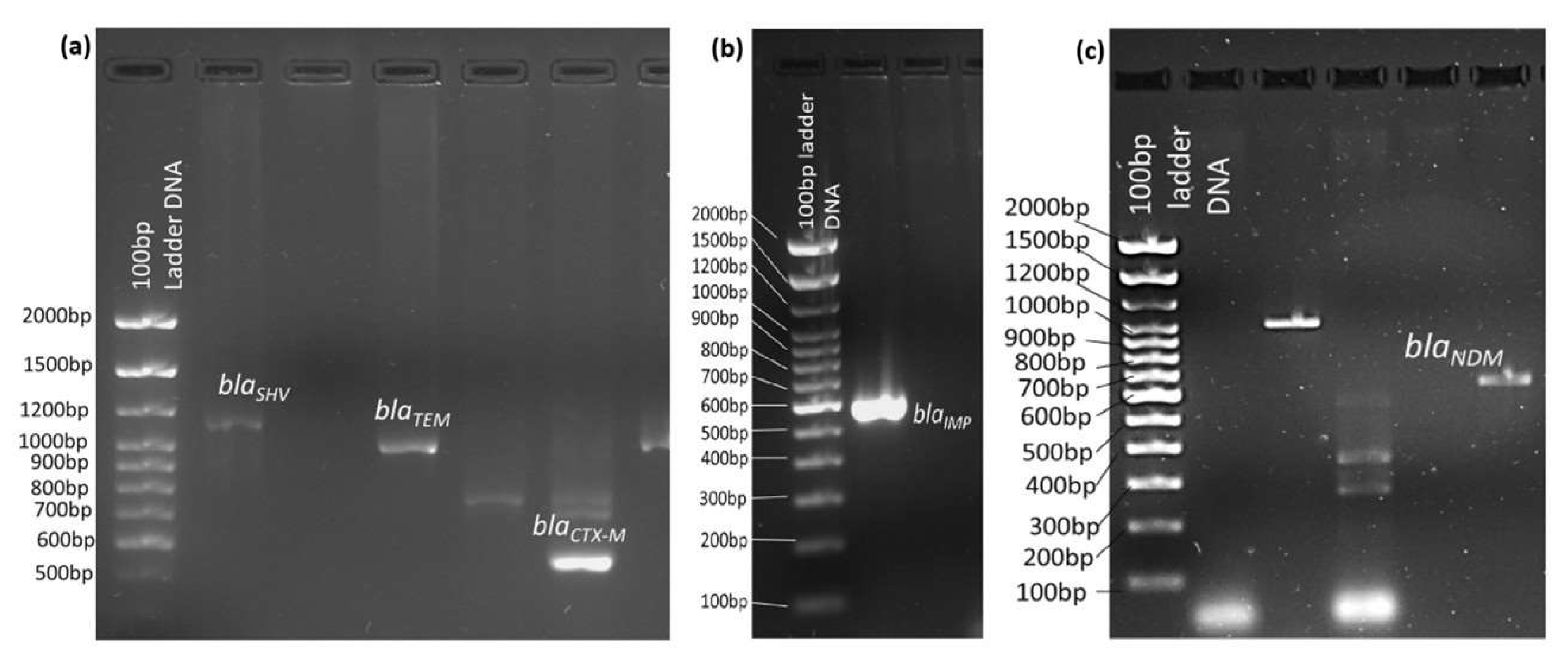
Figure 1. Detection of ESBL genes (a) blaCTX-M, blaTEM, and blaSHV, and carbapenemase genes (b) blaIMP, and (c) blaNDM gene bands and their base pair (bp) sizes using a 100 bp ladder DNA in 1% agarose gel electrophoresis.
Microorganisms 13 01880 g001
Figure 2. Number of Gram-negative and Gram-positive bacterial pathogens isolated from various clinical samples such as urine, blood, pus, and sputum.
Microorganisms 13 01880 g002
Figure 3. Number of different studied bacterial pathogens isolated from various clinical samples.
Microorganisms 13 01880 g003
Figure 4. Number of intermediate and resistant patterns of E. coli, K. pneumonia, P. mirabilis, P. aeruginosa, and A. baumannii to various common antibiotics.
Microorganisms 13 01880 g004
Figure 5. Frequency of single and mixed resistant ESBL and carbapenemase-encoding genes detected in E. coli, K. pneumoniae, P. aeruginosa, P. mirabilis, and A. baumannii.
Microorganisms 13 01880 g005
Figure 6. Frequency of detection of ESBL and carbapenemase-resistant genes in the studied bacterial pathogens.
Microorganisms 13 01880 g006
Figure 7. Cladogram of the phylogenetic tree of the gene blaCTX-M illustrates a 99.74% similarity with other blaCTX-M genes from various E. coli and K. pneumoniae isolates. The software Mega 11 was used to construct the tree.
Microorganisms 13 01880 g007
Table 1. Details of the primer sequences, amplicon size, and annealing temperature of the ESBL and Carbapenem-resistant genes.
Gene TypeAmpliconPrimer Sequence (5′-3′)Amplicon Size (bp)Annealing Temperature °CReference
ESBL
gene primers
blaCTX-MF-ATGTGCAGYACCAGTAARGTKAT GCC
R-TGGGTRARRTARGTSACCAGAAY CAGCGG
59358[15]
blaTEMF-TCGCCGCATACACTATTCTCAGAA TGA
R-ACGCTCACCGGCTCCAGATTTAT
44550
blaSHVF-GGGTTATTCTT ATTTGTCGC,
R-TTAGCGTTGCCAGTGCTC
74758
Carbapenem-resistant gene primersblaNDMF-GGTTTG GCGATCTGGTTTTC
R-CGGAATGGCTCATCACGATC
62157[27]
blaIMPF-CTACCGCAGCAGAGTCTTTG
R-AACCAGTTTTGCCTTACCAT
58756
blaOXA48F-GCGTGGTTAAGGATGAACAC
R-CATCAAGTTCAACCCAACCG
43857[15]
Control primer16S rRNA geneF-AGAGTTTGATCMTGGCTCAG
R-ACGGHTACCTTGTTACGACTT
150055
Table 2. No. of various clinical samples for positive bacterial growth.
Clinical
Samples
n = 3670 (%)
No. of Bacterial Pathogens Isolated n = 1098 (29.9%)No. of Samples for No Bacterial Growth
n = 2572 (70.1%)
p-Value/
Chi-Square Test
Gram-Negative Bacteria
n = 833 (22.7%)
Gram-Positive Bacteria
n = 265 (7.2%)
Urine
n = 1970 (53.7%)
503 (13.7%)37 (1.0%)1430 (39%)<0.001/335 *
Blood
n = 879 (23.9%)
48 (1.3%)71 (1.9%)760 (20.7%)
Pus
n = 469 (12.8%)
111 (3.0%)142 (3.9%)216 (5.9%)
Sputum
n = 352 (9.6%)
171 (4.7%)15 (0.4%)166 (4.5%)
* Chi-square test.
Table 3. Total no. of studied bacteria is isolated from various clinical samples.
No. of
Samples Positive
for Gram-Ve
Bacteria n (%)
No. of Gram-Ve Bacteria Isolated n = 833
E. coli
n = 394 (47.2%)
K. pneumoniae
n = 200 (24%)
P. aeruginosa
n = 129 (15.5%)
P. mirabilis
n = 40 (4.8%)
A. baumannii
n = 70 (8.4%)
Urine
n = 503 (60.3%)
354 (42.5%)116 (13.9%)15 (1.8%)12 (1.4%)6 (0.7%)
Blood
n = 48 (5.7%)
8 (0.9%)15 (1.8%)9 (1.1%)016 (1.9%)
Pus
n = 111 (13.3%)
25 (3.0%)35 (4.2%)19 (2.3%)24 (2.9%)8 (1.0%)
Sputum
n = 171 (20.5%)
7 (0.8%)34 (4.1%)86 (10.3%)4 (0.5%)40 (4.8%)
Table 4. Antibiotic sensitivity pattern of the studied bacteria isolated from various clinical samples.
AntibioticsE. coli
n = 157 (%)
K. pneumoniae
n = 45 (%)
P. mirabilis
n = 17 (%)
P. aeruginosa
n = 20 (%)
A. baumannii
n = 10 (%)
IR IRIRIRIR
Gentamicin 023 (15%)015 (33%)4 (23%)13 (77%)020 (100%)010 (100%)
Imipenem0005 (11%)00020 (100%)010 (100%)
Levofloxacin073 (46%)010 (22%)08 (47%)020 (100%)010 (100%)
Meropenem00010 (22%)00020 (100%)010 (100%)
Nitrofurantoin0005 (11%)0000010 (100%)
Norfloxacin0100 (64%)05 (11%)8 (47%)9 (53%)00010 (100%)
Piperacillin/Tazobactam00010 (22%)007 (35%)13 (65%)00
Tigecycline005 (11%)10 (22%)000000
Tobramycin7 (5%)20 (13%)5 (11%)10 (22%)017 (100%)020 (100%)010 (100%)
Trimethoprim/
Sulfamethoxazole
092 (59%)025 (55%)017 (100%)002 (20%)8 (80%)
Amikacin00010 (22%)013 (77%)020 (100%)010 (100%)
Amoxicillin +
Clavulanate
0145 (92%)040 (89%)017 (100%)0000
Ampicillin/
sulbactam
0153 (97%)045 (100%)4 (23%)13 (77%)003 (30%)7 (70%)
Ampicillin0153 (97%)045 (100%)017 (100%)0000
Aztreonam0153 (97%)031 (69%)017 (100%)13 (65%)7 (35%)00
Cefazolin0153 (97%)045 (100%)017 (100%)0000
Cefepime0153 (97%)045 (100%)017 (100%)020 (100%)010 (100%)
Cefotaxime0157 (100%)5 (11%)40 (89%)017 (100%)0000
Cefoxitin7 (5%)12 (7.6%)010 (22%)000000
Ceftazidime0157 (100%)030 (67%)017 (100%)020 (100%)010 (100%)
Cefuroxime4 (3%)153 (97%)045 (100%)017 (100%)0000
Ciprofloxacin081 (52%)5 (11%)10 (22%)8 (47%)9 (53%)020 (100%)010 (100%)
Ertapenem00010 (22%)000000
Colistin0005 (11%)000003 (30%)
Table 5. Number of ESBL and carbapenem-resistant genes in number of E. coli, K. pneumoniae, P. aeruginosa, P. mirabilis, and A. baumannii isolated from various clinical samples.
No. of Resistant Bacteria in Clinical Samples
n = 249 (%)
Single Gene DetectionMultiple Gene Detection
blaCTX-M
n = 111
blaTEM
n = 85
blaSHV
n = 45
blaNDM
n = 3
blaIMP
n = 6
blaCTX-M
+TEM
n = 33
blaCTX-M
+SHV
n = 26
blaTEM
+SHV
n = 3
blaCTX-M+TEM +SHV
n = 6
blaTEM +NDM
n = 2
blaTEM
+IMP
n = 1
blaCTX-M+TEM+SHV+IMP
n = 2
Urine
n = 156
E. coli
n = 120 (77%)
56
(46.7%)
44
(36.7%)
20
(16.7%)
0011
(9%)
11
(9%)
06
(5%)
000
K. pnuemoniae
n = 28 (18%)
8
(28.6%)
10
(35.7%)
7
(35.7%)
03
(21.4%)
8
(28.6%)
7
(25%)
3
(10.7%)
0000
P. aeruginosa
n = 5 (3%)
2
(40%)
2
(40%)
1
(20%)
000000000
P. mirabilis
n = 3 (2%)
2
(66%)
1
(33%)
0001
(33%)
000000
A. baumannii
n = 0
000000000000
Blood
n = 14
E. coli
n = 7 (50%)
5
(71.4%)
1
(14.3%)
1
(14.3%)
001
(14.3%)
000000
K. pnuemoniae
n = 2 (14.3%)
1
(50%)
1
(50%)
0000000000
P. aeruginosa
n = 2 (14.3%)
1
(50%)
01
(50%)
000000000
P. mirabilis
n = 0
000000000000
A. baumannii
n = 3 (2%)
2
(66%)
1
(33.3%)
0000000000
Pus
n = 49
E. coli
n = 25 (51%)
8 (32%) 10(40%)7 (28%)003 (12%)3 (12%)00000
K. pnuemoniae
n = 7 (14.3%)
5
(71.4%)
1
(14.3%)
1
(14.3%)
003
(43%)
2
(28.6%)
00000
P. aeruginosa
n = 3 (6%)
1
(33%)
1
(33%)
001
(33%)
0000000
P. mirabilis
n = 14 (28.6%)
8
(57%)
4
(43%)
0002
(14.3%)
000000
A. baumannii
n = 0
000000000000
Sputum
n = 30
E. coli
n = 5 (16.7%)
3
(60%)
1
(20%)
1
(20%)
0001
(20%)
00000
K. pnuemoniae
n = 8 (26.6%)
4
(50%)
2
(25%)
1
(12.5%)
1
(12.5%)
03
(37.5%)
1
(12.5%)
001
(12.5%)
00
P. aeruginosa
n = 10 (33%)
3
(42.9%)
5
(28.6%)
1
(14.3%)
01
(14.3%)
1
(14.3%)
1
(14.3%)
00001
(14.3%)
P. mirabilis
n = 0
000000000000
A. baumannii
n = 7 (23%)
2
(28.6%)
1
(14.3%)
1
(14.3%)
2
(28.6%)
1
(14.3%)
00001
(14.3%)
1
(14.3%)
1
(14.3%)
Table 6. Frequency of detection of ESBL and carbapenemase-encoding genes in the studied bacterial pathogens.
Detected
Genes
No. of Bacteria Carrying the Resistant Genes n = 249
E. coli
n = 157 (63%)
K. pnuemoniae
n = 45 (18%)
P. aeruginosa
n = 20 (8%)
P. mirabilis
n = 17 (6.8%)
A. baumannii
n = 10 (4%)
Single geneblaCTX-M n = 11172 (29%) 18 (40%)7 (35%)10 (59%)4 (40%)
blaTEM n = 8656 (35.7%)14 (31%)8 (40%)6 (35.3%)2 (20%)
blaSHV n = 4329 (18.5%)9 (20%)3 (15%)1 (5.9%)1 (10%)
blaNDM n = 301 (2.2%)002 (20%)
blaIMP n = 603 (6.6%)2 (10%)01 (10%)
Mixed genesblaCTX-M+TEM n = 3315 (9.5%)14 (31%)1 (5%)3 (17.7%)0
blaCTX-M+SHV n = 2615 (9.5%)10 (22.2%)1 (5%)00
blaTEM+SHV n = 303 (6.6%)000
blaTEM+NDM n = 201 (2.2%)001 (10%)
blaTEM+IMP n = 100001 (10%)
blaCTX-M+TEM+SHV n = 66 (4%) 000
blaCTX-M+TEM+SHV+IMP n = 2001 (5%)01 (10%)
Disclaimer/Publisher’s Note: The statements, opinions and data contained in all publications are solely those of the individual author(s) and contributor(s) and not of MDPI and/or the editor(s). MDPI and/or the editor(s) disclaim responsibility for any injury to people or property resulting from any ideas, methods, instructions or products referred to in the content.