Article

Comparative Analysis of Soil Microbial Community Structures in Rhizosphere of Two Texture-Differentiated Lotus Root Varieties

1
Guangxi Key Laboratory of Agro-Environment and Agro-Products Safety, National Demonstration Center for Experimental Plant Science Education Guangxi Agricultural College, Guangxi University, Nanning 530004, China
2
Vegetable Research Institute, Guangxi Academy of Agricultural Sciences, Nanning 530007, China
*
Authors to whom correspondence should be addressed.
Microorganisms 2025, 13(7), 1637; https://doi.org/10.3390/microorganisms13071637
Submission received: 13 June 2025 / Revised: 3 July 2025 / Accepted: 9 July 2025 / Published: 10 July 2025
(This article belongs to the Section Plant Microbe Interactions)

Abstract

To investigate the relationship between the rhizosphere microbial community structure and lotus root texture, the biological properties, and the rhizosphere microbial composition of mealy (ML) and crunchy lotus (CL) varieties were all analyzed using traditional and high-throughput sequencing technologies. The results showed that the ML varieties exhibited significantly lower moisture but higher starch contents than those of CL. Meanwhile, the rhizosphere fungal richness of ML was also significantly higher than that of CL. Moreover, the relative abundances of bacterial phyla and genera, such as Nitrospirota, Bacteroidota, Proteobacteria, and Bacillus, alongside fungal phyla and genera, i.e., Ascomycota and Emericellopsis, were enriched in rhizosphere of ML compared to CL. Functional prediction also revealed that elevated nitrogen cycling, polysaccharide degradation and cellulose breakdown functions could be detected in ML, potentially driving starch accumulation and cell wall modification. These results suggest that rhizosphere microbial composition, particularly nitrogen-cycling bacteria and lignocellulose-degrading fungi, may contribute to texture formation between texture-differentiated lotus root varieties.

1. Introduction

Lotus root (Nelumbo nuciferais Gaertn.), a perennial aquatic herb belonging to the family Nymphaeaceae [1], is widely cultivated as a nutrient-rich vegetable in China, Australia, India, and Japan [2]. Its rhizomes contain diverse bioactive compounds, including carbohydrates (starch, dietary fiber, and sugars), proteins, amino acids, minerals, vitamins, and phenolic compounds [3,4,5,6]. These components contribute to various health benefits, such as antidiarrheal [7], antimicrobial [8], hypotensive [9], hypocholesterolemic [10], hypoglycemic [11], hepatic steatosis-alleviating [12], and diuretic effects [13]. With increasing public awareness of its nutritional value, lotus root has gained significant agricultural importance. However, the textural characteristics (e.g., mealy vs. crunchy) of different varieties, which critically influence consumer acceptance and processing, remain insufficiently understood. Investigating the factors driving texture variation—particularly the potential role of rhizosphere microbial communities—is essential for optimizing breeding programs, and enhancing market competitiveness.
Texture is one of the key characteristics of food and is often used as an important indicator for assessing the quality and pricing of vegetables and fruits [14]. This texture is closely related to the structural integrity of plant cell walls, which are primarily composed of cellulose, hemicellulose, lignin, and pectin, along with smaller amount of proteins (such as structural glycoproteins), lipids, and other substances [15]. Among them, cellulose, as a stable polysaccharide, forms the primary structural framework of the cell wall, providing rigidity and resistance, while hemicellulose enhances flexibility by cross-linking cellulose microfibrils. Also, the integrity of the cell wall, maintained by these components, critically influences the mechanical properties of lotus root by resisting cellular rupture during processing [16]. Additionally, the texture is modulated by internal components, such as starch content, starch granule morphology, and moisture content [17]. Based on texture, lotus root can be classified into two types. The first type is mealy lotus root, characterized by a high starch content (predominantly amylopectin) and low moisture content. Upon cooking, the starch fully gelatinizes, forming a soft, sticky, and cohesive texture. This type exhibits high thermal stability, making it ideal for boiling, or steaming. The second type is crunchy lotus root, which contains lower starch but high moisture and soluble sugars. During processing, limiting starch gelatinization occurs due to competitive water binding by sugars, followed by rapid retrogradation. This results in minimal viscosity and a crisp, refreshing texture, suited for cold dishes or quick stir-frying [17,18].
The composition and function of the rhizosphere soil microbial communities are strongly influenced by plant variety [19,20]. Through species-specific root exudates (e.g., organic acids and secondary metabolites), different plant varieties selectively shape microbial assembles in the rhizosphere, leading to distinct community structures and metabolic potentials [21,22,23]. For instance, disease-resistant strawberry cultivars enrich beneficial microbes (e.g., Pseudomonas spp.) that suppress soil-borne pathogens, highlighting the link between microbial recruitment and plant fitness [24]. These microbiota, in turn, interact with plants by producing phytoactive compounds [25]. Certain rhizobacteria synthesize auxins [26] and cytokinins [27], which directly promote root growth, while others modulate plant stress responses through ethylene [28] or abscisic acid [29] signaling. Additionally, microbial synthesis of jasmonic acid analogs or induction of salicylic acid pathways in plants further fine-tunes immune responses [30]. Collectively, these interactions optimize the rhizosphere environment, enhancing nutrient acquisition and stress resilience [31].
Current research on lotus root primarily focuses on cultivation and breeding [32,33], nutritional profiling [34], and physiological functions [35]. However, studies investigating microbial contributions to lotus root quality formation remain scarce. To address this gap, we propose that the textural differences between mealy (ML) and crunchy (CL) lotus root varieties are closely associated with the microbial composition and function in rhizosphere. To test this hypothesis, soil microbial community structures in the rhizosphere of ML and CL root varieties are analyzed. Our goal specifically is to examine how microbial community structures potentially regulate textural differentiation.

2. Materials and Methods

2.1. Field Site Description and Experimental Designs

The experiment was conducted at the Li Jian Scientific Experimental Site of the Guangxi Academy of Agricultural Sciences (108°17′ E, 23°25′ N). The soil at the experimental site was classified as red loam with pH 5.49. The total nitrogen, phosphorus, and potassium contents were 0.86, 0.46, and 2.73 g·kg−1, respectively. Additionally, the available nitrogen, phosphorus, and potassium contents were 54.5, 8.7, and 91.0 mg·kg−1, respectively.
A randomized complete block design with three biological replicates was implemented. And six lotus root varieties were selected based on textural phenotypes: mealy lotus root varieties (Qintang, Red beauty, Baiyuzan; abbreviated, ML group) and crunchy lotus root varieties (Elian 6, Elian 10, Dabaipang; abbreviated, CL group). All varieties were simultaneously sown and grown in the same field on 10 April 2023, i.e., each variety was randomly planted in a 500 m2 pond (length is 100 m and width is 50 m). All varieties were managed under identical conditions.

2.2. Soil Samples Collection

Samples were randomly collected in November 2023. The soil surrounding each plant was loosened using a spade disinfected with 75% ethanol spray, and the entire plant was carefully pulled out by firmly grasping its base and manually extracting it. The roots were then rinsed with sterile water to remove surface debris. Meanwhile, soil samples from the same field, managed under identical conditions but without lotus root plants, were collected as background samples (CK). These samples were placed in sterile plastic bags, packed with ice in a polystyrene foam box and transported to the laboratory as soon as possible and sieved using a 2 mm stainless steel mesh and stored at −80 °C.

2.3. Test Methods

The moisture content was measured using a Nexus FT-IR spectrometer (Thermo Nicolet Corporation, Waltham, MA, USA) within the spectral range of 4000–10,000 cm [36]. Starch was separated using absolute ethanol [37].
Total DNA extraction, PCR amplification, and sequence determination of the soil samples were performed by Shanghai Majorbio Biopharm Technology Co., Ltd., Shanghai, China. High-throughput sequencing was performed using the MiSeq platform.
Total DNA extraction was performed based on the instructions of the FastDNA® Spin Kit for Rhizosphere (MP Biomedicals, Santa Ana, CA, USA), while DNA concentration and purity were measured using a NanoDrop 2000 spectrophotometer (Thermo Fisher Scientific, Waltham, MA, USA). The ABI GeneAmp® type 9700 (ABI, Carlsbad, CA, USA) was used for the PCR, and the products were recovered by 2% agarose gel electrophoresis and purified by an AxyPrep. Their specific primers and sequence types are shown in Table 1.
Illumina MiSeq sequencing: PCR products from the same sample were purified using the AxyPrep DNA Gel Extraction Kit (Axygen Biosciences, Union City, CA, USA), mixed and detected by recovery using a 2% agarose gel. The recovered products were quantified using a Quantus™ Fluorometer (Promega, Madison, WI, USA). Library construction was performed using the NEXTFLEX® Rapid DNA-Seq Kit.
Sequencing was performed using the Illumina Miseq platform (Shanghai Majorbio Bio-pharm Technology Co., Ltd., Shanghai, China). The raw data were uploaded to the NCBI database for comparison.

2.4. Statistical Analyses

The experimental data underwent statistical analysis using Excel 2019 and SPSS Statistics 27 software. Data are presented as the mean ± standard deviation (SD). The significance analyses were conducted using the Wilcoxon rank sum test, with microbial diversity and richness expressed as Shannon and Simpson, Ace, and Chao1 indices, respectively. Online data analysis was performed using the I-sanger cloud data analysis platform provided by Shanghai Majorbio Bio-Pharm Technology Co., Ltd., Shanghai, China.

3. Results

Raw data from sequencing of rhizospheres bacteria and fungi of lotus root have been deposited in the NCBI database under accession numbers PRJNA1275841 and PRJNA1275856, respectively.

3.1. Biological Characteristics of Different Varieties of Lotus Roots

As seen in Figure 1, the moisture content in the meany lotus root varieties (ML) was significantly lower than that in the crunchy lotus root varieties (CL). However, the starch content was significantly higher than that in the crunchy lotus root varieties (CL).

3.2. Soil Microbial Diversity in Rhizospheres in Mealy and Crunchy Lotus Root Varieties

As shown in Table 2, the coverage indices reached over 96%, indicating that the sequencing data were reliable. Comparative analysis also revealed that there were no significant differences in soil bacterial diversity (Shannon and Simpson indices) and richness (Ace and Chao1 indices) between ML and CL. Additionally, no significant differences in rhizosphere between ML and CL regarding soil fungal diversity, whereas soil fungal richness in rhizosphere of ML was significantly higher than that in CL.
At the OTU level, the results of Partial Least Squares Discriminant Analysis (PLS-DA) revealed that distinct clustering of soil bacterial (Figure 2a) and fungal (Figure 2b) communities could all be detected among ML, CL, and CK. These results indicate that soil bacterial and fungal compositions in rhizosphere show significant differences among ML, CL, and CK. In other words, the mealy and crunchy lotus root varieties recruited different soil bacterial and fungal communities in their rhizosphere.

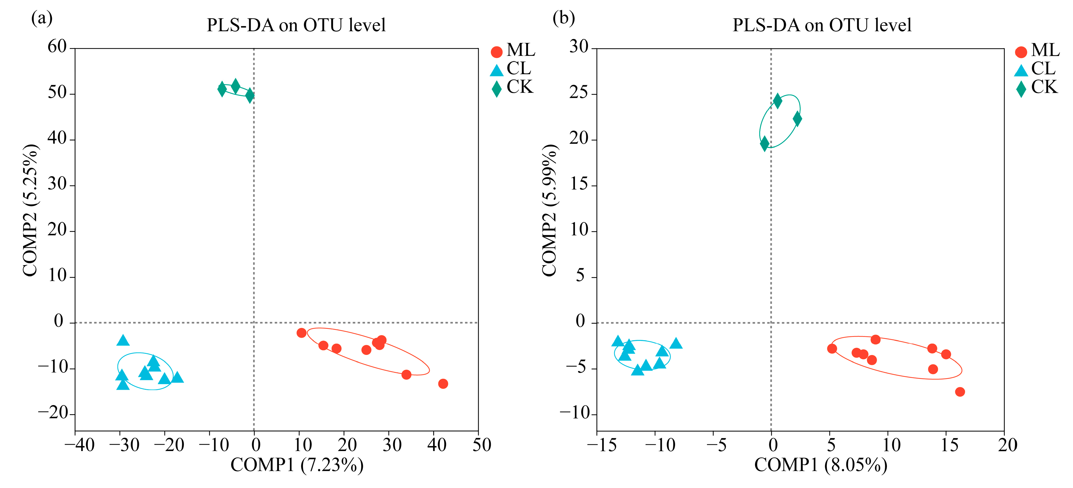
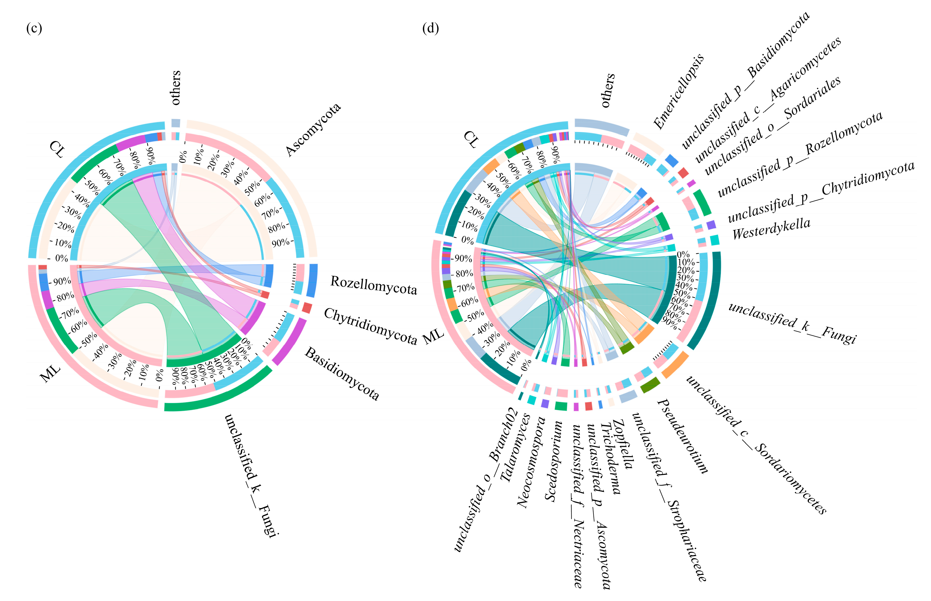
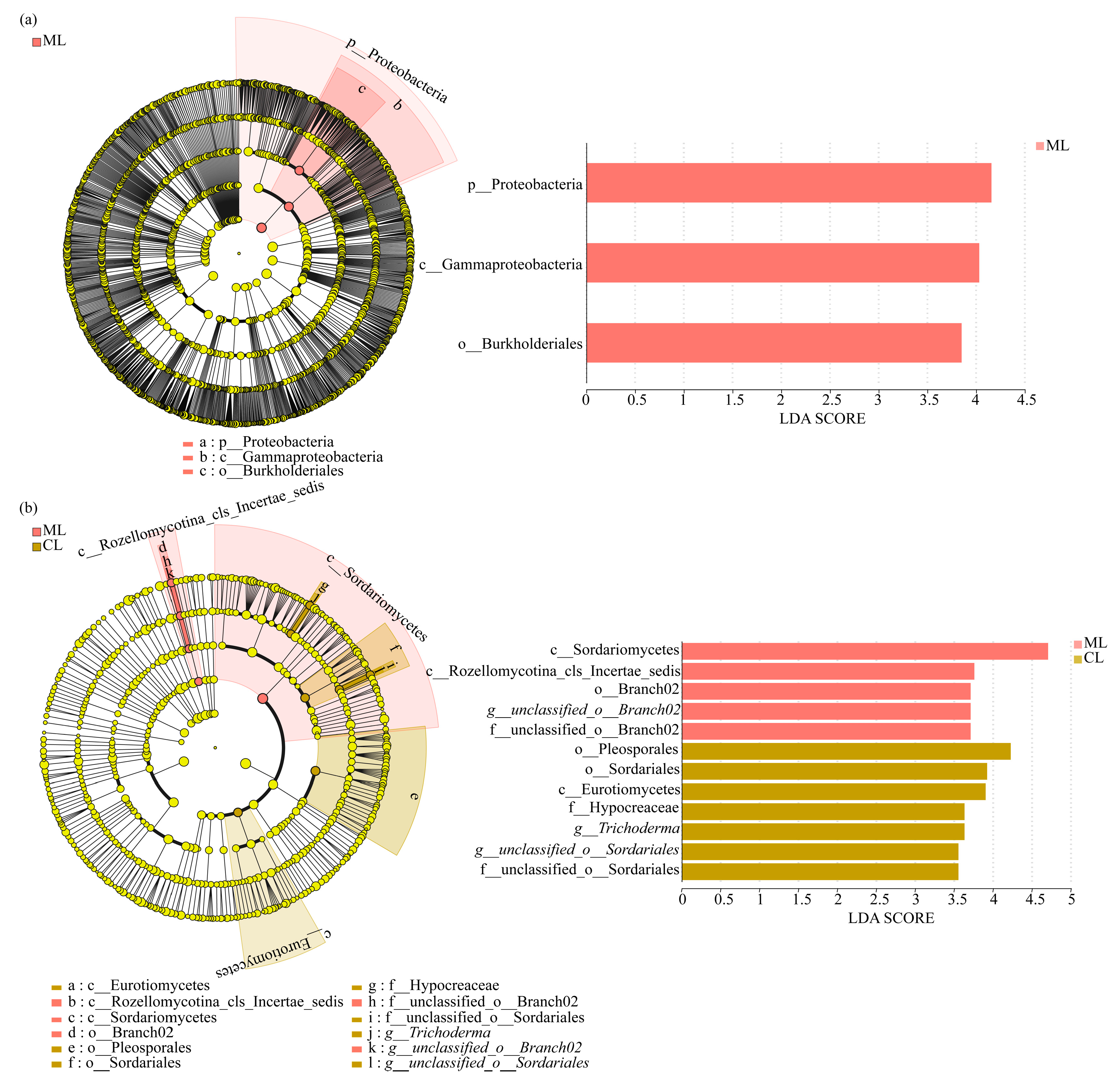
3.3. Soil Microbial Compositions of Rhizospheres in Mealy and Crunchy Lotus Root Varieties

As shown in Figure 3a, Chloroflexi, Actinobacteriota, Proteobacteria, Acidobacteriota, Firmicutes, Myxococcota, Methylomirabilota, Bacteroidota, Gemmatimonadota, Nitrospirota, and unclassified_k__norank_d__Bacteria were the dominant soil bacterial phyla (i.e., relative abundance > 1%) in rhizosphere for both ML and CL. Among the bacterial phyla grouped as “others”, Planctomycetota was the most abundant in the rhizosphere of both ML and CL, although its relative abundance was below the 1% threshold. Moreover, the abundances of Nitrospirota were significantly higher in rhizospheres of ML than those of CL. Furthermore, it is noteworthy that MBNT15 was a unique dominant soil bacterial phylum in ML.
At the genus level, 22 and 24 dominant bacterial genera could be detected in rhizospheres of ML and CL, respectively. Among them, norank_f__norank_o__SBR1031, norank_f__Anaerolineaceae, Intrasporangium, norank_f__norank_o__Vicinamibacterales, norank_f__Vicinamibacteraceae, norank_f__norank_o__Gaiellales, norank_f__67-14, Bacillus, norank_f__Xanthobacteraceae, Gaiella, norank_f__norank_o__RBG-13-54-9, norank_f__norank_o__Rokubacteriales, norank_f__norank_o__norank_c__KD4-9, norank_f__Roseiflexaceae, norank_f__Gemmatimonadaceae, unclassified_k__norank_d__Bacteria, norank_f__norank_o__SJA-15, norank_f__norank_o__IMCC26256, and Mycobacterium were the common dominant bacterial genera in rhizospheres of ML and CL (Figure 3b). Additionally, within the “others” bacterial genera grouped, norank_f__A4b showed the highest relative abundance in both ML and CL rhizospheres. Moreover, norank_f__norank_o__norank_c__norank_p__MBNT15, norank_f__norank_o__norank_c__Thermodesulfovibrionia, and norank_f__SC-I-84 were the unique dominant soil bacterial genera in the rhizosphere of ML. By contrast, unclassified_f__Nocardioidaceae, norank_f__norank_o__norank_c__MB-A2-108, norank_f__Methyloligellaceae, norank_f__norank_o__Subgroup_7, and norank_f__norank_o__Subgroup_17 were the unique dominant soil bacterial genera in the rhizosphere of CL.
Additionally, a total of five dominant fungal phyla, i.e., Ascomycota, unclassified_k__Fungi, Basidiomycota, Rozellomycota, and Chytridiomycota could be detected in rhizospheres of ML and CL (Figure 3c). Among the “others” fungal phyla, Glomeromycota was the most abundant in both ML and CL rhizospheres.
At the genus level, 15 and 16 dominant fungal genera could be detected in rhizospheres of ML and CL, respectively. Among them, unclassified_k__Fungi, unclassified_c__Sordariomycetes, Emericellopsis, unclassified_p__Rozellomycota, Pseudeurotium, unclassified_f__Strophariaceae, Westerdykella, Talaromyces, unclassified_c__Agaricomycetes, unclassified_p__Chytridiomycota, unclassified_p__Ascomycota, and unclassified_f__Nectriaceae were the common dominant fungal genera in the rhizospheres of MS and CL (Figure 3d). In addition, among the “others” fungal genera grouped, Zopfiella showed the highest relative abundance in ML, while unclassified_o__Agaricales was the most abundant in CL. Moreover, Scedosporium, Neocosmospora, and unclassified_o__Branch02 were the unique dominant soil fungal genera in rhizosphere of ML, whereas unclassified_p__Basidiomycota, Zopfiella, unclassified_o__Sordariales, and Trichoderma were the unique dominant soil fungal genera in the rhizosphere of CL.
Linear Discriminant Analysis Effect Size (LEfSe) analysis with a linear discriminant analysis (LDA) threshold of 1.0 was also used to identify significant differences and primary influential biomarker categories in the rhizosphere between ML and CL.
As shown in Figure 4a, at the phylum level, Proteobacteria was significantly clustered in the rhizospheres of ML.
Moreover, unclassified_o__Branch02 was significantly enriched in the rhizospheres of ML, whereas Trichoderma and unclassified_o__Sordariales were significantly enriched in the rhizospheres of CL (Figure 4b).

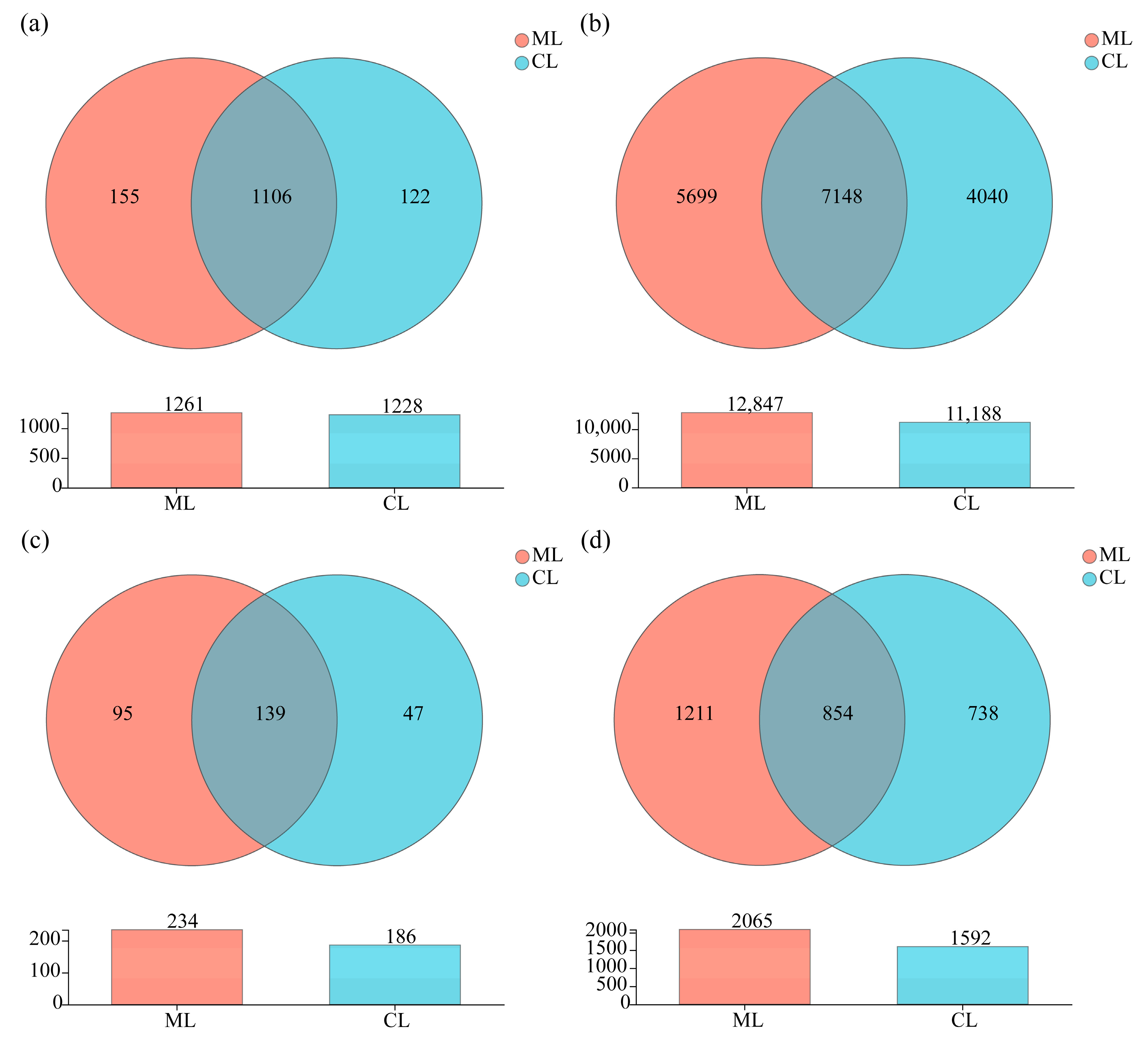
3.4. Venn Diagram Analysis of Soil Microorganisms in Rhizosphere of Mealy and Crunchy Lotus Root Varieties

At the genus level, the numbers of unique soil bacterial genera in the rhizosphere of ML and CL varieties were 155 and 122, respectively, and the total number of corresponding soil bacterial genera was 1261 and 1228 (Figure 5a). Moreover, 12,847 and 11,188 bacterial OTUs were found in ML and CL soil, respectively. Among them, 7148 common soil-bacterial OTUs were detected in both rhizospheres, and 5699 and 4040 unique soil bacterial OTUs were detected in the rhizospheres of ML and CL, respectively (Figure 5b).
Furthermore, the numbers of unique soil fungal genera in the rhizosphere of ML and CL were 95 and 47, respectively, and the total numbers of soil fungi genera in the rhizosphere of ML and CL were 234 and 186, respectively (Figure 5c). A total of 854 common soil fungal OTUs were identified, while 1211 and 738 unique OTUs were found in the rhizosphere of ML and CL, respectively (Figure 5d).
All of the above results suggested that different textural lotus varieties (ML and CL) recruited different soil microorganisms in the rhizosphere. In comparison with CL, higher abundance of bacteria and fungi needed to be recruited in the rhizosphere of ML.

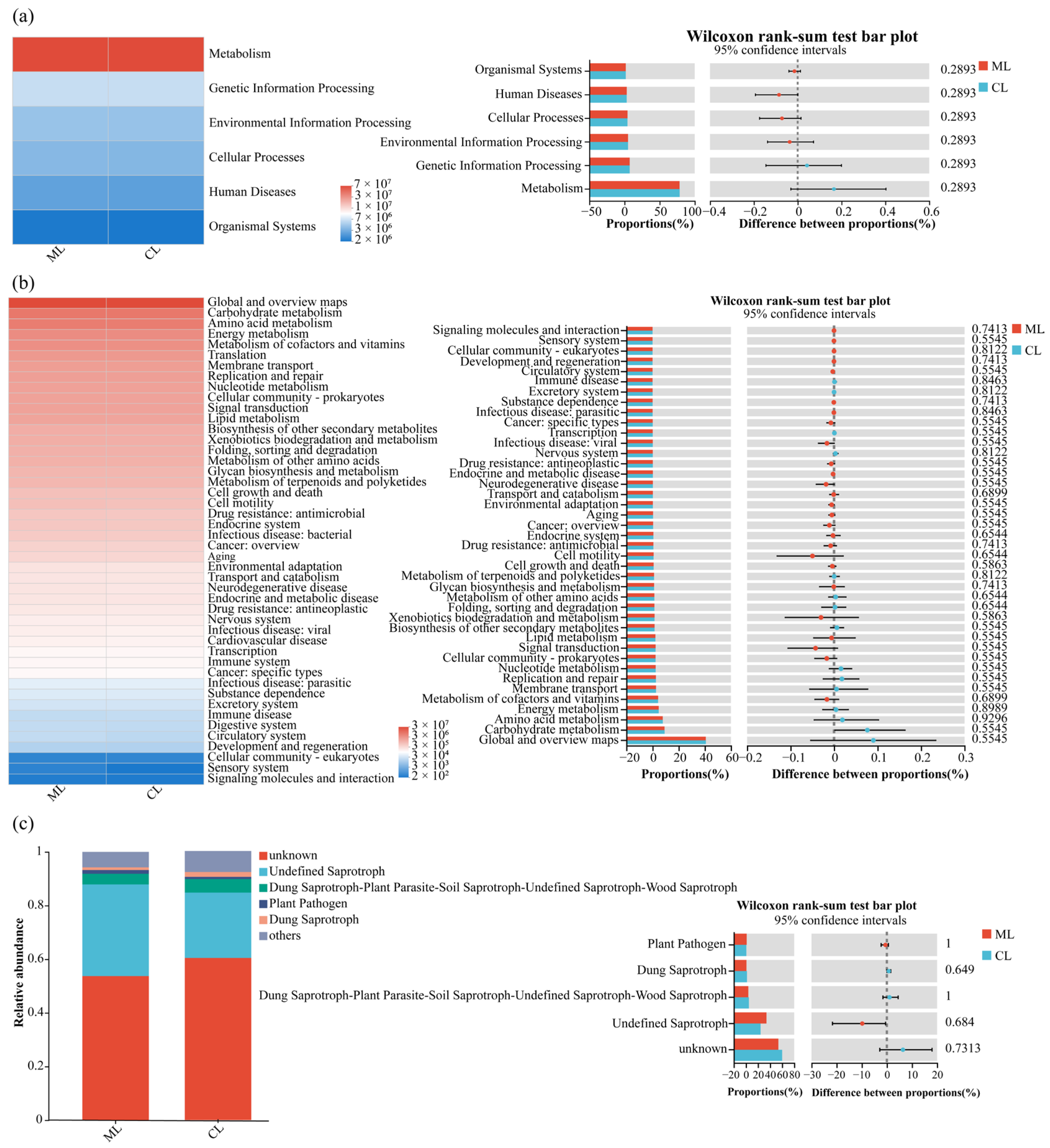
3.5. Functional Predictions of Soil Microbial Communities in Rhizospheres of Mealy and Crunchy Lotus Root Varieties

Based on the KEGG (Kyoto Encyclopedia of Genes and Genomes) database, six metabolic pathways of soil bacterial communities were identified in the rhizospheres of ML and CL, i.e., they were metabolism, genetic information processing, environmental information processing, cellular processes, human diseases, and organismal systems (Figure 6a). However, the Wilcoxon rank-sum test also indicated that there were no significant differences in the relative abundances of these pathways between ML and CL.
Moreover, a total of 46 bacterial metabolic pathways were detected in the rhizospheres of ML and CL. These pathways encompassed global and overview maps, carbohydrate metabolism, amino acid metabolism, energy metabolism, and the metabolism of cofactors and vitamins, among others (Figure 6b). Comparative analysis revealed that the overall functional composition of bacterial communities was largely conserved between the two varieties with no significant enrichment or depletion of specific pathways.
Based on FUNGuild functional predictions, five major fungal ecological categories were identified in the rhizosphere, including undefined saprotrophs, dung saprotroph-plant, parasite-soil saprotroph-undefined saprotroph-wood saprotroph, plant pathogens, and dung saprotrophs (Figure 6c). The Wilcoxon rank-sum test indicated that there were no significant differences in the five ecological functional categories fungal communities in the rhizosphere between ML and CL varieties.

4. Discussions

The textural quality of lotus root, a crucial aquatic vegetable, is predominantly determined by its cellular composition. The cell walls of higher plants are composed of polysaccharides such as pectin, cellulose, and hemicellulose [38]. Kan Juan et al. [39] demonstrated that the enzymatic degradation of pectic backbone and neutral sugars side-chains (e.g., galactose) in cell wall polysaccharides critically contributes to tissue softening through the action of polysaccharide-degrading enzymes. This process is further regulated by differential expression of cell wall-modifying enzyme genes during ripening [40]. Complementing these structural factors, intracellular components markedly influence textural properties. Starch content shows a strong negative correlation with crispness, where mealy lotus roots contain higher starch content compared to lower starch content in crunchy lotus roots [41].
As root exudates and nutrient composition can shape soil microbial communities in the rhizosphere of plants [42]. Our results revealed that not only soil bacterial and fungal composition in the rhizosphere of mealy (ML) were significantly different compared to crunchy lotus (CL) varieties, but also found that soil fungal richness in the rhizosphere of ML was significantly higher than that of CL, i.e., it also indicated that different genotypes of lotus root selectively recruited distinct bacterial and fungal communities in the rhizosphere. In addition, the rhizosphere microbial communities of both ML and CL differed markedly from those in the background soil (CK), highlighting the influence of plant genotype on microbial recruitment. PLS-DA analysis confirmed clear separation among ML, CL, and CK, suggesting that lotus root varieties could significantly shape their rhizosphere microbiota.
In comparison with CL varieties, the relative abundance of Nitrospirota was significantly higher in the rhizosphere of ML (1.74%) than in that of CL (1.24%). Similarly, Bacteroidota also showed a higher abundance in ML (2.15%) compared to CL (1.89%). Meanwhile, LEfSe analysis also revealed that Proteobacteria significantly enriched in the ML rhizosphere. As Nitrospirota participates in nitrogen cycle by oxidizing nitrite to nitrate, with nitrate being one of the nitrogen sources available to plants [43]. Moreover, some strains of Proteobacteria are known nitrifying bacteria, capable of oxidizing ammonia to nitrite and further oxidizing nitrite to nitrate [44]. And adequate nitrogen supply can increase photosynthetic rate and starch accumulation [45]. Bacteroidota is a major degrader of polysaccharides, and its genome contains a large number of carbohydrate-active enzymes (CAZymes), which are arranged in polysaccharide utilization loci (PULs) for degrading plant polysaccharides [46]. Furthermore, Bacillus was the dominant soil bacterial genus in the rhizosphere of ML compared to CL varieties. As Bacillus can produce cellulases with high activity and stability under different environmental conditions, it also suggests that Bacillus may contribute to cellulose degradation [47].
Although Ascomycota, unclassified_k__Fungi, Basidiomycota, Rozellomycota, and Chytridiomycota were all the dominant fungal phyla in the rhizosphere of ML and CL. However, their relative abundances were quite different in ML and CL. For instance, the abundance of Ascomycota was higher in ML (50.47%) than in CL (45.08%). Ascomycota sp. SM2 had been demonstrated to process extremely high carboxymethyl cellulase and filter paper cellulase activities [48]. At genus level, Emericellopsis was also more abundant in the rhizosphere of ML (11.07%) compared to CL (6.39%). Emericellopsis cladophorae can secrete a variety of carbohydrate-active enzymes, such as cellulases, chitinases, and pectinases, which play an important role in degrading polysaccharides, such as starch and cellulose [49].
Additionally, based on the KEGG database, similar metabolic pathways of soil microbial communities, such as carbohydrate metabolism, amino acid metabolism, and energy metabolism were detected in the rhizosphere of ML and CL. FUNGuild functional prediction also found that no significant differences in soil fungal ecological functional categories in rhizosphere between ML and CL. However, the enrichment of special dominant soil microorganisms in the rhizosphere of ML variety also indicated that its potential advantage of soil microorganisms in starch metabolism and cellulose degradation. Notably, rhizosphere microbes in ML enhanced polysaccharide degradation and cellulose breakdown likely weaken cell-wall rigidity (e.g., pectin and hemicellulose networks), facilitating starch granule expansion and a softer, cohesive “mealy” texture.

5. Conclusions

Mealy lotus (ML) varieties exhibited significantly lower moisture but higher starch contents than those of crunchy lotus (CL) varieties. Meanwhile, rhizosphere fungal richness of ML was also significantly higher than that of CL. Moreover, the relative abundances of bacterial phyla and genera, such as Nitrospirota, Bacteroidota, Proteobacteria, and Bacillus, alongside fungal phyla and genera, i.e., Ascomycota and Emericellopsis were enriched in the rhizosphere of ML compared to CL. Functional prediction also revealed that elevated nitrogen cycling, polysaccharide degradation and cellulose breakdown functions could be detected in ML, potentially driving starch accumulation and cell wall modification. These results suggest that rhizosphere microbial composition, particularly nitrogen-cycling bacteria and lignocellulose-degrading fungi may contribute to texture formation between texture-differentiated lotus root varieties.

Author Contributions

Conceptualization, X.L. and S.Y.; methodology, X.L.; software, Q.L.; validation, M.G.; formal analysis, X.L. and Q.L.; investigation, Y.O.; resources, H.J.; data curation, X.L.; writing—original draft preparation, X.L. and S.Y.; writing—review and editing, X.L., H.J., and S.Y.; visualization, Y.H.; supervision, W.J.; project administration, H.J. and S.Y.; funding acquisition, H.J. and S.Y. All authors have read and agreed to the published version of the manuscript.

Funding

This research was funded by the Guangxi Science and Technology pioneer Project (Guinongke alliance 202405) and the Guangxi Qingtang lotus root test station (TS2022012).

Institutional Review Board Statement

Not applicable.

Informed Consent Statement

Not applicable.

Data Availability Statement

The data presented in this study are openly available in [National Center for Biotechnology Information] at [https://www.ncbi.nlm.nih.gov/, accessed on 12 June 2025], reference number [PRJNA1275841, PRJNA1275856].

Conflicts of Interest

The authors declare no conflicts of interest.

References

  1. Xue, J.; Dong, W.; Cheng, T.; Zhou, S. Nelumbonaceae: Systematic position and species diversification revealed by the complete chloroplast genome. J. Syst. Evol. 2012, 50, 477–487. [Google Scholar] [CrossRef]
  2. Thanushree, M.P.; Sudha, M.L.; Crassina, K. Lotus (Nelumbo nucifera) rhizome powder as a novel ingredient in bread sticks: Rheological characteristics and nutrient composition. J. Food Meas. Charact. 2017, 11, 1795–1803. [Google Scholar] [CrossRef]
  3. Xu, S.Y.; Shoemaker, C.F. Gelatinization properties of Chinese water chestnut starch and lotus root starch. J. Food Sci. 1986, 51, 445–449. [Google Scholar] [CrossRef]
  4. Xu, Y. Perspectives on the 21st century development of functional foods: Bridging Chinese medicated diet and functional foods. Int. J. Food Sci. Technol. 2001, 36, 229–242. [Google Scholar] [CrossRef]
  5. Guo, H.B. Cultivation of lotus (Nelumbo nucifera Gaertn. ssp nucifera) and its utilization in China. Genet. Resour. Crop Evol. 2009, 56, 323–330. [Google Scholar] [CrossRef]
  6. Yang, D. Antioxidant Activities of Lotus Rhizome. Ph.D. Thesis, Dissertation of Zhejiang University, Zhejiang, China, 2007. [Google Scholar]
  7. Mukerjee, P.K.; Das, K.; Balasubramanian, R.; Saha, K.; Pal, M.; Saha, B.P. Anti-Diarrhoel evaluation of Nelumbo nucifera rhizome extract. Indian. J. Pharmacol. 1995, 27, 262–264. [Google Scholar]
  8. Mukherjee, P.K.; Balasubramanian, R.; Saha, K.; Saha, B.P.; Pal, M. Antibacterial efficiency of Nelumbo nucifera. Indian Drugs 1995, 32, 274–276. [Google Scholar]
  9. Eon-Hwan, S.; Jae-Geun, K. Effects of Nelumbo nucifera on the regional cerebral blood flow and blood pressure in rats. J. East Asian Soc. Diet. Life 2005, 15, 49–56. [Google Scholar]
  10. Lee, J.; Park, S.; Lee, M. Effect of lotus root (Nelumbo nucifera G) on lipid metabolism in rats with diet-induced hypercholesterolemia. Food Sci. Preserv. 2006, 13, 634–642. [Google Scholar]
  11. Mukherjee, P.K.; Pal, S.K.; Saha, K.; Saha, B.P. Hypoglycaemic activity of Nelumbo nucifera Gaertn. (Fam. Nymphaeaceae) rhizome (methanolic extract) in streptozotocin-induced diabetic rats. Phytother. Res. 1995, 9, 522–524. [Google Scholar] [CrossRef]
  12. Du, H.; Zhao, X.; You, J.; Park, J.; Kim, S.; Chang, K. Antioxidant and hepatic protective effects of lotus root hot water extract with taurine supplementation in rats fed a high fat diet. J. Biomed. Sci. 2010, 17, S39. [Google Scholar] [CrossRef] [PubMed]
  13. Mukherjee, P.K.; Pal, M.; Saha, K.; Saha, B.P.; Das, J. Diuretic activity of extract of the rhizomes of Nelumbo nucifera Gaertn. (Fam. Nymphaeaceae). Phytother. Res. 1996, 10, 424–425. [Google Scholar] [CrossRef]
  14. Chiang, P.; Luo, Y. Effects of pressurized cooking on the relationship between the chemical compositions and texture changes of lotus root (Nelumbo nucifera Gaertn.). Food Chem. 2007, 105, 480–484. [Google Scholar] [CrossRef]
  15. Northcote, D.H. The cell walls of higher plants: Their composition, structure and growth. Biol. Rev. 1958, 33, 53–102. [Google Scholar] [CrossRef]
  16. Wang, H.; Wang, H.; Yi, Y.; Hou, W.; Wang, L.; Ai, Y.; Min, T. Varying concentrations of ethephon induce antioxidant defences and cell wall degradation to regulate storage quality of fresh-cut lotus root. Food Biosci. 2024, 61, 104975. [Google Scholar] [CrossRef]
  17. Cai, Z.; Jiang, Y.; Wang, F.; Liu, J.; Kan, J.; Zhang, M.; Qi, X.; Li, L.; Zhao, S.; Qian, C. Study on Quality and Starch Characteristics of Powdery and Crispy Lotus Roots. Foods 2024, 13, 3335. [Google Scholar] [CrossRef]
  18. Yu, H.; Cheng, L.; Yin, J.; Yan, S.; Liu, K.; Zhang, F.; Xu, B.; Li, L. Structure and physicochemical properties of starches in lotus (Nelumbo nucifera Gaertn) rhizome. Food Sci. Nutr. 2013, 1, 273–283. [Google Scholar] [CrossRef]
  19. Bokulich, N.A.; Thorngate, J.H.; Richardson, P.M.; Mills, D.A. Microbial biogeography of wine grapes is conditioned by cultivar, vintage, and climate. Proc. Natl. Acad. Sci. USA 2014, 111, E139–E148. [Google Scholar] [CrossRef]
  20. Rochefort, A.; Briand, M.; Marais, C.; Wagner, M.; Laperche, A.; Vallee, P.; Barret, M.; Sarniguet, A. Influence of Environment and Host Plant Genotype on the Structure and Diversity of the Brassica napus Seed Microbiota. Phytobiomes J. 2019, 3, 326–336. [Google Scholar] [CrossRef]
  21. Ge, Y.; Xu, S.; Xu, Y. Review on influencing factors of rhizosphere microbiome assemblage. Acta Agric. Zhejiangensis 2019, 31, 2120–2130. [Google Scholar] [CrossRef]
  22. Cregger, M.A.; Veach, A.M.; Yang, Z.K.; Crouch, M.J.; Vilgalys, R.; Tuskan, G.A.; Schadt, C.W. The Populus holobiont: Dissecting the effects of plant niches and genotype on the microbiome. Microbiome 2018, 6, 14. [Google Scholar] [CrossRef] [PubMed]
  23. Igwe, A.N.; Vannette, R.L. Bacterial communities differ between plant species and soil type, and differentially influence seedling establishment on serpentine soils. Plant Soil 2019, 441, 423–437. [Google Scholar] [CrossRef]
  24. Sangiorgio, D.; Cellini, A.; Donati, I.; Ferrari, E.; Tanunchai, B.; Wahdan, S.F.M.; Sadubsarn, D.; Farneti, B.; Checcucci, A.; Buscot, F.; et al. Taxonomical and functional composition of strawberry microbiome is genotype-dependent. J. Adv. Res. 2022, 42, 189–204. [Google Scholar] [CrossRef] [PubMed]
  25. Chanclud, E.; Morel, J. Plant hormones: A fungal point of view. Mol. Plant Pathol. 2016, 17, 1289–1297. [Google Scholar] [CrossRef]
  26. Frankenberger, W.T., Jr.; Brunner, W. Method of detection of auxin-indole-3-acetic acid in soils by high performance liquid chromatography. Soil Sci. Soc. Am. J. 1983, 47, 237–241. [Google Scholar] [CrossRef]
  27. Nieto, K.F.; Frankenberger, W.T., Jr. Biosynthesis of cytokinins in soil. Soil Sci. Soc. Am. J. 1989, 53, 735–740. [Google Scholar] [CrossRef]
  28. Smith, A.M.; Cook, R.J. Implications of ethylene production by bacteria for biological balance of soil. Nature 1974, 252, 703–705. [Google Scholar] [CrossRef]
  29. Tudzynski, B.; Sharon, A. Biosynthesis, Biological Role and Application of Fungal Phytohormones. In Industrial Applications; Osiewacz, H.D., Ed.; Springer: Berlin/Heidelberg, Germany, 2002; pp. 183–211. ISBN 978-3-662-10378-4. [Google Scholar]
  30. Chaves, F.C.; Gianfagna, T.J. Necrotrophic phase of Monihophthora perniciosa causes salicylic acid accumulation in infected stems of cacao. Physiol. Mol. Plant Pathol. 2006, 69, 104–108. [Google Scholar] [CrossRef]
  31. Arshad, M.; Frankenberger, W.T. Plant Growth-Regulating Substances in the Rhizosphere: Microbial Production and Functions. In Advances in Agronomy; Sparks, D.L., Ed.; Academic Press: Cambridge, MA, USA, 1997; Volume 62, pp. 45–151. ISBN 0065-2113. [Google Scholar]
  32. Zhou, P.; Jin, Q.; Qian, P.; Wang, Y.; Wang, X.; Jiang, H.; Yao, D.; Liu, X.; Liu, F.; Li, J.; et al. Genetic resources of lotus (Nelumbo) and their improvement. Ornam. Plant Res. 2022, 2, 5. [Google Scholar] [CrossRef]
  33. Zhao, S.; Deng, K.; Zhu, Y.; Jiang, T.; Wu, P.; Feng, K.; Li, L. Optimization of slow-release fertilizer application improves lotus rhizome quality by affecting the physicochemical properties of starch. J. Integr. Agric. 2023, 22, 1045–1057. [Google Scholar] [CrossRef]
  34. Dong, W.; Liu, X.; Yi, Y.; Wang, L.; Hou, W.; Ai, Y.; Wang, H.; Min, T. Evaluation of Pre-Harvest Nutrient Composition and Functional Active Substances in Various Lotus Roots. Foods 2024, 13, 2297. [Google Scholar] [CrossRef] [PubMed]
  35. Yi, Y.; Huang, X.; Zhong, Z.; Huang, F.; Li, S.; Wang, L.; Min, T.; Wang, H. Structural and biological properties of polysaccharides from lotus root. Int. J. Biol. Macromol. 2019, 130, 454–461. [Google Scholar] [CrossRef] [PubMed]
  36. Salo-väänänen, P.P.; Koivistoinen, P.E. Determination of protein in foods: Comparison of net protein and crude protein (N× 6.25) values. Food Chem. 1996, 57, 27–31. [Google Scholar] [CrossRef]
  37. Zhu, F.; Sun, H.; Wang, J.; Zheng, X.; Wang, T.; Diao, Y.; Hu, Z. Differential expression involved in starch synthesis pathway genes reveal various starch characteristics of seed and rhizome in lotus (Nelumbo nucifera). J. Food Sci. 2022, 87, 4250–4263. [Google Scholar] [CrossRef]
  38. Zhang, P.; Chen, F.; Yang, H.; LI, L.; Gong, B.; Wang, L. Research advances on cell wall disassembly in fruit ripening and softening. Food Sci. Technol. 2010, 35, 62–66. [Google Scholar] [CrossRef]
  39. Kan, J.; Liu, T.; Jin, C.; Xie, H. Degradation of Cell Wall Polysaccharides and Related Enzyme Activities during Non-melting-fleshPeach Fruit Softening. Food Sci. 2011, 32, 268–274. [Google Scholar]
  40. Owino, W.O.; Nakano, R.; Kubo, Y.; Inaba, A. Coordinated expression patterns of genes encoding cell wall modifying enzymes during ripening in distinct anatomical tissue regions of the fig (Ficus carica L.) fruit. Postharvest Biol. Technol. 2004, 32, 253–261. [Google Scholar] [CrossRef]
  41. Yu, S. Study on Extraction and Characteristics of Lotus Root. Master’s Thesis, Wuhan Polytechnic University, Wuhan, China, 2017. [Google Scholar]
  42. Haichar, F.E.Z.; Marol, C.; Berge, O.; Rangel-Castro, J.I.; Prosser, J.I.; Balesdent, J.; Heulin, T.; Achouak, W. Plant host habitat and root exudates shape soil bacterial community structure. ISME J. 2008, 2, 1221–1230. [Google Scholar] [CrossRef]
  43. Jiang, Y.; Han, S.; Luo, X. Role of generalists, specialists and opportunists in Nitrospira community assembly in soil aggregates from nearby cropland and grassland areas in a campus. Appl. Soil Ecol. 2024, 198, 105377. [Google Scholar] [CrossRef]
  44. Zhao, Z.; Luo, J.; Jin, B.; Zhang, J.; Li, B.; Ma, B.; An, X.; Zhang, S.; Shan, B. Analysis of Bacterial Communities in Partial Nitritation and Conventional Nitrification Systems for Nitrogen Removal. Sci. Rep. 2018, 8, 12930. [Google Scholar] [CrossRef]
  45. Xu, C.M.; Zhou, C.N.; Zheng, G.S.; Wang, D.Y.; Zhao, F.; Zhu, X.D.; Zhang, X.F. Effects of Nitrogen Fertilizer Application and Densities on Non-structural Carbohydrates Accumulation, Distribution and Quality of Super Late Rice Tianyouhuazhan. J. Agric. Sci. Technol. 2010, 12, 86–91. [Google Scholar] [CrossRef]
  46. Lapébie, P.; Lombard, V.; Drula, E.; Terrapon, N.; Henrissat, B. Bacteroidetes use thousands of enzyme combinations to break down glycans. Nat. Commun. 2019, 10, 2043. [Google Scholar] [CrossRef] [PubMed]
  47. Bhatt, B.; Bhatt, K.; Lal, S.; Srinivasan, R.; Bhatt, V. Production of a novel cellulase by Bacillus amyloliquefaciens OKB3 isolated from soil: Purification and characterization. Int. J. Biol. Macromol. 2024, 282, 137454. [Google Scholar] [CrossRef] [PubMed]
  48. Ma, Y.N.; Mongkolthanaruk, W.; Riddech, N. Enhancing soil amendment for salt stress using pretreated rice straw and cellulolytic fungi. Sci. Rep. 2024, 14, 13903. [Google Scholar] [CrossRef]
  49. Goncalves, M.F.M.; Hilario, S.; van de Peer, Y.; Esteves, A.C.; Alves, A. Genomic and Metabolomic Analyses of the Marine Fungus Emericellopsis cladophorae: Insights into Saltwater Adaptability Mechanisms and Its Biosynthetic Potential. J. Fungi 2022, 8, 31. [Google Scholar] [CrossRef]
Figure 1. Biological characteristics of different varieties of lotus roots. ML: mealy lotus varieties; CL: crunchy lotus varieties. All data are presented as the mean ± SD (standard deviation). Different letters in the column indicate significant differences among treatments at p < 0.05.
Microorganisms 13 01637 g001
Figure 2. Comparison of the soil bacterial and fungal compositions among ML, CL, and CK. PLS-DA score plot of the soil bacterial (a) and fungal (b) communities among ML, CL, and CK. ML: mealy lotus varieties; CL: crunchy lotus varieties.
Microorganisms 13 01637 g002
Figure 3. (a) Compositions of rhizosphere soil bacterial communities at the phylum level; (b) compositions of rhizosphere soil bacterial communities at the genus level. (c) Compositions of rhizosphere soil fungal communities at the phylum level; (d) compositions of rhizosphere soil fungal communities at the genus level. ML: mealy lotus varieties, CL: crunchy lotus varieties.
Microorganisms 13 01637 g003aMicroorganisms 13 01637 g003b
Figure 4. LEfSe analysis of soil bacteria (a) and fungi (b) in rhizospheres between ML and CL varieties. ML: mealy lotus varieties; CL: crunchy lotus varieties.
Microorganisms 13 01637 g004
Figure 5. (a) Venn diagram analyses of soil bacteria at the genus level. (b) Venn diagram analyses of soil bacteria at the OTU level. (c) Venn diagram analyses of soil fungi at the genus level. (d) Venn diagram analyses of soil fungi at the OTU level. ML: mealy lotus varieties; CL: crunchy lotus varieties.
Microorganisms 13 01637 g005
Figure 6. Functional predictions of soil bacterial and fungal communities in rhizospheres of ML and CL. Compositional variability test for bacterial (a,b) and fungal (c) communities. ML: mealy lotus varieties; CL: crunchy lotus varieties.
Microorganisms 13 01637 g006
Table 1. Sequence types and primer sequences.
Sequence TypePrimer NamePrimer SequenceLength SequencingSequencing Platform
Bacterial 16S rRNA338F5′-ACTCCTACGGGAGGCAGCAG-3′416 bpMiseq PE300
806R5′-GGACTACHVGGGTWTCTAAT-3′
Fungal ITSITS1F5′-CTTGGTCATTTAGAGGAAGTAA-3′251 bpMiSeq PE300
ITS2R5′-GCTGCGTTCTTCATCGATGC-3′
Table 2. Soil microbial diversity in rhizospheres of mealy and crunchy lotus root varieties.
SamplesShannon IndexSimpson IndexAce IndexChao 1 IndexCoverage
BacteriaML7.17 ± 0.07a0.0024 ± 0.0003a5847.07 ± 325.66a5506.40 ± 290.86a0.96
CL7.04 ± 0.26a0.0041 ± 0.0051a5587.18 ± 286.22a5294.94 ± 247.26a0.96
CK7.13 ± 0.01a0.0024 ± 0.0002a5895.46 ± 81.78a5551.98 ± 47.85a0.96
FungiML4.33 ± 0.71a0.069 ± 0.0948a556.56 ± 114.02a559.30 ± 113.97a0.99
CL4.47 ± 0.31a0.0356 ± 0.0186a385.24 ± 89.06b387.66 ± 89.26b0.99
CK4.43 ± 0.35a0.0471 ± 0.0386a417.13 ± 80.85ab419.51 ± 80.97ab0.99
Notes: Data in the table are presented as the means ± SDs; values followed by different lowercase letters indicate significant differences at p < 0.05 among ML, CL, and CK (ML: mealy lotus root, CL: crunchy lotus root, CK: background).
Disclaimer/Publisher’s Note: The statements, opinions and data contained in all publications are solely those of the individual author(s) and contributor(s) and not of MDPI and/or the editor(s). MDPI and/or the editor(s) disclaim responsibility for any injury to people or property resulting from any ideas, methods, instructions or products referred to in the content.

