Article

Waterborne Transmission Driving the Prevalence of Blastocystis sp. in Los Ríos Region, Southern Chile

by
Daniel Sanhueza Teneo
1,*,
Cedric B. Chesnais
2,
Javiera Manzano
1,
María Paz Moll
1,
Analía Téllez
1 and
Guillermo Valenzuela-Nieto
3
1
Instituto de Inmunología y Parasitología, Facultad de Medicina, Universidad Austral de Chile, Valdivia 5110566, Chile
2
TransVIHMI, INSERM Unité, Institut de Recherche pour le Développement (IRD), Montpellier University, 34090 Montpellier, France
3
Facultad de Ciencias para el Cuidado de la Salud, Universidad San Sebastián, Valdivia 5090000, Chile
*
Author to whom correspondence should be addressed.
Microorganisms 2025, 13(7), 1549; https://doi.org/10.3390/microorganisms13071549
Submission received: 22 May 2025 / Revised: 19 June 2025 / Accepted: 23 June 2025 / Published: 1 July 2025
(This article belongs to the Special Issue Water Microorganisms Associated with Human Health, 2nd Edition)

Abstract

Waterborne gastrointestinal infections remain a global health concern, with approximately 1.7 billion diarrhea-related illnesses annually attributable to protozoan parasites. These pathogens are transmitted through contaminated water and exhibit high resistance to chlorination, posing substantial challenges to effective water treatment. This study focused on the most prevalent intestinal parasites in the Los Ríos Region of Chile: Blastocystis sp., Giardia duodenalis, and Entamoeba coli. The objectives were to assess the prevalence of eukaryotic parasites in water samples—covering both drinking and recreational sources—to describe the circulating subtypes of Blastocystis sp. and to identify ecological factors associated with parasite presence. Water samples were analyzed using conventional PCR, next-generation sequencing (NGS) was employed for Blastocystis sp. subtype identification, and the environmental predictors were evaluated using a multivariable logistic regression model. A total of 132 water samples were analyzed, of which 15.2% were positive for Blastocystis sp. and 1.5% for E. coli, while no samples tested positive for G. duodenalis. We identified subtypes ST1–ST4 of Blastocystis sp., along with ST7, ST10, ST14, ST21, and ST23–ST26, the latter being reported for the first time in Chile. Ecological factors significantly associated with Blastocystis sp. presence included higher water temperature and greater rainfall at positive sites. Potable water was associated with significantly lower odds of Blastocystis sp. infection (aOR = 0.04, 95% CI: 0.00–0.87; p = 0.041), while precipitation increased infection odds by 3% per additional millimeter (aOR = 1.03, 95% CI: 1.00–1.06; p = 0.036). Greater distance to the nearest farmhouse was also significantly associated with reduced infection risk, suggesting that proximity to livestock environments may influence Blastocystis sp. transmission. These findings help explain the high prevalence of Blastocystis sp. observed in humans in the Los Ríos Region and highlight the pivotal role of ecological conditions in driving waterborne transmission. To our knowledge, this is the first environmental study in Chile to clearly demonstrate the association between human infection, environmental factors, and the transmission dynamics of Blastocystis sp.

1. Introduction

Nowadays, it is estimated that at least one in six people worldwide is affected by a parasitic disease. The global health burden is disproportionately concentrated in those countries with the greatest social and economic disadvantages [1]. According to the World Health Organization (WHO), waterborne gastrointestinal infections are among the leading causes of morbidity and mortality worldwide, with protozoan parasites responsible for 1.7 billion diarrheal illnesses and 842,000 deaths annually [2].
Intestinal protozoa are agents of widespread global distribution, whose transmission occurs mainly through the fecal-oral route through the consumption of water or food contaminated with their resistant stages, such as cysts or oocysts [3,4]. One of the most prevalent intestinal eukaryotic parasites in the world are Blastocystis sp., with prevalence rates close to 50% in developing countries and up to 20% in developed countries [5]; and Giardia duodenalis, with infection rates of up to 30% in developing countries and up to 8% in developed countries [6,7].
Chile has made a significant progress in the control of parasitic diseases. This can be attributed to the implementation of improvements in basic sanitation, drinking water supply, and sewage systems, as well as food control and the execution of strategies aimed at promoting literacy and health education [8].
However, there are limited current data on the prevalence of intestinal parasites in humans in the country. For example, Barra et al., in 2016 [9], determined the prevalence of intestinal parasites in schoolchildren in the commune of Puerto Montt. The prevalence of Blastocystis sp. was found to be up to 13%, Endolimax nana 12%, and Entamoeba coli 5%. Vidal et al., in 2010 [10], reported a significant increase in blastocystosis, with numbers up to 72.9% between 2005 and 2008. This coincides with a trend observed in the country, where blastocystosis is currently the most frequently diagnosed parasitosis through serial parasitological examination of stools (EPSD) [11].
There is also little data on intestinal parasites in the human population of the Los Ríos Region, southern Chile. In 1997, Torres et al. [12] analyzed stool samples from schoolchildren in different localities in the city of Valdivia and established prevalences of up to 51% for Blastocystis sp., 26.6% for G. duodenalis, 24% for E. coli, 18.4% for E. nana, 13.3% for the geohelminth Trichuris trichiura, and up to 7.2% for Entamoeba histolytica. More recently, Sanhueza-Teneo et al., in 2025 [13], described the prevalence of parasites in urban area of Valdivia, where Blastocystis sp. had 22% prevalence, followed by E. coli at 11.3% and E. nana at 3.5%. Finally, the most recent study of parasite prevalence in rural areas of Los Ríos Region establishes Blastocystis sp. as the most prevalent (29.2%), followed by E. nana (13.4%), E. coli (7.2%), and G. duodenalis (2.8%) [14].
Environmental factors, including suspended solids, oxygen levels, pH, organic matter, and temperature, play a crucial role in the viability and/or development of environmental resistance elements of parasitic stages [15,16]. Intestinal protozoa, such as G. duodenalis and Cryptosporidium spp., are excreted in the feces of infected humans and animals. These protozoa have been documented in numerous gastrointestinal disease outbreaks, predominantly in developing countries, having been transmitted by contaminated waters [17]. In addition, these pathogens possess a very low infectious dose. For example, for G. duodenalis, the intake of just 10 cysts is sufficient to cause infection in humans [7]. In terms of environmental persistence, parasites are characterized by their high resistance to chlorination and filtration, processes used in water treatment, which makes them a significant threat to public health [3]. As for Blastocystis sp. cysts, it has been reported that they can survive one month at room temperature and up to two months at 4 °C [18].
Studies of the genetic diversity of Blastocystis sp., has identified at least 40 subtypes (STs) [19]. Among these, ST1–ST8 have been reported in both humans and animals, while ST10–ST17 have been identified in non-human species [20]. Some subtypes are commonly found in particular hosts, such as ST3 in humans, ST5 in pigs, and ST7 in poultry [21]; however, no subtype is host-specific. Given the broad range of hosts, there have been findings of the same subtype of Blastocystis sp. in humans, their breeding animals, and in rivers adjacent to the housing sector [22].
In Chile, there are limited research in presence of parasites in environmental matrices. Suarez et al., in 2024 [23], identified the protozoa Cryptosporidium parvum using Ziehl–Neelsen staining and direct immunofluorescence assay (IFA) and Blastocystis sp. through conventional PCR in samples of mussels and treated wastewater (disinfected by chlorination) in a rural cove in the city of Concepción, Bío-Bío region. C. parvum and Blastocystis sp. were identified in 40% and 45% of the samples, respectively. Both parasites were also identified in the treated wastewater.
The interest area of this study includes the communes of Corral and Lago Ranco because they are among the communes with the highest prevalence of intestinal parasites in humans in Los Ríos Region. To that purpose, we implemented diagnostic techniques using endpoint PCR for three of the most prevalent parasites in this region: Blastocystis sp., Giardia duodenalis, and Entamoeba coli [14]. Finally, next-generation sequencing (NGS) technique were used to determine the subtypes of Blastocystis sp. in these two communities. The aim of this study was to determine the presence of eukaryotic parasites in water samples from the communes of Corral and Lago Ranco, in order to better understand the transmission pathways of these parasites in the region.

2. Material and Method

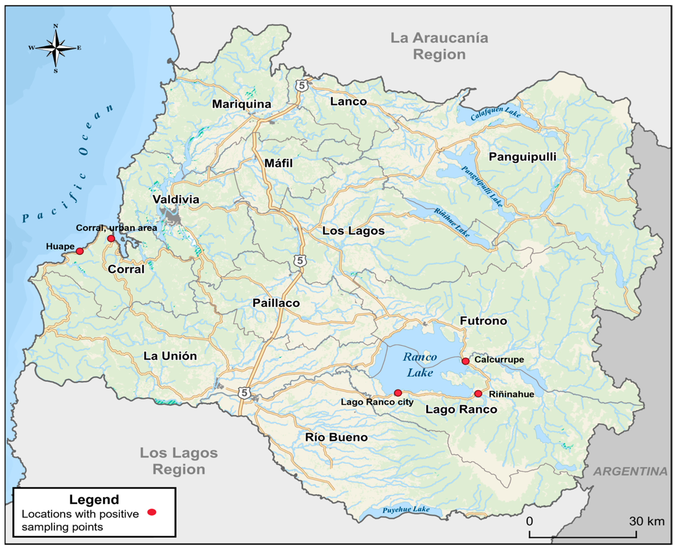
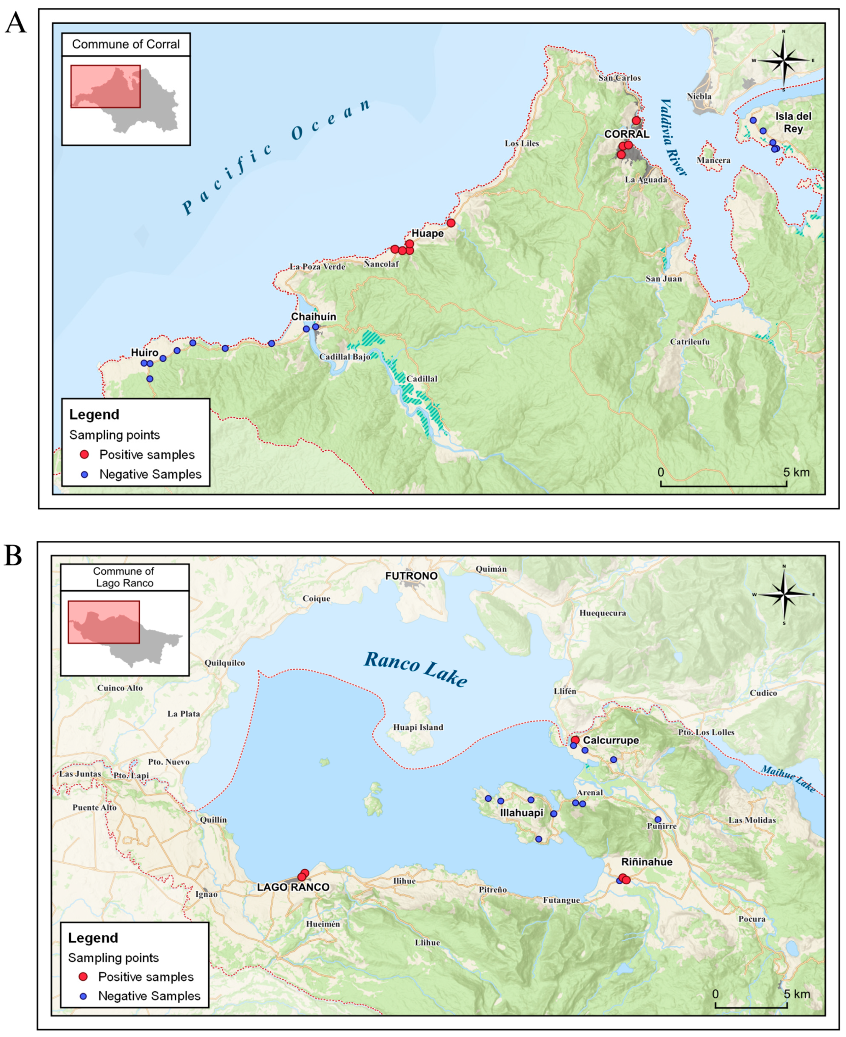
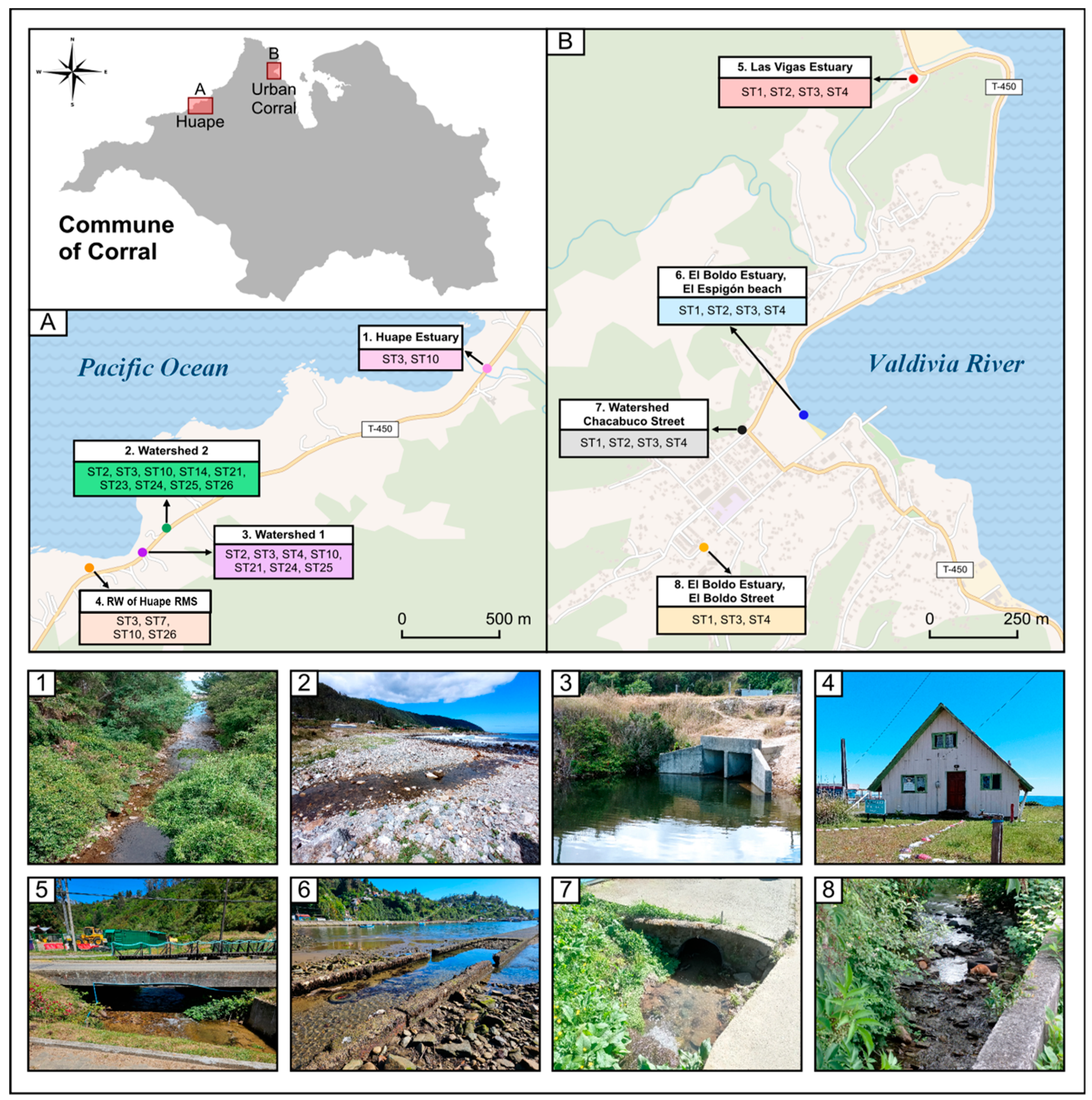
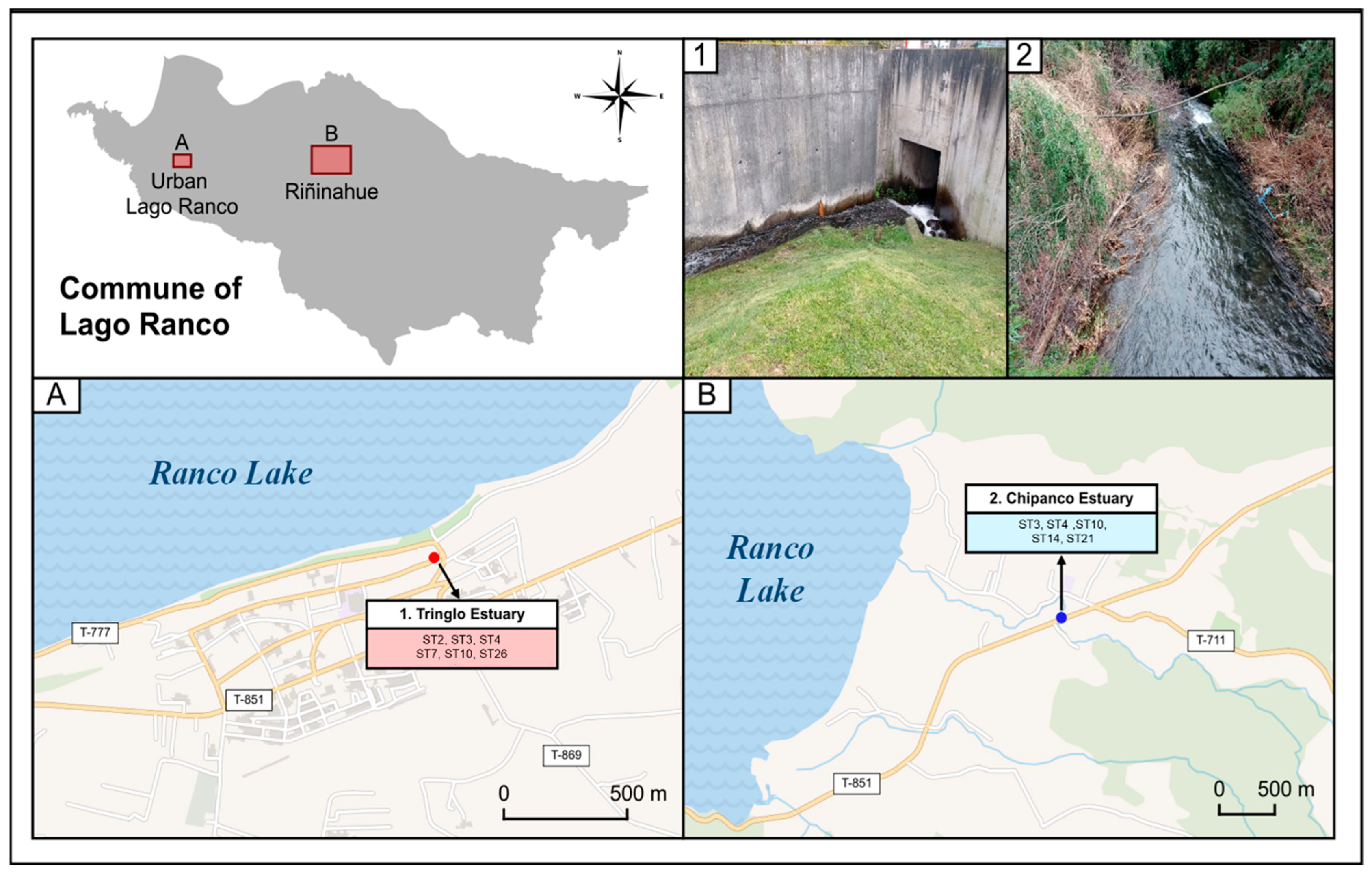
2.1. Characterization of the Study Area

The commune of Corral (39°53′ south latitude and 73°25′ west longitude) has an area of 767 km2 and is located approximately 20 km from the city of Valdivia, capital of the Los Ríos Region [24]. Its population is 5302, of which 65.7% live in urban areas and 34.3% in rural areas. In total, 54.3% of households lack access to basic services [25]. Corral is located at the mouth of the Valdivia River, and its territory includes coastal areas, forests, rivers, and mountain ranges [24]. The climate is temperate and rainy, with a cumulative rainfall of 1983 mm and an average annual temperature of 12.5 °C during the year 2024 [26].
On the other hand, the commune of Lago Ranco (40°19′00″ south latitude and 72°30′00″ west longitude), located in Ranco Province, Los Ríos Region, covers an area of 1763 km2 and has a population of 9896 inhabitants, of which 77.1% live in rural areas, and 36.7% reside in homes lacking basic services [27]. The hydrography of the commune consists of rivers, estuaries, lakes, and lagoons. Lake Ranco, the main body of water in the commune, covers an area of approximately 410 km2. Several rivers flow into the lake, including the Calcurrupe, Riñinahue, and Nilahue rivers [28]. The climate is temperate and rainy, with an annual precipitation of 1371 mm and an average temperature of 10.3 °C in 2024 [26].
The sampling points were georeferenced through Google Maps and subsequently imported into the QGIS Geographic Information System (v. 3.28) for the design of thematic cartographies.

2.2. Sampling

Sampling was conducted between August 2023 and June 2024, with the aim of collecting samples across all four seasons. In both communes of Corral and Lago Ranco, sampling sites were selected in areas with year-round geographic accessibility, ensuring comprehensive coverage of the study area.
According to international protocols (USEPA, WHO), the recommended volume for the detection of helminths and protozoa in water ranges from 1 to 10 L, depending on the type and quality of the sample [29]. However, due to logistical and operational constraints—such as limited time, transportation challenges, and filtration capacity—a sample volume of 3 L was established for stagnant and non-stagnant water bodies (estuaries, rivers, springs, ponds), and 2 L for rural drinking water (RW) samples, as these often required the cooperation of local residents, making the collection of larger volumes more difficult. Samples were collected in 1-L autoclaved glass bottles, protected from light with aluminum foil, and kept at 2–8 °C during transport. Upon arrival at the laboratory, they were stored at 4 °C until processing.
In Corral commune, 24 sampling points were established across five localities: 4 in the urban area of Corral, 5 in Huape, 3 in Chaihuín, 7 in Huiro, and 5 in Isla del Rey. A total of 69 water samples were collected in the study. Table 1 shows the number of samples collected at each site during each season.
In Lago Ranco, 19 sampling points were established across four localities: 2 in the city of Lago Ranco, 9 in Illahuapi, 4 in Riñinahue, and 4 in Calcurrupe. Over the entire sampling period, a total of 63 water samples were collected. Table 2 shows the number of samples collected in each locality during each season.
In total, 132 water samples were collected from both studied communes throughout the sampling period (69 water samples from Corral and 63 from Lago Ranco).
The number of samples collected during each season varied, due to logistical and environmental factors specific to each period, such as difficulties accessing certain areas due to adverse weather conditions or limited transportation availability. This variation was taken into account in the analysis and interpretation of the results.

2.3. Analysis of Abiotic Parameters in Water Samples

Abiotic parameters were measured in situ at the sampling points. Temperature (°C), pH, total dissolved solids (TDSs) (ppm), and electrical conductivity (EC) (mS/cm) were measured using the GrolineHI9814 instrument by Hanna Instruments® (Padova, Italy).

2.4. Water Sample Processing

Sample processing was carried out based on the protocols described by Kumar et al. [30] and Sánchez et al. [31]. Samples were processed within 24 to 48 h of collection. Filtration of the samples was performed through disposable filtration systems connected to a vacuum filtration pump. The sample volume was completely filtered and then the filter was removed with sterilized scalpels and surgical forceps and deposited in a disposable Petri dish. The filter was cut into two equal halves; the first half was stored in the freezer at −20 °C until used for DNA extraction. The second half of the filter was used for observation by optical microscopy; for this, with a scalpel, the filter was cut into smaller segments that were deposited in a 15 mL centrifuge tube, after which they were washed with 13 mL of Tween 80® 0.1% solution by vortex agitation. The washing liquid obtained was centrifuged at 3000 rpm for 10 min, the supernatant was discarded, and the sediment was subjected to the zinc sulfate flotation method described below.

2.5. Analysis of Water Samples by Microscopy

For the microscopic analysis of the water samples, the zinc sulfate flotation method modified from García in 2007 [32] was used. The sediment was resuspended with 1 to 2 mL of zinc sulfate (33 g of zinc sulfate in 67 mL of distilled water) and then zinc sulfate was added again until 11 mL of the tube was completed. It was centrifuged for two minutes at 1.800 rpm and zinc sulfate was added again to form a meniscus, in which, carefully, a 22 × 22 mm coverslip was placed and left to rest for 30 min. Each preparation was observed by light microscopy at 10× and 40×, and 100×. All preparations were performed in duplicate.

2.6. Extracting DNA from Water Samples

DNA extraction and storage were based by the results obtained by Hinlo et al. [33], who evaluated different conditions to maximize the recovery of environmental DNA from water samples. The DNA extraction kits used were QIAGEN®’ (Hilden, Germany)Dneasy Powersoil Pro-Kit and ThermoFisher (Waltham, MA, USA) Thermo Scientific GeneJET Genomic DNA Purification Kit.® DNA extractions were performed following the manufacturer’s instructions.

2.7. PCR

PCR was performed using the ThermoScientific DreamTaq Green PCR Master Mix (2×) enzyme (ThermoFisher). The thermal profile of the amplification reactions included the following steps: initial denaturation at 94 °C for 5 min, followed by 35 cycles for each pair of primers with a denaturation at 94 °C for 1 min, annealing temperature at 50, 57, and 60 °C for Blastocystis sp., G. duodenalis, and E. coli, respectively, for 30 s and extension temperature of 72 °C for 1 min. Finally, final extension at 72 °C for 5 min.
As positive control, DNA samples extracted from human stool samples positive for Blastocystis sp., G. duodenalis and E. coli were used by light microscopy and endpoint PCR. Moreover, as a negative control, nuclease-free water was used.
The primers used for detection of each parasite are described in the Table 3.

2.8. Analysis of Subtypes of Blastocystis sp. by Using Next-Generation Sequencing

The positive samples for Blastocystis sp. by conventional PCR were sent to Austral-OMICS laboratory (Core facility at the Universidad Austral of Chile), but only fourteen positive samples were further analyzed using next-generation sequencing (NGS), targeting the 18rRNA gene. Basically, raw sequences obtained from Illumina MiSeq were quality-filtered and the Amplicon sequence variants (ASVs) were inferred using the DADA2 package in R [37]. Taxonomic mapping was performed using the RDP Naive Bayesian Classifier algorithm and to identify ASVs belonging to Blastocystis sp., a phylogenetic tree was constructed using reference sequences from NCBI. Subtypes were determined by aligning representative sequences of each known Blastocystis sp. subtype with the Blastocystis-assigned ASVs. The relative abundance of ASVs was visualized using bar plots in R. A full description of the technique is provided in the Supplementary Materials.

2.9. Statistical Analyses

Physicochemical and environmental characteristics at the water collection site were used for statistical analysis to explain the presence of Blastocystis sp. only, because the number of positive samples for the other parasites was very low. Water type was categorized into three groups: standing water (ponds, etc.), non-standing water (rivers, streams, etc.), and drinking water. The four seasons were also included. All other variables were used as continuous quantitative variables: water temperature (°C), pH, TDS (ppm), EC (mS/cm), precipitation (mm), relative humidity (%), ambient temperature (°C), and distance from the water collection point to the nearest dwelling (km). The distance was measured using mapping tools from Google Maps.
Categorical variables were compared using the chi-square or Fisher’s exact test as appropriate. Quantitative variables were compared using the non-parametric Kruskal–Wallis test.
We fitted an adjusted logistic regression model including our variables, first examining multicollinearity via the variance inflation factor (VIF). Variables with VIF > 5 were considered to unduly imbalance the model and were excluded; the remaining variables were selected accordingly.

3. Results

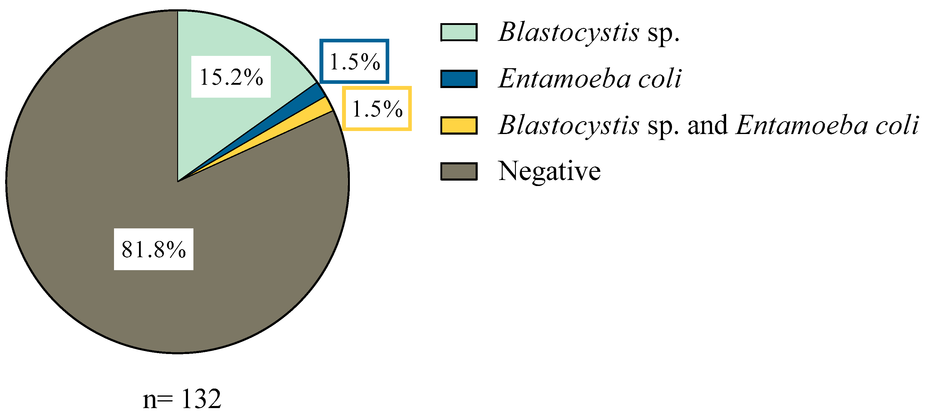
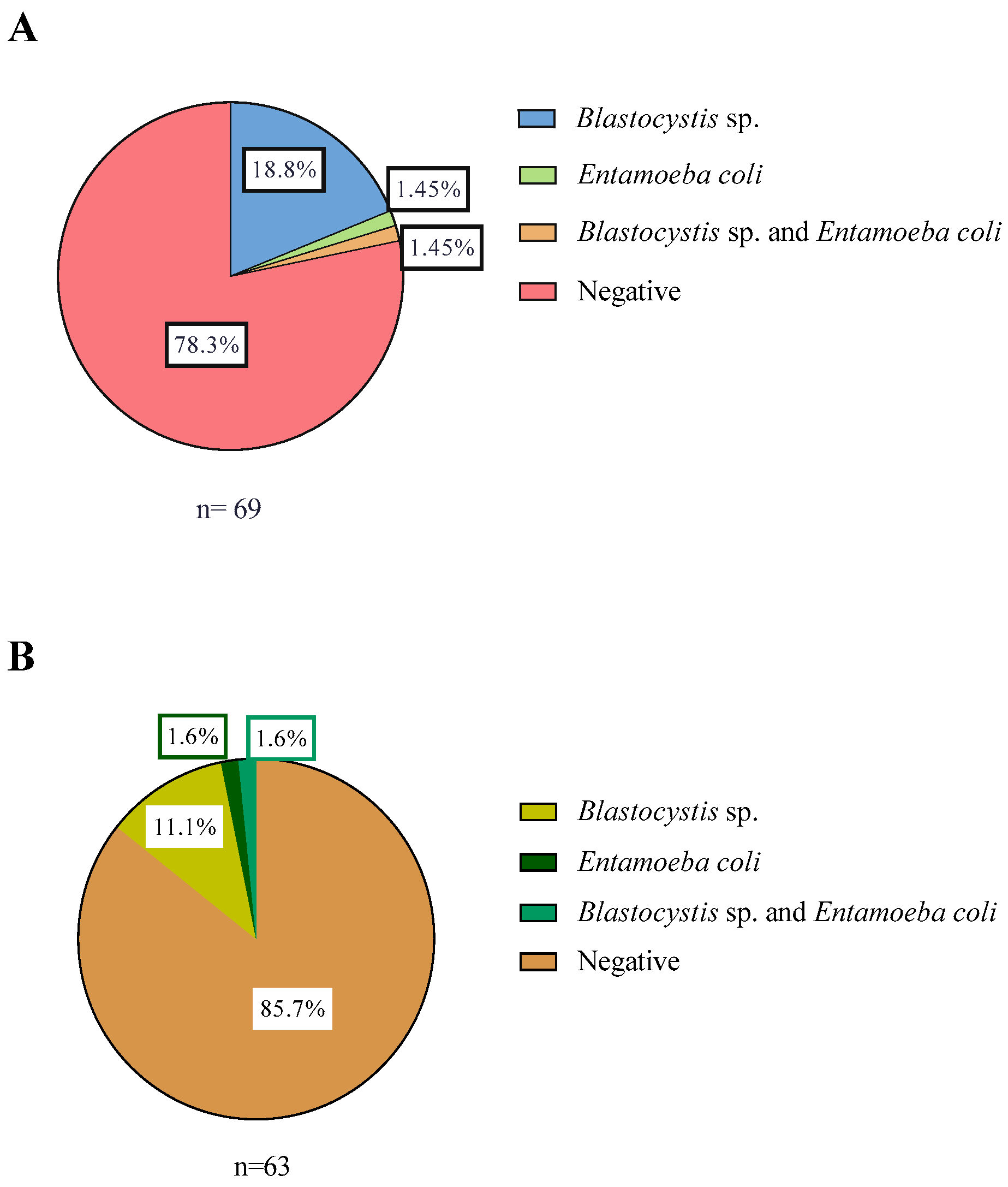
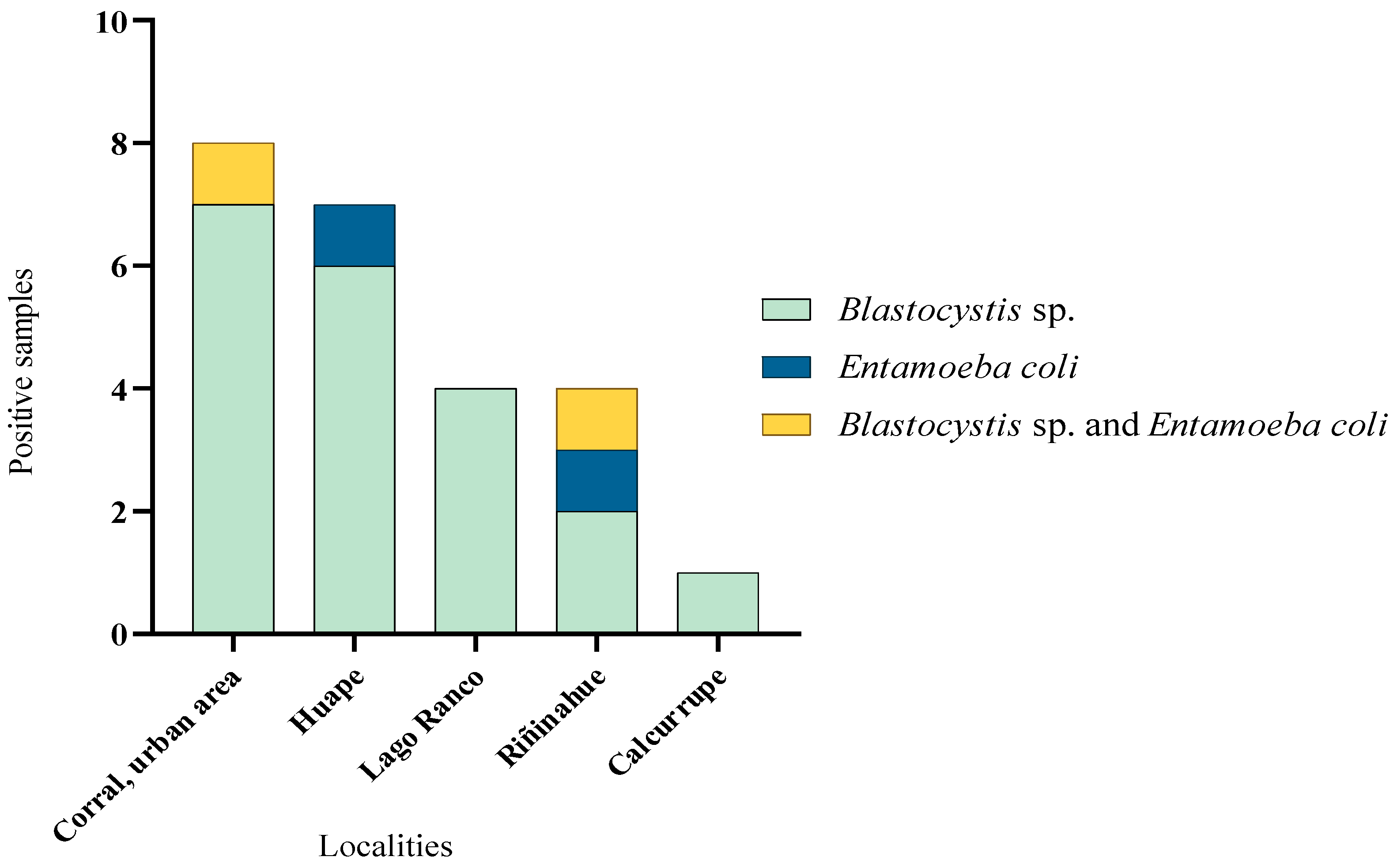
3.1. Distribution of Samples with Parasites

In total, 132 water samples were analyzed by conventional PCR and microscopy. No parasites were observed by light microscopy and 24 samples were positive for at least one parasite by conventional PCR, which represents 18.2%. By parasite, 15.2% (20/132) of the samples had Blastocystis sp., 1.5% (2/132) had Entamoeba coli, and 1.5% (2/132) had both Blastocystis sp. and E. coli. (Figure 1). All samples were negative for Giardia duodenalis. Electrophoreses photographs for each commune are shown in Supplementary Materials.
For Corral, a total of 69 water samples were analyzed by conventional PCR. Here, 21% (15/69) was positive, where 18.8% (13/69) was positive for Blastocystis sp. only, 1.45% (1/69) for E. coli, and 1.45% (1/69) for both Blastocystis sp. and E. coli (Figure 2A). In Lago Ranco, a total of 63 water samples were analyzed by conventional PCR. No parasite DNA was detected in 85.7% (54/63) of the samples. Blastocystis sp. was detected in 11.1% (7/63), 1.6% (1/63) of the samples amplified for E. coli, and 1.6% (1/63) was positive for both Blastocystis sp. and E. coli (Figure 2B).
The geographical distribution of the positive sampling points was Corral (urban area), Huape, Lago Ranco (urban area), Riñinahue, and Calcurrupe (Figure 3).
Blastocystis sp. was found in every positive site and the sampling point with the highest number of positive samples was the Corral urban area (Figure 4).
All of the positive samples of Corral came exclusively from the sectors of Corral (urban area) and Huape. In both locations, at least one positive sample was obtained at all sampling points (Figure 5A). The spatial distribution of the positive samples in the Lago Ranco commune shows that parasites were found in Lago Ranco (urban area), Riñinahue, and Calcurrupe (Figure 5B).

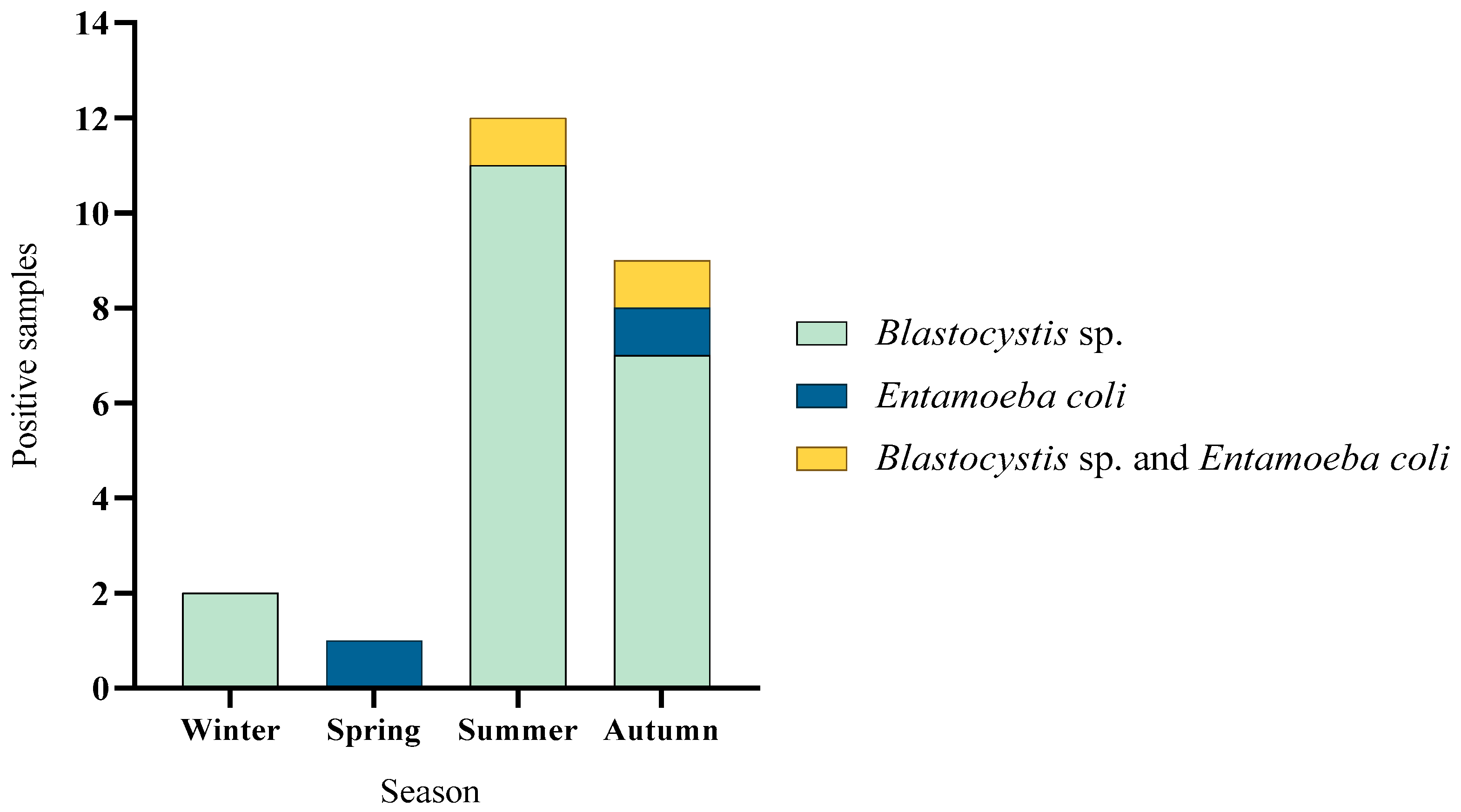
3.2. Seasonal Variation in Parasite Occurrence

Regarding the temporal distribution of parasites, these were found in all seasons of the year, with the highest number of positive samples being summer and autumn (p < 0.001). The highest diversity of parasites was found in autumn (Figure 6).
The seasonal distribution in Corral showed that parasites were found in every season of the year, except spring. The highest number of positive samples were found in summer. The highest diversity of parasites was described in autumn, with Blastocystis sp. and Entamoeba coli found in this season. In winter and summer only, Blastocystis sp. was found in our samples (Figure 7A). In Lago Ranco, parasites were detected in every season of the year. Blastocystis sp. was present all through the year, except in spring, where only E. coli was detected. The highest number of positive samples was described in summer and autumn (Figure 7B).

3.3. Subtypes of Blastocystis sp.

Of the 14 positive samples—12 for Corral and 2 for Lago Ranco—for Blastocystis sp. that were successfully sequenced by Next-Generation Sequencing (NGS), 12 different subtypes were described in both communes. Sequences obtained from the analysis have been deposited in the GenBank database, following the accession number PRJNA1231441. The details of the proportion of the ASVs assigned for each commune is available in the Supplementary Materials.
In the commune of Corral, the subtypes described were ST1, ST2, ST3, ST4, ST7, ST10, ST14, ST21, ST23, ST24, ST25, and ST26. The details of the spatial distributions of each subtype are shown in Figure 8.
In Lago Ranco, the subtypes ST2, ST3, ST4, ST7, ST10, ST14, ST21, and ST26 were found. The details of the spatial distributions of each subtype are shown in Figure 9.

3.4. Environmental Factors Associated with Blastocystis sp. Presence

The descriptive analysis of water-collection site characteristics by Blastocystis sp. infection status is presented in Table 4. Overall, water temperature averaged 12.89 °C (±2.55) but was significantly higher among Blastocystis sp.-positive samples (13.97 ± 2.65 °C) compared with negatives (12.68 ± 2.49 °C; Mann–Whitney p = 0.021). Precipitation at collection sites also differed, with positives experiencing greater rainfall (41.48 ± 34.17mm) than negatives (28.44 ± 33.12mm; p = 0.002). Ambient air temperature was elevated in positive sites (11.97 ± 4.99 °C vs. 9.50 ± 3.38 °C; p = 0.034). Differences in pH, total dissolved solids (TDSs), electrical conductivity (EC), relative humidity, and density of farmhouses did not have statistical significance.
In our multivariable analysis, the season variable exhibited a high variance inflation factor (VIF > 10), reflecting substantial collinearity with other environmental predictors. After removing season from the model, all remaining VIF values fell below 5. The multivariable logistic regression model (Table 5) identified several environmental predictors of Blastocystis sp. presence. Water source emerged as a significant factor: compared with standing water, potable water was associated with a dramatically lower odds of Blastocystis sp. infection (aOR = 0.04, 95% CI 0.00–0.87, p = 0.041), whereas non-standing/river water showed no significant difference (aOR = 0.48, 95% CI 0.04–6.20, p = 0.572). Precipitation was positively associated with infection risk, with each additional millimeter increasing the odds by 3% (aOR = 1.03, 95% CI 1.00–1.06, p = 0.036). Distance to the nearest farmhouses also reached significance: greater distance strongly reduced infection odds (aOR ≈ 5.8 × 10⁻⁹, 95% CI 7.1 × 10⁻¹⁶–0.0472, p = 0.019), suggesting proximity to livestock environments may influence transmission. Other variables—including region (Lago Ranco vs. Corral), TDS, water temperature, pH, electrical conductivity, relative humidity, and ambient temperature—did not show statistically significant associations (p > 0.05), although relative humidity approached significance (aOR = 0.74, 95% CI 0.52–1.04, p = 0.079). These results indicate that water potability, rainfall, and proximity to farmhouses are key drivers of Blastocystis sp. contamination in this setting.

4. Discussion

PCR revealed Blastocystis sp. in 15.2% of samples, making it the most frequently detected parasite. This is the first study in Chile that describes the finding of Blastocystis sp. in recreational and drinking water. Similar prevalences of the parasite have been reported elsewhere: Khalifa et al., in 2014 [38], found 15.8% positivity in Egyptian pond water; Richard, in 2016 [39], found 27.7% in Malaysian drinking-water plants; and Karaman et al., in 2017 [40], found a prevalence of 20.6% in Turkish streams and drinking sources. Falcone et al., in 2024 [41]. reviewed 87 Latin America water-sampling studies (2000–2022) and noted Blastocystis sp. in 18.7% of them. Despite its public-health importance—recognized by the WHO as a parasite of concern in drinking water [42]—information on Blastocystis sp. in water remains scarce globally.
Entamoeba coli was detected in 1.5% of samples. This protozoan has been identified in wastewater samples in Spain [43], Iran [44], and Argentina [45]. Due to a similar mode of transmission, its public health implications in the region are comparable to those previously described for Blastocystis sp., with similar implications for fecal contamination monitoring.
The absence of Giardia lamblia in water samples, despite its well-established public health relevance [46], aligns with its low prevalence previously observed in the local human population [13]. This suggests a limited environmental circulation in the study area, in contrast to Blastocystis sp., which was frequently detected in both human and environmental samples. As for Cryptosporidium sp., although it is also a globally significant pathogen [46], a targeted search in water samples was not performed due to the lack of prior data on its circulation among the local population. In the present study, also no parasites were observed by light microscopy. This result is comparable to those reported by Ramos and Magtibay, 2020 [47], who likewise only found free-living organisms by light microscopy. The lack of parasitic elements by this method may reflect the limited sensitivity of light microscopy or the characteristics of the samples: (i) the small water volumes collected may reduce detection probability [29] and (ii) protozoan cysts and oocysts often occur at very low concentrations in water samples [48]
In terms of genetic diversity, the detection of multiple Blastocystis subtypes (STs) in both humans and animals—mammals, birds, and reptiles—underscores its zoonotic potential [49]. For example, ST7, commonly found in poultry, has been detected in individuals who work closely with these animals, and ST8 has been documented moving between non-human primates and their human caretakers [50]. Water, essential for all life, is therefore a key transmission route. In Nepal, Lee et al., in 2012 [22], found high ST4 prevalence simultaneously in villagers, and nearby rivers, strongly suggesting waterborne zoonotic transmission.
Globally, ST1–4 make up over 90% of human isolates, with ST3 the most common [49], and these subtypes also appear in animals and water. ST9, in contrast, was reported only in humans [42]. Our findings mirror this: ST1–4 were present in water samples from Lago Ranco and Corral, but ST9 was not. Animal-associated subtypes—ST7, ST10, ST12, ST14, and ST16—are less common in people [51] but ST7, ST10, and ST14 were found in our water samples of both communes. We also detected ST21 and ST23–26, typically linked to wild and domestic ruminants [52], though ST23 was first seen in 2021 in humans by Jinatham et al. [50] and ST26 in 2023 in Thai schoolchildren [53,54].
The results obtained in urban Corral are worth discussing compared to Huape. In the urban area of Corral, Blastocystis subtypes, commonly associated with humans (ST1–4), were mainly concentrated, whereas in Huape, a more rural area, a greater diversity of subtypes (ST7, ST10, ST14, ST21, ST23–26) was identified, frequently linked to domestic and wild animals. This difference could suggest a possible influence of zoonotic reservoirs and the existence of more diverse transmission routes in Huape, in contrast to the predominantly anthropogenic transmission in Corral. Meanwhile, in the city of Lago Ranco and Riñinahue, subtypes predominantly associated with humans were identified, along with subtypes linked to animals, in the same water source. This could indicate that these water bodies are exposed to a variety of hosts, suggesting the existence of more diverse transmission routes.
In Chile, human fecal studies have documented ST1–4 [14,55] and more recently ST6 [14]. Our work is the first to report ST1–4 in Chilean water, and to add ST7, ST10, ST14, ST21, and ST23–ST26 description. These environmental findings could help to explain the high 29% human Blastocystis sp. prevalence in the Los Ríos Region [14].
Although our descriptive and multivariable analyses confirmed the importance of natural factors—precipitation and water source—in Blastocystis sp. contamination, anthropogenic influences are equally critical. We found that infection risk decreases significantly as the distance between the sampling point and the nearest dwelling or farm increases. No significant differences were observed between regions, indicating that local characteristics—land use and livestock density—are the main drivers of fecal contamination. Nonetheless, it remains plausible that human population density further modulates risk: positive samples are clustered in more urbanized localities (Corral and Huape), where greater human density and infrastructural complexity can amplify sources of contamination [56,57]. In rural Chile, limited sanitation infrastructure and the proximity of latrines, livestock pens, and homes to watercourses facilitate the transfer of fecal matter into streams. Surface runoff during rainfall can rapidly carry cysts and oocysts to sampling points, while heavy rainfall may also dilute parasite concentrations [16]. These findings illustrate how anthropization—the alteration of natural landscapes by human settlement and land use—creates “hotspots” for Blastocystis sp. introduction and persistence. To mitigate transmission, targeted measures are needed: improving sanitation at water points, establishing buffer zones between latrines or animal enclosures and watercourses, and educating communities on safe water-handling practices.

Study Limitations and Future Directions

The cross-sectional design limits temporal inference, and sample volumes constrained microscopy sensitivity. Future work should combine quantitative PCR with larger volume filtration and incorporate sanitary surveys to more precisely map fecal contamination sources. Moreover, integrating land-use GIS layers with high-resolution sanitation infrastructure maps could refine our understanding of anthropogenic risk factors.

5. Conclusions

Our findings confirm that Blastocystis sp. is a common waterborne parasite in southern Chile and highlight the dual importance of environmental conditions and human-driven landscape changes in its transmission. Enhanced sanitation measures and strategic water-point management are warranted to mitigate fecal pollution and protect community health.

Supplementary Materials

The following supporting information can be downloaded at: https://www.mdpi.com/article/10.3390/microorganisms13071549/s1: Title S1. Description of Next-generation Sequencing (NGS) technique; Figure S1:Electrophoresis of positive samples for Blastocystis sp. from Lago Ranco commune; Figure S2: Electrophoresis of positive samples for Blastocystis sp. from Corral commune; Figure S3: Blastocystis subtypes identified in Lago Ranco commune; Figure S4: Distribution of Blastocystis subtypes in Lago Ranco commune; Figure S5: Blastocystis subtypes identified in Corral commune; Figure S6: Distribution of Blastocystis subtypes in Corral commune. References [37,58,59,60,61] are cited in the Supplementary Materials.

Author Contributions

Conceptualization, D.S.T. and C.B.C.; Methodology, D.S.T., A.T. and G.V.-N.; Validation, D.S.T.; Formal analysis, D.S.T. and C.B.C.; Investigation, D.S.T., J.M., M.P.M., A.T. and G.V.-N.; Resources, D.S.T.; Data curation, C.B.C.; Writing—original draft, D.S.T.; Writing—review and editing, D.S.T., C.B.C., J.M. and M.P.M.; Visualization, D.S.T.; Supervision, D.S.T.; Project administration, D.S.T.; Funding acquisition, D.S.T. All authors have read and agreed to the published version of the manuscript.

Funding

This research was funded by Agencia Nacional de Investigación y Desarrollo (ANID), Chile, Fondecyt, grant number 11220065.

Institutional Review Board Statement

Not applicable.

Informed Consent Statement

Not applicable.

Data Availability Statement

The original contributions presented in this study are included in the article/Supplementary Materials. Further inquiries can be directed to the corresponding author.

Conflicts of Interest

The authors declare no conflicts of interest.

References

  1. Maurice, A.P.; Jenkin, A.; Norton, R.E.; Hamilton, A.; Ho, Y.H. Epidemiology of Parasitic Diseases. In The Surgical Management of Parasitic Diseases; Tsoulfas, G., Hoballah, J.J., Velmahos, G.C., Ho, Y.H., Eds.; Springer: Cham, Switzerland, 2020; pp. 3–21. ISBN 978-3-030-47948-0. [Google Scholar]
  2. Bourli, P.; Eslahi, A.V.; Tzoraki, O.; Karanis, P. Waterborne transmission of protozoan parasites: A review of worldwide outbreaks–An update 2017–2022. J. Water Health 2023, 21, 1421–1447. [Google Scholar] [CrossRef]
  3. Rojo, G.; Cuadros, J. Malaria y Protozoos Intestinales. Enferm. Infecc. Microbiol. Clin. 2016, 34, 191–204. [Google Scholar] [CrossRef] [PubMed]
  4. Theel, E.S.; Pritt, B.S. Parasites. Microbiol. Spectr. 2016, 4. [Google Scholar] [CrossRef]
  5. Ahmed, M. Intestinal Parasitic Infections in 2023. Gastroenterol. Res. 2023, 16, 127–140. [Google Scholar] [CrossRef]
  6. Leung, A.K.C.; Leung, A.A.M.; Wong, A.H.C.; Sergi, C.M.; Kam, J.K.M. Giardiasis: An Overview. Recent Pat. Inflamm. Allergy Drug Discov. 2019, 13, 134–143. [Google Scholar] [CrossRef] [PubMed]
  7. Lalle, M.; Cacciò, S.M. Giardiasis from a One Health Perspective. In Zoonoses: Infections Affecting Humans and Animals; Sing, A., Ed.; Springer: Cham, Switzerland, 2023; pp. 1285–1311. ISBN 978-3-031-27164-9. [Google Scholar] [CrossRef]
  8. Instituto de Salud Pública de Chile Boletines de Vigilancia de Laboratorio. Vigilancia de Diarreas por Agentes Parasitarios en Menores de 5 Años, Chile 2008–2012. 2012. Available online: https://www.ispch.gob.cl/boletin/page/13/ (accessed on 20 May 2025).
  9. Barra, M.; Bustos, L.; Ossa, X. Desigualdad en la prevalencia de parasitosis intestinales en escolares de una escuela urbana y dos rurales de la comuna de Puerto Monnt. Rev. Med. Chile 2016, 144, 886–893. [Google Scholar] [CrossRef] [PubMed]
  10. Vidal, S.; Toloza, L.; Cancino, B. Evolución de la prevalencia de enteroparasitosis en la ciudad de Talca, Región del Maule, Chile. Rev. Chil. Infectología 2010, 27, 336–340. [Google Scholar] [CrossRef]
  11. Apt, W.; Zulantay, I.; Canals, M.; Fredes, F. Visión de las parasitosis humanas en Chile. Parasitol. Latinoam. 2021, 70, 47–67. [Google Scholar]
  12. Torres, P.; Otth, L.; Montefusco, A.; Wilson, G.; Ramírez, C.; Acuña, M. Infección por protozoos y helmintos intestinales en escolares de sectores ribereños, con distintos niveles de contaminación fecal, del río Valdivia, Chile. Bol. Chil. Parasitol. 1997, 52, 3–11. [Google Scholar]
  13. Sanhueza-Teneo, D.; Cerna, O.; Chesnais, C.B.; Cárdenas, D.; Camus, P. High parasite prevalence driven by the human-animal-environment interface: A One Health study in an urban area in southern of Chile. Front. Vet. Sci. 2025, 12, 1536861. [Google Scholar] [CrossRef]
  14. Sanhueza-Teneo, D.; Venegas, T.; Videla, F.; Chesnais, C.B.; Loncoman, C.; Valenzuela-Nieto, G. Prevalence and Genetic Diversity of Parasites in Humans and Pet Dogs in Rural Areas of Los Ríos Region, Southern Chile. Pathogens 2025, 14, 186. [Google Scholar] [CrossRef] [PubMed]
  15. Smith, H.V. Detection of parasites in the environment. Parasitology 1998, 117 (Suppl), S113–S141. [Google Scholar] [CrossRef]
  16. Ajeagah, G.; Njine, T.; Bilong Bilong, C.; Foto, S.; Wouafo, M.; Nola, M. Seasonal Distribution of Enteric Opportunistic Cryptosporidium spp. Oocystis and Giardia spp. Cysts in a Tropical Water Basin, Cameroon. Water 2010, 2, 44–57. [Google Scholar]
  17. Betancourt, W.; Querales, L. Parásitos Protozoarios Entéricos en Ambientes Acuáticos: Métodos de concentración y detección. Interciencia 2008, 33, 418–423. [Google Scholar]
  18. Del Coco, V.F.; Molina, N.B.; Basualdo, J.A.; Córdoba, M.A. Blastocystis spp.: Avances, controversias y desafíos futuros. Rev. Argent. Microbio 2017, 49, 110–118. [Google Scholar] [CrossRef]
  19. Koehler, A.V.; Herath, H.M.P.D.; Hall, R.S.; Wilcox, S.; Gasser, R.B. Marked genetic diversity within Blastocystis in Australian wildlife revealed using a next generation sequencing–phylogenetic approach. Int. J. Parasitol. Parasites Wildl. 2024, 23, 100902. [Google Scholar] [CrossRef] [PubMed]
  20. Barati, M.; Karimi-Pour-Saryazdi, A.; Rahmanian, V.; Bahadory, S.; Abdoli, A.; Rezanezhad, H. Global prevalence and subtype distribution of Blastocystis sp. in rodents, birds, and water supplies: A systematic review and meta-analysis. Prev. Vet. Med. 2022, 208, 105770. [Google Scholar] [CrossRef]
  21. Adao, D.E.V.; Rivera, W.L. Subtype–host patterns and genetic differentiation of Blastocystis sp. in the Philippines. Heliyon 2024, 10, e29019. [Google Scholar] [CrossRef] [PubMed]
  22. Lee, L.I.; Chye, T.T.; Karmacharya, B.M.; Govind, S.K. Blastocystis sp.: Waterborne zoonotic organism, a possibility? Parasit. Vectors 2012, 5, 130. [Google Scholar] [CrossRef]
  23. Suarez, P.; Vallejos-Almirall, A.; Fernández, I.; Gonzalez-Chavarria, I.; Alonso, J.L.; Vidal, G. Identification of Cryptosporidium parvum and Blastocystis hominis subtype ST3 in Cholga mussel and treated sewage: Preliminary evidence of fecal contamination in harvesting area. Food Waterborne Parasitol. 2024, 34, e00214. [Google Scholar] [CrossRef]
  24. Aguilera, N.; Villagra, P. Territorial and multidimensional contrasts between urban and rural community resilience to disaster in the Corral Commune. Rev. De Urban. 2023, 49, 66–92. [Google Scholar] [CrossRef]
  25. Biblioteca del Congreso Nacional de Chile (BCN). Corral Reporte Comunal. 2023. Available online: https://www.bcn.cl/siit/reportescomunales/comunas_v.html?anno=2023&idcom=14102 (accessed on 20 March 2025).
  26. Instituto de Investigaciones Agropecuarias (INIA). 2024. Available online: https://agrometeorologia.cl/PP_TOTAL# (accessed on 15 May 2025).
  27. Biblioteca del Congreso Nacional de Chile (BCN). Lago Ranco Reporte Comunal. 2023. Available online: https://www.bcn.cl/siit/reportescomunales/comunas_v.html?anno=2023&idcom=14203 (accessed on 20 March 2025).
  28. Oyarzo, C.; Efectos del Cambio Climático en la Evolución de los Niveles de Agua de Lago Ranco. Universidad Andrés Bello. 2022. Available online: https://repositorio.unab.cl/xmlui/handle/ria/50860 (accessed on 20 May 2025).
  29. Amoah, I.D.; Singh, G.; Stenström, T.A.; Reddy, P. Detection and quantification of soil-transmitted helminths in environmental samples: A review of current state-of-the-art and future perspectives. Acta Trop. 2017, 169, 187–201. [Google Scholar] [CrossRef]
  30. Kumar, T.; Onichandran, S.; Lim, Y.A.L.; Sawangjaroen, N.; Ithoi, I.; Andiappan, H. Comparative Study on Waterborne Parasites between Malaysia and Thailand: A New Insight. Am. Soc. Trop. Med. Hyg. 2014, 90, 682–689. [Google Scholar] [CrossRef] [PubMed]
  31. Sánchez, C.; López, M.C.; Galeano, L.A.; Qvarnstrom, Y.; Houghton, K.; Ramírez, J.D. Molecular detection and genotyping of pathogenic protozoan parasites in raw and treated water samples from southwest Colombia. Parasit. Vectors 2018, 11, 563. [Google Scholar] [CrossRef]
  32. García, L. Diagnostic Medical Parasitoly, 5th ed.; ASM Press: Washington, DC, USA, 2007. [Google Scholar]
  33. Hinlo, R.; Gleeson, D.; Lintermans, M.; Furlan, E. Methods to maximise recovery of environmental DNA from water samples. PLoS ONE 2017, 12, e0179251. [Google Scholar] [CrossRef] [PubMed]
  34. Santín, M.; Gómez-Muñoz, M.T.; Solano-Aguilar, G.; Fayer, R. Development of a new PCR protocol to detect and subtype Blastocystis spp. from humans and animals. Parasitol. Res. 2011, 109, 205–212. [Google Scholar] [CrossRef]
  35. Maloney, J.G.; Molokin, A.; Santin, M. Assessment of next generation amplicon sequencing of the beta-giardin gene for the detection of Giardia duodenalis assemblages and mixed infections. Food Waterborne Parasitol. 2020, 21, e00098. [Google Scholar] [CrossRef]
  36. Rattaprasert, P.; Nitatsukprasert, C.; Thima, K.; Chavalishewinkoon-Permitr, P. Development of nested PCR for identification of Entamoeba coli in stools samples. Res. Sq. 2021. preprint. [Google Scholar] [CrossRef]
  37. Callahan, B.J.; McMurdie, P.J.; Rosen, M.J.; Han, A.W.; Johnson, A.J.A.; Holmes, S.P. DADA2: High-resolution sample inference from Illumina amplicon data. Nat. Methods 2016, 13, 581–583. [Google Scholar] [CrossRef]
  38. Khalifa, R.M.A.; Ahmad, A.K.; Abdel-Hafeez, E.H. Present Status of Protozoan Pathogens Causing Water—Borne Disease in Northern Part of El-Minia Governorate, Egypt. J. Egypt. Soc. Parasitol. 2014, 44, 559–566. [Google Scholar] [CrossRef]
  39. Richard, R.L.; Ithoi, I.; Abd- Majid, M.A.; Wan-Sulaiman, W.Y.; Tan, T.C.; Nissapatorn, V. Monitoring of Waterborne Parasites in Two Drinking Water Treatment Plants: A Study in Sarawak, Malaysia. Int. J. Environ. Res. Public. Health 2016, 13, 641. [Google Scholar] [CrossRef] [PubMed]
  40. Karaman, Ü.; Kolören, Z.; Seferoğlu, O.; Ayaz, E.; Demirel, E. Presence of Parasites in Environmental Waters in Samsun and Its Districts. Turk. Parazitol. Derg. 2017, 41, 19–21. [Google Scholar] [CrossRef] [PubMed]
  41. Falcone, A.C.; Sanguino-Jorquera, D.G.; Unzaga, J.M. Review of the concentration procedures used to determine the parasitological water quality in Latin America (2000–2022). Water Pract. Technol. 2024, 19, 3578. [Google Scholar] [CrossRef]
  42. Attah, A.O.; Sanggari, A.; Li, L.I.; Nik-Him, N.A.I.I.; Ismail, A.H.; Meor-Termizi, F.H. Blastocystis occurrence in water sources worldwide from 2005 to 2022: A review. Parasitol. Res. 2023, 122, 1–10. [Google Scholar] [CrossRef]
  43. Moreno-Mesonero, L.; Amorós, I.; Moreno, Y.; Alonso, J.L. Simultaneous detection of less frequent waterborne parasitic protozoa in reused wastewater using amplicon sequencing and qPCR techniques. J. Environ. Manag. 2022, 314, 115029. [Google Scholar] [CrossRef]
  44. Hatam-Nahavandi, K.; Mahvi, A.H.; Mohebali, M.; Keshavarz, H.; Mobedi, I.; Rezaeian, M. Detection of parasitic particles in domestic and urban wastewaters and assessment of removal efficiency of treatment plants in Tehran, Iran. J. Environ. Health Sci. Eng. 2015, 13, 4. [Google Scholar] [CrossRef]
  45. Basualdo, J.; Pezzani, B.; Luca, M.; Córdoba, A.; Apezteguía, M. Screening of the municipal water system of La Plata, Argentina, for human intestinal parasites. Int. J. Hyg. Environ. Health 2000, 203, 177–182. [Google Scholar] [CrossRef]
  46. Kumar, T.; Majid, M.A.A.; Onichandran, S.; Jaturas, N.; Andiappan, H.; Salibay, C.C. Presence of Cryptosporidium parvum and Giardia lamblia in water samples from Southeast Asia: Towards an integrated water detection system. Infect. Dis. Poverty 2016, 5, 3. [Google Scholar] [CrossRef]
  47. Ramos, M.R.; Magtibay, C.M. Detection and Identification of Waterborne Parasites in Taguibo Watershed Forest Reserve, Butuan City. Asia Pac. J. Allied Health Sci. 2020, 3, 21–28. [Google Scholar]
  48. Adamska, M.; Sawczuk, M.; Kolodziejczyk, L.; Skotarczak, B. Assessment of molecular methods as a tool for detecting pathogenic protozoa isolated from water bodies. J. Water Health 2015, 13, 953–959. [Google Scholar] [CrossRef]
  49. Guilavogui, T.; Gantois, N.; Even, G.; Desramaut, J.; Dautel, E.; Denoyelle, C. Detection, Molecular Identification and Transmission of the Intestinal Protozoa Blastocystis sp. in Guinea from a Large-Scale Epidemiological Study Conducted in the Conakry Area. Microorganisms 2022, 10, 446. [Google Scholar] [CrossRef]
  50. Jinatham, V.; Maxamhud, S.; Popluechai, S.; Tsaousis, A.D.; Gentekaki, E. Blastocystis One Health Approach in a Rural Community of Northern Thailand: Prevalence, Subtypes and Novel Transmission Routes. Front. Microbiol. 2021, 12, 746340. [Google Scholar] [CrossRef]
  51. Fusaro, C.; Bernal, J.E.; Baldiris-Ávila, R.; González-Cuello, R.; Cisneros-Lorduy, J.; Reales-Ruiz, A. Molecular Prevalence and Subtypes Distribution of Blastocystis spp. in Humans of Latin America: A Systematic Review. Trop. Med. Infect. Dis. 2024, 9, 38. [Google Scholar] [CrossRef] [PubMed]
  52. Abarca, N.; Santín, M.; Ortega, S.; Maloney, J.G.; George, N.S.; Molokin, A. Molecular Detection and Characterization of Blastocystis sp. and Enterocytozoon bieneusi in Cattle in Northern Spain. Vet. Sci. 2021, 8, 191. [Google Scholar] [CrossRef] [PubMed]
  53. McCain, A.; Gruneck, L.; Popluechai, S.; Tsaousis, A.D.; Gentekaki, E. Circulation and colonisation of Blastocystis subtypes in schoolchildren of various ethnicities in rural northern Thailand. Epidemiol. Infect. 2023, 151, e77. [Google Scholar] [CrossRef] [PubMed]
  54. Yang, F.; Gou, J.; Yang, B.; Du, J.; Yao, H.; Ren, M. Prevalence and Subtype Distribution of Blastocystis in Tibetan Sheep in Qinghai Province, Northwestern China. Protist 2023, 174, 125948. [Google Scholar] [CrossRef]
  55. Peña, S.; Carrasco, G.; Rojas, P.; Castillo, D.; Ozaki, L.S.; Mercado, R. Determination of subtypes of Blastocystis sp. in Chilean patients with and without inflammatory bowel syndrome, A preliminary report. Parasite Epidemiol. Control 2020, 8, e00125. [Google Scholar] [CrossRef]
  56. Cable, J.; Barber, I.; Boag, B.; Ellison, A.R.; Morgan, E.R.; Murray, K. Global change, parasite transmission and disease control: Lessons from ecology. Philos. Trans. R. Soc. Lond. Ser. B Biol. Sci. 2017, 372, 20160088. [Google Scholar] [CrossRef]
  57. Simon-Oke, I.A.; Afolabi, O.J.; Obimakinde, E.T. Parasitic contamination of water sources in Akure, Ondo State, Nigeria. J. Basic Appl. Zool. 2020, 81, 50. [Google Scholar] [CrossRef]
  58. Bolger, A.M.; Lohse, M.; Usadel, B. Trimmomatic: A flexible trimmer for Illumina sequence data. Bioinformatics 2014, 30, 2114–2120. [Google Scholar] [CrossRef]
  59. Schmieder, R.; Edwards, R. Quality control and preprocessing of metagenomic datasets. Bioinformatics 2011, 27, 863–864. [Google Scholar] [CrossRef] [PubMed]
  60. Wang, Q.; Garrity, G.M.; Tiedje, J.M.; Cole, J.R. Naive Bayesian classifier for rapid assignment of rRNA sequences into the new bacterial taxonomy. Appl. Environ. Microbiol. 2007, 73, 5261–5267. [Google Scholar] [CrossRef] [PubMed]
  61. Quast, C.; Pruesse, E.; Yilmaz, P.; Gerken, J.; Schweer, T.; Yarza, P.; Peplies, J.; Glöckner, F.O. The SILVA ribosomal RNA gene database project: Improved data processing and web-based tools. Nucleic Acids Res. 2013, 41, D590–D596. [Google Scholar] [CrossRef] [PubMed]
Figure 1. Positive samples with parasites in the communes of Lago Ranco and Corral.
Microorganisms 13 01549 g001
Figure 2. Percentage of samples with each parasite species described in water by conventional PCR: (A) Samples of Corral commune; (B) Samples of Lago Ranco commune.
Microorganisms 13 01549 g002
Figure 3. Geographic distribution of locations with positive sampling points in the communes of Corral and Lago Ranco.
Microorganisms 13 01549 g003
Figure 4. Number of water samples with parasites in all the positives sampling sites in Corral and Lago Ranco.
Microorganisms 13 01549 g004
Figure 5. Geographical distribution of positive and negative samples: (A) Sampling points of Corral, commune; (B) Sampling points of Lago Ranco commune.
Microorganisms 13 01549 g005
Figure 6. Seasonal distribution of positive samples with parasites in the communes of Lago Ranco and Corral.
Microorganisms 13 01549 g006
Figure 7. Seasonal distribution of positive samples with parasites: (A) Samples of Corral commune; (B) Samples of Lago Ranco commune.
Microorganisms 13 01549 g007
Figure 8. Description of spatial distribution of each subtype of Blastocystis sp. in Corral. (A) Subtypes described in the Huape sector and (B) subtypes described in Corral (Urban Area). Photos 1 to 8 show the exact sampling point in the commune of Corral (RW: Rural Drinking Water; RMS: Rural Medical Station).
Microorganisms 13 01549 g008
Figure 9. Description of spatial distribution of each subtype of Blastocystis sp. in Lago Ranco. (A) Subtypes described in Tringlo estuary sector and (B) subtypes described in Chipanco estuary. Photos 1 and 2 show the exact sampling points in the commune of Lago Ranco.
Microorganisms 13 01549 g009
Table 1. Distribution of water samples collected in the Corral commune by season and sampling locality.
LocalitiesWinterSpringSummerAutumn
Corral, urban area2444
Huape4555
Chaihuín3322
Huiro2766
Isla del Rey-5--
Total samples11241717
Total samples in Corral commune69
Table 2. Distribution of water samples collected in the Lago Ranco commune by season and sampling locality.
LocalitiesWinterSpringSummerAutumn
Lago Ranco, city2222
Illahuapi4937
Riñinahue4444
Calcurrupe4444
Total samples14191317
Total samples in Lago Ranco commune63
Table 3. Primers sequences used in this study.
ParasiteGenGenBank CodeOligonucleotide Sequence
5′ to 3′
PCR Product
Size
Reference
Blastocystis sp.18sXM_013038637.1FS-GGAGGTAGTGACAATAAATC
AS-GCTTTCGCACTTGTTCATC
538 bpSantín et al., 2011 [34]
Giardia duodenalisβ-giardinXM_001705373.1FS-GAACGAGATCGAGGTCCG
AS-CTCGACGAGCTTCGTGTT
455 bpMaloney et al., 2020 [35]
Entamoeba coliSSU RrnaAF149915.1FS-CTAAGCACAAAGTCCTAGTATGATG
AS-CCTCATCGATTACACTCCCAG-AC
166 bpRattaprasert et al., 2021 [36]
Table 4. Mean and standard deviation of environmental variables according to Blastocystis sp. status.
VariableTotal
Mean ± SD
Blastocystis
Mean ± SD
Blastocystis +
Mean ± SD
p-Value
(Mann–Whitney)
Temperature (°C)12.89 ± 2.5512.68 ± 2.4913.97 ± 2.650.021
pH6.94 ± 0.586.95 ± 0.606.91 ± 0.480.831
TDS (ppm)163.18 ± 479.52186.18 ± 522.5348.18 ± 22.810.120
EC (mS/cm)0.34 ± 0.960.39 ± 1.040.11 ± 0.050.096
Precipitation (mm)30.61 ± 33.5228.44 ± 33.1241.48 ± 34.170.002
Relative Humidity (%)84.85 ± 7.3285.36 ± 7.2282.59 ± 7.480.095
Ambient Temperature (°C)10.03 ± 3.909.50 ± 3.3811.97 ± 4.990.034
Farmhouses distance (km)0.09 ± 0.190.10 ± 0.210.05 ± 0.040.707
Table 5. Logistic model explaining the presence of Blastocystis sp.
VariableaOR (IC 95%)p-Value
Lago Ranco region (Ref. Corral)0.24 [0.02–2.39]0.222
Type of water (Ref. standing water)
No standing water/river0.48 [0.04–6.20]0.572
Potable water0.04 [0.00–0.87]0.041
TDS1.02 [0.86–1.21]0.831
Water temperature (°C)1.15 [0.76–1.73]0.510
Ph0.35 [0.03–3.62]0.379
EC (mS/cm)8.91 × 10−9 [1.29 × 10−47–6.16 × 1030]0.685
Precipitations (mm)1.03 [1.00–1.06]0.036
Relative humidity (%)0.74 [0.52–1.04]0.079
Ambient temperature (°C)0.69 [0.39–1.23]0.205
Farmhouses distance (km)5.81 × 10⁻⁹ [7.14 × 10⁻¹⁶–0.0472]0.019
Disclaimer/Publisher’s Note: The statements, opinions and data contained in all publications are solely those of the individual author(s) and contributor(s) and not of MDPI and/or the editor(s). MDPI and/or the editor(s) disclaim responsibility for any injury to people or property resulting from any ideas, methods, instructions or products referred to in the content.

