Article

Identification of a Novel Regulatory Gene, trmE, that Orchestrates Salmonella Flagellar Synthesis and Virulence

1
Key Laboratory of Prevention and Control of Biological Hazard Factors (Animal Origin) for Agrifood Safety and Quality, Ministry of Agriculture of China, Yangzhou University, Yangzhou 225009, China
2
Jiangsu Key Laboratory of Zoonosis, Yangzhou University, Yangzhou 225009, China
3
Jiangsu Co-Innovation Center for Prevention and Control of Important Animal Infectious Diseases and Zoonoses, Yangzhou University, Yangzhou 225009, China
4
Joint International Research Laboratory of Agriculture and Agri-product Safety of the Ministry of Education, Yangzhou University, Yangzhou 225009, China
5
School of Veterinary Medicine, University of Surrey, Daphne Jackson Road, Guildford GU2 7AL, UK
*
Author to whom correspondence should be addressed.
These authors contributed equally to this work.
Microorganisms 2025, 13(7), 1455; https://doi.org/10.3390/microorganisms13071455
Submission received: 28 April 2025 / Revised: 17 June 2025 / Accepted: 18 June 2025 / Published: 23 June 2025
(This article belongs to the Section Molecular Microbiology and Immunology)

Abstract

It is well established that flagella play a critical role in bacterial motility and virulence, and the genes associated with flagellar synthesis and regulation have been extensively characterized. In this study, we identified the trmE gene as a novel modulator of flagellar synthesis in Salmonella Enteritidis. A transposon (Tn5) mutant library of Salmonella Enteritidis (SE) was constructed through bacterial conjugation, followed by screening for motility-deficient mutants. Among 1321 mutants screened, C50041trmE::Tn5 exhibited reduced motility. To validate this phenotype, we constructed C50041ΔtrmE mutants and complementary strains C50041ΔtrmE::trmE. Compared to parental strain SE(C50041), C50041ΔtrmE displayed significantly lower mRNA levels of flagellar synthesis-related genes as determined via quantitative real-time PCR and the few visible flagella observed via transmission electron microscopy (TEM). Function studies assessing virulence also showed results that matched this phenotype; specifically, C50041ΔtrmE demonstrated decreased adhesion and invasion capabilities towards macrophages. Furthermore, C50041ΔtrmE induced impaired apoptosis and pyroptosis in macrophages, while exhibiting reduced mortality in BALB/c mice along with diminished mRNA levels of pro-inflammatory cellular factors within murine spleen. This study provides compelling evidence that the trmE gene in Salmonella Enteritidis is involved in flagellar synthesis.

1. Instruction

Salmonella spp. are critical zoonotic pathogens that are predominantly transmitted via the fecal–oral route in animals and humans [1]. They can survive in macrophages in vivo after being engulfed, and subsequently escape to cause systemic infections [2,3]. This process establishes the Salmonella infection cycle, triggering a robust inflammatory response leading to severe disease.
In Salmonella’s pathogenesis, flagella play a crucial role in various processes, such as biofilm formation [4], inflammation induction [5], growth chemotaxis [6], and intracellular escape [7], which contribute to their survival in the host environment. The structural components of flagella include three main elements: the basal body, hook, and filament [8]. Many regulators and chaperones are known to take part in its synthesis [9]; almost 50 genes have been manipulated by more than 17 operons to synthesize flagella in Salmonella typhimurium [10] to date. Protein synthesis is a highly complex process, and in addition to those mentioned above, there are many constraints, such as DNA transcription, mRNA modification, and protein translation. We believe that a few regulators—beyond the controlling genes of the flagella themselves—remain undiscovered, but identifying these regulators will necessitate specialized methods.
Salmonella enterica may show flagellar phase variation; its flagellin filament protein has two forms, either type B or C. This switching is achieved through the stochastic inversion of a promoter that produces both type B flagellin (FljB) and an inhibitor (FljA) of type C flagellin formation. When the fljB-fljA operon is expressed, only type B flagella are produced; when the operon is not transcribed, the gene for type C flagellin (FliC) is released from inhibition and forms type C flagella [11].
In addition, Salmonella flagella do not always exist during the pathogen’s life cycle. When Salmonella enters host cells, flagella synthesis is quickly turned off to escape the host immune system. Two EAL-like proteins in Salmonella, STM1344 and STM1697, are involved in the cooperative regulation of flagellar synthesis [12].
Although most genes have been identified in different bacteria, flagellar synthesis systems of Escherichia coli and Salmonella enterica exhibit a significant level of genetic and functional synteny, although they may not function in precisely the same manner [13].
We believe that some flagella-synthesized gene(s) remain unidentified. Transposon mutagenesis is a powerful reverse genetic technique that can be used to find such function-related genes based on the principle of randomly inserting transposons into the bacterial genome [14]. We have discovered many function-related genes in Salmonella using this technique. This method has the special advantage of offering a great number of mutants in the transposon mutant library, and a lot of target mutants can be quickly screened out one by one so many function-related genes can be identified.
This study employed this advanced technique to find genes regulating flagellar synthesis. One SE(C50041) mutant library was constructed with a plasmid pUT miniTn5, and mutants with weak bacterial motility were screened out on semi-solid LB media. The selected mutants underwent whole genome-sequencing to identify target genes using NCBI BLAST using transposon Tn5-flanking sequences. Flagella were observed via TEM to further confirm motility phenotype. Subsequently, function analysis was conducted to further confirm bacterial attenuated virulence in both cellular and murine models.

2. Materials and Methods

2.1. Bacterial Strains, Plasmids, and Cells

The bacterial strains, plasmids, and cells used in this study are listed in Table A1. The defined bacterial mutants were constructed according to the method of genome genetic modification [15].

2.2. Mice and Animal Ethics

Specific pathogen-free (SPF) female BALB/c mice (8 week; 20 ± 2 g) were obtained from the Comparative Medical Center of Yangzhou University (Yangzhou, China). Animal experiments were approved by the Animal Welfare and Ethics Committees of Yangzhou University (SYXK[Su] 2023-0089) and were conducted in accordance with the guidelines of the Yangzhou University Institutional Animal Care and Use Committee (IACUC).

2.3. Construction of SE Transposon Mutant Library

A Tn5 mutant library was constructed by conjugating donor E. coli χ7213 (Tn5), which is growth-dependent to DAP (diaminopimelic acid) and resistant to chloramphenicol and kanamycin, with recipient SE C50041, which is sensitive to kanamycin. A suicide vector pUT with mini-Tn5 transposon in E. coli χ7213, in which a kanamycin-resistant gene as a transposon can be inserted into bacterial chromosome through transposase. Each transconjugant was isolated on LB agar containing 50 µg/mL chloramphenicol and 100 µg/mL kanamycin and no DAP [16], because E. coli χ7213 (Tn5) cannot grow in LB media without DAP and SE C50041 cannot grow in LB media with kanamycin, and so only transconjugant as a mutant can grow in LB media with kanamycin and without DAP.

2.4. Motility-Deficient Mutant Screening and Identification using Semi-Solid Plate and U-Tube

Each mutant was inoculated into LB liquid medium, the concentration was adjusted to OD600 = 1 using PBS, and then 10 μL of bacterial suspension was pipetted onto the center of the semi-solid plate containing 0.5% agar. After the bacterial suspension was dried, the plate was incubated in a constant-temperature incubator for 20 h at 37 °C. Salmonella motility can be statistically analyzed using the diameter of the bacterial halo [16].
Subsequently, the mutant with weak motility was evaluated again using a U-tube assay. A sterile needle with a freshly cultured single colony of SE mutant was punctured into semi-solid LB medium from one side of a U-tube. The U-tube was placed in a constant-temperature incubator for 20 h, and bacterial growth was observed on the LB surface from another side of the U-tube.

2.5. Gene Identification of Sequence Flanking Tn5 Inserted in Bacterial Genome

The confirmed mutant with weak motility underwent bacterial whole-genome sequencing, where the bacterial genome was sequenced on an Illumina HiSeq platform (Annaroad Gene Technology, Beijing, China) and assembled using MicrobeTrakr Plus 0.9.1. Tn5 (kanamycin-resistant gene) and its flanking sequence were found, and a Tn5-inserted target gene was identified through homology searches using the public databases BLASTn and BLASTx at http://www.ncbi.nlm.nih.gov (accessed on 22 June 2022).

2.6. Construction of C50041∆trmE and Its Complemented Strain C50041∆ trmE::trmE

According to the protocol based on pGMB152 suicide plasmid [17], C50041∆trmE was constructed with the chloramphenicol resistance gene replacing the trmE gene, and the complemented strain C50041∆trmE::trmE was generated with pBR322-trmE [18].

2.7. Biological Characteristics of SEΔtrmE

2.7.1. Growth and Biochemical Characteristics

C50041ΔtrmE and SE(C50041) were inoculated into 4 mL of LB liquid media and incubated at 37 °C with continuous shaking (180 rpm) for 8–10 h. Bacterial cultures were adjusted to an OD600 of approximately 0.05 using LB medium. The adjusted suspensions were then transferred to a 96-well plate (200 μL per well), with three replicate wells per strain. Measurements were performed using the Spark multimode microplate reader (Switzerland) with the incubation conditions set to 37 °C and 180 rpm shaking. Bacterial growth was monitored once per 30 min for 12 h through the systematic measurement of OD600 values, and the recorded data were subsequently used for the growth curve.
API 20E biochemical identification strips (BioMérieux, Craponne, France) were utilized to assess the biochemical characteristics of C50041ΔtrmE and SE(C50041). A fresh single colony was picked using an inoculating loop to a 0.85% saline solution to be cultured to OD600 = 0.6–0.8. According to the manufacturer’s protocol, bacterial suspension was added to the reaction wells of the test strip, and the reaction wells of URE, LDC, ADH, H2S, and ODC needed be sealed with liquid paraffin. Then, the test strip was placed on honeycomb in a sealed metal box, with 5 mL of distilled water added to fill its recesses for a humid environment. After incubation at 30 °C for 24 h, chromogenic reagents were added, and color changes were observed [19].

2.7.2. Antimicrobial Susceptibly Test

Antimicrobial susceptibility testing was performed using the CLSI disk diffusion method. Bacterial colonies from MH agar were suspended in 4.5% NaCl to achieve the 0.5 McFarland standard. The suspension was swabbed onto MH plates using three-directional streaking. In total, 12 antibiotic disks (Oxoid) were aseptically applied, and the plates were incubated at 37 °C (16–18 h). Inhibition zones were measured using digital calipers and interpreted per CLSI Enterobacteriaceae guidelines. Quality control strains (E. coli ATCC 25922, K. pneumoniae ATCC 700603) validated each assay [20].

2.8. mRNA Level of Flagellar Synthesis-Related Gene and Flagella Observation via TEM

2.8.1. mRNA Level of Flagellar Synthesis-Related Gene via qRT-PCR

C50041ΔtrmE and SE(C50041) were cultured for almost 10 h, bacterial RNA was extracted using an RNA extraction kit (TianGen Biotech (Beijing) Co., Ltd., Beijing, China), and the concentration and purity of the RNA were assessed using a micro-spectrophotometer. According to the instructions in the PrimeScript™ RT reagent kit (Takara Company, Beijing, China), cDNA was synthesized, and the mRNA level of flagellar synthesis-related genes was detected via qRT-PCR based on the cDNA, with the bacterial 16S rRNA gene used as an internal reference gene [21].

2.8.2. Flagella Observation via TEM

A fresh single bacterial colony was cultured statically for 10 h, suspended with PBS, and negatively stained with 0.1% phosphotungstic acid solution for 1 min; flagella were observed via TEM [16].

2.9. In Vitro and In Vivo Assay for Virulence Analysis

2.9.1. Cell Adhesion, Invasion, and Intracellular Proliferation

Cell Adhesion. Murine macrophages RAW264.7 and J774A.1 were cultured in complete DMEM medium and maintained in a 37 °C incubator with 5% CO2. After viability assessment, they were digested with pre-warmed trypsin and incubated at 37 °C for 1 min (RAW264.7) or 5 min (J774A.1) and then seeded at a density of 2.0 × 10⁵ cells per well in a 24-well plate for 12 h. The medium was discarded and replaced with fresh DMEM, and 100 μL of SE suspension at a multiplicity of infection (MOI) of 100:1 was then added. After a 30 min adhesion, the medium was discarded again, and the cell wells were washed twice with sterile PBS. Subsequently, 1 mL of 0.2% Triton X-100 was added to lyse the cells at 37 °C for 10 min. Diluted bacteria were coated on the LB plate and counted, and the adhesion rate of the mutant relative to SE(C50041) was calculated; three replicates were carried out for each mutant.
Cell Invasion. The preliminary steps were identical to those in the adhesion experiment. After 30 min for adhesion, the medium was removed, the cell wells were washed with sterile PBS, and 1 mL of fresh DMEM (Dulbecco’s Modified Eagle’s Medium) was added with 100 μg/mL gentamicin. Then, 1 h later, the medium was discarded, the cell wells were washed twice with sterile PBS and lysed with 0.2% Triton X-100, diluted bacteria were coated on the LB plate, bacterial colonies were counted, and the invasion rate of the mutants relative to SE(C50041) was calculated, with three replicates carried out for each mutant.
Intracellular Proliferation. The preliminary steps were consistent with those in the invasion experiment. After incubating the cells in DMEM containing 100 μg/mL gentamicin for one hour, the cells were washed twice with sterile PBS and then cultured in 1 mL of DMEM containing 10 μg/mL gentamicin, and then the cells at 1 h, 5 h, 10 h, and 20 h post cell culture were lysed to count the number of intracellular bacteria and plot the proliferation curve, with three replicates carried out for each mutant.

2.9.2. LDH for Cytotoxicity

The cytotoxicity of trmE mutants was evaluated using the LDH (Lactate dehydrogenase) release from Salmonella-infected RAW264.7 macrophages. Cell culture and bacterial infection were performed as described above. DMEM with 10 µg/mL gentamicin was added for 6 h, and the LDH level in the cell media was detected using an LDH cytotoxicity detection kit (Beyotime, Nantong, China), with three replicates.

2.9.3. Apoptosis Based on Dual Staining with FITC and PI

After the RAW264.7 cells were infected with Salmonella strains, the degree of macrophages apoptosis was analyzed via flow cytometry using the Annexin V-FITC Kit (Miltenyi, Bergisch Gladbach, Germany) for labeling cells. After RAW264.7 cells were infected with Salmonella for 3 h, they were collected and counted. Following the protocol in the Annexin V-FITC Kit, the cells were stained with FITC and PI and then analyzed via flow cytometry to calculate the rate of macrophage apoptosis [22].

2.9.4. Pyroptosis Based on Caspase-1 Protein

After RAW264.7 cells were infected with Salmonella strains, the degree of macrophages pyroptosis was assessed by measuring the protein level of Caspase-1. Salmonella strains were cultured in liquid LB for 12 h. Following a wash with sterile PBS, the optical density was adjusted to OD600 = 1 for further use. J774A.1 cells were digested with trypsin and seeded at a density of 5.0×10⁵ cells per 500 μL in a 12-well plate for culturing 12 h. After the cells were pre-stimulated with 1 mg/mL LPS for 5 h, the culture media were then replaced with fresh OPTI-MEM media, and bacteria were added at MOI = 100:1. After 30 min for adhesion, the media were replaced with OPTI-MEM containing 50 µg/mL gentamicin for an additional 3 h. Cell media were collected and cells were lysed using 500 µL of lysis buffer. After protein extraction and concentration from the collected cell media and cell lysate, concentrated products were resuspended in 40 μL of 1 × SDS sample buffer and incubated at 95 °C for 10 min for full dissolution. Finally, Caspase-1 protein in the samples was analyzed using Western blot [23].

2.9.5. LD50 in Mice

C50041ΔtrmE, C50041trmE::Tn5, and SE(C50041) were inoculated into 4 mL of LB liquid medium and incubated overnight at 37 °C with shaking at 180 rpm, and then bacterial cultures were washed with sterile PBS and adjusted to an optical density of OD600 = 1. Then, 10-fold serial dilutions from 1.0 × 10⁴ CFU to 1.0 × 10⁸ CFU were administered orally to 8-week-old female BALB/c mice per mouse (n = 5/group), while a PBS group was established as a blank control [24]. The mice were monitored for two weeks, during which their survival was recorded and LD50 was calculated. The logarithmic value of the maximum dose group, the logarithmic ratio of adjacent high and low doses, and the total mortality rate across all groups were denoted as Xm, i, and ΣP, respectively. Then, the formula for calculating LD50 is LD50 = log − 1[Xm − i(ΣP − 0.5)].

2.9.6. Persistence In Vivo

After oral administration, mice (n = 3 per group) were euthanized at days 1, 4, and 7 post Salmonella infection. Spleens and livers were aseptically harvested, weighed, and then homogenized in 1 mL of sterile PBS using a homogenizer at 4500 rpm for 15 s, two times. The homogenate was serially diluted, and three appropriate dilutions were coated on an XLT4 plate for 12 h incubation until black colonies emerged. Bacterial counts were performed on the plates to analyze Salmonella persistence in vivo [16].

2.9.7. Lesions and mRNA Level of Inflammatory Cytokine in Murine Spleen

After the Salmonella strains orally infected the mice, murine spleens were collected on days 1, 4, and 7. One tissue part was fixed in 10% formalin and processed into paraffin sections for lesion analysis, while another part was homogenized using a homogenizer to extract total mRNA, and its cDNA was analyzed via qPCR, as described in Section 2.8.1. The relative mRNA levels of inflammatory cytokines were compared to those of murine GAPDH, which served as an internal reference gene. The primers used are listed in Table A2 and Table A3.

2.10. Statistical Analysis

The data regarding bacterial CFUs, mice survival, and motility analysis were analyzed using GraphPad Prism 10 (GraphPad Software, LaJolla, CA, USA). An analysis of variance (ANOVA) was performed by comparing mutant groups to the SE(C50041) control and blank control. All results are expressed as the mean ± SEM. Statistical significance was assigned at p < 0.05 (*), <0.01 (**), <0.001 (***), and <0.0001 (****) based on Student’s t-test.

3. Results

3.1. Phenotype Exhibited trmE::Tn5 Mutant Without Motility

A total of 1321 mutants were screened from the Tn5 mutant library of SE(C50041), which was estimated to contain more than 20,000 mutants, and 11 mutants showed motility deficiencies. Through BLAST analysis based on the bacterial whole-genome sequence, a Tn5-inserted gene was identified through homology searches of the Tn5 flanking sequence. Eight genes were identified, as shown in Table 1: trmE gene (tRNA modification GTPase TrmE), fliD (two mutants), fliP and fliA genes (flagella synthesis), rfbK and rfaL (two mutants) genes (lipopolysaccharide synthesis), csrD gene (two mutants) (global regulation). Although most Tn5-inserted genes were annotated as components or regulators associated with the flagella, the trmE gene was focused on because it has not been previously reported for bacterial motility and flagella synthesis.

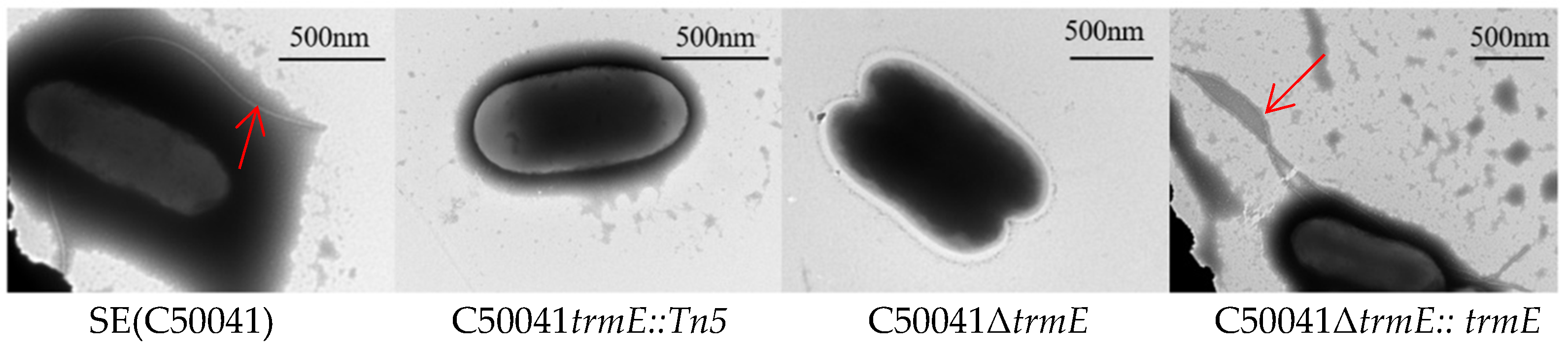
3.2. SE∆trmE Reconfirmed This Phenotype Without Motility

To confirm the phenotype, C50041∆trmE and C50041∆trmE::trmE were constructed and compared to their motility utilizing the semi-solid LB plate and U-tube. Compared to C50041trmE::Tn5, C50041∆trmE exhibited the same phenotype without motility on a semi-solid plate, while C50041∆trmE::trmE displayed a phenotype with strong motility, which was similar to that of SE (C50041) (Figure 1). The diameter of the SEΔtrmE halo was measured at 23 ± 1 mm, while that of C50041trmE::Tn5 was 15 ± 1 mm and that of C50041∆trmE::trmE was 36 ± 2 mm. As a control, that of the SE(C50041) halo was up to 48 ± 2 mm. These results indicate that SEΔtrmE motility was significantly weaker than that of SE(C50041) (p < 0.05).
This was assessed again using U-tube assays. The needles were punctured into LB media from the A side of the U-tube, and bacterial growth was observed on the LB surface from the B side. C50041ΔtrmE and C50041trmE::Tn5 showed no bacterial growth, while C50041∆trmE::trmE and SE(C50041) grew on the surface at B side of the U-tube, which indicates that C50041ΔtrmE had no motility (Figure 1) by trmE loss.

3.3. Biological Characteristics of C50041ΔtrmE Without Flagella

3.3.1. No Change in Biochemical Characteristics

After biochemical identification card API 20E was employed, the biochemical characteristics of C50041ΔtrmE were confirmed to be consistent with those of SE(C50041), and C50041∆trmE::trmE, ONPG, ADH), LDC, CIT, URE, TDA, IND, VP, GEL), INO, SAC, MEL, and AMY were tested to be negative (−), while ODC, H2S, GLU, MAN, SOR, RHA, and ARA were detected to be positive (+). This indicates that the trmE gene did not affect the biochemical properties of SE(C50041).

3.3.2. No Change in Antibiotic Resistance

Antimicrobial susceptibility in SE(C50041), C50041ΔtrmE, and C50041∆trmE::trmE was tested using 12 antibiotic disks (Oxoid, Basingstoke, UK) with the following specifications: kanamycin (KAN, 20 μg), ciprofloxacin (CIP, 5 μg), meropenem (MEM, 10 μg), cefotaxime (CTX, 5 μg), ampicillin (AMP, 10 μg), chloramphenicol (CHL, 30 μg), tetracycline (TET, 30 μg), nalidixic acid (NAL, 30 μg), florfenicol (FFC, 30 μg), streptomycin (STR, 10 μg), gentamicin (GEN, 10 μg), and trimethoprim-sulfamethoxazole (SXT, 10 μg). This demonstrated that C50041ΔtrmE did not alter bacterial antibiotic resistance.

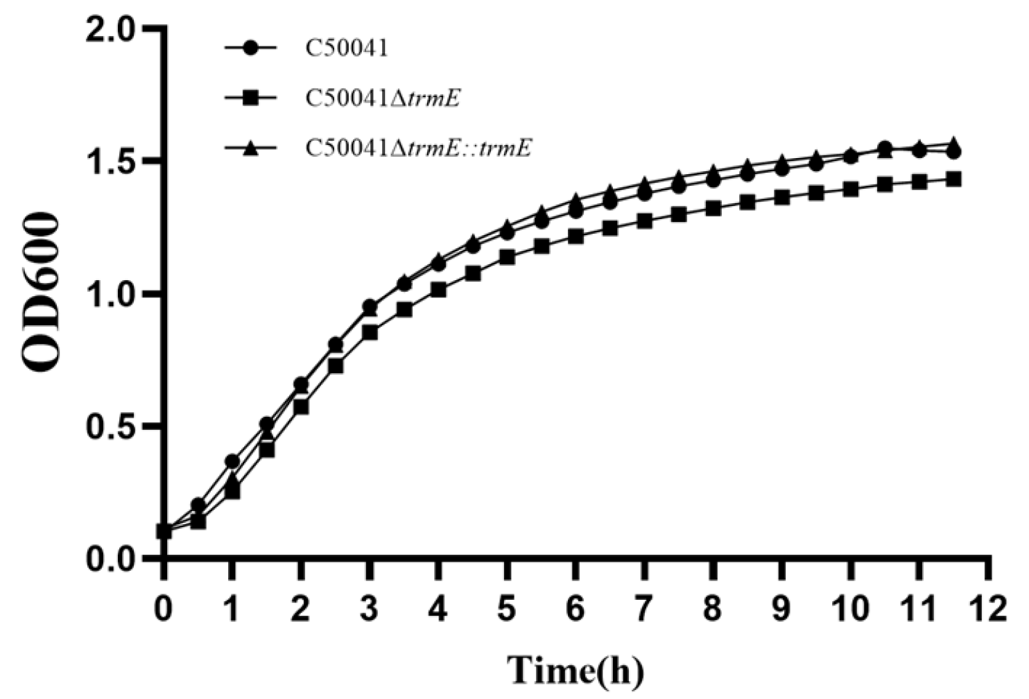
3.3.3. No Change in Bacterial Growth Speed

The growth speed of C50041ΔtrmE was analyzed by measuring its absorbance at 600 nm for the growth curve, indicating that the growth rate of C50041ΔtrmE showed no significant difference at the same time points from 1 to 12 h compared to SE(C50041) and C50041∆trmE::trmE (Figure 2).

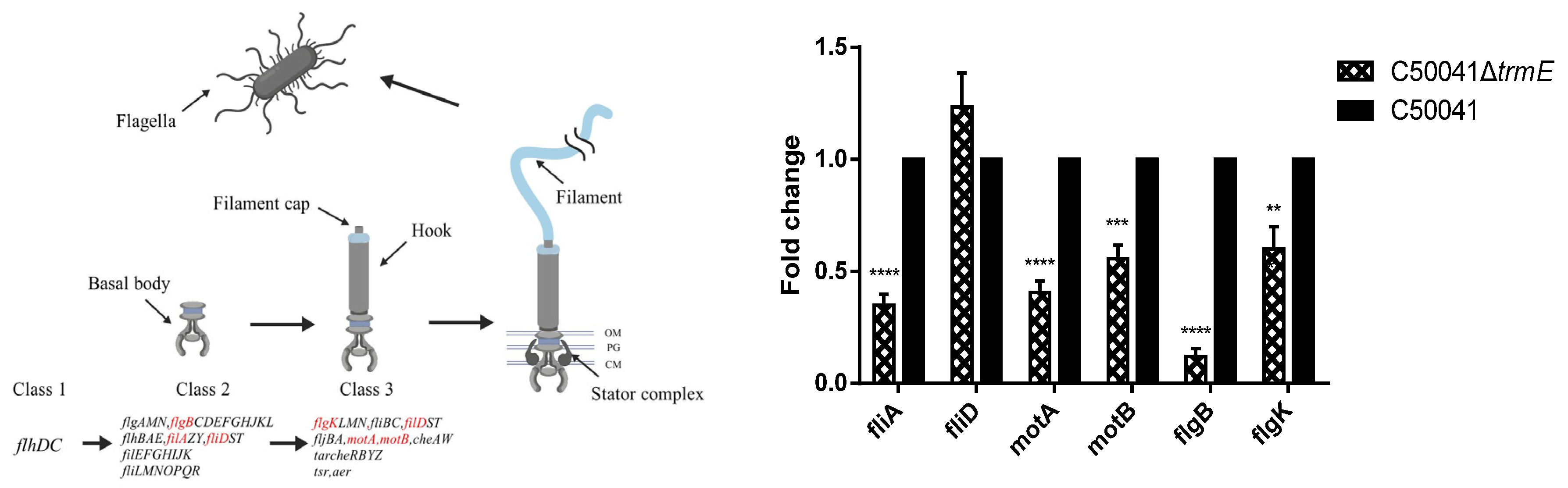
3.4. Low mRNA Level of Flagella Synthesis-Related Genes of C50041ΔtrmE

The mRNA level of the flagella synthesis-related genes of C50041ΔtrmE was analyzed via qRT-PCR. C50041ΔtrmE was indicated to be significantly downregulated in terms of the mRNA level of the flagellar synthesis σ factor fliA (p < 0.05), flagellar motor proteins motA (p < 0.05) and motB (p < 0.05), and flagellar biosynthesis proteins flgB (p < 0.05) and flgK (p < 0.05). This evidence demonstrates that trmE loss significantly inhibited flagellar synthesis in C50041ΔtrmE (Figure 3).
Further, Salmonella strains were observed for flagella via TEM: C50041ΔtrmE exhibited few flagella on the bacterial surface compared to SE(C50041) and C50041ΔtrmE:: trmE, which showed many flagella (Figure 4).

3.5. Low-Virulence In Vitro and In Vivo Assay of C50041ΔtrmE

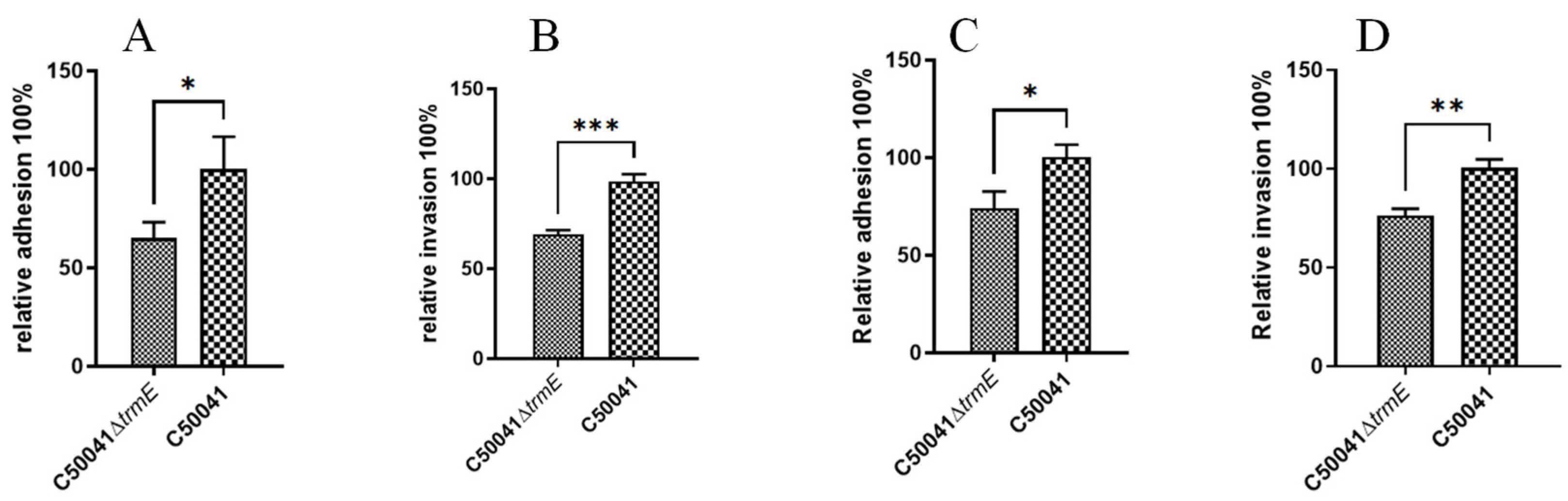
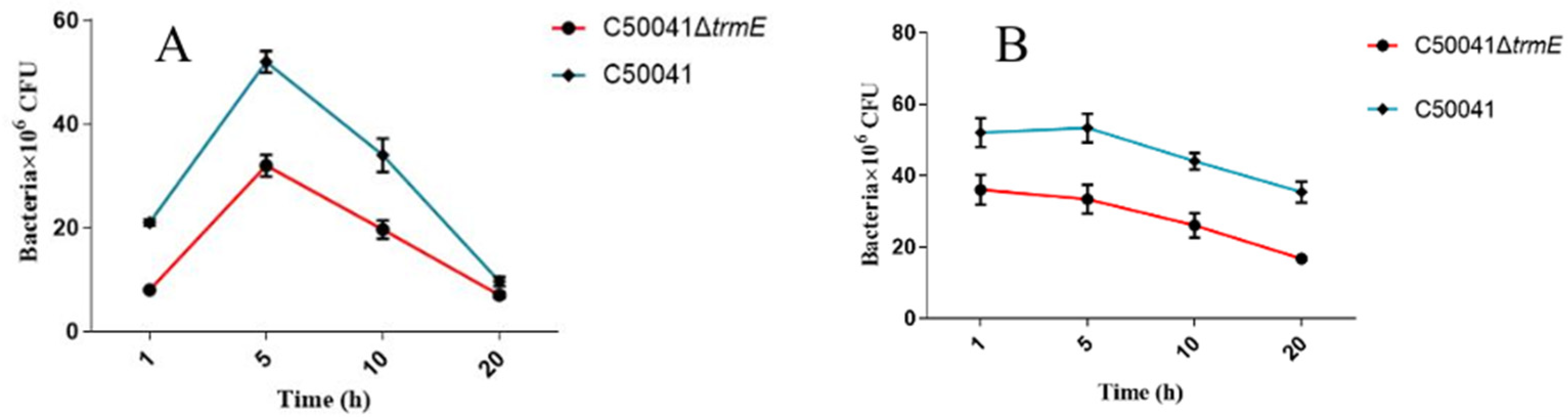
3.5.1. Low Ability to Infect Macrophages

RAW264.7 and J774A.1 cells were infected with Salmonella strains at MOI = 100:1. Adhesion and invasion abilities were evaluated after 30 min adhesion and then 1 h invasion. C50041ΔtrmE exhibited a significant decrease in adhesion rates to RAW264.7 (p < 0.05) and J774A.1 (p < 0.001) and in invasion rates to RAW264.7 (p < 0.05) and J774A.1 (p < 0.01) compared to SE(C50041) (Figure 5), which suggests that the trmE gene significantly affected Salmonella adhesion and invasion abilities to RAW264.7 and J774A.1 cells.
Subsequently, the proliferation rate of C50041ΔtrmE was assessed by counting the intracellular bacterial numbers at 1, 5, 10, and 20 h post infection using dilution plating. The results demonstrate that the loads of C50041ΔtrmE were significantly low at every time point compared to those of SE(C50041) because the primary intracellular bacterial numbers were different. However, the change trend was similar, indicating that the trmE gene did not affect the proliferation capacity of SE(C50041) (Figure 6).

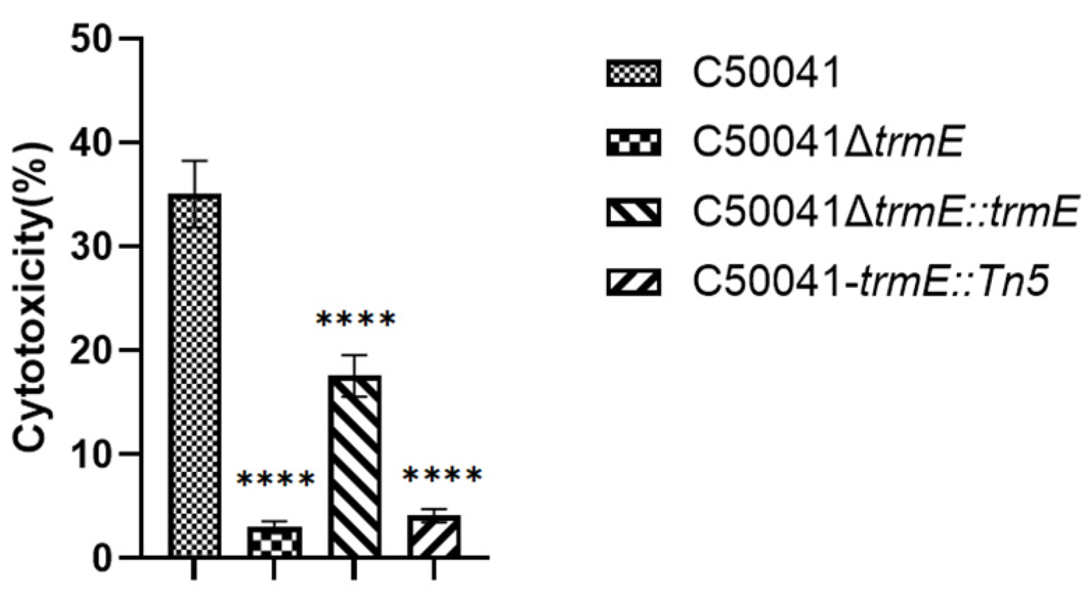
3.5.2. LDH Assay for Cytotoxicity

The cytotoxic activity of C50041ΔtrmE was assessed against RAW264.7 using a lactate dehydrogenase (LDH) release assay (Figure 7). C50041ΔtrmE could induce a statistically significant reduction in LDH release from RAW264.7 cells (p < 0.0001) compared to SE (C50041), which demonstrates that trmE loss attenuated SE(C50041) cytotoxicity.

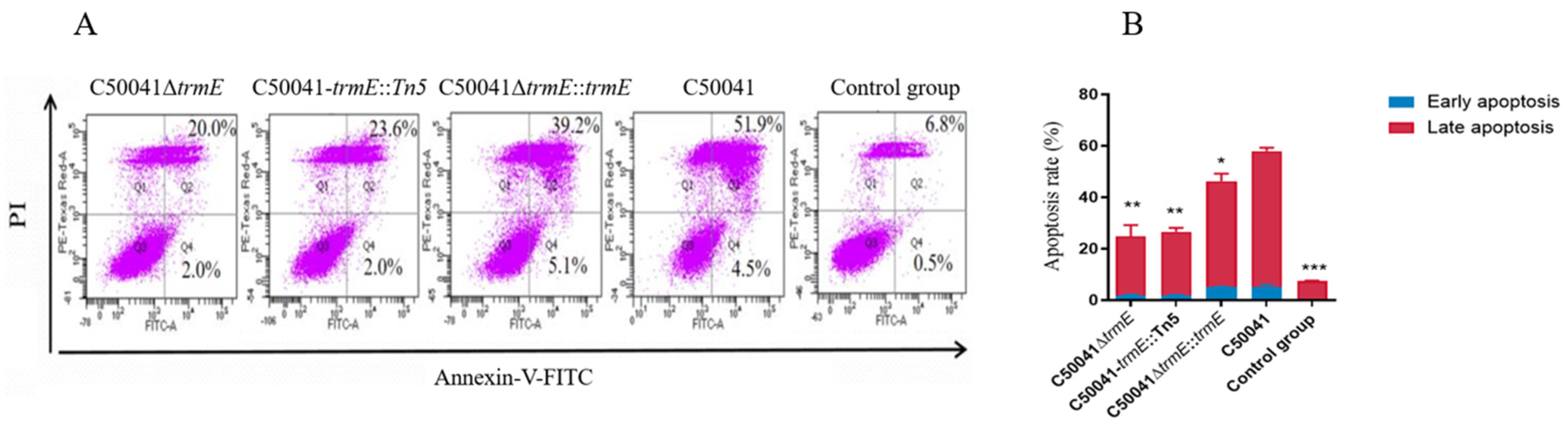
3.5.3. Decreased Ability to Induce Macrophage Apoptosis

After 3 h of RAW264.7 cells being infected by Salmonella strains, the early and late macrophage apoptosis level was assessed via flow cytometry using the Annexin V-FITC Kit. In addition to live cells (Annexin V/PI), the early apoptosis (Annexin V+/PI) rate of RAW264.7 cells was low from 2.0% to 5.1%, due to different Salmonella strains, but the late apoptosis (Annexin V+/PI+) rate with C50041ΔtrmE was 20.0%, which is same as that of C50041trmE::Tn5(23.6%) (p < 0.05) and significantly lower than that of C50041ΔtrmE::trmE (39.2%) (p < 0.05) and SE(C50041) (51.9%) (p < 0.01) (Figure 8). This suggests that trmE loss significantly reduced the ability of SE-induced macrophage apoptosis.

3.5.4. Decreased Ability to Induce Macrophage Pyroptosis Based on Caspase-1 Level

Caspase-1 level was assessed in RAW264.7 cells after Salmonella infection using Western blot, which reflected Salmonella’s ability to induce macrophage pyroptosis. The results indicate that the Caspase-1 level with C50041ΔtrmE was similar to that of C50041trmE::Tn5, significantly reduced compared to that with SE(C50041) (p = 0.0016), which demonstrates that trmE loss significantly inhibited SE to induce macrophage pyroptosis (Figure 9).

3.5.5. Low Mortality to Mice Based on LD50

LD50 was determined for C50041ΔtrmE and SE(C50041). Ten-fold gradient concentrations from 1.0 × 10⁴ CFU to 1.0 × 10⁸ CFU per mouse were orally administered to 8-week-old female BALB/c mice (n = 5), which were continuously observed for 2 weeks to record their survival. LD50 was calculated using the method described by Kouchi. The LD50 of C50041ΔtrmE increased by over 1500 folds compared to that of SE(C50041). This indicates that trmE loss significantly reduced SE virulence.

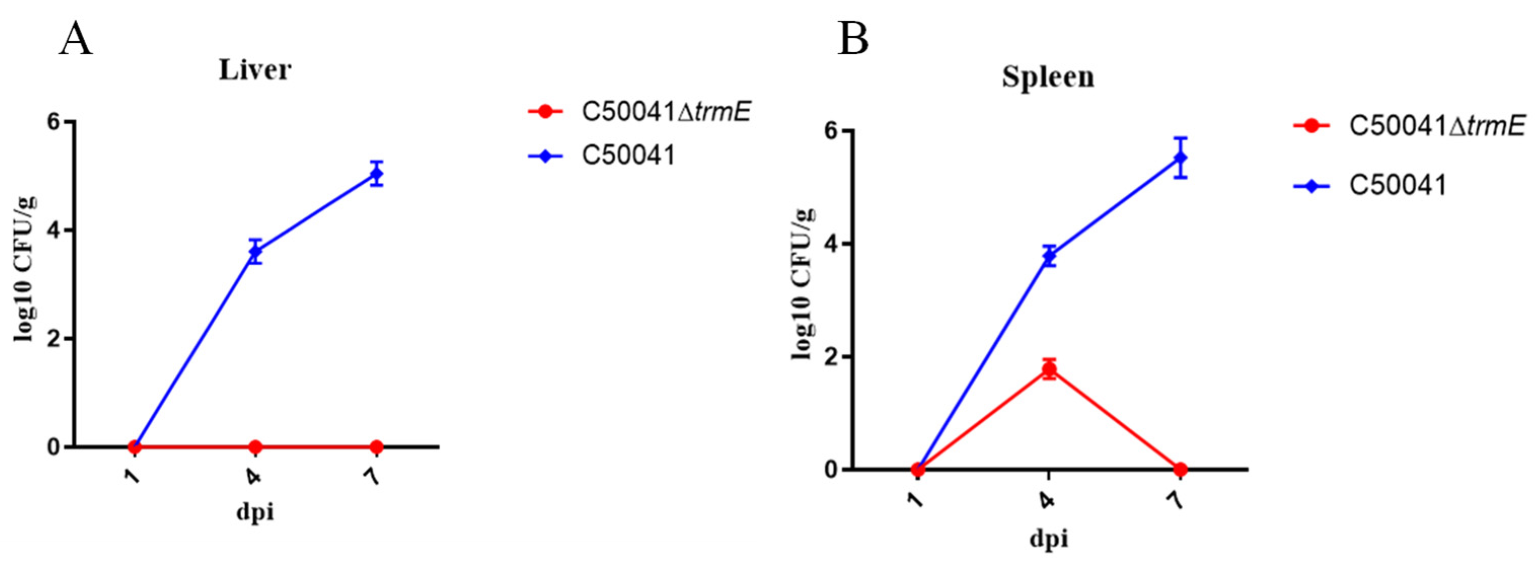
3.5.6. Low Persistence Ability In Vivo

According to LD50, a high dose of 1.0 × 10⁷ CFU in 100 µL C50041∆trmE was orally administered to mice (n = 3). The in vivo dynamics showed that no bacteria in the liver and only a few in the spleen were isolated, which shows that trmE loss decreased SE persistence ability in vivo (Figure 10).

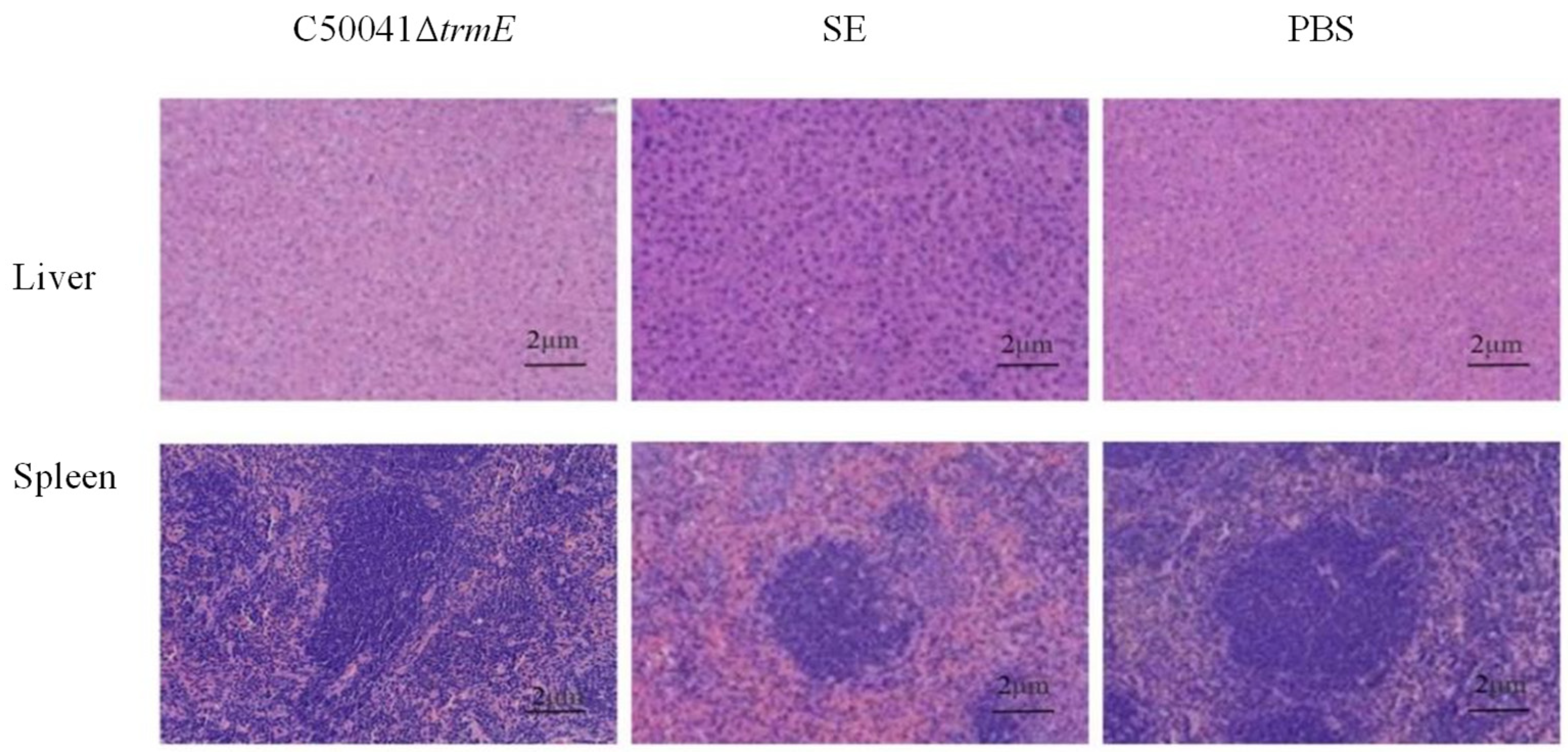
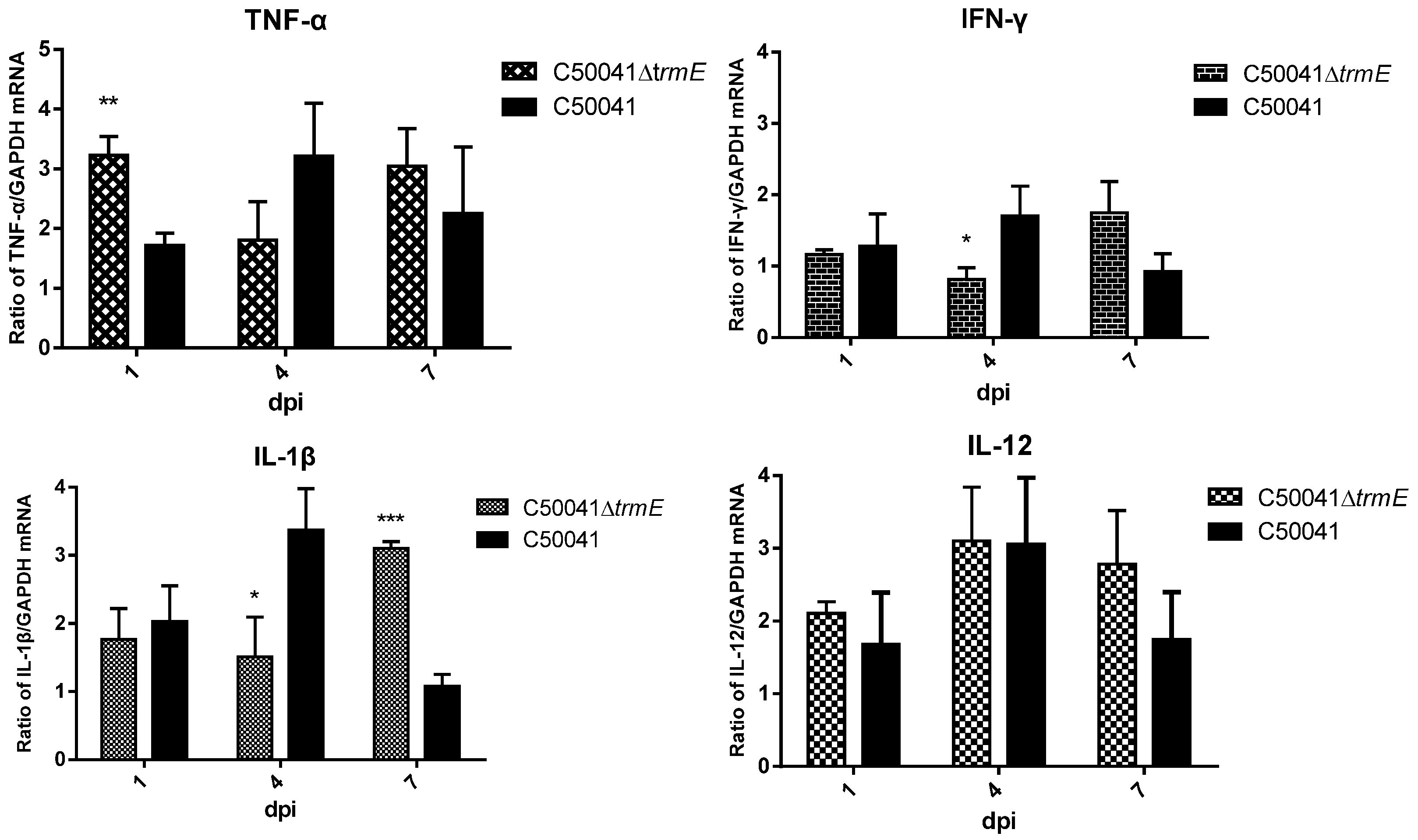
3.5.7. No Obvious Tissue Lesions and Changed mRNA Level of Pro-Inflammatory Cytokines

On day 7 after the mice (n = 3) were infected with Salmonella strains, pathological sections of the spleen and liver were taken and analyzed. No significant lesions occurred in either organ in the C50041ΔtrmE group, but there were severe lesions with inflammatory cell infiltration in the SE(C50041) group as a control and local necrotic foci. This proves that the trmE loss largely reduced Salmonella virulence (Figure 11).
In addition to tissue lesions, the mRNA of the pro-inflammatory factor also changed. After C50041ΔtrmE infection, there was a significant increase in the mRNA of TNF-α (p < 0.01) on day 1 and IL-1β (p < 0.001) on day 7, but the mRNA of IL-1β (p < 0.05) and IFN-γ (p < 0.05) was significantly reduced on day 4 compared to that of SE (C50041). IL-12 had no difference compared to that of SE(C50041) (Figure 12).

4. Discussion

The flagella of Salmonella play a crucial role in the bacteria’s bacterial motility and virulence. Utilizing its flagella, Salmonella increases the likelihood of being captured by resident macrophages in tissues [26], thereby breaching the blood–intestinal barrier and entering the internal environment. Once engulfed by macrophages, Salmonella proliferates within the Salmonella-containing vacuole (SCV) and activates the inflammasome through flagellin, leading to the activation of Caspase-1 and the initiation of the classical pyroptosis pathway. This ultimately results in macrophage pyroptosis and contributes to systemic infection [27]. Research on the regulatory genes of flagella holds significant importance for comprehensive understanding of Salmonella pathogenicity and virulence.
To identify all genes related to function or phenotype, a mutant library must be constructed and high-throughput screening must be performed. Theoretically, the Salmonella ssp. genome has more than 4000 ORFs; a minimum number of mutants equal to or two times greater than the number of ORFs is required for a mutant library to be considered representative. Although high-throughput screening is performed, not all genes related to function or phenotype are screened out in one experiment. In this study, we aimed to find novel gene(s) related to flagella synthesis; however, this is difficult to achieve because most genes have been found. In total, 11 motility-deficient mutants were screened out, and 8 genes were identified because 1 gene was possibly identified in two or more mutants. The other seven genes except trmE were confirmed to be involved in flagellar synthesis. A motility-deficient trmE mutant was accidentally identified from 1321 mutants in one transposon mutation library. Currently, there is no research reporting that the loss of the trmE gene results in the loss of Salmonella flagella. A thorough analysis of this mutant based on the motility phenotype and virulence function confirmed that the trmE gene regulates flagellar synthesis to reduce bacterial virulence.
The trmE gene was inactivated through its insertion with a Tn5 transposon, which resulted in SE(C50041) with weak motility. Thus, we constructed C50041ΔtrmE and C50041ΔtrmE::trmE to demonstrate their ability to regulate flagellar synthesis to prove their close correlation. At last, a few flagella on the surface of C50041ΔtrmE were observed via TEM as direct evidence, but their growth speed and biochemical characteristics did not change compared to with SE(C50041). Then, virulence analysis showed that the adhesion and invasion abilities of C50041ΔtrmE were decreased, as well as the intramacrophage proliferation capacity, cytotoxicity, cell apoptosis and pyroptosis, LD50, and tissue lesions.
Studies have indicated that region VIII of the fliC gene, which encodes flagellin, serves as a primary site for the induction of cytokines such as TNF-α and IFN-γ in CD4+ T cells [28]. Flagella can also stimulate PBMCs to release cytokines such as IL-1β and IL-12 [29]. Thus, we analyzed pathological changes in murine liver and spleen and the mRNA level of inflammatory factors in murine spleens in order to prove that C50041ΔtrmE successfully infected and induced localized tissue lesions, revealing that the absence of the trmE gene significantly affected the degree of inflammatory response in mice. However, the mRNA level of inflammatory factors in murine spleens did not show regularity. The absence of flagella does not lead to a comprehensive reduction in the expression of pro-inflammatory cytokines. For example, the levels of these pro-inflammatory cytokines were consistently elevated in the C50041ΔtrmE group compared to the wild-type control group on day 7 post Salmonella infection, and the mRNA level of IL-12 showed no change on days 1, 4, or 7, which did not match theoretical expectations, suggesting the potential existence of other compensatory mechanisms that need further investigation.
The trmE gene’s involvement in bacterial virulence has been studied. During protein translation, the wobble effect occurs, and the third (5′) base of the anticodon can typically pair with either member of the purine or pyrimidine pair in the codon. The trmE gene has been studied to decode TrmE to modify U34. It is also referred to as mnmE or thdF, and it spans 1364 base pairs. The trmE gene encodes the tRNA-modifying GTPase TrmE, which introduces a carboxymethyl-amino-methyl (cmnm) modification at the swing position (U34) of selected tRNAs, leading to the creation of tRNA-cmnm5s2U34 [30] for translational misreading [31]. The TrmE protein exhibits a tripartite structural domain and is highly conserved across eukaryotic and bacterial species; it functions as a molecular switch, adopting various conformations based on GTP or GDP binding. To date, researchers have identified over 20 homologs of TrmE in both prokaryotes and eukaryotes. Additionally, trmE genes may play a role in bacterial survival under low-temperature conditions. Singh et al. demonstrated that low temperatures induce the trmE gene and its promoter, noting that the Pseudomonas syringae CSM1 mutant, which lacks a functional trmE gene, exhibits cold sensitivity [32,33]. Gong investigated the involvement of the trmE gene in E. coli acid tolerance, revealing that this effect depends on glutamate and requires the activation of the gadE gene [34]. Currently, there are no documented studies indicating that no expression of the trmE gene leads to the loss of Salmonella flagella.
Although we have confirmed that the trmE gene is positively correlated with flagellar synthesis, we also found one question asking why it significantly affects flagella synthesis: since TrmE affects the function of tRNA, it should have a global impact, but no significant effect was observed on its growth speed, biochemical characteristics, or antibiotic resistance. This bias and its mechanism will be elucidated in the future to further explore the mysteries of bacterial life.

5. Conclusions

In conclusion, our study provides strong evidence, from the phenotype and function levels, that trmE is a novel gene involved in the regulation of flagella synthesis and bacterial virulence.

Author Contributions

Conceptualization, H.G., L.L., and J.Z.; methodology, H.G., L.L., and J.G.; software, H.G. and J.Z.; formal analysis, H.G., L.L., and J.G.; data curation, H.G. and L.L.; supervision, P.B. and S.G.; project administration, S.G. All authors have read and agreed to the published version of the manuscript.

Funding

This work was supported by the National Key Research and Development Program Special Project (2016YFD0501607), the Natural Science Foundation of Jiangsu Province of China (BK20151306), and the Special Project on Science and Technology in North Jiangsu (SZ-SQ2021046).

Institutional Review Board Statement

Animal experiments were approved by the Animal Welfare and Ethics Committee of Yangzhou University (SYXK[Su] 2023-0089) on 26 February 2024 and were conducted in accordance with the guidelines of the Yangzhou University Institutional Animal Care and Use Committee (IACUC).

Informed Consent Statement

Not applicable.

Data Availability Statement

Data are contained within the article.

Acknowledgments

The authors thank all colleagues who made suggestions and revisions to this paper. All individuals included in this section have consented to this acknowledgement.

Conflicts of Interest

All authors declare that they have no known competing financial interests or personal relationships that could have appeared to influence the work reported in this paper.

Appendix A

Table A1. Bacterial strains, plasmids, and cells used in this study.
Strains or PlasmidsCharacteristicReference
Strains
SE C50041Wild-type Salmonella Enteritidis Lab collection
C50041-trmE::Tn5C50041 with Tn5 inserted in trmE geneThis study
C50041∆trmEC50041 with a defined deletion of the trmE geneThis study
C50041∆trmE::trmEC50041∆trmE with pBR322 expressing the trmE geneThis study
E.coli χ7213Its growth for pGMB152 with DAP, as conjugal donor[15]
Plasmids
pUT mini-Tn5Km2Transposon delivery vector, Cmr, Kmr[17]
pGMB152pGMB151 derivative, suicide vector, Ampr, Smr, LacZYA[15]
pBR322For construction of SE∆trmE::trmE, Cmr[18]
pBR322-trmEpPR322 derivative containing trmE, Ampr and TetrThis study
Cells
RAW264.7Murine macrophagesThis study
J774A.1Murine macrophagesThis study
Table A2. Primers used in this study.
Primer NamePrimer Sequences (5′-3′)Target
trmE-up-FCCCCCCCTGCAGGTCGACGTGGTTCCCGTCAGGTCTConstruction for C50041∆ trmE
trmE-up-RCAGCCTACACAATCGCTCAAGGTTAGTCTCAACTTTGTTGCAAT
trmE-Cm-FATTGCAACAAAGTTGAGACTAACCTTGAGCGATTGTGTAGGCTG
trmE-Cm-RTTTGTAGGCCCGGTAAGCATATGGGAATTAGCCATGGTCC
trmE-down-FGGACCATGGCTAATTCCCATATGCTTACCGGGCCTACAAA
trmE-down-RCTTATCGATACCGTCGACCAGGTAAACGGAGAAGGCGA
trmE-NYZ-FAAACTCGTTACAGGGGGCAT
trmE-NYZ-RTACGCTCAACTTCGTCGCTG
pGMB152-FCGTGGAGGCCATCAAACCAC
pGMB152-RCGCGAAATAAACGACCGGGA
C-trmE-FTTATCATCGATAAGCTTATGAGCCATAACGACACTATCGTCConstruction of complemented mutant C50041∆ trmE:: trmE
C-trmE-RTCCGGCGTAGAGGATCCTTGTAGGCCCGGTAAGCATC
Note: The underlined segments indicate the homologous sequences for the one-step ligation of recombinant plasmid.
Table A3. Primers used in this study.
Primer NamePrimer Sequences (5′-3′)Size (bp)
IL-1β-FTGGCCTTCAAAGGAAAGAATCTATACCTGTCC167
IL-1β-RGTTGGGGAACTCTGCAGACTCAAACTCCAC
IL-12-FTGCCCCCACAGAAGACGTCTTTGATGAT138
IL-12-RGATGGCCACCAGCATGCCCTTGTC
TNF-α-FCAGGCCTTCCTACCTTCAGACCTTTCCAGAT
TNF-α-RACACCCCGCCCTTCCAAATAAATACATTCAT122
IFN-γ-FGCCAAGACTGTGATTGCGGGGTTGTATCT
IFN-γ-RTAAAGCGCTGGCCCGGAGTGTAGACA198
GAPDH-FCAGCCTCGTCCCGTAGACAA
GAPDH-RACCCCGTCTCCGGAGTCCATCACAAT156

References

  1. Shaji, S.; Selvaraj, R.K.; Shanmugasundaram, R. Salmonella Infection in Poultry: A Review on the Pathogen and Control Strategies. Microorganisms 2023, 11, 2814. [Google Scholar] [CrossRef] [PubMed]
  2. Dai, Y.; Zhang, M.; Liu, X.; Sun, T.; Qi, W.; Ding, W.; Chen, Z.; Zhang, P.; Liu, R.; Chen, H.; et al. Salmonella manipulates macrophage migration via SteC-mediated myosin light chain activation to penetrate the gut-vascular barrier. EMBO J. 2024, 43, 1499–1518. [Google Scholar] [CrossRef]
  3. Santos, R.L.; Zhang, S.; Tsolis, R.M.; Kingsley, R.A.; Adams, L.G.; Bäumler, A.J. Animal models of Salmonella infections: Enteritis versus typhoid fever. Microbes Infect. 2001, 3, 1335–1344. [Google Scholar] [CrossRef]
  4. Magariyama, Y.; Sugiyama, S.; Kudo, S. Bacterial swimming speed and rotation rate of bundled flagella. FEMS Microbiol. Lett. 2001, 199, 125–129. [Google Scholar] [CrossRef]
  5. Vilas Boas, D.; Castro, J.; Araújo, D.; Nóbrega, F.L.; Keevil, C.W.; Azevedo, N.F.; Vieira, M.J.; Almeida, C. The Role of Flagellum and Flagellum-Based Motility on Salmonella Enteritidis and Escherichia coli Biofilm Formation. Microorganisms 2024, 12, 232. [Google Scholar] [CrossRef] [PubMed]
  6. Cohen, H.; Hoede, C.; Scharte, F.; Coluzzi, C.; Cohen, E.; Shomer, I.; Mallet, L.; Holbert, S.; Serre, R.F.; Schiex, T.; et al. Intracellular Salmonella Paratyphi A is motile and differs in the expression of flagella-chemotaxis, SPI-1 and carbon utilization pathways in comparison to intracellular S. Typhimurium. PLoS Pathog. 2022, 18, e1010425. [Google Scholar] [CrossRef] [PubMed]
  7. Wang, M.; Qazi, I.H.; Wang, L.; Zhou, G.; Han, H. Salmonella Virulence and Immune Escape. Microorganisms 2020, 8, 407. [Google Scholar] [CrossRef]
  8. Minamino, T.; Macnab, R.M. Components of the Salmonella flagellar export apparatus and classification of export substrates. J. Bacteriol. 1999, 181, 1388–1394. [Google Scholar] [CrossRef]
  9. Chilcott, G.S.; Hughes, K.T. Coupling of flagellar gene expression to flagellar assembly in Salmonella enterica serovar typhimurium and Escherichia coli. Microbiol. Mol. Biol. Rev. MMBR 2000, 64, 694–708. [Google Scholar] [CrossRef]
  10. Das, C.; Mokashi, C.; Mande, S.S.; Saini, S. Dynamics and Control of Flagella Assembly in Salmonella typhimurium. Front. Cell. Infect. Microbiol. 2018, 8, 36. [Google Scholar] [CrossRef]
  11. Aldridge, P.D.; Wu, C.; Gnerer, J.; Karlinsey, J.E.; Hughes, K.T.; Sachs, M.S. Regulatory protein that inhibits both synthesis and use of the target protein controls flagellar phase variation in Salmonella enterica. Proc. Natl. Acad. Sci. USA 2006, 103, 11340–11345. [Google Scholar] [CrossRef] [PubMed]
  12. Yue, Y.; Wang, W.; Ma, Y.; Song, N.; Jia, H.; Li, C.; Wang, Q.; Li, H.; Li, B. Cooperative Regulation of Flagellar Synthesis by Two EAL-Like Proteins upon Salmonella Entry into Host Cells. Microbiol. Spectr. 2023, 11, e0285922. [Google Scholar] [CrossRef] [PubMed]
  13. Albanna, A.; Sim, M.; Hoskisson, P.A.; Gillespie, C.; Rao, C.V.; Aldridge, P.D. Driving the expression of the Salmonella enterica sv Typhimurium flagellum using flhDC from Escherichia coli results in key regulatory and cellular differences. Sci. Rep. 2018, 8, 16705. [Google Scholar] [CrossRef] [PubMed]
  14. Saenz, H.L.; Dehio, C. Signature-tagged mutagenesis: Technical advances in a negative selection method for virulence gene identification. Curr. Opin. Microbiol. 2005, 8, 612–619. [Google Scholar] [CrossRef]
  15. Geng, S.; Tian, Q.; An, S.; Pan, Z.; Chen, X.; Jiao, X. High-Efficiency, Two-Step Scarless-Markerless Genome Genetic Modification in Salmonella enterica. Curr. Microbiol. 2016, 72, 700–706. [Google Scholar] [CrossRef]
  16. Gao, R.; Zhang, J.; Geng, H.; Wang, Y.; Kang, X.; Geng, S.; Jiao, X.; Barrow, P. The Loss of focA Gene Increases the Ability of Salmonella Enteritidis to Exit from Macrophages and Boosts Early Extraintestinal Spread for Systemic Infection in a Mouse Model. Microorganisms 2022, 10, 1557. [Google Scholar] [CrossRef]
  17. Geng, S.; Jiao, X.; Barrow, P.; Pan, Z.; Chen, X. Virulence determinants of Salmonella Gallinarum biovar Pullorum identified by PCR signature-tagged mutagenesis and the spiC mutant as a candidate live attenuated vaccine. Vet. Microbiol. 2014, 168, 388–394. [Google Scholar] [CrossRef]
  18. Geng, S.; Wang, Y.; Xue, Y.; Wang, H.; Cai, Y.; Zhang, J.; Barrow, P.; Pan, Z.; Jiao, X. The SseL protein inhibits the intracellular NF-κB pathway to enhance the virulence of Salmonella Pullorum in a chicken model. Microb. Pathog. 2019, 129, 1–6. [Google Scholar] [CrossRef]
  19. Neubauer, H.; Sauer, T.; Becker, H.; Aleksic, S.; Meyer, H. Comparison of systems for identification and differentiation of species within the genus Yersinia. J. Clin. Microbiol. 1998, 36, 3366–3368. [Google Scholar] [CrossRef]
  20. Wikler, M.A.; Cockerill, F.R.; Bush, K.; Dudley, M.N.; Eliopoulos, G.M.; Hardy, D.; Hecht, D.W.; Ferraro, M.J.; Swenson, J.M.; Hindler, J.F.; et al. Methods for Dilution Antimicrobial Susceptibility Tests for Bacteria That Grow Aerobically: Approved Standard; Clinical and Laboratory Standards Institute: Wayne, PA, USA, 2000. [Google Scholar]
  21. Taylor, S.C.; Nadeau, K.; Abbasi, M.; Lachance, C.; Nguyen, M.; Fenrich, J. The Ultimate qPCR Experiment: Producing Publication Quality, Reproducible Data the First Time. Trends Biotechnol. 2019, 37, 761–774. [Google Scholar] [CrossRef]
  22. Wang, W.; Yuan, X.; Mu, J.; Zou, Y.; Xu, L.; Chen, J.; Zhu, X.; Li, B.; Zeng, Z.; Wu, X.; et al. Quercetin induces MGMT+ glioblastoma cells apoptosis via dual inhibition of Wnt3a/β-Catenin and Akt/NF-κB signaling pathways. Phytomed. Int. J. Phytother. Phytopharm. 2023, 118, 154933. [Google Scholar] [CrossRef] [PubMed]
  23. Boise, L.H.; Collins, C.M. Salmonella-induced cell death: Apoptosis, necrosis or programmed cell death? Trends Microbiol. 2001, 9, 64–67. [Google Scholar] [CrossRef] [PubMed]
  24. Yin, J.; Cheng, Z.; Wang, X.; Xu, L.; Li, Q.; Geng, S.; Jiao, X. Evaluation of the Salmonella enterica Serovar Pullorum Pathogenicity Island 2 Mutant as a Candidate Live Attenuated Oral Vaccine. Clin. Vaccine Immunol. CVI 2015, 22, 706–710. [Google Scholar] [CrossRef] [PubMed]
  25. Minamino, T.; Morimoto, Y.V.; Kinoshita, M.; Namba, K. Multiple Roles of Flagellar Export Chaperones for Efficient and Robust Flagellar Filament Formation in Salmonella. Front. Microbiol. 2021, 12, 756044. [Google Scholar] [CrossRef]
  26. Haiko, J.; Westerlund-Wikström, B. The role of the bacterial flagellum in adhesion and virulence. Biology 2013, 2, 1242–1267. [Google Scholar] [CrossRef]
  27. Pandeya, A.; Zhang, Y.; Cui, J.; Yang, L.; Li, J.; Zhang, G.; Wu, C.; Li, Z.; Wei, Y. Inflammasome activation and pyroptosis mediate coagulopathy and inflammation in Salmonella systemic infection. Microbiol. Res. 2023, 275, 127460. [Google Scholar] [CrossRef]
  28. McSorley, S.J.; Cookson, B.T.; Jenkins, M.K. Characterization of CD4+ T cell responses during natural infection with Salmonella typhimurium. J. Immunol. 2000, 164, 986–993. [Google Scholar] [CrossRef]
  29. Wyant, T.L.; Tanner, M.K.; Sztein, M.B. Salmonella typhi flagella are potent inducers of proinflammatory cytokine secretion by human monocytes. Infect. Immun. 1999, 67, 3619–3624. [Google Scholar] [CrossRef] [PubMed]
  30. Cabedo, H.; Macián, F.; Villarroya, M.; Escudero, J.C.; Martínez-Vicente, M.; Knecht, E.; Armengod, M.E. The Escherichia coli trmE (mnmE) gene, involved in tRNA modification, codes for an evolutionarily conserved GTPase with unusual biochemical properties. EMBO J. 1999, 18, 7063–7076. [Google Scholar] [CrossRef]
  31. Brégeon, D.; Colot, V.; Radman, M.; Taddei, F. Translational misreading: A tRNA modification counteracts a +2 ribosomal frameshift. Genes Dev. 2001, 15, 2295–2306. [Google Scholar] [CrossRef]
  32. Singh, A.K.; Pindi, P.K.; Dube, S.; Sundareswaran, V.R.; Shivaji, S. Importance of trmE for growth of the psychrophile Pseudomonas syringae at low temperatures. Appl. Environ. Microbiol. 2009, 75, 4419–4426. [Google Scholar] [CrossRef] [PubMed]
  33. Shippy, D.C.; Fadl, A.A. tRNA modification enzymes GidA and MnmE: Potential role in virulence of bacterial pathogens. Int. J. Mol. Sci. 2014, 15, 18267–18280. [Google Scholar] [CrossRef] [PubMed]
  34. Gong, S.; Ma, Z.; Foster, J.W. The Era-like GTPase TrmE conditionally activates gadE and glutamate-dependent acid resistance in Escherichia coli. Mol. Microbiol. 2004, 54, 948–961. [Google Scholar] [CrossRef] [PubMed]
Figure 1. Motility analysis of SE trmE mutants using semi-solid plate and U-tube. Note: A is the puncture end, and B is the observation end. p < 0.05 (*), <0.01 (**), <0.001 (***).
Microorganisms 13 01455 g001
Figure 2. Growth curves of Salmonella strains.
Microorganisms 13 01455 g002
Figure 3. mRNA level of part C50041ΔtrmE flagella synthesis-related genes and the locations of these genes (marked in red) [25]. p < 0.01 (**), <0.001 (***), <0.0001 (****).
Microorganisms 13 01455 g003
Figure 4. Flagellar observation of SE trmE mutants via TEM. Note: The red arrows point to Salmonella flagella.
Microorganisms 13 01455 g004
Figure 5. Adhesion and invasion ability of C50041ΔtrmE to RAW264.7 (A,B) and J774A.1 (C,D) cells (MOI = 100). p < 0.05 (*), <0.01 (**), <0.001 (***).
Microorganisms 13 01455 g005
Figure 6. Proliferation ability of C50041ΔtrmE in RAW264.7 (A) and J774A.1 (B) cells.
Microorganisms 13 01455 g006
Figure 7. Cytotoxicity of Salmonella to RAW264.7 based on LDH assay. p <0.0001 (****).
Microorganisms 13 01455 g007
Figure 8. Ability analysis of Salmonella to induce macrophage apoptosis. (A): Flow cytometry; (B): statistical analysis. p < 0.05 (*), <0.01 (**), <0.001 (***).
Microorganisms 13 01455 g008
Figure 9. Caspase-1 level during Salmonella C50041ΔtrmE inducing macrophage pyroptosis. (A): Western blot analysis; (B): gray analysis. Note: The original image was submitted. p < 0.05 (*), <0.0001 (****).
Microorganisms 13 01455 g009
Figure 10. Dynamic analysis of Salmonella strains in murine liver (A) and spleen (B).
Microorganisms 13 01455 g010
Figure 11. Lesions in murine liver and spleen from Salmonella infection (200×). Note: Compared with the C50041ΔtrmE and PBS group, the SE group exhibits a splenic nodule architecture that is markedly disorganized, exhibiting significant depletion of small lymphocytes within the nodules. Cellular arrangement appears loose, with effacement of the red-white pulp demarcation, accompanied by diffuse inflammatory cell infiltration.
Microorganisms 13 01455 g011
Figure 12. mRNA level of murine cytokines induced by Salmonella strains in spleen (using three mice, three replicates per sample, and a t-test). p < 0.05 (*), <0.01 (**), <0.001 (***).
Microorganisms 13 01455 g012
Table 1. Information regarding the insertion of Tn5 into genes.
Identified GeneNumber of Mutant Gene FunctionSimilarity
trmE1tRNA modification GTPase TrmE100%
fliD2synthesis of flagellum 100%
fliP1synthesis of flagellum 100%
rfbK1synthesis of lipopolysaccharides (LPS)100%
fliA1flagellar biosynthesis sigma factor100%
cpsG1synthesis of lipopolysaccharides (LPS)100%
rfaL2synthesis of lipopolysaccharides (LPS)100%
csrD2Global regulation100%
Disclaimer/Publisher’s Note: The statements, opinions and data contained in all publications are solely those of the individual author(s) and contributor(s) and not of MDPI and/or the editor(s). MDPI and/or the editor(s) disclaim responsibility for any injury to people or property resulting from any ideas, methods, instructions or products referred to in the content.

