Article

Virulence and Antibiotic Resistance of Pathogenic Aeromonas caviae from Diseased Macrobrachium rosenbergii

College of Animal Science and Technology, Yangzhou University, Yangzhou 225009, China
*
Author to whom correspondence should be addressed.
Microorganisms 2025, 13(6), 1343; https://doi.org/10.3390/microorganisms13061343
Submission received: 13 April 2025 / Revised: 2 June 2025 / Accepted: 7 June 2025 / Published: 10 June 2025
(This article belongs to the Section Veterinary Microbiology)

Abstract

In recent years, viral and bacterial diseases have posed serious challenges to the sustainable development of Macrobrachium rosenbergii (giant freshwater prawn) aquaculture, resulting in considerable economic losses across China. Among the bacterial pathogens, Aeromonas caviae has emerged as a notable opportunistic agent capable of causing large-scale mortality in various aquatic species. In this study, a highly virulent strain of A. caviae (designated GMRS4) was isolated from diseased M. rosenbergii exhibiting mass mortality in Yangzhou, Jiangsu Province. The isolate, a Gram-negative bacillus, was identified as the causative agent based on morphological, molecular, and histopathological analyses. Pathogenicity was confirmed through experimental infection, with the strain displaying marked virulence, evidenced by an LD50 of 1.91 × 10⁶ CFU/mL at 96 h. Whole-genome sequencing of GMRS4 revealed 4078 coding sequences, including a suite of virulence-associated genes encoding extracellular enzymes (DNase, hemolysin, caseinase, and lecithinase) and toxins (serine protease, elastase, and flagellin). Antimicrobial susceptibility testing indicated resistance to several antibiotics, particularly those in the penicillin and sulfonamide classes, while maintaining sensitivity to quinolones. Genomic analysis further revealed multiple antibiotic resistance genes and virulence genes, offering insights into the pathogenic mechanisms and resistance profile of the strain. This study underscores the threat posed by A. caviae to freshwater prawn aquaculture and provides a genetic basis for developing targeted disease management strategies.

1. Introduction

As a commercially valuable aquaculture species, Macrobrachium rosenbergii, is one of the most important freshwater cultured crustacean species in Asia, with a high annual production of 177,836 tons in 2023 in China (China Fishery Statistical Yearbook, 2024). Such a significant increase in breeding output is primarily due to improvements in seedling and changes in aquaculture models [1,2]. High-density culture is currently the primary farming model of M. rosenbergii, resulting in a rising incidence of disease outbreaks caused by bacterial and viral infections [3,4]. Reports indicate that some bacterial pathogens affecting M. rosenbergii include Vibrio spp. (non-O1/O139 Vibrio cholerae, Vibrio vulnificus, and Vibrio alginolyticus) [5,6,7], Enterobacter spp. (Enterobacter cloacae) [8], and Aeromonas spp. (Aeromonas sobria and Aeromonas veronii) [9,10]. Generally, outbreaks caused by viral pathogens include infectious precocious virus (IPV) [11], M. rosenbergii nodavirus (MrNV) [12], extra small virus (XSV) [13], and decapod iridescent virus 1 (DIV1) [14]. These pathogens cause mass mortalities, significantly diminishing the annual production of cultured M. rosenbergii and resulting in significant financial losses for M. rosenbergii aquaculture. Therefore, it is crucial to monitor the emergence of pathogens in aquaculture species and to understand their pathogenic mechanisms, as this knowledge can inform the development of effective control measures. This study investigated a mass mortality event of M. rosenbergii in Yangzhou, Jiangsu Province. The dominant bacterial strain, GMRS4, isolated from the diseased prawns, was identified as Aeromonas caviae.
A. caviae is a Gram-negative bacterium belonging to the Aeromonas genus and is ubiquitously distributed in various water environments, including freshwater and brackish water [15]. As an opportunistic pathogen, A. caviae typically exists harmlessly in the environment or within its host but can cause infections when the host’s immune system is compromised, such as under conditions of stress, overcrowding, or poor water quality [16,17]. In recent years, A. caviae has been recognized as an emerging pathogen in aquaculture, causing serious diseases in aquatic animals, such as Clarias batrachus [18], Gelidium pusillum [19], Clarias batrachus [20], Oreochromis niloticus (Linnaeus, 1758) [21], Micropterus salmoides [22]. Additionally, the extensive application of antibiotics in aquaculture and clinical settings has led to a notable increase in A. caviae resistance [23]. Thus, it is imperative to promptly investigate and analyze the pathogenic mechanisms and drug resistance factors of bacterial pathogens to effectively prevent and control this disease in aquaculture.
As is commonly acknowledged, the pathogenic potential of bacteria is largely determined by a range of virulence factors, including colonization, invasiveness, exotoxin, endotoxin, and extracellular enzymes [24]. These factors facilitate the pathogen’s effective colonization, tissue invasion, immune evasion, replication, and pathogenesis in the host [25]. In addition to virulence factors, bacterial survival in hostile environments and resistance to antimicrobial treatment are largely facilitated by resistance mechanisms, such as efflux pumps and β-lactamase production, which are encoded by specific antibiotic resistance genes [26,27]. Unlike single mechanisms, the synergistic interaction of multiple virulence and resistance factors significantly enhances the ability of pathogens to invade the host and survive in the presence of an antimicrobial agent [28]. Consequently, genes encoding both virulence and resistance factors are frequently used to evaluate the pathogenic potential and antimicrobial resistance profiles of bacterial pathogens. Recent advancements in NGS technologies, coupled with the development of sophisticated bioinformatics tools, have significantly enhanced the rapid and precise analysis of entire genomes of various bacterial pathogens. Whole-genome sequencing (WGS) plays a critical role in species identification, gene function annotation, the identification of resistance genes, and the analysis of genomic structural variations, which contribute to understanding the pathogenicity and drug resistance of different pathogens [29,30].
In this study, A. caviae GMRS4 was isolated from the hepatopancreas of diseased M. rosenbergii, and the pathogenic characteristics of GMRS4 were evaluated through artificial infection, histopathological examination, and analysis of virulence genes. Furthermore, the complete genome of A. caviae GMRS4 was sequenced, allowing for the identification of virulence-associated and antibiotic resistance genes to elucidate its pathogenic mechanisms and resistance traits. The present results indicate that A. caviae GMRS4 is the causative agent responsible for the mass mortalities observed in M. rosenbergii aquaculture, and the genomic analysis will provide insights into the pathogenic mechanisms and antimicrobial resistance of A. caviae, which aims to provide a theoretical basis for disease prevention and control in the M. rosenbergii industry.

2. Materials and Methods

2.1. Prawn Disease Description and Pathogen Examination

In August 2023, cultured M. rosenbergii (weight: 22.94 ± 0.93 g; length: 128.86 ± 2.48 mm) experienced mass mortality within a few days across several farms in Gaoyou, Jiangsu Province, under a cultural condition of 28 °C in freshwater. The disease has been prevalent for an extended period, with peak outbreaks occurring during the summer, and prawn mortalities reaching approximately 40–50%. Specimens of M. rosenbergii exhibiting high mortality rates were obtained from affected farms for pathogen analysis. Bacterial examination was conducted following Gao et al. [31], viral examination was based on Qian et al. [14], and parasites were observed using a light microscope. No viruses or parasites were detected; nevertheless, the primary bacterial strain was isolated from the hepatopancreas and designated as GMRS4. The dominant single colony was subsequently re-streaked onto a Luria–Bertani (LB) agar slant (Hope Bio. Co., Ltd., Qingdao, China), and the purified culture was stored in LB medium supplemented with 30% glycerol at −80 °C for further studies.

2.2. Morphological Observation of Aeromonas caviae GMRS4
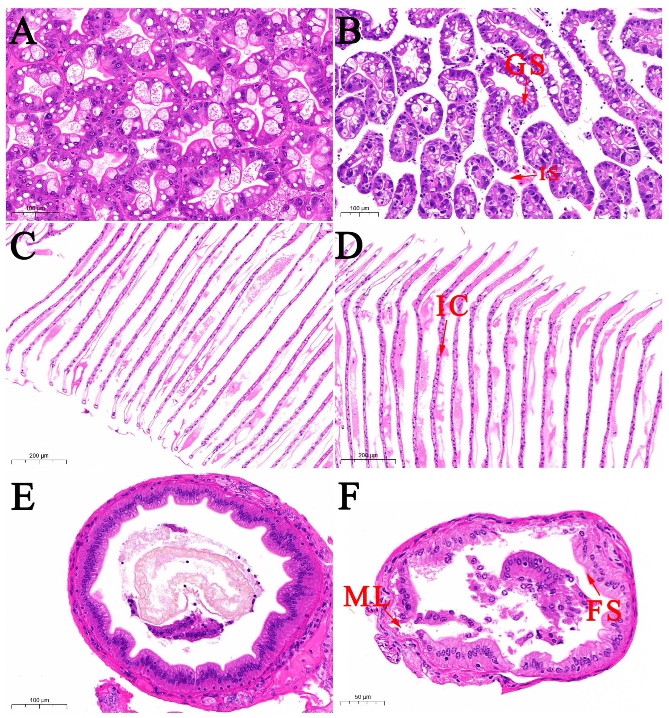
GMRS4 was cultured in LB broth at 28 °C with shaking at 180 rpm for 18 h. Subsequently, the pure GMRS4 cells were harvested via centrifugation at 6000× g for 10 min at 4 °C and washed three times with sterile phosphate-buffered saline (PBS). The bacterial samples were fixed with 0.25 g/L glutaraldehyde at 4 °C and stained with osmium tetroxide (0.1 M dimethylarsinate buffer). The samples were washed with PBS, and excess stain was removed. Subsequently, the samples were gradually dehydrated using a series of ethanol solutions (50%, 70%, 90%, and 100%). After embedding and polymerizing in epoxy resin, the samples were sectioned and placed in a palladium–gold alloy. Flagellated cells were examined using a Zeiss EM10 transmission electron microscope (Zeiss, Oberkochen, Germany) to analyze the types and sizes of the flagella.

2.3. Histopathology

Samples from the hepatopancreas, gills, and intestines of naturally infected M. rosenbergii were fixed in 10% neutral buffered formalin, dehydrated through an ethanol series, and embedded in paraffin. Subsequently, 6 μm sections were cut and stained with hematoxylin and eosin (H&E) for examination under a light microscope [14]. Tissue samples from healthy prawns were set as a control group.

2.4. Identification of Bacterial Isolates

Twenty-two kinds of biochemical reaction tubes (Hangzhou Binhe Microorganism Reagent Co., Ltd., Hangzhou, China) were selected for the biochemical tests, and the phenotypic characteristics of strain GMRS4 were assessed and compared with descriptions in Bergey’s Manual of Systematic Bacteriology [32]. The 16S rRNA gene of GMRS4 was amplified via PCR using primers specified by Zhang et al. [33]. The amplifications were performed in a 25 μL reaction mixture containing 12.5 μL of Easy Taq PCR Super® Mix (TransGen Biotech, Beijing, China), 0.5 μL of forward and reverse primers (10 mM), 1 μL of DNA template (57.93 ng/μL), and 10.5 μL of ddH2O. Following sequencing at Shanghai Sangon Biotech Co., Ltd. (Shanghai, China), the 16S rRNA sequence homology of GMRS4 was analyzed using BLAST on NCBI. Using the maximum likelihood approach, phylogenetic trees were generated with MEGA 7.0 software [34].

2.5. Bacterial Virulence Assay

Healthy M. rosenbergii specimens (weight: 2.16 ± 0.32 g) were sourced from a prawn farm in Gaoyou County, Jiangsu Province, China, and reared in a closed-containment aquaculture freshwater system at a constant temperature of 30 °C and dissolved oxygen levels maintained at ≥5 ppm. The prawns were fed twice daily with commercial pellet feed, and water was renewed twice daily, with one-fourth of the total volume replaced each time. After a 7-day acclimation period, the prawns were confirmed to be free of pathogen infections. All animal handling procedures adhered strictly to the guidelines set forth by the Animal Experiment Ethics Committee of Yangzhou University.
For the challenge experiment, strain GMRS4 was incubated in LB broth at 28 °C with continuous shaking at 180 rpm for 18 h. Following incubation, the bacterial culture was serially diluted in sterile PBS (pH 7.4) to achieve final concentrations ranging from 2.4 × 10⁸ to 2.4 × 10⁴ CFU/mL. Prawns were allocated into experimental and control groups, with each group comprising three replicates. In each challenge group, 50 prawns were intraperitoneally injected with 100 μL of bacterial suspension at concentrations of 2.4 × 10⁸, 2.4 × 10⁷, 2.4 × 10⁶, and 2.4 × 10⁵ CFU/mL. Control prawns received 100 μL of sterile PBS. Survival rate was observed daily over a seven-day period, and cumulative mortality data were collected to determine the LD50 value of GMRS4 for M. rosenbergii using the method of Behreans and Kärber [35].

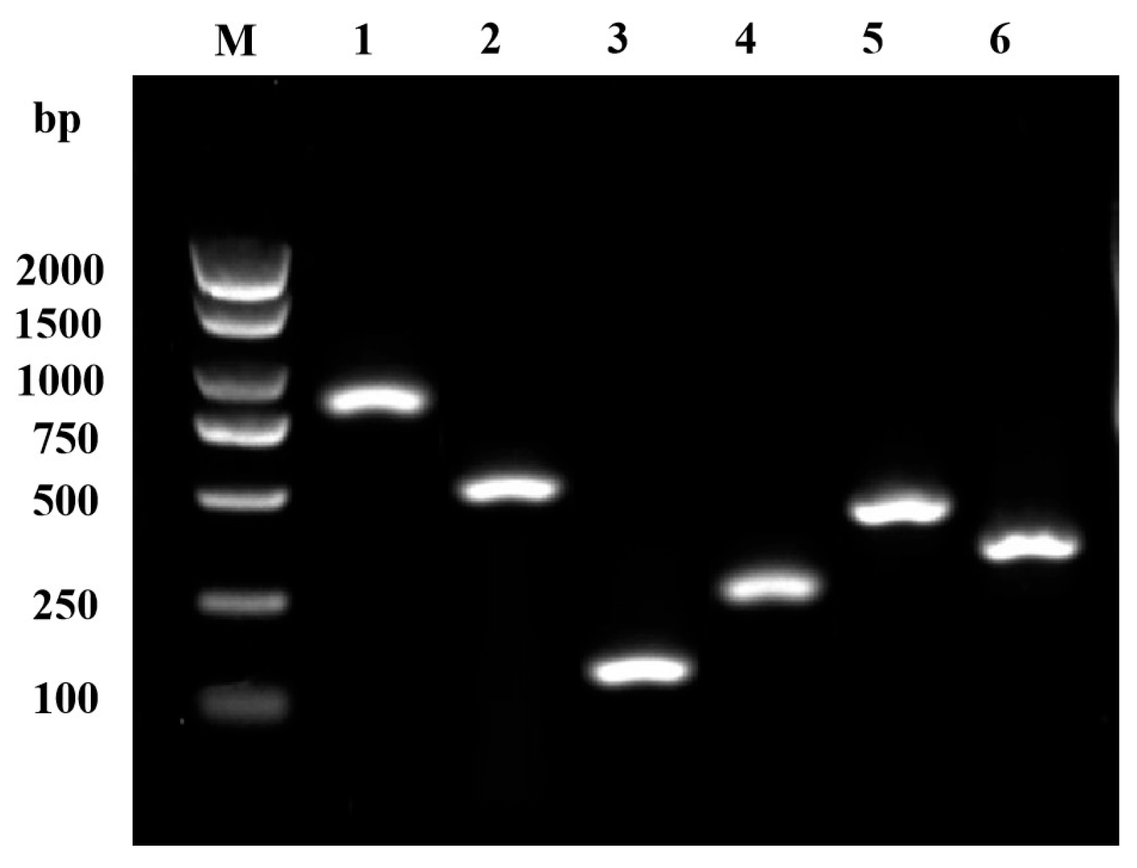
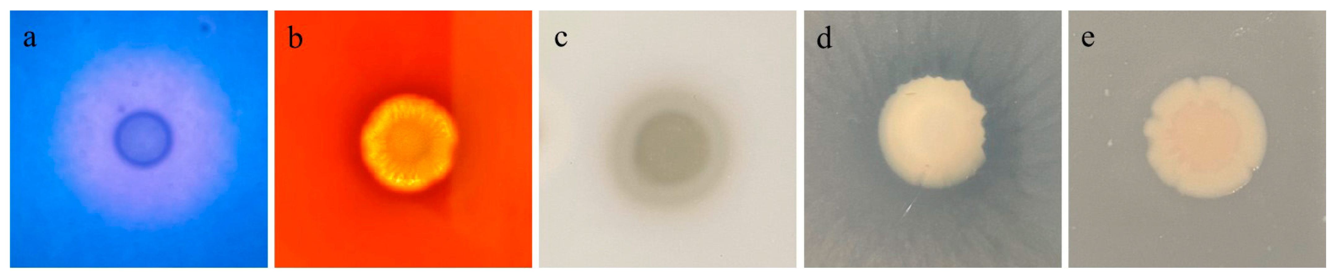
2.6. Determination of Virulence-Related Factors

Exoenzyme activities, including DNase, hemolysin, caseinase, phospholipase, and amylase, were assessed according to Gao et al. [31]. A 5 µL aliquot of bacterial suspension was carefully inoculated onto the center of each plate, followed by incubation at 28 °C for 18 h in triplicate. The presence of extracellular enzyme activity in isolate GMRS4 would be indicated by the formation of a clear lytic halo surrounding the colonies.
The polymerase chain reaction was performed using Easy Taq PCR Super® Mix (Tolo Biotech Co., Ltd., Shanghai, China) to detect the presence of virulence-related genes in the isolate GMRS4, including ahp, ahyB, flgM, hly, alt, and aer. Specific primers were designed based on genomic data (Table 1). PCR conditions were the same as described in Section 2.4. PCR products were analyzed using 1% agarose gel electrophoresis.

2.7. Antibiotic Sensitivity Test

To evaluate the antibiotic sensitivity of isolate GMRS4, the Kirby–Bauer disk diffusion method was employed. The bacterial culture was grown overnight and subsequently diluted in sterile saline to reach a final concentration of 1.8 × 10⁸ CFU/mL. A 100 μL aliquot of the prepared bacterial suspension was then evenly distributed across the surface of an LB agar plate. Antibiotic discs were carefully placed at the center of plates, and the plate was placed in an inverted orientation and incubated at 28 °C for a duration of 24 h. Following incubation, the inhibition zone diameters (mm) around each antibiotic disc were recorded and analyzed. The susceptibility of GMRS4 to different antibiotics would be compared following the guidelines provided by Hangzhou Binhe Biological Company.

2.8. Whole-Genome Sequencing and Assembly

Genomic DNA was extracted from 1 g of pure bacteria utilizing an optimized SDS-based extraction protocol (Oxford Nanopore Technologies, Oxford, UK). Agarose gel electrophoresis was also used to check the quality of genomic DNA. Following quality assessment of the sample using a NanoDrop One spectrophotometer (NanoDrop Technologies, Wilmington, DE, USA) and a Qubit 3.0 Fluorometer (Life Technologies, Carlsbad, CA, USA), a sequencing library was prepared using the SQK-LSK110 kit (Oxford Nanopore Technologies, Oxford, UK) according to the instructions specified by the manufacturer. Concurrently, a small-fragment library was generated employing the VAHTS® Universal Plus DNA Library Prep Kit for MGI V2/for Illumina V2 (Vazyme, Nanjing, China). After assessing the library quality, sequencing was performed using both the Nanopore PromethION and Illumina NovaSeq 6000 platforms. Raw sequencing reads obtained from the Nanopore system were subjected to quality control, retaining only those with Q scores of 7 or higher and lengths of at least 1600 bp to ensure valid data output. Raw data from second-generation sequencing were filtered using fastp [36], and Unicycler software (https://github.com/rrwick/Unicycler) was utilized to assemble the filtered reads, followed by depth statistics for second-generation sequencing. BWA was used to align Illumina short reads to the assembled genome [37], while the depth statistics for third-generation sequencing were obtained using Minimap2 to align long reads to the assembled genome [38]. Finally, the average sequencing depth for both second- and third-generation sequencing data was calculated using the Samtools depth tool, reflecting the read coverage across different regions [39].

2.9. Gene Prediction and Database Annotation

The assembled genome was annotated for coding genes using Prokka [40]. Gene islands present in the genome were predicted using IslandPath [41]. Phage-like sequences in the genome were predicted using the PhiSpy website. The prediction and analysis of genome promoter sequences were conducted utilizing PromPredict software (https://bio.tools/prompredict). To ensure a comprehensive understanding of gene functionality, annotations were carried out based on information from eight key databases, including UniProt [42], KEGG [43], GO [44], Pfam [45], COG [46], TIGRFams [47], RefSeq, and NR. Annotations with an E-value threshold of 1 × 10⁻⁵ were selected based on the highest sequence similarity. Using the BLAST function in DIAMOND software (https://www.crystalimpact.com/diamond/), sequences were analyzed against the CARD resistance gene database with a stricter E-value threshold of 1 × 10⁻¹⁰. BLASTP was used to annotate the target protein sequences based on the VFDB, PHI, ARDB, and CARD databases, and annotations with E < 1 × 10⁻⁵ were selected using DIAMOND software, in order to analyze virulence-associated genes and antibiotic resistance–related genes.

3. Results

3.1. Electron Microscopic Observation of the Isolate GMRS4

TEM revealed that GMRS4 was rod-shaped with blunt, rounded ends and possessed a single flagellum. The size of the bacteria ranged from 1.4 to 1.8 μm in length and 0.7 to 1.1 μm in width (Figure 1).

3.2. Histological Observation

Tissue sections showed significant changes in the hepatopancreatic tissue of the diseased prawns. Compared to the control group, the hepatopancreatic tubule lumens and intratubular spaces in naturally infected prawns were notably enlarged, with increased vacuolization of hepatocytes (Figure 2A,B). The glandular structure was disrupted, and the cellular arrangement was disordered, indicating significant damage to the hepatopancreatic tissue. Additionally, a large infiltration of inflammatory cells was observed in the gill tissue of the diseased prawns, and gill cells exhibited swelling, shedding, or necrosis (Figure 2C,D). In the intestinal tissue of naturally infected prawns, the intestinal wall showed large fissures, reduced fold structures, and separation between the columnar epithelium and connective tissue. The muscle layer was ruptured, the connective tissue was atrophied, and there was separation between the muscle and connective tissues, along with disorganization of the villous structures (Figure 2E,F).

3.3. Physiological and Biochemical Characterization

The physiological and biochemical traits of isolate GMRS4 are displayed in Table 2. The isolate can utilize cellobiose, xylose, sucrose, rhamnose, mannose, trehalose, aesculin, salicylic acid, glucosamine, arginine dihydrolase, α-methyl-D-glucosamine, and β-galactoside. However, it cannot utilize lactose, arabinose, citrate, myo-inositol, glucuronic acid, melezitose, tartrate, lysine, hydrogen sulfide, or peptone water.

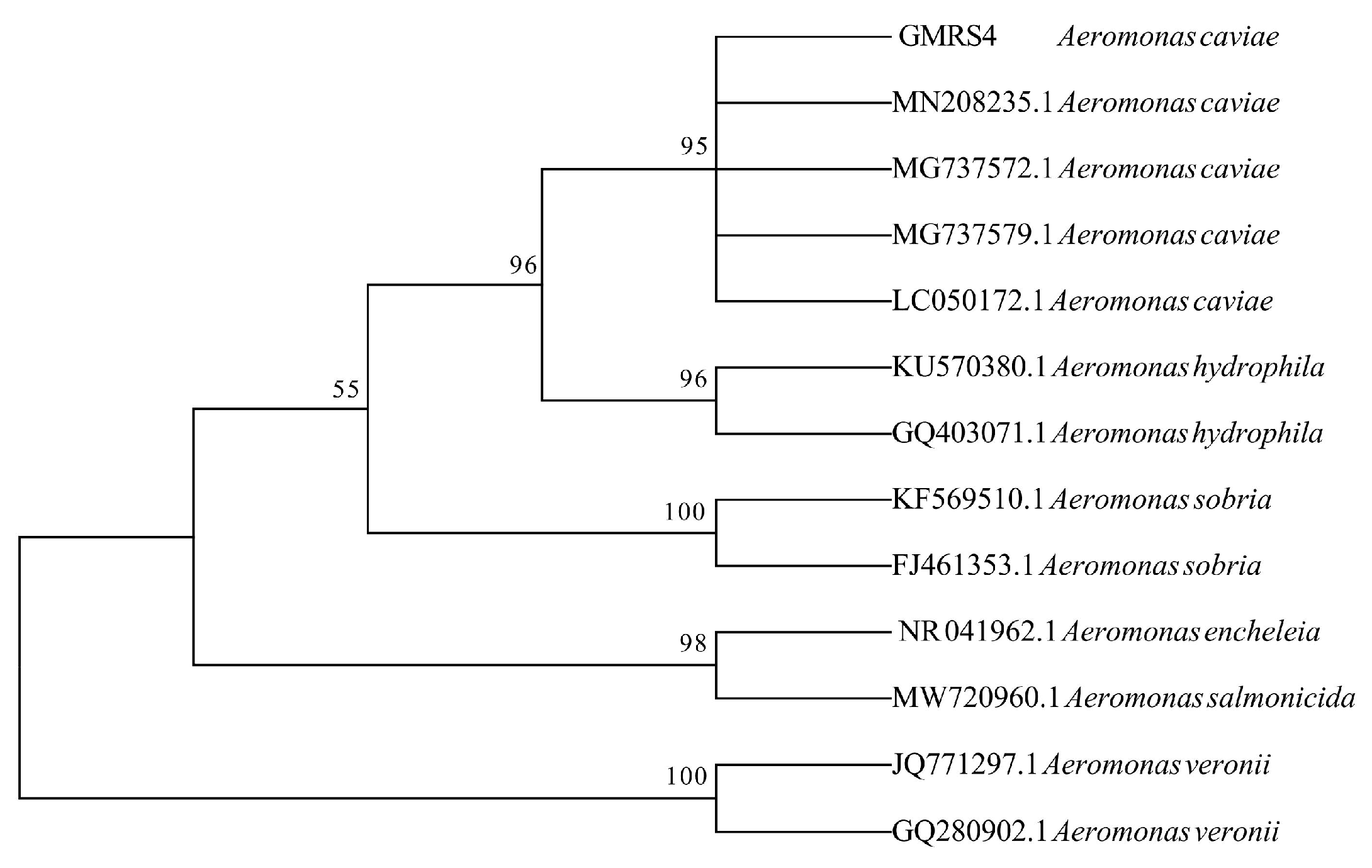
3.4. Molecular Identification

The 16S rRNA gene of GMRS4 was sequenced by Sengon Biotech and assembled using Seqman software (https://www.dnastar.com/software/lasergene/seqman-ngen/) following PCR amplification. Comparison of the 16S rRNA sequence of isolate GMRS4 revealed a 95% sequence identity with A. caviae strains (accession number: OM943772.1) in the NCBI Reference RNA Sequences (refseq rna). Phylogenetic analysis further confirmed that isolate GMRS4 is classified as A. caviae (Figure 3).

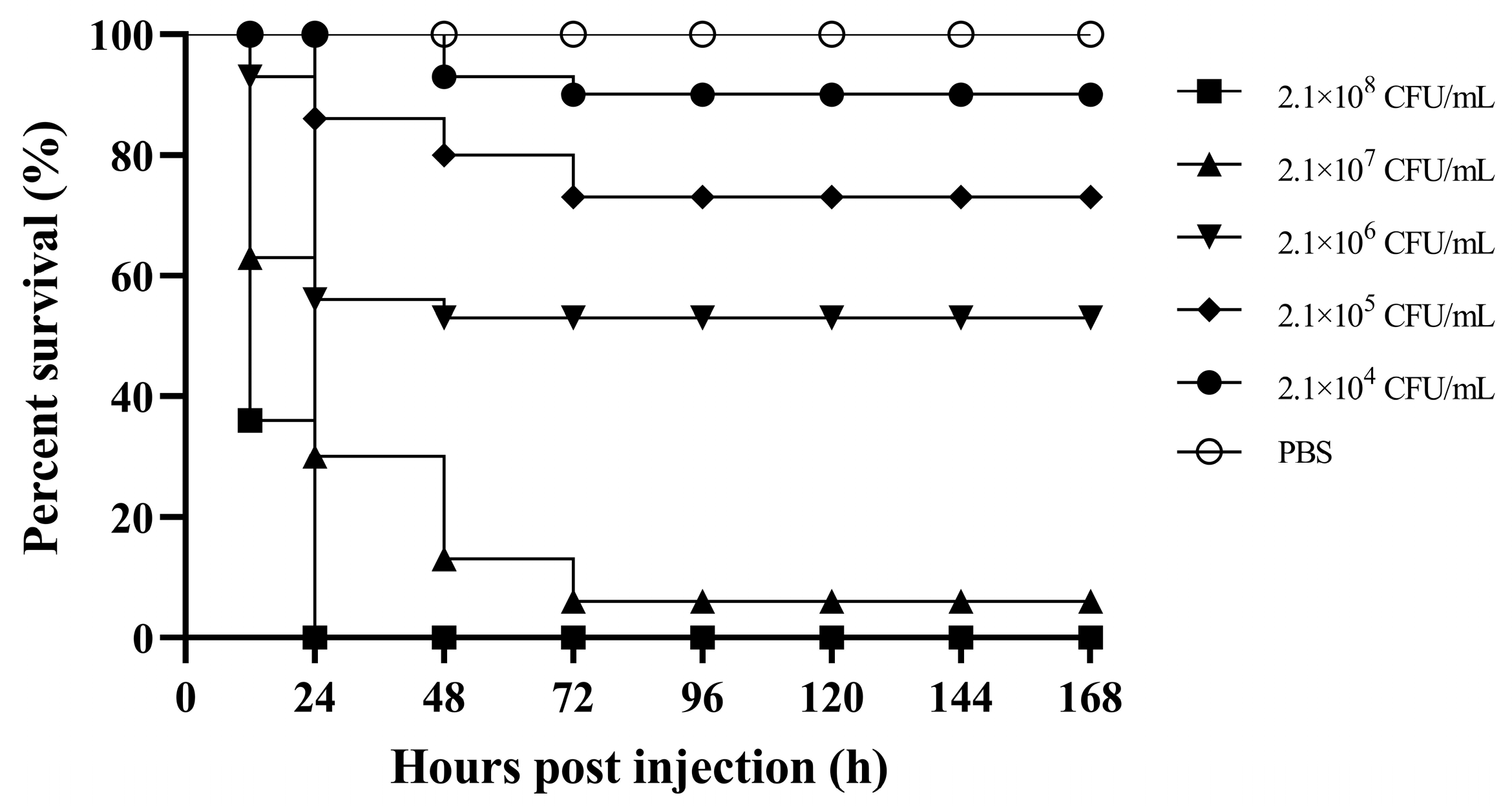
3.5. Virulence of A. caviae GMRS4

After being infected with different concentrations of A. caviae GMRS4, the experimental group of M. rosenbergii began to show mortality starting from the first day. Infection with GMRS4 at concentrations of 2.4 × 10⁸, 2.4 × 10⁷, 2.4 × 10⁶, 2.4 × 10⁵, and 2.4 × 10⁴ CFU/mL resulted in 0%, 7%, 53%, 73%, and 90% survival rate at 96 hpi, respectively, while all prawns in the control group survived (Figure 4). Based on these results, the LD50 value of A. caviae GMRS4 for M. rosenbergii was calculated to be 1.91 × 10⁶ CFU/mL at 96 h.

3.6. Virulence Factors of GMRS4

PCR analysis was used to identify several virulence-related genes, including serine protease (ahp), elastase (ahyB), flagellin (flgM), hemolysin (hly), heat-labile enterotoxin (alt), and aerolysin (aer). The enzymatic activities of extracellular proteins produced by GMRS4 are presented in Figure 5. GMRS4 exhibits DNase activity, hemolysin activity, caseinase activity, and lecithinase activity, but does not show amylase activity (Figure 6).

3.7. Antibiotic Sensitivity of GMRS4

The drug susceptibility of GMRS4 against 35 antibiotics is shown in Table 3. The isolate exhibited low sensitivity to penicillin-class antibiotics, amphenicol-class antibiotics, and sulfonamides but was highly sensitive to quinolone-class antibiotics such as levofloxacin, ofloxacin, and ciprofloxacin. GMRS4 showed moderate sensitivity to macrolide-class and nitrofuran-class antibiotics.

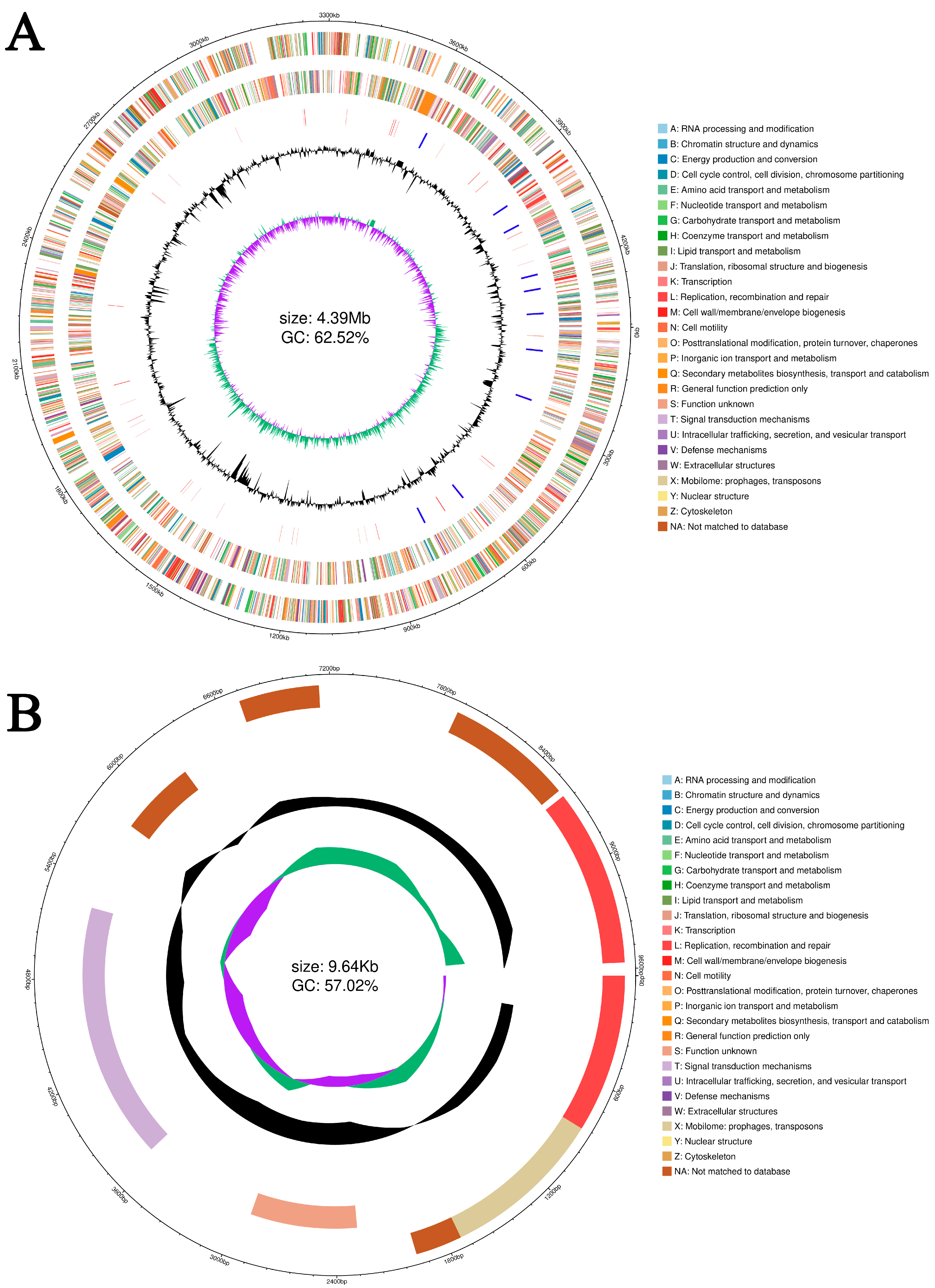
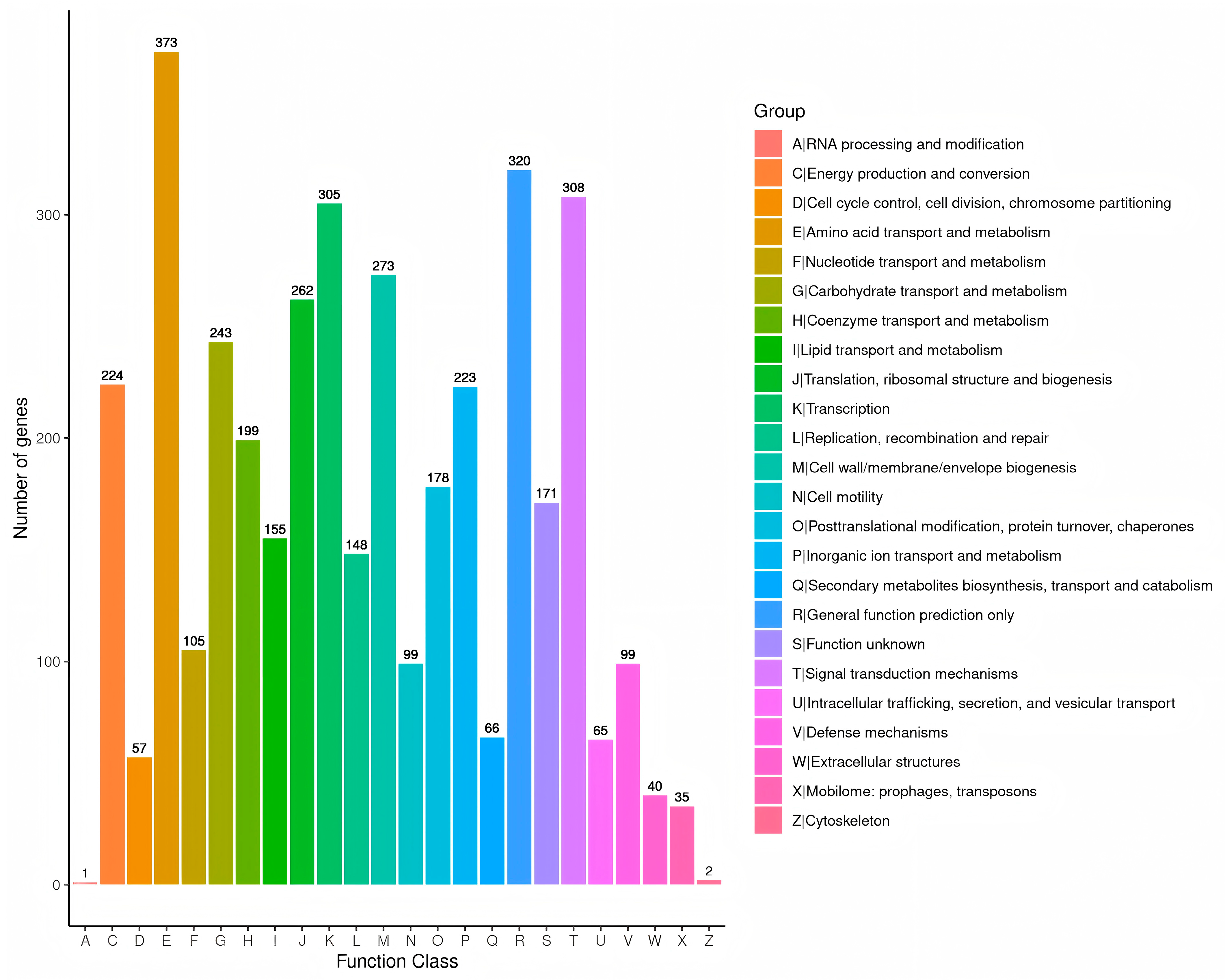
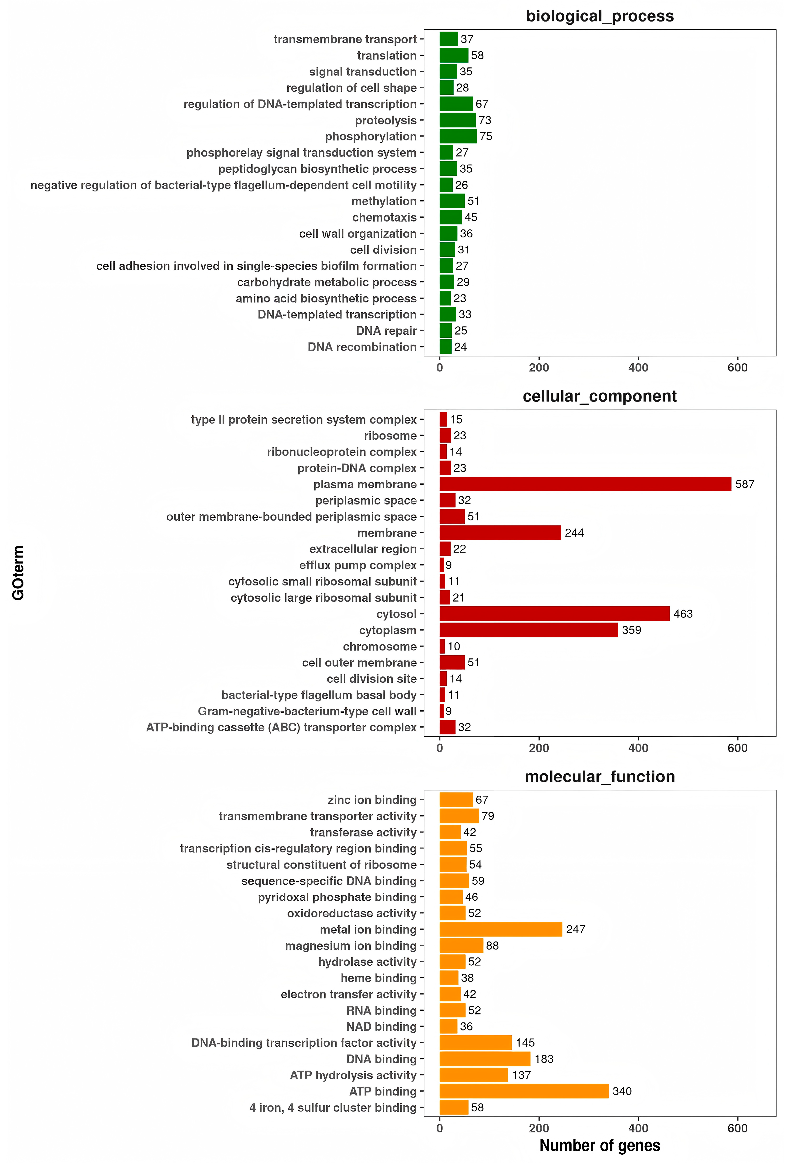
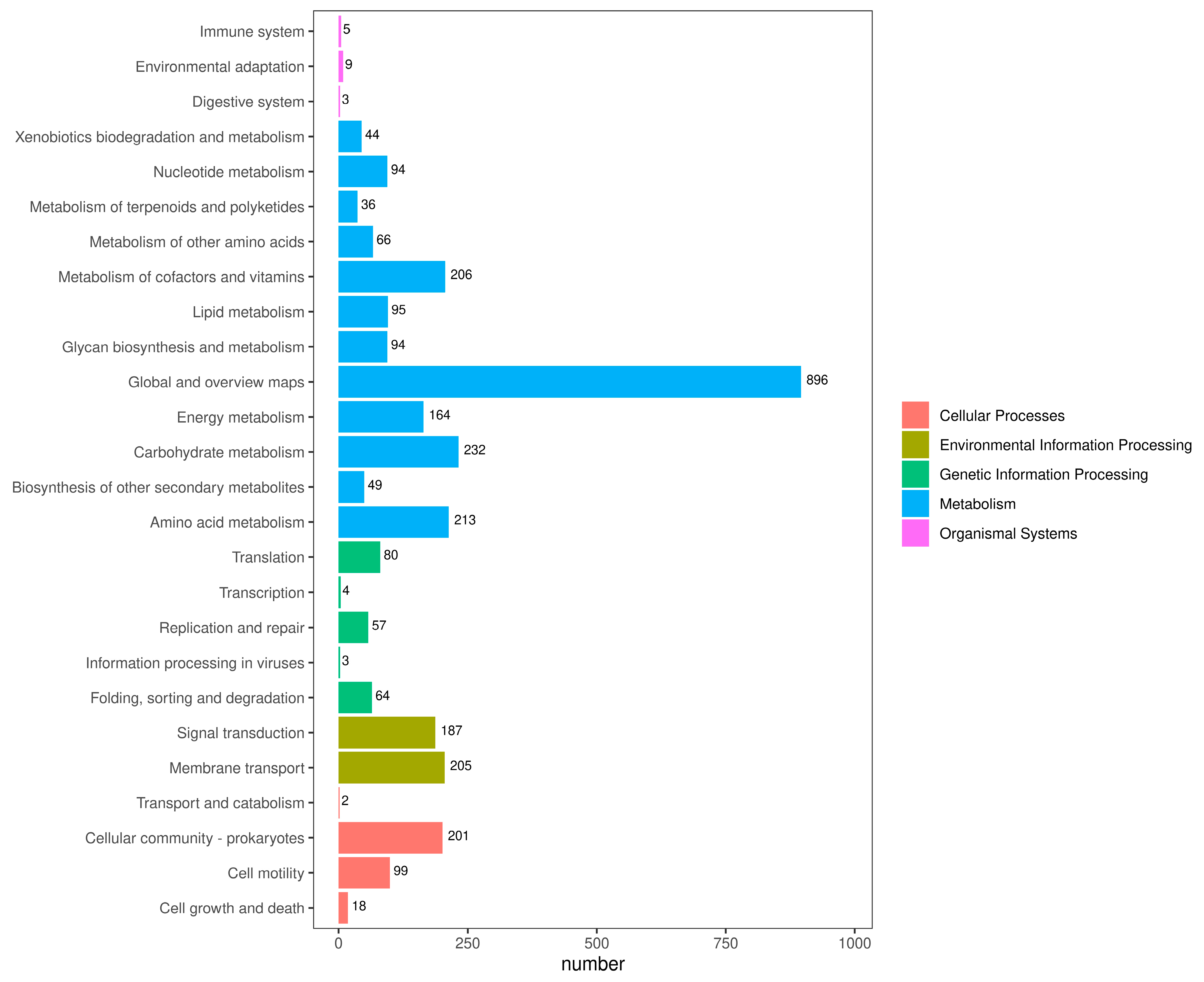
3.8. Genome Structure and General Features of A. caviae GMRS4 Genome

After quality filtration, the entire genome of A. caviae GMRS4 was assembled into one circular chromosome and a circular plasmid (Figure 7). The chromosome of GMRS4 is 4,387,439 bp in length with a G + C content of 62.52%, whereas the plasmid spans 9639 bp with a G + C content of 57% (accession number: PRJNA1247916). In total, 4078 coding genes were identified, accounting for 3,871,119 bp of the genome.
In addition, the genome also contained 124 tRNA genes, 33 rRNA genes, and 45 miscRNA genes. The genome of strain GMRS4 contained 379 pseudogenes, 10 CRISPR, and 6 genomic islands. A total of 3951 genes that were annotated according to the COG analysis were divided into 24 functional groups (Figure 8); 4688 genes that were annotated according to the KEGG analysis were divided into five categories and 26 functional groups (Figure 9); and 10,522 genes that were annotated according to GO were divided into three categories and 2445 GO terms (Figure 10).

3.9. Prediction of Virulence Genes of A. caviae GMRS4

Analysis compared using the Virulence Factor Database (VFDB) revealed 1184 coding sequences in the GMRS4 genome that are potential virulence genes. Among these, the majority are associated with polar flagella (75 genes, 6.33%), capsules (65 genes, 5.49%), and pyoverdine (47 genes, 3.97%) (Table 4). Additionally, 1806 genes were identified in the PHI database, which may be associated with pathogen–host interactions. Among them, 21 genes were classified as “lethal”, 130 genes were classified as “increased virulence”, 323 genes were classified as “unaffected pathogenicity”, and 1019 genes were classified as “reduced virulence”.

3.10. Prediction of Resistance Genes of A. caviae GMRS4

To gain deeper insights into the drug resistance profile of strain GMRS4, the key resistance genes were identified using predictions based on the ARDB and CARD databases. Two resistance genes (baca: ctg_03842; qnrs: ctg_04073) were detected in the ARDB database, which predicts that GMRS4 is resistant to bacitracin and fluoroquinolone (Table 5). Additionally, according to the CARD database annotation, the GMRS4 genome contains eight drug-resistant genes related to fluoroquinolone antibiotic, tetracycline antibiotic, cephalosporin, penam, glycopeptide antibiotic, diaminopyrimidine antibiotic, phenicol antibiotic, and elfamycin antibiotic. The primary function of these genes is to mediate antibiotic resistance through various mechanisms such as antibiotic efflux, antibiotic inactivation, antibiotic target alteration, and antibiotic target protection (Table 6).

4. Discussion

As one of the most common pathogenic bacteria in aquaculture, Aeromonas species are widely distributed in both freshwater and marine environments and can cause a variety of diseases in aquatic animals, including fish, prawns, and crabs. The most common species, Aeromonas hydrophila, infects a variety of aquatic organisms and has been extensively studied to develop therapeutic strategies. It is endemic in several countries and is considered the most frequent Aeromonas species in Japan [48]. However, there are few reports on A. caviae in aquaculture, and most of them focus on fish. Therefore, it is crucial to study A. caviae infection in economically important prawns for developing strategies to combat its spread. In this study, a representative dominant strain, GMRS4, was isolated from diseased M. rosenbergii, with severe, serious histopathological signs, including hepatopancreatic tubule lumen and intertubular space expansion, increased hepatocyte vacuolization, severe gill inflammation, and large fissures with reduced fold structures in the intestinal wall. Then, the strain was identified as A. caviae. In vivo experimental infection revealed that the LD50 of A. caviae GMRS4 for M. rosenbergii was 1.91 × 10⁶ CFU/mL at 96 h, indicating its high virulence toward this species. Previous research has also confirmed that A. caviae is a highly virulent pathogen affecting various aquatic animals. According to Xue et al., A. caviae exhibited high virulence toward M. salmoides, leading to acute mortality in aquaculture, with an LD50 of A. caviae WH21406 recorded at 3.46 × 10⁵ CFU/mL [22]. Similarly, Wu et al. reported that A. caviae demonstrated β-hemolysis, and the LD50 of strain L2 for C. gibelio was determined to be 1.33 × 10⁶ CFU/mL [49]. Furthermore, A. caviae can infect not only fish but also crustaceans, causing significant mortality. Zhou et al. revealed the strong pathogenicity of A. caviae to Eriocheir sinensis through experimental infection, with an LD50 value of 1.6 × 10⁶ CFU/mL [50]. Zeng et al. confirmed A. caviae as a pathogen of yellow leg disease in P. vannamei [51].
The high pathogenicity of bacteria results from a complex interplay of factors that enable them to invade, colonize, and cause disease in the host. These factors are typically categorized into virulence factors, environmental influences, and host factors, with virulence factors playing a key role [52]. The virulence factors of Aeromonas spp. mainly consist of adhesion factors (fimbriae/pili and adhesins), toxins (exotoxins, endotoxins, and hemolysins), extracellular enzymes (proteases, phospholipases, gelatinase, and urease), etc. [53]. In this study, several extracellular enzymes and toxins, including DNase activity, hemolysin activity, caseinase activity, and lecithinase activity, were detected in A. caviae GMRS4. By degrading host DNA, DNase helps Aeromonas spp. escape immune traps like NETs, facilitate biofilm formation and dispersal, promote tissue damage, acquire nutrients, and even acquire new genetic material through horizontal gene transfer [54]. Caseinase facilitates nutrient acquisition by degrading casein and enhances virulence by promoting tissue invasion, immune evasion, and inflammation (Esteve). By degrading lecithin in host cell membranes, lecithinase disrupts membrane integrity, leading to cell lysis and contributing to tissue damage. This enables Aeromonas spp. to penetrate deeper into tissues, promoting the spread of infection. Hemolysins secreted by Aeromonas spp. contribute to pathogen damage in aquatic animals by lysing red blood cells, thereby releasing iron and other nutrients that support bacterial growth [55]. This disruption of host cell membranes also triggers inflammation, facilitates bacterial spread, and impairs the host’s immune defense, ultimately leading to systemic infection and tissue damage.
The pathogenesis of A. caviae is a complex process depends on multiple factors, including polar flagella, capsule, and pyoverdine. These items were annotated in the A. caviae GMRS4 genome. Polar flagella in A. caviae are crucial for its virulence, enabling motility that allows the bacteria to reach infection sites and adhere to host tissues. Additionally, flagella facilitate biofilm formation and immune evasion, enhancing bacterial persistence and resistance to host defenses [56]. The capsule of Aeromonas caviae plays a critical role in its virulence by preventing phagocytosis, allowing the bacterium to evade the host immune system [57]. Additionally, the capsule aids in biofilm formation, enhancing bacterial persistence on host tissues and providing resistance to both immune defenses and antimicrobial treatments [58]. Pyoverdine, a siderophore synthesized by A. caviae, is essential for its virulence, as it facilitates iron acquisition from the host—an essential element for bacterial proliferation and survival [59]. By binding to iron and transporting it into the bacterial cell, pyoverdine facilitates the pathogen’s proliferation in iron-limited environments, such as the host bloodstream or infected tissues, and contributes to the bacterium’s ability to establish and maintain infection [60]. Additionally, pyoverdine may influence the host immune response, promoting inflammation that supports bacterial survival while potentially causing tissue damage.
In aquaculture systems, antibiotics remain one of the most rapid and effective measures for controlling bacterial diseases [61]. However, overuse of antibiotics is a common issue, often resulting in residual antibiotics in both water and sediment. These residues can exert selective pressure, facilitating the emergence and persistence of antibiotic-resistant pathogens [62]. In this study, the GMRS4 strain exhibited a certain degree of antibiotic resistance. In the CARD database annotation, multiple resistance genes were identified, primarily associated with resistance to fluoroquinolones, tetracyclines, cephalosporins, carbapenems, and other antibiotics. These genes are involved in various resistance mechanisms, including antibiotic efflux, antibiotic inactivation, target modification, and target protection, through which bacteria can control intracellular antibiotic levels via complex biochemical pathways or enzymatic degradation of antibiotics [63]. However, in the ARDB database annotation, only two resistance genes, baca and qnrs, were identified. The drug resistance mechanism associated with the baca gene operates through the synthesis of β-lactamase enzymes, which degrade the β-lactam ring in antibiotics, thereby neutralizing their efficacy [64]. Moreover, the mechanism of drug resistance mediated by the qnrs resistance gene involves the modification of target enzymes, such as DNA gyrase and topoisomerase IV, preventing the binding of quinolone antibiotics and reducing their efficacy [65]. However, the antimicrobial susceptibility tests revealed that GMRS4 exhibited higher sensitivity to penicillin and lower sensitivity to quinolones. This discrepancy may be attributed to the possible dysfunction of the efflux pump, lack of expression of resistance genes, or enzyme malfunction, which suggests that the presence of antibiotic resistance genes does not necessarily correlate with the manifestation of an antibiotic-resistant phenotype [66].

5. Conclusions

In conclusion, this study identified A. caviae GMRS4 as a highly virulent pathogen affecting M. rosenbergii. Whole-genome sequencing uncovered multiple genes associated with virulence and antibiotic resistance, as annotated in public databases. These findings contribute to a deeper molecular-level understanding of the genetic characteristics, pathogenic potential, antibiotic resistance, and virulence factors of highly pathogenic A. caviae.

Author Contributions

X.Z. (Xinhai Zhu), data curation, formal analysis, methodology, validation, writing—original draft, writing—review and editing, and funding acquisition; Q.Q., data curation, investigation, methodology, and software; A.C., data curation, investigation, and methodology; L.Z., methodology; X.G., investigation; Q.J., supervision; Y.Z., supervision; X.Z. (Xiaojun Zhang), project administration, funding acquisition, supervision, validation, and writing—review and editing. All authors have read and agreed to the published version of the manuscript.

Funding

This research was funded by the earmarked fund for the National Natural Science Fund (31972830, 32202982), the “JBGS” Project of Seed Industry Revitalization in Jiangsu Province (JBGS [2021] 120), the Jiangsu Agricultural Science and Technology Innovation Fund (CX(23)1006); Postgraduate Research & Practice Innovation Program of Jiangsu Province (KYCX23_3589).

Institutional Review Board Statement

All treatments involving animals were carried out under the strict guidelines of the Animal Experiment Ethics Committee of Yangzhou University (7 March 2024, No. 202503142).

Informed Consent Statement

Not applicable.

Data Availability Statement

The whole-genome sequencing data of A. caviae GMRS4 have been deposited in the NCBI Sequence Read Archive (SRA) under the accession number SRR33018161 (https://trace.ncbi.nlm.nih.gov/Traces?run=SRR33018161, accessed on 28 May 2025).

Conflicts of Interest

The authors declare no conflicts of interest.

References

  1. Chavanne, H.; Janssen, K.; Hofherr, J.; Contini, F.; Haffray, P.; Consortium, A.; Komen, H.; Nielsen, E.E.; Bargelloni, L. A comprehensive survey on selective breeding programs and seed market in the European aquaculture fish industry. Aquac. Int. 2016, 24, 1287–1307. [Google Scholar] [CrossRef]
  2. Regan, T.; Bean, T.P.; Ellis, T.; Davie, A.; Carboni, S.; Migaud, H.; Houston, R.D. Genetic improvement technologies to support the sustainable growth of UK aquaculture. Rev. Aquac. 2021, 13, 1958–1985. [Google Scholar] [CrossRef]
  3. Li, J.; Xu, S.; Cai, Y.; Jiang, Y.; Chen, H.; Lin, L.; Lv, X. The effects of net-chasing training on survival and growth are related to stocking density in the freshwater prawn Macrobrachium rosenbergii. Aquaculture 2022, 561, 738621. [Google Scholar] [CrossRef]
  4. Tan, K.; Wang, W. The early life culture and gonadal development of giant freshwater prawn, Macrobrachium rosenbergii: A review. Aquaculture 2022, 559, 738357. [Google Scholar] [CrossRef]
  5. Gao, X.; Miao, Z.; Li, X.; Chen, N.; Gu, W.; Liu, X.; Yang, H.; Wei, W.; Zhang, X. Pathogenicity of non-O1/O139 Vibrio cholerae and its induced immune response in Macrobrachium rosenbergii. Fish. Shellfish. Immun. 2019, 92, 300–307. [Google Scholar] [CrossRef]
  6. Li, X.; Zhou, Y.; Jiang, Q.; Yang, H.; Pi, D.; Liu, X.; Gao, X.; Chen, N.; Zhang, X. Virulence properties of Vibrio vulnificus isolated from diseased zoea of freshness shrimp Macrobrachium rosenbergii. Microb. Pathog. 2019, 127, 166–171. [Google Scholar] [CrossRef]
  7. Jayaprakash, N.; Pai, S.; Philip, R.; Singh, I. Isolation of a pathogenic strain of Vibrio alginolyticus from necrotic larvae of Macrobrachium rosenbergii (de Man). J. Fish. Dis. 2006, 29, 187–191. [Google Scholar] [CrossRef]
  8. Gao, X.; Zhang, H.; Jiang, Q.; Chen, N.; Li, X.; Liu, X.; Yang, H.; Wei, W.; Zhang, X. Enterobacter cloacae associated with mass mortality in zoea of giant freshwater prawns Macrobrachium rosenbergii and control with specific chicken egg yolk immunoglobulins (IgY). Aquaculture 2019, 501, 331–337. [Google Scholar] [CrossRef]
  9. Gao, X.; Chen, Z.; Zhang, Z.; Qian, Q.; Chen, A.; Qin, L.; Tang, X.; Jiang, Q.; Zhang, X. Pathogenicity of Aeromonas veronii Isolated from Diseased Macrobrachium rosenbergii and Host Immune-Related Gene Expression Profiles. Microorganisms 2024, 12, 694. [Google Scholar] [CrossRef]
  10. Sung, H.H.; Hwang, S.F.; Tasi, F.M. Responses of giant freshwater prawn (Macrobrachium rosenbergii) to challenge by two strains of Aeromonas spp. J. Invertebr. Pathol. 2000, 76, 278–284. [Google Scholar] [CrossRef]
  11. Xu, W.; Zhao, C.; Pan, X.; He, X.; Liu, X.; Hou, J.; Pang, J. Transmission Routes and a New Freshwater Crustacean Host for Infectious Precocity Virus (IPV). Transbound. Emerg. Dis. 2023, 1, 3950107. [Google Scholar] [CrossRef]
  12. Low, C.F.; Yusoff, M.R.; Kuppusamy, G.; Ahmad, N.F. Molecular biology of Macrobrachium rosenbergii nodavirus infection in giant freshwater prawn. J. Fish. Dis. 2018, 41, 1771–1781. [Google Scholar] [CrossRef] [PubMed]
  13. Hooper, C.; Debnath, P.P.; Stentiford, G.D.; Bateman, K.S.; Salin, K.R.; Bass, D. Diseases of the giant river prawn Macrobrachium rosenbergii: A review for a growing industry. Rev. Aquac. 2023, 15, 738–758. [Google Scholar] [CrossRef]
  14. Qian, Q.; Zhou, Y.; Chen, Z.; Zhu, Y.; Xu, J.; Gao, X.; Jiang, Q.; Wang, J.; Zhang, X. Pathogenesis and complete genome sequence of Decapod iridescent virus 1 (DIV1) associated with mass mortality in Macrobrachium rosenbergii. Aquaculture 2023, 566, 739220. [Google Scholar] [CrossRef]
  15. Jones, B.L.; Wilcox, M.H. Aeromonas infections and their treatment. J. Antimicrob. Chemoth. 1995, 35, 453–461. [Google Scholar] [CrossRef]
  16. De Schryver, P.; Vadstein, O. Ecological theory as a foundation to control pathogenic invasion in aquaculture. ISME J. 2014, 8, 2360–2368. [Google Scholar] [CrossRef] [PubMed]
  17. Cholewińska, P.; Moniuszko, H.; Wojnarowski, K.; Pokorny, P.; Szeligowska, N.; Dobicki, W.; Polechoński, R.; Górniak, W. The occurrence of microplastics and the formation of biofilms by pathogenic and opportunistic bacteria as threats in aquaculture. Int. J. Environ. Res. Public Health 2022, 19, 8137. [Google Scholar] [CrossRef] [PubMed]
  18. Mulia, D.S.; Isnansetyo, A.; Pratiwi, R.; Asmara, W. Molecular characterizations of Aeromonas caviae isolated from catfish (Clarias sp.). AACL Bioflux 2020, 13, 2717–2732. [Google Scholar]
  19. Agarwal, P.; Kayala, P.; Chandrasekaran, N.; Mukherjee, A.; Shah, S.; Thomas, J. Antioxidant and antibacterial activity of Gelidium pusillum (Stackhouse) against Aeromonas caviae and its applications in aquaculture. Aquac. Int. 2021, 29, 845–858. [Google Scholar] [CrossRef]
  20. Thomas, J.; Madan, N.; Nambi, K.S.N.; Majeed, S.A.; Basha, A.N.; Hameed, A.S. Studies on ulcerative disease caused by Aeromonas caviae-like bacterium in Indian catfish, Clarias batrachus (Linn). Aquaculture 2013, 376, 146–150. [Google Scholar] [CrossRef]
  21. Roy, A.; Singha, J.; Abraham, T.J. Histopathology of Aeromonas caviae infection in challenged Nile tilapia Oreochromis niloticus (Linnaeus, 1758). Int. J. Aquac. 2018, 8, 151. [Google Scholar] [CrossRef]
  22. Xue, M.; Xiao, Z.; Li, Y.; Jiang, N.; Liu, W.; Meng, Y.; Fan, Y.D.; Zeng, L.B.; Zhou, Y. Isolation, identification and characteristics of Aeromonas caviae from diseased largemouth bass (Micropterus salmoides). Fishes 2022, 7, 119. [Google Scholar] [CrossRef]
  23. Dai, Y.; Gao, J.; Jiang, M. Case Report: A rare infection of multidrug-resistant Aeromonas caviae in a pediatric case with acute lymphoblastic leukemia and review of the literature. Front. Pediatr. 2024, 12, 1233600. [Google Scholar] [CrossRef] [PubMed]
  24. Tyagi, A.; Sharma, C.; Srivastava, A.; Kumar, B.N.; Pathak, D.; Rai, S. Isolation, characterization and complete genome sequencing of fish pathogenic Aeromonas veronii from diseased Labeo rohita. Aquaculture 2022, 553, 738085. [Google Scholar] [CrossRef]
  25. Casadevall, A.; Pirofski, L.A. Host-pathogen interactions: Redefining the basic concepts of virulence and pathogenicity. Infect. Immun. 1999, 67, 3703–3713. [Google Scholar] [CrossRef]
  26. Liao, C.; Huang, X.; Wang, Q.; Yao, D.; Lu, W. Virulence factors of Pseudomonas aeruginosa and antivirulence strategies to combat its drug resistance. Front. Cell. Infect. Microbiol. 2022, 12, 926758. [Google Scholar] [CrossRef]
  27. Zhang, L.; Wang, F.; Jia, L.; Yan, H.; Gao, L.; Tian, Y.; Su, X.; Zhang, X.; Zhang, X.; Lv, C.; et al. Edwardsiella piscicida infection reshapes the intestinal microbiome and metabolome of big-belly seahorses: Mechanistic insights of synergistic actions of virulence factors. Front. Immunol. 2023, 14, 1135588. [Google Scholar] [CrossRef]
  28. Haslinger-Löffler, B.; Kahl, B.C.; Grundmeier, M.; Strangfeld, K.; Wagner, B.; Fischer, U.; Cheung, A.L.; Peters, G.; Schulze, O.K.; Sinha, B. Multiple virulence factors are required for Staphylococcus aureus-induced apoptosis in endothelial cells. Cell. Microbiol. 2005, 7, 1087–1097. [Google Scholar] [CrossRef]
  29. Nguyen, Q.; Nguyen, T.T.N.; Pham, P.; Chau, V.; Nguyen, L.P.H.; Nguyen, T.D.; Ha, T.T.; Le, N.T.Q.; Vu, D.T.; Baker, S.; et al. Genomic insights into the circulation of pandemic fluoroquinolone-resistant extra-intestinal pathogenic Escherichia coli ST1193 in Vietnam. Microb. Genom. 2021, 7, 000733. [Google Scholar] [CrossRef]
  30. Zhu, L.; Lian, Y.; Lin, D.; Huang, D.; Yao, Y.; Ju, F.; Wang, M. Insights into microbial contamination in multi-type manure-amended soils: The profile of human bacterial pathogens, virulence factor genes and antibiotic resistance genes. J. Hazard. Mater. 2022, 437, 129356. [Google Scholar] [CrossRef]
  31. Gao, X.; Zhou, Y.; Zhu, X.; Tang, H.; Li, X.; Jiang, Q.; Wei, W.; Zhang, X. Enterobacter cloacae: A probable etiological agent associated with slow growth in the giant freshwater prawn Macrobrachium rosenbergii. Aquaculture 2021, 530, 735826. [Google Scholar] [CrossRef]
  32. Brenner, D.J.; Krieg, N.R.; Staley, J.T. Bergey’s Manual of Systematic Bacteriology, 2nd ed.; Springer: Greer, SC, USA, 2021; Volume 2, Part B, pp. 404–406. [Google Scholar]
  33. Zhang, X.J.; Bai, X.S.; Yan, B.L.; Bi, K.R.; Qin, L. Vibrio harveyi as a causative agent of mass mortalities of megalopa in the seed production of swimming crab Portunus trituberculatus. Aquac. Int. 2014, 22, 661–672. [Google Scholar] [CrossRef]
  34. Kumar, S.; Stecher, G.; Tamura, K. MEGA7: Molecular evolutionary genetics analysis version 7.0 for bigger datasets. Mol. Biol. Evol. 2016, 33, 1870–1874. [Google Scholar] [CrossRef]
  35. Behreans, A.; Karber, L. Determination of LD50. In Screening in Pharmacology; Academic Press: New York, NY, USA, 1953. [Google Scholar]
  36. Chen, S.; Zhou, Y.; Chen, Y.; Gu, J. fastp: An ultra-fast all-in-one FASTQ preprocessor. Bioinformatics 2018, 34, i884–i890. [Google Scholar] [CrossRef]
  37. Li, H. Aligning sequence reads, clone sequences and assembly contigs with BWA-MEM. arXiv 2013, arXiv:1303.3997. [Google Scholar]
  38. Li, H. Minimap and miniasm: Fast mapping and de novo assembly for noisy long sequences. Bioinformatics 2016, 32, 2103–2110. [Google Scholar] [CrossRef] [PubMed]
  39. Li, H.; Handsaker, B.; Wysoker, A.; Fennell, T.; Ruan, J.; Homer, N.; Marth, G.; Abecasis, G.; Durbin, R. 1000 Genome Project Data Processing Subgroup The sequence alignment/map format and SAMtools. Bioinformatics 2009, 25, 2078–2079. [Google Scholar] [CrossRef] [PubMed]
  40. Seemann, T. Prokka: Rapid prokaryotic genome annotation. Bioinformatics 2014, 30, 2068–2069. [Google Scholar] [CrossRef]
  41. Bertelli, C.; Brinkman, F.S. Improved genomic island predictions with IslandPath-DIMOB. Bioinformatics 2018, 34, 2161–2167. [Google Scholar] [CrossRef]
  42. The UniProt Consortium. UniProt: The universal protein knowledgebase. Nucleic Acids Res. 2018, 46, 2699. [Google Scholar] [CrossRef]
  43. Kanehisa, M.; Goto, S.; Kawashima, S.; Okuno, Y.; Hattori, M. The KEGG resource for deciphering the genome. Nucleic Acids Res. 2004, 32 (Suppl. S1), D277–D280. [Google Scholar] [CrossRef] [PubMed]
  44. Ashburner, M.; Ball, C.A.; Blake, J.A.; Botstein, D.; Butler, H.; Cherry, J.M.; Davis, A.P.; Dolinski, K.; Dwight, S.S.; Eppig, J.T.; et al. Gene ontology: Tool for the unification of biology. Nat. Genet. 2000, 25, 25–29. [Google Scholar] [CrossRef]
  45. Bateman, A.; Coin, L.; Durbin, R.; Finn, R.D.; Hollich, V.; Griffiths-Jones, S.; Khanna, A.; Marshall, M.; Moxon, S.; Sonnhammer, E.; et al. The Pfam protein families database. Nucleic Acids Res. 2004, 32 (Suppl. S1), D138–D141. [Google Scholar] [CrossRef] [PubMed]
  46. Tatusov, R.L.; Galperin, M.Y.; Natale, D.A.; Koonin, E.V. The COG database: A tool for genome-scale analysis of protein functions and evolution. Nucleic Acids Res. 2000, 28, 33–36. [Google Scholar] [CrossRef]
  47. Haft, D.H.; Selengut, J.D.; White, O. The TIGRFAMs database of protein families. Nucleic Acids Res. 2003, 31, 371–373. [Google Scholar] [CrossRef]
  48. Kimura, M.; Araoka, H.; Yoneyama, A. Aeromonas caviae is the most frequent pathogen amongst cases of Aeromonas bacteremia in Japan. Scand. J. Infect. Dis. 2013, 45, 304–309. [Google Scholar] [CrossRef]
  49. Wu, R.; Shen, J.; Tian, D.; Yu, J.; He, T.; Yi, J.; Li, Y. A potential alternative to traditional antibiotics in aquaculture: Yeast glycoprotein exhibits antimicrobial effect in vivo and in vitro on Aeromonas caviae isolated from Carassius auratus gibelio. Vet. Med. Sci. 2020, 6, 639–648. [Google Scholar] [CrossRef]
  50. Zhou, L.; Jang, Z.; Qian, Q.; Chen, Z.; Gu, S.; Li, J.; Ji, P.; Gao, X.; Jang, O.; Zhang, X. Identification and pathogenicity of pathogenic Aeromonas caviae isolated from Eriocheir sinensis. Freshw. Fish. 2022, 52, 58–65. [Google Scholar]
  51. Zeng, X.; Cao, H.; Mu, C. Isolation and antibiotic susceptibility ofan Aeromonas caviae pathogen from yellow legdisease-infected Penaeus vannamei. J. Aquac. 2020, 41, 28–33. [Google Scholar]
  52. Roberts, A.J.; Wiedmann, M. Pathogen, host and environmental factors contributing to the pathogenesis of listeriosis. Cell. Mol. Life Sci. 2003, 60, 904–918. [Google Scholar] [CrossRef]
  53. Beaz-Hidalgo, R.; Figueras, M.J. Aeromonas spp. whole genomes and virulence factors implicated in fish disease. J. Fish Dis. 2013, 36, 371–388. [Google Scholar] [CrossRef] [PubMed]
  54. Pemberton, J.M.; Kidd, S.P.; Schmidt, R. Secreted enzymes of Aeromonas. FEMS Microbiol. Lett. 1997, 152, 1–10. [Google Scholar] [CrossRef]
  55. Wang, G.; Clark, C.G.; Liu, C.; Pucknell, C.; Munro, C.K.; Kruk, T.M.; Caldeira, R.; Woodward, D.; Rodgers, F.G. Detection and characterization of the hemolysin genes in Aeromonas hydrophila and Aeromonas sobria by multiplex PCR. J. Clin. Microbiol. 2003, 41, 1048–1054. [Google Scholar] [CrossRef]
  56. Rabaan, A.A.; Gryllos, I.; Tomás, J.M.; Shaw, J.G. Motility and the polar flagellum are required for Aeromonas caviae adherence to HEp-2 cells. Infect. Immun. 2001, 69, 4257–4267. [Google Scholar] [CrossRef] [PubMed]
  57. Hanchanachai, N.; Chumnanpuen, P.; E-kobon, T. Effect of conditioned media from Aeromonas caviae on the transcriptomic changes of the porcine isolates of Pasteurella multocida. BMC Microbiol. 2022, 22, 272. [Google Scholar] [CrossRef] [PubMed]
  58. Pugazhendhi, A.S.; Wei, F.; Hughes, M.; Coathup, M. Bacterial adhesion, virulence, and biofilm formation. In Musculoskeletal Infection; Springer International Publishing: Cham, Switzerland, 2022; pp. 19–64. [Google Scholar]
  59. Ghssein, G.; Ezzeddine, Z. A review of Pseudomonas aeruginosa metallophores: Pyoverdine, pyochelin and pseudopaline. Biology 2022, 11, 1711. [Google Scholar] [CrossRef]
  60. Cornelis, P.; Tahrioui, A.; Lesouhaitier, O.; Bouffartigues, E.; Feuilloley, M.; Baysse, C.; Chevalier, S. High affinity iron uptake by pyoverdine in Pseudomonas aeruginosa involves multiple regulators besides Fur, PvdS, and FpvI. Biometals 2023, 36, 255–261. [Google Scholar] [CrossRef]
  61. Cherian, T.; Ragavendran, C.; Vijayan, S.; Kurien, S.; Peijnenburg, W.J. A review on the fate, human health and environmental impacts, as well as regulation of antibiotics used in aquaculture. Environ. Adv. 2023, 13, 100411. [Google Scholar] [CrossRef]
  62. Inuwa, A.B.; Pervez, A.; Nazir, R. Microalgae-based wastewater treatment system: Current state, antibiotic resistant bacteria and antibiotic resistance genes reduction potentials. Int. J. Environ. Sci. Technol. 2023, 20, 14053–14072. [Google Scholar] [CrossRef]
  63. Baran, A.; Kwiatkowska, A.; Potocki, L. Antibiotics and bacterial resistance—A short story of an endless arms race. Int. J. Mol. Sci. 2023, 24, 5777. [Google Scholar] [CrossRef]
  64. Nijland, M.; Lefebvre, S.N.; Thangaratnarajah, C.; Slotboom, D.J. Bidirectional ATP-driven transport of cobalamin by the mycobacterial ABC transporter BacA. Nat. Commun. 2024, 15, 2626. [Google Scholar] [CrossRef] [PubMed]
  65. Zhao, Y.; Cao, Z.; Cui, L.; Hu, T.; Guo, K.; Zhang, F.; Wang, X.; Peng, Z.; Liu, Q.; Dai, M. Enrofloxacin promotes plasmid-mediated conjugation transfer of fluoroquinolone-resistance gene qnrS. Front. Microbiol. 2022, 12, 773664. [Google Scholar] [CrossRef] [PubMed]
  66. Zhu, X.; Qin, L.; Zhu, Y.; Qian, Q.; Gao, X.; Jiang, Q.; Wang, J.; Liu, G.; Zhang, X. Characteristics and complete genome analysis of a pathogenic Aeromonas Veronii SJ4 from diseased Siniperca Chuatsi. Mar. Biotechnol. 2023, 25, 966–982. [Google Scholar] [CrossRef] [PubMed]
Figure 1. Electron micrograph of GMRS4, scale bar = 0.5 μm. The bacterium appears rod-shaped with blunt, rounded ends and a single polar flagellum. Cell dimension range: length, 1.4–1.8 μm; width, 0.7–1.1 μm.
Microorganisms 13 01343 g001
Figure 2. Histological changes in the Hepatopancreas (A,B), gills (C,D), and intestines (E,F) of M. rosenbergii infected with isolate GMRS4. GS represents disrupted glandular structure; IS represents enlarged intratubular spaces; IC represents inflammatory cells; FS represents reduced fold structures; ML represents ruptured muscle layer.
Microorganisms 13 01343 g002
Figure 3. Phylogenetic tree of Aeromonas species based on 16S rRNA sequences. Bootstrap values are shown beside the clades. The names of bacteria are indicated beside the accession numbers.
Microorganisms 13 01343 g003
Figure 4. Survival rates of M. rosenbergii challenged by GMRS4 at concentrations of 2.1 × 10⁸, 2.1 × 10⁷, 2.1 × 10⁶, 2.1 × 10⁵, and 2.1 × 10⁴ CFU/mL. PBS was set as the control group. Each group included three parallel replicates.
Microorganisms 13 01343 g004
Figure 5. Virulence genes of A. caviae GMRS4 via PCR amplification. M, marker; 1, ahp; 2, ahyB; 3, flgM; 4, hly; 5, alt; 6, aer.
Microorganisms 13 01343 g005
Figure 6. Extracellular enzyme test results of A. caviae GMRS4. (a) DNase activity; (b) hemolysin activity; (c) caseinase activity; (d) lecithinase activity; (e) amylase activity.
Microorganisms 13 01343 g006
Figure 7. Circular genome map (A) and plasmid map (B) of A. caviae GMRS4. There are coding genes, COG databases, genome size, GC content, and the distribution of GC-skew value from outside to inside.
Microorganisms 13 01343 g007
Figure 8. Clusters of Orthologous Gene (COG) functional annotation of the whole genome of A. caviae GMRS4. A total of 3951 genes has a COG classification among the 24 categories. The x-axis indicates the COG categories, and the y-axis indicates the number of genes.
Microorganisms 13 01343 g008
Figure 9. Gene Oncology (GO) functional annotation of the whole genome of A. caviae GMRS4. All annotated genes were grouped into 3 functional subcategories: biological process, cellular component, and molecular function.
Microorganisms 13 01343 g009
Figure 10. Gene distribution based on Kyoto Encyclopedia of Genes and Genomes (KEGG) classification of A. caviae GMRS4. All annotated genes were grouped into 5 functional subcategories: cellular process, environmental information processing, genetic information processing, metabolism, and organismal systems.
Microorganisms 13 01343 g010
Table 1. The primers used for the PCR.
Sequence IDTemperature/°C;Product Length (bp)Primer Sequences (5′-3′)Gene
ctg_0168455911ATTGGATCCCTGCCTAahp
GCTAAGCTTGCATCCG
ctg_0079655513ACACGGTCAAGGAGATCAACahyB
ACACGGTCAAGGAGATCAAC
ctg_0147263194GCTACTGTCAAGCTGGACTCflgM
AGATTGGCCTCGAAACTG
ctg_0218963289CGGACGATTATCAGGATGGhly
CAAGAACGAGTTTCAGTGGC
ctg_0316163442TGACCCAGTCCTGGalt
GGTGATCGATCACC
ctg_0407463301AACCGAACTCTCCATaer
CGCCTTGTCCTTGTA
Table 2. Physical and chemical properties of isolate GMRS4.
Aeromonas caviae *GMRS4Characteristics
++Cellobiose
++Xylose
++Sucrose
++Rhamnose
++Mannose
Lactose
Arabinose
++D-Trehalose anhydrous
++Escin
Citrate
Inositol
Dacron
Raffinose
Tartrate
++Salicin
Lysine
Hydrogen sulfide
++Ammonium Gluconate
++Arginine dihydrolase
++α-Methyl-d-glucose ammonium
Peptone
Note: “+” indicates positive, “−” indicates negative. “*” refers to data taken from Bergey’s Manual of Systematic Bacteriology.
Table 3. Antimicrobial sensitivity of pathogenic A. caviae to 35 drugs.
SensitivityDiameter of Inhibition Zone/mmDrug Content of Paper Disc/µg/discDrugDrug Classification
R830CefazolinCephalosporins
R830Ceftriaxone
I1630Cefuroxime
S2275Cefoperazone
S3030Cefotaxime
S2330Ceftriaxone
S1930Cefepime
S20.530Ceftazidime
S830Cefoxitin
S1930AmikacinAminoglycosides
S1610Streptomycin
I13.510Tobramycin
S18100Spectinomycin
I1730Kanamycin
S1810Gentamicin
I13.530Neomycin
R81OxacillinPenicillin
R810Penicillin G
R810Ampicillin
R14100Piperacillin
I1415ErythromycinMacrolides
I1615Clarithromycin
R82Clindamycin
S1930TetracyclineTetracyclines
S2030Minocycline
S2610NorfloxacinQuinolones
S245Ciprofloxacin
S235Levofloxacin
S255Ofloxacin
S2610NorfloxacinPolypeptide
S245CiprofloxacinFuran
S235LevofloxacinAmphenicols
S255OfloxacinMonobactams
I11.530Polymyxin BAmides
R823.75Trimethoprim/sulfamethoxazoleSulfonamides
Note: S sensitive; I intermediate; R resistant.
Table 4. VFDB database annotation in the complete genome of A. caviae GMRS4.
NumberRelated GeneVF NameVF Category
65bcs1, kpsD, cpsA/uppS, lipA, kpsF, etc.CapsuleImmune modulation
39lpxC, lpxH, galE, licA, lpxB, msbB, waaQ, etc.LOS
11sadH, fadD13, adhD, etc.MymA operon
11ddrA, ppsC, etc.PDIM
27lpxA/glmU, flmF2, etc.LPS
14YPO_RS16495, YPTB_RS05510, etc.O-antigen
15fbpB, fbpC, etc.FbpABC
22hitC, hitB, hitA, etc.HitABC
16sugC, sugA, sugB, etc.Trehalose-recycling ABC transporter
14hemX, hemD, hemL, hemG, hemN, hemB, etc.Heme biosynthesis
11allB, allS, allR, etc.Allantion utilization
47pvdQ, PA2383, pvdN, pvdE, pvdI, ptxR, etc.Pyoverdine
17pdxJ, CFF8240_RS05385, fleQ, flgP, fliL, etc.FlagellaMotility
75flgB, flrC, fliE, flaA, fliK, flgM, flgA, cheY, etc.Polar flagella
17tapV, tapP, ASA_RS18105, tapU, tppF, etc.Tap type IV piliAdherence
16mshN, mshC, mshJ, mshP, mshI1, etc.MSHA type IV pili
20frpC, etc.RTX protein
25rpoN, pilS, pilH, etc.Type IV pili
14vpdC, lirB, LPG_RS00105, lidL, etc.Dot/Icm T4SS secreted effectors
14exeM, exeN, exeK, exeH, exeA, exeB, exeL, etc.Exe T2SS
10ETAE_RS04220, MAFF_RS25805, etc.T3SS
12CBUK_RS04750, coxDFB4, rimP, etc.T4SS secreted effectors
14mucD, algW, mucC, algQ, mucP, algU, etc.Alginate
10bopD, etc.BopD
34papR, etc.PlcR-PapR quorum sensing
17rtxA, etc.RtxAExotoxin
23acfB, etc.ACF
Table 5. ARDB database annotation in the complete genome of A. caviae GMRS4.
NumberSeqListResistance ProfileDescriptionResistance Type
1ctg_03842bacitracinUndecaprenyl pyrophosphate phosphatase, which consists in the sequestration of Undecaprenyl pyrophosphate.baca
1ctg_04073fluoroquinolonePentapeptide repeat family, which protects DNA gyrase from the inhibition of quinolones.qnrs
Table 6. CARD database annotation in the complete genome of A. caviae GMRS4.
AMR Gene FamilyResistance MechanismDrug ClassAROBest_Hit_AROORF_ID
resistance-nodulation-cell division (RND) antibiotic efflux pumpantibiotic effluxfluoroquinolone antibiotic; tetracycline antibiotic3000777adeFctg_01323
resistance-nodulation-cell division (RND) antibiotic efflux pumpantibiotic effluxfluoroquinolone antibiotic; tetracycline antibiotic3000777adeFctg_01375
TRU beta-lactamaseantibiotic inactivationcephalosporin; penam3004450TRU-1ctg_02948
glycopeptide resistance gene cluster; vanTantibiotic target alterationglycopeptide antibiotic3002972vanT gene in vanG clusterctg_02455
resistance-nodulation-cell division (RND) antibiotic efflux pumpantibiotic effluxfluoroquinolone antibiotic; diaminopyrimidine antibiotic; phenicol antibiotic3005069rsmActg_00475
elfamycin resistant EF-Tuantibiotic target alterationelfamycin antibiotic3003369Escherichia coli EF-Tu mutants conferring resistance to Pulvomycinctg_03757
elfamycin resistant EF-Tuantibiotic target alterationelfamycin antibiotic3003369Escherichia coli EF-Tu mutants conferring resistance to Pulvomycinctg_03776
quinolone resistance protein (qnr)antibiotic target protectionfluoroquinolone antibiotic3002791QnrS2ctg_04073
Disclaimer/Publisher’s Note: The statements, opinions and data contained in all publications are solely those of the individual author(s) and contributor(s) and not of MDPI and/or the editor(s). MDPI and/or the editor(s) disclaim responsibility for any injury to people or property resulting from any ideas, methods, instructions or products referred to in the content.

