Case Report

Central Nervous System Infections Caused by Bacillus Calmette–Guerin: Case Report and Narrative Literature Review

1
Department of Infectious Diseases, University Hospital of Modena, University of Modena and Reggio Emilia, 41125 Modena, Italy
2
Department of Surgery, Medicine, Dental Medicine and Morphological Sciences, University of Modena and Reggio Emilia, 41125 Modena, Italy
3
School of Neurosurgery, Department of Biomedical, Metabolic and Neural Sciences, University of Modena and Reggio Emilia, 41125 Modena, Italy
4
Department of Neurosurgery, Hospital of Baggiovara, University Hospital of Modena, 41126 Modena, Italy
5
Neurosurgery Unit, Azienda Unita Sanitaria Locale di Reggio Emilia (AUSL RE) IRCCS, 42122 Reggio Emilia, Italy
*
Authors to whom correspondence should be addressed.
Microorganisms 2025, 13(6), 1283; https://doi.org/10.3390/microorganisms13061283
Submission received: 10 March 2025 / Revised: 2 May 2025 / Accepted: 24 May 2025 / Published: 30 May 2025
(This article belongs to the Special Issue Mycobacterial Tuberculosis Pathogenesis and Vaccine Development)

Abstract

Bacillus Calmette–Guerin (BCG) central nervous system (CNS) infections are one of the rarest complications following BCG exposure. A 77-year-old male, with bladder cancer previously treated with BCG instillation, presented with fever, confusion, and brain magnetic resonance imaging (MRI) consistent with encephalitis one month after the last BCG instillation. Cerebrospinal fluid (CSF) showed marked hypoglycorrhachia, hyperproteinorrachia, and lymphocytic pleocytosis. Despite CSF culture negativity, the presentation was considered suggestive of BCG-related encephalitis, and the empirical standard antitubercular treatment (rifampin, isoniazid and ethambutol), plus dexamethasone, was initiated. Following initial improvement, gait ataxia and hemiplegia were observed at the 4-month follow-up. MRI revealed an excluded enlarged left lateral ventricle with signs of ventriculitis, requiring surgical drainage. CSF collected during neurosurgery resulted positive on PCR for M. tuberculosis complex. Adjunctive linezolid was initiated, replaced by levofloxacin due to adverse events after 2 weeks. The patient was discharged following a normal CSF analysis. Oral antitubercular therapy was prescribed for 14 months and there were no signs of relapse at the 24-month follow-up. Previously, 16 cases of CNS BCGitis have been reported, without any cases of clinical relapse during antitubercular treatment. Furthermore, our study reports the use of linezolid as a 4th antitubercular drug for CNS BCGitis.

1. Introduction

Immunotherapy with Mycobacterium bovis and Bacillus Calmette–Guerin (BCG) is recommended by guidelines as an adjuvant intravesical treatment for non-muscle-invasive bladder cancer (NMIBC) to reduce the incidence of recurrence following the endoscopic removal of NMIBC [1,2,3,4]. Despite data suggesting that BCG intravesical immunotherapy is effective in the prevention of local NMIBC recurrence, side effects, mostly mild and self-resolving, are frequently reported [4].
The cumulative incidence of post-intravesical BCG-disseminated infections ranges from 1% to 4.8% [4,5,6,7,8,9]. The pattern of organ involvement varies, with pulmonary infections being the most common (25–50%) [4,5,6], followed by osteoarticular infections (19.9%), mycotic aneurysms (5.7%), and granulomatous hepatitis (5.7%) [4,5,6]. The median time from the last BCG instillation and the onset of symptoms is reported to be 13 days, with systemic infections generally presenting earlier than genitourinary infections [4].
In literature, disseminated BCG infections are heterogeneously defined; a large literature review published in 2014 defined disseminated BCG infections as “mycobacterial infection occurring beyond the genitourinary system” in patients who received one or more BCG instillations, responded to antitubercular therapy and without any other plausible clinical explanation [4]. However, there is no consensus on whether pathogen isolation from the affected site is necessary to define a BCG infection [4,5,6].
As CNS infections following BCG intravesical immunotherapy are rare, estimated at 0.4%, only a few case reports are available [4,10,11,12,13,14,15,16,17,18,19,20,21]. Diagnostic and therapeutic guidelines are missing.
We report a case of microbiologically confirmed BCG ventriculitis following BCG intravesical immunotherapy, as well as infection relapse despite antitubercular treatment. We include a current literature review of CNS infections associated with BCG intravesical immunotherapy or vaccination.

2. Case Presentation

2.1. Past Medical History and Presentation of Symptoms

We present the case of a 77-year-old male diagnosed with a BCG encephalitis/ventriculitis. In 2017, he was diagnosed with prostate adenocarcinoma (Gleason score 3 + 3), which was managed with ablative radiotherapy, completed in November 2017. No prostate cancer recurrence has been observed. In 2020, following the onset of hematuria, the patient was diagnosed with high-grade bladder cancer. A transurethral resection of the bladder was subsequently performed, followed by intravesical BCG immunotherapy, consisting of two cycles of induction (6 + 6 instillations) and one cycle of maintenance (3 instillations). The most recent invasive procedure was a cystoscopy, performed during an oncological bladder cancer follow-up 5 months before the patient presented with clinical symptoms of infection.
Thirty-three days after the final instillation of BCG immunotherapy, a persistent low-grade fever, a transient state of confusion, asthenia, headache, and weight loss (8 kg in a month) were observed. His general practitioner prescribed amoxicillin/clavulanate 875/125 mg twice daily per os for 7 days, but symptoms persisted despite therapy. An altered mental status emerged and the patient was admitted to the local hospital.

2.2. First Hospital Admission

Upon admission, the patient was tachycardic, asthenic, and febrile (38 °C). No meningeal signs were present. Blood chemistry tests were within normal ranges, with the exception of hyponatremia (134 mEq/L; reference range: 136–146 mEq/L). Additional blood and urine cultures, chest X-ray, QuantiFERON, and HIV and syphilis tests all resulted negative. IgG for B. burgdorferi resulted positive (35 UA/mL; reference range: >15 positive) with negative IgM (<0.9 index; reference range: >1.1 positive), confirmed with a Western blot assay (positivity in VIsE, Osp17, and p39).
During the hospital stay, his mental status further deteriorated. A lumbar puncture (LP) was performed, and cerebrospinal fluid (CSF) analysis showed lymphocytic pleocytosis (leucocytes 237 cell/mm3, mainly lymphocytes; reference range: >4 cell/mcL abnormal), hypoglycorrhachia (17 mg/dL; reference range: 40–80 mg/dL), and hyperproteinorrachia (250 mg/dL; reference range: 20–50 mg/dL). An empirical treatment with acyclovir ev 10 mg/kg three times a day, ampicillin ev 3g four times a day, and ceftriaxone ev 2 g two times a day was initiated due to suspicion of meningoencephalitis. The patient was transferred to our center for specialized management.
Brain magnetic resonance imaging (MRI) was prescribed, and imaging showed vasogenic edema in the hippocampus and temporal lobe regions, consistent with encephalitis (Figure 1). Electroencephalography (EEG) showed diffuse slow basal activity with no epileptiform anomaly.
CSF microbiological tests (multiplex PCR, bacterial and mycobacterial cultures) all resulted negative. A Western blot assay for B. burgdorferi, performed on CSF, confirmed IgG positivity (11.0 AU/mL; reference range: >5.5 positive). Acyclovir and ampicillin were discontinued. Due to suspicion of neuroborreliosis, ceftriaxone was maintained for a total of 14 days. No clinical improvement was shown during therapy.
Tumor and autoimmune markers were tested on plasma and CSF to rule out paraneoplastic encephalitis; results were negative. A total-body computed tomography (CT) scan and positron emission tomography did not show any signs of active neoplasia.
Due to persistently altered mental status, an LP was repeated, and CSF showed persistent hypoglycorrhachia, lymphocytic pleocytosis and hyperproteinorrachia. Urine culture was positive for M. bovis BCG strain, while mycobacterial cultures on CSF, blood and feces resulted negative.
During his hospital stay, the patient developed severe hyponatremia (Na+ 124 mEq/L). Liquid restriction was initiated, under the suspicion of syndrome of inappropriate antidiuretic hormone secretion (SIADH), but it was not sufficient in correcting the hyponatremia, and oral tolvaptan was started.
Despite the negativity of CSF mycobacterial microscopy, PCR and cultures, the persistent hypoglycorrhachia, the lymphocytic pleocytosis, and the patient’s past medical history were considered suggestive of BCG-related encephalitis. Furthermore, SIADH is a condition commonly associated with tubercular meningitis/encephalitis [22]. An empirical treatment with rifampin 600 mg/day, ethambutol 1500 mg/day, and isoniazid 300 mg/day, plus dexamethasone 32 mg/day, was started. The clinical status of the patient improved abruptly after therapy initiation. A second MRI showed reduced inflammation on the temporal lobe and EEG findings improved.
The patient was discharged and instructed to continue antitubercular therapy for 12 months, with steroid treatment to be tapered over an 8-week course. At the scheduled 2-week follow-up visit, the patient was in a generally good clinical state.

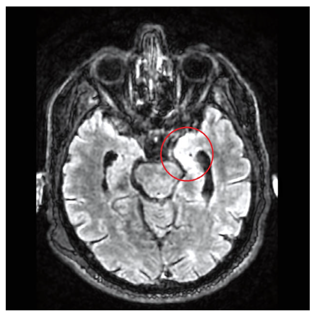
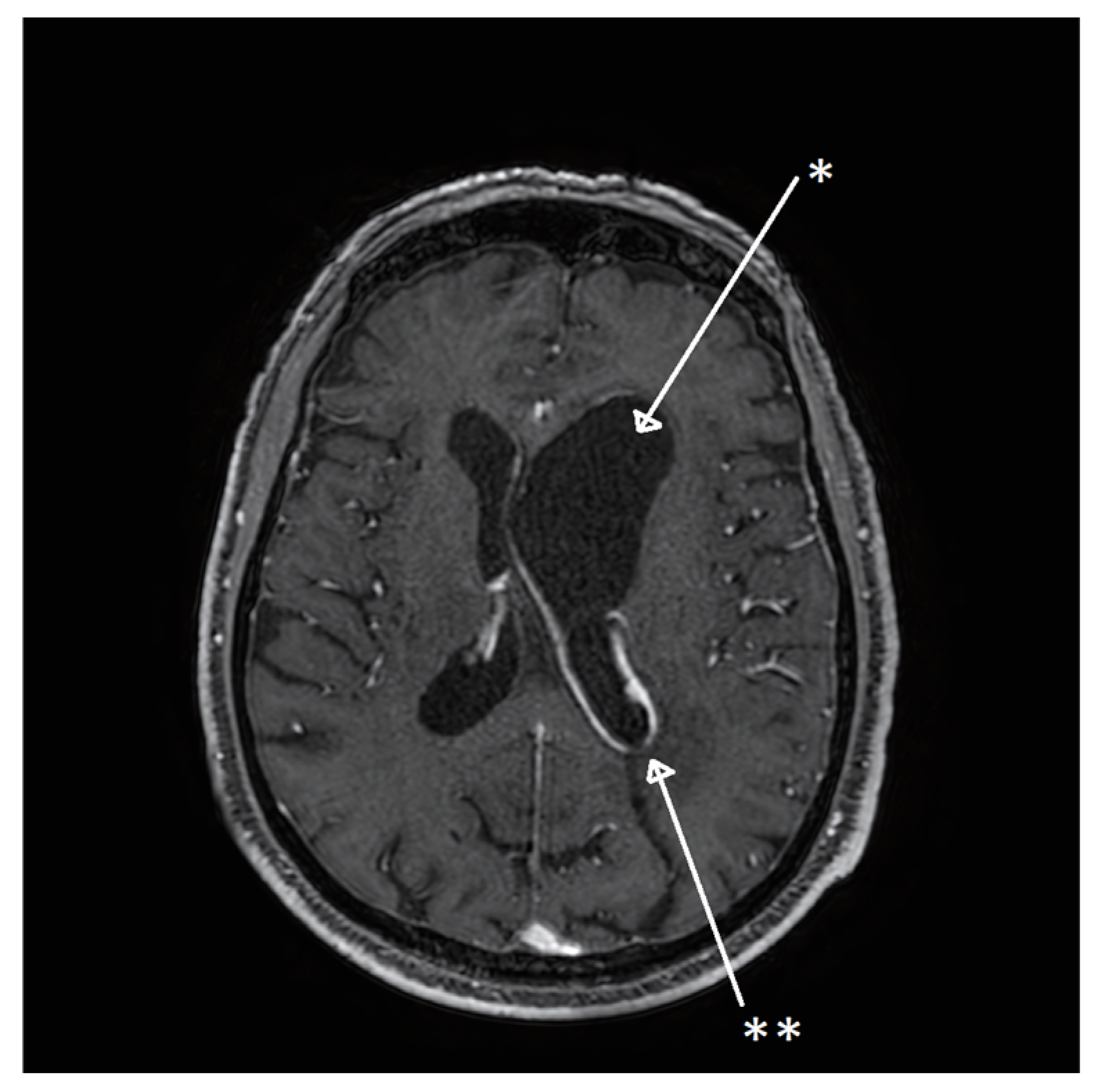
2.3. Second Hospital Admission

One month after discharge, the patient developed intermittent serotine fever (37.5 °C). At 6 weeks from discharge, the patient presented to the emergency room of our center with altered mental state, gait palsy, aphasia, and strength reduction in the superior right limb. A cerebral MRI with gadolinium revealed an excluded enlarged left lateral ventricle, with a natural hyperintensity in the right frontal lobe presenting in the T2 and FLAIR sequences, as well as in a leptomeningeal contrast enhancement of the walls of the excluded ventricle, suggesting ventriculitis and leptomeningeal inflammation (Figure 2). An emergency external ventricular drain (EVD) was placed in the left lateral ventricle medial wall for intracranial pressure control and CSF examination.
The patient began corticosteroid treatment (dexamethasone 32 mg/day) with clinical improvement. CSF analysis showed severe hypoglycorrhachia (19 mg/dL) and hyperproteinorrachia (106 mg/dL) with a normal white blood cell count. All bacterial cultures resulted negative; mycobacterial PCR and an acid-fast bacilli (AFB) smear also resulted negative. To restore CSF circulation within the contralateral ventricles, an endoscopic septostomy through the right frontal horn and an endoscopic exploration of the occipital horn through a left occipital burr hole were performed under neuronavigation. The ependyma appeared to be completely covered by filaments and synechiae (Figure 3), and no communication with the rest of the ventricular system was detectable. A catheter was left in place and connected to an Ommaya reservoir to drain CSF over the next few days. During surgery, a CSF sample was collected directly from the excluded ventricle, and PCR for Mycobacterium tuberculosis complex resulted positive, with no molecular resistance.
Antitubercular therapy was switched from an oral to an intravenous formulation. Linezolid ev 1200 mg/day was added as a fourth antitubercular therapy; this therapy was discontinued after 2 weeks, due to thrombocytopenia, and replaced with levofloxacin 750 mg/day. Dexamethasone was tapered over an 8-week course. A scheduled post-operative CT scan showed resolution of ventricular inflammation and cerebral edema (Figure 4). Before discharge, an LP was repeated, and CSF showed hypoglycorrhachia resolution (45 mg/dL), with a mycobacterial PCR and an AFB smear both negative. Clinically, the patient improved, with some persistent confusion and fatigue, but with fever resolution. The patient was discharged with oral levofloxacin, rifampicin, ethambutol, and isoniazid.

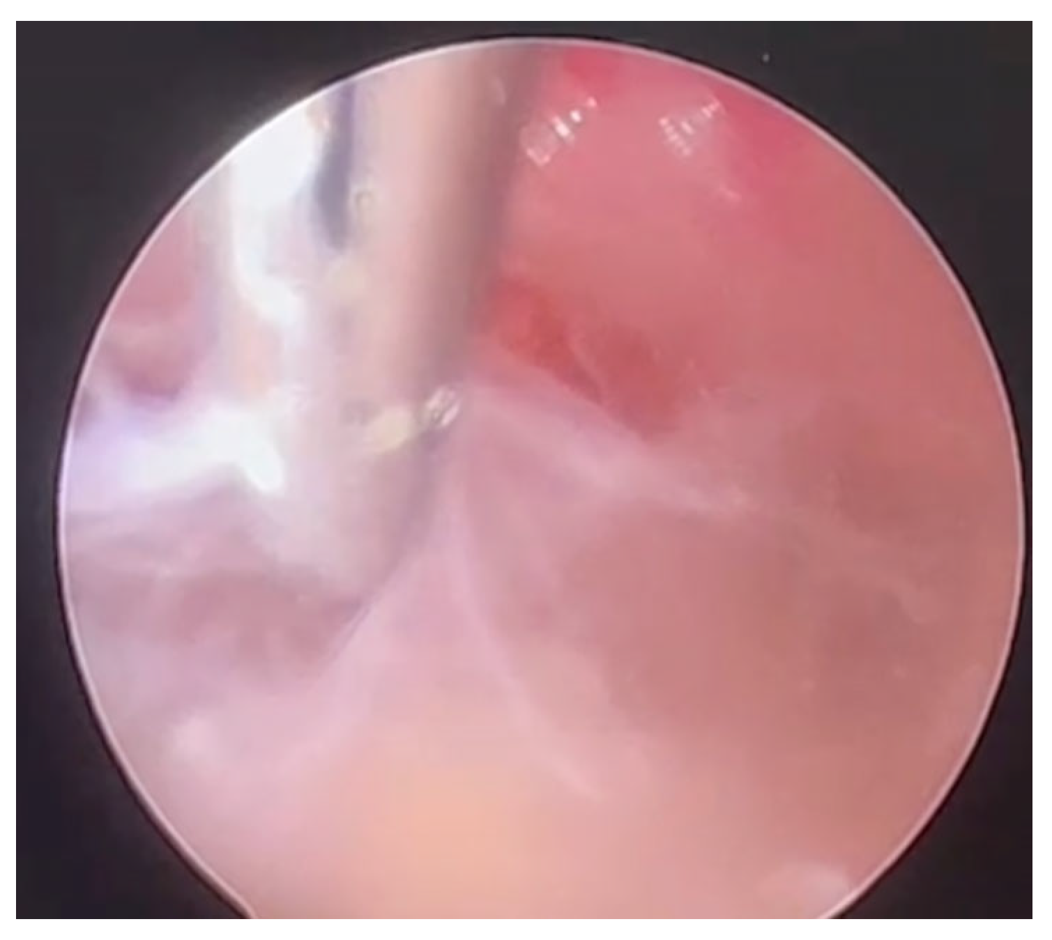
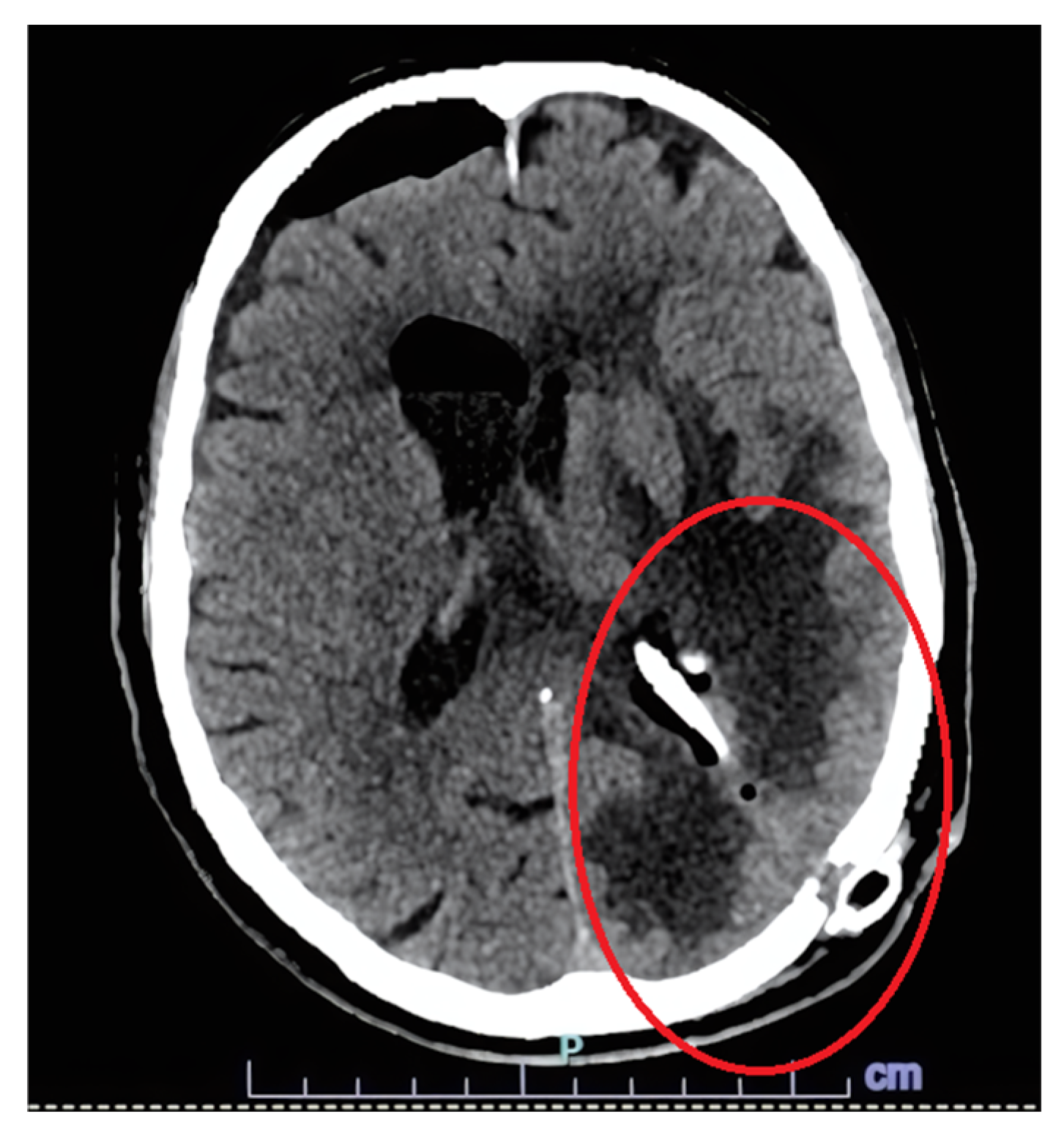
2.4. Third Hospital Admission
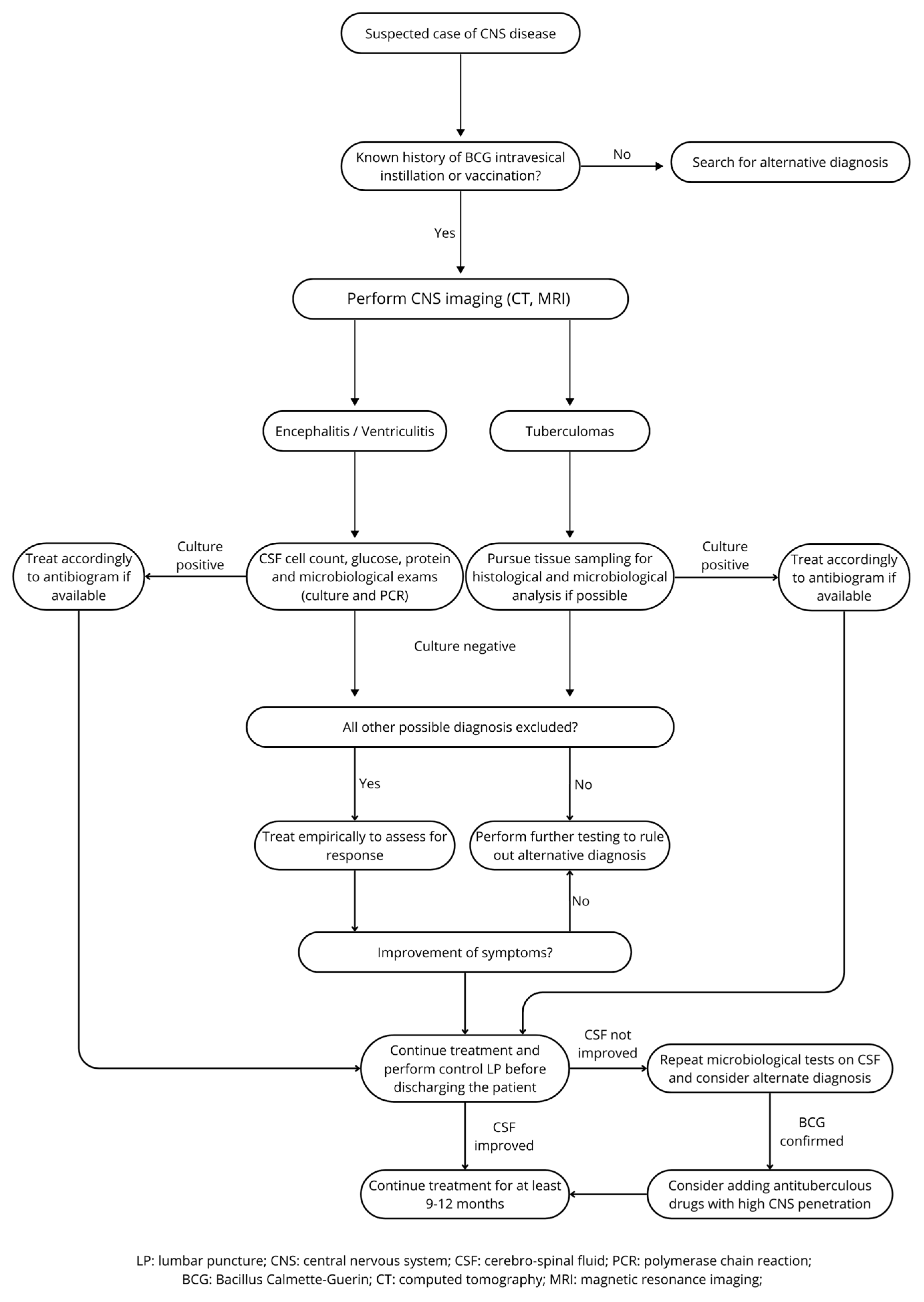
The patient was admitted again six months after the second hospital discharge due to clinical symptoms of aphasia and confusion. A cerebral CT scan noted hydrocephalus and cysts in the frontal horn of the left lateral ventricle. The Ommaya drain was obstructed and the patient underwent urgent surgery with marsupialization of the frontal horn and cisternostomy. CSF analysis was normal, with negative mycobacterial PCR and cultures. He was discharged with some remaining confusion and behavior disinhibition that resolved in several weeks. He completed 14 months of antitubercular therapy (levofloxacin, rifampicin, ethambutol, and isoniazid). A return to baseline functional status was clinically confirmed at 2 months and maintained at the most recent 10-month follow-up.

3. Discussion

As guidelines for BCG immunotherapy-associated CNS infection management are missing, our case report emphasizes the current complexity of the unguided diagnosis and management of CNS BCG infections and provides a comprehensive review of the common management principles available in the literature. Our case is characterized by the microbiological isolation of BCG in the CSF only when collected during surgery, as well as the clinical relapse of symptoms with the development of ventricle obstruction, despite a three-drug antitubercular therapy. A switch to an intravenous four-drug regimen and surgical source control achieved the resolution of symptoms, and clinical cure was sustained up to the 10-month follow-up.
CNS involvement in BCGitis is only anecdotally described, mainly as single case reports. No reviews of CNS BCG infections are currently available. We reviewed all articles indexed in the PubMed, Google Scholar, Embase, ResearchGate, JSTOR, and Science Direct databases from 1988 to March 2025, using the following keywords: Mycobacteria, Bacillus Calmette-Guerin, BCG, encephalitis, meningitis, brain abscess, central nervous system infection; all articles not in English or with missing relevant data were excluded. We identified 15 case reports of CNS BCGitis, plus the one described in this article. All cases have been accurately described in Table 1. Eight cases were related to intravesical instillation [4,10,11,12,13,14,15], six to BCG vaccination [16,17,18,19,20] and two were probably related to LP contamination [21]. Patients diagnosed with CNS BCGitis were predominantly male (94%) [4]. As expected, the median patient age associated with BCG intravesical instillation was higher than that for vaccination-associated BCG infection (74 years versus 3 years, p < 0.01).
Interestingly, only 31% of the patients identified with our review were reported to be immunosuppressed. This observation invites clinicians to consider BCGitis independently of a patient’s immunocompetence [4,18,20,21]. In the literature, worse outcomes and disseminated disease have been extensively linked with primary immunodeficiencies [23]. More recently, evidence has emerged linking invasive mycobacterial infections to uncommon causes of immunosuppression, such as anti-cytokine antibodies, which are not routinely performed in clinical practice [24,25,26]. The detection of anti-cytokine antibodies may explain disseminated disease in otherwise immunocompetent patients. Since our patient had no immunosuppression history, we tested his convalescent plasma for anti-IL23, anti-IFN-gamma, anti-GM-CSF, and anti-IFN-alpha autoantibodies, which were all negative.
BCG infections are tested with various diagnostic tools, with different diagnostic performance rates. The estimated rates for AFB staining are around 25%, mycobacterial cultures and PCR assays are around 41% each, and overall microbiological diagnoses are around 30% [4]. Histological examination, when possible, reveals granulomatous inflammation in more than 80% of cases [4]. BCG urine isolation cannot be considered diagnostic, since BCG can be isolated in the urine for more than a year after the final instillation of immunotherapy, even in asymptomatic patients [27].
Clinical isolation of BCG in meningitis or ventriculitis is most frequently identified with CSF cultures (n = 15; 94%). Conversely, in patients that developed cerebral abscesses or tuberculomas, BCG isolation was obtained from biopsied cerebral tissue, with CSF cultures often negative. Six out of nine cases of meningitis or ventriculitis showed hypoglycorrhachia, similar to our case [13,16,18,19,21]. In patients that developed cerebral abscesses or tuberculomas, unremarkable or slightly abnormal CSF was usually observed, without any cases of hypoglycorrhachia [10,11,12,17]. In the current patient, as microbiological confirmation of BCG infection was initially missing, a review of the patient’s history and risk factors (including treatment with BCG, the SIADH, and marked hypoglycorrhachia) strengthened our suspicion of a mycobacterial infection [28]. This observation was critical for early BCG-specific empirical antimicrobial therapy, 4 months prior to diagnostic confirmation. In the current patient, a positive PCR for M. tuberculosis was achieved only after analyzing the CSF drained during neurosurgery in the excluded ventricle, where we suspected a high bacterial burden.
BCG, being a modified strain of M. bovis, is intrinsically resistant to pyrazinamide, cycloserine, and all macrolides, except for clarithromycin [29]. No specific guidelines exist on BCG infection treatment management. Usually, empirical antitubercular therapy in the suspect of systemic BCG infection relies upon the combination of isoniazid, rifampicin, and ethambutol [30,31]. In our review of CNS BCGitis cases, this standard therapy regimen was administered in only 27% of cases [12,16,19], showing a high heterogeneity in therapy regimens with the administration of additional drugs.
Following patient relapse, our therapy regimen included linezolid in combination with standard antitubercular therapy. Linezolid is an antimycobacterial drug suggested for the treatment of MDR- and XDR-resistant TB [32] and has previously been successfully used in non-CNS disseminated BCGitis, but not in CNS BCGitis [33]. We selected linezolid based on its capacity to penetrate the blood–brain barrier and achieve high concentrations in the brain parenchyma [34]. While our patient showed clinical improvement after initiating linezolid, the drug was replaced with levofloxacin after two weeks due to drug-related thrombocytopenia. The choice of fluoroquinolone was driven by evidence that this drug is also effective in achieving a high level in the CNS [35,36]. Inclusion of a fluoroquinolone in the therapy regimen has been reported in other similar cases [4,10,11,13,20].
Notably, we want to emphasize that, even though the BCG strain used for vaccination and intravesical instillation is standardized by the manufacturer, two strains of isolated BCG identified in the literature exhibited unexpected resistance to isoniazid (n = 1) and ethambutol (n = 1). The first patient was treated with rifampicin, ethambutol, and moxifloxacin [10], whereas the second received rifampicin, isoniazid, clofazimine, clarithromycin, ciprofloxacin, and amikacin (for 2 weeks) [20]. These unexpected resistances highlight the importance of conducting antitubercular susceptibility testing for BCG isolates, in case of mycobacterial culture positivity. In our patient’s isolate, no antimicrobial resistance was found.
The use of corticosteroids as adjunctive therapy in BCG infection is controversial. Our review proves that it is administered in less than half of CNS BCGitis cases reported in the literature (6 of 16 patients received corticosteroids). In treating our patient, we decided to follow the indication of tuberculous meningitis guidelines [37,38] and administered high doses of dexamethasone (0.4 mg/kg/day), followed by a taper over 8 weeks.
Despite appropriate oral therapy with three antitubercular drugs, commonly administered in cases of BCG infection (32% in the review by Pèrez et al. [4]), the patient developed obstructive hydrocephalus, requiring endoscopic septostomy and the placement of an Ommaya drain. CSF analysis at the second admission showed a normal white blood cell count (4 cell/mm3 vs. 237 cell/mm3), but a persistently reduced glycorrhachia, suggesting the persistence of the mycobacteria in the ventricles, responsible for inflammation and ventricle exclusion. We hypothesized that the worsening of symptoms and the formation of the synechiae, observed during surgery, were likely due to inadequate CSF penetration of the three-drug antitubercular therapy initiated during the first admission.
In the absence of clear guidelines, our study group developed a proposed flowchart for optimized CNS BCGitis diagnosis and patient management, see Figure 5.

4. Conclusions

CNS BCG infections are multi-faceted diseases. Diagnosis requires a careful history review to identify the patient’s exposure to BCG, exclude alternative diagnoses, and pursue invasive tissue sampling for microbiological investigations. Our patient was the only case described in the literature that had a clinical relapse while on standard antitubercular therapy, with persistent evidence of active infection. A risk–benefit analysis of repeat LP before patient discharge should be investigated as a therapeutic confirmation of CNS BCGitis therapy efficacy. Linezolid may be an alternative choice as a fourth drug in CNS BCGitis, due to optimal CNS penetration, despite the need for strict follow-up for the early identification of possible adverse events.

Author Contributions

Conceptualization, D.C. and E.F.; literature review, D.C.; writing—original draft preparation, D.C., M.A., S.E., E.G., G.F. and L.L.; writing—review and editing, D.C., E.F. and J.C.; supervision, C.M., C.I., G.P. and G.O. All authors have read and agreed to the published version of the manuscript.

Funding

This research received no external funding.

Institutional Review Board Statement

For case reports, approval by our ethics committee is not required.

Informed Consent Statement

We collected informed consent from the patient.

Data Availability Statement

The data presented in this study are available on request from the corresponding author, due to privacy restrictions.

Acknowledgments

We kindly thank Aristine Cheng and her team for their suggestions and for testing our patient’s blood for anti-cytokine antibodies.

Conflicts of Interest

The authors declare no conflicts of interest.

Abbreviations

The following abbreviations are used in this manuscript:
BCGBacillus Calmette–Guerin
MRIMagnetic resonance imaging
CSFCerebrospinal fluid
NMIBCNon-muscle-invasive bladder cancer
CNSCentral nervous system
EEGElectroencephalography
LPLumbar puncture
CTComputed tomography
SIADHInappropriate antidiuretic hormone secretion
EVDExternal ventricular drain
AFBAcid-fast bacilli
AIDSAcquired immunodeficiency syndrome
RIFRifampicin
EMBEthambutol
MOXMoxifloxacin
INHIsoniazid
PZAPyrazinamide
ETIEthionamide
STPStreptomycin
LEVLevofloxacin
ALLAcute lymphoblastic leukemia
SCIDSevere combined immunodeficiency
CFZClofazimine
CAMClarithromycin
CIPCiprofloxacin
N.R.Not reported
AMKAmikacin
N.A.Not applicable
SRSymptom resolution

References

  1. Burger, M.; Catto, J.W.F.; Dalbagni, G.; Grossman, H.B.; Herr, H.; Karakiewicz, P.; Kassouf, W.; Kiemeney, L.A.; La Vecchia, C.; Shariat, S.; et al. Epidemiology and Risk Factors of Urothelial Bladder Cancer. Eur. Urol. 2013, 63, 234–241. [Google Scholar] [CrossRef] [PubMed]
  2. Babjuk, M.; Burger, M.; Capoun, O.; Cohen, D.; Compérat, E.M.; Dominguez Escrig, J.L.; Gontero, P.; Liedberg, F.; Masson-Lecomte, A.; Mostafid, A.H.; et al. European Association of Urology Guidelines on Non–Muscle-Invasive Bladder Cancer (Ta, T1, and Carcinoma in Situ). Eur. Urol. 2022, 81, 75–94. [Google Scholar] [CrossRef] [PubMed]
  3. Chang, S.S.; Boorjian, S.A.; Chou, R.; Clark, P.E.; Daneshmand, S.; Konety, B.R.; Pruthi, R.; Quale, D.Z.; Ritch, C.R.; Seigne, J.D.; et al. Diagnosis and Treatment of Non-Muscle Invasive Bladder Cancer: AUA/SUO Guideline. J. Urol. 2016, 196, 1021–1069. [Google Scholar] [CrossRef]
  4. Pérez-Jacoiste Asín, M.A.; Fernández-Ruiz, M.; López-Medrano, F.; Lumbreras, C.; Tejido, Á.; San Juan, R.; Arrebola-Pajares, A.; Lizasoain, M.; Prieto, S.; Aguado, J.M. Bacillus Calmette-Guérin (BCG) Infection Following Intravesical BCG Administration as Adjunctive Therapy for Bladder Cancer: Incidence, Risk Factors, and Outcome in a Single-Institution Series and Review of the Literature. Medicine 2014, 93, 236–254. [Google Scholar] [CrossRef]
  5. González-Del Vecchio, M.; Ruíz-Serrano, M.J.; Gijón, P.; Sánchez-Somolinos, M.; de Egea, V.; García de Viedma, D.; Sánchez Fresneda, M.N.; Bouza, E. Differences between a Probable and Proven BCG Infection Following Intravesical Instillations: 16 Years Experience in a Tertiary Care Hospital. Diagn. Microbiol. Infect. Dis. 2016, 85, 338–343. [Google Scholar] [CrossRef]
  6. Larsen, E.S.; Nordholm, A.C.; Lillebaek, T.; Holden, I.K.; Johansen, I.S. The Epidemiology of Bacille Calmette-Guérin Infections after Bladder Instillation from 2002 through 2017: A Nationwide Retrospective Cohort Study. BJU Int. 2019, 124, 910–916. [Google Scholar] [CrossRef] [PubMed]
  7. Steg, A.; Leleu, C.; Debré, B.; Boccon-Gibod, L.; Sieard, D. Systemic Bacillus Calmette-Guérin Infection, “BCGitis”, in Patients Treated by Intravesical Bacillus Calmette-Guérin Therapy for Bladder Cancer. Eur. Urol. 1989, 16, 161–164. [Google Scholar] [CrossRef]
  8. Lamm, D.L.; van der Meijden, P.M.; Morales, A.; Brosman, S.A.; Catalona, W.J.; Herr, H.W.; Soloway, M.S.; Steg, A.; Debruyne, F.M. Incidence and Treatment of Complications of Bacillus Calmette-Guerin Intravesical Therapy in Superficial Bladder Cancer. J. Urol. 1992, 147, 596–600. [Google Scholar] [CrossRef]
  9. Nummi, A.; Järvinen, R.; Sairanen, J.; Huotari, K. A Retrospective Study on Tolerability and Complications of Bacillus Calmette-Guérin (BCG) Instillations for Non-Muscle-Invasive Bladder Cancer. Scand. J. Urol. 2019, 53, 116–122. [Google Scholar] [CrossRef]
  10. Golub, V.; Malhotra, P.; Patel, S. Mycobacterial Brain Tuberculomas Due to Bacille Calmette-Guérin Intravesical Chemotherapy for Bladder Cancer: A Case Report and Literature Review. Can. J. Infect. Dis. Med. Microbiol. 2011, 22, 104–106. [Google Scholar] [CrossRef]
  11. Parent, M.-E.; Richer, M.; Liang, P. The First Case of Bacillus Calmette-Guérin-Induced Small-Vessel Central Nervous System Vasculitis. Clin. Rheumatol. 2018, 37, 2297–2302. [Google Scholar] [CrossRef] [PubMed]
  12. Sheron, M.W.; Holt, S.L.; Ingram, C.W. Mycobacterium Bovis Cerebellar Abscess Following Treatment with Bacillus Calmette-Guérin. J. Pharm. Pract. 2017, 30, 378–380. [Google Scholar] [CrossRef] [PubMed]
  13. Tariq, M.; Clarke, N.; Chen, M. Mycobacterial Meningitis as an Early Complication of Bacille Calmette-Guerin Intravesical Chemotherapy in the Setting of a Systemic Infection. Neurology 2022, 98 (Suppl. S18), 112. [Google Scholar] [CrossRef]
  14. Shoskes, A.; Amin, M. Central Nervous System Miliary Mycobacterium Bovis Infection Secondary to Intravesical BCG Instillation. Neurology 2021, 96 (Suppl. S15), 2719. [Google Scholar] [CrossRef]
  15. Schwartz, S.; Amaya, S.; Kirby, J.; Varma, H. Mycobacterium Bovis Infection of the Brain Following Intravesical BCG Treatment. J. Neuropathol. Exp. Neurol. 2023, 82, 583–584. [Google Scholar]
  16. Furuichi, M.; Mori, F.; Uejima, Y.; Sato, S.; Kurihara, J.; Kawano, Y.; Suganuma, E. A Case of Mycobacterium Bovis Bacillus Calmette-Guérin (BCG) Strain Meningitis and Ventriculitis Following BCG Vaccination. Int. J. Infect. Dis. 2020, 100, 373–376. [Google Scholar] [CrossRef]
  17. Sharifi, G.; Mousavinejad, S.A.; Moradian, K.; Ebrahimzadeh, K.; Samadian, M.; Zerehpoosh, F.B.; Rezaei, O. Pineal Region Tuberculoma Caused by Mycobacterium bovis as a Complication of Bacille Calmette-Guérin Vaccine: Case Report and Review of the Literature. World Neurosurg. 2020, 133, 416–418. [Google Scholar] [CrossRef]
  18. van Deutekom, H.; Smulders, Y.M.; Roozendaal, K.J.; van Soolingen, D. Bacille Calmette-Cuerin (BCG) Meningitis in an AIDS Patient 12 Years After Vaccination with BCG. Clin. Infect. Dis. 1996, 22, 870–871. [Google Scholar] [CrossRef]
  19. Tardieu, M.; Truffot-Pernot, C.; Carriere, J.P.; Dupic, Y.; Landrieu, P. Tuberculous Meningitis Due to BCG in Two Previously Healthy Children. Lancet 1988, 331, 440–441. [Google Scholar] [CrossRef]
  20. Eser, I.; Uyan, Z.S.; Kilic, S.C.; Uyur, E.; Arisoy, E.S.; Tezcan, G.; Aydogan, M. Intracranial Calcification after Probable Disseminated Bacillus Calmette–Guérin Infection in a Child with Severe Combined Immunodeficiency. J. Pediatr. Infect. Dis. 2018, 15, 48–52. [Google Scholar] [CrossRef]
  21. Stone, M.M.; Vannier, A.M.; Storch, S.K.; Peterson, C.; Nitta, A.T.; Zhang, Y. Brief Report: Meningitis Due to Iatrogenic BCG Infection in Two Immunocompromised Children. New Engl. J. Med. 1995, 333, 561–563. [Google Scholar] [CrossRef] [PubMed]
  22. Misra, U.K.; Kalita, J.; Tuberculous Meningitis International Research Consortium. Mechanism, Spectrum, Consequences and Management of Hyponatremia in Tuberculous Meningitis. Wellcome Open Res. 2019, 4, 189. [Google Scholar] [CrossRef]
  23. Bernatowska, E.A.; Wolska-Kusnierz, B.; Pac, M.; Kurenko-Deptuch, M.; Zwolska, Z.; Casanova, J.-L.; Piatosa, B.; Van Dongen, J.; Roszkowski, K.; Mikoluc, B.; et al. Disseminated Bacillus Calmette-Guérin Infection and Immunodeficiency. Emerg. Infect. Dis. 2007, 13, 799–801. [Google Scholar] [CrossRef]
  24. Barcenas-Morales, G.; Cortes-Acevedo, P.; Doffinger, R. Anticytokine Autoantibodies Leading to Infection: Early Recognition, Diagnosis and Treatment Options. Curr. Opin. Infect. Dis. 2019, 32, 330–336. [Google Scholar] [CrossRef] [PubMed]
  25. Martínez-Barricarte, R.; Markle, J.G.; Ma, C.S.; Deenick, E.K.; Ramírez-Alejo, N.; Mele, F.; Latorre, D.; Mahdaviani, S.A.; Aytekin, C.; Mansouri, D.; et al. Human IFN-γ Immunity to Mycobacteria Is Governed by Both IL-12 and IL-23. Sci. Immunol. 2018, 3, eaau6759. [Google Scholar] [CrossRef] [PubMed]
  26. Cheng, A.; Kashyap, A.; Salvator, H.; Rosen, L.B.; Colby, D.; Ardeshir-Larijani, F.; Loehrer, P.J.; Ding, L.; Lugo Reyes, S.O.; Riminton, S.; et al. Anti–Interleukin-23 Autoantibodies in Adult-Onset Immunodeficiency. N. Engl. J. Med. 2024, 390, 1105–1117. [Google Scholar] [CrossRef]
  27. Bowyer, L.; Hall, R.R.; Reading, J.; Marsh, M.M. The Persistence of Bacille Calmette-Guérin in the Bladder after Intravesical Treatment for Bladder Cancer. Br. J. Urol. 1995, 75, 188–192. [Google Scholar] [CrossRef]
  28. Solomons, R.S.; Visser, D.H.; Donald, P.R.; Marais, B.J.; Schoeman, J.F.; van Furth, A.M. The Diagnostic Value of Cerebrospinal Fluid Chemistry Results in Childhood Tuberculous Meningitis. Child’s Nerv. Syst. 2015, 31, 1335–1340. [Google Scholar] [CrossRef]
  29. Durek, C.; Rüsch-Gerdes, S.; Jocham, D.; Böhle, A. Sensitivity of BCG to Modern Antibiotics. Eur. Urol. 2000, 37 (Suppl. S1), 21–25. [Google Scholar] [CrossRef]
  30. Pommier, J.D.; Ben Lasfar, N.; Van Grunderbeeck, N.; Burdet, C.; Laouénan, C.; Rioux, C.; Pierre-Audigier, C.; Meybeck, A.; Choudat, L.; Benchikh, A.; et al. Complications Following Intravesical Bacillus Calmette-Guerin Treatment for Bladder Cancer: A Case Series of 22 Patients. Infect. Dis. 2015, 47, 725–731. [Google Scholar] [CrossRef]
  31. Audenet, F.; Sotto, A.; Roumiguié, M.; Allory, Y.; Andrejak, C.; Leon, P.; Loriot, Y.; Masson-Lecomte, A.; Pradère, B.; Seisen, T.; et al. Recommandations Des Comités de Cancérologie (CC-AFU) et d’infectiologie (CI-AFU) de l’Association Française d’urologie Pour La Prise En Charge Effets Indésirables et Complications Du BCG. Prog. Urol. 2022, 32, 165–176. [Google Scholar] [CrossRef] [PubMed]
  32. Nahid, P.; Mase, S.R.; Migliori, G.B.; Sotgiu, G.; Bothamley, G.H.; Brozek, J.L.; Cattamanchi, A.; Cegielski, J.P.; Chen, L.; Daley, C.L.; et al. Treatment of Drug-Resistant Tuberculosis. An Official ATS/CDC/ERS/IDSA Clinical Practice Guideline. Am. J. Respir. Crit. Care Med. 2019, 200, e93–e142. [Google Scholar] [CrossRef] [PubMed]
  33. Roglić, S.; Dickson, D.; Miše, B.; Višković, K.; Katalinić-Janković, V.; Rutherford, G.; Begovac, J. Successful Treatment of Disseminated Bacillus Calmette-Guérin Disease in an HIV-Infected Child with a Linezolid-Containing Regimen. Case Rep. Infect. Dis. 2016, 2016, 1528981. [Google Scholar] [CrossRef]
  34. Nau, R.; Sörgel, F.; Eiffert, H. Penetration of Drugs through the Blood-Cerebrospinal Fluid/Blood-Brain Barrier for Treatment of Central Nervous System Infections. Clin. Microbiol. Rev. 2010, 23, 858–883. [Google Scholar] [CrossRef]
  35. Thwaites, G.E.; Bhavnani, S.M.; Chau, T.T.H.; Hammel, J.P.; Török, M.E.; Van Wart, S.A.; Mai, P.P.; Reynolds, D.K.; Caws, M.; Dung, N.T.; et al. Randomized Pharmacokinetic and Pharmacodynamic Comparison of Fluoroquinolones for Tuberculous Meningitis. Antimicrob. Agents Chemother. 2011, 55, 3244–3253. [Google Scholar] [CrossRef] [PubMed]
  36. Nahid, P.; Dorman, S.E.; Alipanah, N.; Barry, P.M.; Brozek, J.L.; Cattamanchi, A.; Chaisson, L.H.; Chaisson, R.E.; Daley, C.L.; Grzemska, M.; et al. Official American Thoracic Society/Centers for Disease Control and Prevention/Infectious Diseases Society of America Clinical Practice Guidelines: Treatment of Drug-Susceptible Tuberculosis. Clin. Infect. Dis. 2016, 63, e147–e195. [Google Scholar] [CrossRef]
  37. WHO Consolidated Guidelines on Tuberculosis: Module 4: Treatment: Drug-Susceptible Tuberculosis Treatment. Available online: https://www.who.int/publications/i/item/9789240048126 (accessed on 21 March 2025).
  38. Thwaites, G.E.; Nguyen, D.B.; Nguyen, H.D.; Hoang, T.Q.; Do, T.T.O.; Nguyen, T.C.T.; Nguyen, Q.H.; Nguyen, T.T.; Nguyen, N.H.; Nguyen, T.N.L.; et al. Dexamethasone for the Treatment of Tuberculous Meningitis in Adolescents and Adults. N. Engl. J. Med. 2004, 351, 1741–1751. [Google Scholar] [CrossRef]
Figure 1. First MRI in FLAIR sequence showing vasogenic edema of the temporal lobe.
Microorganisms 13 01283 g001
Figure 2. MRI in T1 sequence, with gadolinium contrast enhancement, showing an enlarged left lateral ventricle * and contrast uptake of the excluded ventricle **.
Microorganisms 13 01283 g002
Figure 3. Endoscopic image showing synechiae found in the left excluded ventricle.
Microorganisms 13 01283 g003
Figure 4. CT scan showing the resolution of hydrocephalus after endoscopic septostomy and placement of Ommaya drain.
Microorganisms 13 01283 g004
Figure 5. CNS BCGitis management flowchart.
Microorganisms 13 01283 g005
Table 1. Clinical and microbiological characteristics of patients with CNS BCGitis found in literature.
Author
[Reference]
Age, Yrs/
Sex
Clinical PresentationLesion SiteBCG Infection TypeBCG Isolation SiteCSF AlterationsTherapyImmuno-
Deficient
Adjunctive
Corticosteroids
Therapy Duration, MonthsOutcome
BCG origin: Intravesical instillations
1Golub et al. [10]73/MHeadaches, dizziness, right hand tremorFrontal, temporal, and basal gangliaTuberculomasBrain tissue cultureLymphocytic Pleocytosis and hyperproteinorrachiaRIF + EMB + MOXNoNo12SR
2Parent et al. [11]89/MRight upper limb hyposthenia, dysarthriaMultiple left-sided white matter lesionsVasculitis, endophthalmitisBrain tissue, humor vitreoPleocytosisINH + RIF + MOX + EMBNoYesN.R.SR
3Sheron et al. [12]74/MDizziness, vertigo, gait ataxiaCerebellumAbscessBrain tissue cultureN.R.INH + RIF + EMBNoYes12SR
4Tariq et al. [13]80/MSevere ataxia, dysmetriaNodular leptomeningeal enhancement involving cranial nerves, brainstem, and superior cerebellumMeningitisCSFLymphocytic Pleocytosis, hypoglycorrhachia, and hyperproteinorrachiaRIF + EMB + MOXNoYes12Improvement of symptoms; remaining dysmetria
5Shoskes et al. [14] 67/MFever, encephalopathy, respiratory failureHyperintense lesions in cortex, subcortical white matter, and brainstemMiliary infectionBiopsyN.R.RIF + INH + EMB + AZMNoYes6SR
6Schwartz et al. [15]88/MAphasiaFrontotemporal lesionsTuberculomasTissue biopsyN.R.NoneNoNo0Death
7Pérez-Jacoiste et al. [4]73/MN.R.Inflammation of meningesLymphocytic meningitisOnly urineN.R.RIF + INH + LEVYesN.R.6SR
8Our case77/MFever, confusionRight frontal lobe hyperintensity and left lateral ventricle exclusion.Encephalitis and ventriculitisCSFPleocytosis, hypoglycorrhachia, and hyperproteinorrachiaINH + RIF + EMB + LEV; LIN (2 weeks)NoYes14SR
BCG origin: Vaccination
9Furuichi et al. [16]1.3/Mgait disturbancesthird ventricleVentriculitisCSF and brain tissueLymphocytic Pleocytosis, hypoglycorrhachia, and hyperproteinorrachiaINH + RIF + EMBNoYes24Improvement of symptoms; remaining gait disturbances
10Sharifi et al. [17]0.5/MPoor feeding, nausea, vomitingPineal glandTuberculomaBrain tissue cultureWithin rangeN.R.NoN.R.N.R.SR
11Van Deutekom et al. [18]31/MFever, neck stiffness, headacheN.A.MeningitisCSFHyperproteinorrachia and pleocytosisINH + RIF + PZAYes (AIDS)No0.3Death
12Tardieu et al. [19]5/MFever, rigorHyperintensity of subarachnoid space exclusion of fourth ventricleVentriculitisCSFPleocytosis, hypoglycorrhachia, and hyperproteinorrachiaINH + RIF + EMBNoNo18SR
13Tardieu et al. [19]4/MFever, rigorN.A.MeningitisCSFPleocytosis, hypoglycorrhachia, and hyperproteinorrachiaINH + RIF + EMBNoNo18SR
14Eser et al. [20]0.25/MAfebrile seizuresCalcification of the subcortical and periventricular regionsCalcifications, TuberculomasLymph nodes *Within rangeINH + RIF + CAM + CFZ + CIP + AMK (2 mo)Yes (SCID)No12Microcephaly and persistence of axial hypotonia
BCG origin: Unknown (LP contamination?)
15Stone et al. [21] 3/FHeadacheN.A.MeningitisCSFPleocytosisINH + RIF + ETI + STPYes (ALL in active chemotherapy *)No12SR
16Stone et al. [21]5/MHeadache, fever, weight lossN.A.MeningitisCSFPleocytosis, hypoglycorrhachia, and hyperproteinorrachiaINH + RIF + STP + ETIYes (ALL in active chemotherapy *)No12SR
BCG: Bacillus Calmette–Guerin; CSF: cerebrospinal fluid; RIF: rifampicin; EMB: ethambutol; MOX: moxifloxacin; CIP: ciprofloxacin; AMK: amikacin; CAM: clarithromycin; CFZ: clofazimine; INH: isoniazid; PZA: pyrazinamide; ETI: ethionamide; STP: streptomycin; LEV: levofloxacin; AZM: azithromycin; LIN: linezolid; N.R.: not reported; N.A.: not applicable; SR: symptom resolution; ALL: acute lymphoblastic leukemia; SCID: severe combined immunodeficiency. * see comment in the main text.
Disclaimer/Publisher’s Note: The statements, opinions and data contained in all publications are solely those of the individual author(s) and contributor(s) and not of MDPI and/or the editor(s). MDPI and/or the editor(s) disclaim responsibility for any injury to people or property resulting from any ideas, methods, instructions or products referred to in the content.

