Article

Occurrence and Genetic Diversity of Cryptosporidium spp. and Giardia intestinalis from Yaks (Bos grunniens) in Ganzi, Sichuan Province, China

College of Veterinary Medicine, Northwest A&F University, Yangling 712100, China
*
Author to whom correspondence should be addressed.
These authors contributed equally to this work.
Current Address: Animal Husbandry Science Institute of Ganzi Tibetan Autonomous Prefecture, Kangding 626000, China.
Microorganisms 2025, 13(6), 1261; https://doi.org/10.3390/microorganisms13061261
Submission received: 27 April 2025 / Revised: 20 May 2025 / Accepted: 28 May 2025 / Published: 29 May 2025

Abstract

Cryptosporidium spp. and Giardia intestinalis are important zoonotic protozoa that are closely related to diarrhea and cause considerable economic losses in the livestock breeding industry. Ganzi is one of the main production areas for yaks in China, but there have been few reports on the occurrence of Cryptosporidium spp. and G. intestinalis in yaks. This study used PCR-based sequencing techniques to survey the prevalence and species/genotypes of Cryptosporidium spp. and G. intestinalis in faecal samples from 223 yaks in Ganzi, Sichuan Province. The positive rate of Cryptosporidium spp. was 7.2% (16/223), with the highest positive rate of yaks found in Yajiang (37%, 10/27), which was significantly higher than that in Litang (3.1%, 4/130) and Seda (3%, 2/66). The positive rate of Cryptosporidium spp. in young animals aged <6 months (20.5%, 8/39) was significantly higher than that in older animals aged 12–23 months (0; 0/43) and >24 months (3.3%, 3/90). Three Cryptosporidium species were found by sequence analysis of 18S rRNA locus, namely C. andersoni, C. ryanae, and C. bovis. The total positive rate of G. intestinalis was 15.7% (35/223), with significant differences identified between Yajiang (40.7%, 11/27), Litang (17.7%, 23/130), and Seda (1.5%, 1/66). One genotype (assemblage E) was found by analyzing the sequence of gdh, bg, and tpi loci. Meanwhile, co-infection of Cryptosporidium spp. and Giardia intestinalis was identified in five samples. The present study explores the infection of Cryptosporidium spp. and G. intestinalis from yaks in Ganzi, aiming to enrich our understanding of the occurrence of these protozoa in livestock.

1. Introduction

Cryptosporidium spp. and Giardia intestinalis are common zoonotic protozoa leading to diarrhea in quite a few animals and humans [1,2]. These two protozoa can be transmitted by foodborne and waterborne pathways or direct/indirect contact with infected hosts, threatening the health of humans and the development of the breeding industry [1,3,4]. Oocysts of the two pathogens can be excreted in faeces from the hosts, and then contaminate food and water. Humans and animals are usually infected by ingesting polluted food and water or coming into contact with faeces containing oocysts [1,3,4]. As important opportunistic pathogens, Cryptosporidium spp. and G. intestinalis have led to multiple outbreaks of diarrhea in both humans and bovine animals, causing great economic losses within the breeding industry and affecting human health [1]. Meanwhile, they are listed as pathogens that must be tested for in domestic water due to their high pathogenicity.
Knowledge about the distribution and composition of Cryptosporidium spp. and G. intestinalis helps with the recognition of the transmission of these pathogens. To date, more than 40 valid species have been reported in various animals [5]. Of these, over 20 species/genotypes are zoonotic, while others are usually identified in limited hosts [6]. Host adaption exists in the presence of Cryptosporidium spp., as reflected by the major occurrence of 1–4 species in one host [6]. Sequence analysis based on the triosephosphate isomerase (tpi), glutamate dehydrogenase (gdh), and β-giardin (bg) loci of G. intestinalis indicates there are eight genotypes with different host preferences, namely assemblage A-H [1,7,8]. Assemblages A and B can infect humans and many mammals. Assemblages C and D are usually recognized in canids, while assemblages E-H are usually identified in ruminants, felines, rodents, and aquatic animals, respectively [1,7,8].
Yaks are mainly distributed in the plateau region centered around the Qinghai–Tibet Plateau. Previous studies have reported the occurrence of Cryptosporidium spp. and G. intestinalis in yaks in Qinghai, Tibet, Gansu, and some other regions in China with positive rates of 1.4–30% [9,10,11,12] and 1.7–6% [10,12,13], respectively. Genetic diversity analysis indicated 12 species/genotypes (C. parvum, C. canis, C. bovis, C. struthionis, C. ryanae, C. baileyi, C. andersoni, C. ubiquitum, C. hominis, C. xiaoi, C. suis-like and Cryptosporidium new genotype) and three genotypes (assemblages A, B and E) for Cryptosporidium spp. and G. intestinalis in yaks, respectively [14,15,16]. Ganzi is one of the major important yak-breeding areas in Sichuan in China, located in the southeastern Tibetan plateau. As a common economic animal in Ganzi, yaks are kept in enclosures and have close contact with farmers, but the occurrence of Cryptosporidium spp. and G. intestinalis in yaks in this region was seldom explored. Considering the high pathogenicity of the two pathogens in yaks and the lack of information on their distribution and transmission in yaks in Ganzi, this study investigated the occurrence and genetic diversity of Cryptosporidium and G. intestinalis in yaks from three regions in Ganzi, China, which could enrich our understanding of the occurrence of these two pathogens and provide a foundation for understanding their transmission and zoonotic potential in yaks in Ganzi.

2. Materials and Methods

2.1. Sample Collection

Faecal specimens were sampled from 223 yaks kept in Seda, Yajiang, and Litang in Ganzi, China, as indicated in Figure 1. Seda, Yajiang, and Litang are the main production areas of yaks in Ganzi, and some yaks in these three regions experienced diarrhea according to the description of farmers. Those faecal specimens were randomly collected and accounted for 10.9% (66/603), 14.3% (27/189) and 10% (130/1296) of the total number of yaks in Seda, Yajiang, and Litang, respectively. Yaks in those investigated regions were housed, and did not mix with other livestock. Faecal specimens were directly and randomly collected from the rectum of yaks, and placed in disposable sampling bags marked with the yaks’ location, age, gender and diarrhea condition. Age groups were defined according to the growth and development condition of the yaks and verified by ear tags. The samples were then transmitted to the laboratory as soon as possible at a low temperature and preserved at −20 °C.

2.2. Genomic DNA Extraction

A commercial DNA isolation kit (Cate No. D4015-02; Omega Bio-Tek, Norcross, GA, USA) was used to isolate genomic DNA from 200 mg faecal specimens without pre-treatment as per the instructions. All the genomic DNA samples were preserved at −20 °C.

2.3. PCR Amplification

The occurrence and species of Cryptosporidium spp. were recognized by applying a nested PCR based on the 18S rRNA gene with the size of ~830 bp [17]. The occurrence and genotypes of G. intestinalis were identified by using three nested PCRs targeting the bg [18], gdh [19], and tpi [20] loci with lengths of ~511 bp, ~392 bp and ~530 bp, respectively. The reaction system of PCRs (25 μL) contained 1 × Rapid Taq Master Mix (Cate No. P222-01, Vazyme, Nanjing, China), 0.4 μΜ of each primer (Table 1), 1 μL of gDNA for the primary PCR/1 μL primary PCR product for the secondary PCR under the PCR reaction conditions: denaturing occurred at 94 °C for 5 min, followed by 35 cycles of 94 °C for 45 s, annealing at the same temperature (Table 1) for 45 s, and 68 °C for 1 min, and an additional extension at 68 °C for 7 min. For each PCR, genomic samples of Cryptosporidium xiaoi and G. intestinalis assemblage C were used as positive controls for Cryptosporidium spp. and G. intestinalis PCR, respectively, and a negative control with ddH2O was also included. Positive secondary PCR products were confirmed by gel electrophoresis and then applied for sequencing at both directions using primers as PCRs.

2.4. Sequence Analysis

All the sequences were assemblaged, edited, and aligned by ChromasPro V1.5, BioEdit V7.0.5.3 and Clustal X V1.81, respectively. Phylogenetic trees for each locus were constructed using the Maximum Likelihood method with the bootstrap evaluation of 1000 replicates in MEGA V6.06.

2.5. Statistical Analysis

Differences in the occurrence of Cryptosporidium spp. and G. intestinalis among various factors were analyzed by applying an χ2 test within SPSS V22.0. Significant differences were recognized if the p value was less than 0.05. The OR (odds ratio) and RR (risk ratio) with 95% CIs (confidence intervals) were analyzed for the identification of factors associated with Cryptosporidium spp. and G. intestinalis infection.

2.6. Nucleotide Sequence Accession Numbers

Representative sequences for the 18S rRNA, bg, gdh, and tpi loci found were submitted to GenBankTM with the numbers PV151463-PV151469, PV157997-PV158007, PV297768-PV297771 and PV297772-PV297776, respectively.

3. Results

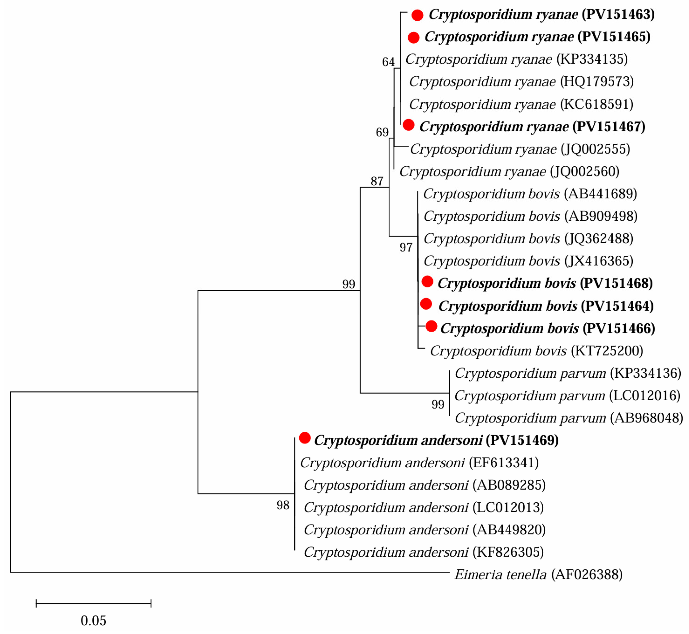
3.1. Occurrence of Cryptosporidium spp. in Yaks in Ganzi

The positive rate of Cryptosporidium spp. was 7.2% (16/223) in yaks in Ganzi. Among those three locations, the highest rate was found in yaks in Yajiang (37%, 10/27), followed by Seda (3%, 2/66) and Litang (3.1%, 4/130), and significant differences were found for the positive rates of Cryptosporidium spp. among the three locations (χ2 = 41.1316, df = 2, p < 0.0001). Meanwhile, significant differences were also recognized for the positive rates of Cryptosporidium spp. among yaks of different ages (χ2 = 14.7828, df = 3, p = 0.002), with the highest observed in yaks under 6 months of age (20.5%, 8/39), followed by yaks aged 6–12 months (7.8%, 4/51), >24 months (4.4%, 4/90), and 12–24 months (0, 0/43). Although a higher positive rate was found in male yaks and non-diarrheal yaks compared with female yaks and diarrheal yaks, respectively, no significant difference for the positive rate was found in relation to gender and diarrhea (Table 2).
Sequence analysis of the 18S rRNA locus identified three Cryptosporidium species in the 16 positive specimens, namely C. bovis (9), C. ryanae (6), and C. andersoni (1) (Table 2 and Figure 2). Among them, C. bovis was found in three locations and three age groups, C. ryanae in two locations and three age groups, while C. andersoni was found in only one location and one age group.

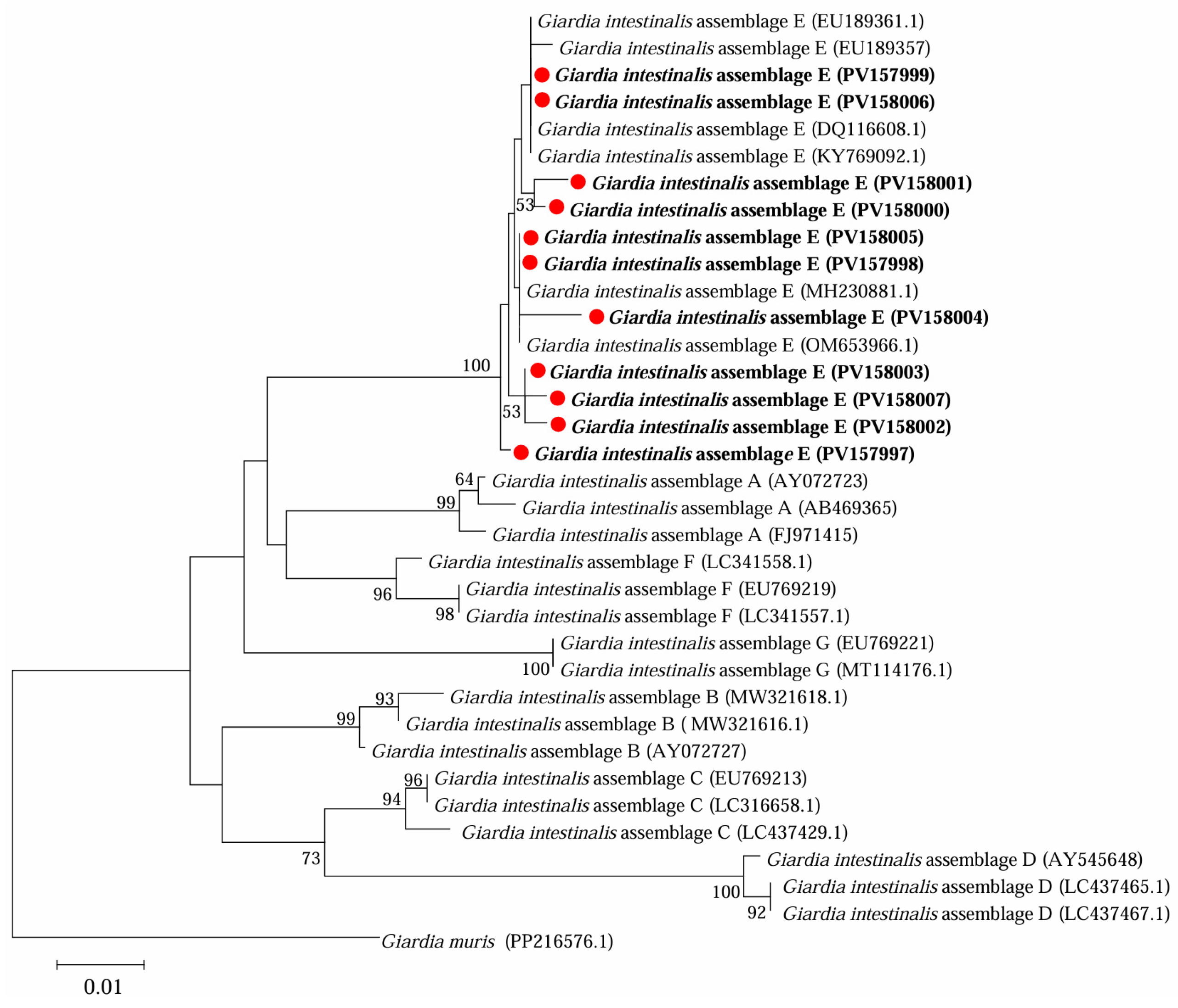
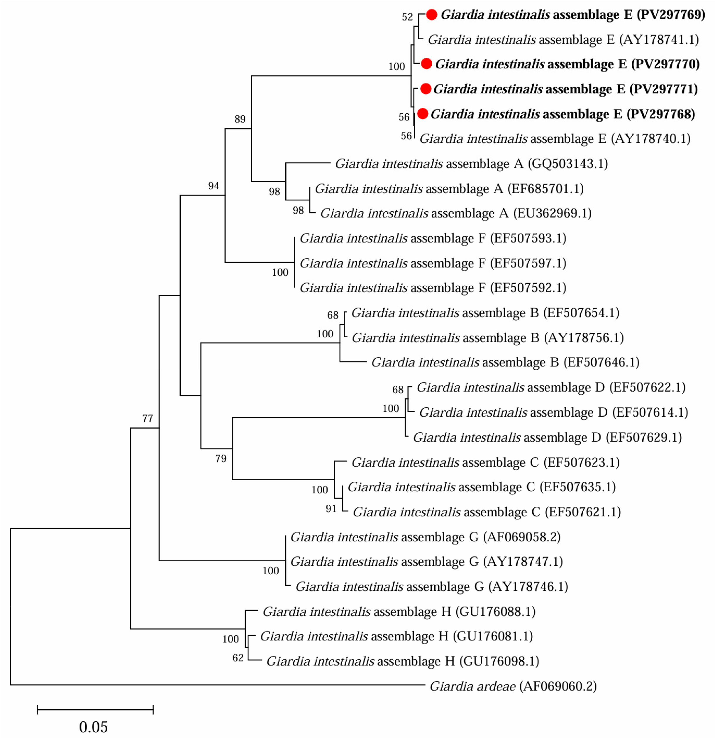
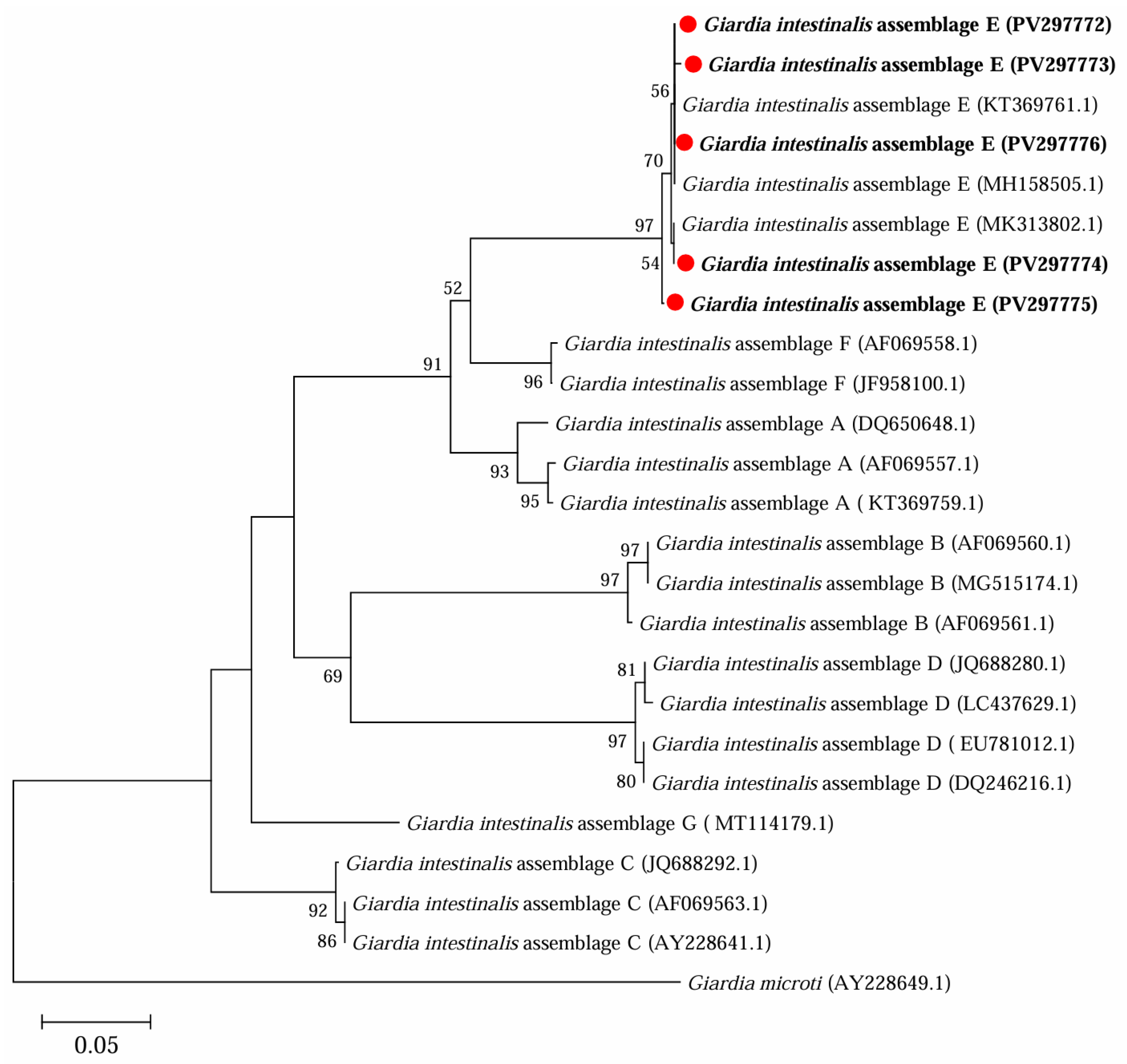
3.2. Occurrence of G. intestinalis in Yaks in Ganzi

The positive rate of G. intestinalis was 15.7% (35/223) in yaks in Ganzi, with 15.7% (35/223), 13.9% (31/223), and 7.2% (16/223) at gdh, bg, and tpi loci, respectively. Significant differences were found for the positive rates among three locations (χ2 = 23.2214, df = 2, p < 0.0001), with the highest in Yajiang (40.7%, 11/27), followed by Litang (17.7%, 23/130), and Seda (1.5%, 1/66).
The highest positive rate of G. intestinalis was found in yaks aged <6 months (35.9%, 14/39), followed by those aged 6–12 months (23.5%, 12/51), 12–24 months (11.6%, 5/43), and >24 months (4.4%, 4/90), and significant differences were found for the positive rates of G. intestinalis among the four age groups (χ2 = 23.5424, df = 3, p < 0.0001). However, there were no significant differences for G. intestinalis in terms of gender and diarrheal condition (Table 3).
Further phylogenetic analysis based on sequences of bg (Figure 3), gdh (Figure 4) and tpi (Figure 5) loci from the present study and referenced sequences downloaded from NCBI indicated that there existed one genotype (assemblage E) for G. intestinalis in yaks in the present study.

3.3. Co-Infection of Cryptosporidium spp. and G. intestinalis in Yaks in Ganzi

Notably, co-infection of Cryptosporidium spp. and G. intestinalis was found in five yaks. Among them, four yaks were co-infected with C. ryanae and G. intestinalis, and one with C. bovis and G. intestinalis.

4. Discussion

Cryptosporidium spp. and G. intestinalis are two common zoonotic protozoa that are closely related to diarrhea, threatening the health of humans and the breeding industry. This study applied nested PCR-based sequencing techniques to explore Cryptosporidium spp. and G. intestinalis infections in yaks in Ganzi, China, which could contribute to the understanding of the occurrence and zoonotic potential of those pathogens in yaks.
Several techniques have been applied in epidemiological studies of Cryptosporidium spp. and G. intestinalis. Traditional morphological methods are considered to be the “gold standard”, but these techniques are time-consuming, lack sensitivity under low infection intensity, and should be performed by an experienced person. PCR-based molecular methods are high-throughput and can make up for the deficiencies of traditional morphological methods to a certain extent [1], but can easily be polluted if the operation is not standardized. Therefore, it is necessary to follow a standard operation procedure and set suitable positive and negative controls in the PCR test.
The total positive rate of Cryptosporidium spp. in yaks was 7.2% (16/223) in Ganzi, which was higher than that in Tibet (1.4%, 8/577) [10], Gansu (5.26%, 4/76) [21], and the central western region of China (4%, 22/545) [12], but lower than that in Qinghai (30%, 98/327) [11]. Differences in the positive rates among regions were likely caused by several factors, such as geographic location, sample size, age, and animal condition. There existed significant differences related to the occurrence of Cryptosporidium spp. in yaks among different age groups, and the positive rate decreased with the increase in age. Similar results have also been reported in yaks in Qinghai [9,11], reflecting Cryptosporidium infection likely related to immunity state, and younger yaks were found to be more susceptible to Cryptosporidium compared with older ones. Considering the greater susceptibility of younger yaks to Cryptosporidium and potential transmission from older yaks to younger ones, it is better to provide separate pens according to age, avoid unnecessary contact with older yaks, and enhance daily management to reduce the possibility of infection.
Further sequence analysis highlighted three Cryptosporidium species (C. bovis, C. andersoni and C. ryanae), with C. bovis being the dominant one. Except for those three common species in bovine animals, several common zoonotic species (e.g., C. parvum and C. canis) [15] have also been reported in yaks, indicating yaks’ zoonotic potential for the transmission of Cryptosporidium. In this study, no zoonotic Cryptosporidium species were found, which was likely due to the differences in multifaceted factors, such as sampling size, climate, and management. Investigations on a wider scale are needed to comprehensively understand the composition and transmission of Cryptosporidium species. Considering the close contact between farmers and yaks in the investigated regions and the potential zoonotic transmission of C. parvum, C. canis, and other species, interventions based on WASH (water, sanitation, and hygiene) are needed to reduce the possibility of Cryptosporidium species being transmitted between humans and yaks.
The positive rate of G. intestinalis was 15.7% (35/223) in yaks in Ganzi, which was higher than that in Tibet (1.7%, 10/577) [10] and the central western region of China (6%, 16/545) [13]. Several factors, e.g., sampling size, age, and management, may have led to the differences in the occurrence of G. intestinalis in yaks among the regions. Meanwhile, significant differences in the positive rates of G. intestinalis in yaks among different age groups were identified, and the positive rate of G. intestinalis in yaks decreased with age. Considering the similarities in the age-related prevalence patterns of Cryptosporidium infection in yaks, similar interventions to those performed for Cryptosporidium are needed to reduce the possibility of G. intestinalis infections in younger animals with incomplete immunity.
Sequence analysis indicated that there was one genotype (assemblage E) of G. intestinalis in yaks in this study, which has also been identified in yaks in Tibet [10], Qinghai [14], and the central western region of China [12]. Assemblage E was commonly found in ruminants (e.g., dairy cattle, camels, sheep, and goats) [22,23,24,25]. In addition to assemblage E, zoonotic assemblages A and B have been found in yaks in Tibet [16] and Qinghai [14], respectively, indicating the possible zoonotic potential for the spread of G. intestinalis in yaks. The zoonotic potential for G. intestinalis in yaks in Ganzi needs to be further explored in more animals located in different geographic regions.
Notably, co-infection of Cryptosporidium spp. and G. intestinalis was observed in yaks. Previous studies have reported that mixed infection of Cryptosporidium spp. and G. intestinalis was also found in other ruminants, such as sheep and cattle [26,27,28]. Both Cryptosporidium spp. and G. intestinalis infection could lead to diarrhea and intestinal damage, which could provide convenient conditions and thus cause secondary infection of other intestinal pathogens, exacerbating intestinal damage. Moreover, previous studies found that co-infection with multiple intestinal pathogens likely caused more severe diarrhea compared with a single infection [29]. Thus, the relationship between co-infection of Cryptosporidium spp. and G. intestinalis and the severity of diarrhea and intestinal damage in yaks needs to be investigated in further studies to help improve our understanding of the pathogenesis of these two pathogens in yaks.

5. Conclusions

This study investigated the colonization of Cryptosporidium spp. and G. intestinalis in yaks from Ganzi, China, and C. bovis, C. ryanae, C. andersoni and G. intestinalis (assemblage E) identified. Significantly higher positive rates of Cryptosporidium spp. and G. intestinalis were found in yaks in Yajiang and yaks aged under 6 months, which indicated that location and age were likely to be risk factors. The results of the recent studies could enrich our knowledge about Cryptosporidium spp. and G. intestinalis infections in yaks in Ganzi, offering reference data which will be useful for understanding the transmission of those two pathogens in Ganzi and other related regions.

Author Contributions

Conceptualization, X.Y. and Y.F.; methodology, Y.F., G.H. and D.Y.; software, X.H. and M.Z.; data curation, Y.N. and Z.W.; writing—original draft preparation, Y.F.; writing—review and editing, X.Y.; visualization, X.Y. and Y.F.; supervision, X.Y.; funding acquisition, X.Y. and Y.F. All authors have read and agreed to the published version of the manuscript.

Funding

This study was founded by the National Natural Science Foundation of China (32202838), the State Key Laboratory for Animal Disease Control and Prevention Foundation (SKLADCPKFKT202411), the Natural Science Foundation of Shaanxi Province (2024JC-YBQN-0173), and the Postdoctoral Research Project of Shaanxi Province (2023BSHEDZZ138).

Institutional Review Board Statement

This study was conducted under the approval and instructions of the ethics committee of Northwest A&F University (DY2022048, Approval date 8 April 2022).

Informed Consent Statement

Not applicable.

Data Availability Statement

Data is contained within the article.

Conflicts of Interest

The authors declare no conflicts of interest.

References

  1. Xiao, L.; Feng, Y. Molecular epidemiologic tools for waterborne pathogens Cryptosporidium spp. and Giardia duodenalis. Food Waterborne Parasitol. 2017, 8–9, 14–32. [Google Scholar] [CrossRef] [PubMed]
  2. Hijjawi, N.; Zahedi, A.; Al-Falah, M.; Ryan, U. A review of the molecular epidemiology of Cryptosporidium spp. and Giardia duodenalis in the Middle East and North Africa (MENA) region. Infect. Genet. Evol. 2022, 98, 105212. [Google Scholar] [CrossRef]
  3. Ali, M.; Ji, Y.; Xu, C.; Hina, Q.; Javed, U.; Li, K. Food and waterborne cryptosporidiosis from a one health perspective: A comprehensive review. Animals 2024, 14, 3287. [Google Scholar] [CrossRef] [PubMed]
  4. Meng, X.Z.; Kang, C.; Wei, J.; Ma, H.; Liu, G.; Zhao, J.P.; Zhang, H.S.; Yang, X.B.; Wang, X.Y.; Yang, L.H.; et al. Meta-analysis of the prevalence of Giardia duodenalis in cattle in China. Foodborne Pathog. Dis. 2023, 20, 17–31. [Google Scholar] [CrossRef] [PubMed]
  5. Ryan, U.M.; Feng, Y.; Fayer, R.; Xiao, L. Taxonomy and molecular epidemiology of Cryptosporidium and Giardia—A 50 year perspective (1971–2021). Int. J. Parasitol. 2021, 51, 1099–1119. [Google Scholar] [CrossRef]
  6. Feng, Y.; Ryan, U.M.; Xiao, L. Genetic diversity and population structure of Cryptosporidium. Trends Parasitol. 2018, 34, 997–1011. [Google Scholar] [CrossRef] [PubMed]
  7. Feng, Y.; Xiao, L. Zoonotic potential and molecular epidemiology of Giardia species and giardiasis. Clin. Microbiol. Rev. 2011, 24, 110. [Google Scholar] [CrossRef]
  8. Cai, W.; Ryan, U.; Xiao, L.; Feng, Y. Zoonotic giardiasis: An update. Parasitol. Res. 2021, 120, 4199–4218. [Google Scholar] [CrossRef]
  9. Li, P.; Cai, J.; Cai, M.; Wu, W.; Li, C.; Lei, M.; Xu, H.; Feng, L.; Ma, J.; Feng, Y.; et al. Distribution of Cryptosporidium species in Tibetan sheep and yaks in Qinghai, China. Vet. Parasitol. 2016, 215, 58–62. [Google Scholar] [CrossRef]
  10. Wu, Y.; Chang, Y.; Zhang, X.; Chen, Y.; Li, D.; Wang, L.; Zheng, S.; Wang, R.; Zhang, S.; Jian, F.; et al. Molecular characterization and distribution of Cryptosporidium spp., Giardia duodenalis, and Enterocytozoon bieneusi from yaks in Tibet, China. BMC Vet. Res. 2019, 15, 417. [Google Scholar] [CrossRef]
  11. Ma, J.; Cai, J.; Ma, J.; Feng, Y.; Xiao, L. Occurrence and molecular characterization of Cryptosporidium spp. in yaks (Bos grunniens) in China. Vet. Parasitol. 2014, 202, 113–118. [Google Scholar] [CrossRef] [PubMed]
  12. Qi, M.; Cai, J.; Wang, R.; Li, J.; Jian, F.; Huang, J.; Zhou, H.; Zhang, L. Molecular characterization of Cryptosporidium spp. and Giardia duodenalis from yaks in the central western region of China. BMC Microbiol. 2015, 15, 108. [Google Scholar] [CrossRef] [PubMed]
  13. Song, G.Y.; Qin, S.Y.; Zhao, G.H.; Zhu, X.Q.; Zhou, D.H.; Song, M.X. Molecular characterization of Giardia duodenalis from white yaks in China. Acta Parasitol. 2016, 61, 397–400. [Google Scholar] [CrossRef]
  14. Wang, G.; Wang, G.; Li, X.; Zhang, X.; Karanis, G.; Jian, Y.; Ma, L.; Karanis, P. Prevalence and molecular characterization of Cryptosporidium spp. and Giardia duodenalis in 1–2-month-old highland yaks in Qinghai Province, China. Parasitol. Res. 2018, 117, 1793–1800. [Google Scholar] [CrossRef]
  15. Geng, H.L.; Ni, H.B.; Li, J.H.; Jiang, J.; Wang, W.; Wei, X.Y.; Zhang, Y.; Sun, H.T. Prevalence of Cryptosporidium spp. in yaks (Bos grunniens) in China: A systematic review and meta-analysis. Front. Cell. Infect. Microbiol. 2021, 11, 770612. [Google Scholar] [CrossRef] [PubMed]
  16. Wu, Y.; Chen, Y.; Chang, Y.; Zhang, X.; Li, D.; Wang, L.; Zheng, S.; Wang, R.; Zhang, S.; Li, J.; et al. Genotyping and identification of Cryptosporidium spp., Giardia duodenalis and Enterocytozoon bieneusi from free-range Tibetan yellow cattle and cattle-yak in Tibet, China. Acta Trop. 2020, 212, 105671. [Google Scholar] [CrossRef]
  17. Xiao, L.; Escalante, L.; Yang, C.; Sulaiman, I.; Escalante, A.A.; Montali, R.J.; Fayer, R.; Lal, A.A. Phylogenetic analysis of Cryptosporidium parasites based on the small-subunit rRNA gene locus. Appl. Environ. Microb. 1999, 65, 1578–1583. [Google Scholar] [CrossRef]
  18. Lalle, M.; Pozio, E.; Capelli, G.; Bruschi, F.; Crotti, D.; Cacciò, S.M. Genetic heterogeneity at the β-giardin locus among human and animal isolates of Giardia duodenalis and identification of potentially zoonotic subgenotypes. Int. J. Parasitol. 2005, 35, 207–213. [Google Scholar] [CrossRef]
  19. Cacciò, S.M.; Beck, R.; Lalle, M.; Marinculic, A.; Pozio, E. Multilocus genotyping of Giardia duodenalis reveals striking differences between assemblages A and B. Int. J. Parasitol. 2008, 38, 1523–1531. [Google Scholar] [CrossRef]
  20. Sulaiman, I.M.; Fayer, R.; Bern, C.; Gilman, R.H.; Trout, J.M.; Schantz, P.M.; Das, P.; Lal, A.A.; Xiao, L. Triosephosphate isomerase gene characterization and potential zoonotic transmission of Giardia duodenalis. Emerg. Infect. Dis. 2003, 9, 1444–1452. [Google Scholar] [CrossRef]
  21. Qin, S.Y.; Zhang, X.X.; Zhao, G.H.; Zhou, D.H.; Yin, M.Y.; Zhao, Q.; Zhu, X.Q. First report of Cryptosporidium spp. in white yaks in China. Parasite Vector 2014, 7, 230. [Google Scholar] [CrossRef]
  22. Wang, Y.; Cao, J.; Chang, Y.; Yu, F.; Zhang, S.; Wang, R.; Zhang, L. Prevalence and molecular characterization of Cryptosporidium spp. and Giardia duodenalis in dairy cattle in Gansu, northwest China. Parasite 2020, 27, 62. [Google Scholar] [CrossRef] [PubMed]
  23. Elmahallawy, E.K.; Köster, P.C.; Dashti, A.; Alghamdi, S.Q.; Saleh, A.; Gareh, A.; Alrashdi, B.M.; Hernández-Castro, C.; Bailo, B.; Lokman, M.S.; et al. Molecular detection and characterization of Cryptosporidium spp., Giardia duodenalis, and Enterocytozoon bieneusi infections in dromedary camels (Camelus dromedaries) in Egypt. Front. Vet. Sci. 2023, 10, 1139388. [Google Scholar] [CrossRef]
  24. Wang, P.; Zheng, L.; Liu, L.; Yu, F.; Jian, Y.; Wang, R.; Zhang, S.; Zhang, L.; Ning, C.; Jian, F. Genotyping of Cryptosporidium spp., Giardia duodenalis and Enterocytozoon bieneusi from sheep and goats in China. BMC Vet. Res. 2022, 18, 361. [Google Scholar] [CrossRef] [PubMed]
  25. Ruiz, A.; Foronda, P.; González, J.F.; Guedes, A.; Abreu-Acosta, N.; Molina, J.M.; Valladares, B. Occurrence and genotype characterization of Giardia duodenalis in goat kids from the Canary Islands, Spain. Vet. Parasitol. 2008, 154, 137–141. [Google Scholar] [CrossRef]
  26. Zhao, J.Q.; Fan, Y.Y.; Lei, Y.D.; Liu, D.; Wang, J.W.; Yang, X.; Song, J.K.; Zhao, G.H. Molecular characterization of common zoonotic protozoan parasites and bacteria causing diarrhea in dairy calves in Ningxia Hui Autonomous Region, China. Parasite 2024, 31, 60. [Google Scholar] [CrossRef] [PubMed]
  27. Ma, Y.; Zan, X.Q.; Liu, J.B.; Xu, L.H.; Zhao, H.X. Molecular detection and characterization of Cryptosporidium spp. and Giardia duodenalis in dairy calves from Ningxia, China. Acta Parasitol. 2024, 69, 1876–1885. [Google Scholar] [CrossRef]
  28. Qi, M.Z.; Fang, Y.Q.; Wang, X.T.; Zhang, L.X.; Wang, R.J.; Du, S.Z.; Guo, Y.X.; Jia, Y.Q.; Yao, L.; Liu, Q.D.; et al. Molecular characterization of Cryptosporidium spp. in pre-weaned calves in Shaanxi Province, north-western China. J. Med. Microbiol. 2015, 64, 111–116. [Google Scholar] [CrossRef]
  29. Zhang, S.X.; Zhou, Y.M.; Xu, W.; Tian, L.G.; Chen, J.X.; Chen, S.H.; Dang, Z.S.; Gu, W.P.; Yin, J.W.; Serrano, E.; et al. Impact of co-infections with enteric pathogens on children suffering from acute diarrhea in southwest China. Infect. Dis. Poverty 2016, 5, 64. [Google Scholar] [CrossRef]
Figure 1. Distribution of three sampling sites (Seda, Yajiang, and Litang) of yak faeces in Ganzi. Red solid circles indicate sampling sites. The number in each bracket refers to the sample size.
Microorganisms 13 01261 g001
Figure 2. Phylogenetic analysis of representative sequences for the 18S rRNA locus of Cryptosporidium species in this study with referenced sequences, obtained via Maximum Likelihood analysis. The sequences obtained in this study are marked with red solid circles and highlighted in bold font. Eimeria tenella (AF026388) was used as the outgroup; bootstrap values over 50% are indicated.
Microorganisms 13 01261 g002
Figure 3. Phylogenetic analysis of representative sequences for the bg locus of G. intestinalis in this study with referenced sequences, obtained using Maximum Likelihood analysis. The sequences obtained in this study are marked with red solid circles and highlighted in bold font. Giardia muris (PP216576.1) was used as the outgroup; bootstrap values over 50% are indicated.
Microorganisms 13 01261 g003
Figure 4. Phylogenetic analysis of representative sequences for the gdh locus of G. intestinalis in this study with referenced sequences obtained using Maximum Likelihood analysis. The sequences obtained in this study are marked with red solid circles and highlighted in bold font. Giardia ardeae (AF069060.2) was used as the outgroup; bootstrap values over 50% are indicated.
Microorganisms 13 01261 g004
Figure 5. Phylogenetic analysis of representative sequences for the tpi locus of G. intestinalis in this study with referenced sequences obtained using Maximum Likelihood analysis. Those sequences obtained in this study are marked with red solid circles and highlighted in bold font. Giardia microti (AY228649.1) was used as the outgroup; bootstrap values over 50% are indicated.
Microorganisms 13 01261 g005
Table 1. Sequence information of the primers used for PCRs in this study.
TargetsPrimer NamesSequence (5′-3′)Annealing Temperature
18S rRNA [17]18S-F1TTCTAGAGCTAATACATGCG55 °C
18S-R1CCCATTTCCTTCGAAACAGGA
18S-F2GGAAGGGTTGTATTTATTAGATAAAG55 °C
18S-R2CTCATAAGGTGCTGAAGGAGTA
bg [18]bg-F1AAGCCCGACGACCTCACCCGCAGTGC65 °C
bg-R1GAGGCCGCCCTGGATCTTCGAGACGAC
bg-F2GAACGAGATCGAGGTCCG55 °C
bg-R2CTCGACGAGCTTCGTGTT
gdh [19]gdh-F1TTCCGTGTCCAGTACAACTC50 °C
gdh-R1GCCAGCTTCTCCTCGTTGAA
gdh-F2CGCTTCCACCCCTCTGTCAAT50 °C
gdh-R2TGTTGTCCTTGCACATCTC
tpi [20]tpi-F1AATAAATIATGCCTGCTCGTCG54 °C
tpi-R1ATGGACITCCTCTGCCTGCTC
tpi-F2CCCTTCATCGGIGGTAACTTCAA58 °C
tpi-R2GTGGCCACCACICCCGTGCC
Table 2. Occurrence of Cryptosporidium spp. in yaks in Ganzi, Sichuan Province.
FactorsNo. TestedNo. Positive
(%, 95% CI)
RR (95% CI)OR (95% CI)pSpecies (n)
Location
Yajiang2710 (37.0, 0.2153–0.5577)0.0831 (0.0281–0.2454)0.054 (0.0152–0.1913)<0.0001C. bovis (7), C. ryanae (3)
Seda662 (3.0, 0.0083–0.1039)1.0154 (0.1909–5.4012)1.0159 (0.1812–5.6951) C. bovis (1), C. andersoni (1)
Litang1304 (3.1, 0.012–0.0765)ReferenceReference C. ryanae (3), C. bovis (1)
Age (Months)
<6398 (20.5, 0.1078–0.3553)0.2167 (0.0693–0.6774)0.1802 (0.0507–0.6408)0.002C. bovis (5), C. ryanae (3)
6–12514 (7.8, 0.0309–0.185)0.5667 (0.148–2.1699)0.5465 (0.1307–2.2855) C. bovis (3), C. ryanae (1)
12–24430 (0, 0–0.082)-- -
>24904 (4.4, 0.0174–0.1087)ReferenceReference C. ryanae (2), C. bovis (1), C. andersoni (1)
Gender
Male623 (4.8, 0.0166–0.1329)0.3444 (0.0368–3.22)0.3333 (0.0337–3.2976)0.3253C. ryanae (2), C. bovis (1)
Female601 (1.7, 0.003–0.0886)ReferenceReference C. ryanae (1)
NA10112 (11.9, 0.0693–0.1963) C. bovis (8), C. ryanae (3), C. andersoni (1)
Diarrhea
Yes403 (5.0, 0.0258–0.1986)1.0558 (0.3155–3.5331)1.0603 (0.2876–3.9092)0.9299C. bovis (1), C. ryanae (1), C. andersoni (1)
No18313 (7.1, 0.042–0.1177)ReferenceReference C. bovis (8), C. ryanae (5)
Total22316 (7.2, 0.0446–0.1133)-- C. bovis (9), C. ryanae (6), C. andersoni (1)
NA: not available; CI: confidence interval; RR: risk ratio; OR: odds ratio; Reference: reference group for pairwise comparison.
Table 3. Occurrence and risk factors of G. intestinalis infection in yaks in Ganzi, Sichuan Province.
FactorsNo. TestedNo. Positive (%, 95% CI)RR (95% CI)OR (95% CI)pGenotype (n)
gdhtpibggdhtpibg
Location
Yajiang2711 (40.7, 0.2451–0.5927)4 (14.8, 0.0591–0.3247)10 (37, 0.2153–0.5577)0.4343 (0.2415–0.781)0.3127 (0.1284–0.7614)<0.0001E (11)E (4)E (10)
Seda661 (1.5, 0.0027–0.081)0 (0, 0–0.055)0 (0, 0–0.055)11.6769 (1.612–84.583)13.972 (1.8428–105.9317) E (1)--
Litang13023 (17.7, 0.1209–0.2515)12 (9.2, 0.0536–0.1544)21 (16.2, 0.1081–0.2343)ReferenceReference E (23)E (12)E (21)
Age (Months)
<63914 (35.9, 0.2274–0.5158)8 (20.5, 0.1078–0.3553)13 (33.3, 0.2063–0.4902)0.1238 (0.0435–0.3523)0.0831 (0.0251–0.275)<0.0001E (14)E (8)E (13)
6–125112 (23.5, 0.1401–0.3676)5 (9.8, 0.0426–0.2097)12 (23.5, 0.1401–0.3676)0.1889 (0.0643–0.5552)0.1512 (0.0458–0.4985) E (12)E (5)E (12)
12–24435 (11.6, 0.0507–0.2448)2 (4.7, 0.0128–0.1545)4 (9.3, 0.0368–0.216)0.3822 (0.108–1.3524)0.3535 (0.0899–1.3899) E (5)E (2)E (4)
>24904 (4.4, 0.0174–0.1087)1 (1.1, 0.002–0.0603)2 (2.2, 0.0061–0.0774)ReferenceReference E (4)E (1)E (2)
Gender
Male6210 (16.1, 0.09–0.2721)4 (6.5, 0.0254–0.1545)10 (16.1, 0.09–0.2721)1.1367 (0.5214–2.478)1.1673 (0.4555–2.9917)0.7471E (10)E (4)E (10)
Female6011 (18.3, 0.1056–0.2992)3 (5, 0.0171–0.137)9 (15, 0.081–0.2611)ReferenceReference E (11)E (3)E (9)
NA10114 (13.9, 0.0844–0.2193)9 (8.9, 0.0476–0.1607)12 (11.9, 0.0693–0.1963)-- E (14)E (9)E (12)
Diarrhea
Yes406 (15.0, 0.0706–0.2907)2 (5, 0.0138–0.165)2 (5, 0.0138–0.165)1.0565 (0.4701–2.3743)1.0671 (0.4109–2.7711)0.8939E (6)E (2)E (2)
No18329 (15.9, 0.1127–0.2184)14 (7.7, 0.0461–0.1243)29 (15.9, 0.1127–0.2184)ReferenceReference E (29)E (14)E (29)
Total22335 (15.7, 0.1151–0.2105)16 (7.2, 0.0446–0.1133)31 (13.9, 0.0997–0.1905)-- E (35)E (16)E (31)
NA: not available; CI: confidence interval; RR: risk ratio; OR: odds ratio; Reference: reference group for pairwise comparison.
Disclaimer/Publisher’s Note: The statements, opinions and data contained in all publications are solely those of the individual author(s) and contributor(s) and not of MDPI and/or the editor(s). MDPI and/or the editor(s) disclaim responsibility for any injury to people or property resulting from any ideas, methods, instructions or products referred to in the content.

