Article

Porcine Teschovirus 2 3Cpro Evades Host Antiviral Innate Immunity by Inhibiting the IFN-β Signaling Pathway

1
Wenzhou Key Laboratory for Virology and Immunology, Institute of Virology, Wenzhou University, Wenzhou 325035, China
2
College of Animal Sciences, Institute of Preventive Veterinary Medicine, Zhejiang University, Hangzhou 310000, China
3
College of Veterinary Medicine, Northwest A&F University, Yangling 712100, China
*
Author to whom correspondence should be addressed.
These authors contributed equally to this work.
Microorganisms 2025, 13(6), 1209; https://doi.org/10.3390/microorganisms13061209
Submission received: 29 March 2025 / Revised: 19 May 2025 / Accepted: 22 May 2025 / Published: 26 May 2025
(This article belongs to the Special Issue The Pathogenic Epidemiology of Important Swine Diseases)

Abstract

:
Porcine teschovirus (PTV) circulates in pig populations, causing clinical diseases such as poliomyelitis, reproductive disorders, and pneumonia. However, the molecular mechanisms underlying the pathogenesis of PTV infection have not been fully elucidated. Here, we found that PTV infection does not activate the promoters of NF-κB or IFN-β. The expression of PTV 3Cpro inhibits the promoter activity of NF-κB and IFN-β stimulated by SeV and inhibits the downstream transcription of NF-κB and IFN-β by blocking the phosphorylation and nuclear translocation of NF-κB. Coimmunoprecipiation (co-IP) experiments demonstrated that 3Cpro and NF-κB interact. The degradation of NF-κB was unaffected by inhibitors targeting lysosomes (NH4Cl), proteasomes (MG132), or caspases (Z-VAD-FMK). The protease activity of 3Cpro, which relies on its catalytic active site, is vital for NF-κB cleavage and degradation. Loss of proteolytic activity in mutants abolished NF-κB degradation, impairing the ability of 3Cpro to suppress SeV-induced innate immunity and restore VSV-GFP replication, thereby underscoring its critical role in immune evasion by targeting NF-κB. This study reveals novel mechanisms underlying PTV-mediated suppression of host innate immunity.

1. Introduction

Porcine teschovirus (PTV) infection can lead to polioencephalomyelitis, reproductive disorders in sows, enteric diseases, and pneumonia, constituting a substantial threat to swine production [1]. PTV is a single-stranded positive-sense RNA virus belonging to the Teschovirus genus in the Picornaviridae family. The virus particles are spherical, 20–30 nanometers in diameter, and lack an envelope [2]. The genome is approximately 7.2 kb long and has an open reading frame encoding a polyprotein that is cleaved into four structural proteins (VP1-VP4) and eight nonstructural proteins (Lpro, 2A-2C, 3A-3D) [3]. In 2019, the International Committee on Taxonomy of Viruses (ICTV) proposed a revision of the teschovirus taxonomy; PTV currently comprises at least 15 PTV genotypes, which has expanded with the recent discoveries of novel genotypes [4]. In 1929, the first cases of infection with PTV were documented in Teschen, Czechoslovakia, which was later recognized as Teschen disease [5]. Pigs are the only hosts, with piglets being the most susceptible. The virus primarily resides in the swine encephalon and spinal cord, and infection can result in up to 100% mortality in piglets. The disease spreads mainly through subclinical infections, making early detection difficult [6]. In the past twenty years, PTV has spread progressively throughout Asia, including countries such as Japan [7], India [8], and China [9]. Moreover, outbreaks have been documented in North American regions, including Canada [10], the United States [11], and Haiti [12]. However, effective treatments are still lacking, highlighting the significance of investigating and preventing PTV infections.
A great research question is how PTV can evade innate immunity and spread through replication. Research has shown that Picornaviruses have evolved strategies to counteract the host’s innate immune system [13]. The picornavirus 3C protease functions as a chymotrypsin-like enzyme that cleaves viral proteins to form mature proteins, playing a crucial role in viral replication. It can also combat innate immunity by regulating host factors and promoting viral replication [14]. In addition, 3Cpro broadly suppresses type I interferon signaling, facilitating viral immune evasion [15]. Seneca Valley virus (SVA) 3Cpro induces caspase-3-dependent NF-κB-p65 proteolysis, disrupting survival signaling and triggering apoptotic cell death [16]. SVA 3Cpro degrades RIG-I, thereby inhibiting its ability to recognize viral RNA and suppress IFN-β-mediated innate immunity [17]. Enterovirus 71 (EV71) 3Cpro cleaves TIR-domain-containing adapter-inducing IFN-β (TRIF) at Q312, impacting NF-κB activation and IFN-β production [18]. Encephalomyocarditis virus (EMCV) 3Cpro cleaves TANK and disrupts the TANK–TBK1–IKKε–IRF3 complex, which results in a reduction in IRF3 phosphorylation and IFN-β production [19].
Previous studies have shown that PTV VP1 targets melanoma differentiation-associated gene 5 (MDA5) through its 2CARD and Hel domains to effectively inhibit IFN-β production and block NF-κB activation [20]. PTV2 infection can induce incomplete autophagy, which impedes viral propagation within PK-15 cells [21]. Although significant progress has been made in understanding the pathogenesis and immune evasion strategies of PTV, further investigation is still needed. Given the crucial role of picornaviral 3Cpro in viral replication and host antiviral pathway suppression, elucidating its functional mechanisms in PTV immune evasion holds important theoretical significance and research value. Therefore, this study aimed to elucidate the functional significance of PTV 3Cpro in interfering with key antiviral pathways. By exploring these mechanisms, we hope to provide new insights into PTV pathogenesis and contribute to the development of targeted interventions against this important swine pathogen.

2. Materials and Methods

2.1. Cells, Viral Strains, and Plasmids

ST cells (ATCC, CRL-1746, Manassas, VA, USA), HEK-293T cells (ATCC, CRL-11268), Dulbecco’s modified Eagle’s medium (Gibco, Carlsbad, CA, USA), and fetal bovine serum (Cellmax, SA101.02, Beijing, China) were used. PTV strain type 2 GX/2020, Sendai virus (SeV), and recombinant vesicular stomatitis virus expressing green fluorescent protein (VSV-GFP) were stored at the Wenzhou Key Laboratory for Virology and Immunology. The NF-κB plasmid (NCBI, NM_001114281.1) was constructed by Xin-yu Zhang, and the H-NF-κB, NF-κB-Luc, IFN-β-Luc, and PRL-TK plasmids were also maintained in the laboratory.

2.2. Antibodies

Anti-Flag (Bioss, bsm-33346 M, Beijing, China), anti-NF-κB p65 rabbit polyclonal (Beyotime, AF0246, Shanghai, China), anti-phospho-NF-κB p65 (Ser536) (93H1), anti-Myc (Bioss, bsm-51003 M), rabbit anti-Lamin B1 (Bioss, bs-34005R), anti-rabbit IgG (H + L), F(ab’)2 fragment (Cell Signaling Technology, 8889s, Shanghai, China), anti-mouse IgG (H + L), F(ab’)2 fragment (CST, 4408S, Shanghai, China) and GAPDH (Proteintech, 60004-1-Ig, Wuhan, China) antibodies were used.

2.3. Luciferase Reporter Assay

Briefly, 293T or ST cells were seeded in 12-well plates and cotransfected with reporter plasmids encoding NF-κB-Luc or IFN-β-Luc, along with TK-Renilla and other necessary expression plasmids, using ExFect Transfection Reagent (Vazyme, T101, Nanjing, China). An empty vector served as a negative control. A TransDetect® Double-Luciferase Reporter Assay Kit (TransGen, Lot# R30906, Beijing, China) was used for dual luciferase detection. The luciferase reaction reagent and luciferase reaction reagent II were sequentially added according to the manufacturer’s instructions. The luminescence values of Luc and TK were measured using a microplate reader (BioTek, Synergy H1, Winooski, VT, USA), and the luciferase activity was normalized to firefly luciferase and Renilla luciferase values. All data from the reporter gene assays were derived from three independent experiments.

2.4. RNA Extraction and Plasmid Construction

Following the manufacturer’s protocol, RNA was isolated from cells using a total RNA extractor (Sangon, B511311, Shanghai, China). cDNA synthesis was subsequently performed with an RT–PCR system (TaKaRa, RR014A, Beijing, China). PCR amplification was conducted using the primer pairs detailed in Table 1. According to the instructions included with the high-fidelity PCR Master Mix (Vazyme, P525), the PCR protocol was set as follows: initial heat activation at 95 °C for 3 min, followed by 35 cycles of amplification (each cycle consisting of 95 °C denaturation for 15 s, 58 °C annealing for 15 s, and 72 °C extension for 1 min), with a final extension at 72 °C for 5 min.
The amplified product was purified using a gel extraction system (US Everbright, UE-GX-250, Suzhou, China). The purified DNA fragment was then cloned and inserted into the digested vector using a cloning system (Vazyme, C115). Following transformation into Fast T1 (Vazyme, C505) by heat shock treatment, positive transformants were selected using colony PCR after 12 h of incubation and subsequently verified by sequencing analysis (Sangon).

2.5. Real-Time PCR Assay

After reverse transcription of RNA into cDNA, quantitative PCR amplification was performed with a qPCR Master Mix (Vazyme, Q712) and specific primers (Table 2) on an ABI 7500 Fast Real-Time PCR instrument, with GAPDH used as the internal control. qPCR was carried out after preparing a reaction mixture consisting of 10.0 μL of qPCR Master Mix, 0.4 μL of each primer, and 1.0 μL of cDNA, with nuclease-free water added to achieve a final volume of 20.0 μL. qPCR amplification was carried out with an initial denaturation step at 95 °C for 30 s, followed by 40 cycles consisting of denaturation at 95 °C for 10 s and combined annealing/extension at 60 °C for 30 s. The 2−ΔΔCT method was employed to calculate the relative mRNA expression levels.

2.6. Coimmunoprecipitation (Co-IP), Nuclear and Cytoplasmic Protein Extraction, and Western Blotting

Following 24 h of incubation after PTV 3Cpro transfection, the ST cells were subjected to 12 h of SeV infection. The cytoplasmic and nuclear proteins were separately extracted according to the instructions of the Nuclear and Cytoplasmic Protein Extraction Kit (Beyotime, P0027).
To perform the co-IP experiments, NP-40 lysis buffer containing PMSF was prepared. The cells were lysed on ice for 10 min. After centrifugation at 12,000 rpm for 10 min, the supernatant was collected and mixed with specific antibodies, followed by overnight incubation at 4 °C. At room temperature, a 60-min incubation was performed with the antibody-bound proteins and magnetic beads (ThermoScientific™ 88802, Waltham, MA, USA). The eluted beads were mixed with 1× SDS buffer, boiled, and used for western blot analysis.
Equal amounts of protein samples were separated using SDS–PAGE and transferred onto PVDF membranes. The membranes were then blocked with 5% skim milk at room temperature for 1 h and incubated with the corresponding primary antibodies overnight at 4 °C. After washing, the membranes were incubated for 60 min at room temperature with HRP-conjugated secondary antibodies, followed by chemiluminescent detection (ChemiDoc™ XRS+, Bio-Rad, Hercules, CA, USA).
Grayscale analysis was performed using ImageJ1.54m software. Images were converted to 8-bit grayscale mode, and background noise was removed using default parameters. After the image was inverted, the area, mean gray value, and integrated density were measured. The data were normalized for subsequent statistical analysis.

2.7. Gene Site-Directed Mutagenesis Experiment

Table 3 summarizes the oligonucleotides designed for introducing specific mutations. PCR was carried out on the 3Cpro plasmid using either the sense primer PTV-3C-F together with an antisense primer harboring the target mutation, or vice versa. This reaction yielded a DNA fragment encompassing the mutated region. The PCR amplification was performed using the same protocol as described in Section 2.3, except with an annealing temperature of 65 °C. The DNA fragment was extracted from the gel and subsequently utilized as a primer in PCR amplification to generate the complete mutated 3Cpro gene. After the mutated gene was connected to the vector, it was transformed into chemically competent FAST T1 cells, and plasmid preparation and identification were performed to obtain genes with site-specific mutations.. Single-gene site-directed mutagenesis experiments revealed that the 3CDM double mutant plasmid was successfully constructed by repeating the aforementioned experimental procedures, such as introducing the C158 mutation into the H49A plasmid or the H49 mutation into the C158A plasmid.

2.8. Indirect Immunofluorescence Assay (IFA)

Following seeding in glass dishes, the ST cells were transfected with the NF-κB and 3Cpro plasmids and then exposed to SeV infection 24 h later, with samples collected at 12 hpi. Following fixation with 4% paraformaldehyde (30 min), the cells were permeabilized with 0.5% Triton X-100 (10 min) and blocked with 5% BSA (1 h). Primary antibody incubation was performed overnight at 4 °C, followed by incubation with secondary antibodies (1 h, RT). Nuclear staining with DAPI (5 min) preceded fluorescence microscopy imaging.

2.9. VSV Infection Experiment

Briefly, 293T cells in 12-well plates were transfected for 24 h with either the empty vector or the 3Cpro plasmid. Following medium replacement with 2% serum solution, VSV-GFP and poly(I:C) were introduced. After 12 h, the viral fluorescence inside the cells was observed under a fluorescence microscope and photographed. The cells were gently scraped off with a cell scraper and centrifuged at 4000 rpm for 5 min. The supernatant was discarded, and the cells were collected and gently suspended in PBS. Flow cytometry was performed, and the results were analyzed and processed using the professional software FlowJo10.10.

2.10. Statistics

All the experiments were performed in triplicate. The data from the bar charts were analyzed using GraphPad Prism 9 and are expressed as the means ± standard deviations (SDs). Statistical significance was determined at p < 0.05; * p < 0.05, ** p < 0.01, *** p < 0.001, and **** p < 0.0001; NS indicates no significant difference.

3. Results

3.1. PTV Infection Inhibits IFN-β Promoter Activation

Interferons (IFNs) are crucial cytokines with diverse antiviral and cellular regulatory functions. IFN-β plays a pivotal role in antiviral defense by triggering downstream pathways to stimulate an antiviral response. However, the mechanisms by which PTV evades innate immunity and the effects of IFN-β remain elusive. We evaluated whether PTV infection can induce the promoter activity of NF-κB and IFN-β. According to the dual-luciferase reporter assay, PTV infection failed to activate the NF-κB or IFN-β pathways (Figure 1A). Further studies revealed that PTV infection could inhibited the SeV-induced activity of the NF-κB and IFN-β promoters (Figure 1B). These findings suggest that PTV is capable of evading the host immune response.

3.2. PTV 3Cpro Decreases NF-κB and IFN-β mRNA Levels and Inhibits Promoter Activity

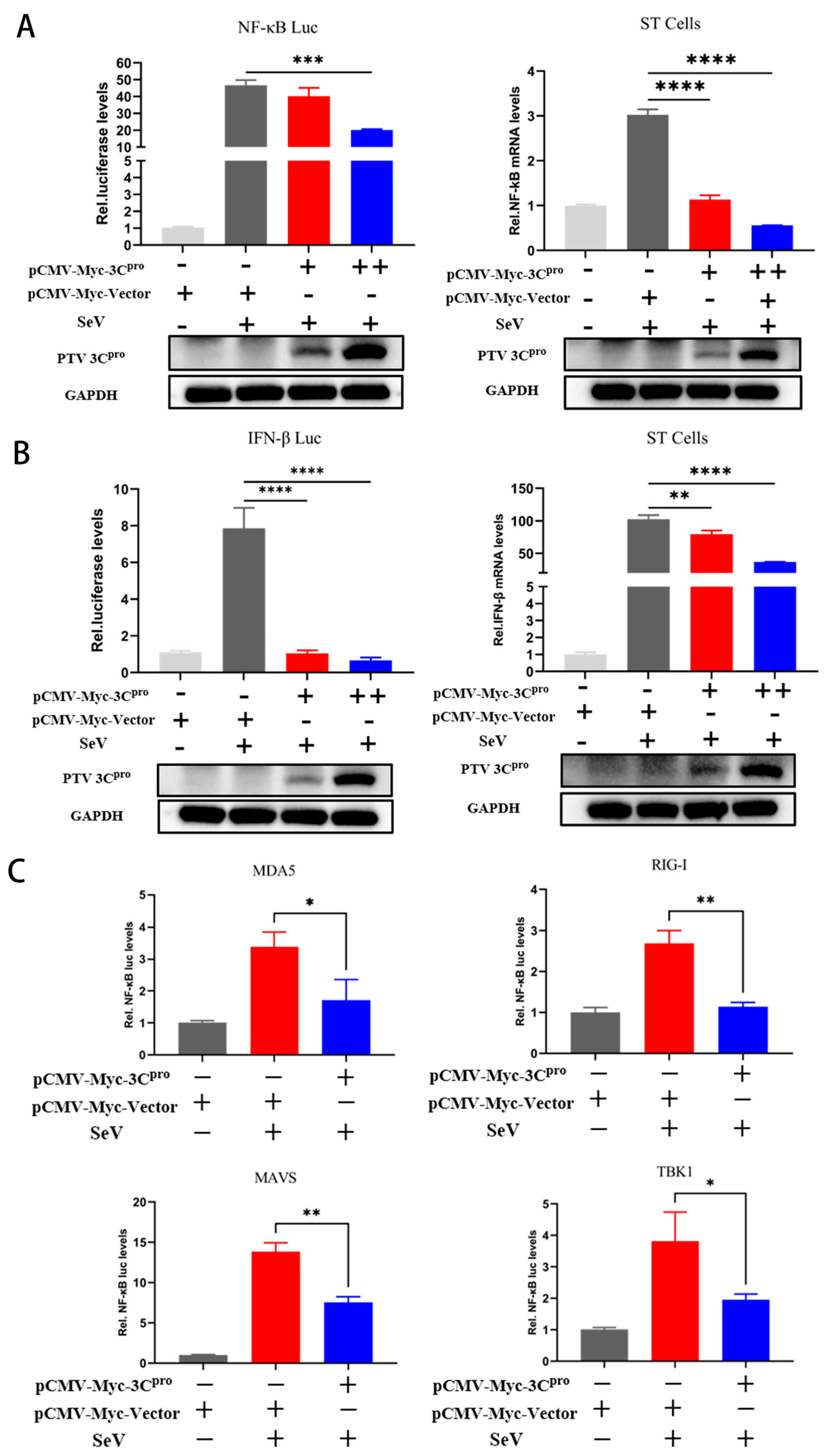
The 3Cpro protease in various Picornaviridae strains usually has an inhibitory effect on the IFN-β pathway. To investigate whether PTV 3Cpro similarly possesses this ability, we conducted a series of experimental analyses. A dual-luciferase reporter assay was used to evaluate the impact of 3Cpro on the activity of the NF-κB and IFN-β promoters, which revealed that PTV 3Cpro inhibited the NF-κB and IFN-β promoter activity induced by SeV stimulation (Figure 2A). In addition, the results of fluorescent quantitative PCR revealed that under SeV stimulation, the mRNA levels of NF-κB and IFN-β in 3Cpro-transfected cells were lower than those in empty vector-transfected cells (Figure 2B). Furthermore, the effects of 3Cpro on NF-κB promoter activity induction by upstream signaling components was analyzed using a dual-luciferase reporter system. The NF-κB promoter activity induced by MDA5, RIG-I, MAVS, and TBK1 was similarly inhibited by PTV 3Cpro (Figure 2C). These data collectively establish PTV 3Cpro as a transcriptional suppressor of NF-κB, and we proceeded to evaluate whether it exerts its inhibitory effects by disrupting NF-κB phosphorylation and nuclear translocation.

3.3. PTV 3Cpro Inhibits the Phosphorylation and Translocation of NF-κB

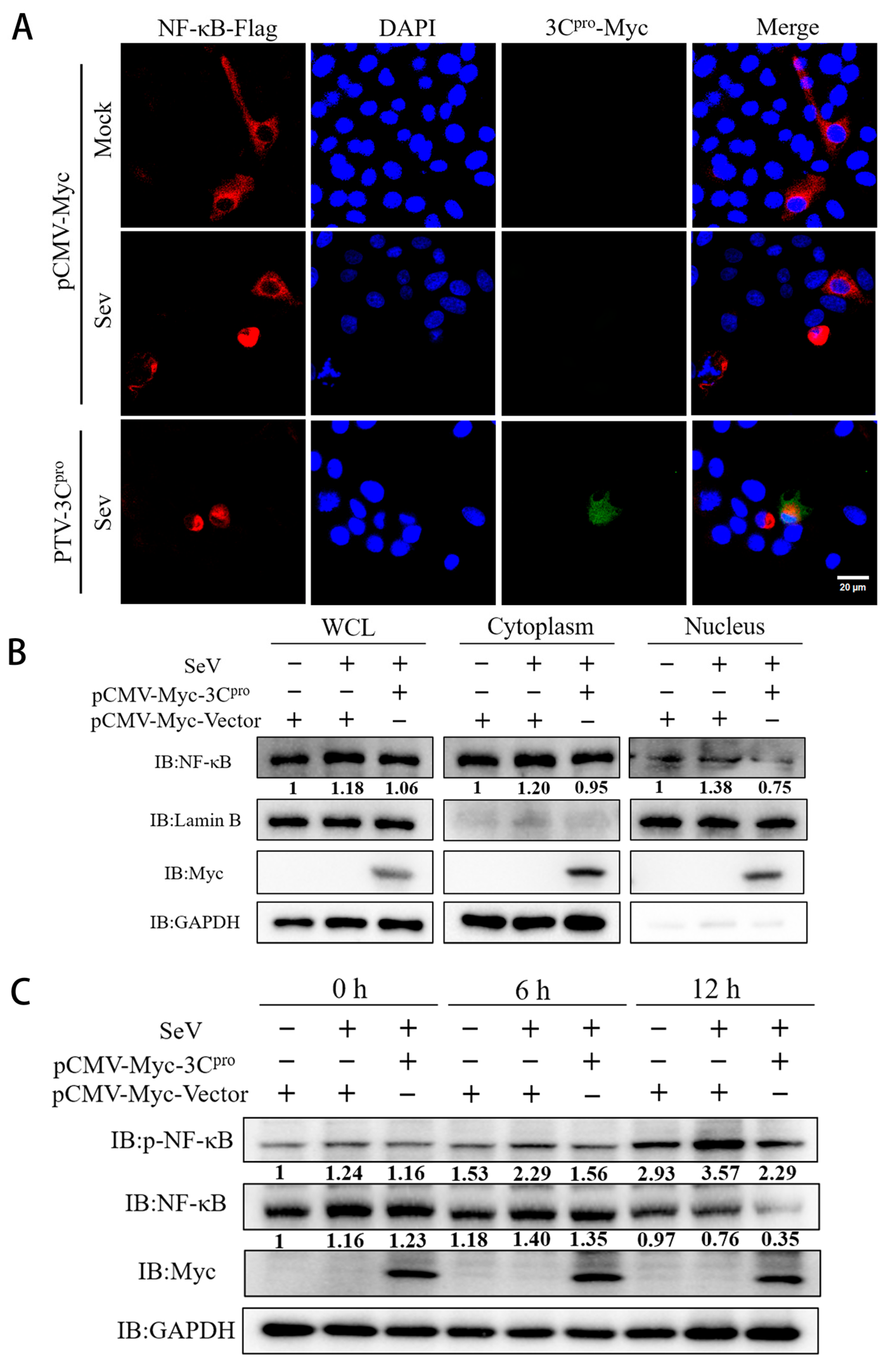
Typically, NF-κB activation involves its phosphorylation and translocation to the nucleus and binding to specific promoter sequences to exert its effects. Through indirect immunofluorescence experiments, 3Cpro inhibited the nuclear translocation of NF-κB induced by SeV stimulation (Figure 3A). An examination of NF-κB levels in the cytoplasm and nucleus revealed a significant reduction in NF-κB levels in both compartments (Figure 3B). The phosphorylation of NF-κB at Ser536, triggered by diverse stimuli, facilitates its nuclear translocation and increases target gene expression. After PTV 3Cpro was transfected into ST cells, the protein levels of NF-κB and phosphorylated NF-κB were analyzed using western blotting at different time points after SeV stimulation. The results showed that PTV 3Cpro decreased the protein levels of NF-κB and phosphorylated NF-κB (Figure 3C). Multiple experiments have demonstrated that 3Cpro can reduce the level of phosphorylated NF-κB, thus preventing its nuclear translocation.

3.4. Mechanism of 3Cpro-Mediated Degradation of NF-κB

In previous experiments, we reported that PTV 3Cpro decreases NF-κB protein levels. To verify the species specificity of PTV 3Cpro for NF-κB, PTV 3Cpro was transfected into 293T cells, where it was found to cleave the human NF-κB protein (H-NF-κB) (Figure 4A). Under the assumption that 3Cpro interacts with NF-κB or H-NF-κB, co-IP experiments revealed the interaction between NF-κB or H-NF-κB and 3Cpro (Figure 4B). The degradation and cleavage pathways were subsequently explored. The primary protein degradation pathways in eukaryotic cells include lysosomal, ubiquitin-proteasome, and caspase-mediated pathways. Cotransfection of 3Cpro and NF-κB (or H-NF-κB) was performed, and 24 h later, NH4Cl, MG132, and Z-VAD-FMK—which target lysosomes, proteasomes, and caspases, respectively—were added. The results revealed that NF-κB was inhibited in both species, but that the mechanisms of inhibition were different. NH4Cl, MG132, and Z-VAD-FMK did not block NF-κB degradation, whereas Z-VAD-FMK prevented H-NF-κB degradation but did not affect H-NF-κB cleavage (Figure 4C). Moreover, 3Cpro has serine/cysteine protease characteristics and features an active site comprising Cys-His residues. By comparing the amino acid sequences of PTV 3Cpro with those of other members of the Picornaviridae family, the catalytic site of PTV 3Cpro was identified (Figure 4D). Three mutant variants—H49A, C158A, and a double mutant (DM)—were subsequently generated for further investigation. The catalytic residues H49 and C158 were conserved across all the analyzed PTV genotypes (Figure S1). Next, PTV 3Cpro and its mutants were cotransfected with NF-κB (or H-NF-κB) into 293T cells. According to the western blotting results, the PTV 3Cpro mutant no longer cleaved or inhibited H-NF-κB nor NF-κB (Figure 4E). These findings suggest that the capacity of PTV 3Cpro to inhibit NF-κB depends on its protease activity.

3.5. The PTV 3Cpro Mutant Loses the Ability to Evade Innate Immunity

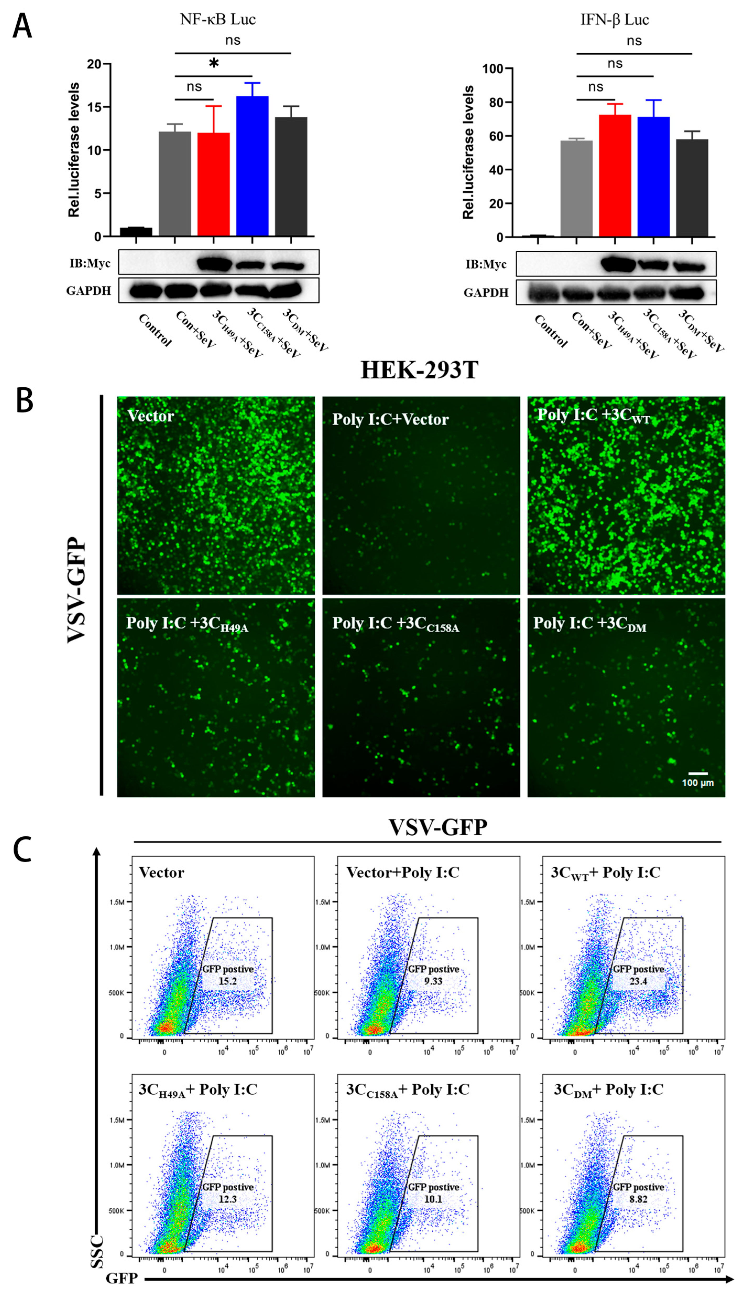
To further elucidate the immunomodulatory function of the 3Cpro mutant, the impact of the PTV 3Cpro mutant on IFN-β or NF-κB promoter activity was studied using a dual-luciferase reporter assay. The experimental findings revealed that site-directed mutagenesis at the enzymatic active site abrogated the ability of PTV 3Cpro to suppress both IFN-Luc and NF-κB-Luc promoter transcriptional activities (Figure 5A). Virus replication was assessed by measuring the fluorescence intensity of vesicular stomatitis virus (VSV-GFP). A significant reduction in GFP fluorescence intensity was observed in cells treated with poly(I:C), which activates NF-κB and IFN-β, suggesting that VSV-GFP replication was inhibited. However, GFP fluorescence intensity was recovered in cells transfected with 3Cpro, suggesting a partial restoration of VSV-GFP replication, whereas no recovery was observed when the mutant was expressed. Fluorescent cells were quantified using flow cytometry, and the results also revealed that cells transfected with PTV 3Cpro had greater fluorescence intensity than cells transfected with the mutant (Figure 5B,C). These results demonstrate that 3Cpro can suppress the activation of innate immunity by poly(I:C), but the mutant cannot suppress NF-κB activation and inhibit the innate immune response, thereby losing the ability to evade innate immunity and aid in virus replication.

4. Discussion

PTV is widely distributed among swine populations worldwide and is commonly found in the feces of asymptomatic pigs, revealing its ability to establish intestinal colonization without inducing clinical symptoms [22,23]. However, highly pathogenic PTV strains can cause severe clinical symptoms, including moderate to severe neurological symptoms, respiratory diseases, and inflammatory responses [22]. The primary mode of PTV transmission is through oral ingestion, but exposure to urine and feces can also lead to infection. Following oral infection, PTV primarily replicates in the tonsils and intestines [1]. Characteristic signs after 1–2 d of PTV infection include elevated body temperature, diarrhea, and flaccid and spasmodic paralysis lasting for approximately 10–11 d; respiratory failure represents the primary factor leading to mortality in infected hosts [24]. Moreover, the humoral immune response mediated by IgG and IgM antibodies plays a major role in combating PTV infections; locally produced IgA antibodies have been shown to have a protective effect when the virus enters through the oral cavity. The primary antibody class involved in fetal responses to the PTV is IgM, with IgG appearing as the next most common type [25,26].
In China, swine-related diseases caused by PTV were first reported in 2003 with the isolation of swine/CH/IMH/03 [27]. Since then, PTV strains have been sporadically discovered in several provinces in China, and infected swine typically had mild symptoms. Historically, high mortality from PTV infections has been associated mainly with the PTV-1 strain [12,28]. In 2021, a PTV-1 outbreak was reported in Northeast China, with an approximately 10% mortality rate among piglets [27,29]. Additionally, a significant outbreak of PTV-2 was documented by Wenqi Liang in 2023 at a pig farm located in Henan Province, China [30]. The emergence of the HeNZ1 strain resulted in severe neurological symptoms and diarrhea among piglets, along with very high morbidity and mortality rates among suckling pigs, marking the first reported serious outbreak caused by the PTV-2 strain [30]. Research on how PTV-2 evades the innate immune system response can help gain insights into its pathogenic mechanisms, leading to the development of effective treatment strategies to combat potential future outbreaks. Picornaviridae 3Cpro is a protease-like protein that contains a conserved His-Asp-Cys catalytic triad. This triad can cleave viral proteins to form mature proteins and promote viral replication by antagonizing host factors and innate immunity [15]. Although PTV has been causing substantial economic impacts on swine production for decades, investigations into its pathogenic mechanisms, particularly how it evades host innate immune responses, remain rare. In light of this gap, the present study focused on the potential immunomodulatory function of PTV 3Cpro in suppressing antiviral defense pathways.
We found that PTV infection does not increase the mRNA levels or activate the promoters of NF-κB and IFN-β in cells. Additionally, 3Cpro inhibits the promoter activity of NF-κB induced by upstream factors. Normally, phosphorylation of NF-κB at Ser536 leads to its nuclear translocation and binding to specific promoter sequences, activating the transcription of target genes [31]. This activation leads to the formation of a complex and the initiation of a partially overlapping transcriptional program, which in turn suppresses viral replication [32,33]. The pivotal role of the antiviral innate immune response in protecting cells from viral infections has been extensively demonstrated. For example, IFN-β is essential for the regulation of virus replication and has been shown to effectively reduce the viral load in cardiomyocytes infected with coxsackievirus B3 [34,35]. However, our experiments revealed that 3Cpro inhibits the phosphorylation and nuclear translocation of NF-κB through degradation. By studying the degradation mechanism of NF-κB, we found that PTV 3Cpro has different inhibitory effects on NF-κB in different species, such as cleaving human NF-κB and degrading porcine NF-κB. While Z-VAD-FMK, a pancaspase inhibitor, suppresses the degradation of H-NF-κB, it does not block its cleavage. The degradation of porcine NF-κB is unaffected by the lysosome inhibitor NH4CL, the proteasome inhibitor MG132, and the caspase inhibitor Z-VAD-FMK. Co-IP experiments revealed that 3Cpro interacts with both forms of NF-κB. The mechanism of PTV 3Cpro-mediated caspase 3 cleavage of NF-κB is species specific, suggesting that human NF-κB has a cleavage site that is recognized by 3Cpro, whereas the sequence of porcine NF-κB differs at this site.
Previous studies have highlighted the essential role of proteolytic activity in maintaining the functional integrity of the 3C protein. For example, inactivation of PSV 3Cpro prevents MAVS cleavage and abolishes its ability to degrade TBK1 and MDA5 [36]. Similarly, SVA 3Cpro loses both cleavage and degradation activities toward RIG-I and MAVS upon inactivation [21]. To explore this mechanism in PTV, we performed multiple sequence alignments between PTV 3Cpro and other members of the Picornaviridae family to identify conserved catalytic residues. On the basis of these findings, we generated site-directed mutants lacking protease activity. Functional assays revealed that inactivated PTV 3Cpro could no longer cleave or induce the degradation of NF-κB. Moreover, VSV-GFP experiments demonstrated that the mutant protein failed to suppress IFN-β signaling activation. Dual-luciferase assays revealed that the PTV 3Cpro mutant no longer inhibited the activity of the IFN-β promoter. Future research should validate these findings in in vivo porcine models to assess how 3Cpro-mediated immune suppression impacts viral pathogenesis and tissue tropism. Comparative analyses of PTV-1 and PTV-2 strains, including the HeNZ1 variant, are critical for determining whether genotype-specific 3Cpro activity is correlated with virulence and immune evasion efficiency. Reverse genetic approaches should generate 3Cpro-mutant PTV viruses to evaluate their impact on immune evasion; however, prior to assessing immune evasion, the effect of 3Cpro inactivation on viral replication must be established.
In summary, our findings demonstrate that PTV 3Cpro plays a critical role in evading the host innate immune response by binding with NF-κB, inhibiting its phosphorylation and nuclear translocation. Notably, we show that PTV 3Cpro-induced degradation of NF-κB is species specific. The results of mutagenesis studies further confirmed that the protease activity of 3Cpro is essential for these immunosuppressive functions. These results provide novel insights into the immune evasion strategies of PTV and highlight the importance of 3Cpro as a potential target for antiviral intervention.

Supplementary Materials

The following supporting information can be downloaded at: https://www.mdpi.com/article/10.3390/microorganisms13061209/s1, Figure S1: Cross-genotypic sequence alignment.

Author Contributions

Methodology, X.-Y.Z., Y.-Y.L. and W.-C.S.; software, X.-Y.Z., Y.-Y.L. and W.C.; validation, X.-Y.Z., Y.-Y.L., W.C., L.-L.X. and Y.-Q.H.; formal analysis, X.-Y.Z., Y.-M.Z. and W.C.; resources, W.-C.S. and T.L.; data curation, X.-Y.Z. and W.-C.S.; writing—original draft preparation, X.-Y.Z. and Y.-M.Z.; writing—review and editing, Y.-Q.H., Y.Q., H.-X.H., L.Z., T.L. and W.-C.S.; visualization, X.-Y.Z., Y.-M.Z., L.-L.X. and Y.-Q.H.; supervision, T.L. and W.-C.S.; project administration, W.-C.S.; funding acquisition, W.-C.S. All authors have read and agreed to the published version of the manuscript.

Funding

This work was supported by the Wenzhou Basic Medical Health Science and Technology Project (Grant Number Y20220136).

Institutional Review Board Statement

Not applicable.

Informed Consent Statement

Not applicable.

Data Availability Statement

The data that support the findings of this study are available from the corresponding author upon reasonable request.

Conflicts of Interest

The authors declare no conflict of interest.

References

  1. Chiu, S.C.; Hu, S.C.; Chang, C.C.; Chang, C.-Y.; Huang, C.-C.; Pang, V.F.; Wang, F.-I. The role of porcine teschovirus in causing diseases in endemically infected pigs. Vet. Microbiol. 2012, 161, 88–95. [Google Scholar] [CrossRef] [PubMed]
  2. Patel, S.K.; Agrawal, A.; Pathak, M.; Singh, A.; Varshney, R.; Rana, J.; Saikumar, G. Detection of porcine enteric picornaviruses from faecal samples of Indian pigs. Virusdisease 2022, 33, 102–107. [Google Scholar] [CrossRef] [PubMed]
  3. Villanova, F.; Cui, S.; Ai, X.; Leal, É. Analysis of full-length genomes of porcine teschovirus (PTV) and the effect of purifying selection on phylogenetic trees. Arch. Virol. 2016, 161, 1199–1208. [Google Scholar] [CrossRef] [PubMed]
  4. Yang, T.; Lu, Y.; Zhang, L.; Li, X. Identification of novel genotypes belonging to the species Teschovirus A from indigenous pigs in western Jiangxi, China. Arch. Virol. 2020, 165, 993–1001. [Google Scholar] [CrossRef]
  5. Trefny, L. Massive illness of swine in Teschen area. Zveroleki Obz. 1930, 23, 235–236. [Google Scholar]
  6. Izawa, H.; Bankowski, R.A.; Howarth, J.A. Porcine enteroviruses. I. Properties of three isolates from swine with diarrhea and one from apparently normal swine. Am. J. Vet. Res. 1962, 23, 1131–1141. [Google Scholar]
  7. Yamada, M.; Nakamura, K.; Kaku, Y.; Yoshii, M.; Haritani, M.; Kozakura, R.; Ikegami, R. Enterovirus encephalomyelitis in pigs in Japan caused by porcine teschovirus. Vet. Rec. 2004, 155, 304–306. [Google Scholar] [CrossRef]
  8. Ray, P.K.; Desingu, P.A.; Anoopraj, R.; Singh, R.K.; Saikumar, G. Identification and genotypic characterization of porcine teschovirus from selected pig populations in India. Trop. Anim. Health Prod. 2020, 52, 1161–1166. [Google Scholar] [CrossRef]
  9. Feng, L.; Shi, H.Y.; Liu, S.W.; Wu, B.P.; Chen, J.F.; Sun, D.B.; Tong, Y.E.; Fu, M.S.; Wang, Y.F.; Tong, G.Z. Isolation and molecular characterization of a porcine teschovirus 1 isolate from China. Acta Virol. 2007, 51, 7–11. [Google Scholar]
  10. Salles, M.W.; Scholes, S.F.; Dauber, M.; Strebelow, G.; Wojnarowicz, C.; Hassard, L.; Acton, A.C.; Bollinger, T.K. Porcine teschovirus polioencephalomyelitis in western Canada. J. Vet. Diagn. Investig. 2011, 23, 367–373. [Google Scholar] [CrossRef]
  11. Bangari, D.S.; Pogranichniy, R.M.; Gillespie, T.; Stevenson, G.W. Genotyping of Porcine teschovirus from nervous tissue of pigs with and without polioencephalomyelitis in Indiana. J. Vet. Diagn. Investig. 2010, 22, 594–597. [Google Scholar] [CrossRef] [PubMed]
  12. Deng, M.Y.; Millien, M.; Jacques-Simon, R.; Flanagan, J.K.; Bracht, A.J.; Carrillo, C.; Barrette, R.W.; Fabian, A.; Mohamed, F.; Moran, K.; et al. Diagnosis of Porcine teschovirus encephalomyelitis in the Republic of Haiti. J. Vet. Diagn. Investig. 2012, 24, 671–678. [Google Scholar] [CrossRef] [PubMed]
  13. Samuel, C.E. Antiviral actions of interferons. Clin. Microbiol. Rev. 2001, 14, 778–809. [Google Scholar] [CrossRef] [PubMed]
  14. Mondal, S.; Sarvari, G.; Boehr, D.D. Picornavirus 3C Proteins Intervene in Host Cell Processes through Proteolysis and Interactions with RNA. Viruses 2023, 15, 2413. [Google Scholar] [CrossRef]
  15. Ng, C.S.; Stobart, C.C.; Luo, H. Innate immune evasion mediated by picornaviral 3C protease: Possible lessons for coronaviral 3C-like protease? Rev. Med. Virol. 2021, 31, 1–22. [Google Scholar] [CrossRef]
  16. Fernandes, M.H.V.; Maggioli, M.F.; Otta, J.; Joshi, L.R.; Lawson, S.; Diel, D.G. Senecavirus A 3C Protease Mediates Host Cell Apoptosis Late in Infection. Front. Immunol. 2019, 10, 363. [Google Scholar] [CrossRef]
  17. Wen, W.; Yin, M.; Zhang, H.; Liu, T.; Chen, H.; Qian, P.; Hu, J.; Li, X. Seneca Valley virus 2C and 3C inhibit type I interferon production by inducing the degradation of RIG-I. Virology 2019, 535, 122–129. [Google Scholar] [CrossRef]
  18. Lei, X.; Sun, Z.; Liu, X.; Jin, Q.; He, B.; Wang, J. Cleavage of the adaptor protein TRIF by enterovirus 71 3C inhibits antiviral responses mediated by Toll-like receptor 3. J. Virol. 2011, 85, 8811–8818. [Google Scholar] [CrossRef]
  19. Huang, L.; Xiong, T.; Yu, H.; Zhang, Q.; Zhang, K.; Li, C.; Hu, L.; Zhang, Y.; Zhang, L.; Liu, Q.; et al. Encephalomyocarditis virus 3C protease attenuates type I interferon production through disrupting the TANK-TBK1-IKKε-IRF3 complex. Biochem. J. 2017, 474, 2051–2065. [Google Scholar] [CrossRef]
  20. Li, Y.; Zhang, X.; Zhao, B.; Zhao, C.; Lei, X.; Huang, H.; Li, C.; Zheng, M.; Lan, T.; Sun, W.; et al. The VP1 Protein of Porcine Teschovirus Inhibits the Innate Immune Response to Viral Infection by Blocking MDA5 Activation. Transbound. Emerg. Dis. 2024, 2024, 6649669. [Google Scholar] [CrossRef]
  21. Gu, Y.; Zhou, Y.; Shi, X.; Xin, Y.; Shan, Y.; Chen, C.; Cao, T.; Fang, W.; Li, X. Porcine teschovirus 2 induces an incomplete autophagic response in PK-15 cells. Arch. Virol. 2018, 163, 623–632. [Google Scholar] [CrossRef] [PubMed]
  22. Vreman, S.; Caliskan, N.; Harders, F.; Boonstra, J.; Peperkamp, K.; Ho, C.K.Y.; Kuller, W.; Kortekaas, J. Two novel porcine teschovirus strains as the causative agents of encephalomyelitis in the Netherlands. BMC Vet. Res. 2020, 16, 51. [Google Scholar] [CrossRef]
  23. Hammerschmitt, M.E.; de Almeida, P.R.; de Cecco, B.S.; Lorenzett, M.P.; Schwertz, C.I.; da Cruz, R.A.S.; Caprioli, R.A.; Schuh, D.T.; Demoliner, M.; Eisen, A.K.A.; et al. Swine polioencephalomyelitis in Brazil: Identification of Teschovirus A, Sapelovirus A, and Enterovirus G in a farm from Southern Brazil. Braz. J. Microbiol. 2021, 52, 1617–1622. [Google Scholar] [CrossRef] [PubMed]
  24. Stadler, J.; Junker, S.; Gründl, J.; Fröhlich, S.; Beisl, M.; Zöls, S.; Ritzmann, M.; Eddicks, M.; Palzer, A.; Sehl, J.; et al. Hinterhandlähmungen bei Mastschweinen im Zusammenhang mit einem neuen Stamm des porzinen Teschovirus A11 [Hind limb paralysis in fattening pigs due to a new strain of porcine Teschovirus A11]. Tierarztl. Prax. Ausg. G. Grosstiere Nutztiere. 2022, 50, 59–67. (In German) [Google Scholar] [PubMed]
  25. Wang, J.T.; Dunne, H.W.; Griel, L.C.; Hokanson, J.F.; Murphy, D.M. Mortality, antibody development, and viral persistence in porcine fetuses incubated in utero with SMEDI (entero-) virus. Am. J. Vet. Res. 1973, 34, 785–791. [Google Scholar] [CrossRef]
  26. Huang, J.; Gentry, R.F.; Zarkower, A. Experimental infection of pregnant sows with porcine enteroviruses. Am. J. Vet. Res. 1980, 41, 469–473. [Google Scholar] [CrossRef] [PubMed]
  27. Ma, H.; Zhang, M.; Wu, M.; Ghonaim, A.; Fan, S.; He, Q. Isolation and genetic characteristics of a neurotropic teschovirus variant belonging to genotype 1 in northeast China. Arch. Virol. 2021, 166, 1355–1370. [Google Scholar] [CrossRef]
  28. Kaku, Y.; Murakami, Y.; Sarai, A.; Wang, Y.; Ohashi, S.; Sakamoto, K. Antigenic properties of porcine teschovirus 1 (PTV-1) Talfan strain and molecular strategy for serotyping of PTVs. Arch. Virol. 2007, 152, 929–940. [Google Scholar] [CrossRef]
  29. Yang, T.; Yu, X.; Luo, B.; Yan, M.; Li, R.; Qu, T.; Ren, X. Epidemiology and molecular characterization of porcine teschovirus in Hunan. China Transbound. Emerg. Dis. 2018, 65, 480–490. [Google Scholar] [CrossRef]
  30. Liang, W.; Wu, X.; Ding, Z.; Zhong, S.; Qian, X.; Ye, P.; Liu, H.; Chen, Z.; Zhang, J.; Cao, H.; et al. Identification of a novel porcine Teschovirus 2 strain as causative agent of encephalomyelitis in suckling piglets with high mortality in China. BMC Vet. Res. 2023, 19, 2. [Google Scholar] [CrossRef]
  31. Huang, B.; Yang, X.D.; Lamb, A.; Chen, L.F. Posttranslational modifications of NF-kappaB: Another layer of regulation for NF-kappaB signaling pathway. Cell. Signal. 2010, 22, 1282–1290. [Google Scholar] [CrossRef] [PubMed]
  32. Chiang, J.J.; Davis, M.E.; Gack, M.U. Regulation of RIG-I-like receptor signaling by host and viral proteins. Cytokine Growth Factor Rev. 2014, 25, 491–505. [Google Scholar] [CrossRef] [PubMed]
  33. Schneider, W.M.; Chevillotte, M.D.; Rice, C.M. Interferon-stimulated genes: A complex web of host defenses. Annu. Rev. Immunol. 2014, 32, 513–545. [Google Scholar] [CrossRef] [PubMed]
  34. Pestka, S.; Krause, C.D.; Walter, M.R. Interferons, interferon-like cytokines, and their receptors. Immunol Rev. 2004, 202, 8–32. [Google Scholar] [CrossRef]
  35. Burke, J.D.; Sonenberg, N.; Platanias, L.C.; Fish, E.N. Antiviral effects of interferon-β are enhanced in the absence of the translational suppressor 4E-BP1 in myocarditis induced by Coxsackievirus B3. Antivir. Ther. 2011, 16, 577–584. [Google Scholar] [CrossRef]
  36. Yin, M.; Wen, W.; Wang, H.; Zhao, Q.; Zhu, H.; Chen, H.; Li, X.; Qian, P. Porcine Sapelovirus 3Cpro Inhibits the Production of Type I Interferon. Front. Cell. Infect. Microbiol. 2022, 12, 852473. [Google Scholar] [CrossRef]
Figure 1. PTV infection inhibits the activity of the NF-κB and IFN-β promoters. (A) ST cells were cotransfected with NF-κB-Luc or IFN-β-Luc (1000 ng) and PRL-TK (50 ng), followed by 24 h of infection with PTV (MOI = 1, 5) or SeV (MOI = 1), and the fluorescence intensity was measured. (B) ST cells were cotransfected with NF-κB-Luc or IFN-β-Luc (1000 ng) and PRL-TK (50 ng), followed by 24 h of induction with SeV (MOI = 1) and 24 h of infection with PTV (MOI = 1, 5), and the fluorescence intensity was measured. **** p < 0.0001, ns indicates no significant difference.
Microorganisms 13 01209 g001
Figure 2. PTV 3Cpro inhibits NF-κB and IFN-β mRNA expression and promoter activity. (A,B) Cotransfection of NF-κB-Luc or IFN-β-Luc (1000 ng) with PRL-TK (50 ng) and 3Cpro (200 ng, 500 ng) into 293T cells, followed by 12 h of induction with SeV (MOI = 1) and measurement of firefly luciferase activity, normalized to Renilla luciferase activity. 3Cpro (200 ng, 500 ng) was transfected into ST cells, which were then induced with SeV (MOI = 1) for 12 h, and the NF-κB or IFN-β mRNA levels were measured using fluorescence quantitative PCR. (C) NF-κB-Luc (1000 ng) with PRL-TK (50 ng) and 3Cpro (500 ng) were cotransfected into 293T cells, followed by cotransfection of MDA5, RIG-I, TBK1, and MAVS (700 ng) to induce promoter activity. After 24 h, firefly luciferase activity, normalized to Renilla luciferase activity, was measured. * p < 0.05, ** p < 0.01, *** p < 0.001, and **** p < 0.0001.
Microorganisms 13 01209 g002
Figure 3. PTV 3Cpro inhibits NF-κB phosphorylation and nuclear translocation. (A) NF-κB (500 ng) and PTV 3Cpro (500 ng) were introduced into ST cells that were infected with SeV after 24 h, and protein fluorescence was observed through an indirect immunofluorescence assay. (B) PTV 3Cpro (500 ng) was introduced into ST cells infected with SeV after 24 h, and cytoplasmic and nuclear proteins were extracted to measure the levels of NF-κB. GAPDH and Lamin B proteins were used as internal references. Quantitative display is achieved through standardization of the ratio of NF-κB/internal references. (C) PTV 3Cpro (500 ng) was transfected into ST cells, followed by SeV (MOI = 1) stimulation after 12 h, and the cell lysate was collected at 0 h, 6 h, and 12 h. Western blotting was performed to detect the protein levels of NF-κB and phosphorylated NF-κB. GAPDH protein was used as the internal reference. Quantitative display is achieved through standardization of the ratio of p-NF-κB (NF-κB) to GAPDH.
Microorganisms 13 01209 g003
Figure 4. Mechanism of 3Cpro-mediated degradation of NF-κB. (A) PTV 3Cpro (1000 ng) was transfected into ST or 293T cells, which were subsequently incubated for 24 h, and NF-κB protein levels were detected using western blotting. The asterisk (*) denotes the band corresponding to H-NF-κB cleaved by PTV 3Cpro. (B) 293T cells were cotransfected with NF-κB (or H-NF-κB) (1000 ng) and the 3Cpro plasmid (300 ng), and co-IP and western blotting were subsequently performed to measure the protein levels after 24 h of incubation. (C) 293T cells were cotransfected with NF-κB (or H-NF-κB) (1000 ng) and the 3Cpro plasmid (1000 ng), followed by 12 h of inhibition treatment and western blotting detection of protein levels. (D) Comparative sequence analysis of PTV 3Cpro with other members of the Picornaviridae family revealed the conserved catalytic site. On the basis of the enzyme activity sites of other viruses, H49 and C158 were predicted to be the enzyme activity sites of PTV 3Cpro. The asterisks (*) were used to separate consecutive 10-sequence intervals. (E) NF-κB (or H-NF-κB) (1000 ng), PTV 3Cpro, and its mutant (1000 ng) were transfected into 293T cells, which were then cultured for 24 h, after which NF-κB expression was assessed using western blotting. The asterisk (*) denotes the band corresponding to H-NF-κB cleaved by PTV 3Cpro.
Microorganisms 13 01209 g004
Figure 5. The PTV 3Cpro mutant cannot antagonize innate immunity. (A) In 293T cells, NF-κB-Luc or IFN-β-Luc (1000 ng) and PRL-TK (50 ng) were cotransfected with 3CH49A, 3CC158A, or 3CDM (500 ng), followed by SeV induction for 12 h prior to dual-luciferase activity measurement. (B,C) PTV 3Cpro and the variant (1000 ng) were transfected into 293T cells, which were subsequently subjected to VSV infection and poly(I:C) stimulation for 12 h, fluorescence observation, and quantification using fluorescence microscopy and flow cytometry. * p < 0.05, ns indicates no significant difference.
Microorganisms 13 01209 g005
Table 1. Primers for plasmid construction.
PrimerSequence
NF-κB-FgatgacgacgataaggaattcATGGACGACCTCTTCCCCC
NF-κB-RattaagatctgctagctcgagTTAGGAGCTGATCTGACTCAGAAGG
PTV-3C-FtctgaagaggacttggaattcGGACCAAAGGGACAAGCTAACA
PTV-3C-RccgcggccgcggtacctcgagTTGAAATTCCAAAAATCCCTCAA
Table 2. Primers for SYBR Green I qPCR assays.
PrimerSequence
Q-GAPDH-FAGCAACAGGGTGGTGGACCT
Q-GAPDH-RCTGGGATGGAAACTGGAAGT
Q-NF-κB-FCCTGAGGCTATAACTCGCTTGG
Q-NF-κB-RGTCCGCAATGGAGGAGAAGT
Q-IFN-β-FCATCCTCCAAATCGCTCTCC
Q-IFN-β-RACATGCCAAATTGCTGCTCC
Table 3. Gene-specific mutagenesis primers.
PrimerSequence
PTV-H49A-FTCTGATCAATACTgcTATTTTGACAGGTATAAA
PTV-H49A-RTTTATACCTGTCAAAATAgcAGTATTGATCAGA
PTV-C158A-FACGGCTATgcCGGCTCTGTGATGGTTGCGGATGCTGGAG
PTV-C158A-RCTCCAGCATCCGCAACCATCACAGAGCCGgcATAGCCGT
Disclaimer/Publisher’s Note: The statements, opinions and data contained in all publications are solely those of the individual author(s) and contributor(s) and not of MDPI and/or the editor(s). MDPI and/or the editor(s) disclaim responsibility for any injury to people or property resulting from any ideas, methods, instructions or products referred to in the content.