Article

Phycobacteria Biodiversity, Selected Isolation, and Bioactivity Elucidation of New Bacterial Species of Highly Toxic Marine Dinoflagellate Alexandrium minutum amtk4

by
Xiaoling Zhang
,
Zekang Pan
,
Jinkai Zhang
,
Bingqian Liu
and
Qiao Yang
*
ABI Group, Phycosphere Microbiology Laboratory, College of Marine Science and Technology, Zhejiang Ocean University, Zhoushan 316022, China
*
Author to whom correspondence should be addressed.
Microorganisms 2025, 13(6), 1198; https://doi.org/10.3390/microorganisms13061198
Submission received: 18 April 2025 / Revised: 8 May 2025 / Accepted: 22 May 2025 / Published: 24 May 2025

Abstract

:
Phycosphere niches host rich, unique microbial consortia that harbor complex algae–bacteria interactions with fundamental significance in underpinning most functions of aquatic ecological processes. Therefore, harvesting the uncultured phycobacteria is crucial for understanding the intricate mechanisms governing these dynamic interactions. Here, we characterized and compared microbial community composition of the phycosphere microbiota from six harmful algal bloom-forming marine dinoflagellates, Alexandrium spp., and their bacterial associations. Furthermore, based on a combinational enhanced cultivation strategy (CECS) procedure for the selected isolation for cultivable phycobacteria, a new yellow-pigmented bioactive bacterium designated ABI-6-9 was successfully recovered from cultivable phycobacteria of the highly toxic A. minutum strain amtk4. An additional phylogenomic analysis fully identified this new isolate as a potential novel species of the genus Mameliella within the family Roseobacteraceae. The bioactivity evaluation observed that strain ABI-6-9 can significantly promote the cell growth of its algal host and altered the gonyautoxin accumulation profiles in the co-culture circumstance. Additionally, the bacterial production of active bioflocculanting exopolysaccharides (EPSs) by strain ABI-6-9 was also measured after culture optimization. Thus, these findings revealed the potential environmental and biotechnological implications of this new microalgae growth- promoting phycobacterium.

1. Introduction

Billion-year-old algae are pivotal primary producers in global aquatic ecosystems and fundamental to life on Earth [1]. Hence, the phycosphere niche, as a microscopic-scale and fluid environment, hosts unique, closely associated microbial inhabitants surrounding the algal cell [2]. They are a unique and widely distributed microbial community, and thus form the algal microbiome within the phytoplankton holobiont called the phycobiont as one core concept of phycosphere microbiology [3]. The phycosphere microbial consortium harbors complex algae–bacteria interactions (ABI), in which nutrient trafficking and signaling molecules are central to these dynamic relationships [4]. Within this metabolic hot spot and interactive interface, exopolysaccharides or extracellular polysaccharides (EPSs) secreted by both interactive sides are regarded as vital component of the algae–bacteria joint extracellular matrix (ECM), which embeds proliferating cells, promotes the formation of a complex and interconnected ECM, facilitates aggregation both during phytoplankton blooms in the nature and in laboratory cultures [5] and colony formation, and physically defines the phycosphere [6]. These interactions usually span mutualism, commensalism, antagonism, parasitism, and competition [7]. The interplaying process often involves cross-kingdom, inter- or intra-species exchanges of nutrients, secondary metabolites, infochemicals, and ever vital gene transfer agents (GTAs). Thus, it underpins most functions of aquatic ecological processes, including primary production, marine snow formation, biogeochemical cycles, and the microbial loop in the oceans [8]. Moreover, it is also a critical player in regulating the biosynthesis of the phycotoxins such as paralytic shellfish poisoning toxins (PSTs) derived from various dinoflagellate species during harmful algal blooms (HABs) [9,10,11]. Regarding those HAB-forming dinoflagellates, the genus Alexandrium is a well-known group due to its significant impacts on both environmental and human health because of phycotoxins such as PSTs [12]. Moreover, it is worth noting that, due to chemical complexity and bioactivity with remarkable selectivity and ligand affinity toward varied pharmacological targets, these natural neurotoxins provide a successful and promising future for new drug development, such as analgesic pharmaceuticals [7,8,13,14,15].
Microorganisms with a huge biomass and species are an indispensable part of mankind’s production and the life of human civilization [16]. Although pure microbial culture was invented nearly one and a half centuries ago, the phenomenon of “the great plate count anomaly” remains the main obstacle for successfully transferring rich microbial resources to fully serve human society’s development [17]. Currently, there are still only a minority of microorganisms that can be cultured under laboratory conditions, thus leaving the vast bioprospecting potential of the uncultured microorganisms, which are called microbial dark matter (MDM), largely unexplored [18,19,20]. Modern multiomic techniques, such as genomics, transcriptomics, proteomics and metabolomics, etc., have gained increasingly rapid development; however, they still cannot fully help to answer those basic and pivotal questions about the microbes, such as what nutrients the bacteria need, what metabolites they produce, and how they interact with other living organisms in nature [21,22,23,24,25]. In order to find satisfactory answers to these questions, microbiologists are still dedicated to first isolate and culture these microorganisms in the laboratory [26]. So, continuous progress was achieved for microbial culturability enhancement, and new cultivation technologies were successively invented and applied, including co-culture [27,28,29], high-throughput culture [30], trap in situ culture screening [31], and soil matrix membrane system [32]. The invention and application of these innovative technologies significantly increased microbial culturability, resulted in the discovery of huge number of new microbial species, and consequently improved microbial species diversity and promoted greater microbial resource development and wide practical applications during the development of phycosphere microbiology [33].
For the phycobacteria community, it is crucial to expand the ability to cultivate phycobacteria to enhance the understanding of the underlying interactive mechanisms between algae and phycobacteria and promote their multiple applications for human welfare [34]. Previously, we have performed the Phycosphere Microbiome Project (PMP) to characterize the microbial consortium composition of diverse HAB-forming marine dinoflagellates [35,36,37,38,39,40,41,42,43]. In this study, we presented the comparative study of the characterizations of the phycosphere microbiota (PM) of six marine dinoflagellate Alexandrium spp. and identified potential bacterial associations. Furthermore, we isolated and conducted a phylogenetic study of a new bacterium designated ABI-6-9 using the combinational enhanced cultivation strategy (CECS) for the selected isolation of cultivable phycobacteria [20,21,22,23,24,25,26]. Additionally, various bioactivities of strain ABI-6-9 were measured, and revealed its multiple roles as a new microalgae growth-promoting bacterium (MGPB) [12,35] and a producer of bacterial bioflocculanting EPSs derived from the phycosphere niche.

2. Materials and Methods

2.1. Algal Strains and Culture

Six strains of the marine dinoflagellate Alexandrium spp. were used in this study. These algal strains were all sampled during the occurrence of natural HABs [44]. A. tamarense strain AT260 was obtained from the Yangtze Estuary in the East China Sea [45], A. minutum amtk4 and AMKS5 were isolated from southern Taiwan [46,47,48]; A. catenella ACHQ and ACHK were originally isolated from the Qingdao sea area in the East China Sea and Hongkong in South China Sea, respectively. A. tamarense strain AT11 was isolated from Ireland, UK. The dinoflagellates used in this study were cultured at 20–25 °C in F/2 media with 12L/12D cycles under a light intensity of approximately 25 μmol photons m−2 s−1 [49,50]. Obtaining the axenic culture of A. minutum amtk4 for the algae–bacteria co-culture experiment was performed by repeated washing, lysozyme, SDS, and multiple antibiotic treatments, as described previously [51,52,53].

2.2. 16S rRNA Pyrosequencing Analysis

Total genomic DNA was extracted using Fast Bacterial Genomic DNA Extraction Kits (Merck, Shanghai, China) according to manufacturer’s instructions, and the extracted DNA concentration and purity were evaluated by a NanoDrop UV-Vis spectrophotometer (BioTek, Winooski, VT, USA) [54]. A general bacterial primer pair 338F/806R was used to amplify the V3-V4 variable region of bacterial 16S rRNA genes [54]. PCR products were purified by 2% (w/v) agarose gel electrophoresis. The library was constructed using a TruSeq® DNA PCR-Free Sample Preparation Kit (Illumina, San Diego, CA, USA) purchased from Illumina, and then sequenced using NovaSeq 6000 PE250 platform [55]. Data analysis was performed using qiime2 document (https://docs.qiime2.org, accessed on 3 March 2025). The parameters used were all the default settings. Based on the relative abundance of the main microbial species in the samples, the co-occurrence network analysis was used to calculate the Spearman rank correlation coefficient. The network diagram was drawn using the “igraph” package [56].

2.3. Isolation of Cultivable Phycobacteria Using the CECS Procedure

For cultivable phycobacterial isolation, different isolation media were prepared for the combinational enhanced cultivation strategy (CECS), as previously described [57]. Briefly, all isolation media contained 5.24 g/L HEPES (Merck, Shanghai, China) as a buffering agent and 300 mL/L concentrated artificial seawater and were adjusted to pH 7.2 ± 0.1. After sterilization by autoclaving, the media were supplemented with 5.0 mL/L respective algal culture filter [58]. The nanoscale designed composite gel (DCG) was pre-treated with varied micro-nutrients according to the procedure previously described [59]. Each of the triplicate of 1 mL micro-wells were seeded with 10 μL of diluted bacterial cultures which were serially diluted of 1:10 by the dilution-to-extinction technique [60]. These microplates were then incubated at 28 °C for at least 3–4 weeks until visible bacterial colonies growing on the isolation plates were observed. The isolated strains were further purified, maintained on marine agar (MA; Difco, BD, Franklin Lakes, NJ, USA), and preserved at 4 °C as a solid slant for short-term preservation or as a glycerol suspension (25%, v/v) at −80 °C for long-term preservation.

2.4. Phylogenetic Analysis of the 16S rRNA Gene of Strain ABI-6-9

The phylogenetic identification and calculation of pairwise similarities of 16S rRNA gene sequences of the new bacterial strains were achieved using the EzTaxon-e server (http://eztaxon-e.ezbiocloud.net, accessed on 2 March 2025). The sequence alignments were performed using CLUSTAL_X (http://www.clustal.org, accessed on 1 March 2025). Phylogenetic trees were reconstructed with the neighbor-joining (NJ) algorithm using MEGA software (version 7.0) (https://www.megasoftware.net, accessed on 6 March 2025).

2.5. Phylogenomic Analysis by ANI, AAI, and dDDH Calculations

Phylogenomic calculations based on average nucleotide identity (ANI) analyses and average amino acid identity (AAI) were performed using the ANI/AAIMatrix Genome-based distance matrix calculator (http://enve-omics.ce.gatech.edu/g-matrix, accessed on 12 March 2025). The digital DNA-DNA hybridization (dDDH) analysis was performed using the Genome-to-Genome Distance Calculator (GGDC 2.1) (http://ggdc.dsmz.de, accessed on 10 March 2025). The phylogenomic tree was constructed using the UBCG pipeline [61].

2.6. Bacterial Growth Measurement

For bacterial culture optimization, 1 mL of a 24 h fresh bacterial culture of strain ABI-6-9 was taken and mixed with 24 mL of fresh 2216 medium. Then, 150 μL of the mixture was added into the wells of the 96-well microplate containing different carbon sources, and then cultured with shaking at 60 rpm/min [62]. During different culture periods, the change in bacterial growth was measured at OD600nm every four hours. Ten carbon sources, including cellobiose, fructose, galactose, glucose, glycerol, lactose, maltose, mannose, sucrose, and trehalose, and various pH values (5.0–9.0) were used for the measurements [58,62].

2.7. Characterization of the Monosaccharides of Bacterial EPSs

The extraction of bacterial EPSs and chemical elucidation of monosaccharides of crude polysaccharides were performed as previously described using an Agilent 1100 HPLC system (Agilent Technologies, Santa Clara, CA, USA) [63]. Analytical standards (HPLC purity ≧ 99%) of six monosaccharides were purchased from Merck (Shanghai, China).

2.8. Evaluation of Bioflocculation and MGP Bioactivity

Bioflocculating activity measurements of bacterial EPSs were performed according to the procedures reported previously [64]. The prepared EPSs were dissolved in distilled water for further measurements. The kaolin clay suspension flocculation (KCSF) assay calculated the bioflocculation rate (BR, %) and was used for the bioactivity measurement using 96-well microplates with at least six replicates per sample [65]. For the MGP activity assay, the preparation of axenic amtk4 cells for co-culture was performed as described previously [66]. Bacterial growth was recorded by monitoring OD600nm changes using a SpectraMax M2 model 96-well microplate reader (Molecular Devices, LLC, San Jose, CA, USA). Measuring the changes in cell numbers of algal strain amtk4 was performed using a Millicell® Disposable hemocytometer (Merck KGaA, Darmstadt, Germany).

2.9. Statistical Analysis

All the results are expressed as the means ± SDs. The correlation coefficients (R2) of the monosaccharide portions with bioflocculanting activities and the statistical significance were performed using Spearman’s rank order correlation analysis and plotted with the OriginPro (Version 10.0) (OriginLab Corp., Northampton, MA, USA). All the obtained results were expressed as means ± SDs. A p-value of less than 0.05 was considered to be statistically significant for all analyses.

3. Results and Discussion

3.1. Characterization of the PM Compositions of Six Alexandrium spp.

Based on the pyrosequencing analysis, the PM compositions of six Alexandrium spp. were compared. It could inferred that proteobacteria was the predominant group at the phylum level, with a relative abundance range of 32.2–97.2%. However, for A. tamarense AT.11, firmicutes was the most prevalent and constituted about 53.6% of the total PMs (Figure S1A). At family level, the Rhodobacteraceae was the most prevalent and occupied 10.1–62.1% of total PMs (Figure S1B). With the exception of toxic A. catenella ACHK, the family Flammeovirgaceae dominated the total PMs (25.5%), followed by Rhodobacteraceae (6.5%). Furthermore, among the top ten genera, Mameliella, Sulfitobacter, Dinoroseobacter, and Marivita showed significant differences between the toxic and non-toxic groups among the six Alexandrium spp. Regarding the second-dominant group of cyanobacteria, only one genus, Microcystis was found. It may suggest that cryptic cyanobacterial lineages may be concealed within the dinoflagellate host [54]. Additionally, it is noteworthy that about 16–64% of OTUs were classified as unidentified taxonomic groups (Figure S1B). It clearly indicates that the six PMs of Alexandrium spp. harbored great potential for discovering novel species with promising ecological and biological functions.
For the PM of highly toxic A. minutum amtk4, proteobacteria (80.9%) was identified as the dominant fraction, followed by bacteroidetes (17.9%) (Figure S1A). At family level, Alteromonadaceae occupied about 24.9% of the total PM, followed by rhizobiaceae (21.1%), rhodobacteraceae (16.4%), cyclobacteriaceae (16.3%), unidentified cohaesibacter (2.6%), and sphingomonadaceae (1.5%) (Figure S1B). Moreover, four dominant genera were identified: Mameliella (30.1%), Alteromonas (24.9%), Cohaesibacter (20.5%), and Ekhidna (15.8%) (Figure S1C). In particular, Mameliella exhibited a higher abundance. In addition, the highest portion of 64% of OTUs were classified as unidentified groups in the six PMs of Alexandrium spp. This considerable observation highlights the need for further investigation to fully understand complete diversity and potential roles of these PMs in the phycosphere ecosystem of HAB-causing marine Alexandrium spp.

3.2. Analysis of Bacterial Associations in the PMs of Six Alexandrium spp.

The co-occurrence of microorganisms can be modeled using a network analysis to illustrate complex microbial relationships and responses to variations in operational factors and suggest the clustering of sub-communities, and more detailed information also can be provided to reveal microbial interactions and their functional correlations with ecosystems [67]. Based on the co-occurrence network analysis, it is noteworthy that Mameliella, which was one abundant genera of the six PMs, showed significant positive correlations with Roseovarius, Thalassobacter, Phaeobacter, and Muricauda. Moreover, three dominant genera—Mameliella, Roseovarius, and Muricauda—demonstrated close positive associations with each other. Interestingly, it is also found that Mameliella demonstrated a relatively low abundance (<1.0%) in the non-toxic group (AT-11, AMKS5, and ACHQ). Otherwise, it was processed as absolutely abundant portions in the toxic group (AMTK4, AT 260, and ACHK). Considering the potential function of the Mameliella species during algae–bacteria interactions [68], the reason governing for this specific distribution pattern of the Mameliella is well worth further investigation.

3.3. CECS-Based Selected Isolation of Cultivable Phycobacterial Strains

The realization of pure cultures of phycobacteria provides a vital basis and necessary prerequisite for the successful construction of co-culture system and further explaining the detailed mechanisms governing algae–bacteria interactions [9,10,11]. To achieve this purpose, a selected isolation procedure named CECS was proposed in our previous study [25]. Based on CECS isolation, total 16 phycobacterial strains were recovered from the A. minutum amtk4 culture (Figure S3A). To determine the phylogenetic relationship, a phylogenetic tree using the 16S rRNA gene was constructed (Figure S3A). Strain ABI-6-9 was clustered with strain LZ-21, which was isolated from the toxic dinoflagellate A. catenella LZT-09 [25]. Among these new isolates, eight bacterial strains have been identified as novel species [13,35,40,41,42,43,44] (Figure S3B). This findings indicated that the proposed CECS procedure is indeed effective for harvesting the uncultured phycobacteria. Thereafter, it will lay solid basis for furthering studies on algae–bacteria interactions to fully understand their intricate mechanisms.

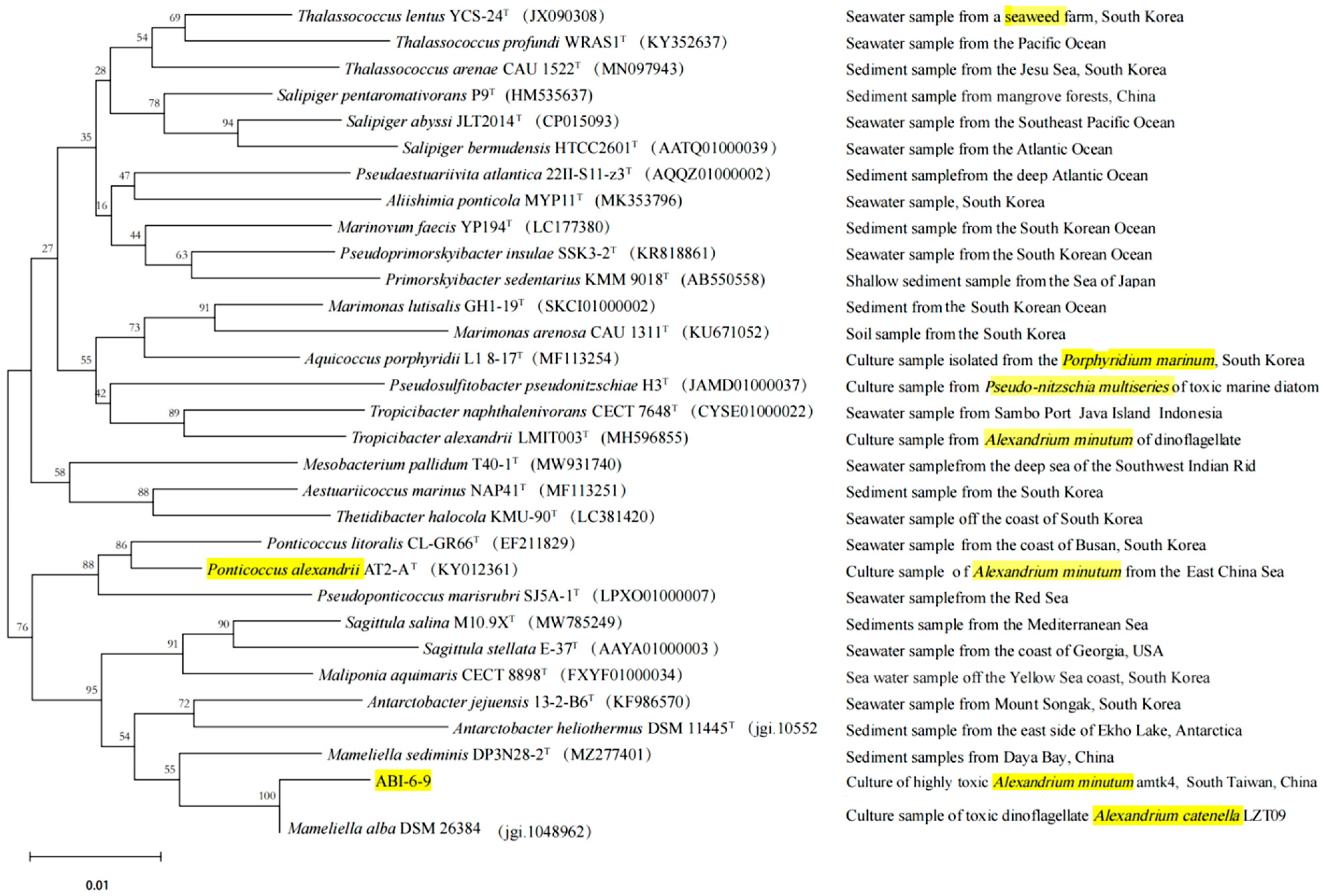
3.4. Phylogenetic Analysis of Strain ABI-6-9

The phylogenetic analysis based on the 16S rRNA gene revealed that strain ABI-6-9 exhibited the gene similarity of 97.98% with Mameliella alba JLT354-WT [68]. The value was less than the threshold value (98.65%) for novel species identification [58]. The phylogenetic tree was then constructed. It can be seen that strain ABI-6-9 formed a distinct separate phylogenetic line and clustered with M. alba JLT354-WT (Figure 1). Based on these data, it can be inferred that strain ABI-6-9 represents a potentially new species within the genus Mameliella of family Roseobacteraceae. Additionally, it is interesting to note that among the closely related type species of strain ABI-6-9, four bacteria, including Thalassococcus lentus YCS-24T, Pseudosulfitobacter pseudonitzschiae H3T, Tropicibacter alexandrii LMIT003T, and Ponticoccus alexandrii AT2-AT, were also isolated from phycosphere niches.

3.5. Phylogenomic Characterization of Strain ABI-6-9

To further infer the phylogenetic relationship of strain ABI-6-9, a phylogenomic tree using an up-to-date bacterial core gene set (UBCG) was performed. As shown in Figure S4, strain ABI-6-9 was phylogenetically attributed to the genus Mameliella and clustered with the type species of M. alba JLT354-WT. Additionally, three main phylogenomic parameters, including ANI, AAI, and dDDH values, were calculated as 93.7%, 92.1%, and 65.5%, respectively. All the values were below the thresholds (95–96% for ANI, 97% for AAI, and 70% for dDDH) generally accepted for new species delineations [34]. Accordingly, it confirmed that strain ABI-6-9 was a new member and a potential novel species of the genus Mameliella.

3.6. Microalgae Growth-Promoting Potential Analysis of Strain ABI-6-9

Recently, MGPBs have received considerable attention due to their potential to develop microalgae–bacteria co-culture strategies for improved efficiency and sustainability of the water–energy–environment nexus [69,70]. Based on MGP bioactivity assays, the cell numbers of amtk4 increased significantly when directly co-cultured with strain ABI-6-9, and demonstrated an MGP efficiency of at least 16.5 ± 3.8% (Figure 2). This observation clearly suggests potential close associations between strain ABI-6-9 and its algal host, while the precise nature of the mechanism remains unresolved. Previous studies have revealed that Mameliella alba can alleviate vitamin limitations of its algal host Karenia brevis, a toxigenic marine HAB-forming dinoflagellate, by providing vitamins B1, B7, and B12 and thus greatly contributes to algal growth dynamics [30,48]. Current ongoing research exploring associations under algae–bacteria co-culture conditions using multiomics analysis is expected to provide new insights into the mechanisms governing the interactions between strain ABI-6-9 and its algal host.

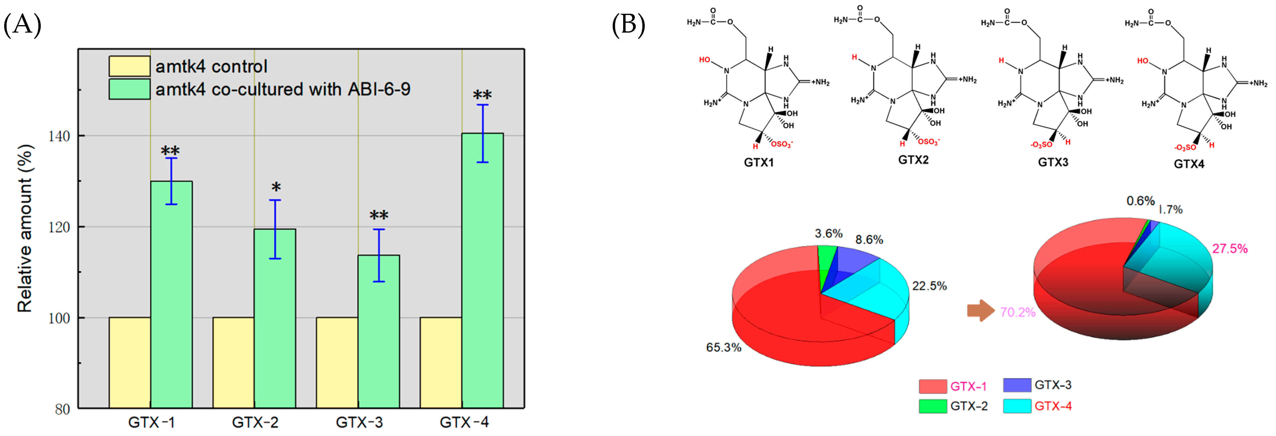
3.7. Promotion of the Accumulation of Algal GTXs by Algal–Bacterial Co-Culture

The HPLC analysis of algal GTX toxin production indicated that the total toxin production by algal amtk4 was about (1.83 ± 0.05) × 10−14 mol/cell, and that of the algal–bacterial co-culture was determined as about (2.48 ± 0.34) × 10−14 mol/cell (Figure 3A). It showed that the co-culture manner obviously promoted algal GTX accumulation up to about 1.36 times compared with that of algae alone. Furthermore, it is observed that algal–bacterial co-culture also changed the individual portions of algal GTXs (Figure 3B). Among the four algal GTX toxins produced, the contents of GTX-1 and GTX-4 were much higher than those of GTX-3 and GTX-2 under both culture conditions. However, in algal–bacterial co-culture, the contents of the GTX-1 and GTX-4 toxins (more toxic) increased from 87.8% to 97.7% (about 1.13 fold, p < 0.01) among the total toxins, Accordingly, those of the GTX-2 and GTX-3 toxins (less toxic) were accordingly decreased from 12.2% to 2.3% in the total (p < 0.01). These findings may provide a useful clue to help optimize and enhance the biological production of those promising phycotoxins [65,66]. It also may help us to understand the implications for the environmental toxicity of phycotoxins such as PSTs [6,7,8,9,10].

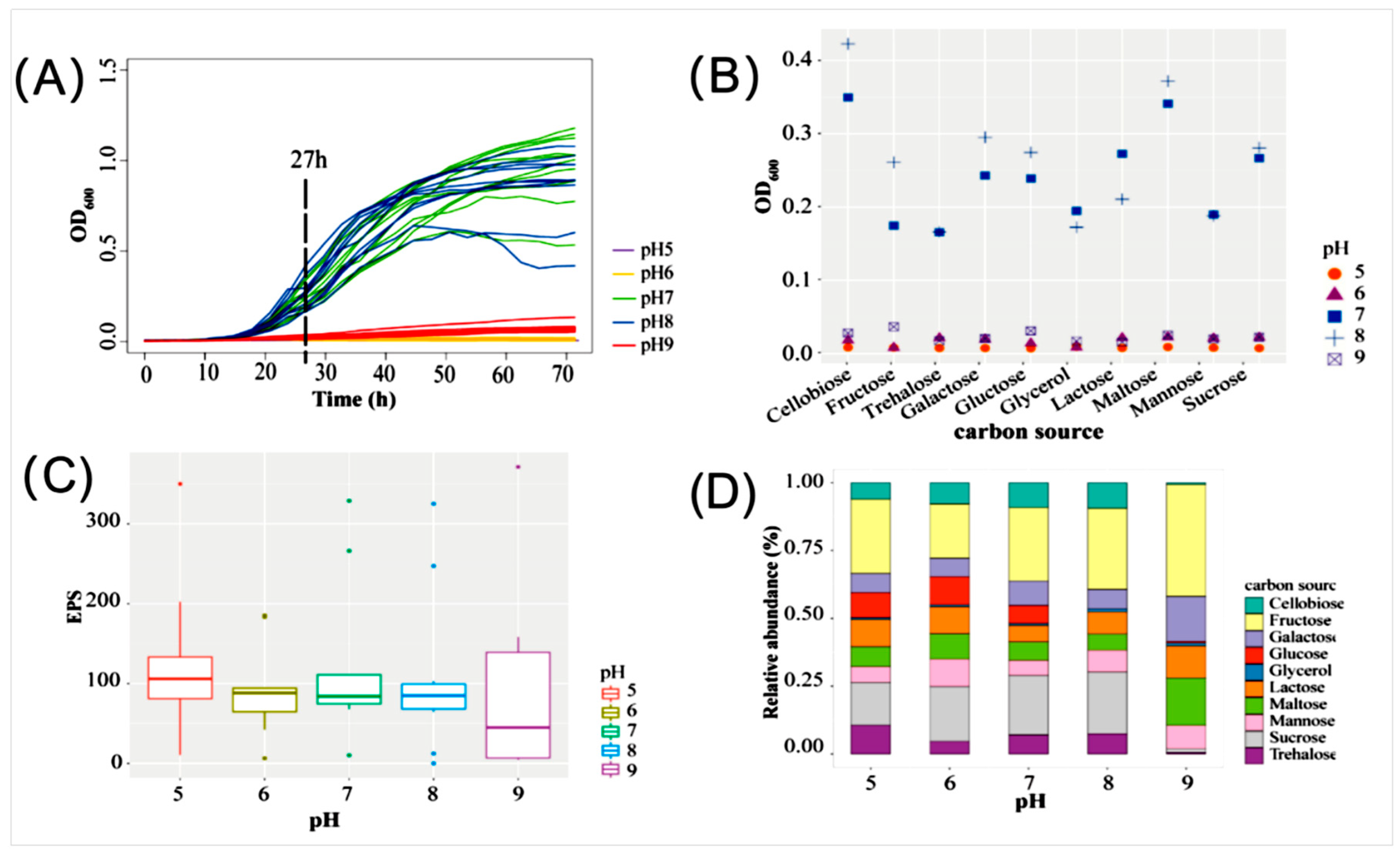
3.8. Culture Optimization of EPS Production by Strain ABI-6-9

EPSs are regarded as vital component of the algae–bacteria joint extracellular matrix [6,7,8,9]. A preliminary investigation indicated that the pH and carbon source in the media were the primary factors influencing EPS accumulation [24,70]. In this study, ten selected carbon sources, including cellobiose, fructose, galactose, glucose, glycerol, lactose, maltose, mannose, sucrose, and trehalose, and a pH range (5.0–9.0) were used for culture optimization. The obtained result showed that strain ABI-6-9 exhibited better growth when cultured at a pH ranging from 7.0 to 8.0 (Figure 4A). Additionally, the fastest bacterial growth of strain ABI-6-9 was achieved when cellobiose was used as the sole carbon source in culture medium and cultured at pH 8.0 (Figure 4B). However, the EPS accumulation indicated that the highest EPS yield was obtained when cultured at pH 9.0 (Figure 4C). Consequently, under the optimized conditions, the highest EPS yield of 115.6 ± 17.8 μg/mL was achieved when cellobiose (10 g/L) was used as the carbon source and bacteria were cultured at 28 °C, pH 9.0 (Figure 4D). It indicated that the increasing the medium pH may enhance bacterial EPS accumulation in strain ABI-6-9. It is speculated that in response to shifts in extracellular pH, bacteria usually secrete extra EPSs to cover the cellular surface, thereby mitigating adverse effects on cellular survival and function [10,11,12].

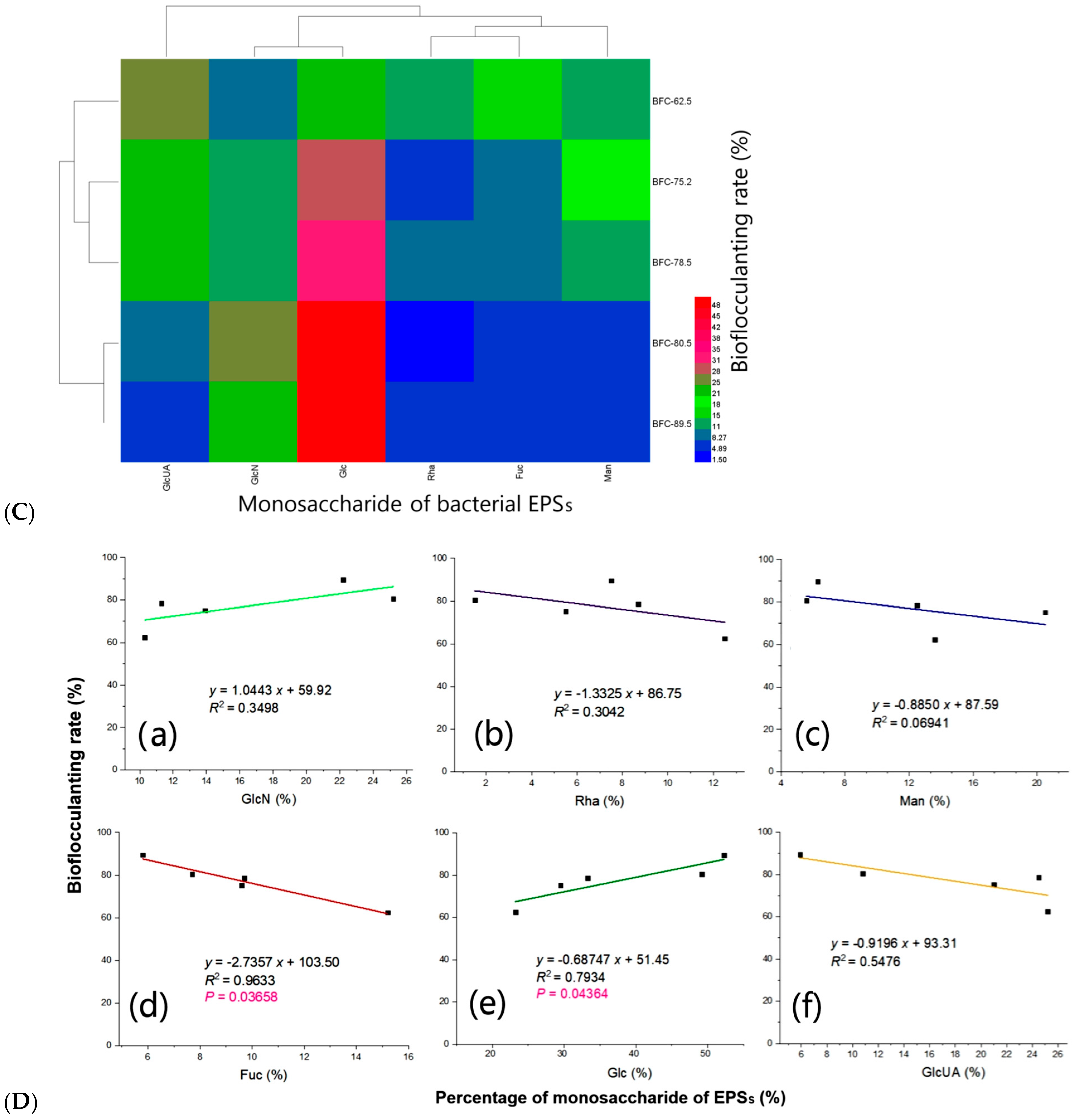
3.9. Bioflocculanting Bioactivity of EPSs Produced by Strain ABI-6-9

Polysaccharides play a fundamental roles in the phycosphere niche either by providing necessary valuable carbon sources for the phycobacteria community [5,6] or ensuring the formation of a favorable microenvironment for attachment, maintaining exoenzyme activity, sequestering nutrients, and protecting against toxins [7]. Additionally, they demonstrated promising bioflocculanting activity [6,7,8,9]. Phycobacterial candidates acting as natural EPS producers also possess varied overwhelming advantages, such as fast bacterial growth, flexible environmental adaptability, and convenient engineering operation for genetic modification with an easily accessible and clearer genetic background for biological and industrial applications [6].
In this study, to evaluate the production capacity of bacterial bioflocculants, the EPSs produced by strain ABI-6-9 were extracted and then subjected to a bioflocculanting activity evaluation. A comparison of the bioflocculanting effect under a series of EPS concentrations was performed. As shown in Figure 5A, the bioflocculanting efficiency of bacterial EPS showed a concentration-dependent manner, and it reached a maximum of 95.6 ± 7.5% (mean ± SD) when the EPS concentration was 0.70 g/L [39,40]. Previously, we found that the composition of EPSs varied considerably between different phycobacterial strains, and potentially reflected their fate in the extracellular environment during algae–bacteria interactions [12,22,23,32]. To further infer whether the type and composition of monosaccharides of EPSs were related to the bioflocculanting activity, characterizations of the monosaccharide composition (Figure 5B) and the correlations (Figure 5C) with bioflocculanting activity were performed. Based on the constructed heatmap, the relative portions of glucose demonstrated a significantly positive correlation with the bioflocculanting capacities, with correlation coefficient of (R2) of 0.7934 (p = 0.04364), while the portion of fucose showed an obvious negative correlation with a high R2 value of 0.9633 (p = 0.03658). In addition, the other three monosaccharides (Rha, Man, and GlcUA) were found to be negatively correlated, and GlcN had a positive correlation, but all were without statistical significance (Figure 5D). Despite these findings, the detailed chemical structures of bacterial EPSs remain to be further elucidated. Thus, these finding indicated that the new isolate, strain ABI-6-9, may serve as a new bacterial candidate with natural potential for the production of promising microbial bioflocculants derived from the phycosphere niche [35,36,37,38,39,40,41,42,71,72,73,74].

4. Conclusions

Characterizing the microbial composition of the phycosphere microbiota of six marine HAB-forming dinoflagellate Alexandrium spp. revealed they harbored vast portions of unidentified taxonomic groups and may offer attractive potential for discovering novel species with promising ecological and biological functions. Some cryptic cyanobacterial lineages within the dinoflagellate host were also found within the phycosphere community. To expand the number of new cultivable phycobacteria, a yellow-pigmented bacterium designated ABI-6-9 was recovered from highly toxic A. minutum amtk4 based on the CEFC procedure for the selected isolation of the phycobacteria. The phylogenomic analysis confirmed the phylogenetic position of this new isolate to represent a potential novel species of the genus Mameliella within the family Roseobacteraceae. Strain ABI-6-9 demonstrated an obvious promoting potential toward algal cell growth, both of the gonyautoxins’ accumulation profiles and their individual portions. The bacterial accumulation of active bioflocculanting EPSs indicated that the portions of glucose had a positive correlation with the bioflocculanting capacity, and fructose had a negative association. This study highlights the environmental and biotechnological implications of a newly discovered microalgae growth-promoting bacterium recovered from the phycosphere niche.

Supplementary Materials

The following supporting information can be downloaded at https://www.mdpi.com/article/10.3390/microorganisms13061198/s1: Figure S1. Distribution patterns of bacterial abundance at the phylum and family levels of the phycosphere microbiota of six Alexandrium spp. (A) and the relative bacterial abundance distributions of the top 10 genera of the family Rhodobacteraceae (B). Samples: the non-toxic group, A. tamarense AT-11, A. minutum AMKS5, and A. catenella ACHQ, and the toxic group: A. minutum AMTK4, A. tamarense AT 260, and A. catenella ACHK. Figure S2. Constructed co-occurrence networks at the genus level of the PM of highly toxic A. minutum amtk4. Circle size reflects the number of connections of one specific OTUs with the others. Green lines were for positive associations, and the red lines were for the negative. The genera of Mameliella was indicated in red. Figure S3. The constructed phylogenetic tree based on bacterial 16S rRNA gene sequences of cultivable phycobacterial strains isolated from Alexandrium spp. (A), and type species established by our laboratory (B). Bacterial strains isolated from highly-toxic A. catenella amtk4 were indicated in yellow color. Figure S4. Constructed phylogenomic tree based on the up-to-date bacterial core gene (UBCG) set between strain ABI-6-9 and closely related type strains within the families Rhodobacteraceae and Roseobacteraceae. Five typical genome parameters including genome size, GCC%, and the numbers of rRNA, tRNA and coding DNA sequence (CDS) were also shown in the right. Bar: 0.1 nt substitutions per site.

Author Contributions

Conceptualization, Q.Y.; methodology, Z.P., J.Z. and B.L.; software, X.Z. and Z.P.; validation, X.Z. and Q.Y.; formal analysis, X.Z., Z.P. and J.Z.; investigation, Z.P., J.Z. and B.L.; resources, X.Z. and Q.Y.; data curation, X.Z. and B.L.; writing—original draft preparation, X.Z. and Q.Y.; writing—review and editing, X.Z. and Q.Y.; visualization, X.Z.; supervision, Q.Y.; project administration, X.Z. and Q.Y.; funding acquisition, X.Z. and Q.Y. All authors have read and agreed to the published version of the manuscript.

Funding

This work was supported by the Natural Science Foundation of Zhejiang (LY23D060005), the Fundamental Research Funds for Zhejiang Provincial Universities (2024J006), the NSF of China (41876114), the Project from the Municipal Science and Technology Bureau of Zhoushan (2022C41018), the Industry–University Collaborative Education Project of Zhejiang, the Undergraduate Teaching Reform Project of Zhejiang (JGBA2024256), and the Project from the Zhejiang Provincial Association for Higher Education (KT2024099).

Institutional Review Board Statement

Not applicable.

Informed Consent Statement

Not applicable.

Data Availability Statement

The original contributions presented in this study are included in the article. Further inquiries can be directed to the corresponding author.

Conflicts of Interest

The authors declare no conflicts of interest.

Abbreviations

The following abbreviations are used in this manuscript:
AAIaverage amino acid Identity
ABIalgae-bacteria interactions
ANIaverage nucleotide identity
CECScombinational enhanced cultivation strategy
dDDHdigital DNA-DNA hybridization
DCGdesigned composite gel
ECMextracellular matrix
EPSexopolysaccharide
GTAsgene transfer agents
GTXgonyautoxin
HABsharmful algal blooms
HEPES 4-(2-hydroxyethyl)-1-piperazineethanesulfonic acid
IAAindole-3-acetic acid
MAmarine agar
MDMmicrobial dark matter
MGPmicroalgae growth-promoting
MGPBmicroalgae growth-promoting bacterium
ODoptical density
OTUsoperational taxonomic units
PMphycosphere microbiota
PMPPhycosphere Microbiome Project
PSPparalytic shellfish poison
PSTsparalytic shellfish poisoning toxins
UBCGup-to-date bacterial core gene set

References

  1. Shimizu, Y. Microalgal metabolites. Curr. Opin. Microbiol. 2003, 6, 236–243. [Google Scholar] [CrossRef] [PubMed]
  2. Seymour, J.R.; Simó, R.; Ahmed, T.; Stocker, R. Chemoattraction to dimethylsulfoniopropionate throughout the marine microbial food web. Science 2010, 329, 342–345. [Google Scholar] [CrossRef]
  3. Thoré, E.S.J.; Muylaert, K.; Bertram, M.G.; Brodin, T. Microalgae. Curr. Biol. 2023, 33, R91–R95. [Google Scholar] [CrossRef]
  4. Scott, G.D. Lichen terminology. Nature 1957, 179, 486–487. [Google Scholar] [CrossRef]
  5. Sutherland, I.W. Biofilm exopolysaccharides: A strong and sticky framework. Microbiology 2001, 147, 3–9. [Google Scholar] [CrossRef]
  6. Li, S.N.; Zhang, C.; Li, F.; Ren, N.Q.; Ho, S.H. Recent advances of algae-bacteria consortia in aquatic remediation. Crit. Rev. Environ. Sci. Technol. 2023, 53, 315–339. [Google Scholar] [CrossRef]
  7. Seymour, J.R.; Amin, S.A.; Raina, J.B.; Stocker, R. Zooming in on the phycosphere: The ecological interface for phytoplankton-bacteria relationships. Nat. Microbiol. 2017, 2, 17065. [Google Scholar] [CrossRef]
  8. Amin, S.A.; Parker, M.S.; Armbrust, E.V. Interactions between diatoms and bacteria. Microbiol. Mol. Biol. Rev. 2012, 76, 667–684. [Google Scholar] [CrossRef]
  9. Kouzuma, A.; Watanabe, K. Exploring the potential of algae/bacteria interactions. Curr. Opin. Biotechnol. 2015, 33, 125–129. [Google Scholar] [CrossRef]
  10. Cirri, E.; Pohnert, G. Algae-bacteria interactions that balance the planktonic microbiome. New Phytol. 2019, 223, 100–106. [Google Scholar] [CrossRef]
  11. Yang, Q.; Ge, Y.M.; Iqbal, N.M.; Yang, X.; Zhang, X.L. Sulfitobacter alexandrii sp. nov.; a new microalgae growth-promoting bacterium with exopolysaccharides bioflocculanting potential isolated from marine phycosphere. Antonie Van Leeuwenhoek 2021, 114, 1091–1106. [Google Scholar] [CrossRef] [PubMed]
  12. Landry, Z.C.; Vergin, K.; Mannenbach, C.; Block, S.; Yang, Q.; Blainey, P.; Carlson, C.; Giovannoni, S.J. Optofluidic Single-Cell Genome Amplification of Sub-Micron Bacteria in the Ocean Subsurface. Front. Microbiol. 2018, 9, 1152. [Google Scholar] [CrossRef]
  13. Ramanan, R.; Kang, Z.; Kim, B.H.; Cho, D.H.; Jin, L.; Oh, H.M.; Kim, H.S. Phycosphere bacterial diversity in green algae reveals an apparent similarity across habitats. Algal Res. 2015, 8, 140–144. [Google Scholar] [CrossRef]
  14. Chen, Z.; Zakrzewska, S.; Hajare, H.S.; Alvarez-Buylla, A.; Abderemane-Ali, F.; Bogan, M.; Ramirez, D.; O’Connell, L.A.; Du Bois, J.; Minor, D.L., Jr. Definition of a saxitoxin (STX) binding code enables discovery and characterization of the anuran saxiphilin family. Proc. Natl. Acad. Sci. USA 2022, 119, e2210114119. [Google Scholar] [CrossRef] [PubMed]
  15. Schwenk, D.; Nohynek, L.; Rischer, H. Algae-bacteria association inferred by 16S rDNA similarity in established microalgae cultures. MicrobiologyOpen 2014, 3, 356–368. [Google Scholar] [CrossRef]
  16. Rinke, C.; Schwientek, P.; Sczyrba, A.; Ivanova, N.N.; Anderson, I.J.; Cheng, J.F.; Darling, A.; Malfatti, S.; Swan, B.K.; Gies, E.A.; et al. Insights into the phylogeny and coding potential of microbial dark matter. Nature 2013, 499, 431–437. [Google Scholar] [CrossRef]
  17. Zhang, X.L.; Tian, X.Q.; Ma, L.Y.; Feng, B.; Liu, Q.H.; Yuan, L.D. Biodiversity of the symbiotic bacteria associated with toxic marine dinoflagellate Alexandrium tamarense. J. Biosci. Med. 2015, 3, 23–28. [Google Scholar] [CrossRef]
  18. Seyedsayamdost, M.R.; Case, R.J.; Kolter, R.; Clardy, J. The Jekyll-and-Hyde chemistry of Phaeobacter gallaeciensis. Nat. Chem. 2011, 3, 331–335. [Google Scholar] [CrossRef]
  19. Zhang, X.L.; Ma, L.Y.; Tian, X.Q.; Huang, H.L.; Yang, Q. Biodiversity study of intracellular bacteria closely associated with paralytic shellfish poisoning dinoflagellates Alexandrium tamarense and A. minutum. Int. J. Environ. Resour. 2015, 4, 23–27. [Google Scholar] [CrossRef]
  20. Zhu, W.Z.; Ge, Y.M.; Dai, J.; Zhang, X.L.; Yang, Q. Alexandriicola marinus gen. nov.; sp. nov.; a new member of the family Rhodobacteraceae isolated from marine phycosphere. Antonie van Leeuwenhoek 2022, 115, 473–486. [Google Scholar] [CrossRef]
  21. Feng, X.M.; Mo, Y.X.; Han, L.; Nogi, Y.; Zhu, Y.H.; Lv, J. Mameliella sediminis gen. nov.; sp. nov.; a member of the family Erythrobacteraceae isolated from subterrestrial sediment. Int. J. Syst. Evol. Microbiol. 2015, 65, 3658–3665. [Google Scholar] [CrossRef]
  22. Zhang, X.L.; Li, G.X.; Ge, Y.M.; Iqbal, N.M.; Yang, X.; Cui, Z.D.; Yang, Q. Sphingopyxis microcysteis sp. nov.; a novel bioactive exopolysaccharides-bearing Sphingomonadaceae isolated from the Microcystis phycosphere. Antonie Van Leeuwenhoek 2021, 114, 845–857. [Google Scholar] [CrossRef] [PubMed]
  23. Silva, E.S. Intracellular bacteria: The origin of dinoflagellate toxicity. J. Environ. Pathol. Toxicol. Oncol. 1990, 10, 124–128. [Google Scholar]
  24. Ren, C.Z.; Gao, H.M.; Dai, J.; Zhu, W.Z.; Xu, F.F.; Ye, Y.; Zhang, X.L.; Yang, Q. Taxonomic and Bioactivity Characterizations of Mameliella alba Strain LZ-28 Isolated from Highly Toxic Marine Dinoflagellate Alexandrium catenella amtk4. Mar. Drugs 2022, 20, 321. [Google Scholar] [CrossRef]
  25. Liu, J.; Yang, J.; Ge, Y.; Yang, Q.; Sun, J.; Yu, X. Acute effects of CH3NH3PbI3 perovskite on Scenedesmus obliquus and Daphnia magana in aquatic environment. Ecotoxicol. Environ. Saf. 2021, 208, 111677. [Google Scholar] [CrossRef]
  26. Singh, R.P.; Reddy, C.R.K. Seaweed-microbial interactions: Key functions of seaweed-associated bacteria. FEMS Microbiol. Ecol. 2014, 88, 213–230. [Google Scholar] [CrossRef]
  27. Zhang, H.; Zhao, W.; Yang, W.; Zhang, H.; Qian, X.; Sun, K.; Yang, Q.; Shen, X.; Zhang, L. Autoinducer-2 enhances defence of Vibrio furnissii against oxidative stress and DNA damage by modulation of c-di-GMP signaling via a two-component system. mBio 2025, 16, 02922–02924. [Google Scholar] [CrossRef]
  28. Li, S.; Liu, Q.; Duan, C.; Li, J.; Sun, H.; Xu, L.; Yang, Q.; Wang, Y.; Shen, X.; Zhang, L. c-di-GMP inhibits the DNA binding activity of H-NS in Salmonella. Nat. Commun. 2023, 14, 7502. [Google Scholar] [CrossRef]
  29. Sokolovskaya, O.M.; Shelton, A.N.; Taga, M.E. Sharing vitamins: Cobamides unveil microbial interactions. Science 2020, 369, eaba0165. [Google Scholar] [CrossRef]
  30. Thume, K.; Gebser, B.; Chen, L.; Meyer, N.; Kieber, D.J.; Pohnert, G. The metabolite dimethylsulfoxonium propionate extends the marine organosulfur cycle. Nature 2018, 563, 412–415. [Google Scholar] [CrossRef]
  31. Gao, H.M.; Xie, P.F.; Zhang, X.L.; Yang, Q. Isolation, Phylogenetic and Gephyromycin Metabolites Characterization of New Exopolysaccharides-Bearing Antarctic Actinobacterium from Feces of Emperor Penguin. Mar. Drugs 2021, 19, 458. [Google Scholar] [CrossRef] [PubMed]
  32. Tschitschko, B.; Esti, M.; Philippi, M.; Kidane, A.T.; Littmann, S.; Kitzinger, K.; Speth, D.R.; Li, S.; Kraberg, A.; Tienken, D.; et al. Rhizobia-diatom symbiosis fixes missing nitrogen in the ocean. Nature 2024, 630, 899–904. [Google Scholar] [CrossRef] [PubMed]
  33. Magdouli, S.; Brar, S.K.; Blais, J.F. Co-culture for lipid production: Advances and challenges. Biomass Bioenerg. 2016, 92, 20–30. [Google Scholar] [CrossRef]
  34. Yang, Q.; Feng, Q.; Zhang, B.P.; Gao, J.J.; Sheng, Z.; Xue, Q.P.; Zhang, X.L. Marinobacter alexandrii sp. nov.; a novel yellow-pigmented and algae growth-promoting bacterium isolated from marine phycosphere microbiota. Antonie Van Leeuwenhoek 2021, 114, 709–718. [Google Scholar] [CrossRef]
  35. Yang, Q.; Jiang, Z.; Zhou, X.; Zhang, R.; Xie, Z.; Zhang, S.; Wu, Y.; Ge, Y.; Zhang, X. Haliea alexandrii sp. nov.; isolated from phycosphere microbiota of the toxin-producing dinoflagellate Alexandrium catenella. Int. J. Syst. Evol. Microbiol. 2020, 70, 1133–1138. [Google Scholar] [CrossRef]
  36. Duan, Y.; Jiang, Z.; Wu, Z.; Sheng, Z.; Yang, X.; Sun, J.; Zhang, X.; Yang, Q.; Yu, X.; Yan, J. Limnobacter alexandrii sp. nov.; a thiosulfate-oxidizing, heterotrophic and EPS-bearing Burkholderiaceae isolated from cultivable phycosphere microbiota of toxic Alexandrium catenella amtk4. Antonie Van Leeuwenhoek 2020, 13, 1689–1698. [Google Scholar] [CrossRef]
  37. Zhang, L.; Yang, Q.; Luo, X.; Fang, C.; Zhang, Q.; Tang, Y. Knockout of crtB or crtI gene blocks the carotenoid biosynthetic pathway in Deinococcus radiodurans R1 and influences its resistance to oxidative DNA-damaging agents due to change of free radicals scavenging ability. Arch. Microbiol. 2007, 188, 411–419. [Google Scholar] [CrossRef]
  38. Yang, Q.; Jiang, Z.; Zhou, X.; Zhang, R.; Wu, Y.; Lou, L.; Ma, Z.; Wang, D.; Ge, Y.; Zhang, X.; et al. Nioella ostreopsis sp. nov.; isolated from toxic dinoflagellate, Ostreopsis lenticularis. Int. J. Syst. Evol. Microbiol. 2020, 70, 759–765. [Google Scholar] [CrossRef]
  39. Siddharth, T.; Sridhar, P.; Vinila, V.; Tyagi, R.D. Environmental applications of microbial extracellular polymeric substance (EPS): A review. J. Environ. Manag. 2021, 287, 112307. [Google Scholar] [CrossRef]
  40. Zhang, X.L.; Qi, M.; Li, Q.H.; Cui, Z.D.; Yang, Q. Maricaulis alexandrii sp. nov.; a novel active bioflocculants-bearing and dimorphic prosthecate bacterium isolated from marine phycosphere. Antonie Van Leeuwenhoek 2021, 114, 1195–1203. [Google Scholar] [CrossRef]
  41. Wang, X.; Ye, Y.; Xu, F.; Duan, Y.; Xie, P.; Yang, Q.; Zhang, X. Maritimibacter alexandrii sp. nov., a New Member of Rhodobacteraceae Isolated from Marine Phycosphere. Curr. Microbiol. 2021, 78, 3996–4003. [Google Scholar] [CrossRef] [PubMed]
  42. Yang, Q.; Jiang, Z.; Zhou, X.; Xie, Z.; Wang, Y.; Wang, D.; Feng, L.; Yang, G.; Ge, Y.; Zhang, X. Saccharospirillum alexandrii sp. nov.; isolated from the toxigenic marine dinoflagellate Alexandrium catenella amtk4. Int. J. Syst. Evol. Microbiol. 2020, 70, 820–826. [Google Scholar] [CrossRef] [PubMed]
  43. Durham, B.P.; Boysen, A.K.; Carlson, L.T.; Groussman, R.D.; Heal, K.R.; Cain, K.R.; Morales, R.L.; Coesel, S.N.; Morris, R.M.; Ingalls, A.E.; et al. Sulfonate-based networks between eukaryotic phytoplankton and heterotrophic bacteria in the surface ocean. Nat. Microbiol. 2019, 4, 1706–1715. [Google Scholar] [CrossRef]
  44. Yang, X.; Xiang, R.; Iqbal, M.N.; Duan, Y.; Zhang, X.; Wang, L.; Yu, L.; Li, J.; Sun, M.; Yang, Q.; et al. Marinobacter shengliensis subsp. alexandrii subsp. nov., isolated from cultivable phycosphere microbiota of highly toxic dinoflagellate Alexandrium catenella LZT09 and description of Marinobacter shengliensis subsp. shengliensis subsp. nov. Curr. Microbiol. 2021, 78, 1648–1655. [Google Scholar]
  45. Yang, X.; Jiang, Z.W.; Zhang, J.; Zhou, X.; Zhang, X.L.; Wang, L.; Yu, T.; Wang, Z.; Bei, J.; Dong, B. Mesorhizobium alexandrii sp. nov.; isolated from phycosphere microbiota of PSTs-producing marine dinoflagellate Alexandrium minutum amtk4. Antonie Van Leeuwenhoek 2020, 113, 907–917. [Google Scholar] [CrossRef]
  46. Yang, Q.; Jiang, Z.W.; Huang, C.H.; Zhang, R.N.; Li, L.Z.; Yang, G.; Feng, L.J.; Yang, G.F.; Zhang, H.; Zhang, X.L. Hoeflea prorocentri sp. nov.; isolated from a culture of the marine dinoflagellate Prorocentrum mexicanum PM01. Antonie Van Leeuwenhoek 2018, 111, 1845–1853. [Google Scholar] [CrossRef]
  47. Croft, M.T.; Lawrence, A.D.; Raux-Deery, E.; Warren, M.J.; Smith, A.G. Algae acquire vitamin B12 through a symbiotic relationship with bacteria. Nature 2005, 438, 90–93. [Google Scholar] [CrossRef]
  48. Yan, Z.; Chen, B.; Yang, Y.; Yi, X.; Wei, M.; Ecklu-Mensah, G.; Buschmann, M.M.; Liu, H.; Gao, J.; Liang, W.; et al. Multi-omics analyses of airway host-microbe interactions in chronic obstructive pulmonary disease identify potential therapeutic interventions. Nat. Microbiol. 2022, 7, 1361–1375. [Google Scholar] [CrossRef]
  49. Guérin, H.; Kulakauskas, S.; Chapot-Chartier, M.P. Structural variations and roles of rhamnose-rich cell wall polysaccharides in Gram-positive bacteria. J. Biol. Chem. 2022, 298, 102488. [Google Scholar] [CrossRef]
  50. Qin, L.; Liu, L.; Wang, Z.; Chen, W.; Wei, D. The mixed culture of microalgae Chlorella pyrenoidosa and yeast Yarrowia lipolytica for microbial biomass production. Bioprocess Biosyst. Eng. 2019, 42, 1409–1419. [Google Scholar] [CrossRef]
  51. Nakayama, T.; Nomura, M.; Takano, Y.; Tanifuji, G.; Shiba, K.; Inaba, K.; Inagaki, Y.; Kawata, M. Single-cell genomics unveiled a cryptic cyanobacterial lineage with a worldwide distribution hidden by a dinoflagellate host. Proc. Natl. Acad. Sci. USA 2019, 116, 15973–15978. [Google Scholar] [CrossRef] [PubMed]
  52. Su, J.; Yang, X.; Zheng, T.; Hong, H. An efficient method to obtain axenic cultures of Alexandrium tamarense—a PSP-producing dinoflagellate. J. Microbiol Methods 2007, 69, 425–430. [Google Scholar] [CrossRef] [PubMed]
  53. Xiang, T.; Hambleton, E.A.; DeNofrio, J.C.; Pringle, J.R.; Grossman, A.R. Isolation of clonal axenic strains of the symbiotic dinoflagellate Symbiodinium and their growth and host specificity. J. Phycol. 2013, 49, 447–458. [Google Scholar] [CrossRef]
  54. Ge, Y.; Xue, Y.; Yang, Q.; Xing, W.; Zhu, S.; Jiang, W.; Liu, J. Benzalkonium chloride disinfection increases the difficulty of controlling foodborne pathogens identified in aquatic product processing. J. Hazard. Mater. 2025, 488, 137140. [Google Scholar] [CrossRef]
  55. Pennisi, E. SYMBIOSIS. A lichen ménage à trois. Science 2016, 353, 337. [Google Scholar] [CrossRef]
  56. Jiang, Z.; Duan, Y.; Yang, X.; Yao, B.; Zeng, T.; Wang, X.; Feng, Q.; Qi, M.; Yang, Q.; Zhang, X.L. Nitratireductor alexandrii sp. nov.; from phycosphere microbiota of toxic marine dinoflagellate Alexandrium tamarense. Int. J. Syst. Evol. Microbiol. 2020, 70, 4390–4397. [Google Scholar] [CrossRef]
  57. Zhang, X.; Xu, J.; Dai, J.; Zhang, L.; Feng, L.; Tian, X.; Yang, Q. Taxonomic, Phylogenomic and Bioactivity Profiling of Novel Phycosphere Bacterium from Model Cyanobacterium Synechococcus elongatus PCC 7942. Mar. Drugs. 2024, 22, 36. [Google Scholar] [CrossRef]
  58. Ramanan, R.; Kim, B.H.; Cho, D.H.; Oh, H.M.; Kim, H.S. Algae-bacteria interactions: Evolution, ecology and emerging applications. Biotechnol. Adv. 2016, 34, 14–29. [Google Scholar] [CrossRef]
  59. Zhang, X.L.; Yang, Q. Phycosphere Microbiology: An emerging interdiscipline comprehensively promoting sustainable development. Acta Microbiol. Sin. 2025, 65, 1831–1848. [Google Scholar]
  60. Zhou, J.; Lyu, Y.; Richlen, M.L.; Anderson, D.M.; Cai, Z. Quorum sensing is a language of chemical signals and plays an ecological role in algal-bacterial interactions. Crit. Rev. Plant Sci. 2016, 35, 81–105. [Google Scholar] [CrossRef]
  61. Yang, Q.; Zhang, X.; Bao, X.; Lu, H.; Zhang, W.; Wu, W.; Miao, H.; Jiao, B. Single cell determination of nitric oxide release using capillary electrophoresis with laser-induced fluorescence detection. J. Chromatogr. A 2008, 1201, 120–127. [Google Scholar] [CrossRef]
  62. Zilber-Rosenberg, I.; Rosenberg, E. Role of microorganisms in the evolution of animals and plants: The hologenome theory of evolution. FEMS Microbiol. Rev. 2008, 32, 723–735. [Google Scholar] [CrossRef]
  63. Zipfel, C.; Oldroyd, G. Plant signalling in symbiosis and immunity. Nature 2017, 543, 328–336. [Google Scholar] [CrossRef]
  64. Segev, E.; Wyche, T.P.; Kim, K.H.; Petersen, J.; Ellebrandt, C.; Vlamakis, H.; Barteneva, N.; Paulson, J.N.; Chai, L.; Clardy, J.; et al. Dynamic metabolic exchange governs a marine algal-bacterial interaction. eLife 2016, 5, e17473. [Google Scholar] [CrossRef] [PubMed]
  65. Sadiq, F.A.; Hansen, M.F.; Burmølle, M.; Heyndrickx, M.; Flint, S.; Lu, W.; Chen, W.; Zhang, H. Trans-kingdom interactions in mixed biofilm communities. FEMS Microbiol. Rev. 2022, 46, fuac024. [Google Scholar] [CrossRef]
  66. Moran, M.A.; Belas, R.; Schell, M.A.; González, J.M.; Sun, F.; Sun, S.; Binder, B.J.; Edmonds, J.; Ye, W.; Orcutt, B.; et al. Ecological genomics of marine Roseobacters. Appl. Environ. Microbiol. 2007, 3, 4559–4569. [Google Scholar] [CrossRef]
  67. Zheng, Q.; Chen, C.; Yan, X.; Wang, Y.; Zeng, Y.; Hao, L.; He, W.; Jiao, N. Mameliella alba gen. nov., sp. nov., a marine bacterium of the Roseobacter clade in the order Rhodobacterales. Int. J. Syst. Evol. Microbiol. 2010, 60, 953–957. [Google Scholar] [CrossRef]
  68. Hasegawa, Y.; Martin, J.L.; Giewat, M.W.; Rooney-Varga, J.N. Microbial community diversity in the phycosphere of natural populations of the toxic alga, Alexandrium fundyense. Environ. Microbiol. 2007, 9, 3108–3121. [Google Scholar] [CrossRef]
  69. Kramer, J.; Özkaya, Ö.; Kümmerli, R. Bacterial siderophores in community and host interactions. Nat. Rev. Microbiol. 2020, 18, 152–163. [Google Scholar] [CrossRef]
  70. Martin, F.M.; Uroz, S.; Barker, D.G. Ancestral alliances: Plant mutualistic symbioses with fungi and bacteria. Science 2017, 356, eaad4501. [Google Scholar] [CrossRef]
  71. Kuhlisch, C.; Shemi, A.; Barak-Gavish, N.; Schatz, D.; Vardi, A. Algal blooms in the ocean: Hot spots for chemically mediated microbial interactions. Nat. Rev. Microbiol. 2024, 22, 138–154. [Google Scholar] [CrossRef] [PubMed]
  72. Wang, Z.; Xie, P.; Dai, J.; Zhang, L.; Yang, Q.; Zhang, X.; Yang, X. Variation in Structure and Functional Diversity of Surface Bacterioplankton Communities in the Eastern East China Sea. J. Mar. Sci. Eng. 2024, 12, 69. [Google Scholar] [CrossRef]
  73. Kiers, E.T.; West, S.A. Evolutionary biology. Evolving new organisms via symbiosis. Science 2015, 348, 392–394. [Google Scholar] [CrossRef]
  74. Fu, H.; Uchimiya, M.; Gore, J.; Moran, M.A. Ecological drivers of bacterial community assembly in synthetic phycospheres. Proc. Natl. Acad. Sci. USA 2020, 117, 3656–3662. [Google Scholar] [CrossRef]
Figure 1. Phylogenetic tree constructed based on the bacterial 16S rRNA gene of strain ABI-6-9 and the closely related type strains of the families Rhodobacteraceae and Roseobacteraceae, and the isolation sources. Those phycobacterial strains were indicated in yellow color.
Microorganisms 13 01198 g001
Figure 2. Evaluation of the MGP potential of strain ABI-6-9 on algal host A. minutum amtk4 cells.
Microorganisms 13 01198 g002
Figure 3. Enhanced effect of algal–bacterial co-culture on the accumulation (A) and changes in the individual portions (B) of four algal GTX toxins, which are indicated with their chemical structures. Notes: *, p < 0.05 and **, p < 0.01.
Microorganisms 13 01198 g003
Figure 4. Culture optimization of bacterial growth and EPS production of strain ABI-6-9. Effects of different pHs and carbon sources on bacterial growth recorded at OD600nm (A,B) and EPS production (C,D).
Microorganisms 13 01198 g004
Figure 5. Bioflocculanting activity profiles of bacterial EPSs produced by strain ABI-6-9 (A), the monosaccharide portion of bacterial EPSs (B), their overall associations with bioflocculanting activity visualized in a clustered heat map (C), and the correlation analysis of the relative portions (%) of six monosaccharides (D), including D-galactosamine (GlcN, panel a), L-rhamnose (Rha, panel b), mannose (Man, panel c), D-fucose (Fuc, panel d), glucose (Glc, panel e), and D-galacturonic acid (GlcUA, panel f), with the bioflocculanting rate (%).
Microorganisms 13 01198 g005aMicroorganisms 13 01198 g005b
Disclaimer/Publisher’s Note: The statements, opinions and data contained in all publications are solely those of the individual author(s) and contributor(s) and not of MDPI and/or the editor(s). MDPI and/or the editor(s) disclaim responsibility for any injury to people or property resulting from any ideas, methods, instructions or products referred to in the content.

