Article

Population Genomics, Virulence Traits, and Antimicrobial Resistance of Streptococcus suis Isolated in China

1
National Key Laboratory of Agricultural Microbiology, College of Veterinary Medicine, Huazhong Agricultural University, Wuhan 430070, China
2
Hubei Hongshan Laboratory, Wuhan 430070, China
3
The Cooperative Innovation Center for Sustainable Pig Production, Wuhan 430070, China
4
College of Biomedicine and Health, Huazhong Agricultural University, Wuhan 430070, China
*
Authors to whom correspondence should be addressed.
Microorganisms 2025, 13(6), 1197; https://doi.org/10.3390/microorganisms13061197
Submission received: 20 March 2025 / Revised: 30 April 2025 / Accepted: 20 May 2025 / Published: 23 May 2025
(This article belongs to the Section Veterinary Microbiology)

Abstract

Streptococcus suis is a significant zoonotic pathogen of public health importance. In this study, whole-genome sequencing of 177 isolates of Streptococcus suis, isolated from diseased swine across 15 provinces in China between 2017 and 2019, was performed. A total of 23 serotypes and 28 ST types were identified, with serotypes 2 and 3 comprising 50.8% of the isolates, and sequence types ST353 and ST117 accounting for 23.7%. Clustering analysis based on known virulence-associated factors (VAFs) resulted in the identification of four distinct clusters, and virulence was assessed using animal models, including a unique, highly virulent cluster designated as cluster I. Drug susceptibility testing indicated that 97.7% of the isolates were multidrug-resistant. A total of 26 resistance-associated genes were identified within the genome, 18 of which were associated with integrative and conjugative elements (ICEs) and/or integrative mobilizable elements (IMEs). Nevertheless, our understanding of suis virulence in terms of phylogeny remains incomplete. This study contributes to the understanding of the population structure and genetic characteristics of suis, provides a framework and novel partitioning approach for future investigations into its virulence and pathogenicity, and complements the data on antibiotic resistance.

1. Introduction

Streptococcus suis is a globally recognized zoonotic pathogen that significantly impacts the swine industry, leading to considerable economic losses. In swine, this bacterium is primarily responsible for conditions such as pneumonia, septicaemia, and meningitis [1]. In humans, Streptococcus suis infection is associated with disease primarily through exposure to contaminated byproducts or through occupational hazards [2,3]. China experienced major outbreaks of human infections in 1998, 2005, and 2016, all attributed to Streptococcus suis [4]. Between 2002 and 2013, over 1500 cases of human infection were documented across 34 countries [5]. Furthermore, Streptococcus suis serves as a significant reservoir for antimicrobial resistance (AMR) genes [6]. This bacterium exhibits a high prevalence of resistance to lincosamides [7], macrolides, and tetracyclines, with an increasing trend in resistance to sulfonamides, aminoglycosides, and fluoroquinolones over time [8]. Sequencing studies of Streptococcus suis have revealed the widespread presence of AMR-associated genes, many of which are located on integrative and conjugative elements (ICEs) and integrative mobilizable elements (IMEs) [9]. Additionally, Streptococcus suis has the capacity to transfer drug resistant genes to other pathogens, thereby exacerbating the issue of antimicrobial resistance [10].
Recent advancements in sequencing technology have led to a significant increase in the genetic data available for Streptococcus suis, primarily due to the whole-genome sequencing (WGS) of numerous isolates [11,12,13]. Multi Locus Sequence Typing (MLST) [14], which determines sequence types (STs) through the analysis of seven housekeeping genes, is widely employed to assess the genetic diversity and conduct epidemiological studies of Streptococcus suis. The most commonly isolated sequence types include ST1, ST25, ST28 [5], and ST7. Notably, ST1 is the predominant disease-associated sequence type in Europe, Asia, Africa, and South America [15], whereas ST25 and ST28 are more prevalent in North American isolates [16,17]. Conversely, ST7 is only endemic to mainland China [18,19]. In another study, a separate investigation analyzed SNPs from 1634 Streptococcus suis genomes utilizing Bayesian analysis of population structure (BAPS), resulting in the construction of a maximum-likelihood phylogenetic tree that delineates three primary clades: the human-associated clade (HAC), the diseased-pig clade (DPC), and the healthy-pig clade (HPC). The HAC includes several isolates that exhibit high virulence in experimental infection models [4].
Currently, 29 serotypes of Streptococcus suis have been identified on the basis of the antigenicity of capsular polysaccharide (CPS), specifically serotypes 1–19, 21, 23–25, 27–31, and 1/2 [20]. Recently, 28 new serotypes, referred to as NCL and Chz, have recently been recognized [21,22,23,24]. There are notable geographical variations in the distribution of these serotypes; however, serotype 2 predominates among isolates from various countries [5,25]. Serotypes 1, 3, 7, and 9 are the most frequently isolated serotypes from diseased pigs [26,27]. Serotypes and MLST types are often used to analyze the virulence phenotype of suis. Serotype 2 has been described as the most virulent serotype [5]. Conversely, it has been shown that isolates of serotype 2 prevalent in North America are less virulent [28]. In animal models of infection, ST1 was more virulent than ST25, and ST28 was the least virulent [16,29]. However, ST28 has reported different clinical presentationss in the United States [30]. Currently, more than 100 virulence-associated factors (VAFs) for Streptococcus suis have been proposed or validated, although the majority lack experimental validation [28,31]. Muramidase-released protein (MRP) [32], extracellular protein factor (EPF) [33], and suilysin (SLY) [34,35] are the most extensively studied VAFs believed to play a role in the pathogenesis of S. suis, and they are frequently utilized to predict the potential virulence of S. suis isolates. Nevertheless, the genotypes of mrp, epf, and/or sly do not definitively determine the virulence classification of suis, as strains lacking expression of these factors have still been found to exhibit virulent phenotypes [36]. In summary, Streptococcus suis appears to be a pathogen characterized by redundant virulence factors, making the identification of key virulence traits exceedingly challenging [28,37].
In this study, we performed genome sequencing on 177 isolates of Streptococcus suis obtained from diseased swine across 15 provinces in China during the period from 2017 to 2019. This analysis elucidated the population structure of suis and evaluated its potential virulence and antibiotic resistance. The 177 isolates were categorized into HAC, HPC, and DPC according to a previously reported scheme [4]. Furthermore, we examined these isolates in relation to 104 previously documented virulence-associated factors [31,38,39,40,41]. The virulence phenotypes of the distinct clusters within the three evolutionary clades were subsequently validated using a murine model. Overall, this study provides a framework for identifying potentially virulent strains among isolates and enhances the existing knowledge regarding the antibiotic resistance profiles of Streptococcus suis in China. Since the virulence phenotypes of Streptococcus suis cannot be fully characterized by a single or limited set of virulence factors [28], we wished to further understand the virulence of suis through phylogenetic analyses.

2. Materials and Methods

2.1. Streptococcus suis Isolate Collection

In this study, a total of 177 strains of Streptococcus suis were collected from various provinces across China between the years 2017 and 2019. Detailed information regarding the sources and characteristics of these strains can be found in the Supplementary Table S1. Efforts were made to obtain Streptococcus suis isolates from as many provinces as possible, ensuring that none of the isolates were duplicates derived from the same pig. The isolates were inoculated and cultured for 24 h on TSA supplemented with 10% bovine serum. Subsequently, monoclonal strains were selected and transferred to 5 mL of TSB medium, where they were incubated for 16 h at 37 °C in a 5% CO2 atmosphere.

2.2. Phylogenetic Analysis

To perform a wide-ranging phylogenetic analysis, 463 whole-genome sequences of S. suis were obtained from NCBI. In total, 640 S. suis sequences were collected for phylogenetic analysis. The sequences were first aligned to the reference genome (BM407) using MUMmer v. 3.23, and a total of 550,691 SNP sites were identified. In this study, indels and adjacent mismatches were not considered to be true SNPs. These SNPs of 640 Streptococcus suis sequences were subsequently used to construct the phylogenetic tree through IQ-TREE v. 2.2.0.3 with with the GTR + GAMMA model and 1000 bootstrap replicates. The clade information of each sequence was manually marked based on the phylogenetic tree and previous research. Finally, the phylogenetic tree was visualized by TVBOT and retouched using Adobe Illustrator (CC 2019).

2.3. MLST Analysis and Molecular Serotype Prediction

The sequence type of each isolate was annotated by SRST2 with default parameters. In addition, S. suis genome sequences were analyzed by BLASTn against a nucleotide database of serotype-specific genes in the CPS synthesis locus for molecular serotyping [42]. This identified 29 classical serotypes, with the exception of two pairs of serotypes: 1/14 and 2/ 1 2 .

2.4. Identification of Virulence Factors (VFs) and Antimicrobial Resistance Genes (ARGs)

The VFs for each S. suis genome were determined using BLASTn against the database collected in this study by reviewing the references (Supplementary Table S3). The BLASTn minimum identity was set to 90%, and the minimum coverage was set to 80%. The ARGs of each strain were identified by RGI v. 6.0.3 using reference data from the Comprehensive Antibiotic Resistance Database (CARD). Here, Python 3.10.13 scripts were used to collect matching gene fragments for further analysis and visualization.

2.5. Annotation and Visualization of Bacterial Integrative and Conjugative Elements (ICEs)

The gene fragments of each strain identified as VFs or ARGs were selected for annotation with the ICEberg 3.0 database as ICE, ACE, IME, or CIME. In this study, BLASTn was employed to align each gene fragment with the ICEberg 3.0 database, setting the minimum identity at 90% and the minimum coverage at 80%. The resulting matches between gene fragments and ICE were then collected using Python scripts. Moreover, the datasets comprising VFs, ARGs, and ICEs from 177 S. suis genomes were visualized using the ggplot2 3.4.2 and ComplexHeatmap packages 2.15.4 in R 4.3.0, with subsequent editing conducted in Adobe Illustrator.

2.6. Antimicrobial Susceptibility Testing

The assessment of resistance was conducted by determining the minimum inhibitory concentration (MIC) values of Streptococcus suis isolates in relation to various antibiotic agents. The microbroth dilution method, as recommended by the Clinical & Laboratory Standards Institute (CLSI) in its 28th edition of M100, was employed for this purpose, and the results were interpreted in accordance with the established guidelines. A total of twelve antibiotics were utilized in this investigation, which were Ampicillin, Penicillin G, Cefepime, Florfenicol, Levofloxacin, Enrofloxacin, Minocycline, Clindamycin, Erythromycin, Clarithromycin, and Daptomycin.

2.7. Mouse Model of Infection

In this study, six-week-old female BALB/c mice were utilized and randomly assigned to various experimental groups. All procedures were sanctioned by the Ethics Review Committee for Animal Experimentation at Huazhong Agricultural University, adhering to stringent ethical guidelines to minimize animal suffering, which included the euthanasia of mice at the conclusion of the experiments. Bacterial cultures were prepared as previously described, inoculated in TSB medium supplemented with 10% bovine serum, and incubated at 37 °C in a 5% CO2 atmosphere until reaching the logarithmic growth phase. The bacterial suspensions were subsequently washed with phosphate-buffered saline (PBS), diluted appropriately in sterile PBS, and plated on tryptic soy agar (TSA) to quantify the viable bacteria after a 24 h incubation at 37 °C. For the mouse survival model, the mice were infected via intraperitoneal injection with 5 × 10⁸ colony-forming units (CFUs) and monitored daily for survival over a minimum period of five days. For the infection experiments, an intraperitoneal dose of 1 × 10⁸ CFU was administered as previously described. Following a 12 h infection period, the mice were euthanized through CO2 inhalation, and blood samples were collected via cardiac puncture. Lung tissues were harvested for the assessment of the bacterial load. Plasma was isolated from the blood through centrifugation at 4 °C for the analysis of cytokines, including TNF-α, IL-6, IL-12 p70, IFN-γ, and CXCL 1 (KC). Lung tissues were subsequently fixed in 4% paraformaldehyde, paraffin-embedded, and sectioned to 2–4 μm thickness for histopathological examination after hematoxylin and eosin staining.

2.8. Statistical Analysis

If not individually labelled, the data were analyzed using two-tailed, unpaired t-tests. The bacterial load, cytokine, and chemokine assays were all repeated at least three times, and the data are presented as the means. For all tests, a value of p < 0.05 was considered significant.

3. Results

This section may be divided by subheadings. It should provide a concise and precise description of the experimental results and their interpretations, as well as the experimental conclusions that can be drawn.

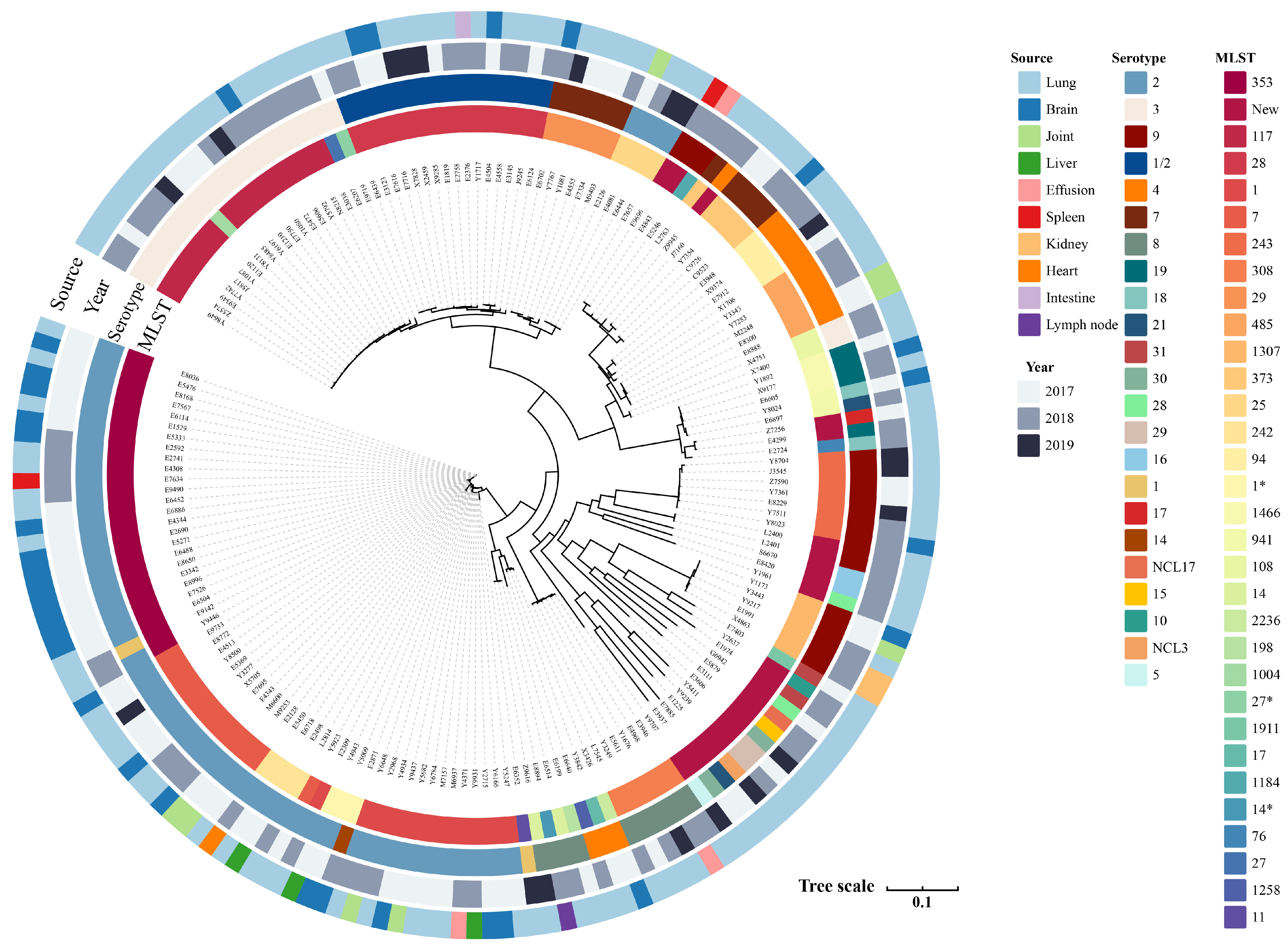
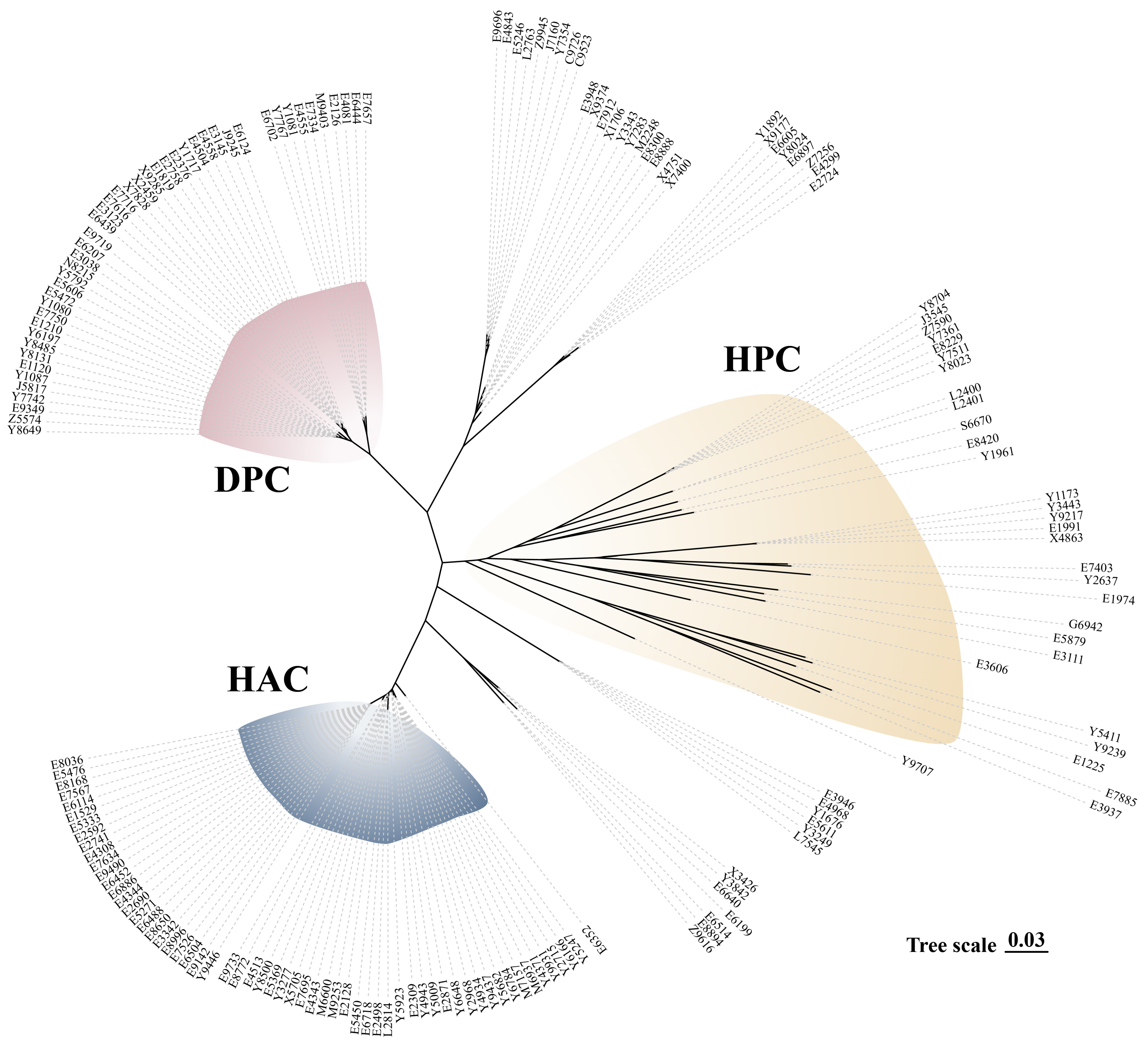
3.1. Serotype Distribution, MLST Identification, and Differential Clades

The 177 porcine isolates recovered in this study were obtained from clinical disease swine collected between 2017 and 2019, with each isolate originating from a different animal. The diseased tissues were obtained from 15 provinces in China, mainly from Hubei (n = 93/177) and Henan (n = 40/177) provinces (Table S2 and Figure S1). Isolates were recovered from the following anatomical sites: 70.0% (n = 124/177) from the lung; 18.0% (n = 32/177) from the brain; 4.5% (n = 8/177) from the joints; 1.7% (n = 3/177) each from the liver and effusion; 1.1% (n = 2/177) each from the spleen and kidney; and 0.5% (n = 1/177) each from the heart, intestine, and lymph nodes. Twenty-three serotypes were identified in the 177 isolates, with the majority of isolates being serotype 2 (n = 68, 38%), followed by those of serotype 3 (n = 22, 12%) and serotype 9 (n = 17, 12%). The remaining serotypes were distributed as serotypes 4 (n = 13), 7 (n = 11), 8 (n = 10), 1/2 (n = 9), and 19 (n = 4). The number of isolates in the remaining serotypes was less than or equal to 2 (Figure 1). According to the new typing scheme of the novel CPS loci (NCL) serotypes, one strain each of serotypes NCL17 and NCL3 on the new scheme was found. Both NCL17 and NCL3 were recovered from the lungs.
The top five isolates included ST353 (n = 25, 14.1%), ST117 (n = 17, 9.6%), ST1 (n = 17, 9.6%), ST28 (n = 16, 9.0%), and ST7 (n = 13, 7.3%), and the remaining STs were fewer in number (n < 10). In the following analysis, we identified 28 STs from the isolates, 22 of which were novel unique allele combinations (Figure 1). Serotype 2 consisted of six STs: ST1 (n = 16), ST7 (n = 13), ST25 (n = 4), ST28 (n = 7), ST242 (n = 4), and ST353 (n = 24). Serotype 3 consisted of four STs: ST27 (n = 2), ST108 (n = 2), ST117 (n = 17), and ST1004 (n = 1). Serotype 9 consisted of three STs and four novel STs; the known STs were ST243 (n = 7), ST1184 (n = 1), and ST1307 (n = 5). The phylogenetic tree (Figure 1) revealed that the serotype and MLST of Streptococcus suis isolates presented a high degree of diversity.
As previously defined by Dong et al. [4], large-scale genomic analysis on 1634 isolates of Streptococcus suis from a well-defined source has revealed three distinct bacterial clades, i.e., the healthy-pig clade, diseased-pig clade, and human-associated clade (HAC). There were still some isolates that had no clear clade (unclassified). Analyses using the reported protocols revealed in similar clades, i.e., HAC, DPC, and HPC (Figure 2).

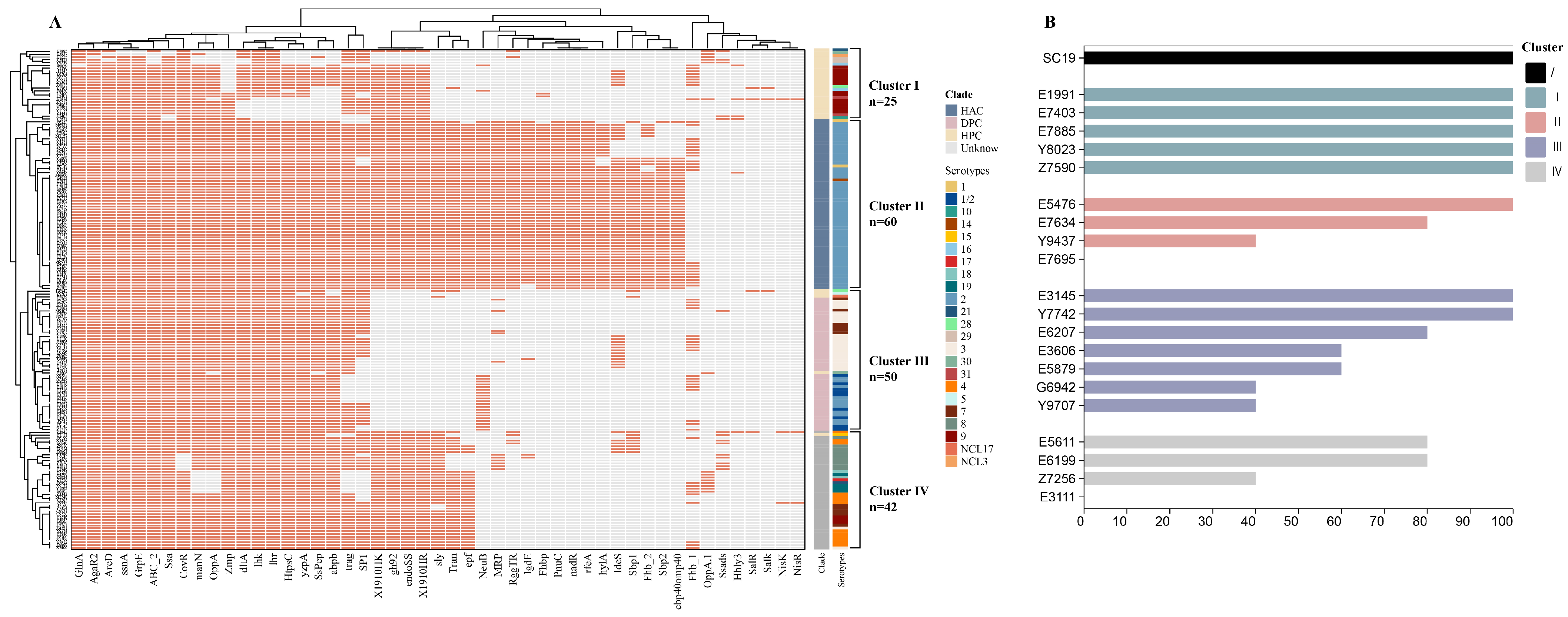
3.2. Analysis of Virulence-Related Genes and Virulence Phenotypes

To further examine the relationship that could exist between virulence genes and virulence phenotypes of isolates, we characterized the carriage of VAFs in the genomes of Streptococcus suis isolates (n = 177). Among the virulence-associated factors of Streptococcus suis, mrp, epf, and sly are the most studied genes. The classical VAFs, mrp, epf, and sly, were identified in 72 (40.6%), 90 (50.8%), and 102 (57.6%) of the 177 isolates, respectively. We further examined the distribution of combinations of mrp, epf, and sly genes (Supplemental Materials). These genes were dominated by mrp+epf+sly+ (60/177) and mrpepfsly (68/177), followed by mrpepf+sly+ (28/177) and mrp+epfsly+ (7/177), with no combined distribution of mrp+epf+sly.
Given the limited distribution of classical VAFs (mrp, epf, and sly) among the isolates, we used 104 previously published possible VAF genes (including mrp, epf, and sly) in pigs and humans (Table S3). Fifty-four VAFs were present in all 177 isolates, and the distribution of the presence of the remaining VAF genes is shown (Figure 3A). Clustering analysis was used to determine the relationship between the distribution of VAF and the isolates. The analysis of previously published possible VAF genes in pigs and humans identified four clusters (clusters I–IV) (Figure S3). Cluster II predominantly consisted of serotype 2 isolates, and 56% in cluster I were serotype 9. There was a high degree of overlap between cluster II and the HAC clade after clustering of the isolates, with all of cluster II (n = 60) occurring in the HAC clade. Cluster I (n = 25) appeared only in the HPC, and cluster III was distributed in the DPC (n = 46) and HPC (n = 4). Only one strain in cluster IV was from the HPC, and the remaining isolates (n = 41) were from the unknown clade (Figure S3). Interestingly, isolates with 100% mortality in HPC (Figure S3) all appeared in cluster I, while isolates with 40–60% mortality appeared in cluster III of HPC, and the isolates that did not cause mouse death were in cluster IV of HPC (n = 1).
To understand the virulence profile of the strains in the different clusters, we selected five isolates in cluster Ⅰ (Table S7), four isolates assigned to cluster II, seven isolates assigned to cluster III, and four strains assigned to cluster Ⅳ, for a total of twenty isolates (Figure 3B). The survival of mice inoculated intraperitoneally with a 5 × 10⁸ CFU dose (n = 5) of the different isolates until 7 days post-infection was assessed. We found that 18 isolates caused the death of the mice (Figure 3B), and that all infected mice presented clinical signs; the strains in the subgroups that did not cause death in mice were from cluster II and cluster Ⅳ. All of the mice died within 72 h of intraperitoneal injection of Streptococcus suis isolates (Table S7), during which mortality was the result of sepsis and/or septic shock. Streptococcus suis isolates that caused 100% mortality were present in clusters Ⅰ, II, and III. Five isolates of cluster Ⅰ caused 100% mortality in mice, and there was no difference from SC19.

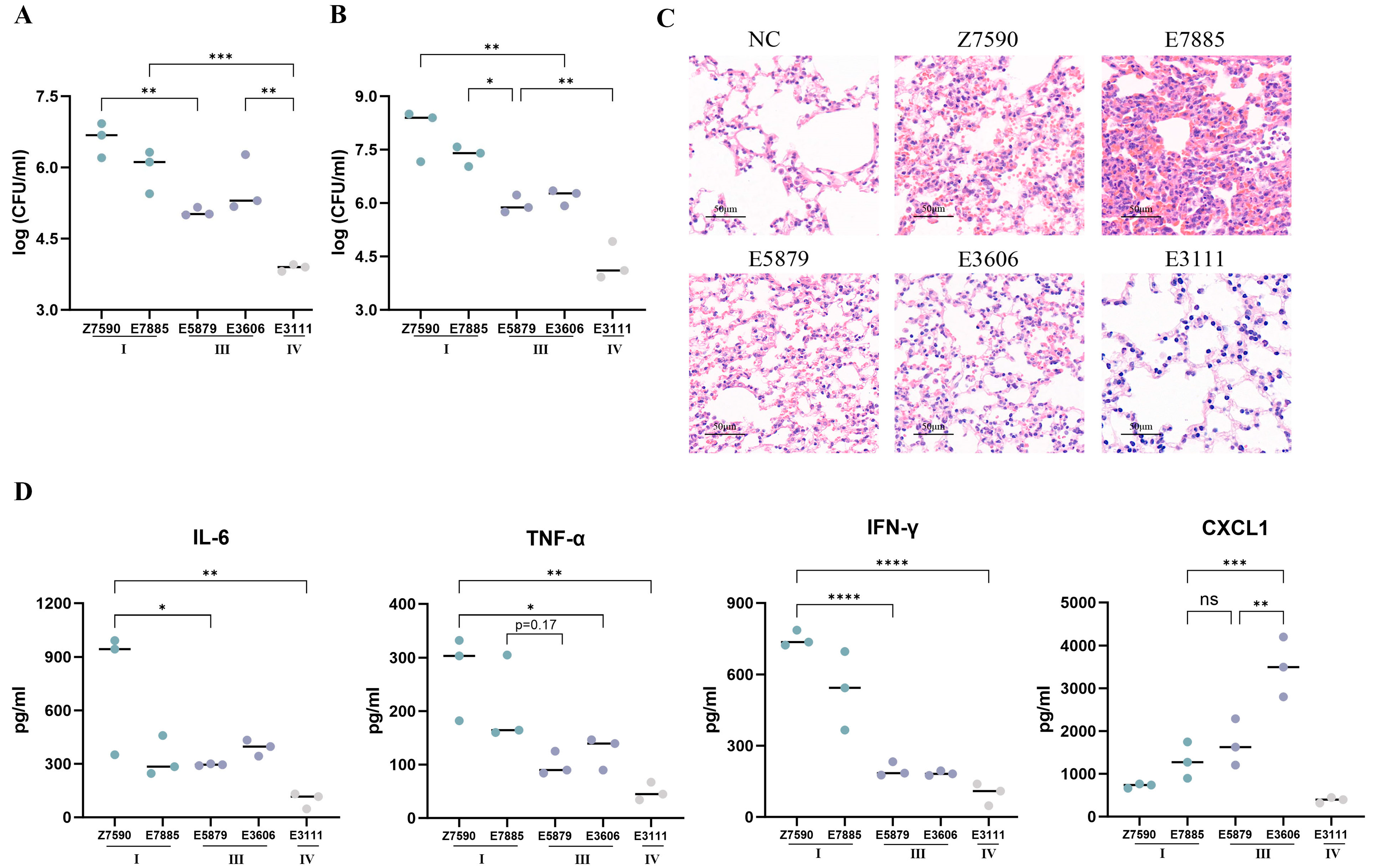
3.3. Mouse Model of Infection

The mortality caused by Streptococcus suis isolates may be the result of a combination of high blood/tissue bacterial titers and a systemic inflammatory response. Consequently, the bacterial burden in the blood, lungs, and brain, and the levels of cytokines in the serum were measured at 12 h post-infection. Significant differences were observed between cluster I-infected mice and cluster III-infected mice in lung tissue and blood (Figure 4B), and bacterial titers in cluster IV-infected mice were lower than those in the other two groups. The bacterial number of cluster I-infected mice was high, the number of cluster III-infected mice was intermediate, and the bacterial number of cluster IV-infected mice was low (Figure 4A). Infection of mice with cluster I strains (Z7590 and E7885) resulted in significant organ damage and signs of congestion and infiltration of inflammatory cells into the lungs (Figure 4C). One of the strains, strain E7885, was more clearly demonstrated in the lung tissue. The lung tissue damage and inflammatory cell infiltration caused by the cluster III isolates were not as severe as those caused by cluster I isolates, and no significant tissue damage or inflammatory cell infiltration was observed with the cluster IV isolates.
Excessive inflammation may also lead to death in mice. Cytokines and chemokines were measured in the plasma 12 h after the different strains were used to infect the mice (Figure 4D). The strains of cluster I all induced higher levels of interferon (IFN)-γ than those of cluster III (p < 0.001). Strain Z7590 in cluster I and both strains in cluster III showed significant differences in interleukin (IL)-6 and tumor necrosis factor (TNF)-α (p < 0.05), whereas strain E7885 did not. There were no significant differences in strains between the same VAF clusters. The chemokine C-X-C motif ligand (CXCL)1 was also detected. Interestingly, mice in cluster III with moderate mortality produced higher levels of CXCL1; specifically, strain E3111 produced 3 × higher CXCL1 expression than strain E7885 (p < 0.001). E3111 induced the lowest levels of IFN-γ, TNF-α, IL-6, and CXCL1.

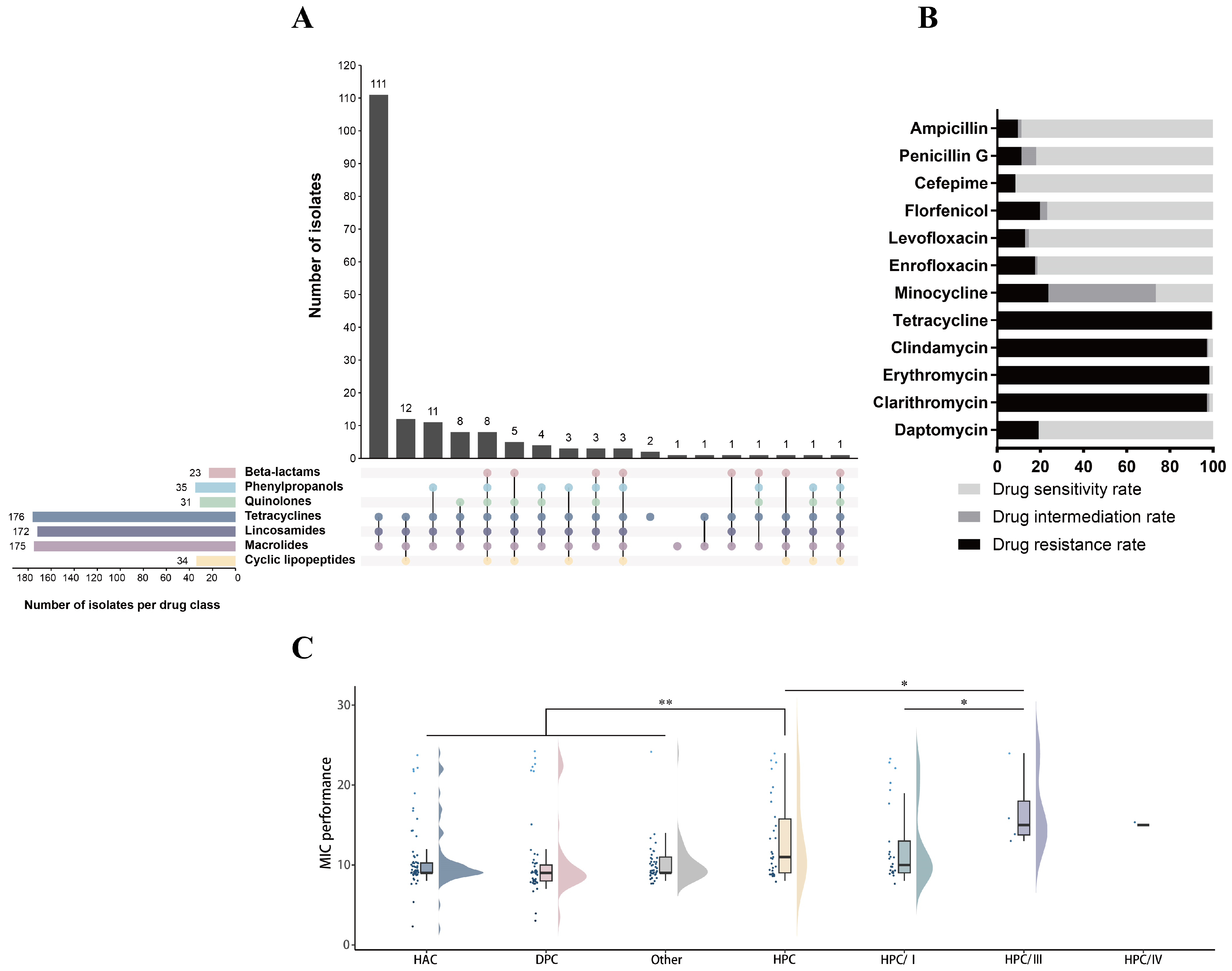
3.4. Antibiotic Resistance Phenotypes and Genes

To assess the antibiotic resistance of the collected Streptococcus suis isolates, we conducted susceptibility tests against twelve antibiotics of seven different species of these isolates. All of the isolates were resistant to at least one antibiotic, and no isolates resistant to all twelve antibiotics were found at this time. Multi-resistant (resistant to antibiotics from at least three classes) strains accounted for 97.7% of the isolates, with 62.7% of the isolates (n = 111) being resistant to three classes of antibiotics (Figure 5A,B). A large proportion of these strains were resistant to Tetracycline (99.4%, n = 176), Clindamycin (97.1%, n = 172), Erythromycin (98.3%, n = 174), and Clarithromycin (97.1%, n = 172), and a small proportion of them were resistant to Ampicillin (9.6%, n = 17) and Cefepime (8.4%, n = 15). Further, we analyzed the resistance phenotypes of isolates classified as HAC, HPC, and DPC (Figure 5C). Isolates assigned to the HPC clade showed significant differences in drug resistance from the other clades. In the HPC clade, cluster III with intermediate mortality was more resistant than cluster I with high mortality, and they were significantly different from each other (p < 0.05). No significant differences in resistance phenotypes were observed among isolates from the HAC, DPC, and unknown clades.
To understand the genomic basis of the drug resistance phenotype of Streptococcus suis, we identified the presence of ARGs in the genomic sequence (Figure 6A). We found that the Streptococcus suis genome is abundant in antibiotic resistance genes, with a total of 26 ARGs identified (Table S4), 18 of which were carried by ICE and/or IME (Figure 6B and Table S5). The ARGs were identified as six aminoglycoside genes, (APH(3)-IIIa, SAT-4, aad(6), ANT(6)-Ia, AAC(6)-Ie-APH(2)-Ia, and ANT(9)-Ia), and seven tetracycline genes, (tet(O), tet(40), tet(M), tet(W), tet(32), tet(O/W/32/O), and tet(L)). All 177 strains carried the resistance gene for Quinolones was patB. The Gene ErmB for Macrolides accounted for 92.0% in all isolates, tet(O) for Tetracyclines accounted for 90.3%, the Gene patA for Quinolones accounted for 89.2% in all isolates, and other resistance genes were detected in less than 50% of cases. The most resistance genes carried by ICE and IME were ErmB and tet(O). Moreover, 92.9% of the strains identified as carrying tet(40) were serotype 2, with APH(3)-IIIa in 90%, SAT-4 in 92.3%, and aad(6) in 97.1%.

4. Discussion

Streptococcus suis is a zoonotic pathogen with a worldwide presence [43]. Streptococcus suis is a highly diverse species [44], with the current findings identifying 23 serotypes and 28 sequence types among the isolates. Notably, serotypes 2 and 3 were the most frequently isolated from clinical cases in Canada [5], whereas another investigation indicated that serotypes 4 and 7 were more prevalent during the period of 2015–2016 compared to 1996–2004 [44]. ST7 was found only in China [19], with all ST7 strains classified as serotype 2. Additionally, four of the twenty-two strains representing novel sequence types were identified as serotype 9, whereas the remaining strains included two each of serotypes 16, 28, 29, and 30. Serotype 9 exhibited considerable genetic heterogeneity and diversity [45,46].
Previous studies have delineated three distinct clades of Streptococcus suis: the healthy-pig clade (HPC), the diseased-pig clade (DPC), and the human-associated clade (HAC) [4]. Analysis of isolates with known origins revealed that 96% of human isolates were classified within the HAC, 72.2% of isolates from healthy pigs clustered in the HPC, and 69% of isolates from diseased pigs were categorized in the DPC [4]. The isolates of Streptococcus suis obtained from sick pigs in this study were similarly distributed among the HAC (34.5%), HPC (16.9%), and DPC (26%) clades. Furthermore, genomic data from tonsils of clinically healthy pigs [12] revealed a distribution of 2.7% in HAC, 90.1% in HPC, and 2.2% in DPC (Figure S2). These findings suggest that isolates derived from diseased pigs may possess a richer genomic repertoire.
Muramidase-released protein (mrp), extracellular protein factor (epf), and suilysin (sly) represent the most extensively researched virulence-associated factors (VAFs) and have been utilized as indicators of the virulence profile of Streptococcus suis [28]. In clinical disease samples collected in Canada, isolates were found to be negative for mrp [11], and the absence of mrp was predominantly noted in serotype 9 isolates from affected pigs [47]. While mrp is correlated with virulent strains of ST1, the results are not the same for other serotype 2 STs [16]. Additionally, studies utilizing mouse intraperitoneal infection models indicated that bacterial burdens were significantly greater in sly-positive strains [48,49], and when employing an intranasal mouse model for mucosal colonization, the bacterial burden of an isogenic sly mutant did not differ significantly from that of the wild-type strain [50]. Findings from the porcine intranasal infection model suggest that mrp functions more as a “virulence marker” rather than a definitive “virulence factor” [51]. In the present study, despite the isolates being derived from diseased pigs, mrp was absent in 40.7% of the isolates, epf was absent in 49.1%, and sly was absent in 42.4%, with a combined mrpepfsly genotype present in 38.4% of cases. In contrast, numerous isolates from healthy pigs presented the epf+mrp+sly+ genotype [12]. Importantly, for many pathogens, the knockout of specific genes or proteins does not yield significant phenotypic changes, and in some instances, the functions may be preserved, thereby complicating the analysis of virulence factors due to redundancy [28]. In conclusion, Streptococcus suis is a pathogen characterized by virulence factor redundancy, and virulence-associated factors in conjunction with transcriptomic and proteomic methods can more effectively elucidate the regulatory mechanisms of virulence in future studies.
On the other hand, the virulence of Streptococcus suis exhibits notable heterogeneity, necessitating infection experiments to validate the virulence of various strains [28]. In this study, our isolates were categorized into clusters I–IV based on virulence-associated factor (VAF) cluster analysis. Cluster II completely overlaps with the HAC, with serotype 2—recognized as the most prevalent serotype responsible for human infections—comprising 86.4% of this cluster. Cluster II may be linked to human infections caused by suis, and it is characterized by the greatest number of identified virulence factors. While the genomic size of human-associated suis isolates is diminishing, there is a tendency for the encoding of known or putative virulence factors to occur at high frequencies in pathogenic isolates [4,52]. The virulence-related genes, hylA, PnuC, rfeA, Sbp1, and Sbp2, only appeared in cluster II, and these genes are considered as putative zoonotic virulence factors [39]. The HPC not only illustrates evolutionary diversity within the phylogenetic tree but also encompasses three additional clusters beyond cluster II in the VAFs. Cluster I, which is exclusive to the HPC, demonstrates high virulence and is associated with elevated mortality rates in animal models. The 1901HK/RR, endoSS, and GH 92 genes were present in cluster I but not detected in cluster III. The 1901HK/RR is associated with adherence and invasion, aiding suis in surviving in the bloodstream [53]. EndoSS-inserted suis exhibit a higher proliferative capacity, while GH 92 can degrade N-glycans to generate endoSS substrates [54]; these VAFs may facilitate higher loads of suis in host blood and tissues. Although the number of VAFs identified in cluster I was not the highest, this may be due to differences in the expression of genes highly correlated with virulence [38], or it is also possible that the lack of transcriptional regulators, combined with an increase in genes encoding DNA replication and repair proteins in the genome [55], resulting in a massive increase in bacterial populations within the host [52,55] and ultimately the death of the host. Compared with those from clusters III and IV, strains Z7590 and E7885 from cluster I presented significantly greater bacterial loads in the lungs and bloodstream and induced elevated levels of IL-6, TNF-α, and IFN-γ in the lungs and blood of infected mice, which, in turn, leads to the development of inflammatory diseases, such as fever and septic shock [56]. An excessive inflammatory response and high bacterial load may trigger sepsis [57], which is a leading cause of death [58]. Strains Z7590 and E7885 induced high levels of proinflammatory mediators. However, the medium mortality strain E3606 induced increased expression of CXCL1 chemokines. CXCL1 is known to activate neutrophils and enhance their phagocytic and bactericidal capabilities [59], and this may result in the premature clearance of E3606 by neutrophils before it can cause damage to the lungs or other tissues, thereby enhancing survival in mice. Significant tissue damage and cellular infiltration were observed in tissue sections following infection with strain E7885, thereby increasing the likelihood of mortality in mice [60]. Although the HPC represents an evolutionary clade associated with the colonization of healthy pigs, the strains classified within it retain the potential to cause disease and include highly virulent cluster I strains.
The findings from the Minimum Inhibitory Concentration (MIC) analysis revealed that a significant proportion of Streptococcus suis isolates are categorized as multi-drug resistant (MDR). Notably, strains within the HPC exhibit heightened resistance levels, and the presence of antibiotic pressure appears to facilitate conditions conducive to host colonization [61]. The HPC clade demonstrates increased resistance within cluster III, which is associated with moderate virulence, while strains exhibiting lower virulence may confer certain advantages [62]. Of particular interest is the strain E7885 from cluster I lineage, which is characterized by both high virulence and resistance to ten different antibiotics. Streptococcus suis is a repository of resistance genes [8], and more than half of the resistance genes in Streptococcus suis are carried by ICE and/or IME [6,9]. In this study, 73.1% of the resistance genes identified were located in ICE and or IME. Notably, despite the prohibition of chloramphenicol usage in animal production facilities in China since 2002 [63], the chloramphenicol resistance genes, catA8, fexA, and cat-TC, were identified. The data presented in this report underscore the genetic diversity present within Streptococcus suis. This study also assessed the virulence-associated factors (VAFs) and resistance genes among various isolates. The MIC analysis established the resistance profiles of the strains and corroborated the virulence of the isolates in animal models.

Supplementary Materials

The following supporting information can be downloaded at https://www.mdpi.com/article/10.3390/microorganisms13061197/s1, Figure S1. The map of China showing Streptococcus suis from different provinces. Figure S2. Phylogenetic tree of Streptococcus suis isolates of different origins. Tips in blue were obtained from the tonsils of clinically healthy pigs (n = 223), tips in red for the isolates in this study (n = 177), and tips in green from the database (n = 240). Figure S3. Phylogenetic tree (unrooted) of evolutionary clade and VAF clade. Tips are colored according to clade, blue for HAC, red for DPC, and yellow for HPC. Table S1. The information of 177 suis isolates in this study. Table S2. Serotypes, MLST, clades, and VF clusters of the 177 suis used in this study. Table S3. The matrix of the presence or absence of VAFs in 177 suis genomes. Table S4. The matrix of the presence or absence of ARGs in 177 suis genomes. Table S5. The matrix of the presence or absence of ARGs carried by ICE and IME. Table S6. The MIC values and folding points of the 12 antibiotics. Table S7. The data of the BALB/c mice infection model.

Author Contributions

Y.L. drafted the manuscript; Y.L., X.J., S.N., G.C. and X.Z. carried out the experiments; B.M. and Y.W. generated the data; Y.L. and B.M. analyzed the data; C.T., H.J., J.L. and B.M. reviewed the manuscript; C.T. and H.J. designed the project. All authors have read and agreed to the published version of the manuscript.

Funding

This study has been supported by the National Key Research and Development Program (2022YFD1800901), the China Agriculture Research System of MOF, and MARA (CARS-35).

Institutional Review Board Statement

The animal study protocols were conducted with strict adherence to the guidelines for the Care and Use of Laboratory Animals Monitoring Committee of Hubei Province, China, and the protocol was approved by the Scientific Ethics Committee of Huazhong Agricultural University (ethical approval date: 10 December 2024).

Informed Consent Statement

Not applicable.

Data Availability Statement

The original contributions presented in the study are included in the article/Supplementary Materials, and further inquiries can be directed to the corresponding authors.

Conflicts of Interest

The authors declare no conflicts of interest.

References

  1. Bojarska, A.; Molska, E.; Janas, K.; Skoczyńska, A.; Stefaniuk, E.; Hryniewicz, W.; Sadowy, E. Streptococcus suis in invasive human infections in Poland: Clonality and determinants of virulence and antimicrobial resistance. Eur. J. Clin. Microbiol. Infect. Dis. 2016, 35, 917–925. [Google Scholar] [CrossRef] [PubMed]
  2. Dutkiewicz, J.; Zajac, V.; Sroka, J.; Wasinski, B.; Cisak, E.; Sawczyn, A.; Kloc, A.; Wójcik-Fatla, A. Streptococcus suis: A re-emerging pathogen associated with occupational exposure to pigs or pork products. Part II–Pathogenesis. Ann. Agric. Environ. Med. 2018, 25, 186–203. [Google Scholar] [CrossRef] [PubMed]
  3. Ferrando, M.L.; De Greeff, A.; van Rooijen, W.J.; Stockhofe-Zurwieden, N.; Nielsen, J.; Wichgers Schreur, P.J.; Pannekoek, Y.; Heuvelink, A.; Van Der Ende, A.; Smith, H. Host-pathogen interaction at the intestinal mucosa correlates with zoonotic potential of Streptococcus suis. J. Infect. Dis. 2015, 212, 95–105. [Google Scholar] [CrossRef]
  4. Dong, X.; Chao, Y.; Zhou, Y.; Zhou, R.; Zhang, W.; Fischetti, V.A.; Wang, X.; Feng, Y.; Li, J. The global emergence of a novel Streptococcus suis clade associated with human infections. EMBO Mol. Med. 2021, 13, e13810. [Google Scholar] [CrossRef]
  5. Goyette-Desjardins, G.; Auger, J.-P.; Xu, J.; Segura, M.; Gottschalk, M. Streptococcus suis, an important pig pathogen and emerging zoonotic agent—An update on the worldwide distribution based on serotyping and sequence typing. Emerg. Microbes Infect. 2014, 3, 1–20. [Google Scholar] [CrossRef]
  6. Libante, V.; Nombre, Y.; Coluzzi, C.; Staub, J.; Guédon, G.; Gottschalk, M.; Teatero, S.; Fittipaldi, N.; Leblond-Bourget, N.; Payot, S. Chromosomal conjugative and mobilizable elements in Streptococcus suis: Major actors in the spreading of antimicrobial resistance and bacteriocin synthesis genes. Pathogens 2019, 9, 22. [Google Scholar] [CrossRef] [PubMed]
  7. Arndt, E.R.; Farzan, A.; MacInnes, J.I.; Friendship, R.M. Antimicrobial resistance of Streptococcus suis isolates recovered from clinically ill nursery pigs and from healthy pigs at different stages of production. Can. Vet. J. 2019, 60, 519–522. [Google Scholar]
  8. Uruén, C.; García, C.; Fraile, L.; Tommassen, J.; Arenas, J. How Streptococcus suis escapes antibiotic treatments. Vet. Res. 2022, 53, 91. [Google Scholar] [CrossRef]
  9. Huang, J.; Ma, J.; Shang, K.; Hu, X.; Liang, Y.; Li, D.; Wu, Z.; Dai, L.; Chen, L.; Wang, L. Evolution and diversity of the antimicrobial resistance associated mobilome in Streptococcus suis: A probable mobile genetic elements reservoir for other streptococci. Front. Cell. Infect. Microbiol. 2016, 6, 118. [Google Scholar] [CrossRef]
  10. Huang, J.; Liang, Y.; Guo, D.; Shang, K.; Ge, L.; Kashif, J.; Wang, L. Comparative genomic analysis of the ICE Sa 2603 family ICEs and spread of erm (B)-and tet (O)-carrying transferable 89K-subtype ICEs in swine and bovine isolates in China. Front. Microbiol. 2016, 7, 55. [Google Scholar] [CrossRef]
  11. Aradanas, M.; Poljak, Z.; Fittipaldi, N.; Ricker, N.; Farzan, A. Serotypes, virulence-associated factors, and antimicrobial resistance of Streptococcus suis isolates recovered from sick and healthy pigs determined by whole-genome sequencing. Front. Vet. Sci. 2021, 8, 742345. [Google Scholar] [CrossRef]
  12. Zou, G.; Zhou, J.; Xiao, R.; Zhang, L.; Cheng, Y.; Jin, H.; Li, L.; Zhang, L.; Wu, B.; Qian, P. Effects of environmental and management-associated factors on prevalence and diversity of Streptococcus suis in clinically healthy pig herds in China and the United Kingdom. Appl. Environ. Microbiol. 2018, 84, e02590-17. [Google Scholar] [CrossRef] [PubMed]
  13. Uruen, C.; Gimeno, J.; Sanz, M.; Fraile, L.; Marín, C.M.; Arenas, J. Invasive Streptococcus suis isolated in Spain contain a highly promiscuous and dynamic resistome. Front. Cell. Infect. Microbiol. 2024, 13, 1329632. [Google Scholar] [CrossRef]
  14. King, S.J.; Leigh, J.A.; Heath, P.J.; Luque, I.; Tarradas, C.; Dowson, C.G.; Whatmore, A.M. Development of a multilocus sequence typing scheme for the pig pathogen Streptococcus suis: Identification of virulent clones and potential capsular serotype exchange. J. Clin. Microbiol. 2002, 40, 3671–3680. [Google Scholar] [CrossRef] [PubMed]
  15. Callejo, R.; Zheng, H.; Du, P.; Prieto, M.; Xu, J.; Zielinski, G.; Auger, J.-P.; Gottschalk, M. Streptococcus suis serotype 2 strains isolated in Argentina (South America) are different from those recovered in North America and present a higher risk for humans. JMM Case Rep. 2016, 3, e005066. [Google Scholar] [CrossRef]
  16. Fittipaldi, N.; Xu, J.; Lacouture, S.; Tharavichitkul, P.; Osaki, M.; Sekizaki, T.; Takamatsu, D.; Gottschalk, M. Lineage and virulence of Streptococcus suis serotype 2 isolates from North America. Emerg. Infect. Dis. 2011, 17, 2239–2244. [Google Scholar] [CrossRef]
  17. Estrada, A.A.; Gottschalk, M.; Rossow, S.; Rendahl, A.; Gebhart, C.; Marthaler, D.G. Serotype and genotype (multilocus sequence type) of Streptococcus suis isolates from the United States serve as predictors of pathotype. J. Clin. Microbiol. 2019, 57, e00377-19. [Google Scholar] [CrossRef]
  18. Huang, J.; Shang, K.; Kashif, J.; Wang, L. Genetic diversity of Streptococcus suis isolated from three pig farms of China obtained by acquiring antibiotic resistance genes. J. Sci. Food Agric. 2015, 95, 1454–1460. [Google Scholar] [CrossRef] [PubMed]
  19. Ye, C.; Zhu, X.; Jing, H.; Du, H.; Segura, M.; Zheng, H.; Kan, B.; Wang, L.; Bai, X.; Zhou, Y. Streptococcus suis sequence type 7 outbreak, Sichuan, China. Emerg. Infect. Dis. 2006, 12, 1203–1208. [Google Scholar] [CrossRef]
  20. Okura, M.; Osaki, M.; Nomoto, R.; Arai, S.; Osawa, R.; Sekizaki, T.; Takamatsu, D. Current taxonomical situation of Streptococcus suis. Pathogens 2016, 5, 45. [Google Scholar] [CrossRef]
  21. Zheng, H.; Ji, S.; Liu, Z.; Lan, R.; Huang, Y.; Bai, X.; Gottschalk, M.; Xu, J. Eight novel capsular polysaccharide synthesis gene loci identified in nontypeable Streptococcus suis isolates. Appl. Environ. Microbiol. 2015, 81, 4111–4119. [Google Scholar] [CrossRef] [PubMed]
  22. Qiu, X.; Bai, X.; Lan, R.; Zheng, H.; Xu, J. Novel capsular polysaccharide loci and new diagnostic tools for high-throughput capsular gene typing in Streptococcus suis. Appl. Environ. Microbiol. 2016, 82, 7102–7112. [Google Scholar] [CrossRef] [PubMed]
  23. Zheng, H.; Qiu, X.; Roy, D.; Segura, M.; Du, P.; Xu, J.; Gottschalk, M. Genotyping and investigating capsular polysaccharide synthesis gene loci of non-serotypeable Streptococcus suis isolated from diseased pigs in Canada. Vet. Res. Commun. 2017, 48, 10. [Google Scholar] [CrossRef]
  24. Huang, J.; Liu, X.; Chen, H.; Chen, L.; Gao, X.; Pan, Z.; Wang, J.; Lu, C.; Yao, H.; Wang, L. Identification of six novel capsular polysaccharide loci (NCL) from Streptococcus suis multidrug resistant non-typeable strains and the pathogenic characteristic of strains carrying new NCL s. Transbound. Emerg. Dis. 2019, 66, 995–1003. [Google Scholar] [CrossRef]
  25. Segura, M. Streptococcus suis research: Progress and challenges. Pathogens 2020, 9, 707. [Google Scholar] [CrossRef]
  26. Petrocchi-Rilo, M.; Martínez-Martínez, S.; Aguarón-Turrientes, Á.; Roca-Martínez, E.; García-Iglesias, M.-J.; Pérez-Fernández, E.; González-Fernández, A.; Herencia-Lagunar, E.; Gutiérrez-Martín, C.-B. Anatomical site, typing, virulence gene profiling, antimicrobial susceptibility and resistance genes of Streptococcus suis isolates recovered from pigs in Spain. Antibiotics 2021, 10, 707. [Google Scholar] [CrossRef] [PubMed]
  27. Gottschalk, M.; Lacouture, S.; Bonifait, L.; Roy, D.; Fittipaldi, N.; Grenier, D. Characterization of Streptococcus suis isolates recovered between 2008 and 2011 from diseased pigs in Quebec, Canada. Vet. Microbiol. 2013, 162, 819–825. [Google Scholar] [CrossRef]
  28. Segura, M.; Fittipaldi, N.; Calzas, C.; Gottschalk, M. Critical Streptococcus suis virulence factors: Are they all really critical? Trends Microbiol. 2017, 25, 585–599. [Google Scholar] [CrossRef]
  29. Athey, T.B.; Auger, J.-P.; Teatero, S.; Dumesnil, A.; Takamatsu, D.; Wasserscheid, J.; Dewar, K.; Gottschalk, M.; Fittipaldi, N. Complex population structure and virulence differences among serotype 2 Streptococcus suis strains belonging to sequence type 28. PLoS ONE 2015, 10, e0137760. [Google Scholar] [CrossRef]
  30. Auger, J.-P.; Fittipaldi, N.; Benoit-Biancamano, M.-O.; Segura, M.; Gottschalk, M. Virulence studies of different sequence types and geographical origins of Streptococcus suis serotype 2 in a mouse model of infection. Pathogens 2016, 5, 48. [Google Scholar] [CrossRef]
  31. Estrada, A.A.; Gottschalk, M.; Rendahl, A.; Rossow, S.; Marshall-Lund, L.; Marthaler, D.G.; Gebhart, C.J. Proposed virulence-associated genes of Streptococcus suis isolates from the United States serve as predictors of pathogenicity. Porc. Health Manag. 2021, 7, 22. [Google Scholar] [CrossRef]
  32. Li, Q.; Fu, Y.; Ma, C.; He, Y.; Yu, Y.; Du, D.; Yao, H.; Lu, C.; Zhang, W. The non-conserved region of MRP is involved in the virulence of Streptococcus suis serotype 2. Virulence 2017, 8, 1274–1289. [Google Scholar] [CrossRef]
  33. Xia, X.; Qin, W.; Zhu, H.; Wang, X.; Jiang, J.; Hu, J. How Streptococcus suis serotype 2 attempts to avoid attack by host immune defenses. Immunol. Infect. 2019, 52, 516–525. [Google Scholar] [CrossRef] [PubMed]
  34. Lun, S.; Perez-Casal, J.; Connor, W.; Willson, P. Role of suilysin in pathogenesis of Streptococcus suis capsular serotype 2. Microb. Pathog. 2003, 34, 27–37. [Google Scholar] [CrossRef] [PubMed]
  35. He, Z.; Pian, Y.; Ren, Z.; Bi, L.; Yuan, Y.; Zheng, Y.; Jiang, Y.; Wang, F. Increased production of suilysin contributes to invasive infection of the Streptococcus suis strain 05ZYH33. Mol. Med. Rep. 2014, 10, 2819–2826. [Google Scholar] [CrossRef] [PubMed]
  36. Smith, H.E.; Vecht, U.; Wisselink, H.J.; Stockhofe-Zurwieden, N.; Biermann, Y.; Smits, M.A. Mutants of Streptococcus suis types 1 and 2 impaired in expression of muramidase-released protein and extracellular protein induce disease in newborn germfree pigs. Infect. Immun. 1996, 64, 4409–4412. [Google Scholar] [CrossRef]
  37. O’Connor, T.J.; Boyd, D.; Dorer, M.S.; Isberg, R.R. Aggravating genetic interactions allow a solution to redundancy in a bacterial pathogen. Science 2012, 338, 1440–1444. [Google Scholar] [CrossRef]
  38. Wang, J.; Liang, P.; Sun, H.; Wu, Z.; Gottschalk, M.; Qi, K.; Zheng, H. Comparative transcriptomic analysis reveal genes involved in the pathogenicity increase of Streptococcus suis epidemic strains. Virulence 2022, 13, 1455–1470. [Google Scholar] [CrossRef]
  39. Roodsant, T.J.; Van Der Putten, B.C.; Tamminga, S.M.; Schultsz, C.; Van Der Ark, K.C. Identification of Streptococcus suis putative zoonotic virulence factors: A systematic review and genomic meta-analysis. Virulence 2021, 12, 2787–2797. [Google Scholar] [CrossRef]
  40. Segura, M.; Calzas, C.; Grenier, D.; Gottschalk, M. Initial steps of the pathogenesis of the infection caused by Streptococcus suis: Fighting against nonspecific defenses. FEBS Lett. 2016, 590, 3772–3799. [Google Scholar] [CrossRef]
  41. Arenas, J.; Zomer, A.; Harders-Westerveen, J.; Bootsma, H.J.; De Jonge, M.I.; Stockhofe-Zurwieden, N.; Smith, H.E.; De Greeff, A. Identification of conditionally essential genes for Streptococcus suis infection in pigs. Virulence 2020, 11, 446–464. [Google Scholar] [CrossRef] [PubMed]
  42. Athey, T.B.; Teatero, S.; Lacouture, S.; Takamatsu, D.; Gottschalk, M.; Fittipaldi, N. Determining Streptococcus suis serotype from short-read whole-genome sequencing data. BMC Microbiol. 2016, 16, 162. [Google Scholar] [CrossRef]
  43. Lun, Z.-R.; Wang, Q.-P.; Chen, X.-G.; Li, A.-X.; Zhu, X.-Q. Streptococcus suis: An emerging zoonotic pathogen. Lancet Infect. Dis. 2007, 7, 201–209. [Google Scholar] [CrossRef] [PubMed]
  44. Prüfer, T.L.; Rohde, J.; Verspohl, J.; Rohde, M.; De Greeff, A.; Willenborg, J.; Valentin-Weigand, P. Molecular typing of Streptococcus suis strains isolated from diseased and healthy pigs between 1996–2016. PLoS ONE 2019, 14, e0210801. [Google Scholar] [CrossRef]
  45. Scherrer, S.; Rosato, G.; Spoerry Serrano, N.; Stevens, M.J.; Rademacher, F.; Schrenzel, J.; Gottschalk, M.; Stephan, R.; Peterhans, S. Population structure, genetic diversity and pathotypes of Streptococcus suis isolated during the last 13 years from diseased pigs in Switzerland. Vet. Res. 2020, 51, 85. [Google Scholar] [CrossRef]
  46. Dong, W.; Zhu, Y.; Ma, Y.; Ma, J.; Zhang, Y.; Yuan, L.; Pan, Z.; Wu, Z.; Yao, H. Multilocus sequence typing and virulence genotyping of Streptococcus suis serotype 9 isolates revealed high genetic and virulence diversity. FEMS Microbiol. Lett. 2017, 364, fnx192. [Google Scholar] [CrossRef] [PubMed]
  47. Blume, V.; Luque, I.; Borge, C.; Maldonado, A.; Domínguez Rodríquez, L.; Tarradas, C.; Fernández-Garayzábal Fernández, J.F.; Vela Alonso, A.I. Genetic and virulence-phenotype characterization of serotypes 2 and 9 of Streptococcus suis swine isolates. Int. Microbiol. 2009, 12, 161–166. [Google Scholar]
  48. Allen, A.G.; Bolitho, S.; Lindsay, H.; Khan, S.; Bryant, C.; Norton, P.; Ward, P.; Leigh, J.; Morgan, J.; Riches, H. Generation and characterization of a defined mutant of Streptococcus suis lacking suilysin. Infect. Immun. 2001, 69, 2732–2735. [Google Scholar] [CrossRef]
  49. Takeuchi, D.; Akeda, Y.; Nakayama, T.; Kerdsin, A.; Sano, Y.; Kanda, T.; Hamada, S.; Dejsirilert, S.; Oishi, K. The contribution of suilysin to the pathogenesis of Streptococcus suis meningitis. J. Infect. Dis. 2014, 209, 1509–1519. [Google Scholar] [CrossRef]
  50. Seitz, M.; Beineke, A.; Singpiel, A.; Willenborg, J.; Dutow, P.; Goethe, R.; Valentin-Weigand, P.; Klos, A.; Baums, C. Role of capsule and suilysin in mucosal infection of complement-deficient mice with Streptococcus suis. Infect. Immun. 2014, 82, 2460–2471. [Google Scholar] [CrossRef]
  51. Baums, C.G.; Valentin-Weigand, P. Surface-associated and secreted factors of Streptococcus suis in epidemiology, pathogenesis and vaccine development. Anim. Health Res. Rev. 2009, 10, 65–83. [Google Scholar] [CrossRef] [PubMed]
  52. Weinert, L.A.; Chaudhuri, R.R.; Wang, J.; Peters, S.E.; Corander, J.; Jombart, T.; Baig, A.; Howell, K.J.; Vehkala, M.; Välimäki, N. Genomic signatures of human and animal disease in the zoonotic pathogen Streptococcus suis. Nat. Commun. 2015, 6, 6740. [Google Scholar] [CrossRef] [PubMed]
  53. Yuan, F.; Tan, C.; Liu, Z.; Yang, K.; Zhou, D.; Liu, W.; Duan, Z.; Guo, R.; Chen, H.; Tian, Y. The 1910HK/RR two-component system is essential for the virulence of Streptococcus suis serotype 2. Microb. Pathog. 2017, 104, 137–145. [Google Scholar] [CrossRef]
  54. Ma, J.; Zhang, Z.; Pan, Z.; Bai, Q.; Zhong, X.; Zhu, Y.; Zhang, Y.; Wu, Z.; Liu, G.; Yao, H. Streptococcus suis uptakes carbohydrate source from host glycoproteins by N-glycans degradation system for optimal survival and full virulence during infection. Pathogens 2020, 9, 387. [Google Scholar] [CrossRef]
  55. Merhej, V.; Georgiades, K.; Raoult, D. Postgenomic analysis of bacterial pathogens repertoire reveals genome reduction rather than virulence factors. Brief. Funct. Genom. 2013, 12, 291–304. [Google Scholar] [CrossRef] [PubMed]
  56. Mariathasan, S.; Weiss, D.S.; Newton, K.; McBride, J.; O’Rourke, K.; Roose-Girma, M.; Lee, W.P.; Weinrauch, Y.; Monack, D.M.; Dixit, V.M. Cryopyrin activates the inflammasome in response to toxins and ATP. Nature 2006, 440, 228–232. [Google Scholar] [CrossRef]
  57. Jacobi, J. Pathophysiology of sepsis. Am. J. Health-Syst. Pharm. 2002, 59, S3–S8. [Google Scholar] [CrossRef]
  58. Nedeva, C.; Menassa, J.; Puthalakath, H. Sepsis: Inflammation is a necessary evil. Front. Cell Dev. Biol. 2019, 7, 108. [Google Scholar] [CrossRef]
  59. Hackert, N.S.; Radtke, F.A.; Exner, T.; Lorenz, H.-M.; Müller-Tidow, C.; Nigrovic, P.A.; Wabnitz, G.; Grieshaber-Bouyer, R. Human and mouse neutrophils share core transcriptional programs in both homeostatic and inflamed contexts. Nat. Commun. 2023, 14, 8133. [Google Scholar] [CrossRef]
  60. Lin, L.; Xu, L.; Lv, W.; Han, L.; Xiang, Y.; Fu, L.; Jin, M.; Zhou, R.; Chen, H.; Zhang, A. An NLRP3 inflammasome-triggered cytokine storm contributes to Streptococcal toxic shock-like syndrome (STSLS). PLoS Pathog. 2019, 15, e1007795. [Google Scholar] [CrossRef]
  61. Liang, Z.; Wu, H.; Bian, C.; Chen, H.; Shen, Y.; Gao, X.; Ma, J.; Yao, H.; Wang, L.; Wu, Z. The antimicrobial systems of Streptococcus suis promote niche competition in pig tonsils. Virulence 2022, 13, 781–793. [Google Scholar] [CrossRef] [PubMed]
  62. da Silva, G.J.; Mendonça, N. Association between antimicrobial resistance and virulence in Escherichia coli. Virulence 2012, 3, 18–28. [Google Scholar] [CrossRef] [PubMed]
  63. MOA. Announcement of the Ministry of Agriculture and Rural People’s Republic of China No. 193. 2002. Available online: http://www.moa.gov.cn/ztzl/ncpzxzz/flfg/200709/t20070919_893091.htm (accessed on 1 September 2023).
Figure 1. Phylogenetic tree of 177 Streptococcus suis isolates. This phylogenetic tree was generated utilizing genome-wide SNPs. The legends, arranged from the innermost to the outermost ring, include the following information: ST type, serotype, isolation time, and tissue source. Those containing at least one mismatched gene are marked with an asterisk.
Microorganisms 13 01197 g001
Figure 2. Phylogenetic tree (unrooted) showing the clades of 177 isolates of Streptococcus suis isolates, with HAC denoted in blue, HPC in yellow, DPC in red, and the remaining isolates classified as an unknown clade, indicated by the absence of color.
Microorganisms 13 01197 g002
Figure 3. (A) Virulence associated factors (VAFs) in 177 isolates of Streptococcus suis. The heatmap illustrates the presence or absence of 104 previously identified virulence associated factors (VAFs) across 177 isolates of Streptococcus suis. The color coding indicates presence in orange and absence in grey. The right column delineates the clades and serotypes of the isolates, which are categorized into four distinct clusters (clusters Ⅰ–Ⅳ) based on their VAFs. (B) The mouse infection model was generated via intraperitoneal injection, involving a total of 20 isolates, which included 5 isolates from cluster Ⅰ, 4 from cluster II, 7 from cluster III, and 4 from cluster Ⅳ.
Microorganisms 13 01197 g003
Figure 4. Bacterial load and lung tissue of mice infected with suis of cluster I, cluster III. and cluster IV in HPC. The mice were injected peritoneally with 5 × 10⁸ CFU for 12 h. Bacterial loads in (A) blood and (B) lungs. (C) Histological changes in the lungs. (D) Cytokines and chemokines in cluster I, cluster III, and cluster IV of HPC Streptococcus suis-infected mice. Mice (n = 3) were injected peritoneally with 5 × 10⁸ CFUs, and IL-6, TNF-α, IFN-γ, and CXCL1 levels were quantified via ELISA 12 h later. Each dot represents one mouse. In the figure, ‘*’ represents p < 0.05, ‘**’ represents p < 0.01, ‘***’ represents p < 0.001 and ‘* ***’ represents p < 0.0001.
Microorganisms 13 01197 g004
Figure 5. (A) Antibiotic resistance upset plot for Streptococcus suis, with horizontal bars showing antibiotic species and the number of isolates. The colored circles represent resistance to one antibiotic. Vertical bars represent the number of isolates with a particular combination of resistance. (B) Resistance ratios (black), mediator ratios (grey), and susceptibility ratios (light grey) are shown for each antibiotic drug. (C) Resistance phenotypes for each of the clades. Resistant = 2, mediator = 1, and sensitive = 0. The vertical axis is the sum of the values. A single asterisk (*) indicates p < 0.05 and double asterisks (**) indicates p < 0.01.
Microorganisms 13 01197 g005
Figure 6. (A) Heatmap showing the presence (blue) or absence (grey) of resistance genes. The horizontal axis represents the drug resistance genes. (B) Resistance genes carried by ICE and IME. The columns on the right represent serotypes.
Microorganisms 13 01197 g006
Disclaimer/Publisher’s Note: The statements, opinions and data contained in all publications are solely those of the individual author(s) and contributor(s) and not of MDPI and/or the editor(s). MDPI and/or the editor(s) disclaim responsibility for any injury to people or property resulting from any ideas, methods, instructions or products referred to in the content.

