Review

Characterization of Antibiotic Resistance in Shewanella Species: An Emerging Pathogen in Clinical and Environmental Settings

1
School of the Environment, Florida A&M University, Tallahassee, FL 32307, USA
2
U.S. Department of Agriculture, Agricultural Research Service, Delaware State University, Dover, DE 19901, USA
3
Department of Agriculture, Food and Resource Science, University of Maryland Eastern Shore, Princess Anne, MD 21853, USA
*
Author to whom correspondence should be addressed.
Microorganisms 2025, 13(5), 1115; https://doi.org/10.3390/microorganisms13051115
Submission received: 31 March 2025 / Revised: 1 May 2025 / Accepted: 7 May 2025 / Published: 13 May 2025

Abstract

:
Antibiotic resistance is increasing at an alarming rate worldwide, in large part due to their misuse and improper disposal. Antibiotics administered to treat human and animal diseases, including feed supplements for the treatment or prevention of disease in farm animals, have contributed greatly to the emergence of a multitude of antibiotic-resistant pathogens. Shewanella is one of many bacteria that have developed antibiotic resistance, and in some species, multiple-antibiotic resistance (MAR). Shewanella is a rod-shaped, Gram-negative, oxidase-positive, and H2S-producing bacterium that is naturally found in the marine environment. In humans, Shewanella spp. can cause skin and soft tissue infections, septicemia, cellulitis, osteomyelitis, and ear and wound infections. Some Shewanella have been shown to be resistant to a variety of antibiotics, including beta-lactams, aminoglycoside, quinolones, third- or fourth-generation cephalosporins, and carbapenems, due to the presence of genes such as the blaOXA-class D beta-lactamase-encoding gene, blaAmpC-class-C beta-lactamase-encoding gene, and the qnr gene. Bacteria can acquire and transmit these genes through different horizontal gene-transmission mechanisms such as transformation, transduction, and conjugation. The genes for antibiotic resistance are present on Shewanella chromosomes and plasmids. Apart from this, heavy metals such as arsenic, mercury, cadmium, and chromium can also increase antibiotic resistance in Shewanella due to co-selection processes such as co-resistance, cross resistance, and co-regulation mechanisms. Antibiotics and drugs enter Shewanella spp. through pores or gates in their cell wall and may be ejected from the bacteria by efflux pumps, which are the first line of bacterial defense against antibiotics. Multiple-drug resistant Shewanella can be particularly difficult to control. This review focuses on the phenotypic and genomic characteristics of Shewanella that are involved in the increase in antimicrobial resistance in this bacterium.

1. Introduction

One of the defining moments in the history of human disease treatment was the discovery of antibiotics, which have saved billions of lives. Most antibiotics are produced naturally by some species of bacteria and fungi, while others are semi-synthetic or synthetic [1,2]. The period from about 1930 to 1960 was a golden era of antibiotics due to a rise in their discovery [3]. The different classes of antibiotics are based on their modes of action, e.g., the inhibition of protein, nucleic acid, or cell wall synthesis in bacterial cells [4,5]. Apart from these, there is another mechanism in which antibiotics work to alter the biosynthetic processes in bacteria [6]. The most abundantly used antibiotic classes are penicillins, macrolides, cephalosporins, and fluoroquinolones [7].
Antimicrobial resistance is an alarming public health concern that affects the treatment of infectious diseases and the global healthcare system. Currently, resistance against antibiotics is increasing in Gram-negative bacteria at a high rate [8]. Antibiotic resistance genes (ARGs) are found in every type of environment including in the ancient sediments of permafrost [9]. The environmental reservoirs for ARGs still remain unclear [10]. Shewanella spp. are commonly isolated from aquatic environments that are also important reservoirs for ARGs [11,12,13]. Anthropogenic release of antibiotics and other contaminants, including heavy metals, has been reported to increase the selection of antibiotic resistance among native microbiotas, resulting in exposures of large numbers of humans and animals to antimicrobial-resistant pathogens [10,14,15]. The contamination of estuaries with these kinds of contaminants can increase the number of antibiotic-resistant bacteria as well as ARGs [16,17]. Due to increases in multiple-drug resistance (MDR) in Shewanella spp., it is necessary to understand the pathways and processes through which this occurs in different types of environments [18].

2. Background of Shewanella

The first report of the genus Shewanella, Shewanella putrefaciens, described its isolation from putrid butter in 1931 (Dearby and co-workers) and it was assigned to the Achromobacter genus [11]. Over the years, the species underwent several re-classifications and nomenclature changes. In 1941, Long and Hammer re-classified the species as Pseudomonas putrefaciens due to its rod shape, motility, and non-fermentative behavior [19]. This bacterium was actually Shewanella algae, but due to limitations at the time in biochemical identification, it was misidentified as S. putrefaciens [20,21]. In 1941, it was placed in Pseudomonas groups III and IV [22]. Due to its low G + C content (43–53%), as compared to other Pseudomonas species (58–72%), the Shewanella were assigned to the genus Alteromonas [11]. In 1985, MacDonnell and Colwell created a new genus for these organisms based on 5S rDNA and named it Shewanella in honor of Dr. James Mackay Shewan, in recognition of his contributions to the field of fishery microbiology [23,24]. At that time, S. putrefaciens, Shewanella benthica, and Shewanella hanedai were placed in this genus under the family Vibrionaceae, which has common phenotypic characteristics [25]. With the emergence of 16S rRNA sequence analysis, and better information on the relatedness among different isolates, a new family, Alteromonadaceae, was established, to which Shewanella was assigned. The family was further divided into eight families due to the large number of organisms listed, including the Shewanellaceae family, in which Shewanella is a separate genus [26]. This Shewanella is in the family of Gamma proteobacteria and in the order Alteromonadales. Currently, 114 species have been identified in this genus as per GenBank data accessed in November 2024.
The genus Shewanella has taxonomically been classified as Pseudomonas, Alteromonas, Achromobacter, and Shewanella [27,28]. The Shewanella spp. can be used as a potential agent of bioremediation against halogenated organic compounds [29]. Shewanella algae, being an oppurtunistic pathogen, frequently found in aquatic ecosystems, has been shown in various clinical cases in people who ingest raw seafood and are exposed to the marine environment [30]. Morover, it has been implicated in infective arthritis, infective endocarditis, peritonitis, and ear and eye infection [30]. S. xiamenensis is a zoonotic pathogen and can cause intra-abdominal infections in human beings [31], S. haliotis can cause severe soft tisse infection [32] and S. upenei can cause bacteremia and lung infections [33]. Shewanella putrefaciens, isolated from food and environmental samples, is a human pathogen that has been isolated from cases of septicemia (a combination of bacteremia and toxemia) [18]. It is a rod-shaped, Gram-negative, oxidase-positive, and H2S-producing bacterium [34]. It was first isolated in 1995, from rabbit fish, Siganus rivulatus, and has been implicated as a cause of fish spoilage [35]. The role of genes in antibiotic-resistant pathogens in aquatic environments is a matter of great concern. Antibiotics (tetracyclines, sulfonamides, and fluoroquinolones) can persist in soil and aquatic environment for several months [36]. As a result, their residues can affect the communities of microorganisms in sediments and water bodies and give rise to some microorganisms that contain ARGs for the respective antibiotics [37,38]. The existence of antibiotic resistance in S. putrefaciens has been reported in marine environments and can be lethal to marine life such as shellfish and other marine organisms [39].

3. Microbiological Characteristics of Shewanella

Shewanella is a Gram-negative bacterium with a pleomorphic morphology, and it has a length of 1–3 µm and a diameter of 0.5–0.65 µm. It has a straight or curved rod shape with a polar flagellum [40] and does not form spores [22]. The bacteria of this genus are chemoorganotrophic, catalase positive, oxidase positive, can degrade gelatin, and reduce nitrate into nitrite. Shewanella are aerobic or facultative aerobic bacteria. Some species can ferment carbohydrates such as D-glucose and, typically, produce acid without gas formation. Some, but not all, species are H2S positive. API 20E can be used for the identification of Shewanella spp., but in one study 46.8% of bacterial isolates from water and shellfish samples could not be identified on the API 20E database identification system [41]. Some species are psychrophiles and can grow at 4 °C. On growth agar media such as Luria–Bertani agar (Sigma-Aldrich, L3147, St. Louis, MO, USA), Tryptic soy agar (HiMedia Laboratories, M290, Mumbai, MH, India), marine agar (Difco/Becton Dickinson, 2216, Franklin Lakes, NJ, USA), and iron agar (HiMedia Laboratories, M021, Mumbai, MH, India), Shewanella colonies can change color from pale tan to salmon color or orange pink due to the high accumulation of proteins such as cytochrome. On iron agar, it produces black colonies due to iron reduction (Fe3+ to Fe2). The color change may not be observed when cytochrome production is low. The culture age and type of medium influences the color change in species, such as in S. colwelliana and S. hanedai, which can show dark-brown pigments on media amended with L-tyrosine. Shewanella algae and S. colwelliana grow well in media containing seawater due to their requirement for sodium ions [42].

4. Molecular Identification/Characterization of Shewanella

Molecular methods such as sequence analysis of 16S rRNA and gryB gene (encoding the B subunit of the DNA gyrase enzyme) and DNA hybridization have been used to improve species-level identification of Shewanella isolates; 16S rRNA is used most to identify Shewanella spp. For the discrimination of closely related species, gyrB gene sequence analysis works well [43,44]. For the identification of new Shewanella spp., DNA-DNA hybridization has been reported to work well [22,45]. The GC content in Shewanella spp. can be measured by analyzing WGS, values for which are available on the NCBI database. Estimates can also be determined by the thermal renaturation method (Tm) and HPLC. The GC content ranges between 38% and 54%. To date, there are 101 reference genome sequences available in the NCBI database for Shewanella, representing different species having different numbers of genome assemblies. The maximum number of genome assemblies available for the genus Shewanella is 915, which has not yet been characterized for the species level. The first reported genome sequence for Shewanella was S. oneidensis, sequenced in 2002. This species, which is a potential bioremediation organism, has one plasmid along with a single chromosome. Currently, there are five genome assemblies for this bacterium deposited at the NCBI. The two Shewanella species that are most clinically important are S. putrefaciens and S. algae, with genome assemblies of 22 and 230, respectively. The genome assemblies and their sizes for different Shewanella species are shown in Table 1.

5. Heavy Metals’ Roles in Antibiotic Resistance Acceleration

Many studies have documented that heavy metal exposure can increase the transmission of antibiotic resistance through the process of co-selection [46,47]. There are different mechanistic roles in co-selection such as co-resistance, cross resistance, and co-regulation [47]. The process of co-resistance occurs when genes for both heavy metal and antibiotic resistance are in proximity to each other in the same promotor region on the bacterial genome [48]. It has been reported that plasmids for co-resistance may be present in bacteria isolated from humans and in some domestic animals [49]. Another mechanism is cross resistance, which occurs when a single system (e.g., efflux pump) confers resistance to both heavy metals and antibiotics. This efflux system is most common in Shewanella spp. [50]. The efflux pump for multiple drugs is common and contains well-conserved elements in microorganisms, which efflux or pump antimicrobial substances from the cytoplasm of the bacterial cell to the outer environment [51]. These compounds include antibiotics, metals, plant-produced compounds, quorum sensing molecules, and bacterial metabolites and by-products [52]. It is challenging for scientists to determine the regulatory mechanism for these MDR efflux pumps and how they can be regulated. The role of the efflux pump in promoting MDR in Enterobacteriaceae has been reported [53]. The third mechanism through which co-selection can occur is co-regulation, in which the resistance genes for metals and antibiotics are co-regulated by the process of transcription. In Shewanella and Vibrio strains, genes for co-selection phenotypes are on the chromosomes. The homologous gene for the efflux pump indicates that cross resistance and co-regulation contribute to resistance against metals/antibiotics [54].
Bacteria that are resistant to arsenic and mercury are more resistant to antibiotics than bacteria that are sensitive to these metals [55]. In a study of the intestinal microbiota of mummichog (a small killifish found along the Atlantic coast of Canada and the US), Shewanella spp. made up a sizable fraction of the intestinal flora. Analysis of the whole genome sequence of Shewanella found that the genes for heavy metals and antibiotic resistance are co-selected by the same promoter region [54]. Shewanella spp., such as Shewanella putrefaciens and Shewanella algae, are opportunistic pathogens that can cause human infections and serve as a vehicle for MDR, which is a public health concern because of ARGs [11,56].

6. Antibiotic Reservoirs in the Aquatic Environment

Antibiotic resistance is increasing on a global scale and at an alarming rate for bacteria found in humans and other organisms [57]. The aquaculture sector cannot be ignored when considering the reservoirs of antibiotic resistance [58]. Veterinary medicine is the major cause for the increase in antibiotic usage in aquaculture, where antibiotics are used for the treatment of illnesses in animals, disease prevention, and promoting the growth of animals. Shewanella spp., including some antibiotic-resistant strains, are naturally found in the marine environment and may be a reservoir for antibiotic-resistant genes. They can be isolated from seawater, marine sediments, fish, shellfish, animals, sand, and soil [41,59]. Some Shewanella spp. have been reported to show resistance to β-lactam antibiotics and carbapenems, which disrupt the integrity of the bacterial cell wall, and are used to treat hospital-acquired infections of Gram-negative pathogens [60]. Human infections by Shewanella spp. resulting from direct and indirect contact with seawater or seafood are sporadic and opportunistic [61,62].
The whole genome sequence of a multi-drug-resistant Shewanella sp. isolated from the gastrointestinal tract of mummichog fish (Fundulus heteroclitus) was analyzed [54]. Phenotypically, the isolated strain showed resistance to various β-lactam antibiotics and to multiple metals including mercury and arsenic, but the genome analysis only detected the resistance gene for carbapenemase (blaOXA-48). This indicates that the phenotypic co-selection of antibiotics and metals is regulated by genes present on chromosomal DNA. Additionally, the homologs of efflux pump genes were identified and may play a role in the efflux of antibiotics to convey bacterial resistance to the bacterium. Based on that study, it was suggested that the gut microbiota of mummichog fish could be a reservoir for clinical strains of antibiotic resistant Shewanella spp.

7. Shewanella in Human Infections

Shewanella is present in sediment and aquatic environments [63]. Although human infections due to Shewanella spp. are rare, the bacteria of this genus are potential emerging pathogens, and in recent decades have received attention because of the emergence of antibiotic resistance in some species, complicating treatment with traditional antibiotics [18]. Most of the bacterial strains belonging to Shewanella spp. produce hemolysis of red blood cells, which are potential signs of Shewanella pathogenicity [13,64,65,66]. Shewanella species have been reported to cause skin and soft tissue infections, and septicemia [67,68]. Other diseases in which Shewanella have been reported include hepatobiliary disease, otitis media, and pneumonia [69]. In immunocompromised individuals with low immunity, renal failure, neutropenia, hepatobiliary disease, and diabetes can be associated with Shewanella infection [18,40,69].
Several clinical studies of Shewanella infections in hospitals have been reported. In one such study conducted in a regional hospital in Hong Kong, 128 patients were identified with Shewanella species infections over a 10-year period between April 2010 and December 2020 [40]. Among the patients, 61.7% were male in the age range between 65 and 87. Shewanella algae accounted for 92.2% of the infected patients, with 7.8% infected with Shewanella putrefactions [40]. More than 93% of isolates were sensitive to ceftazidime, gentamicin, and ciprofloxacin and 76.6% isolates were sensitive to imipenem. In a study at the University Hospital of Gran Canaria, Spain, 31 patients with Shewanella infections were recorded between 2001 and 2016 [43]. Of these, most were males, with eight females. The patients’ age ranged between 15 and 87, with a mean of 50.7 years. Fifteen of the patients were infected with S. putrefactions and sixteen were with S. algae. Eighteen isolates were obtained from skin and soft tissue infections, with eight from blood infections, two from peritoneal lesions, one from bronchial aspirate, one from bile, and one from an ear swab [42]. In a study of a retrospective analysis of medical and laboratory examinations of patients infected with Shewanella species over the last 10 years published in 2024 [70], 68.8% of 51 cases were identified as S. putrefactions and 31.4% as S. algae. The isolates were resistant to ticarcillin-clavulanic acid (23.5%), cefoperazone-sulbactam (19.6%), cefotaxime (17.6%), and ciprofloxacin (17.6%).
Infections due to Shewanella can be treated with a variety of antibiotics, including β-lactams, aminoglycosides, and quinolones, third- or fourth-generation cephalosporins, carbapenems, β-lactamase inhibitors, aminoglycosides, chloramphenicol, erythromycin, and aztreonam [11,19,22]. Bacteria have acquired some resistance genes that have made some of these antibiotics less effective [71]. These include the blaOXA-Class D β-lactamases encoding gene, blaAmpC-class-C β-lactamases encoding gene, and qnr gene, which confer resistance against different antibiotics such as carbapenems, cephalosporin, and quinolones [43]. The last-resort antibiotic in human medicine, colistin or polymyxin, used to treat MDR bacteria, is becoming ineffective against Shewanella-related infections due to the presence of the chromosomal eptA gene, which encodes for lipid-A phosphoethanolamine transferase [44]. This enzyme changes the lipid A portion of lipopolysaccharide/lipooligosaccharide by the addition of phosphoethanolamine, which results in the reduction in the overall negative charge of the outer membrane. The reduction in overall negative charge reduces the ability of cationic antibiotics to bind effectively with the outer membrane of the bacteria [58].
In recent years, there has been more attention focused on antibiotic resistance genes in the genus Shewanella due to its potential role in the transmission of antibiotic resistance determinants or genes [58]. Genes for antibiotic resistance have been detected in S. algae, S. putrefaciens, S. xiamenensis, S. oneidensis, and S. frigidimarina [11]. Initially, these resistance genes were identified on chromosomes in Shewanella spp., but not on mobile genetic elements (MGEs). However, recent studies have detected multiple resistance genes in MDR plasmids that are different from integrons and transposons, the mobile genetic elements [58]. A recent review of Shewanella covered the microbiological and infectious aspects of these bacteria [34]. The present review here will provide information regarding various aspects of the genus Shewanella, but with a strong focus on antimicrobial resistance elements and mechanisms.

8. Mechanisms of Antibiotic Resistance

The different bacterial resistance mechanisms against antibiotics are shown in Figure 1.
Porins: The cell membrane of Gram-negative bacteria plays a role in their survival and environmental adaptation. Porins are small holes or channels in the outer membrane of Gram-negative bacteria that regulate the influx of nutrients and antibiotics into the cell [72]. Some porins are very specific, such as LamB and FepA, which are involved in the uptake of maltose and iron, respectively [72]. Antibiotics gain access to targets inside the cell through porins. If the porins are modified (changed), closed, or reduced, antibiotics cannot enter the cell [73]. There are five common porin proteins—OmpA, OmpC, OmpF, OmpW, and OmpX—that have role in porin permeability [73]. In Shewanella oneidensis, the role of major porins remains unexplored. Two porins, OmpS38 and OmpA, do not influence the natural resistance to β-lactam antibiotics [74]. A PacBio sequence analysis in S. algae revealed that pdsS is a sensor histidine kinase which is further associated with pdsO (outer membrane-like protein). These pdsO proteins have a role in membrane integrity and beta-lactam antibiotic resistance [75].
Alterations in targeted sites: Antibiotics have very specific target sites in bacterial cells. Alteration or mutation in these target sites can reduce the antibiotic’s ability to attach. In Enterobacteriaceae, the resistance against quinolones is due to a mutation in the genes gyrA and parC, which encode DNA gyrase and topoisomerase II protein [76]. These DNA gyrase and topoisomerase II are the target sites for fluoroquinolones antibiotics. Mutation in the genes gyrA and parC can alter the respective proteins. As a result, fluoroquinolones cannot properly bind with the protein and the antibiotic becomes ineffective [76]. The alteration in target sites often occurs due to spontaneous mutation in bacterial genes present on the chromosome as, e.g., in the mutation in RNA polymerase and DNA gyrase resulting in the resistance to the rifamycins and quinolones, respectively [77]. In one of the studies regarding S. algae strains, a mutation in the chromosome-encoded gyrase subunit (GyrA) led to resistance against quinolone antibiotics such as ciprofloxacin [78]. Qnr, a quinolone resistance protein, binds with topoisomerases (a target site for quinolones), protecting them from quinolone antibiotics [79]. This resistance mechanism is not well studied in Shewanella spp. and could be a valuable area for further research by microbiologists.
Enzyme deactivation: Extra-cellular and intra-cellular enzymes can also play a part in antibiotic resistance mechanisms in Shewanella. β-lactamase production in Gram-negative bacteria deactivates β-lactams and Shewanella species serve as a reservoir of antibiotic resistance by producing β-lactamases against β-lactam antibiotics [80]. In Shewanella xiamenensis strains, the (AAC(6′)-Ib-cr) protein can deactivate the quinolones by acetylating the amino nitrogen on their piperazinyl ring [79]. The qnrA gene in S. algae is located on chromosomal DNA, and this species acts as a reservoir for the qnrA gene, leading to a four-to-eight-fold increase in resistance against fluoroquinolone antibiotics, as measured by MIC [76]. Another study found that resistance genes for quinolone (qepA and oqxAB) are present on plasmids and these encode intracellular enzymes which expel the quinolones [79]. The probable progenitors of blaOXA-48-like genes are present on chromosomes and encode for beta-lactamases in Shewanella species, but their dissemination is unclear [80]. Shewanella oneidensis contains seven genes expected to encode β-lactamases, including AmpC and class D β-lactamase BlaA (also referred to as OXA-54). BlaA, along with its analog from S. xiamenensis (OXA-181), has been proposed as the progenitor of carbapenem-hydrolyzing oxacillinases, which enhance the effectiveness of carbapenems in clinically related Gram-negative pathogens [81,82]. While the physiological relevance of AmpC is yet to be determined, BlaA is the key enzyme conferring S. oneidensis resistance to some β-lactams [83]. The S. oneidensis blaA gene is inducible by ampicillin at high levels. Like the ampC genes in other enterobacteria, the blaA is different from ampC in gene regulation mechanisms. Also, the gene encoding an AmpR homolog is absent in the S. oneidensis genome, linked to the probability of an uncommon regulatory mechanism [83]. Siderophores, iron-binding molecules secreted by bacteria, have been shown to be involved in the transport of sequestered toxic metals and the modulation of antibiotic activity [84]. Shewanella oneidensis MR-1 can degrade chloramphenicol along with the metal oxide due to the presence of antibiotic resistance genes that encode specific degrading enzymes [85]. Another study found that Shewanella spp. can express multiple resistance genes, including beta-lactamases and aminoglycoside modification enzymes, and can sequester iron from the environment, which could be an important virulence factor in Shewanella spp. [77].
Metabolic bypass: Bacteria develop resistance to antibiotics like sulfonamides and trimethoprim by acquiring genes encoding drug-insensitive variants of dihydrofolate reductase (DHFR) and dihydropteroate synthase (DHPS), fundamental enzymes in the folate biosynthetic pathway. Such mechanisms allow bacteria to bypass the inhibitory effects of these antibiotics [86]. Metabolic bypass can play a role in the antibiotic resistance of Shewanella species. A study proposed that under anaerobic or oxygen-limited conditions, S. oneidensis MR-1 utilizes the serine-isocitrate lyase pathway, common to many methylotrophic anaerobes. In this pathway, formaldehyde is produced from pyruvate and condenses with glycine to form serine, which then enters the TCA cycle. This adaptation indicates metabolic flexibility that could potentially bypass certain antibiotic targets [87]. A multi-omics approach revealed that S. algae downregulate genes involved in amino acid metabolism and ubiquinol-8 biosynthesis while upregulating virulence genes. Additionally, the “one-carbon pool by folate” pathway was significantly enriched in E. coli OP50 compared to S. algae, suggesting differences in folate metabolism that could influence antibiotic susceptibility [88].
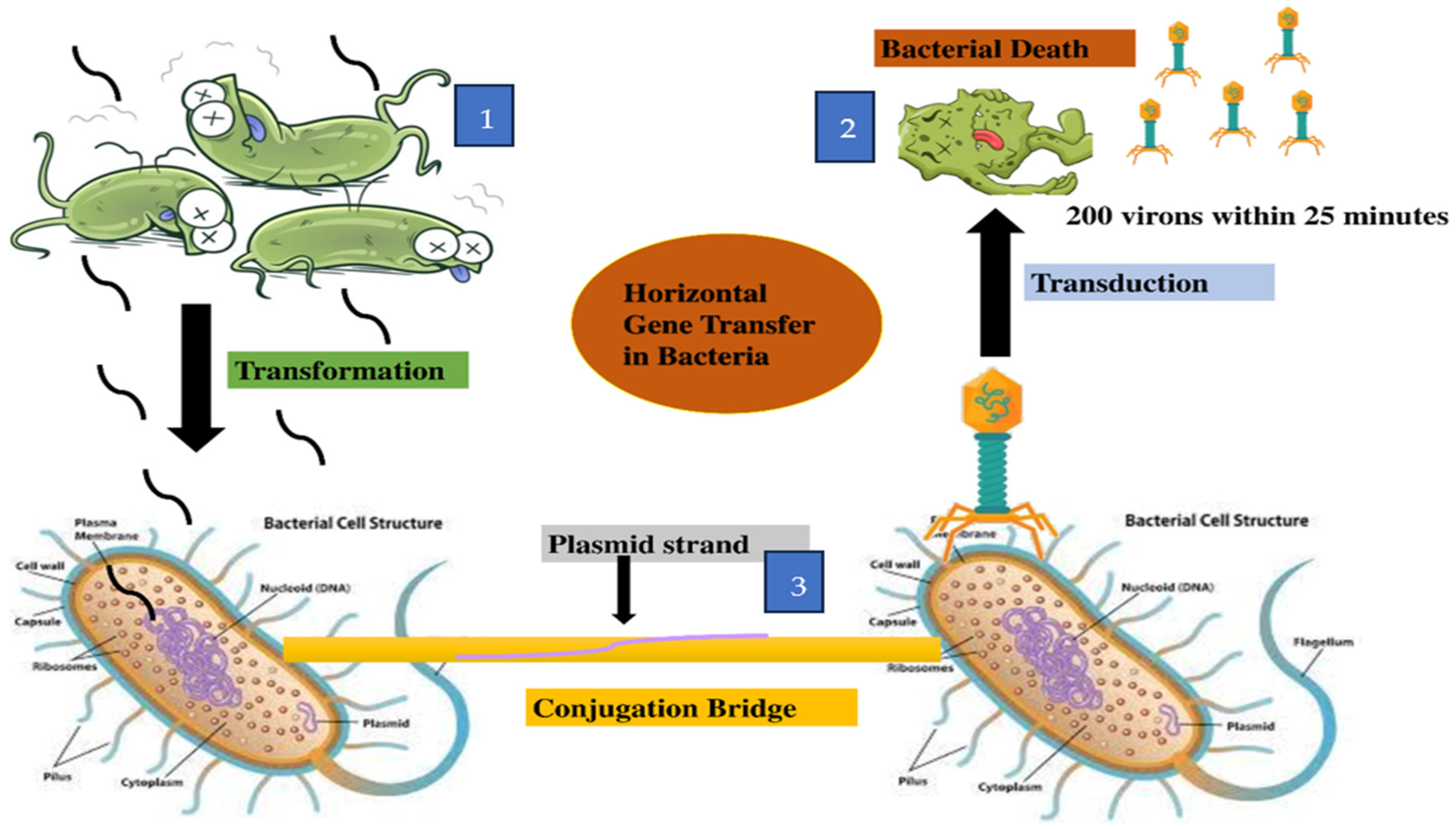
Efflux Pump: The efflux pump is a resistance mechanism by which bacteria pump out antibiotics or drugs that have already entered inside the cell. These may be the first line of defense against antibiotics in some bacteria. The presence of efflux pumps in MDR S. algae can be identified by WGS analysis [59]. There are five different structural classifications or families of efflux pumps enabling MDR in these bacteria, which include the adenosine triphosphate (ATP)-binding cassette (ABC) superfamily, MTE family (multidrug and toxic compound extrusion), SMR (small multidrug resistance) family, MFS (major facilitator superfamily), and the RND (resistance/nodulation/division) superfamily [11,63,65]. All these families work to export drugs or antibiotics from inside the cell to the outside. The efflux pump is common in Gram-negative bacteria, including Shewanellaceae. In Shewanellaceae, integrative and conjugative elements (ICEs) belong to the SXT/R391 family, and mobile genes associated with antimicrobial resistance, heavy metal resistance, and virulence are the main mobile elements that are contributing to the transmission of antibiotic resistance to other bacteria in the environment. Cimmino et al. studied the resistance genes and virulence factors of S. algae strain MARS 14 through whole genome sequencing. This study revealed that the bacteria contained several efflux pumps for multiple-drug resistance and contained beta-lactamases class C and D, along with genes for hemolysins and biofilm formation [59]. In S. oneidensis strain MR-1, the mexF gene encodes the MDR pump, which provides resistance against tetracycline and chloramphenicol [78]. The expression of mexEF operon is regulated by the TetR regulatory protein and the presence of mexF indicates that the bacteria expel antibiotics, which is necessary for their survival [78]. As with many other bacteria, Shewanella antibiotic resistance genes can be transferred by three different mechanisms: transformation, transduction, and conjugation [63]. These mechanisms can occur in aquatic environmental bacteria and in human pathogenic bacteria, as shown in Figure 2.

9. Transmission of Resistance Genes

There are three different mechanisms through which antibiotic resistance genes get transferred in the Shewanella genus. These mechanisms include transformation, transduction, and conjugation. Most recently, a very significant alternation was observed in the resistance pattern of S. xiamenensis. These resistance genes were evaluated through illumina sequencing, which showed that bacteria acquired these genes externally. Twenty-one strains of S. xiamenensis were declared as MDR, with 10 different kinds of antibiotic resistance genes with mobile genetic elements that can disseminate resistance genes into the environment [89]. In 15 different genomes of S. xiamenensis strains, a type IV secretion system was discovered, each with several sequence structures, suggesting it originated from different donors through horizontal gene transfer, which include transformation, transduction, and conjugation [89]. In transformation, bacterial cell lysis results in DNA being released into the environment, including aquatic bodies. The DNA may contain ARGs. This exogenous DNA may be taken up by other bacteria present in the surrounding environment where the recipient bacteria, if naturally competent, can integrate the exogenous DNA into their chromosomes. Shewanella baltica electrocompetent cells were prepared and transformation of P1 and ColE1-like plasmids in S. baltica showed that the plasmid’s stability was lower without antibiotic selection pressure [90]. The transformation efficacy in S. baltica can be estimated by taking the number of S. baltica colonies on antibiotic agar plates, with and without antibiotic stress. It was observed that the transformation efficiency was two to three times lower than in E. coli [90]. Groh et al. showed that mini-Tn10 transposon-bearing plasmid, pBSL180, was transformed, and observed that it can efficiently and randomly mutagenize S. oneidensis strain MR-1 [78]. The probable progenitors of blaOXA-48-like genes are present on chromosomes and encoded beta-lactamases in Shewanella species, but their dissemination is unclear [80].
The second mechanism for gene transmission is transduction, in which bacteriophages or bacterial viruses play a significant role in shaping and regulating communities of bacteria [91]. In this process, bacteriophages may contain ARGs and other genes that play a role in virulence and pathogenicity. In the infection process, the phages may take fragments of such genes from the host bacterium and transfer them to the next bacterium it infects. As a result, the recipient bacterium becomes resistant to the antibiotics. In China, from Qingdao sewage, Shewanella KR11-infecting phage vB_SbaS_Y11 was isolated. This phage has an icosahedral-shaped head with a long tail, as revealed by transmission electron microscopy. This phage may play a role in transduction of genes in Shewanella KR11 [92].
The third process of gene transmission is conjugation, in which Gram-negative bacteria attach by their cell surface pili to recipient cells. The pili forms a bridge or conjugation tube through which the donor cell transfers to a plasmid, which contains integrons, insertion sequences, or transposons carrying ARGs, that confer resistance to the recipient bacterial cell. In 91 strains of Shewanella collected from various samples, SXT/R391 integrative conjugative elements (ICEs) were detected in different species [93]. In the same study, different antibiotic genes such as strB, floR, strA, and sul2 were detected in ICESupCHN110003 (101). Conjugation does occur in aquatic environments [94].
A study conducted in Algeria identified plasmid pSx1 as having a size of 268.4 kb in the widely drug-resistant Shewanella xiamenensis T1 isolated from hospital waste. The isolated bacterium contained ARGs and the novel Tn1696 derivative (Tn6297), a transposon that carries a resistance gene for carbapenems, and blaOXA-48-like gene (blaOXA-416) on the plasmid and chromosome [11]. Initially, the ARGs were identified by WGS analysis of S. xiamenensis T1. Subsequently, the resistance genes were electroporated into an E. coli competent strain for confirmation [11]. In Shewanella xiamenensis, the qnrA1 gene was found through DNA hybridization and it is encoded on chromosomes. It is part of a class 1 integron, followed by ISCR1. This integron is closely related to In825, which has four genes’ cassettes, aacA3, catB11c, dfrA1z, and A2az. IS26 was also found in a genome which provides resistance against macrolides. The presence of resistance genes in S. xiamenensis suggests that this species could serve as a vehicle for the dissemination of resistance genes in other bacteria [95].
Shewanella xiamenensis NUITM-VS2, showing resistance to tigecycline and carbapenem, was isolated from a drainage system in Vietnam in 2012 [61]. It also showed resistance to multiple antibiotics such as aminoglycosides, cephalosporins, carbapenems, and fluoroquinolone. Genome sequencing revealed one chromosome and five plasmids. ResFinder (database for ARGs) and Plasmid Finder were used for the identification of ARGs and plasmid replicons inside the genome of the isolate. tet(X4) and tmexC3.2D3.2-toprJ1, the tigecycline resistance genes, are present on the IncC plasmid pNUITM-VS2_2 (152.2 kb). Similarly, a 24.8 kb untypeable plasmid, pNUITM-VS2_4, contained the carbapenemase blaND-1 gene [61]. By conjugation, pNUITM-VS2_2 was transferred to E. coli and conferred high-level resistance to numerous antibiotics, including tigecycline. This discovery of RND-type efflux pump gene cluster tmexCD-toprJ in Shewanella species was first reported by Dao and co-workers [57].
Another study reported that different strains of Shewanella algae (353M, 178CP, 146bCP, 144bCP, 219VB, 82CP, 38LV, 57CP, 60CP, and 83CP) were resistant to multiple antibiotics, including amoxicillin (AMX); amikacin (AKN); cephalothin (CEF); colistin (CS); Fosfomycin (FOS); cefoxitin (CX); imipenem (IPM); and sulfonamides (SUL) [63].
An analysis of a colistin-resistant clinical isolate of S. algae strain MARS 14, obtained from the broncho-alveolar lavage of a hospitalized patient, was performed to determine the molecular basis for the resistance. A functional genomics method was employed in which E. coli TOP10 was used to construct a genomic expression library based on the pZE21 MCS-1 plasmid. The library’s estimated size was 1.30 × 10⁸ bp. LB-agar with 8 mg/L of colistin was used for the functional screening of colistin-resistant clones. Then, the genomic expression library was thoroughly screened and five clones that were resistant to colistin were obtained. Following examination of DNA sequencing results of these colonies using the Integrated Microbial Genomes (IMG) and KEGG databases, an analysis of this unique gene’s amino acid sequence revealed that it encodes for ethanolamine phosphotransferase (EptA, or PmrC). Overexpression of EptA, an essential enzyme for the arrangement of outer membrane lipopolysaccharides, was observed to be related to resistance to colistin in S. algae MARS 14 (27-fold increase) [69].
Apart from antibiotic resistance, some Shewanella spp. also show resistance to heavy metals such as Cd, Cr, Cu, Co, and Zn, which are toxic for many organisms. It has been reported that two (SY1 and SY2) of fifteen strains were resistant to metals and to antibiotics, including gentamicin, rifampicin, erythromycin, vancomycin, cephalothin, ampicillin, and streptomycin [96]. The genes for antibiotics and heavy metals are usually present on bacterial plasmids. There is good evidence that the development of antibiotic resistance may be associated with selective pressure from heavy metal contamination, suggesting a process of co-resistance [97,98]. Bacteria play a role in bioaccumulation, bioabsorption, and biotransformation of heavy metals in aquatic and other environments [99]. The structure and function of microbial communities can be modified or altered in the presence of these heavy metals. Bacteria may play a role in metal bioaccumulation, bioadsorption, and biotransformation of heavy metals in aquatic and other environments.

10. Conclusions

Shewanella spp. are ubiquitously distributed across environments, including aquatic ecosystems. Some species can cause infections in humans, which may be difficult to treat due to antibiotic resistance. ARGs are part of mobile genetic elements (MGEs) such as integrons, and insertion sequences present on chromosomal DNA and plasmids. Shewanella can acquire or transmit these genes through the process of transformation, conjugation, and transduction. The presence of these ARGs and efflux pumps supports the hypothesis that Shewanella is an emerging pathogen for humans with the ability to develop resistance against various types of antibiotics. There is a need for more research to evaluate other virulence factors and genes that may play an important role in diseases in humans and in aquatic animals such as oysters and shellfish. This can be better evaluated by using whole genome sequencing supported by transcriptomics and proteomics analyses to detect the products of ARGs.

Author Contributions

S.S., G.P.R., S.P. and H.N.W. equally contributed to the conceptualization, writing, and revision of this manuscript. All authors have read and agreed to the published version of the manuscript.

Funding

This research received no external funding.

Institutional Review Board Statement

Not applicable.

Informed Consent Statement

Not applicable.

Data Availability Statement

No new data were created or analyzed in this study. Data sharing is not applicable to this article.

Acknowledgments

Thanks to Kristal L. Williams for preliminary review and editorial assistance.

Conflicts of Interest

The use of trade names or commercial products in this publication is solely for the purpose of providing specific information and does not imply recommendation or endorsement by the U.S. Department of Agriculture (USDA). The USDA is an equal opportunity provider and employer.

References

  1. Aslam, B.; Wang, W.; Arshad, M.I.; Khurshid, M.; Muzammil, S.; Rasool, M.H.; Qamar, M.U. Antibiotic resistance: A rundown of a global crisis. Infect. Drug Resist. 2018, 11, 1645–1658. [Google Scholar] [CrossRef] [PubMed]
  2. Hashem, A.H.; Attia, M.S.; Kandil, E.K.; Fawzi, M.M.; Abdelrahman, A.S.; Khader, M.S.; Abdelaziz, A.M. Bioactive compounds and biomedical applications of endophytic fungi: A recent review. Microb. Cell Fact. 2023, 22, 107. [Google Scholar] [CrossRef] [PubMed]
  3. Nocentini, A.; Capasso, C.; Supuran, C.T. Carbonic anhydrase inhibitors as Novel antibacterials in the era of antibiotic resistance: Where are we now? Antibiotics 2023, 12, 142. [Google Scholar] [CrossRef]
  4. Kümmerer, K. Antibiotics in the aquatic environment—A review—Part I. Chemosphere 2009, 75, 417–434. [Google Scholar] [CrossRef]
  5. Yekani, M.; Azargun, R.; Sharifi, S.; Nabizadeh, E.; Nahand, J.S.; Ansari, N.K.; Soki, J. Collateral sensitivity: An evolutionary trade-off between antibiotic resistance mechanisms, attractive for dealing with drug-resistance crisis. Health Sci. Rep. 2023, 6, e1418. [Google Scholar] [CrossRef]
  6. Iu, H.T.V.; Fong, P.M.; Yam, H.C.B.; Gao, P.; Yan, B.; Lai, P.M.; Ng, K.H.K. Identification of a small molecule compound active against antibiotic-tolerant Staphylococcus aureus by boosting ATP synthesis. Int. J. Mol. Sci. 2023, 24, 6242. [Google Scholar] [CrossRef]
  7. Mahmoudi, L.; Mahdavinia, A.; Khajooi, S.; Zargari-Samadnejad, A.; Karimzadeh, I. Antibiotic use pattern of surgical site infection prophylaxis in surgical wards of a teaching hospital in Shiraz, Iran. Trends Pharm. Sci. 2023, 9, 87–92. [Google Scholar]
  8. Dyary, H.; Faraj, G.; Saeed, N. History, current situation, and future perspectives on antibiotics and antibiotic Resistance. In One Health Triad; Unique Scientific Publishers: Faisalabad, Pakistan, 2023; Volume 2, pp. 109–118. [Google Scholar]
  9. Ren, Z.; Luo, W. Metagenomic analysis reveals the diversity and distribution of antibiotic resistance genes in thermokarst lakes of the Yellow River Source Area. Environ. Pollut. 2022, 313, e120102. [Google Scholar] [CrossRef] [PubMed]
  10. Despotovic, M.; de Nies, L.; Busi, S.B.; Wilmes, P. Reservoirs of antimicrobial resistance in the context of One Health. Curr. Opin. Microbiol. 2023, 73, 102291. [Google Scholar] [CrossRef]
  11. Yousfi, K.; Bekal, S.; Usongo, V.; Touati, A. Current trends of human infections and antibiotic resistance of the genus Shewanella. Eur. J. Clin. Microbiol. Infect. Dis. 2017, 36, 1353–1362. [Google Scholar] [CrossRef]
  12. Azizi, S.M.M.; Haffiez, N.; Zakaria, B.S.; Elbeshbishy, E.; Dhar, B.R. Nano-and microplastics as carriers for antibiotics and antibiotic resistance genes. In Curr. Dev. Biotechnol. Bioengin. 2023, 361–385. [Google Scholar]
  13. Johnson, T.; Richards, G.P.; Jacobs, J.; Townsend, H.; Almuhaideb, E.; Rosales, D.; Parveen, S. Prevalence and pathogenic potential of Shewanella species in oysters and seawater collected from the Chesapeake Bay and Maryland Coastal Bays. Front. Microbiol. 2025, 16, e1502443. [Google Scholar] [CrossRef] [PubMed]
  14. Nguyen, C.C.; Hugie, C.N.; Kile, M.L.; Nawab-Daneshmand, T. Association between heavy metals and antibiotic-resistant human pathogens in environmental reservoirs: A review. Front. Environ. Sci. Eng. 2019, 13, 46. [Google Scholar] [CrossRef]
  15. Zhu, Y.G.; Zhao, Y.; Li, B.; Huang, C.L.; Zhang, S.Y.; Yu, S.; Su, J.Q. Continental-scale pollution of estuaries with antibiotic resistance genes. Nat. Microbiol. 2017, 2, 16270. [Google Scholar] [CrossRef]
  16. Lu, Z.; Na, G.; Gao, H.; Wang, L.; Bao, C.; Yao, Z. Fate of sulfonamide resistance genes in estuary environment and effect of anthropogenic activities. Sci. Total Environ. 2015, 527, 429–438. [Google Scholar] [CrossRef]
  17. Nogueira, A.F.; Nunes, B. Effects of low levels of the antibiotic ciprofloxacin on the polychaete Hediste diversicolor: Biochemical and behavioural effects. Environ. Toxicol. Pharmacol. 2020, 80, e103505. [Google Scholar] [CrossRef]
  18. Yu, K.; Huang, Z.; Xiao, Y.; Wang, D. Shewanella infection in humans: Epidemiology, clinical features and pathogenicity. Virulence 2022, 13, 1515–1532. [Google Scholar] [CrossRef]
  19. Janda, J.M.; Abbott, S.L. The genus Shewanella: From the briny depths below to human pathogen. Crit. Rev. Microbiol. 2014, 40, 293–312. [Google Scholar] [CrossRef]
  20. Dominguez, H.; Vogel, B.F.; Gram, L.; Hoffmann, S.; Schaebel, S. Shewanella alga bacteremia in two patients with lower leg ulcers. Clin. Infect. Dis. 1996, 22, 1036–1039. [Google Scholar] [CrossRef]
  21. Holt, H.M.; Gahrn-Hansen, B.; Bruun, B. Shewanella algae and Shewanella putrefaciens: Clinical and microbiological characteristics. Clin. Microbiol. Infect. 2005, 11, 347–352. [Google Scholar]
  22. Satomi, M. The family Shewanellaceae. In The Prokaryotes; Springer: Berlin/Heidelberg, Germany, 2014; pp. 597–625. [Google Scholar]
  23. MacDonell, M.; Colwell, R. Phylogeny of the Vibrionaceae, and recommendation for two new genera, Listonella and Shewanella. Syst. Appl. Microbiol. 1985, 6, 171–182. [Google Scholar] [CrossRef]
  24. Nouioui, I.; Tarhriz, V.; Kim, H.M.; Montazersaheb, S.; Hejazi, M.A.; Jeon, C.O.; Hejazi, M.S. Shewanella azerbaijanica sp. nov. a novel aquatic species with high bioremediation abilities. Arch. Microbiol. 2022, 204, 496. [Google Scholar] [CrossRef]
  25. Martín-Rodríguez, A.J.; Meier-Kolthoff, J.P. Whole genome-based taxonomy of Shewanella and Parashewanella. Int. J. Syst. Evol. Microbiol. 2022, 72, 005438. [Google Scholar] [CrossRef]
  26. Ortega, R.C.M.H.; Tabugo, S.R.M.; Martinez, J.G.T.; Padasas, C.S.; Balcázar, J.L. Occurrence of Aeromonas Species in the Cutaneous Mucus of Barbour’s Seahorses (Hippocampus barbouri) as Revealed by High-Throughput Sequencing. Animals 2023, 13, 1241. [Google Scholar] [CrossRef] [PubMed]
  27. Kang, C.H.; Shin, Y.; Jeon, H.; Choi, J.H.; Jeong, S.; So, J.S. Antibiotic resistance of Shewanella putrefaciens isolated from shellfish collected from the west sea in Korea. Mar. Pollut. Bull. 2013, 76, 85–88. [Google Scholar] [CrossRef]
  28. De Marco, G.; Cappello, T.; Maisano, M. Histomorphological changes in fish gut in response to prebiotics and probiotics treatment to improve their health status: A review. Animals 2023, 13, 2860. [Google Scholar] [CrossRef] [PubMed]
  29. Hassan, S.; Sabreena, K.Z.; Bhat, S.A.; Kumar, V.; Ameen, F.; Ganai, B.A. Marine bacteria and omic approaches: A novel and potential repository for bioremediation assessment. J. Appl. Microbiol. 2022, 133, 2299–2313. [Google Scholar] [CrossRef]
  30. Tseng, S.Y.; Liu, P.Y.; Lee, Y.H.; Wu, Z.Y.; Huang, C.C.; Cheng, C.C.; Tung, K.C. The pathogenicity of Shewanella algae and ability to tolerate a wide range of temperatures and salinities. Can. J. Infect. Dis. Med. Microbiol. 2018, 1, e6976897. [Google Scholar]
  31. Wang, J.H.; Tseng, S.Y.; Tung, K.C. Genomic investigation of emerging zoonotic pathogen Shewanella xiamenensis. Tzu Chi Med. J. 2020, 32, 162–166. [Google Scholar]
  32. Poovorawan, K.; Chatsuwan, T.; Lakananurak, N.; Chansaenroj, J.; Komolmit, P.; Poovorawan, Y. Shewanella haliotis associated with severe soft tissue infection, Thailand, 2012. Emerg. Infect. Dis. 2013, 19, 1019. [Google Scholar] [CrossRef]
  33. Zhang, F.; Fang, Y.; Pang, F.; Liang, S.; Lu, X.; Kan, B.; Wang, D. Rare Shewanella spp. associated with pulmonary and bloodstream infections of cancer patients, China: A case report. BMC Infect. Dis. 2018, 18, 1–5. [Google Scholar] [CrossRef]
  34. Müller, S.; von Bonin, S.; Schneider, R.; Krüger, M.; Quick, S.; Schröttner, P. Shewanella putrefaciens, a rare human pathogen: A review from a clinical perspective. Front. Cell. Infect. Microbiol. 2023, 12, e1033639. [Google Scholar] [CrossRef] [PubMed]
  35. Pande, A.; Brosnahan, C.; Jones, B.; Ross, A.; Phiri, B.; Pal, C.; Bestbier, M. Tail Fan Necrosis in New Zealand Red Rock Lobster, Jasus edwardsii. MPI Tech. Pap. 2021, 33, 1–29. [Google Scholar]
  36. Gomes, M.P. The convergence of antibiotic contamination, resistance, and climate dynamics in freshwater ecosystems. Water 2024, 16, 2606. [Google Scholar] [CrossRef]
  37. Islam, S.S.; Midya, S. Growth Regulatory Pattern of Zooplankton in Herbicide and Antibiotic Contaminated Aquatic Ecosystem: An overview. Watershed Ecol. Environ. 2023, 5, 153–160. [Google Scholar] [CrossRef]
  38. Zhang, C.; Chen, Y.; Chen, S.; Guan, X.; Zhong, Y.; Yang, Q. Occurrence, risk assessment, and in vitro and in vivo toxicity of antibiotics in surface water in China. Ecotoxicol. Environ. Saf. 2023, 255, e114817. [Google Scholar] [CrossRef]
  39. Liang, J.; She, J.; Fu, J.; Wang, J.; Ye, Y.; Yang, B.; Tao, H. Advances in natural products from the marine-sponge-associated microorganisms with antimicrobial activity in the last decade. Mar. Drugs 2023, 21, 236. [Google Scholar] [CrossRef]
  40. Samantha, A.; Vrielink, A. Lipid A phosphoethanolamine transferase: Regulation, structure and immune response. J. Mol. Biol. 2020, 432, 5184–5196. [Google Scholar] [CrossRef]
  41. Richards, G.P.; Watson, M.A.; Crane, E.J.; Burt, I.G.; Bushek, D. Shewanella and Photobacterium spp. in oysters and seawater from the Delaware Bay. Appl. Environ. Microbiol. 2008, 74, 3323–3327. [Google Scholar] [CrossRef]
  42. Yousfi, K.; Touati, A.; Bekal, S. Complete genome sequence of an extensively drug-resistant Shewanella xiamenensis strain isolated from Algerian hospital effluents. Genome Announc. 2016, 4, e01236-16. [Google Scholar] [CrossRef]
  43. Satomi, M.; Oikawa, H.; Yano, Y. Shewanella marinintestina sp. nov., Shewanella schlegeliana sp. nov. and Shewanella sairae sp. nov., novel eicosapentaenoic-acid-producing marine bacteria isolated from sea-animal intestines. Int. J. Syst. Evol. Microbiol. 2003, 53, 491–499. [Google Scholar] [CrossRef] [PubMed]
  44. Yu, K.; Huang, Z.; Li, Y.; Fu, Q.; Lin, L.; Wu, S.; Lan, R. Establishment and application of matrix-assisted laser desorption/ionization time-of-flight mass spectrometry for detection of Shewanella genus. Front. Microbiol. 2021, 12, 625821. [Google Scholar] [CrossRef]
  45. Mateos, G.; Bonilla, A.M.; de Francisco de Polanco, S.; Martínez, J.M.; Escudero, C.; Rodríguez, N.; Amils, R. Shewanella sp. T2. 3D-1.1 a novel microorganism sustaining the iron cycle in the deep subsurface of the Iberian Pyrite Belt. Microorganisms 2022, 10, 1585. [Google Scholar] [CrossRef]
  46. Gupta, S.; Graham, D.W.; Sreekrishnan, T.; Ahammad, S.Z. Effects of heavy metals pollution on the co-selection of metal and antibiotic resistance in urban rivers in UK and India. Environ. Pollut. 2022, 306, e119326. [Google Scholar] [CrossRef]
  47. Li, Z.; Junaid, M.; Chen, G.; Wang, J. Interactions and associated resistance development mechanisms between microplastics, antibiotics and heavy metals in the aquaculture environment. Rev. Aquacult. 2022, 14, 1028–1045. [Google Scholar] [CrossRef]
  48. Bosch, J.; Bezuidenhout, C.; Coertze, R.; Molale-Tom, L. Metal-and antibiotic-resistant heterotrophic plate count bacteria from a gold mine impacted river: The Mooi River system, South Africa. Environ. Sci. Pollut. Res. 2023, 30, 31605–31619. [Google Scholar] [CrossRef] [PubMed]
  49. Fu, Y.; M’ikanatha, N.M.; Whitehouse, C.A.; Tate, H.; Ottesen, A.; Lorch, J.M.; Dudley, E.G. Low occurrence of multi-antimicrobial and heavy metal resistance in Salmonella enterica from wild birds in the United States. Environ. Microbiol. 2022, 24, 1380–1394. [Google Scholar] [CrossRef]
  50. Dao, T.D.; Takemura, T.; Kasuga, I.; Hirabayashi, A.; Nga, N.T.; Anh, P.H.Q.; Shibayama, K. Emergence of plasmid-mediated RND-type efflux pump gene cluster tmexCD-toprJ in Shewanella xiamenensis in a water environment. bioRxiv 2015. [Google Scholar]
  51. Huang, L.; Wu, C.; Gao, H.; Xu, C.; Dai, M.; Huang, L.; Cheng, G. Bacterial multidrug efflux pumps at the frontline of antimicrobial resistance: An overview. Antibiotics 2022, 11, 520. [Google Scholar] [CrossRef]
  52. Blanco, P.; Hernando-Amado, S.; Reales-Calderon, J.A.; Corona, F.; Lira, F.; Alcalde-Rico, M.; Martinez, J.L. Bacterial multidrug efflux pumps: Much more than antibiotic resistance determinants. Microorganisms 2016, 4, 14. [Google Scholar] [CrossRef]
  53. Athar, M.; Gervasoni, S.; Catte, A.; Basciu, A.; Malloci, G.; Ruggerone, P.; Vargiu, A.V. Tripartite efflux pumps of the RND superfamily: What did we learn from computational studies? Microbiology 2023, 169, e001307. [Google Scholar] [CrossRef]
  54. Lloyd, N.A.; Nazaret, S.; Barkay, T. Whole genome sequences to assess the link between antibiotic and metal resistance in three coastal marine bacteria isolated from the mummichog gastrointestinal tract. Mar. Pollut. Bull. 2018, 135, 514–520. [Google Scholar] [CrossRef] [PubMed]
  55. Kothari, A.; Kumar, P.; Gaurav, A.; Kaushal, K.; Pandey, A.; Yadav, S.R.M.; Omar, B.J. Association of antibiotics and heavy metal arsenic to horizontal gene transfer from multidrug-resistant clinical strains to antibiotic-sensitive environmental strains. J. Hazard. Mater. 2023, 443, e130260. [Google Scholar] [CrossRef]
  56. Prasad, P.; Reshi, S.; Nataraj, G. Topic: Shewanella Species: Case Series on a Rare Emerging Pathogen. Indian J. Microbiol. Res. 2019, 6, 303–307. [Google Scholar]
  57. Madrid, N.Y.; Mejia, L.F.; Urrego, J.F.G. Left knee septic monoarthritis in a pediatric patient due to Shewanella putrefaciens: Case report and literature review. Clin. Microbiol. Antimicrob. 2023, 23, 43. [Google Scholar] [CrossRef] [PubMed]
  58. Zago, V.; Veschetti, L.; Patuzzo, C.; Malerba, G.; Lleo, M.M. Resistome, mobilome and virulome analysis of Shewanella algae and Vibrio spp. strains isolated in italian aquaculture centers. Microorganisms 2020, 8, 572. [Google Scholar] [CrossRef]
  59. Cimmino, T.; Olaitan, A.O.; Rolain, J.M. Whole genome sequence to decipher the resistome of Shewanella algae, a multidrug-resistant bacterium responsible for pneumonia, Marseille, France. Expert Rev. Anti Infect. Ther. 2016, 14, 269–275. [Google Scholar] [CrossRef]
  60. Paździor, E.; Pękala-Safińska, A.; Wasyl, D. Phenotypic diversity and potential virulence factors of the Shewanella putrefaciens group isolated from freshwater fish. J. Vet. Res. 2019, 63, 321. [Google Scholar] [CrossRef]
  61. Jousset, A.B.; Dabos, L.; Bonnin, R.A.; Girlich, D.; Potron, A.; Cabanel, N.; Naas, T. CTX-M-15-producing Shewanella species clinical isolate expressing OXA-535, a chromosome-encoded OXA-48 variant, putative progenitor of the plasmid-encoded OXA-436. Antimicrob. Agents Chemother. 2018, 62, e01879-17. [Google Scholar] [CrossRef]
  62. Kalathuru, S.; Singla, A.; Kumar, A.; Swami, A. Shewanella algae, an emerging pathogen, causing skin and soft tissue infections. Infect. Dis. Clin. Pract. 2023, 31, e1286. [Google Scholar] [CrossRef]
  63. Muñoz-Gallego, I.; Chaves, F.; Orellana, M.A. Epidemiological and clinical characteristics of Shewanella spp. infections in a tertiary hospital in Madrid. Infect. Dis. 2016, 48, 760–762. [Google Scholar] [CrossRef] [PubMed]
  64. Ng, W.W.S.; Shum, H.P.; To, K.K.W.; Sridhar, S. Emerging infections due to Shewanella spp.: A case series of 128 cases over 10 years. Front. Med. 2022, 9, e850938. [Google Scholar] [CrossRef]
  65. Martín-Rodríguez, A.J.; Martín-Pujol, O.; Artiles-Campelo, F.; Bolaños-Rivero, M.; Römling, U. Shewanella spp. infections in Gran Canaria, Spain: Retrospective analysis of 31 cases and a literature review. JMM Case Rep. 2017, 4, e005131. [Google Scholar] [CrossRef]
  66. Li, Y.; Qi, R.; Yang, H.; Zhang, X.L.; Wu, Y.; Huang, B.H.; Zhao, Q.; Gu, Y.F. Analysis of clinical characteristics of infections caused by Shewanella species. Indian J. Med. Microbiol. 2024, 49, e100574. [Google Scholar] [CrossRef]
  67. Luo, B.; Su, J.Y.; Wang, Y.N.; Guan, L.; Dong, K.S.; Niu, H.J.; Li, Y. Shewanella subflava sp. nov., a novel multi-resistant bacterium, isolated from the estuary of the Fenhe River into the Yellow River. Antonie Van Leeuwenhoek 2023, 116, 511–519. [Google Scholar] [CrossRef] [PubMed]
  68. Wahid, J. Study of the Distribution of Antimicrobial Resistance in Organic Crops and Their Environment. 2023. Available online: https://riunet.upv.es/handle/10251/197455 (accessed on 31 March 2025).
  69. Telke, A.A.; Rolain, J.M. Functional genomics to discover antibiotic resistance genes: The paradigm of resistance to colistin mediated by ethanolamine phosphotransferase in Shewanella algae MARS 14. Int. J. Antimicrob. Agents 2015, 46, 648–652. [Google Scholar] [CrossRef]
  70. Kühn, M.J.; Edelmann, D.B.; Thormann, K.M. Polar flagellar wrapping and lateral flagella jointly contribute to Shewanella putrefaciens environmental spreading. Environ. Microbiol. 2022, 24, 5911–5923. [Google Scholar] [CrossRef] [PubMed]
  71. Ghotbi, M.; Kelting, O.; Blümel, M.; Tasdemir, D. Gut and gill-associated microbiota of the flatfish European plaice (Pleuronectes platessa): Diversity, metabolome and bioactivity against human and aquaculture pathogens. Mar. Drugs. 2012, 20, 573. [Google Scholar] [CrossRef]
  72. Fernández, L.; Hancock, R.E. Adaptive and mutational resistance: Role of porins and efflux pumps in drug resistance. Clin. Microbiol. Rev. 2012, 25, 661–681. [Google Scholar] [CrossRef]
  73. Zhou, G.; Wang, Q.; Wang, Y.; Wen, X.; Peng, H.; Peng, R.; Shi, Q.; Xie, X.; Li, L. Outer membrane porins contribute to antimicrobial resistance in gram-negative bacteria. Microorganisms 2023, 11, 1690. [Google Scholar] [CrossRef]
  74. Gao, T.; Ju, L.; Yin, J.; Gao, H. Positive regulation of the Shewanella oneidensis OmpS38, a major porin facilitating anaerobic respiration, by Crp and Fur. Sci. Rep. 2015, 5, e14263. [Google Scholar] [CrossRef]
  75. Huang, Y.T.; Liu, P.Y. Emergence of carbapenem resistance in persistent Shewanella algae bacteremia: The role of pdsS G547W mutation in adaptive subpopulation dynamics. Ann. Clin. Microbiol. Antimicrob. 2024, 23, 102. [Google Scholar] [CrossRef]
  76. Poirel, L.; Rodriguez-Martinez, J.M.; Mammeri, H.; Liard, A.; Nordmann, P. Origin of plasmid-mediated quinolone resistance determinant QnrA. Antimicrob. Agents Chemother. 2005, 49, 3523–3525. [Google Scholar] [CrossRef]
  77. Lambert, P.A. Bacterial resistance to antibiotics: Modified target sites. Adv. Drug Deliv. Rev. 2005, 57, 1471–1485. [Google Scholar] [CrossRef] [PubMed]
  78. Tseng, C.H.; Cheng, J.F.; Chen, S.Y.; Chen, W.H.; Shi, Z.Y.; Lin, Y.H.; Tsai, C.A.; Lin, S.P.; Chen, Y.C.; Lin, Y.C.; et al. Detection of S83V GyrA mutation in quinolone-resistant Shewanella algae using comparative genomics. J. Microbiol. Immunol. Infect. 2021, 54, 658–664. [Google Scholar] [CrossRef]
  79. Azargun, R.; Gholizadeh, P.; Sadeghi, V.; Hosainzadegan, H.; Tarhriz, V.; Memar, M.Y.; Pormohammad, A.; Eyvazi, S. Molecular mechanisms associated with quinolone resistance in Enterobacteriaceae: Review and update. Trans. R. Soc. Trop. Med. Hyg. 2020, 114, 770–781. [Google Scholar] [CrossRef] [PubMed]
  80. Tacão, M.; Araujo, S.; Vendas, M.; Alves, A.; Henriques, I. Shewanella species as the origin of blaOXA-48 genes: Insights into gene diversity, associated phenotypes and possible transfer mechanisms. Int. J. Antimicrob. Agents 2018, 51, 340–348. [Google Scholar] [CrossRef]
  81. Poirel, L.; Héritier, C.; Nordmann, P. Chromosome-encoded Ambler class D β-lactamase of Shewanella oneidensis as a progenitor of carbapenem-hydrolyzing oxacillinase. Antimicrob. Agents Chemother. 2004, 48, 348–351. [Google Scholar] [CrossRef] [PubMed]
  82. Potron, A.; Poirel, L.; Nordmann, P. Origin of OXA-181, an emerging carbapenem-hydrolyzing oxacillinase, as a chromosomal gene in Shewanella xiamenensis. Antimicrob. Agents Chemother. 2011, 55, 4405–4407. [Google Scholar] [CrossRef]
  83. Yin, J.; Sun, L.; Dong, Y.; Chi, X.; Zhu, W.; Qi, S.H.; Gao, H. Expression of blaA underlies unexpected ampicillin-induced cell lysis of Shewanella oneidensis. PLoS ONE 2013, 8, e60460. [Google Scholar] [CrossRef]
  84. Liu, L.; Wang, W.; Wu, S.; Gao, H. Recent advances in the siderophore biology of Shewanella. Front. Microbiol. 2022, 13, e823758. [Google Scholar] [CrossRef]
  85. Li, X.; Zheng, S.; Li, Y.; Ding, J.; Qin, W. Effectively facilitating the degradation of chloramphenicol by the synergism of Shewanella oneidensis MR-1 and the metal-organic framework. J. Hazard. Mater. 2023, 454, e131545. [Google Scholar] [CrossRef] [PubMed]
  86. Van Hoek, A.H.; Mevius, D.; Guerra, B.; Mullany, P.; Roberts, A.P.; Aarts, H.J. Acquired antibiotic resistance genes: An overview Front. Microbiol. 2011, 2, e203. [Google Scholar] [CrossRef] [PubMed]
  87. Tang, Y.J.; Meadows, A.L.; Kirby, J.; Keasling, J.D. Anaerobic central metabolic pathways in Shewanella oneidensis MR-1 reinterpreted in the light of isotopic metabolite labeling. J. Bacteriol. 2007, 189, 894–901. [Google Scholar] [CrossRef]
  88. Xue, Y.; Xue, B.; Zhang, L. Multi-Omics Integrative Analysis to Reveal the Impacts of Shewanella algae on the development and lifespan of marine nematode litoditis marina. Int. J. Mol. Sci. 2024, 25, 9111. [Google Scholar] [CrossRef]
  89. Wang, H.; Xia, F.; Xia, Y.; Li, J.; Hu, Y.; Deng, Y.; Zou, M. Pangenome analysis of Shewanella xiamenensis revealed important genetic traits concerning genetic diversity, pathogenicity and antibiotic resistance. BMC Genom. 2024, 25, 1–14. [Google Scholar] [CrossRef]
  90. Milewska, K.; Węgrzyn, G.; Szalewska-Pałasz, A. Transformation of Shewanella baltica with ColE1-like and P1 plasmids and their maintenance during bacterial growth in cultures. Plasmid 2015, 81, 42–49. [Google Scholar] [CrossRef] [PubMed]
  91. Lopez-Perez, M.; Haro-Moreno, J.M.; Gonzalez-Serrano, R.; Parras-Molto, M.; Rodriguez-Valera, F. Genome diversity of marine phages recovered from Mediterranean metagenomes: Size matters. PLoS Genet. 2017, 13, e1007018. [Google Scholar] [CrossRef]
  92. Wang, H.; Zheng, K.; Wang, M.; Ma, K.; Ren, L.; Guo, R.; Ma, L.; Zhang, H.; Liu, Y.; Xiong, Y.; et al. Shewanella phage encoding a putative anti-CRISPR-like gene represents a novel potential viral family. Microbiol. Spectrum. 2024, 12, e0336723. [Google Scholar] [CrossRef]
  93. Fang, Y.; Wang, Y.; Li, Z.; Liu, Z.; Li, X.; Diao, B.; Kan, B.; Wang, D. Distribution and genetic characteristics of SXT/R391 integrative conjugative elements in Shewanella spp. from China. Front. Microbiol. 2018, 9, 920. [Google Scholar] [CrossRef]
  94. Amirfard, K.D.; Moriyama, M.; Suzuki, S.; Sano, D. Effect of environmental factors on conjugative transfer of antibiotic resistance genes in aquatic settings, J. Appl. Microbiol. 2024, 135, lxae129. [Google Scholar]
  95. Zhao, J.Y.; Mu, X.D.; Zhu, Y.Q.; Xi, L.; Xiao, Z. Identification of an integron containing the quinolone resistance gene qnrA1 in Shewanella xiamenensis. FEMS Microbiol. Lett. 2015, 362, p.fnv146. [Google Scholar] [CrossRef] [PubMed]
  96. Kang, C.H.; So, J.S. Antibiotic and heavy metal resistance in Shewanella putrefaciens strains isolated from shellfishes collected from West Sea, Korea. Mar. Pollut. Bull. 2016, 112, 111–116. [Google Scholar] [CrossRef] [PubMed]
  97. Berg, J.; Tom-Petersen, A.; Nybroe, O. Copper amendment of agricultural soil selects for bacterial antibiotic resistance in the field. Lett. Appl. Microbiol. 2005, 40, 146–151. [Google Scholar] [CrossRef] [PubMed]
  98. Akinbowale, O.L.; Peng, H.; Grant, P.; Barton, M.D. Antibiotic and heavy metal resistance in motile Aeromonads and Pseudomonads from rainbow trout (Oncorhynchus mykiss) farms in Australia. Int. J. Antimicrob. Agents. 2007, 30, 177–182. [Google Scholar] [CrossRef]
  99. Nivetha, N.; Srivarshine, B.; Sowmya, B.; Rajendiran, M.; Saravanan, P.; Rajeshkannan, R.; Dragoi, E.N. A comprehensive review on bio-stimulation and bio-enhancement towards remediation of heavy metals degeneration. Chemosphere 2022, 312, e137099. [Google Scholar] [CrossRef]
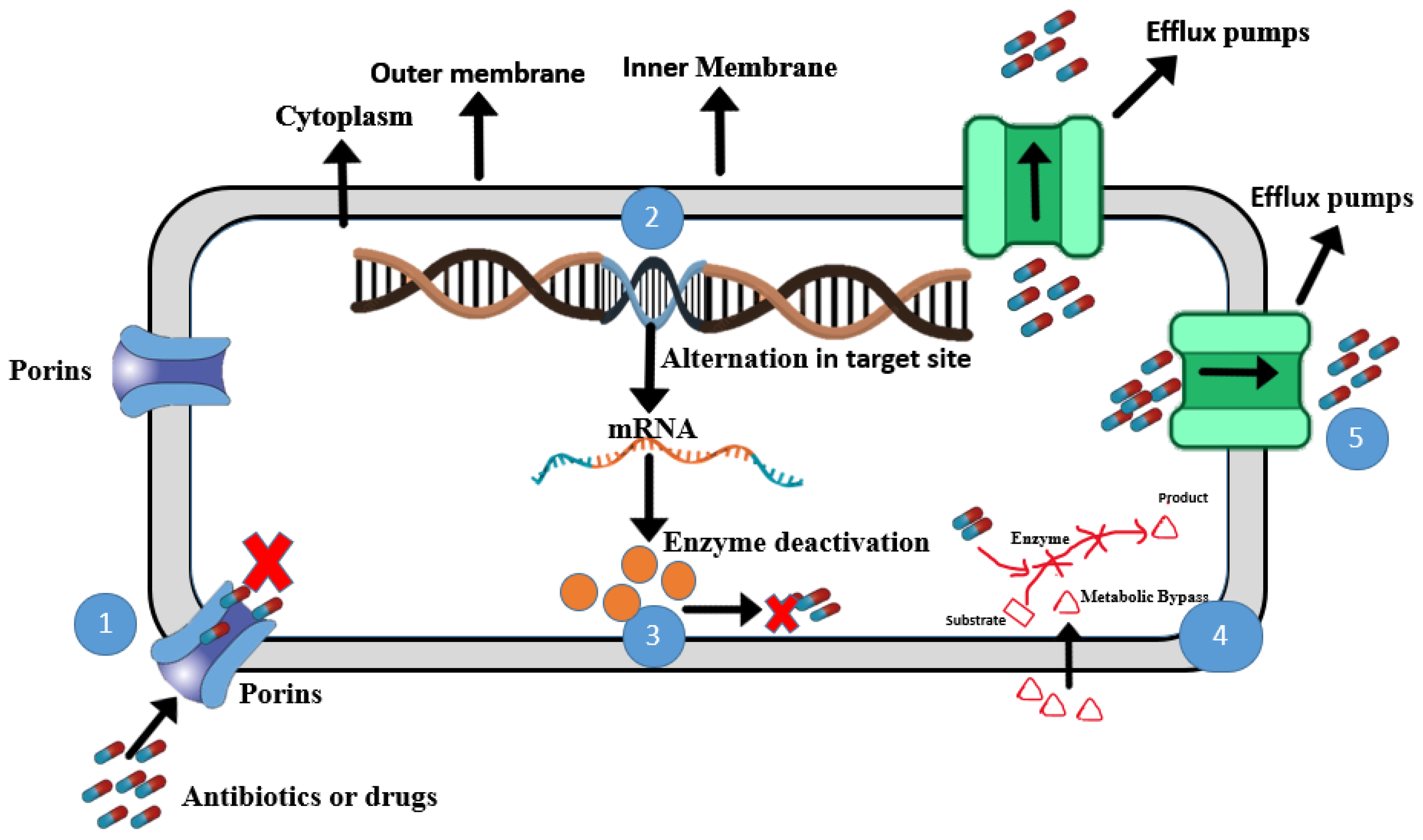
Figure 1. Six antibiotic resistance mechanisms in Shewanella. (1) Porins: alternation in porins limits entrance of antibiotics in cell. (2) Alteration in targeted sites: antibiotic no longer binds with altered targeted site. (3) Enzyme deactivation: intracellular enzymes in bacteria modify antibiotics. (4) Metabolic bypass: bacteria produce enzymes which bypass antibiotic action. (5) Efflux pump: actively transports antibiotics from inside to outside of cell to prevent antibiotic accumulation in the cell. (Figure created by ChemBioDraw, ultra version 13).
Microorganisms 13 01115 g001
Figure 2. Three mechanisms for the transmission of antibiotic resistance genes in Shewanella: (1) transformation: bacterial cells taking DNA from surroundings; (2) transduction: bacteriophages being released after the death of a phage-infected bacterial cell; (3) conjugation: genetic material being transferred from one bacterium to another via a conjugation tube. (Figure created by ChemBioDraw).
Microorganisms 13 01115 g002
Table 1. Genome assemblies and their sizes in different Shewanella spp., all with 1 chromosome (NCBI search from 2024), https://www.ncbi.nlm.nih.gov/datasets/genome/ (accessed on 10 January 2024). ND means not determined.
Shewanella SpeciesGenome AssembliesNo. of Chromo-SomesNo. of PlasmidsGenome Size (Mb)GC
(%)
No. of Genes
(Avg)
No. of Proteins
(Avg)
First Genome
Release Date
Shewanella putrefaciens22134.38–5.0544.30–47.904055382001/10/2014
Shewanella algae230114.60–5.2052.50–53.24185445004/01/2014
Shewanella baltica58143.26–5.5545.99–47.504414458702/20/2007
Shewanella benthica41ND4.03–5.7145.8–463431325911/28/2007
Shewanella woodyi41ND5.82–5.9443.60–43.704874508503/13/2008
Shewanella halifaxensis21ND5.23–5.4642.60–42.804674449402/07/2008
Shewanella oneidensis7113.71–5.1345.93–46.504438426109/12/2002
Shewanella colwelliana8NDND4.47–4.8145.30–45.604135401401/10/2014
Shewanella vesiculosa121ND4.46–4.7841–60–41.704068384011/19/2018
Shewanella chilikensis161ND4.39–4.9152.20–52.503945384512/12/2017
Shewanella indica151ND4.38–4.9952.20–52.604089389709/11/2020
Shewanella profunda11ND4.7444.94080430305/09/2022
Shewanella glacialimarina11ND4.4641.13763391510/15/2021
Shewanella algicola3NDND4.85–4.9742.304267445909/11/2020
Shewanella saliphila2NDND4.62–4.6342.503926408609/11/2020
Disclaimer/Publisher’s Note: The statements, opinions and data contained in all publications are solely those of the individual author(s) and contributor(s) and not of MDPI and/or the editor(s). MDPI and/or the editor(s) disclaim responsibility for any injury to people or property resulting from any ideas, methods, instructions or products referred to in the content.