Article

Identifying Root-Associated Endophytic Fungi and Bacteria in Festuca and Lolium Grasses from a Site in Lithuania

by
Violeta Stakelienė
1,2,*,
Izolda Pašakinskienė
1,2,
Saulė Matijošiūtė
1,2,
Justas Martūnas
1 and
Gitana Štukėnienė
1
1
Botanical Garden, Vilnius University, Kairėnų 43, 10239 Vilnius, Lithuania
2
Life Sciences Centre, Vilnius University, Saulėtekio 7, 10221 Vilnius, Lithuania
*
Author to whom correspondence should be addressed.
Microorganisms 2025, 13(4), 799; https://doi.org/10.3390/microorganisms13040799
Submission received: 26 February 2025 / Revised: 21 March 2025 / Accepted: 26 March 2025 / Published: 31 March 2025
(This article belongs to the Special Issue Rhizosphere Bacteria and Fungi That Promote Plant Growth)

Abstract

:
This study investigates the diversity and distribution of root endophyte fungi and bacteria across Festuca and Lolium grasses, including open-grassland and forest species. The species examined include perennials such as Festuca arundinacea, F. gigantea, F. pratensis, Lolium perenne, and L. perenne × F. gigantea hybrids and the annuals L. temulentum and L. multiflorum. A total of 21 fungal species (60 isolates) and 26 bacterial taxa (59 isolates) were recovered in the culture (PDA medium for fungi and LB for bacteria) from the root cuttings of these grasses. Microdochium bolleyi fungi and Bacillus sp. bacteria were the most prevalent endophytes, with each being identified in five of the seven plant species examined. The annuals L. multiflorum and L. temulentum exhibited a higher abundance of endophytes than that in their perennial relatives, suggesting the benefits of microbial associations in supporting their short life cycles. The woodland F. gigantea demonstrated the highest fungal endophyte diversity, with six species identified. In contrast, the open-grassland perennials F. arundinacea, F. pratensis, and L. perenne hosted only one to two species. Two Basidiomycota, Coprinellus disseminatus and Sistotrema brinkmannii, were exclusively obtained from the roots of the forest grass F. gigantea. Notably, the open-grassland perennial F. arundinacea exhibited the highest bacterial diversity, with nine species present. However, it showed the lowest fungal diversity, with only one species detected. Overall, our study reveals distinct patterns of fungal and bacterial endophyte diversity in the roots of Festuca and Lolium grasses, with variations linked to host species, growth type traits, and ecological adaptations. Among the root-derived endophytes isolated, several fungi and bacteria are potential candidates for plant growth promotion and biocontrol. Therefore, the findings of this study provide potential implications for improved grassland management and crop breeding strategies aimed at specific climate and/or soil conditions.

1. Introduction

Endophytic microorganisms comprising fungi and bacteria are ubiquitous in plants, colonizing the above-ground parts and roots of the plants without causing apparent harm. Endophytic fungi are common in meadows, the Alps, deserts, and forests of the middle zones [1,2,3,4,5,6]. Many well-known endophytes of the anamorphic Ascomycota phylum are characterized by melanized septate hyphae. Due to this feature, these fungi are assigned into a special group of dark septate endophytes, DSEs [7,8]. These endophytic microorganisms can provide nutritional and protective benefits to their hosts. Numerous endophytic fungi have a beneficial impact on plants, helping them to resist biotic and abiotic stress, fighting against pests, and promoting their growth and development [5,9,10]. Just like fungi, diverse bacteria have been shown to promote plant growth and have been assigned as plant-growth-promoting rhizobacteria or plant-growth-promoting bacteria [11,12].
Numerous studies indicate that the majority of grasses are hosts to endophytic fungi [4,13,14,15]. In this respect, Festuca/Lolium (Poaceae) plants have been extensively studied for the Epichloë/Neotyphodium endophyte associations in their foliar parts and seeds [16,17,18,19,20,21,22,23,24]. These fungi systemically colonize the leaves and stems of host plants but not the roots. In contrast to the wide research on Epichloë/Neotyphodium in the above-ground parts, there is only limited knowledge of the root-associated endophytes in Festuca/Lolium grasses so far, which are cultivated worldwide in pastures and meadows, providing nutrition for livestock. Up until now, the fungal and bacterial endophytes in the roots of Festuca spp. from the Schedonorus group have only been described for Festuca gigantea [25], while for F. arundinacea, specifically, root-derived bacterial endophytes have been studied [26]. In the foliar parts, an analysis conducted by Przemieniecki et al. [27] revealed specific patterns of fungal endophytes colonizing the forage grasses Lolium perenne and Phleum pratense.
A limited number of studies has examined fungal and bacterial endophytes within the same experiment, either in relation to a specific plant or an ecosystem [15,25,28,29,30]. Toju and co-authors [30] examined more than 100 grassland plant species and showed that a plant’s below-ground microbiome has different features and dynamics compared to those in the above-ground microbiome. The root system is essential for anchoring the plant in the soil and for absorbing the water and minerals necessary for its growth and survival. Additionally, studies have shown that the roots play a critical role in sensing environmental factors. They act as a sensory hub, influencing plant growth and morphogenesis in response to changes in its surroundings [31,32]. Given that the roots host a diverse range of bacteria and fungi, research into the complexity of the microbial communities in these hidden parts of plants is crucial for advancing our understanding of plant life.
This study aims to fill in a knowledge gap by assessing the diversity and prevalence of the fungal and bacterial communities in the roots of grasses through an isolated culture approach. Overall, its results provide insights into the endophyte distribution patterns across Festuca and Lolium species, which show contrasts based on their ecological habitats and seasonal growth characteristics. These findings contribute to a deeper understanding of plant–microbe interactions in agricultural and ecological contexts.

2. Materials and Methods

2.1. Root Sampling and Sterilization

The plants were grown from young tiller transplants grown in the experimental field of the Botanical Garden of Vilnius University (Vilnius, Lithuania; 54.7362067° N, 25.403482° E). The species examined included the perennials F. arundinacea, F. gigantea, F. pratensis, L. perenne, and L. perenne × F. gigantea hybrids and the annuals L. temulentum and L. multiflorum. F. gigantea is a woodland species, whereas all of the other species are grasses from open-grassland sites used in meadows and pastures, except for L. temulentum, which is a short-lived weed (Table 1). Plant tillers were collected from the experimental field in May–June. In each species, root samples were obtained from 25–30 plants, with each plant represented by three tillers. For preparation of the root samples, the collected tillers were washed under running tap water, and the old roots were removed. Tillers with no roots were placed in test tubes with added tap water. New healthy roots, 1–2 cm long, were collected and surface-sterilized accordingly using 50% ethanol for 90 s and 1.25% sodium hypochlorite for 90 s; after this, the samples were washed 3 times for 3 min with sterile water. In addition, 200 μL of final wash water was added to three Petri dishes with the Potato Dextrose Agar (PDA) or Luria Broth (Miller’s LB broth) (LB) medium during the fungal and bacterial culture from the roots. This was used as a negative control to confirm that the root sterilization was adequate.

2.2. Microscopic Evaluation and Estimation of the Abundance of Endophytic Fungi in the Grass Roots

For the microscopic evaluation, newly grown roots, as described in Section 2.1 (excluding the sterilization step), were used. Root segments were collected and placed in 1.5 mL Eppendorf test tubes with a fixative of ethanol–glacial acetic acid (3:1) and kept in a refrigerator (at 2–3 °C) until their further use. Prior to microscopy, all of the roots were softened using the following enzyme treatment: the sampled roots were washed twice with citrate buffer (0.1 M, pH 4.8) at 27–28 °C for 10 min and treated with 0.5% Macerozyme R-10 at 37 °C for 25 min.
After the enzyme treatment, the roots were washed with citric citrate buffer and stained with 0.025% Trypan Blue, following the protocol described by Kiheri [33] with some modifications: the roots were stained at 90 °C for 30 min and then bleached with a lactic acid–glycerol (1:1) mixture, repeating this three times. The first two bleaches took place at 37 °C for 30 min, while during the third, they were left at room temperature for 24 h before the microscopic analysis. Lactic acid–glycerol (4:1) was used to prepare the root tip sections on the microscopy slides.
To determine the frequency of fungal endophyte colonization and compare it between species, meristem tip cuttings from N = 120 roots were analyzed microscopically for each species. The presence of fungal structures such as septate and melanized hyphae and agglomerates of fungal spores, as specified in [25], were assessed using a Nikon ECLICE Ci-L phase contrast microscope (Nikon, Tokyo, Japan) in 10 fields of view for each root at ×400 magnification.

2.3. Isolation of the Fungi

To make the PDA medium, 200 g of peeled, sliced potatoes (Lithuanian var. Rasa) was boiled in 1 L of distilled water for 30 min. The potato mass was filtered through a cheesecloth, saving the effluent. The potato infusion was poured into flasks 200 mL at a time. A total of 4 g of dextrose and 4 g of agar were added into each flask. The mixtures were autoclaved at 121 °C for 20 min. Ampicillin sodium salt (final concentration—100 mg/L) and streptomycin sulfate (final concentration—100 mg/L) were added prior to distribution of the agar into the plates to inhibit the bacterial growth selectively.
For cultivation of the fungal endophytes, N = 200–250 root tip fragments per species were used; these samples were collected from 25–30 plants. Five cuttings of the surface-sterilized roots were placed into each Petri dish with the PDA medium. Before isolation, the roots were squashed using a sterile needle to facilitate the proliferation of endophytic fungi. The samples were incubated in the dark at 27 °C. After 7–14 days, we observed the growth of fungal colonies in proximity to the root segments. The fungal isolates obtained were deposited into the laboratory collection of Vilnius University Botanical Garden.

2.4. Isolation of the Bacteria

A total of 25 g of LB powder (Thermo Fisher Scientific Baltics, Vilnius, Lithuania) was dissolved in 1 L of purified water, heated and agitated until it completely dissolved, and then sterilized through autoclaving at 121 °C for 15 min. The surface-sterilized roots were prepared as described in Section 2.1. For cultivation of the bacterial endophytes, N = 100 root tip fragments per species were used; these samples were collected from 25–30 plants. Root cuttings were incubated in Petri dishes with the LB medium at 37 °C in the dark. After 1–2 days, we observed the bacterial colonies in proximity to the root segments.

2.5. DNA Extraction from the Fungi and Bacteria

For the extraction of genomic DNA from the fungi, 10-day-old fungal colonies grown on the PDA medium were used, sampling 100 mg of mycelial biomass. For bacteria, 1- to 2-day-old colonies were sampled and transferred into the liquid LB medium for growth. The next day, genomic DNA was extracted from these cultures.
The fungal and bacterial genomic DNA was isolated using the Quick-DNATM HMW MagBead Kit (Zymo Research, Irvine, CA, USA), following the manufacturer’s guidelines.

2.6. Standard DNA Amplification and Sequencing

For standard DNA amplification, the primer pairs used in the PCR reactions are listed in Table 2 for fungi and bacteria, respectively. The total volume of the PCR mix for amplification was 50 μL. PCR was conducted under a temperature profile of 94 °C for 3 min, followed by 35 cycles of 94 °C for 30 s, 49–61 °C [calculated according to the primer’s Ta = Tm − (0–4 °C)] for 30 s, and 72 °C for 1 min and a final extension at 72 °C for 5 min.
The PCR products were purified using the GeneJET PCR Purification Kit (Thermo Fisher Scientific Baltics, Vilnius, Lithuania). The sequencing was performed by BaseClear B.V., Leiden, the Netherlands. The sequences from the fungal and bacterial isolates were analyzed against NCBI reference data using the BLAST tool (version 2.16.0) at https://blast.ncbi.nlm.nih.gov/Blast.cgi (accessed on 26 August 2024).

2.7. Morphological Characterization of the Endophytic Fungi

The fungal isolates were also characterized according to their morphological features, including their structure, color, and colony edge. A mixture of lactic acid–glycerol (4:1) was used to analyze and photograph the fungal mycelium in each specimen. A microscopic Nikon ECLICE Ci-L phase-contrast microscope was used for viewing.

2.8. Statistical Analysis

Isolation frequency (IF): The percentage of the number of endophytic fungal and bacterial strains isolated out of the total number of isolates was obtained. This parameter was used to determine the dominance of the endophytic species accessed.
Margalef’s index D = (S − 1)/lnN was used to analyze taxon richness. S here is the total number of taxa, while N is the total number of isolates. These parameters were calculated by applying the formula used by Song et al. [37].
The data were analyzed using a one-way ANOVA in STATISTICA® 7.0. The statistical significance of the differences between the means was assessed using a post hoc Tukey’s test. Differences were considered to be significant at p ≤ 0.05. Charts were drawn using MS Excel software 2016.

2.9. Photography

Images of the colonies were taken using a Sony Alpha a6300 camera (Sony Corporation, Tokyo, Japan) with a Sigma 56 mm f/1.4 lens (Sony Corporation, Tokyo, Japan). Root section cuttings and mycelium samples were analyzed under the phase-contrast microscope, and the NIS-Elements D software (version 6.02.01) program was used for the microscopic photography and analysis.

3. Results

3.1. The Frequency of Colonization by Endophytic Fungi in Festuca and Lolium Species and Their Hybrids

In the Festuca species and the hybrids, fungal structures were observed in 44% to 56% of the roots examined (Figure 1). The highest prevalence of endophytic fungal structures was observed in the roots of the annuals L. temulentum (94%) and L. multiflorum (84%) in contrast to the perennial L. perenne (48%). The colonization frequency in the L. perenne × F. gigantea hybrids was at the level observed in their parental species.

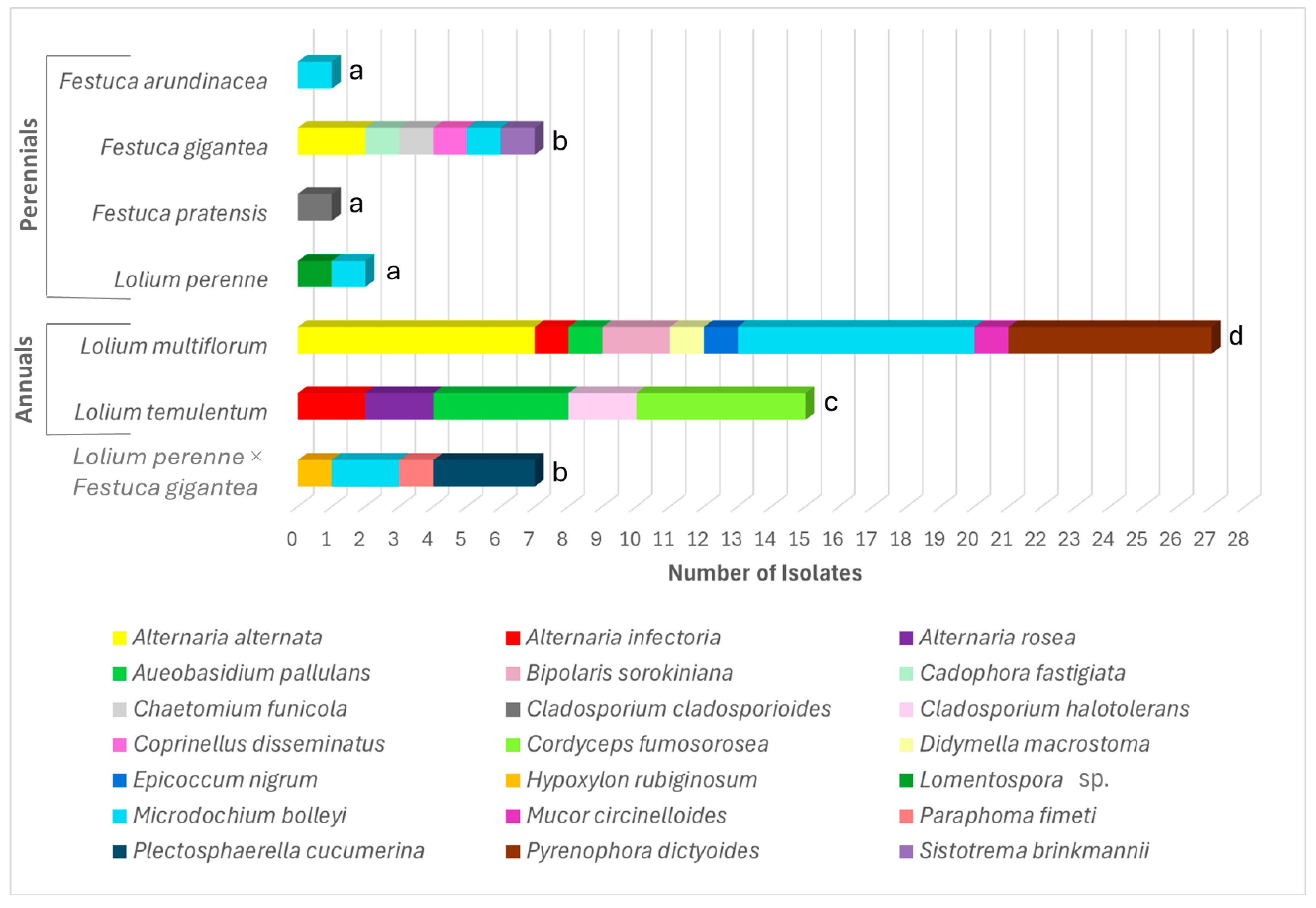
3.2. Diversity and Abundance of the Endophytic Fungi

The fungal endophyte distribution across the species examined is presented in Figure 2. Endophytic fungi were isolated from surface-sterilized fresh root cuttings placed on the PDA medium, sampling N = 200–250 for each of the seven species. No colony growth was detected in the control Petri dishes with the final root wash water after sterilization. The taxonomic assignment was based on the colony morphology and the cytomorphological characteristics of the species, which are shown in Figure S1. In addition, taxonomy was confirmed through the alignment of the PCR-produced ITS RPB2, SSU, and TEF1-a sequences with reference fungal DNA data, following the directions in Vu et al., 2019 [38] (Table S1). It should be noted that our study used previously published data on fungal endophytes in F. gigantea, as described by Pašakinskienė et al. [25]. Detailed descriptions of the fungal morphotypes of the six species detected in F. gigantea can be also found in the paper [25].
The pattern of the fungal endophyte distribution in the roots of the Festuca and Lolium species and their hybrids is shown in Figure 2. A total of 60 fungal isolates, representing 21 species, were obtained from the roots of these grasses. Among these, 18 species were identified as belonging to the phylum Ascomycota—Alternaria alternata, Alternaria infectoria, Alternaria rosea, Aureobasidium pallulans, Bipolaris sorokiniana, Cadophora fastigiata, Chaetomium funicola, Cladosporium cladosporioides, Cladosporium halotolerans, Cordyceps fumosorosea, Didymella macrostoma, Epicoccum nigrum, Hypoxylon rubiginosum, the Lomentospora sp., Microdochium bolleyi, Paraphoma fimeti, Plectosphaerella cucumerina, and Pyrenophora dictyoides—while 2 Basidiomycota species, Coprinellus disseminates and Sistotrema brinkmanii, and Mucor circinelloides from Mucoromycota were also found (Figure 2 and Figure S1, Table S1).
From Basidiomycota, Coprinellus disseminatus and Sistotrema brinkmannii were found in the forest grass F. gigantea (Figure 2), and Mucor circinelloides from Mucoromycota was detected in L. multiflorum.
Among the Lolium species examined, the annual L. multiflorum exhibited the highest fungal diversity, with nine distinct fungal taxa isolated from its roots (Figure 2). Within the Festuca group, the woodland grass F. gigantea showed the greatest fungal diversity, with six fungal endophyte species recovered. A similar rate of fungal diversity by species number was observed in L. temulentum and the L. perenne × F. gigantea hybrids, with five and four taxa, respectively. The lowest diversity was recorded in the open-grassland perennials, namely L. perenne, F. arundinacea, and F. pratensis. From these plants, only one or two endophytic fungal species were recovered, with just one or two isolates obtained (Figure 2). No correlation was found for the presence of fungal taxa when comparing the root endophytes of the parental species L. perenne and F. gigantea with those of their L. perenne × F. gigantea hybrids, except for M. bolleyi, which was obtained in both the parents and the hybrids (Figure 2).
As for the isolation frequency (IF, %), the species of the endophytic fungi are presented hierarchically in Table S2. The most prevalent species was M. bolleyi. It was exceptionally common across the species examined, yielding 12 isolates (IF = 20%), and was found to colonize the roots in five species, except F. pratensis and L. temulentum (Figure 2). Another widespread species was A. alternata, with nine isolates recovered (IF = 15%) (Figure 2). Both M. bolleyi and A. alternata reached the highest number of seven isolates from L. multiflorum as a host species, with each at an IF = 12% (Figure 2). The highest number of 27 isolates was obtained from the roots of L. multiflorum, followed by L. temulentum with 15 isolates, 45% and 25% of the total, respectively (Figure 2). This aligns with the high frequency of fungal colonization observed microscopically in the roots of these two annual species (Figure 1). The woodland grass F. gigantea and the L. perenne × F. gigantea hybrids each yielded seven isolates. In contrast, the open-grassland perennials (F. pratensis, F. arundinacea, and L. perenne) had the lowest frequency, with only one or two isolates obtained per species.

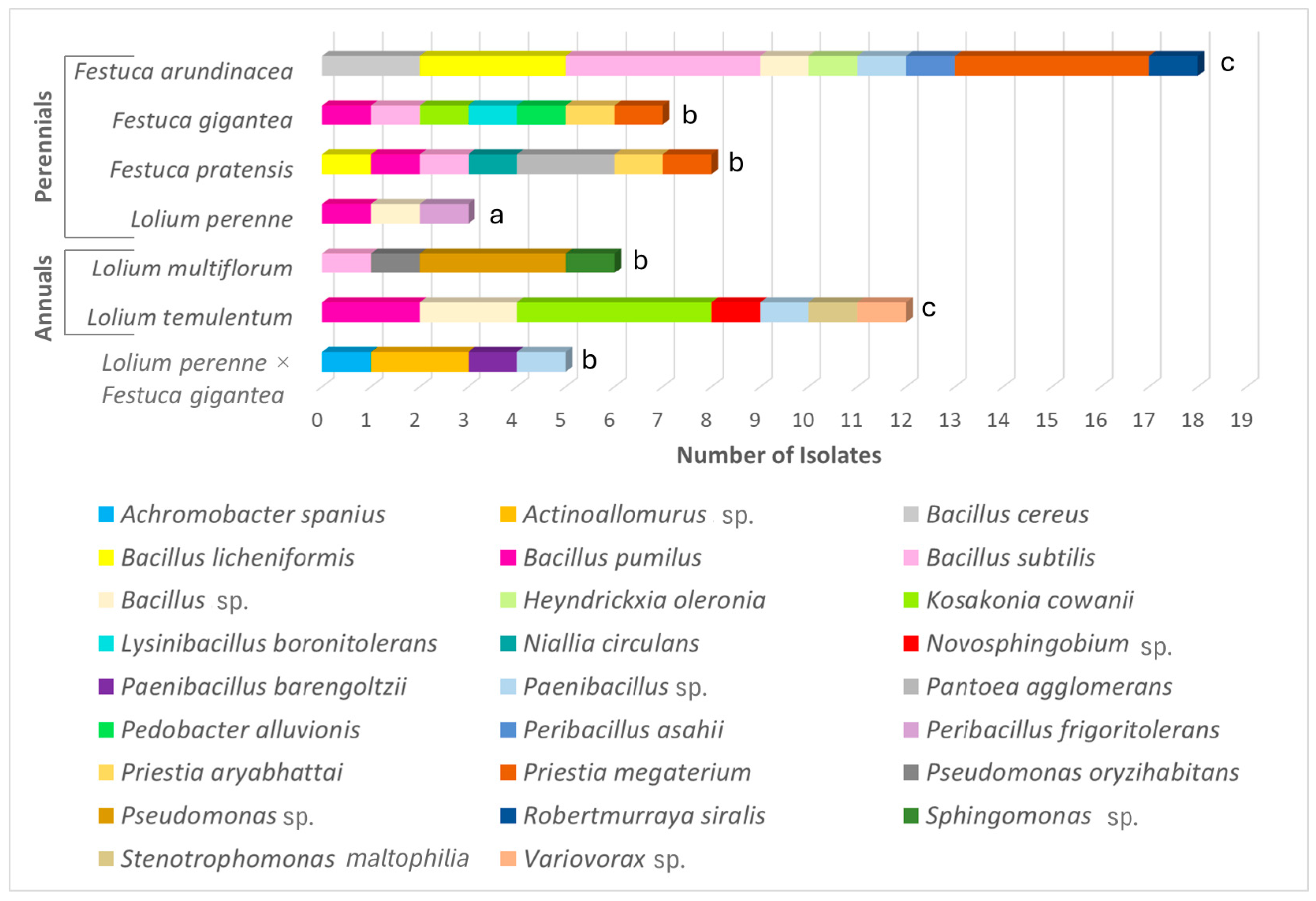
3.3. Diversity and Abundance of the Endophytic Bacteria

The pattern of the endophytic bacterial distribution in the roots of the Lolium and Festuca species and their hybrids is shown in Figure 3. Twenty-six bacteria taxa were identified from a total of 59 isolates in the culture on the LB medium from the roots of Festuca and Lolium species and the L. perenne × F. gigantea hybrids. No bacterial growth was detected in the control Petri dishes with the final root wash water after sterilization. The taxonomic assignment of the bacteria was confirmed by the BLAST results for the 16S rDNA sequences (Table S3). The 16S rDNA sequences obtained from our bacterial isolates were deposited into GenBank (Table S4).
Notably, the open-grassland perennial F. arundinacea stood out with the highest bacterial endophyte diversity, yielding 18 isolates (30% of the total) from nine bacteria species (Figure 3, Table S3). The next species with the highest bacterial endophyte diversity was the annual L. temulentum, with 12 (20%) isolates from seven bacterial taxa. From the roots of F. gigantea and F. pratensis, seven bacterial taxa were obtained from each species, yielding seven (12%) and eight (14%) isolates, respectively. L. multiflorum and L. perenne × F. gigantea yielded four bacterial taxa each. In contrast, L. perenne showed the lowest bacterial associations, with only three endophytic bacterial taxa identified, each represented by a single culture strain.
Similar to the fungal endophyte distribution (Figure 2), the presence of bacterial taxa showed no correlation between the root-derived bacteria of the L. perenne and F. gigantea parents and those of the L. perenne × F. gigantea hybrids (Figure 3).
As for the isolation frequency (IF, %), the genera of the endophytic bacteria are presented hierarchically in Table S2. The most common were Gram-positive, endospore-forming bacteria from Bacillota (synonym Firmicutes); they were represented by 15 species belonging to eight genera, namely the Bacillus, Heyndrickxia, Lysinibacillus, Niallia, Paenibacillus, Peribacillus, Priestia, and Robertmurraya genera (Figure 3). Of these, Bacillus (IF = 37%), Priestia (IF = 14%), and Paenibacillus (IF = 7%) were the most prevalent (Figure 3). The set of 15 Bacillota species was identified as follows: Bacillus cereus, Bacillus licheniformis, Bacillus pumilus, Bacillus subtilis, the Bacillus sp., Heyndrickxia oleronius, Lysinibacillus boronitolerans, Niallia circulans, Paenibacillus barengoltzii, the Paenibacillus sp., Peribaccilus asahii, Peribaccilus frigoritolerans, Priestia aryabhattai, Priestia megaterium, and Robertmurraya siralis.
In addition, nine species of Gram-negative bacteria belonging to the Pseudomonadota phylum were isolated and identified as follows: Achronobacter spanius, Kosakonia cowanii, the Novosphingobium sp., Pantoea agglomerans, Pseudomonas oryzihabitans, Pseudomonas sp., the Sphingomonas sp., Stenotrophomonas maltophilia, and the Variovorax sp. (Table S2, Figure 3). Of these, the K. cowanii and Pseudomonas sp. isolates were recovered at higher rates than a single isolate, with an IF = 8% and an IF = 7%, respectively (Figure 3).
The bacteria from the Actinomycetota and Bacteroidota phyla were rare taxa associated with the studied grasses, with each represented by a single species: Actinoallomurus sp. (Actinomycetales) was obtained from the hybrid (two isolates) and Pedobacter alluvionis (Sphingobacteriales) from F. gigantea (a single isolate), respectively (Figure 3, Table S2).

3.4. The Fungal and Bacterial Taxon Richness Across Plant Species

We calculated the endophyte abundance and taxon richness (D) for the endophytic fungi and bacteria in different plant hosts. Notably, the woodland grass F. gigantea had the highest microbial endophyte richness, namely D = 2.57 for fungi and D = 3.08 for bacteria (Table 3). The next species with the highest fungal endophyte richness was Lolium multiflorum at D = 2.43. For the bacterial associations, all three Festuca species—F. arundinacea, F. gigantea, and F. pratensis—and L. temulentum exhibited high bacterial endophyte richness. The bacterial diversity indices (D) for these species were >2, in contrast to the other host plants, which had diversity indices <2 (Table 3). Perennial polyploid Festuca species of natural hybrid origin, such as F. arundinacea and F. gigantea, exhibited a significantly higher total abundance of microbial endophytes. In contrast, the related diploid species like F. pratensis, and particularly L. perenne, had the lowest abundance (Table 3).

4. Discussion

Our findings highlight significant variations in the colonization frequency, taxonomic diversity, and richness of endophytic fungi and bacteria among Festuca and Lolium grasses, offering insights into the factors that shape the endophyte communities in their roots.

4.1. The Distribution of Fungal Root-Derived Endophytes in Festuca/Lolium Grasses

Numerous studies have demonstrated that the roots of most grasses serve as habitats for endophytic fungi [4,13,14,15]. Research on agricultural crops, including wheat, barley, soybean, corn, rice, and cotton, has also revealed a diverse range of fungal endophytes and their essential roles in supporting host plants [1,2].
In the root cutting culture on the PDA medium, a total of 21 fungal species (60 isolates) were isolated, predominantly from the division Ascomycota (18 species), with a few representatives from Basidiomycota (2) and Mucoromycota (1). This aligns with previous studies showing the dominance of Ascomycota among fungal endophytes, which are common root-associated symbionts and saprotrophs [6,27,30,39,40].
Most of these root fungi are known to be saprophytic or endophytic, and they are found in the soil as decomposers of organic matter or internally in various plant parts. However, some of them are also plant pathogens, namely A. alternata, A. infectoria, B. sorokiniana, P. cucumerina, and P. dictyoides; they cause diseases in cereals and some other crops. Additionally, they can live endophytically, having a neutral effect on plant health.
The most prevalent root endophyte was M. bolleyi, found in five of seven host species at an IF = 20% of the total isolate number. Another widely detected fungus was A. alternata (IF = 15%); it was particularly abundant in L. multiflorum, with the highest number of isolates (seven) recovered. Both M. bolleyi [27,41,42,43] and A. alternata [27,40,44,45] are referred to as plant endophyte generalists and are widely common across diverse plant species.
Along with the grass host generalists, some unusual accessions were isolated from the roots of Festuca/Lolium. Two Basidiomycota, Coprinellus disseminatus (Agaricales) and Sistotrema brinkmannii (Cantharellales), were occasionally present in the forest grass F. gigantea. While C. disseminatus is not typically an endophyte, it has been found in association with orchids in tropical forests [46,47]. In grasses, this endophytic fungus was obtained in a culture of the leaves of timothy (Phleum pratense) grass [27]. S. brinkmannii is not a typical grass endophyte either; it is a wood-rotting fungus widespread on the bark of trees and also found in the soil [48,49]. However, considering the wide distribution of C. disseminatus and S. brinkmannii in terrestrial habitats [49], it can be expected that these endophytes may also be found in the roots of forest grasses.
Cordyceps fumosorosea (Hypocreales) was occasionally present in the annual weed L. temulentum at a high rate (five isolates) of recovery. Cordyceps species are known as parasites that inhabit insect larvae [50,51]. While Cordyceps fungi are best known for their role as endoparasites and entomopathogens, some species have been isolated from plant tissues as well. A list of Cordyceps fungi that naturally infect plants is available in Vega’s review [52]. Notably, in endophyte research, the horizontal transmission of endophytic fungi between insects and plants is a well-known phenomenon, as reviewed by Raman and Suryanarayanan [53]. Given that L. temulentum (a common weed) thrives in a wide range of disturbed ecological sites, such as waste grounds rich in organic decay which attract numerous insects and their larvae, the presence of C. fumosorosea in these environments seems highly possible. Overall, plant endophytes can be transmitted either vertically (directly from the parent via seeds) or horizontally (from the surrounding environment) [54,55]. Further detailed studies are needed, particularly on seed-borne endophyte associations, to clarify the origin of the endophytes found in the roots of Festuca and Lolium plants.
From the fungal endophytes obtained in this study, three root-derived fungi have already been studied for their growth-promoting effects in L. multiflorum and barley (Hordeum vulgare) [10]. In this study, C. fastigiata, P. fimeti, and P. cucumerina promoted the growth of barley and ryegrass, with the most pronounced impact on their root size. In addition, the VOCs (Volatile Organic Compounds) emitted by these fungi exhibited a strong stimulating effect on root growth [10]. Several other fungal endophytes among those obtained have also been identified as growth-promoters. Namely, A. pullulans [56], C. cladosporioides [57], and E. nigrum [58] have demonstrated the ability to produce growth-promoting substances and act as biocontrol agents, helping plants resist pathogens and abiotic stresses.

4.2. Fungal Endophytes Across Grass Growth Types and Habitats

Most grasses are home to endophytic fungi, which play an important role in the ecophysiology of plants. Endophytic fungi are particularly prevalent in high-stress environments and are abundant across various ecosystems [4,13,14,15]. The Festuca/Lolium group examined in our study consists of plants from contrasting environments and of different growth types. Festuca gigantea is a woodland species, whereas all of the other species are grasses from open-grassland sites used in meadows and pastures, except for L. temulentum, which is a short-lived weed. In this tested group of grasses, F. gigantea stays notably distinct from the others—it has adapted to deal with light deficiency in a specific ecological niche rich in decomposing organic litter shaded by the tree canopy [59].
In this study, when comparing the endophyte prevalence across different plant species, the annual L. multiflorum exhibited the highest fungal diversity, with nine distinct fungal taxa isolated from its roots. In terms of the isolates recovered, both annual Lolium species, L. multiflorum and L. temulentum, exhibited the best results. The higher fungal prevalence in the annuals shows the possible benefits of endophytes to these fast-growing Lolium species. High levels of endophytic fungi in the roots are likely to have a positive impact on their nutrient acquisition, supporting the short-season growth dynamics of these plants.
Among the perennials, the greatest fungal diversity was recorded in the roots of the woodland grass F. gigantea. In contrast, the open-grassland perennials F. pratensis, F. arundinacea, and L. perenne exhibited the lowest fungal diversity. This pattern indicates that habitat type plays an important role in shaping fungal endophyte communities, highlighting that forest sites provide more favorable conditions for hosting diverse endophytic fungi than open grasslands.

4.3. The Distribution of Bacterial Root-Derived Endophytes in Festuca/Lolium Grasses

Endophytic bacteria are known for their plant-growth-promoting properties, contribution to plant development, and biocontrol effects, similar to endophytic fungi [15,60]. However, the diversity and functional roles of the root-associated bacteria in Festuca and Lolium species are poorly documented. Among the species examined, F. arundinacea and F. gigantea remain the only species studied in terms of the bacterial endophytes derived from their roots [26,27].
In our study, we identified 26 bacterial species from 59 isolates representing members of four bacterial phyla. The most common were Gram-positive, spore-forming bacteria from the Bacillota (syn. Firmicutes) phylum, yielding 58% of the isolated bacterial cultures.
Bacillus bacteria were the most prevalent, comprising five species and accounting for 38% of the total isolates. Bacillus endophytes were found to be hosted by all of the Lolium and Festuca species examined, except for the hybrids. Bacillus bacteria are widely distributed in natural environments and exhibit remarkable host diversity. These microorganisms perform a broad range of ecological functions, including the decomposition of organic matter, the promotion of plant growth, and the suppression of pathogenic organisms [61,62]. In grasses, the inoculation of bermudagrass with Bacillus sp. strains demonstrated beneficial effects, increasing the nitrogenase activity, phosphate solubilization, and siderophore production [63]. Similarly, P. megaterium exhibited plant defense and growth-promoting responses in Arabidopsis thaliana by elevating the expression of defense-related genes and increasing the accumulation of salicylic acid (SA) [64].
Another group of bacteria was the Gram-negative Pseudomonadota; they accounted for 35% of the isolated bacterial community, with nine species identified. Members of this group exhibit diverse functionalities affecting plant life. For example, Stenotrophomonas maltophilia (Xanthomonadales) is commonly found in agricultural environments and is known for its plant-growth-promoting characteristics [65], as well as its antifungal activity against pathogens in cereals [66]. Pseudomonas oryzihabitans (Pseudomonadales) is described as a soil bacterium that survives in moist, muddy environments and is indigenous to rice paddies [67].

4.4. Bacterial Endophytes Across Grass Growth Types and Habitats

Diverse bacterial communities have been reported in the roots of grasses across many species in different habitats [15,30,60,68,69]. In our study, the highest bacterial diversity was obtained in the open-grassland F. arundinacea, with nine species (18 isolates) recovered, followed by L. temulentum, with seven species (12 isolates). Among the factors influencing more bacterial vs. fungal endophyte hosting could be the dry soil conditions in an open-field grassland compared to the damp habitat established under the forest canopy, the niche of the woodland F. gigantea. In addition, certain plant species may be specifically favorable for hosting diverse endophytes and potentially benefit from interactions that positively affect their growth and stress tolerance. The importance of plant host specificity to the fungal endophyte abundance in the forage grasses Phleum pratense and L. perenne was demonstrated by Przemieniecki et al. [27]. Our results from the roots align with their findings from the leaves, showing that L. perenne has a particularly low endophyte occurrence among that of other grasses used in agricultural grasslands.
Interestingly, the lack of a correlation between the microbial communities of the L. perenne × F. gigantea hybrids and their parental species suggests that endophyte recruitment is influenced by complex host–microbe interactions rather than simple genetic inheritance. Quantitively, these laboratory-produced hybrids did not show an enhanced presence of microbial endophyte taxa. On the other hand, the species of natural hybrid origin, F. arundinacea and F. gigantea, harbored greater microbial diversity compared to that of their diploid F. pratensis and L. perenne relatives, showing a possible link between hybrid-derived genome complexity and increased root microbiota.
A network analysis of microbial communities identified Burkholderiales as consistently present across diverse ecosystem types and as a keystone taxon in grasslands, forests, and agricultural lands [70]. We identified two representatives of Burkholderiales in the grass roots: Achronobacter spanius and the Variovorax sp. Moreover, our analysis detected Pedobacter alluvionis, a member of Sphingobacteriales that, according to Banerjee and co-authors [66], is also among the keystone taxa in woodland and grassland ecosystems. Therefore, although the diversity of the endophytes identified in this study is relatively limited compared to that of the meta-analysis data, our findings confirm the presence of key microbial community members within the roots of Festuca and Lolium grasses.
Our results are based on a location in Lithuania that features common grassland in the European temperate climate zone. The impact of location on endophytic microbial diversification is significant. For instance, a study of the bacterial and fungal endophytes in Elymus nutans (Poaceae) seeds revealed that the fungal community in the seeds varied significantly across four locations on the Qinghai–Tibet Plateau, whereas the bacterial community was not affected by the plant’s growth location [71]. Further investigation into Festuca and Lolium species in different locations and environmental conditions (e.g., drought, cold, and contrasting soils) would provide a deeper understanding of the core endophyte microbiome transmitted across generations and its modulation by environmental factors.

5. Conclusions

Our study reveals distinct patterns of fungal and bacterial endophyte diversity in Festuca and Lolium grasses, with variations linked to the host species, growth type traits, and ecological adaptations. The annual species L. multiflorum and L. temulentum exhibited a greater abundance of endophytes in the roots than that of their perennial relatives, possibly reflecting a need for broader symbiotic interactions to support their short life cycles. In the woodland grass F. gigantea, higher fungal endophyte diversity was observed compared to that in the open-grassland Festuca and Lolium perennials. This implies that environmental factors such as light and humidity, along with access to organic matter, are key players in shaping microbial communities. Differences in the microbial colonization and diversity across Festuca and Lolium grasses—between perennials and annuals as well as between forest-adapted and open-field species—provide valuable insights into the ecological roles of endophytes. Overall, our data from a grassland site in Lithuania suggest that both environmental and genetic factors influence endophyte colonization. Further investigations across contrasting environments and locations are needed to gain a deeper understanding of the core endophyte microbiome of these species and its modulation by environmental factors. Among the root-derived endophytes isolated, several fungi and bacteria are potential candidates for plant growth promotion and biocontrol. These isolates include the fungi Aureobasidium pullulans, Cadophora fastigiata, Epicoccum nigrum, and Plectosphaerella cucumerina, as well as the bacteria Priestia megaterium and Stenotrophomonas maltophilia.

Supplementary Materials

The following supporting information can be downloaded at https://www.mdpi.com/article/10.3390/microorganisms13040799/s1. Figure S1: Cytomorphological images; Table S1: Taxonomic assignment of the endophytic fungi; Table S2: Isolation frequency (IF); Table S3: Taxonomic assignment of the endophytic bacteria; Table S4: The 16S DNA sequences of the bacterial isolates deposited into GenBank.

Author Contributions

Conceptualization: V.S. and I.P. Methodology: V.S., I.P. and J.M. Software: V.S. Validation: V.S. and I.P. Formal analysis: V.S., I.P., S.M. and G.Š. Investigation: V.S., S.M. and J.M. Data curation: V.S. Writing—original draft preparation: V.S., I.P., S.M. and G.Š. Writing—review and editing: V.S. and I.P. Visualization: V.S. Supervision: I.P. All authors have read and agreed to the published version of the manuscript.

Funding

This research received no external funding.

Institutional Review Board Statement

Not applicable.

Informed Consent Statement

Not applicable.

Data Availability Statement

All of the data are contained within the article.

Conflicts of Interest

The authors declare no conflicts of interest.

References

  1. Porras-Alfaro, A.; Herrera, J.; Sinsabaugh, R.L.; Odenbach, K.J.; Lowrey, T.; Natvig, D.O. Novel root fungal consortium associated with a dominant desert grass. Appl. Environ. Microbiol. 2008, 74, 2805–2813. [Google Scholar] [CrossRef] [PubMed]
  2. Mandyam, K.; Loughin, T.; Jumpponen, A. Isolation and morphological and metabolic characterization of common endophytes in annually burned tallgrass prairie. Mycologia 2010, 102, 813–821. [Google Scholar] [CrossRef] [PubMed]
  3. Glynou, K.; Ali, T.; Buch, A.-K.; Haghi Kia, S.; Ploch, S.; Xia, X.; Çelik, A.; Thines, M.; Maciá-Vicente, J.G. The local environment determines the assembly of root endophytic fungi at a continental scale. Environ. Microbiol. 2016, 18, 2418–2434. [Google Scholar] [CrossRef] [PubMed]
  4. Knapp, D.G.; Imrefi, I.; Boldpurev, E.; Csíkos, S.; Akhmetova, G.; Berek-Nagy, P.J.; Otgonsuren, B.; Kovács, G.M. Root-colonizing endophytic fungi of the dominant grass Stipa krylovii from a mongolian steppe grassland. Front. Microbiol. 2019, 10, 2565. [Google Scholar] [CrossRef]
  5. Knapp, D.G.; Németh, J.B.; Barry, K.; Hainaut, M.; Henrissat, B.; Johnson, J.; Kuo, A.; Lim, J.H.P.; Lipzen, A.; Nolan, M.; et al. Comparative genomics provides insights into the lifestyle and reveals functional heterogeneity of dark septate endophytic fungi. Sci. Rep. 2018, 8, 6321. [Google Scholar] [CrossRef]
  6. Toju, H.; Yamamoto, S.; Sato, H.; Tanabe, A.S.; Gilbert, G.S.; Kadowaki, K. Community composition of root-associated fungi in a Quercus-dominated temperate forest: “Codominance” of mycorrhizal and root-endophytic fungi. Ecol. Evol. 2013, 3, 1281–1293. [Google Scholar] [CrossRef]
  7. Mandyam, K.; Jumpponen, A. Seeking the elusive function of the root-colonising dark septate endophytic fungi. Stud. Mycol. 2005, 53, 173–189. [Google Scholar] [CrossRef]
  8. Knapp, D.G.; Pintye, A.; Kovács, G.M. The dark side is not fastidious—Dark septate endophytic fungi of native and invasive plants of semiarid sandy areas. PLoS ONE 2012, 7, e32570. [Google Scholar] [CrossRef]
  9. Andrade-Linares, D.; Franken, P. Fungal endophytes in plant roots: Taxonomy, colonization patterns, and functions. In Symbiotic Endophytes; Springer: Berlin/Heidelberg, Germany, 2013; pp. 311–334. [Google Scholar] [CrossRef]
  10. Pašakinskienė, I.; Stakelienė, V.; Matijošiūtė, S.; Martūnas, J.; Rimkevičius, M.; Būdienė, J.; Aučina, A.; Skridaila, A. Growth-Promoting Effects of Grass Root-Derived Fungi Cadophora fastigiata, Paraphoma fimeti and Plectosphaerella cucumerina on Spring Barley (Hordeum vulgare) and Italian Ryegrass (Lolium multiflorum). Microorganisms 2025, 13, 25. [Google Scholar] [CrossRef]
  11. Backer, R.; Rokem, J.S.; Ilangumaran, G.; Lamont, J.; Praslickova, D.; Ricci, E.; Subramanian, S.; Smith, D.L. Plant growth promoting Rhizobacteria: Context, mechanisms of action, and roadmap to commercialization of biostimulants for sustainable agriculture. Front. Plant Sci. 2018, 9, 1473. [Google Scholar] [CrossRef]
  12. Aloo, B.N.; Nyongesa, B.O.; Were, J.O.; Were, B.A.; Tumuhairwe, J.B. Rhizobacterial biomolecules for sustainable crop production and environmental management: Plausible functions and molecular mechanism. In Microbial Biomolecules Emerging Approach in Agriculture, Pharmaceuticals and Environment Management, 1st ed.; Kumar, A., Bilal, M., Ferreira, L.F.R., Madhuree, K., Eds.; Academic Press: Cambridge, MA, USA, 2023; pp. 1–30. [Google Scholar]
  13. Addy, H.D.; Piercey, M.M.; Currah, R.S. Microfungal endophytes in roots. Can. J. Bot. 2005, 83, 1–13. [Google Scholar] [CrossRef]
  14. Wearn, J.A.; Sutton, B.C.; Morley, N.J.; Gange, A.C. Species and organ specificity of fungal endophytes in herbaceous grassland plants. J. Ecol. 2012, 100, 1085–1092. [Google Scholar] [CrossRef]
  15. Hardoim, P.R.; van Overbeek, L.S.; Berg, G.; Pirttilä, A.M.; Compant, S.; Campisano, A.; Döring, M.; Sessitsch, A. The hidden world within plants: Ecological and evolutionary considerations for defining functioning of microbial endophytes. Microbiol. Mol. Biol. Rev. 2015, 79, 293–320. [Google Scholar] [CrossRef] [PubMed]
  16. Schardl, C.L. The epichloae, symbionts of the grass subfamily Poöideae. Ann. Mo. Bot. Gard. 2010, 97, 646–665. [Google Scholar] [CrossRef]
  17. Tadych, M.; Bergen, M.S.; White, J.F. Epichloë spp. associated with grasses: New insights on life cycles, dissemination and evolution. Mycologia 2014, 106, 181–201. [Google Scholar] [CrossRef]
  18. Saikkonen, K.; Ahlholm, J.; Helander, M.; Lehtimäki, S.; Niemeläinen, O. Endophytic fungi in wild and cultivated grasses in Finland. Ecography 2000, 23, 360–366. [Google Scholar] [CrossRef]
  19. Leyronas, C.; Raynal, G. Presence of Neotyphodium-like endophytes in European grasses. Ann. Appl. Biol. 2001, 139, 119–127. [Google Scholar] [CrossRef]
  20. Müller, C.B.; Krauss, J. Symbiosis between grasses and asexual fungal endophytes. Curr. Opin. Plant Biol. 2005, 8, 450–456. [Google Scholar] [CrossRef]
  21. Soto-Barajas, M.; Azquez-De-Aldana, B.; Alvarez, A.; Zabalgogeazcoa, I. Sympatric Epichloë species and chemotypic profiles in natural populations of Lolium perenne. Fungal Ecol. 2019, 39, 231–241. [Google Scholar] [CrossRef]
  22. Krauss, J.; Vikuk, V.; Young, C.A.; Krischke, M.; Mueller, M.J.; Baerenfaller, K. Epichloë endophyte infection rates and alkaloid content in commercially available grass seed mixtures in Europe. Microorganisms 2020, 8, 498. [Google Scholar] [CrossRef]
  23. Hume, D.E.; Stewart, A.V.; Simpson, W.R.; Johnson, R.D. Epichloë fungal endophytes play a fundamental role in New Zealand grasslands. J. R. Soc. N. Z. 2020, 50, 279–298. [Google Scholar] [CrossRef]
  24. Garces, K.R.; Sage, H.E.; Christian, N.; Emery, S.M. Epichloë increases root fungal endophyte richness and alters root fungal endophyte composition in a changing world. J. Fungi 2022, 8, 1142. [Google Scholar] [CrossRef] [PubMed]
  25. Pašakinskienė, I.; Stakelienė, V.; Matijošiūtė, S.; Martūnas, J. Diversity of Endophytic Fungi and Bacteria Inhabiting the Roots of the Woodland Grass, Festuca gigantea (Poaceae). Diversity 2024, 16, 453. [Google Scholar] [CrossRef]
  26. Del los Santos, M.C.; Taulé, C.; Mareque, C.; Baracochea, M.; Battistoni, F. Identification and characterization of the part of the bacterial community associated with field-grown tall fescue (Festuca arundinacea) cv. SFRO Don Tomás in Uruguay. Ann. Microbiol. 2016, 66, 329–342. [Google Scholar] [CrossRef]
  27. Przemieniecki, S.W.; Damszel, M.; Kurowski, T.P.; Mastalerz, J.; Kotlarz, K. Identification, ecological evaluation and phylogenetic analysis of non-symbiotic endophytic fungi colonizing timothy grass and perennial ryegrass grown in adjacent plots. Grass Forage Sci. 2019, 74, 42–52. [Google Scholar] [CrossRef]
  28. Coombs, J.T.; Franco, C.M.M. Isolation and identification of actinobacteria from surface-sterilized wheat roots. Appl. Environ. Microbiol. 2003, 69, 5603–5608. [Google Scholar] [CrossRef]
  29. Groenewald, J.Z.; Nakashima, C.; Nishikawa, J.; Shin, H.-D.; Park, J.-H.; Jama, A.N.; Groenewald, M.; Braun, U.; Crous, P.W. Species concepts in Cercospora: Spotting the weeds among the roses. Stud. Mycol. 2013, 75, 115–170. [Google Scholar] [CrossRef]
  30. Toju, H.; Kurokawa, H.; Kenta, T. Factors influencing leaf- and root-associated communities of bacteria and fungi across 33 plant orders in a grassland. Front. Microbiol. 2019, 10, 241. [Google Scholar] [CrossRef]
  31. Baluška, F.; Mancuso, S. Root apex transition zone as oscillatory zone. Front. Plant Sci. 2013, 4, 354. [Google Scholar] [CrossRef]
  32. Baskin, T.I. Patterns of root growth acclimation: Constant processes, changing boundaries. Wiley Interdiscip. Rev. Dev. Biol. 2013, 2, 65–73. [Google Scholar] [CrossRef]
  33. Kiheri, H.; Heinonsalo, J.; Timonen, S. Staining and microscopy of mycorrhizal fungal colonization in preserved ericoid plant roots. J. Berry Res. 2017, 4, 231–237. [Google Scholar] [CrossRef]
  34. White, T.J.; Bruns, T.; Lee, S.J.W.T.; Taylor, J. Amplification and direct sequencing of fungal ribosomal RNA genes for phylogenetics. In PCR Protocols: A Guide to Methods and Applications; Academic Press: Cambridge, MA, USA, 1990; pp. 315–322. [Google Scholar]
  35. Sung, G.-H.; Sung, J.-M.; Hywel-Jones, N.L.; Spatafora, J.W. A multi-gene phylogeny of Clavicipitaceae (Ascomycota, fungi): Identification of localized incongruence using a combinational bootstrap approach. Mol. Phylogenet. Evol. 2007, 44, 1204–1223. [Google Scholar] [CrossRef] [PubMed]
  36. Klindworth, A.; Pruesse, E.; Schweer, T.; Peplies, J.; Quast, C.; Horn, M.; Glöckner, F.O. Evaluation of general 16S ribosomal RNA gene PCR primers for classical and next-generation sequencing-based diversity studies. Nucleic Acids Res. 2013, 41, e1. [Google Scholar] [CrossRef] [PubMed]
  37. Song, L.; Huo, J.; Wan, L.; Pan, L.; Jiang, N.; Fu, J.; Wei, S.; He, L. Differences and biocontrol potential of haustorial endophytic fungi from Taxillus chinensis on different host plants. BMC Microbiol. 2023, 23, 128. [Google Scholar] [CrossRef]
  38. Vu, D.; Groenewald, M.; de Vries, M.; Gehrmann, T.; Stielow, B.; Eberhardt, U.; Al-Hatmi, A.; Groenewald, J.Z.; Cardinali, G.; Houbraken, J.; et al. Large-scale generation and analysis of filamentous fungal DNA barcodes boosts coverage for kingdom fungi and reveals thresholds for fungal species and higher taxon delimitation. Stud. Mycol. 2019, 92, 135–154. [Google Scholar] [CrossRef]
  39. Sánchez Márquez, S.; Bills, G.; Zabalgogeazcoa, I. The endophytic mycobiota of the grass Dactylis glomerata. Fungal Divers. 2007, 27, 171–195. [Google Scholar]
  40. Rashmi, M.; Kushveer, J.S.; Sarma, V.V. A worldwide list of endophytic fungi with notes on ecology and diversity. Mycosphere 2019, 10, 798–1079. [Google Scholar] [CrossRef]
  41. Wirsel, S.G.R.; Leibinger, W.; Ernst, M.; Mendgen, K. Genetic diversity of fungi closely associated with common reed. N. Phytol. 2001, 149, 589–598. [Google Scholar] [CrossRef]
  42. Gao, Y.; Ren, G.-C.; Wanasinghe, D.; Xu, J.-C.; Farias, A.; Gui, H. Two new species and a new record of Microdochium from grasses in Yunnan province, South-West China. J. Fungi 2022, 8, 1297. [Google Scholar] [CrossRef]
  43. David, A.S.; Seabloom, E.W.; May, G. Plant host species and geographic distance affect the structure of aboveground fungal symbiont communities, and environmental filtering affects belowground communities in a coastal dune ecosystem. Microb. Ecol. 2016, 71, 912–926. [Google Scholar] [CrossRef]
  44. Thomma, B.P.H.J. Alternaria spp.: From general saprophyte to specific parasite. Mol. Plant Pathol. 2003, 4, 225–236. [Google Scholar] [CrossRef]
  45. Pereira, E.; Vázquez de Aldana, B.R.; San Emeterio, L.; Zabalgogeazcoa, I. A survey of culturable fungal endophytes from Festuca rubra subsp. pruinosa, a grass from marine cliffs, reveals a core microbiome. Front. Microbiol. 2019, 9, 3321. [Google Scholar] [CrossRef] [PubMed]
  46. Yagame, T.; Funabiki, E.; Yukawa, T.; Nagasawa, E. Identification of mycobionts in an achlorophyllous orchid, Cremastra aphylla (Orchidaceae), based on molecular analysis and basidioma morphology. Mycoscience 2018, 59, 18–23. [Google Scholar] [CrossRef]
  47. Gao, Y.; Peng, S.; Hang, Y.; Xie, G.; Ji, N.; Zhang, M. Mycorrhizal fungus Coprinellus disseminatus influences seed germination of the terrestrial orchid Cremastra appendiculata (D. Don) Makino. Sci. Hortic. 2022, 293, 110724. [Google Scholar] [CrossRef]
  48. Potvin, L.R.; Richter, D.L.; Jurgensen, M.F.; Dumroese, R.K. Association of Pinus banksiana Lamb. and Populus tremuloides Michx. seedling fine roots with Sistotrema brinkmannii (Bres.) J. Erikss. (Basidiomycotina). Mycorrhiza 2012, 22, 631–638. [Google Scholar] [CrossRef]
  49. Hallenberg, N. Speciation and distribution in Corticiaceae (Basidiomycetes). Plant Syst. Evol. 1991, 177, 93–110. [Google Scholar] [CrossRef]
  50. Nikoh, N.; Fukatsu, T. Interkingdom host jumping underground: Phylogenetic analysis of entomoparasitic fungi of the genus Cordyceps. Mol. Biol. Evo. 2000, 17, 629–638. [Google Scholar] [CrossRef]
  51. Lei, Y.; Hussain, A.; Guan, Z.; Wang, D.; Jaleel, W.; Lyu, L.; He, Y. Unraveling the Mode of Action of Cordyceps fumosorosea: Potential Biocontrol Agent against Plutella xylostella (Lepidoptera: Plutellidae). Insects 2021, 12, 179. [Google Scholar] [CrossRef]
  52. Vega, F.E. The use of fungal entomopathogens as endophytes in biological control: A review. Mycologia 2018, 110, 4–30. [Google Scholar] [CrossRef]
  53. Raman, A.; Suryanarayanan, T.S. Fungus–plant interaction influences plant-feeding insects. Fungal Ecol. 2017, 29, 123–132. [Google Scholar] [CrossRef]
  54. Gundel, P.E.; Rudgers, J.A.; Ghersa, C.M. Incorporating the process of vertical transmission into understanding of host-symbiont dynamics. Oikos 2011, 120, 1121–1128. [Google Scholar] [CrossRef]
  55. Shade, A.; Jacques, M.S.; Barret, M. Ecological patterns of seed microbiome diversity, transmission, and assembly. Curr. Opin. Microbiol. 2017, 37, 15–22. [Google Scholar] [CrossRef]
  56. Di Francesco, A.; Di Foggia, M.; Corbetta, M.; Baldo, D.; Ratti, C.; Baraldi, E. Biocontrol Activity and Plant Growth Promotion Exerted by Aureobasidium pullulans Strains. J. Plant Growth Regul. 2021, 40, 1233–1244. [Google Scholar] [CrossRef]
  57. Islam, M.T. Current Status and Future Prospects of Cladosporium sp., a Biocontrol Agent for Sustainable Plant Protection. Biocontrol Sci. 2022, 27, 185–191. [Google Scholar] [CrossRef] [PubMed]
  58. Fávaro, L.C.dL.; Sebastianes, F.L.dS.; Araújo, W.L. Epicoccum nigrum P16, a Sugarcane Endophyte, Produces Antifungal Compounds and Induces Root Growth. PLoS ONE 2012, 7, e36826. [Google Scholar] [CrossRef] [PubMed]
  59. Tichý, L.; Axmanová, I.; Dengler, J.; Guarino, R.; Jansen, F.; Midolo, G.; Nobis, M.P.; Van Meerbeek, K.; Aćić, S.; Attorre, F.; et al. Ellenberg-type indicator values for European vascular plant species. J. Veg. Sci. 2023, 34, e13168. [Google Scholar] [CrossRef]
  60. Wemheuer, F.; Kaiser, K.; Karlovsky, P.; Daniel, R.; Vidal, S.; Wemheuer, B. Bacterial endophyte communities of three important agricultural grass species differ in their response towards management regimes. Sci. Rep. 2017, 7, 40914. [Google Scholar] [CrossRef]
  61. Mandic-Mulec, I.; Stefanic, P.; van Elsas, J.D. Ecology of Bacillaceae. Microbiol. Spectr. 2015, 3, 1–24. [Google Scholar] [CrossRef]
  62. Tsotetsi, T.; Nephali, L.; Malebe, M.; Tugizimana, F. Bacillus for Plant Growth Promotion and Stress Resilience: What Have We Learned? Plants 2022, 11, 2482. [Google Scholar] [CrossRef]
  63. Coy, R.M.; Held, D.W.; Kloepper, J.W. Rhizobacterial Colonization of Bermudagrass by Bacillus spp. in a Marvyn Loamy Sand Soil. Appl. Soil Ecol. 2019, 141, 10–17. [Google Scholar] [CrossRef]
  64. Li, Q.; Hou, Z.; Zhou, D.; Jia, M.; Lu, S.; Yu, J. A plant growth-promoting bacteria Priestia megaterium JR48 induces plant resistance to the crucifer black rot via a salicylic acid-dependent signaling pathway. Front. Plant. Sci. 2022, 13, 1046181. [Google Scholar] [CrossRef] [PubMed]
  65. Alexander, A.; Singh, V.K.; Mishra, A.; Jha, B. Plant growth promoting rhizobacterium Stenotrophomonas maltophilia BJ01 augments endurance against N2 starvation by modulating physiology and biochemical activities of Arachis hypogea. PLoS ONE 2019, 14, e0222405. [Google Scholar] [CrossRef]
  66. Sharma, P.; Pandey, R.; Chauhan, N.S. Biofertilizer and biocontrol properties of Stenotrophomonas maltophilia BCM emphasize its potential application for sustainable agriculture. Front. Plant. Sci. 2014, 15, 1364807. [Google Scholar] [CrossRef]
  67. Kodama, K.; Kimura, N.; Komagata, K. Two new species of Pseudomonas: P. oryzihabitans isolated from rice paddy and clinical specimens and P. luteola isolated from clinical specimens. Int. J. Syst. Bacteriol. 1985, 35, 467–474. [Google Scholar] [CrossRef]
  68. Kandel, S.L.; Joubert, P.M.; Doty, S.L. Bacterial endophyte colonization and distribution within plants. Microorganisms 2017, 5, 77. [Google Scholar] [CrossRef]
  69. Li, Y.H.; Zhu, J.N.; Liu, Q.F.; Liu, Y.; Liu, M.; Liu, L.; Zhang, Q. Comparison of the diversity of root-associated bacteria in Phragmites australis and Typha angustifolia L. in artificial wetlands. World J. Microbiol. Biotechnol. 2013, 29, 1499–1508. [Google Scholar] [CrossRef]
  70. Banerjee, S.; Schlaeppi, K.; van der Heijden, M.G.A. Keystone taxa as drivers of microbiome structure and functioning. Nat. Rev. Microbiol. 2018, 16, 567–576. [Google Scholar] [CrossRef]
  71. Guo, J.; Bowatte, S.; Hou, F. Diversity of endophytic bacteria and fungi in seeds of Elymus nutans growing in four locations of Qinghai Tibet Plateau, China. Plant Soil 2021, 459, 49–63. [Google Scholar] [CrossRef]
Figure 1. Colonization frequency of endophytic fungi in root tip cuttings of Lolium and Festuca species and L. perenne × F. gigantea hybrids. Different letters above the bars indicate significant differences between the species (p ≤ 0.05) based on Tukey’s HSD post hoc test.
Microorganisms 13 00799 g001
Figure 2. Distribution of endophytic fungi isolated from the roots of Festuca and Lolium species and their hybrids. All of the species are classified under Ascomycota, except for Coprinellus disseminatus and Sistotrema brinkmannii from Basidiomycota and Mucor circinelloides from Mucoromycota. Different letters above the bars indicate significant differences between the species (p ≤ 0.05) based on Tukey’s HSD post hoc test. Note: The laboratory-produced L. perenne × F. gigantea hybrids are perennial-type plants.
Microorganisms 13 00799 g002
Figure 3. Distribution of endophytic bacteria isolated from Lolium and Festuca species and their hybrids. Different letters above the bars indicate significant differences between the species (p ≤ 0.05) based on Tukey’s HSD post hoc test. Note: The laboratory-produced L. perenne × F. gigantea hybrids are perennial-type plants.
Microorganisms 13 00799 g003
Table 1. List of plant species assessed in the current study.
SpeciesPloidy LevelGrowth TypeHabitat TypeAccession Origin
Festuca arundinacea ‘Monas’2n = 6x = 42PerennialOpen-grasslandLCAFS, IA
Festuca gigantea2n = 6x = 42PerennialForest sitesVU BG, Kairėnai, Vilnius, LT;
Vingis Park, Vilnius, LT
Festuca pratensis ‘Alanta’2n = 2x = 14PerennialOpen-grasslandLCAFS, IA
Lolium perenne ‘Veja’2n = 2x = 14PerennialOpen-grasslandLCAFS, IA
Lolium multiflorum ‘Grazer’2n = 2x = 14AnnualOpen-grasslandLCAFS, IA
Lolium temulentum2n = 2x = 14AnnualOpen-grasslandIPK Leibniz Institute, Gatersleben, DE
Lolium perenne × Festuca gigantea2n = 4x = 28PerennialOpen-grasslandLaboratory-produced, VU BG laboratory collection
Note: LCAFS, IA—Lithuanian Centre for Agricultural and Forestry Sciences, Institute of Agriculture, Akademija, Kėdainiai distr., LT; VU BG—Botanical Garden, Vilnius University; n—the haploid chromosome number: 2n—the total chromosome number in an organism; x—the basic chromosome number: 2x—diploid species, 4x—tetraploid species, 6x—hexaploid species.
Table 2. The list of primers in the PCR reactions for the amplification of fungal and bacterial DNA sequences.
LocusPrimersPrimer Sequences (5′–3′)Tm °CReference
Primers for fungal DNA
ITSITS1
ITS4
TCCGTAGGTGAACCTGCGG
TCCTCCGCTTATTGATATGC
54[34]
TEFaEF1-278F
EF-2
CATCGAGAAGTTCGAGAAGG
GGARGTACCAGTSATCATGTT
54[29]
SSUNS1
NS4
GTAGTCATATGCTTGTCTC
CTTCCGTCAATTCCTTTAAG
49[34]
RPB2RPB2-5F2
fRPB2-7cR
GGGGWGAYCAGAAGAAGGC
CCCATRGCTTGYTTRCCCAT
58[35]
Primers for bacterial DNA
16S rDNA27f CM
1492R
AGAGTTTGATCMTGGCTCAG
TACGGYTACCTTGTTACGACTT
52[28]
16S rDNA704F
765R
GTAGCGGTGAAATGCGTAGA
CTGTTTGCTCCCCACGCTTTC
56[28]
16S rDNAS-D-Bact-0341-b-S-17
S-D-Bact-0785-a-A-21
CCTACGGGNGGCWGCAG
GACTACHVGGGTATCTAATCC
56[36]
Table 3. Endophytic fungal and bacterial taxon richness (D) across different plant species. * No.—number; ** D—Margalef’s index.
Plant SpeciesEndophytic FungiEndophytic BacteriaTotal Microbial Endophyte Abundance
* No. IsolatesNo. Species** DNo. IsolatesNo. SpeciesDNo. IsolatesNo. Species
Perennial species
Festuca arundinacea111.001892.771910
Festuca gigantea762.57773.081413
Festuca pratensis111.00872.8998
Lolium perenne221.44331.8255
Lolium perenne × Festuca gigantea741.54541.86128
Annual species
Lolium multiflorum2792.43641.673313
Lolium temulentum1551.481272.412712
Disclaimer/Publisher’s Note: The statements, opinions and data contained in all publications are solely those of the individual author(s) and contributor(s) and not of MDPI and/or the editor(s). MDPI and/or the editor(s) disclaim responsibility for any injury to people or property resulting from any ideas, methods, instructions or products referred to in the content.

