Article

Exploring the Antimicrobial, Antioxidant and Extracellular Enzymatic Activities of Culturable Endophytic Fungi Isolated from the Leaves of Kirkia acuminata Oliv

by
Sagwadi Kubayi
,
Raymond Tshepiso Makola
and
Khumiso Dithebe
*
Department of Biochemistry, Microbiology and Biotechnology, University of Limpopo, Private Bag X 1106, Sovenga, Polokwane 0727, South Africa
*
Author to whom correspondence should be addressed.
Microorganisms 2025, 13(3), 692; https://doi.org/10.3390/microorganisms13030692
Submission received: 27 January 2025 / Revised: 14 March 2025 / Accepted: 17 March 2025 / Published: 19 March 2025
(This article belongs to the Special Issue Endophytic Fungus as Producers of New and/or Bioactive Substances)

Abstract

:
Fungal endophytes of medicinal plants produce diverse secondary metabolites and extracellular enzymes with therapeutic and biotechnological potential. However, the biological and biotechnological potential of fungal endophytes from South African medicinal plants remain relatively underexplored. In this study, the antimicrobial, antioxidant, anti-inflammatory and extracellular enzymatic capabilities of five fungal endophytes previously isolated from the leaves of Kirkia acuminata Oliv. were investigated. Sequencing of the internal transcribed spacer (ITS) regions revealed that the isolates belonged to the genera Setosphaeria, Diaporthe and Corynespora. The broth micro-dilution assay and the Folin–Ciocalteau reagent method were used to assess the antibacterial activity and the total phenolic content (TPC) of the fungal endophytes’ ethyl acetate crude extracts (CEs), respectively. The antioxidant activity was assessed using the ferric reducing antioxidant power (FRAP) and 1,1-diphenyl-2-picrylhydrazyl (DPPH) free radical scavenging assays. The influence of the CE of the Setosphaeria rostrata KaL-4 on the viability and LPS-induced interleukin-6 (IL-6) production in Raw 264.7 macrophages was assessed using a 3-(4,5-dimethylthiazol-2-yl)-2,5-diphenyltetrazolium bromide (MTT) assay and an ELISA, respectively. The ability of the isolates to produce extracellular proteases, laccases and peroxidases was also determined. The CEs displayed antimicrobial activity with MICs ranging from 0.63 to 1.25 mg/mL and reducing power and scavenging activity ranging from 40% to 18% and from 60% to 48%, respectively. The S. rostrata KaL-4 CE possessed the highest TPC and demonstrated dose-dependent cytotoxicity. The CE further demonstrated a significant reduction in IL-6 production at a concentration of 0.75 µg/mL. Only one isolate demonstrated the ability to produce proteases with an enzymatic index (EI) of 0.66, while laccases (EI range of 0.14 to 1.15) and peroxidases were produced by all of the isolates. These findings suggest that fungal endophytes from South African medicinal plants are promising sources of bioactive compounds and industry-significant extracellular enzymes.

1. Introduction

The emergence of drug-resistant pathogens is one of the major global health challenges. Several microbial pathogens have developed drug resistance mechanisms due to the misuse and/or overuse of antimicrobial drugs [1]. The increase in antimicrobial resistance (AMR) has diminished the number of antimicrobial agents which can be used to effectively treat and clear microbial infections. Microbial infections are a powerful trigger of the innate immune response and inflammation [2]. Prolonged microbial infections may possibly trigger hyper inflammation. While non-steroidal anti-inflammatory drugs (NSAIDs) are widely used to treat inflammation, the prolonged use of NSAIDs may lead to conditions such as renal injury, gastrointestinal toxicity, cardiovascular disorder and hepatoxicity [3], which affect their clinical application. This necessitates the search for novel and effective antimicrobial and anti-inflammatory agents with low toxicity.
Endophytes are microorganisms, mostly fungi and bacteria, which reside in healthy living plant tissue without causing any visible harm to the plant host [4]. The most widely studied group of endophytes, fungal endophytes, produce an array of structurally and chemically diverse secondary metabolites including those that are similar to their hosts [5]. The secondary metabolites produced by endophytic fungi belong to various classes of compounds, including terpenoids, alkaloids, flavonoids, quinones and phenols, which have been reported to possess various bioactivities including antimicrobial, anti-inflammatory, antioxidant and anticancer activities [6,7]. Fungal endophytes are not only excellent sources of secondary metabolites with therapeutic potential, but they also produce various extracellular enzymes [8].
Endophytic fungi use enzymes to penetrate plant tissue to facilitate colonization and obtain nutrition from the host as well as prevent the invasion of the host by phytopathogens [9]. These enzymes, which include cellulases, proteases, laccases, pectinases, lipases and peroxidases, can be applied in several industries, such as the pharmaceutical industry, as well as in food and wastewater treatment [10]. Their ability to withstand harsh conditions, such as temperature and pH, while producing little to no side reactions has made fungal enzymes to be the most sought-after product in industry [11]. Although fungal endophytes of medicinal plants have capabilities that can be utilized for the benefit of human health, the environment and industry, it is estimated that only 1% of fungal endophytes has been studied [12]. Additionally, the fungal endophytes of medicinal plants used in South Africa are relatively underexplored [13]. This leaves room for the exploration of medicinal plants used in South Africa as potential sources of novel fungal endophytes with the ability to produce important bioactive secondary metabolites and industry-significant extracellular enzymes.
The medicinal plant Kirkia acuminata Oliv., commonly known as White seringa, is widespread in tropical areas of sub-Saharan Africa where the fruits, leaves, roots and bark are used to treat abdominal pain, cough, snake bites, cholera and toothache in countries including South Africa [14,15]. This medicinal plant species is reported to be on the decline in the natural environment due to overharvesting by ethno-medicine practitioners looking to utilize the plant for its numerous medicinal uses [14]. Five fungal endophytes were previously isolated from the leaves of K. acuminata Oliv. [16]; however, the identity and the bioactivities and enzyme production potential of the isolates were not assessed.
The aim of this study was to investigate the antimicrobial, antioxidant, anti-inflammatory and extracellular enzymatic activities of the fungal endophytes isolated from K. acuminata Oliv. to assess their bioactive and biotechnological potential.

2. Materials and Methods

2.1. Identification of Endophytic Fungi

Five endophytic fungal isolates previously isolated from Kirkia acuminata Oliv. leaves [16] were plated on potato dextrose agar (PDA, Merck, Johannesburg, South Africa) and incubated for 7 days at 30 °C. The macromorphological characteristics of the cultures were observed to identify the different morphotypes. Molecular identification of the isolates was carried out at Inqaba Biotechnical Industry Ltd., Pretoria, South Africa. The Quick-DNA™ Fungal/Bacterial Miniprep Kit (Zymo Research, Catalogue No. D6005, Irvine, CA, USA) was used to extract genomic DNA from each isolate according to the manufacturer’s instructions. Polymerase chain reaction (PCR) was used to amplify the internal transcribed spacer (ITS) regions 1 and 2 with the forward ITS1 5′-TCCGTAGGTGAACCTGCGG-3′ and reverse ITS4 5′-TCCTCCGCTTATTGATATGC-3′ primers using the OneTaq mastermix (New England BioLabs Inc., Ipswich, MA, USA). The PCR products were visualized on 1% agarose gel (CSL-AG500, Cleaver Scientific Ltd., Warwickshire, UK) stained with EZ-vision® Blue light DNA Dye. The PCR products were enzymatically purified using the ExoSAP procedure (NEB M0293L; NEB M0371; New England BioLabs Inc., Ipswich, MA, USA). The PCR products were purified using the ZR-96 DNA Sequencing Clean-up Kit™ (Zymo Research, Catalogue No. D4050, Irvine, CA, USA) and sequenced in the forward and reverse direction using the ABI 3730xl Genetic Analyzer (Applied Biosystems, Thermo Fisher Scientific, Waltham, MA, USA). The consensus sequence for each isolate was assembled using the CLC Bio Main Workbench, and the basic local alignment search tool (BLAST) analysis was performed on the National Center for Biotechnology Information (NCBI) website (https://blast.ncbi.nlm.nih.gov/Blast.cgi, accessed 15 September 2022) with default parameters [17] to match the sequences with those of known fungi in the GenBank database.

2.2. Fungal Cultivation and Extraction of Metabolites

To produce the secondary metabolites, the pure endophytic fungal isolates were first cultured on potato dextrose agar (PDA, Sigma-Aldrich, Johannesburg, South Africa) plates for 5–7 days at 30 °C. Thereafter, five mycelia plugs (5 mm diameter) of each actively growing culture were used to separately inoculate 500 mL Erlenmeyer flasks containing 200 mL potato dextrose broth (PDB, Sigma-Aldrich, Johannesburg, South Africa). The flasks were incubated at 25 °C for 15 days in the dark with periodical shaking at 150 rpm. Following the incubation period, the fermented broth was filtered using a Whatmann no. 1 filter (Merck, Johannesburg, South Africa) to remove fungal biomass, and the cell-free fermented broth was extracted three times with equal volumes of ethyl acetate in a separation funnel. The upper solvent layers containing the secondary metabolites were pooled together and then evaporated using a vacuum rotary evaporator (Hei-VAP Precision motor-lift; Heidoph, Schwabach, Germany) at 40 °C. The crude extracts (CEs) were re-suspended in dimethyl sulfoxide (DMSO; Sigma-Aldrich, Johannesburg, South Africa) to a final concentration of 10 mg/mL and stored at 4 °C.

2.3. Antibacterial Activity of Endophytic Fungal Crude Extracts

2.3.1. Bacterial Test Organisms

The bacterial strains Pseudomonas aeruginosa (ATCC 25922), Escherichia coli (ATCC 27853) and Klebsiella pneumonia (ATCC 10031) were used as test cultures. The bacterial cultures were first plated on nutrient agar plates and incubated at 37 °C for 24 h.

2.3.2. Minimum Inhibitory Concentration of Crude Extracts

The antibacterial activity of the endophytic fungal CEs was assessed to determine the minimum inhibitory concentration (MIC) of the CEs using the micro-broth dilution assay by Eloff [18] with minor modifications. Briefly, 100 µL nutrient broth was added to each well of a 96-well plate. Thereafter, 100 µL of the 10 mg/mL CEs were transferred to their respective wells and serially diluted by two-folds to achieve a concentration gradient of 2.5–0.02 mg/mL. Chloramphenicol (Merck, Johannesburg, South Africa) and DMSO were used as positive and negative controls, respectively. The 24 h old bacterial test cultures were suspended in nutrient broth to an OD625 value between 0.08 and 0.1 to match a 0.5 McFarland standard. Following this, 100 µL of each inoculum was transferred aseptically into each respective well, and the plates were incubated for 24 h at 37 °C. Following incubation, 40 µL of a 0.2 mg/mL p-iodonitrotetrazolium violet solution (INT, Sigma-Aldrich, Johannesburg, South Africa) was added, and the plates were incubated for 30 min at 37 °C. Viable bacterial cells are able to reduce the yellow INT to the pink-purple iodonitrotetrazolium formazan. The MIC of the CEs was taken as the lowest concentration at which no pink-purple colour change was observed.

2.4. Antioxidant Potential of Endophytic Fungal Crude Extracts

The antioxidant activity of the endophytic fungal CEs was determined using the ferric reducing antioxidant power (FRAP) and 1,1-diphenyl-2-picrylhydrazyl (DPPH; Merck, Johannesburg, South Africa) free radical scavenging assays. The CEs were diluted with sterile distilled water (dH2O) to a concentration of 2.5 mg/mL. All analyses were performed in triplicate, and the average values were used.

2.4.1. Ferric Reducing Antioxidant Power (FRAP) Assay

The reducing potential of the endophytic fungal CEs was assessed using the method described by Arulpriya et al. [19]. Briefly, 100 µL of the CEs was mixed with 250 µL potassium ferricyanide (Sigma-Aldrich, Johannesburg, South Africa) and 250 µL phosphate buffer and incubated at 50 °C for 20 min. Thereafter, 250 µL trichloroacetic acid (Sigma-Aldrich, Johannesburg, South Africa) was added to the mixture and centrifuged at 3000 rpm for 10 min. Following this, 250 µL of the supernatant was mixed with 250 µL of dH2O and 50 µL of freshly prepared ferric chloride solution. The FRAP assay is a calorimetric assay that is dependent on the reduction of ferric iron (Fe3+) to ferrous iron (Fe2+) by electron-donating antioxidant compounds, resulting in the development of a dark blue colour. Absorbance was measured at 700 nm with a Multiskan FC plate reader (Thermo Scientific, Waltham, MA, USA). The ferric chloride and dH2O solution without CEs was used as a blank. Ascorbic acid (2.5 mg/mL; Sigma-Aldrich, South Africa) was used as a positive control.
Reducing power = (Average Absorbance of CE/Average absorbance of ascorbic acid) × 100

2.4.2. DPPH Free Radical Scavenging Assay

The free radical scavenging activity of the CEs was determined using a calorimetric DPPH assay as described by Baliyan et al. [20]. Briefly, 1 mL of the CEs (2.5 mg/mL) was mixed with 1 mL of DPPH [0.1 mM DPPH (Sigma-Aldrich, Johannesburg, South Africa) in 7.5 mL methanol (Rochelle Chemicals, Johannesburg, South Africa)]. The mixtures were shaken and incubated for 30 min in the dark at room temperature (RT). Following the incubation period, the absorbance of the mixture was measured at 517 nm using a Multiskan FC plate reader (Thermo Scientific, Waltham, MA, USA). Similarly, a 2.5 mg/mL ascorbic acid solution was used as a positive control, while water served as the blank. The redox reaction in this assay relied on purple-red-coloured free radical DPPH that turns yellow when reduced (scavenged). The antioxidants react with DPPH and reduce it to DPPH-H, and absorbance decreases. The degree of discoloration indicates the scavenging potential of the antioxidant compounds or extracts in terms of hydrogen donating ability. The scavenging activity of the CEs was illustrated as shown in the formula below:
% Scavenging activity = (Average Absorbance of CE/Average absorbance of ascorbic acid) × 100

2.5. Total Phenolic Content of Endophytic Fungal Crude Extracts

The total phenolic content (TPC) of the CEs was determined using the Folin–Ciocalteau reagent (Sigma-Aldrich, Johannesburg, South Africa) method as described by Tambe and Bhambar [21]. A 10 µL volume of the CEs was added to distilled water to a final volume of 500 µL. Thereafter, 0.25 mL of the Folin–Ciocalteau reagent was added in each test tube, followed by the addition of 1.25 mL of sodium carbonate (Na2CO3, Merck, Johannesburg, South Africa). The mixtures were incubated for 30 min in the dark at RT. The absorbance of the mixtures was measured at 550 nm using the Genesys 10S ultraviolet/visible (UV-VIS) spectrophotometer (Thermo Scientific, Waltham, MA, USA). The same procedure was followed using varying concentrations of gallic acid (1.25–0.08 mg/mL; Merck, Johannesburg, South Africa) to construct a standard curve. The TPC was expressed as milligram gallic acid equivalent/gram of extract (mg GAE/g).

2.6. MTT (3-(4,5-Dimethylthiazol-2-yl)-2,5-diphenyltetrazolium) Assay

The effect of the S. rostrata KaL-4 CEs on the viability of Raw 264.7 macrophages was assessed using a 3-(4,5-dimethylthiazol-2-yl)-2,5-diphenyltetrazolium bromide tetrazolium salt (MTT; Thermo Scientific, Waltham, MA, USA) assay. This is a calorimetric assay that depends on the mitochondrial succinate dehydrogenase enzyme in viable cells [22]. The assay was initiated by seeding Raw 264.7 cells in a 96-well culture plate at a density of 3 × 10⁵ cells/100 µL for 3 h and allowed to adhere. Thereafter, the cells were treated with varying concentrations of CE (3 μg/mL, 1.5 μg/mL, 0.75 μg/mL, 0.375 μg/mL and 0.19 μg/mL) and 50 μg/mL curcumin for 24 h. This was followed by the addition of 5 mg/mL MTT to each well and further incubation for 1 h. Thereafter, the cell culture medium was aspirated, and the formazan was dissolved in 100 µL of DMSO and incubated for 10 min. Absorbance was then measured at 490 nm using a GloMax multiplex (Promega, Fitchburg, WI, USA). The data were plotted using GraphPad Prism 8 software (Sandiego, CA, USA).

2.7. Enzyme-Linked Immunosorbent Assay

The effect of the S. rostrata KaL-4 CE on the production of the pro-inflammatory cytokine, interleukin-6 (IL-6), in the lipopolysaccharide (LPS)-induced Raw 264.7 macrophages was assessed using an enzyme-linked immunosorbent assay (ELISA) using the method described in [23]. The Raw 264.7 cells were seeded in a 6-well plate at 3 × 10⁵ cells/mL for 3 h and allowed to adhere. Thereafter, the cells were stimulated with 10 μg/mL LPS for 3 h, treated with 0.75 μg/mL of the S. rostrata KaL-4 CE and further incubated for 24 h. Following treatment, the supernatant was harvested, and IL-6 production levels were measured using an ELISA according to the manufacturer’s guidelines (Elabscience Bionovation Inc., Houston, TX, USA). The IL-6 capture antibody was diluted with phosphate-buffered saline (PBS) to a concentration of 0.5 µg/mL. Thereafter, 100 µL of the antibody was added to each well and incubated overnight at RT. The unbound excess capture antibody was aspirated, and plates were washed 3× with 300 µL wash buffer (0.05% Tween-20 in 1× PBS). The standard was diluted by 2-fold, and 100 µL of diluted standard was added sequentially in triplicate. On the other hand, the samples were added in triplicate in separate wells and incubated for 2 h at room temperature. After 2 h of incubation, the supernatants were then aspirated, and the wells were washed 3× with wash buffer. The detection antibody was diluted in diluent to 0.5 µg/mL, and 100 µL of the detection antibody was added in each well for 2 h at RT. This was then followed by the aspiration of excess detection antibody, and plates were washed 3× with wash buffer. The 5.5 µL avian peroxidase was then diluted to a ratio of 1:2000; thereafter, 100 µL of this diluted avian peroxidase was added to each well and incubated for 30 min at RT. This solution was aspirated, and wells were washed 3× with wash buffer. Thereafter, 100 µL ABTS substrate solution was added to each well, the plates were incubated for colour development and 100 µL of the stop solution was added. Thereafter, the colour was measured by reading the absorbance at 405 nm with GloMax multiplex (Promega, Fitchburg, WI, USA). The data were plotted using GraphPad Prism 8 software (Sandiego, CA, USA).

2.8. Extracellular Enzymatic Activity of Fungal Endophytes

Endophytic fungal isolates were plated on PDA and incubated at 30 °C for 5–7 days. Thereafter, 5 mm mycelia plugs were used to inoculate glucose yeast extract peptone (GYP) agar media (glucose—1 g/L; yeast extract—0.1 g/L; peptone—0.5 g/L; agar—16 g/L) supplemented with enzyme-specific substrates to determine the ability of the isolates to produce laccases and proteases according to Sunitha et al. [24]. The test tube method was used to determine peroxidase activity using hydrogen peroxide.

2.8.1. Protease Activity

The protease activity was determined by inoculating GYP media supplemented with 1 g gelatin (Sigma-Aldrich, Johannesburg, South Africa) at pH 6. The plates were the incubated for 5–7 days at 30 °C. The plates were then flooded with saturated ammonium sulphate (Merck, Johannesburg, South Africa). The formation of opaque agar and the enhanced clear zone observed around the active colonies indicate the ability of the isolates to produce extracellular proteases [24].

2.8.2. Laccase Activity

Laccase activity was determined using GYP agar containing 0.05 g 1-naphthol/L (Sigma-Aldrich, Johannesburg, South Africa) at pH 6. The plates were incubated for 5–7 days at 30 °C. Following incubation, the colourless media turns blue around the fungal colonies that produce extracellular laccases [25].

2.8.3. Peroxidase Activity

The fungal isolates were tested for peroxidase enzyme activity using a 3% hydrogen peroxide (H2O2; Rochelle solutions, Johannesburg, South Africa) solution in a test tube [26]. A 5 mm mycelia plug of each fungal isolate was separately immersed in 2 mL of the H2O2. The ability of the isolates to produce peroxidases is indicated by fizzing or bubbling in the test tube.

2.8.4. Evaluation of Protease and Laccase Production

The enzymatic index (EI) of proteases and laccases was calculated to determine the relative enzymatic activities of the isolates. The EI was calculated using the formula by Chai et al. [27]:
EI = (diameter of hydrolysis zone − colony diameter)/colony diameter

2.9. Statistical Analysis

The data are presented as the mean of three replicates ± the standard deviation. The statistical significance of the data was analyzed using Student’s t-test, with p < 0.05 considered statistically significant.

3. Results

3.1. Morphological and Molecular Identification of Fugal Endophytes

The morphological characteristics of the endophytic fungal isolates were observed (Figure 1), and the fungal isolates were assigned the codes KaL-1 to KaL-5. ITS sequencing and the subsequent BLAST search results revealed that isolates KaL-1 and KaL-4 are both Setosphaeria rostrata and isolates KaL-2 and KaL-4 are both Diaporthe species, while KaS-3 is Corynespora cassiicola (Table 1).

3.2. Antibacterial Activity of Fungal Crude Extracts

The broth micro-dilution assay revealed that the CEs had varying inhibitory activity against the test pathogens, with MICs ranging from 0.63 to 1.25 mg/mL (Table 2). The CEs of the S. rostrata KaL-1, Diaporthe sp. KaL-2 and Diaporthe sp. KaL-5 isolates had high inhibitory activity against P. aeruginosa with an MIC of 0.63 mg/mL. Furthermore, the Diaporthe sp. KaL-5 isolate displayed high inhibitory activity against E. coli with an MIC of 0.63 mg/mL. All of the CEs had an MIC value of 1.25 mg/mL against K. pneumoniae.

3.3. Antioxidant Activity of Crude Extracts

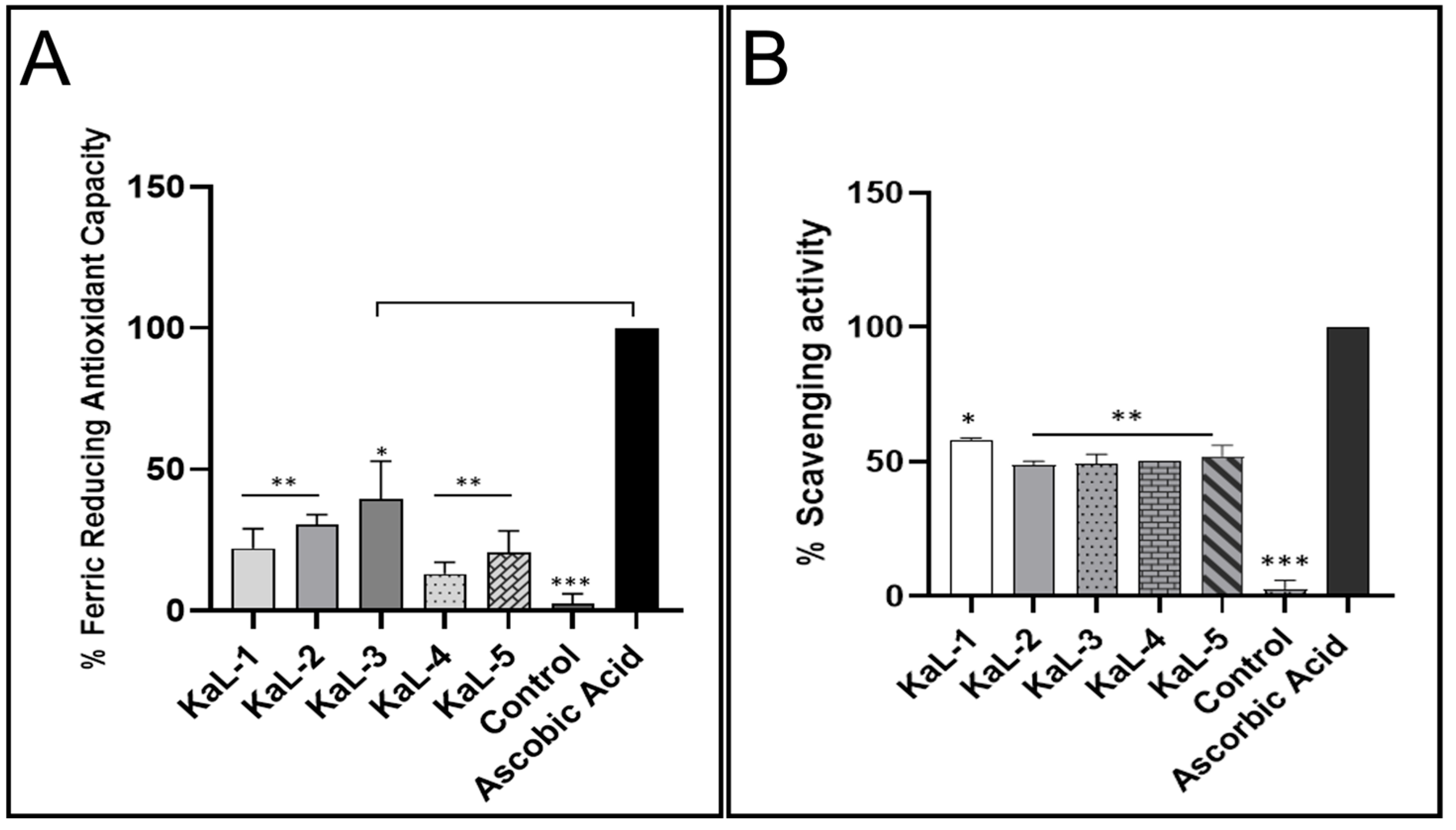
The antioxidant activity (Figure 2) of the endophytic fungal CEs was determined using the FRAP and the DPPH scavenging assays. All CEs were observed to have antioxidant activity using both the FRAP (Figure 2A) and DPPH (Figure 2B) assays. The reducing power ranged from as high as 40% to as low as 18%, while the radical scavenging potential ranged from 60% to 48%. The antioxidant activity for the different CEs was below ascorbic acid control for both assays. The C. cassiicola KaL-3 CE displayed the highest reducing potential of 40%, and the S. rostrata KaL-4 CE displayed the lowest reducing potential of 13%. The highest radical scavenging activity of 60% was observed for Diaporthe sp. KaL-5, while a radical scavenging activity of 48% was observed for C. cassiicola KaL-3, which was the lowest activity.

3.4. Total Phenolic Content of Fungal Endophyte Crude Extracts

The quantification of the TPC of the fungal endophyte CEs using the Folin-Ciocalteau reagent revealed that the CEs had varying levels of phenolic compounds (Table 3). The highest production of phenolic compounds was observed in the CE of the S. rostrata KaL-4 isolate with a TPC of 373.80 ± 12.39 mg GAE/g, while the CE of the S. rostrata KaL-1 isolate had the lowest TPC of 79.64 ± 2.36 mg GAE/g.

3.5. Influence of S. rostrata KaL-4 CE on Viability Profiles and IL-6 Production in RAW 264.7

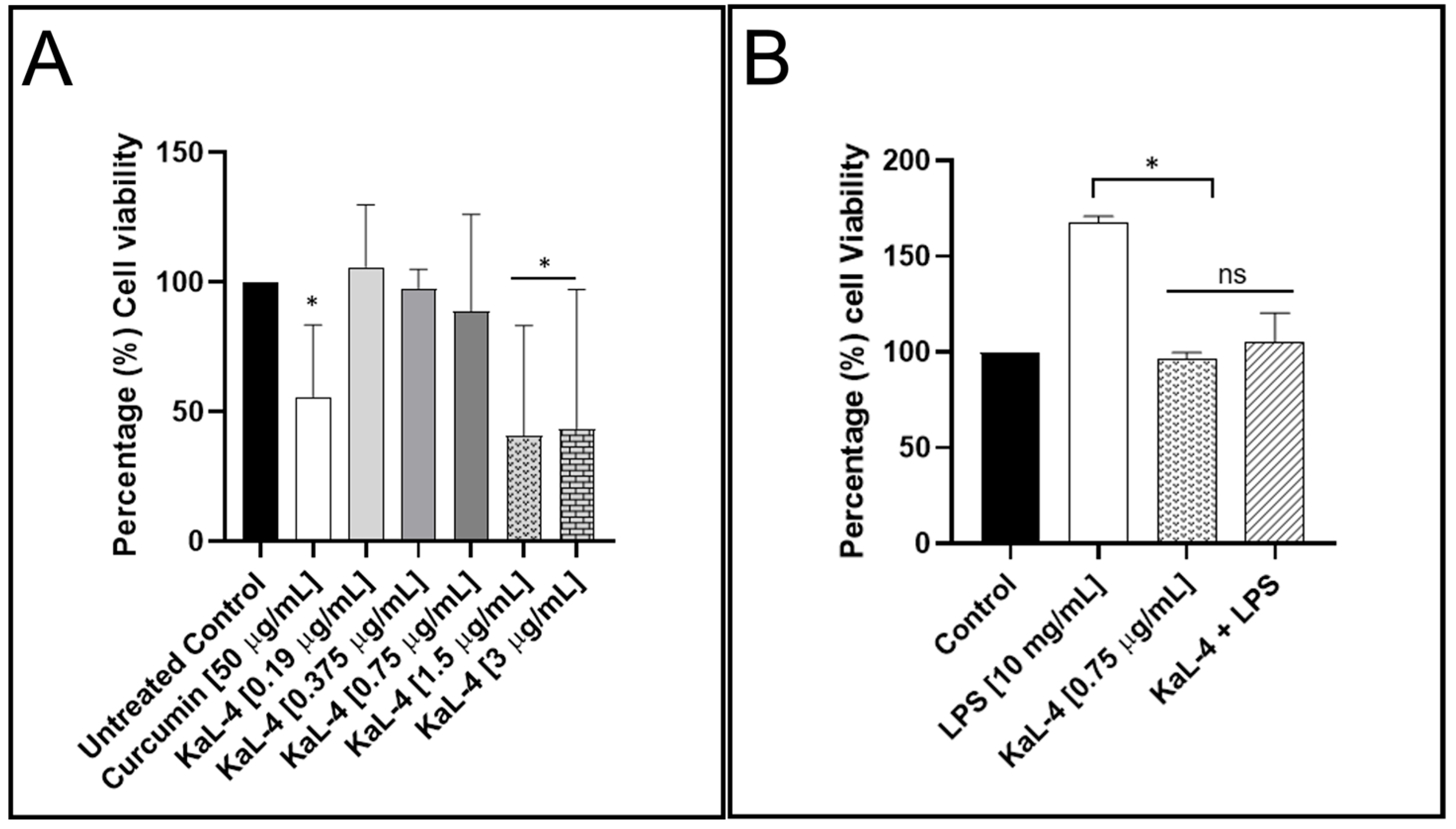
The effect of the S. rostrata KaL-4 CE on the viability profile of the RAW 264.7 macrophages and the production of the inflammatory mediator IL-6 was assessed using the MTT viability assay (Figure 3A) and ELISA (Figure 3B), respectively. The CE showed dose-dependent toxicity, with the viability of the cells being reduced to below 50% at high concentrations (1.5 and 3 µg/mL), while cell viability was observed to be above 90% at low doses (0.375 and 0.75 µg/mL). Interestingly, the CE was observed to enhance the viability of the macrophages at a concentration of 0.19 µg/mL. Moreover, the CE was observed to inhibit the production of IL-6 at the sub-lethal concentration of 0.75 µg/mL.

3.6. Extracellular Enzyme Production Capabilities of Fungal Endophytes

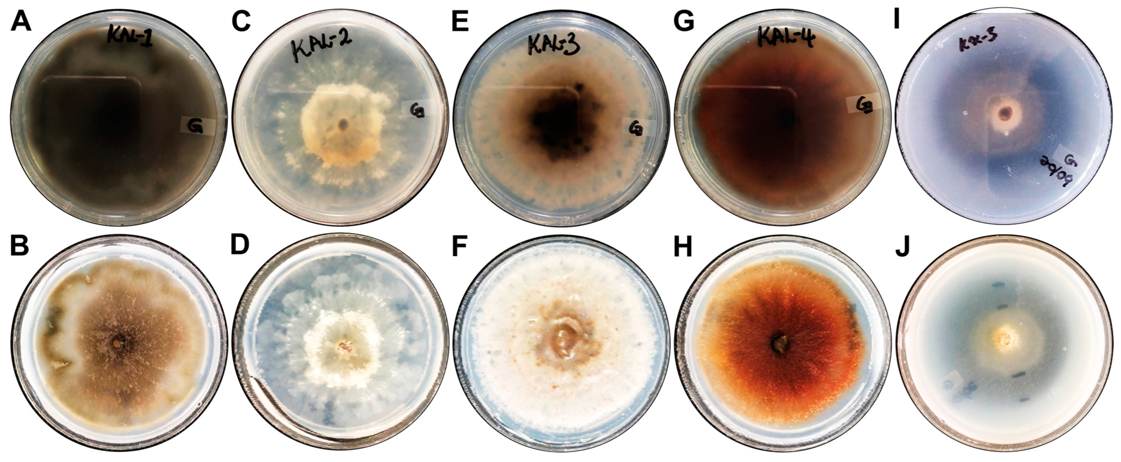
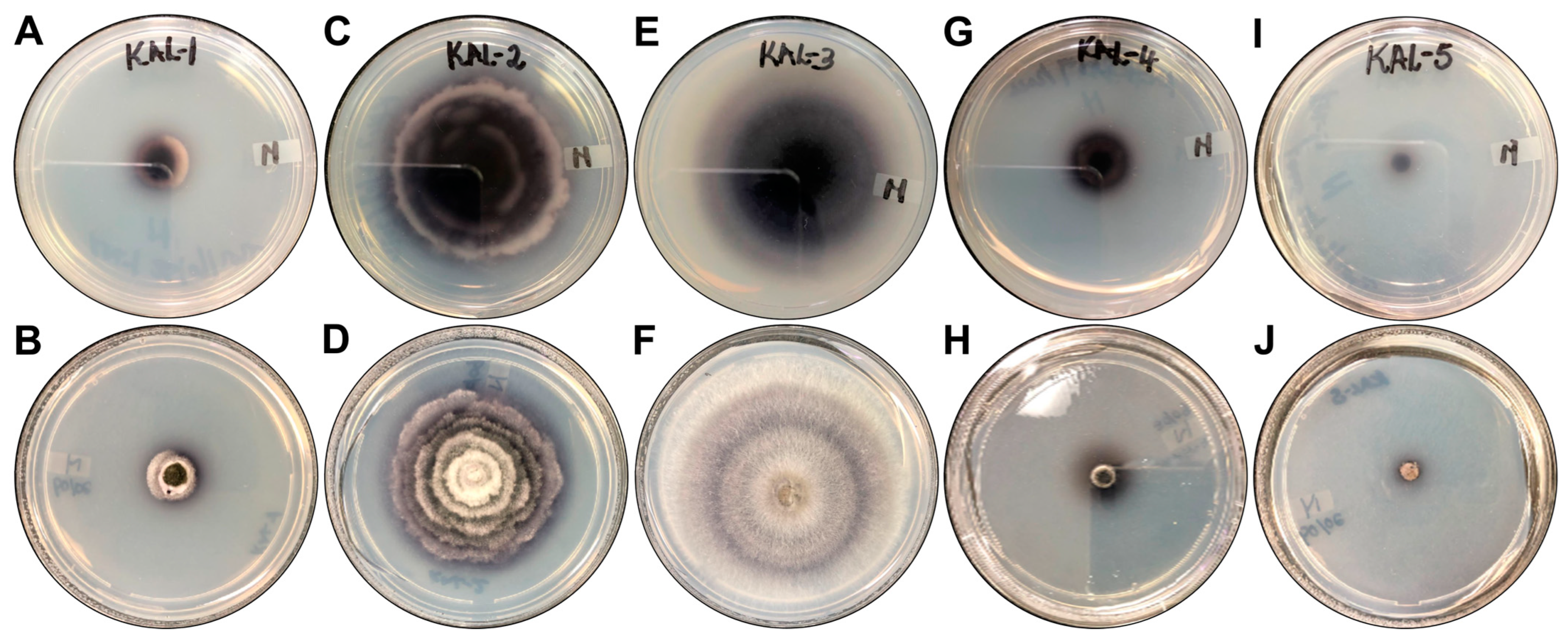
The extracellular protease and laccase activities of the fungal endophytes were determined on enzyme-specific solid media. The production of proteases and laccases varied amongst the isolates (Table 4). Only the Diaporthe sp. KaL-5 isolate displayed any protease activity (Figure 4) with an EI of 0.66 ± 0.05. The formation of a deep blue colour, indicative of laccase production, was observed around all colonies except the colony of the C. cassiicola KaL-3 isolate (Figure 5). However, it was observed that a deep blue colour had developed beneath the colony when the underside of the plate was inspected (Figure 5E,F). The highest laccase activity was observed for the Diaporthe sp. KaL-5 isolate with an EI of 1.15 ± 0.13, while an EI of 0.14 ± 0.01 made the Diaporthe sp. KaL-2 isolate the lowest producer of the laccase enzyme. The use of H2O2 to screen for peroxidase activity revealed that all isolates are able to produce extracellular peroxidases (Table 5).

4. Discussion

Medicinal plants are excellent sources of fungal endophytes with the ability to produce important bioactive secondary metabolites and extracellular enzymes. While numerous studies have focused on the biological activities of K. acuminata Oliv. leaves [28,29,30], there are no studies that have explored the fungal endophytes associated with the leaves of this medicinal plant species. In this study, the assessment of the macro-morphological features of the fungal endophyte colonies previously isolated from the leaves of K. acuminata Oliv. suggested the presence of five morphotypes. Macromorphological and microscopic features have been extensively used to categorize fungal endophytes as morphotypes and isolates; however, the use of macromorphological features to group fungal endophytes into morphotypes does not sufficiently represent the phylogeny of fungal endophytes, particularly for non-sporulating species [31]. This was made apparent by the fact that sequencing of the ITS region (ITS1-5.8S-ITS2) revealed that the five isolates represented three different genera with isolates KaL-1 and KaL-4 identified as S. rostrata, while isolates KaL-2 and KaL-5 were identified as Diaporthe species despite showing differences in their morphology. Despite the inability to delineate genera such as Diaporthe, the ITS region remains the standard DNA marker used for the identification of fungal endophytes [31], and it is important to couple the use of the ITS region to other DNA markers such as β-tubilin II and translation elongation factor 1α [32,33]. However, the selection of the alternative marker depends on the genus in question as the feasibility of the alternative marker cannot be readily transferred from one taxon to another, and an investigation is required for each taxon [33]. The molecular identification of the fungal endophytes revealed that all isolates used in this study belonged to the phylum Ascomycota. This coincides with other studies wherein the majority of fungal endophytes isolated from medicinal plants used in South Africa belonged to the phylum Ascomycota [32,34,35,36]. The low number of isolates obtained in this study correlates with the study by Abdalla et al. [35] wherein a total of five fungal endophytes were isolated from the leaves of three South African medicinal plants, namely, Psychotria zombamontana (Kuntze) Petit, Catha edulis (Vahl) Forssk. ex Endl. and Melianthus comosus Vahl. In contrast, Manganyi et al. [37] and Sishuba et al. [32] isolated 60 and 43 fungal endophytes, respectively, from the South African medicinal plant Sceletium tortuosum (L.). This demonstrates that the number of fungal endophytes obtained can vary from plant to plant.
Owing to the prevailing challenge of the increase in AMR, the secondary metabolites of fungal endophytes have been investigated for their antimicrobial potential [38,39]. In this study, the antibacterial activity of the CEs was tested against Gram-negative bacteria, which account for 90% of all urinary tract infections [40]. Gram-negative bacteria possess an LPS layer in their membranes, which makes them more resistant to antimicrobial agents than Gram-positive bacteria [41]. The S. rostrata KaL-1, the Diaporthe sp. KaL-2 and the Diaporthe sp. KaL-5 strains displayed high inhibitory activity against P. aeruginosa with an MIC of 0.63 mg/mL. Masoko [28] previously reported an MIC of 1.25 mg/mL for the leaf extracts of K. acuminata Oliv. against P. aeruginosa. Interestingly, Mabadahanye et al. [30] did not observe any inhibitory activity for the leaf crude extracts of K. acuminata Oliv. against E. coli using an agar well diffusion assay despite Masoko [28] reporting an MIC of 0.31 mg/mL for the DCM and methanol CEs. This indicates that the ethyl acetate CEs of fungal endophytes are more potent against P. aeruginosa and less potent against E. coli compared to the host plant CEs. Interestingly, the S. rostrata KaL-1 strain displayed higher efficacy against P. aeruginosa than the S. rostrata KaL-4 strain. Similarly, the Diaporthe species KaL-5 strain was more effective against E. coli than the Diaporthe sp. KaL-2 strain. These results suggest that the isolates may be different strains of the same species as their colony morphologies were also different. In contrast to the current study, Gagana et al. [42] reported that the ethyl acetate CEs of the C. casiicola isolate did not display any inhibitory activity against E. coli, K. pneumoniae and P. aeruginosa using the agar well diffusion method. Furthermore, the authors reported that the ethyl acetate CE of the S. rostrata isolate did not inhibit E. coli while displaying inhibitory activity against K. pneumoniae and P. aeruginosa [42]. Even though the MICs obtained in the study can be classified as moderate activity, Nxumalo et al. [43] highlighted that microbial extracts with MIC values lower than 1 mg/mL are considered to be noteworthy. As such, the CEs demonstrating an MIC of 0.63 mg/mL can be further explored for their potential as antimicrobial agents. Interestingly, Masoko [28] and Mabadahanye et al. [30] reported that the leaf extracts of K. acuminata Oliv. displayed inhibitory activity against Staphylococcus aureus. This warrants an investigation of the inhibitory activity of fungal endophyte CEs against Gram-positive bacteria as the CEs may potentially exhibit higher efficacy against Gram-positive bacteria than Gram-negative bacteria. Even though pathogenic bacteria produce biofilms that aid in conferring them with resistance against various antimicrobial agents [41], the CEs of fungal endophytes have been reported to possess anti-biofilm activity against biofilm-forming pathogens [44,45,46]. As such, the anti-biofilm activity of the CEs of the fungal endophytes used in this study needs to be investigated to fully understand the antimicrobial potential of the CEs obtained in the study.
The production of harmful free radicals has been linked with the development of various conditions, including cancer, neurodegenerative disease, diabetes and premature ageing [47]. Fungal endophytes have the ability to produce various secondary metabolites with antioxidant activities. Given that a single antioxidant assay is not adequate to measure the antioxidant efficacy of various crude extracts [48], the FRAP and the DPPH assays were used to assess the antioxidant activities of the CEs. Even though the fungal endophyte CEs displayed antioxidant activity, the reduction power and radical scavenging potential observed were below those of the ascorbic acid control. In general, CEs displayed higher radical scavenging activity than reducing potential. Our findings coincide with the findings by Pan et al. [49] wherein the ethyl acetate fractions of fungal endophytes isolated from the bulbs of Fritillaria unibracteata var. wabuensis were observed to generally display lower reducing power than radical scavenging activity. Antioxidants are known to possess a number of therapeutic properties, including antimicrobial, anti-inflammatory and anticancer properties [50]. Interestingly, the CE of the C. cassiicola KaL-3 strain displayed the highest antioxidant activity despite having an MIC value of 1.25 mg/mL against all tested pathogens.
The antioxidant activity of crude extracts is largely attributed to the presence of phenolic compounds [51]. Mabadahanye et al. [30] reported the presence of phenols in the acetone and methanol extracts but not in the ethyl acetate extracts of K. acuminata Oliv. leaves. In this study, phenols were detected in all ethyl acetate crude extracts albeit at varying concentrations. No significant difference in the TPC was observed for the CEs of S. rostrata KaL-1, Diaporthe sp. KaL-2 and C. cassiicola KaL-3 with a p value of less than 0.05. However, the CE of S. rostrata KaL-4 possessed a significantly higher TPC than all other isolates with a p value of less than 0.05. The significant difference in the TPC for the S. rostrata KaL-1 and KaL-4 strains suggests that they may be different strains of the same species. Similarly, the Diaporthe sp. KaL-5 strain displayed a significantly higher TPC than the Diaporthe sp. KaL-2 strain. This further suggests that the isolates may be different species altogether or different strains of the same species. The TPC recorded for the fungal endophytes in this study are higher than those reported by Ghazi-yaker et al. [52] and Yadav et al. [53], who recorded TPCs ranging from 6.4 to 125.32 mg/g GAE for fungal endophytes isolated from the leaves of Ziziphus lotus L. (Desf) and from 4.46 ± 0.15 to 60.13 ± 0.41 for fungal endophytes isolated from Eugenia jambolana Lam., respectively. Pan et al. [49] reported that the high antioxidant activity observed in the ethyl acetate crude extracts of fungal endophytes correlated with a high TPC. However, this was not the case in the current study as the CE of the S. rostrata KaL-4 strain displayed significantly lower reducing power than the other isolates despite having the highest TPC.
Inflammation is important in the clearance of infections; however, prolonged inflammation leads to oxidative stress, which has been linked with various ailments [54,55]. Secondary metabolites of fungal endophytes have been reported to reduce oxidative stress [56]. While the secondary metabolites of fungal endophytes are generally regarded as safe to use due to these microbes being symbionts of plants [57,58], the cytotoxicity of crude extracts on healthy cells needs to be investigated. The lack of studies on the safety of fungal endophyte crude extract may explain why little progress has been made in the development of commercial products from the secondary metabolites of fungal endophytes. Given that the CE of the S. rostrata KaL-4 strain possessed an overall high TPC, the influence of the CE on the viability of macrophages as well as the ability of the CE to modulate LPS-induced inflammation were assessed. The CE was not only non-cytotoxic and safe to use on normal macrophages at low doses, but it also attenuated the production of the pro-inflammatory cytokine IL-6 at a low concentration of 0.75 µg/mL. This demonstrates that only small doses of CE would be required to affect the requisite reduction in infection-induced inflammation. Recently, Koopklang et al. [59] reported that the endophytic S. rostrata SH8-8 strain isolated from Ipomoea pes-caprae produced a xanthone, ravelin, which demonstrated anti-inflammatory activity by suppressing nitric oxide production in LPS-induced macrophage J774A.1 cells. This demonstrates the potential for secondary metabolites from endophytic S. rostrata strains to be used for the treatment of inflammatory ailments. Although more work is required, the findings in this study suggest that the S. rostrata KaL-4 crude extract may interfere with the inflammatory signalling pathway that regulates the production of inflammatory mediators.
Microbial enzymes have been widely applied in various industries, including baking, food, biofuels and healthcare [60]. Endophytic fungi are well-documented sources of sought-after industry-significant extracellular enzymes with high stability and specificity [9]. In this study, the fungal endophytes were screened for their ability to produce proteases, laccases and peroxidases. Proteases are hydrolytic enzymes that hydrolyse peptide bonds of proteins and are a group of important enzymes that are broadly used in the food, pharmaceutical, leather and detergent industries and in bioremediation processes due to their high stability [60]. In this study, the enzymatic index (EI) of the fungal endophytic strains was determined according to Chai et al. [27] wherein isolates with EIs greater than 1.0 are considered to have strong enzyme production. In addition to being the only isolate to produce proteases, an EI of 0.66 ± 0.05 makes the Diaporthe sp. KaL-5 isolate an intermediate producer of enzymes. Given that the isolates KaL-2 and KaL-5 were identified as Diaporthe species, these results further suggest that they may be different strains or even different species of the same genus. All fungal endophyte isolates produced laccases, albeit at varying levels. Only the isolates Diaporthe sp. KaL-5 demonstrated strong laccase production with an EI of 1.15 ± 0.13, which was significantly higher (p < 0.05) than those of all other isolates. Furthermore, no significant difference was observed for the isolates S. rostrata KaL-1, Diaporthe sp. KaL-2 and S. rostrata KaL-4. While the EI of the C. cassiicola KaL-3 isolate could not be determined, it is important to note that the isolate exhibited the ability to produce the enzyme. Given that the glucose levels in the media may affect laccase production [61], it is possible that reducing the glucose content in the media may result in higher laccase activity for all isolates. Laccase is a dependent oxidative enzyme which has broad biotechnological applications, such as in industrial effluent detoxification, discoloration, the removal of phenolics from wine and pulp bleaching [10]. The production of laccases by the fungal endophyte Diaporthe sp. (syn. Phomopsis sp.) observed in this study correlates with the result obtained by Sunitha et al. [24] who observed extracellular laccase production by a Phomopsis species. Protease [62] and laccase [63] enzymes have also been reported to have antimicrobial activity, which highlights their significance in the pharmaceutical industry. All strains displayed the ability to produce peroxidases. Similarly, Attia et al. [26] observed that the majority of the fungal endophyte isolates from Medicago sativa demonstrated peroxidase activity. Peroxidase enzymes are important in industry for the breakdown of lignocellulosic biomass and the removal of dyes in textiles, and they are used as biosensors [64]. The results obtained in this study further highlight the potential of endophytic fungi to produce industry-significant enzymes.

5. Conclusions

The current study demonstrates that fungal endophytes from the leaves of K. acuminata Oliv. produce bioactive secondary metabolites with promising antimicrobial, antioxidant and anti-inflammatory properties. Thus, the full mycochemical composition of CEs should be determined to identify bioactive compounds. Furthermore, the physicochemical parameters required to enhance the yield and bioactivity of CEs should be investigated. In addition, the underlying molecular mechanisms of action of the CEs should be further explored. Although all fungal endophytes produce laccases and peroxidases, more research is required to understand the yield, stability and activities of the enzymes for economical, pharmaceutical, and industrial production. The findings in this study further highlight the potential of medicinal plant species used in South Africa as sources of endophytic fungi with important biological and biotechnological capabilities.

Author Contributions

Conceptualization, K.D.; Formal Analysis, S.K., R.T.M. and K.D.; Funding Acquisition, K.D.; Investigation, S.K.; Methodology, R.T.M. and K.D.; Project Administration, K.D.; Resources, R.T.M. and K.D.; Supervision, R.T.M. and K.D.; Writing—Original Draft, S.K.; Writing—Review and Editing, R.T.M. and K.D. All authors have read and agreed to the published version of the manuscript.

Funding

This work is based on the research supported wholly by the National Research Foundation of South Africa (Grant Number: 138274), and the APC was funded by the University of Limpopo. All the findings, conclusions and recommendations expressed in this publication are those of the authors, and the NRF accepts no liability whatsoever in this regard.

Institutional Review Board Statement

Not applicable.

Informed Consent Statement

Not applicable.

Data Availability Statement

The original contributions presented in this study are included in the article; further inquiries can be directed to the corresponding author.

Acknowledgments

The authors would like to thank M. Magagula, KS Hlope and MM Nyama for the assistance they provided with the different assays.

Conflicts of Interest

The authors declare no conflicts of interest.

References

  1. Moharram, A.M.; Zohri, A.A.; Omar, H.M.; Abd El-Ghani, O.A. In Vitro Assessment of Antimicrobial and Anti-Inflammatory Potential of Endophytic Fungal Metabolites Extracts. Eur. J. Biol. Res. Res. Artic. Eur. J. Biol. Res. 2017, 7, 234–244. [Google Scholar]
  2. Karin, M.; Lawrence, T.; Nizet, V. Innate Immunity Gone Awry: Linking Microbial Infections to Chronic Inflammation and Cancer. Cell 2006, 124, 823–835. [Google Scholar] [CrossRef] [PubMed]
  3. Arfeen, M.; Srivastava, A.; Srivastava, N.; Khan, R.A.; Almahmoud, S.A.; Mohammed, H.A. Design, Classification, and Adverse Effects of NSAIDs: A Review on Recent Advancements. Bioorg. Med. Chem. 2024, 112, 117899. [Google Scholar] [CrossRef]
  4. Manganyi, M.C.; Ateba, C.N. Untapped Potentials of Endophytic Fungi: A Review of Novel Bioactive Compounds with Biological Applications. Microorganisms 2020, 8, 1934. [Google Scholar] [CrossRef]
  5. Venieraki, A.; Dimou, M.; Katinakis, P. Endophytic Fungi Residing in Medicinal Plants Have the Ability to Produce the Same or Similar Pharmacologically Active Secondary Metabolites as Their Hosts. Hell. Plant Prot. J. 2017, 10, 51–66. [Google Scholar] [CrossRef]
  6. Kaul, S.; Gupta, S.; Ahmed, M.; Dhar, M.K. Endophytic Fungi from Medicinal Plants: A Treasure Hunt for Bioactive Metabolites. Phytochem. Rev. 2012, 11, 487–505. [Google Scholar] [CrossRef]
  7. Gupta, J.; Sharma, S. Endophytic Fungi: A New Hope for Drug Discovery. In New and Future Developments in Microbial Biotechnology and Bioengineering; Elsevier: Amsterdam, The Netherlands, 2020; pp. 39–49. ISBN 9780128210062. [Google Scholar]
  8. Burragoni, S.G.; Jeon, J. Applications of Endophytic Microbes in Agriculture, Biotechnology, Medicine, and Beyond. Microbiol. Res. 2021, 245, 126691. [Google Scholar] [CrossRef]
  9. Sopalun, K.; Iamtham, S. Isolation and Screening of Extracellular Enzymatic Activity of Endophytic Fungi Isolated from Thai Orchids. S. Afr. J. Bot. 2020, 134, 273–279. [Google Scholar] [CrossRef]
  10. Singh, R.; Kumar, M.; Mittal, A.; Mehta, P.K. Microbial Enzymes: Industrial Progress in 21st Century. 3 Biotech 2016, 6, 174. [Google Scholar] [CrossRef]
  11. Shubha, J.; Srinivas, C. Diversity and Extracellular Enzymes of Endophytic Fungi Associated with Cymbidium aloifolium L. African J. Biotechnol. 2017, 16, 2242–2247. [Google Scholar] [CrossRef]
  12. Rai, N.; Kumari Keshri, P.; Verma, A.; Kamble, S.C.; Mishra, P.; Barik, S.; Kumar Singh, S.; Gautam, V. Plant Associated Fungal Endophytes as a Source of Natural Bioactive Compounds. Mycology 2021, 12, 139–159. [Google Scholar] [CrossRef] [PubMed]
  13. Aboobaker, Z.; Viljoen, A.; Chen, W.; Crous, P.W.; Maharaj, V.J.; van Vuuren, S. Endophytic Fungi Isolated from Pelargonium Sidoides DC: Antimicrobial Interaction and Isolation of a Bioactive Compound. S. Afr. J. Bot. 2019, 122, 535–542. [Google Scholar] [CrossRef]
  14. Maroyi, A. Kirkia acuminata Oliv.: A Review of Its Ethnobotany and Pharmacology. Afr. J. Tradit. Complement. Altern. Med. AJTCAM 2017, 14, 217–226. [Google Scholar] [CrossRef]
  15. van Wyk, A.S.; Prinsloo, G. Medicinal Plant Harvesting, Sustainability and Cultivation in South Africa. Biol. Conserv. 2018, 227, 335–342. [Google Scholar] [CrossRef]
  16. Magagula, M. University of Limpopo, Mankweng, South Africa, 2021.
  17. Altschul, S.F.; Madden, T.L.; Schäffer, A.A.; Zhang, J.; Zhang, Z.; Miller, W.; Lipman, D.J. Gapped BLAST and PSI-BLAST: A New Generation of Protein Database Search Programs. Nucleic Acids Res. 1997, 25, 3389–3402. [Google Scholar] [CrossRef]
  18. Eloff, J.N. A Sensitive and Quick Microplate Method to Determine the Minimal Inhibitory Concentration of Plant Extracts for Bacteria. Planta Med. 1998, 64, 711–713. [Google Scholar] [CrossRef]
  19. Arulpriya, P.; Lalitha, P.; Hemalatha, S. Cyclic Voltammetric Assessment of the Antioxidant Activity of Petroleum Ether Extract of Samanea Saman (Jacq.)Merr. Adv. Appl. Sci. Res. 2010, 1, 24–35. [Google Scholar]
  20. Baliyan, S.; Mukherjee, R.; Priyadarshini, A.; Vibhuti, A.; Gupta, A.; Pandey, R.P.; Chang, C.M. Determination of Antioxidants by DPPH Radical Scavenging Activity and Quantitative Phytochemical Analysis of Ficus Religiosa. Molecules 2022, 27, 1326. [Google Scholar] [CrossRef]
  21. Tambe, V.D.; Bhambar, R.S. Estimation of Total Phenol, Tannin, Alkaloid and Flavonoid in Hibiscus Tiliaceus Linn. Wood Extracts. Res. Rev. J. Pharmacogn. Phytochem. 2014, 2, 41–47. [Google Scholar]
  22. van Meerloo, J.; Kaspers, G.J.L.; Cloos, J. Cell Sensitivity Assays: The MTT Assay BT—Cancer Cell Culture: Methods and Protocols; Cree, I.A., Ed.; Humana Press: Totowa, NJ, USA, 2011; pp. 237–245. ISBN 978-1-61779-080-5. [Google Scholar]
  23. Baek, S.; Park, T.; Kang, M.; Park, D. Anti-Inflammatory Activity and ROS Regulation. Molecules 2020, 25, 4089. [Google Scholar] [CrossRef]
  24. Sunitha, V.H.; Nirmala Devi, D.; Srinivas, C. Extracellular Enzymatic Activity of Endophytic Fungal Strains Isolated from Medicinal Plants. World J. Agric. Sci. 2013, 9, 1–9. [Google Scholar] [CrossRef]
  25. Hawar, S.N. Extracellular Enzyme of Endophytic Fungi Isolated from Ziziphus Spina Leaves as Medicinal Plant. Int. J. Biomater. 2022, 2022, 2135927. [Google Scholar] [CrossRef] [PubMed]
  26. Attia, E.Z.; Farouk, H.M.; Abdelmohsen, U.R.; El-Katatny, M.H. Antimicrobial and Extracellular Oxidative Enzyme Activities of Endophytic Fungi Isolated from Alfalfa (Medicago sativa) Assisted by Metabolic Profiling. S. Afr. J. Bot. 2020, 134, 156–162. [Google Scholar] [CrossRef]
  27. Chai, C.H.; Hong, C.F.; Huang, J.W. Identification and Characterization of a Multifunctional Biocontrol Agent, Streptomyces Griseorubiginosus LJS06, Against Cucumber Anthracnose. Front. Microbiol. 2022, 13, 923276. [Google Scholar] [CrossRef]
  28. Masoko, P. Ethnobotanical Study of Some Selected Medicinal Plants Used by Traditional Healers in Limpopo Province (South Africa). Am. J. Res. Commun. 2013, 1, 8–23. [Google Scholar]
  29. Mulholland, D.A.; Cheplogoi, P.; Crouch, N.R. Secondary Metabolites from Kirkia acuminata and Kirkia wilmsii (Kirkiaceae). Biochem. Syst. Ecol. 2003, 31, 793–797. [Google Scholar] [CrossRef]
  30. Mabadahanye, K.; Bhembe, N.L.; Green, E. Crude Extracts Activity of Three Selected Medicinal Plants from the Venda Region against Some Pathogenic Organisms. Afr. Health Sci. 2022, 22, 717–727. [Google Scholar] [CrossRef]
  31. dos Reis, J.B.A.; Lorenzi, A.S.; do Vale, H.M.M. Methods Used for the Study of Endophytic Fungi: A Review on Methodologies and Challenges, and Associated Tips. Arch. Microbiol. 2022, 204, 675. [Google Scholar] [CrossRef]
  32. Sishuba, A.; Leboko, J.; Ateba, C.N.; Manganyi, M.C. First Report: Diversity of Endophytic Fungi Possessing Antifungal Activity Isolated from Native Kougoed (Sceletium tortuosum L.). Mycobiology 2021, 49, 89–94. [Google Scholar] [CrossRef]
  33. Lücking, R.; Aime, M.C.; Robbertse, B.; Miller, A.N.; Ariyawansa, H.A.; Aoki, T.; Cardinali, G.; Crous, P.W.; Druzhinina, I.S.; Geiser, D.M.; et al. Unambiguous Identification of Fungi: Where Do We Stand and How Accurate and Precise Is Fungal DNA Barcoding? IMA Fungus 2020, 11, 14. [Google Scholar] [CrossRef]
  34. Manganyi, M.C.; Regnier, T.; Kumar, A.; Bezuidenhout, C.C.; Ateba, C.N. Biodiversity and Antibacterial Screening of Endophytic Fungi Isolated from Pelargonium sidoide. S. Afr. J. Bot. 2018, 116, 192–199. [Google Scholar] [CrossRef]
  35. Abdalla, M.A.; Aro, A.O.; Gado, D.; Passari, A.K.; Mishra, V.K.; Singh, B.P.; McGaw, L.J. Isolation of Endophytic Fungi from South African Plants, and Screening for Their Antimicrobial and Extracellular Enzymatic Activities and Presence of Type I Polyketide Synthases. S. Afr. J. Bot. 2020, 134, 336–342. [Google Scholar] [CrossRef]
  36. Fanele, A.; Ndlovu, S.I. Endophytic Fungal Species Nigrospora Oryzae and Alternaria Alternata Exhibit Antimicrobial Activity against Gram-Positive and Gram-Negative Multi-Drug Resistant Clinical Bacterial Isolates. BMC Complement. Med. Ther. 2023, 23, 323. [Google Scholar] [CrossRef]
  37. Manganyi, M.C.; Regnier, T.; Tchatchouang, C.D.K.; Bezuidenhout, C.C.; Ateba, C.N. Antibacterial Activity of Endophytic Fungi Isolated from Sceletium tortuosum L. (Kougoed). Ann. Microbiol. 2019, 69, 659–663. [Google Scholar] [CrossRef]
  38. Digra, S.; Nonzom, S. An Insight into Endophytic Antimicrobial Compounds: An Updated Analysis; Springer Nature: Singapore; Berlin/Heidelberg, Germany, 2023; Volume 17, ISBN 1181602300824. [Google Scholar]
  39. Silva, D.P.D.; Cardoso, M.S.; Macedo, A.J. Endophytic Fungi as a Source of Antibacterial Compounds—A Focus on Gram-Negative Bacteria. Antibiotics 2022, 11, 1509. [Google Scholar] [CrossRef]
  40. Mazzulli, T.; Skulnick, M.; Small, G.; Marshall, W.; Hoban, D.J.; Zhanel, G.G.; Finn, S.; Low, D.E. Susceptibility of Community Gram-Negative Urinary Tract Isolates to Mecillinam and Other Oral Agents. Can. J. Infect. Dis. 2001, 12, 289–292. [Google Scholar] [CrossRef]
  41. Breijyeh, Z.; Jubeh, B.; Karaman, R. Resistance of Gram-Negative Bacteria to Current Antibacterial Agents and Approaches to Resolve It. Molecules 2020, 25, 1340. [Google Scholar] [CrossRef]
  42. Gagana, S.L.; Kumaraswamy, B.E.; Shivanna, M.B. Diversity, Antibacterial and Antioxidant Activities of the Fungal Endophytes Associated with Schleichera oleosa (Lour.) Merr. S. Afr. J. Bot. 2020, 134, 369–381. [Google Scholar] [CrossRef]
  43. Nxumalo, C.I.; Ngidi, L.S.; Siyabonga, J.; Shandu, E. Isolation of Endophytic Bacteria from the Leaves of Anredera Cordifolia CIX1 for Metabolites and Their Biological Activities. BMC Complement. Med. Ther. 2020, 20, 300. [Google Scholar]
  44. Barfour, A.F.; Mensah, A.Y.; Asante-Kwatia, E.; Danquah, C.A.; Anokwah, D.; Adjei, S.; Baah, M.K.; Mensah, M.L.K. Antibacterial, Antibiofilm, and Efflux Pump Inhibitory Properties of the Crude Extract and Fractions from Acacia Macrostachya Stem Bark. Sci. World J. 2021, 2021, 5381993. [Google Scholar] [CrossRef]
  45. Attia, E.Z.; Khalifa, B.A.; Shaban, G.M.; Abdelraheem, W.M.; Mustafa, M.; Abdelmohsen, U.R.; El-Katatny, M.H. Discovering the Chemical Profile, Antimicrobial and Antibiofilm Potentials of the Endophytic Fungus Penicillium Chrysogenum Isolated from Artemisia judaica L. Assisted with Docking Studies. S. Afr. J. Bot. 2022, 151, 218–227. [Google Scholar] [CrossRef]
  46. Rabha, J.; Chetri, B.K.; Das, S.; Jha, D.K. In-Vitro and in-Silico Evaluation of Antimicrobial and Antibiofilm Secondary Metabolites of a Novel Fungal Endophyte, Albophoma Sp. BAPR5. S. Afr. J. Bot. 2023, 158, 347–368. [Google Scholar] [CrossRef]
  47. Chaudhary, P.; Janmeda, P.; Docea, A.O.; Yeskaliyeva, B.; Abdull Razis, A.F.; Modu, B.; Calina, D.; Sharifi-Rad, J. Oxidative Stress, Free Radicals and Antioxidants: Potential Crosstalk in the Pathophysiology of Human Diseases. Front. Chem. 2023, 11, 1158198. [Google Scholar] [CrossRef]
  48. Onyekachukwu, R.; Timothy, A.; Chidike, P.; Ezike, T.C.; Uzoechina, J.O. Exploring the Antioxidant Potential of Endophytic Fungi: A Review on Methods for Extraction and Quantification of Total Antioxidant Capacity (TAC). 3 Biotech 2024, 14, 127. [Google Scholar] [CrossRef]
  49. Pan, F.; Su, T.; Cai, S.; Wu, W. Fungal Endophyte-Derived Fritillaria Unibracteata Var. Wabuensis: Diversity, Antioxidant Capacities in Vitro and Relations to Phenolic, Flavonoid or Saponin Compounds. Sci. Rep. 2017, 7, 42008. [Google Scholar] [CrossRef]
  50. Sharaf, M.H.; Abdelaziz, A.M.; Kalaba, M.H.; Radwan, A.A.; Hashem, A.H. Antimicrobial, Antioxidant, Cytotoxic Activities and Phytochemical Analysis of Fungal Endophytes Isolated from Ocimum basilicum. Appl. Biochem. Biotechnol. 2022, 194, 1271–1289. [Google Scholar] [CrossRef]
  51. Vuolo, M.M.; Lima, V.S.; Maróstica Junior, M.R. Phenolic Compounds; Elsevier Inc.: Amsterdam, The Netherlands, 2019; ISBN 9780128147740. [Google Scholar]
  52. Ghazi-yaker, A.; Kraak, B.; Houbraken, J.; Nabti, E.; Cruz, C.; Saadoun, N.; Houali, K. In Vitro Antioxidant and Antibacterial Activities of Ethyl Acetate Extracts of Ziziphus Lotus Leaves and Five Associated Endophytic Fungi. Microorganisms 2024, 12, 2671. [Google Scholar] [CrossRef]
  53. Yadav, M.; Yadav, A.; Yadav, J.P. In Vitro Antioxidant Activity and Total Phenolic Content of Endophytic Fungi Isolated from Eugenia Jambolana Lam. Asian Pac. J. Trop. Med. 2014, 7, S256–S261. [Google Scholar] [CrossRef]
  54. Makola, R.T.; Mbazima, V.G.; Mokgotho, M.P.; Gallicchio, V.S.; Matsebatlela, T.M. The Effect of Lithium on Inflammation-Associated Genes in Lipopolysaccharide-Activated Raw 264.7 Macrophages. Int. J. Inflam. 2020, 2020, 8340195. [Google Scholar] [CrossRef]
  55. Reuter, S.; Gupta, S.C.; Chaturvedi, M.M.; Aggarwal, B.B. Oxidative Stress, Inflammation, and Cancer: How Are They Linked? Free Radic. Biol. Med. 2010, 49, 1603–1616. [Google Scholar] [CrossRef]
  56. Khan, A.L.; Gilani, S.A.; Waqas, M.; Al-Hosni, K.; Al-Khiziri, S.; Kim, Y.H.; Ali, L.; Kang, S.M.; Asaf, S.; Shahzad, R.; et al. Endophytes from Medicinal Plants and Their Potential for Producing Indole Acetic Acid, Improving Seed Germination and Mitigating Oxidative Stress. J. Zhejiang Univ. Sci. B 2017, 18, 125–137. [Google Scholar] [CrossRef] [PubMed]
  57. Chandra, H.; Yadav, A.; Prasad, R.; Kalra, S.J.S.; Singh, A.; Bhardwaj, N.; Gupta, K.K. Fungal Endophytes from Medicinal Plants Acting as Natural Therapeutic Reservoir. The Microbe 2024, 3, 100073. [Google Scholar] [CrossRef]
  58. Uzma, F.; Mohan, C.D.; Hashem, A.; Konappa, N.M.; Rangappa, S.; Kamath, P.V.; Singh, B.P.; Mudili, V.; Gupta, V.K.; Siddaiah, C.N.; et al. Endophytic Fungi-Alternative Sources of Cytotoxic Compounds: A Review. Front. Pharmacol. 2018, 9, 309. [Google Scholar] [CrossRef]
  59. Koopklang, K.; Choodej, S.; Hantanong, S.; Intayot, R.; Jungsuttiwong, S.; Insumran, Y.; Ngamrojanavanich, N.; Pudhom, K. Anti-Inflammatory Properties of Oxygenated Isocoumarins and Xanthone from Thai Mangrove-Associated Endophytic Fungus Setosphaeria Rostrata. Molecules 2024, 29, 603. [Google Scholar] [CrossRef]
  60. Bhadra, F.; Gupta, A.; Vasundhara, M.; Reddy, M.S. Endophytic Fungi: A Potential Source of Industrial Enzyme Producers. 3 Biotech 2022, 12, 86. [Google Scholar] [CrossRef]
  61. Schneider, W.D.H.; Fontana, R.C.; Mendonça, S.; de Siqueira, F.G.; Dillon, A.J.P.; Camassola, M. High Level Production of Laccases and Peroxidases from the Newly Isolated White-Rot Basidiomycete Marasmiellus Palmivorus VE111 in a Stirred-Tank Bioreactor in Response to Different Carbon and Nitrogen Sources. Process Biochem. 2018, 69, 1–11. [Google Scholar] [CrossRef]
  62. Indarmawan, T.; Mustopa, A.Z.; Budiarto, B.R.; Tarman, K. Antibacterial Activity of Extracellular Protease Isolated From an Algicolous Fungus Xylaria Psidii KT30 Against Gram-Positive Bacteria. HAYATI J. Biosci. 2016, 23, 73–78. [Google Scholar] [CrossRef]
  63. Anita, S.H.; Asishe, A.; Syafriana, V.; Febriani, A.; Zulfiana, D.; Oktaviani, M.; Nurhayat, O.D.; Yanto, D.H.Y. Hand Sanitizer Gel Formulation with Laccase Enzyme as an Antibacterial Against Staphylococcus Aureus and Escherichia Coli. Borneo J. Pharm. 2022, 5, 375–383. [Google Scholar] [CrossRef]
  64. de Oliveira, F.K.; Santos, L.O.; Buffon, J.G. Mechanism of Action, Sources, and Application of Peroxidases. Food Res. Int. 2021, 143, 110266. [Google Scholar] [CrossRef]
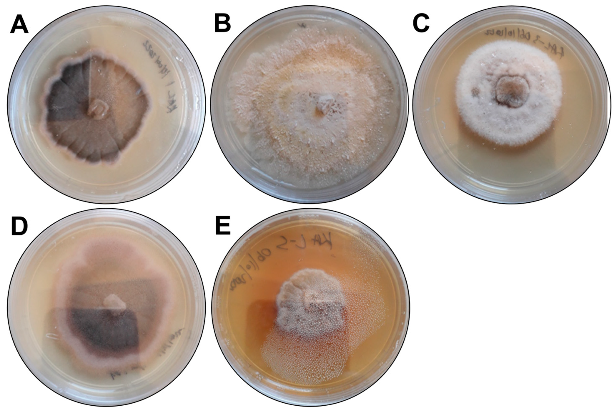
Figure 1. The morphological characteristics of the fungal endophytes growing on potato dextrose agar. The isolates were assigned the following codes: (A) KaL-1, (B) KaL-2, (C) KaL-3, (D) KaL-4 and (E) KaL-5.
Microorganisms 13 00692 g001
Figure 2. The antioxidant activity of the fungal endophyte crude extracts at 2.5 mg/mL. (A) The ferric reducing antioxidant capacity and (B) the free radical scavenging activity of the crude extracts. The data are presented as the mean of three replicates ± the standard deviation. The statistical analysis determines the significant difference between control and samples were p value is: * p < 0.05, ** p < 0.01, *** p < 0.001.
Microorganisms 13 00692 g002
Figure 3. The effect of the S. rostrata KaL-4 crude extract on the viability and production of inflammatory mediators of Raw 264.7 macrophages. The crude extract displayed (A) dose-dependent cytotoxicity against the macrophages and (B) the ability to reduce interleukin-6 production (IL-6) in lipopolysaccharide (LPS)-induced Raw 246.7 cells at 0.75 µg/mL. The data are presented as the mean of three replicates ± the standard deviation. The statistical analysis determines the significant difference between control and samples were p value is: * p < 0.05. ns is non significant.
Microorganisms 13 00692 g003
Figure 4. Extracellular protease activity for fungal endophytes. (A,B) Setosphaeria rostrata KaL-1, (C,D) Diaporthe sp. KaL-2, (E,F) Corynespora cassiicola KaL-3, (G,H) Setosphaeria rostrata KaL-4 and (I,J) Diaporthe sp. KaL-5.
Microorganisms 13 00692 g004
Figure 5. Extracellular laccase activity for fungal endophytes. (A,B) Setosphaeria rostrata KaL-1, (C,D) Diaporthe sp. KaL-2, (E,F) Corynespora cassiicola KaL-3, (G,H) Setosphaeria rostrata KaL-4 and (I,J) Diaporthe sp. KaL-5.
Microorganisms 13 00692 g005
Table 1. Colony morphology and molecular identification of fungal endophytes.
Isolate CodeColony DescriptionPredicted SpeciesGenBank Best Match
Accession Number%
Identity
KaL-1Grey irregular colony with black trim surrounded by white trim. Colony surface is raised and rough.Setosphaeria rostrataLT837843.1100
KaL-2Whitish-brown irregular colony with flat and rough surface.Diaporthe speciesMT355681.199.31
KaL-3White circular colony with raised and soft surface.Corynespora cassiicolaMK139711.1100
KaL-4Grey irregular colony surrounded by black, red and white trims, respectively. Colony surface is raised and rough.Setosphaeria rostrataLT837843.1100
KaL-5Whitish-brown irregular colony with raised and rough surface. Colour diffusing into medium.Diaporthe speciesMZ151534.199.65
Table 2. Minimum inhibitory concentrations (MICs) of fungal endophyte crude extracts (mg/mL).
Endophytic FungiE. coliK. pneumoniaeP. aeruginosa
Setosphaeria rostrata KaL-11.251.250.63
Diaporthe sp. KaL-21.251.250.63
Corynespora cassiicola KaL-31.251.251.25
Setosphaeria rostrata KaL-41.251.251.25
Diaporthe sp. KaL-50.631.250.63
Chloramphenicol0.020.020.02
Table 3. The total phenolic content (TPC) of the fungal endophyte crude extracts.
Endophytic FungiTPC (mg GAE/g)
Setosphaeria rostrata KaL-179.64 ± 2.36
Diaporthe sp. KaL-2102.05 ± 1.77
Corynespora cassiicola KaL-3143.97 ± 7.67
Setosphaeria rostrata KaL-4373.80 ± 12.39
Diaporthe sp. KaL-5231.42 ± 7.08
Table 4. The enzymatic index (EI) of fungal endophytes for the production of proteases and laccases. The data are presented as the mean of three replicates ± the standard deviation.
Endophytic FungiProtease ActivityLaccase Activity
Setosphaeria rostrata0.43 ± 0.29
Diaporthe sp.0.14 ± 0.01
Corynespora cassiicola+
Setosphaeria rostrata0.49 ± 0.02
Diaporthe sp.0.66 ± 0.051.15 ± 0.13
−: negative; +: positive—the isolate displayed laccase activity.
Table 5. Peroxidase production ability of the fungal endophytes.
Endophytic FungiPeroxidase Activity
Setosphaeria rostrata KaL-1+
Diaporthe sp. KaL-2+
Corynespora cassiicola KaL-3+
Setosphaeria rostrata KaL-4+
Diaporthe sp. KaL-5+
+: positive.
Disclaimer/Publisher’s Note: The statements, opinions and data contained in all publications are solely those of the individual author(s) and contributor(s) and not of MDPI and/or the editor(s). MDPI and/or the editor(s) disclaim responsibility for any injury to people or property resulting from any ideas, methods, instructions or products referred to in the content.