Article

Genomic Insights into Selenate Reduction by Anaerobacillus Species

by
Qidong Wang
1,2,
Jian Zhang
1,2,3,
Jinhui Liang
4,5,
Yanlong Wang
1,2,
Chongyang Ren
1,2,
Xinhan Chen
1,2,
Dongle Cheng
1,2,
Huanxin Zhang
1,2,* and
Huaqing Liu
1,2
1
College of Safety and Environmental Engineering, Shandong University of Science and Technology, Qingdao 266590, China
2
Institute of Yellow River Delta Earth Surface Processes and Ecological Integrity, Shandong University of Science and Technology, Qingdao 266590, China
3
School of Geographical Environment, Shandong Normal University, Jinan 250358, China
4
State Environmental Protection Key Laboratory of Land and Sea Ecological Governance and Systematic Regulation, Jinan 250101, China
5
Shandong Academy for Environmental Planning, Jinan 250101, China
*
Author to whom correspondence should be addressed.
Microorganisms 2025, 13(3), 659; https://doi.org/10.3390/microorganisms13030659
Submission received: 18 February 2025 / Revised: 11 March 2025 / Accepted: 11 March 2025 / Published: 14 March 2025
(This article belongs to the Collection Biodegradation and Environmental Microbiomes)

Abstract

:
Selenium (Se), a potentially toxic trace element, undergoes complex biogeochemical cycling in the environment, largely driven by microbial activity. The reduction in selenate or selenite to elemental selenium is an environmentally beneficial process, as it decreases both Se toxicity and mobility. This reduction is catalyzed by enzymes encoded by various related genes. The link between Se reduction gene clusters and specific taxonomic groups is significant for elucidating the ecological roles and processes of Se reduction in diverse environments. In this study, a new species of Se-reducing microorganism belonging to the genus Anaerobacillus was isolated from a mining site. A comparative analysis of the growth characteristics reveals that Anaerobacillus species exhibit notable metabolic versatility, particularly in their fermentation abilities and utilization of diverse electron donors and acceptors. Genome analysis identified a diverse array of gene clusters associated with selenate uptake (sul, pst), selenate reduction (ser), and selenite reduction (hig, frd, trx, and bsh). Since selenate reduction is the first crucial step in Se reduction, genes linked to selenate reductase are the focus. The serA gene clusters analysis suggests that the serA gene is highly conserved across Anaerobacillus species. The surrounding genes of serA show significant variability in both presence and gene size. This evolutionary difference in coenzyme utilization and serA regulation suggests distinct survival strategies among Anaerobacillus species. This study offers insights into Se bio-transformations and the adaptive strategies of Se-reducing microorganisms.

Graphical Abstract

1. Introduction

Selenium (Se), a metalloid in Group VIA of the periodic table, is positioned between arsenic (As) and bromine (Br) in the fourth period. It presents contradictory ecological risks in the environment. Se is an essential component of various proteins known as selenoproteins or selenoenzymes, such as glutathione peroxidase and thioredoxin reductase [1,2]. Organisms ranging from microorganisms to plants and animals require trace amounts of Se, which is incorporated into specialized enzymes for essential metabolic functions [3]. However, Se is also a potent reproductive toxicant and teratogen, and excessive amounts can lead to toxicity in organisms [4]. Mining activities often result in elevated Se levels, causing contamination in surrounding environments. Such Se contamination poses significant threats to human health, wildlife, and microbial diversity [5].
The geochemical cycling and environmental behavior of Se is complex due to its multiple oxidation states (+VI, +IV, 0, and −II) and its presence in various forms, including solid, dissolved, and gaseous phases [6]. The speciation of Se in the environment is closely tied to prevailing redox conditions [7]. For example, the Se oxyanions, selenate (SeO42−) and selenite (SeO32−), are dominant in oxygenated environments [8]. These oxyanions can be reduced to elemental selenium (Se0) in anoxic and anaerobic conditions [9]. Selenium’s oxidation states directly affect its toxicity and mobility. SeO32− is more mobile than SeO42−, while Se0 is considered immobile due to its low solubility. In terms of toxicity, Se species follow the order: SeO32− > SeO42− > Se0 [10]. In natural environments, Se transformations are largely driven by microbial processes, with microorganisms playing a crucial role in Se speciation, toxicity, and mobility [11,12].
The reduction of SeO42− or SeO32− to Se0 is an environmentally advantageous process, as it decreases both the toxicity and mobility of Se [13,14]. Both selenate and selenite serve as terminal electron acceptors during microbial respiration, a process linked to energy conservation. However, the microbial communities responsible for each process differ significantly [15]. For example, selenite was nearly stoichiometrically accumulated during selenate reduction by three isolates belonging to the genus Pseudomonas, indicating that these isolates are selenate reducers but not selenite reducers [16]. Additionally, Rhizobium sp. B1 demonstrated the ability to reduce selenite but not selenate [17]. These Se-reducing microorganisms are isolated from both Se-contaminated and uncontaminated environments. They belong to diverse phylogenetic groups such as Bacillota, Firmicutes, Proteobacteria, and Archaea [6,18,19]. Nevertheless, in comparison to As-reducing microorganisms, the knowledge regarding Se reducers remains limited.
The distinct microbial communities responsible for selenate and selenite reduction may be explained by the differences in the reductases required for each process. Selenate reduction is catalyzed by enzymes encoded by gene clusters such as ser [20], srd [21], nap [22], ynf [23], and nar [24]. In contrast, selenite reduction is facilitated by enzymes encoded by gene clusters including srr [25], fcc [26], nir [27], gsr [28], trx [29], csr [30], hig [31], sir [32], ser [33], frd [34], bsh [35], and pit [36]. Phylogenetically related bacterial species often share similar metabolic pathways, providing key insights into biogeochemical processes [37]. Understanding how Se reduction gene clusters or metabolic pathways are linked to specific taxonomic groups is essential for elucidating the ecological roles and mechanisms of Se reduction across different environments.
In this study, a Se-reducing microorganism from the genus Anaerobacillus was isolated from a mining environment. Anaerobacillus is a genus of spore-forming, anaerobic bacteria capable of surviving in extreme conditions and known for its ability to reduce metals and other compounds [38]. Notably, Anaerobacillus was identified as a significant genus of Se-reducing microorganisms. For example, A. beveridgei, A. arseniciselenatis, and A. selenitireducens, isolated from the arsenic-rich Mono Lake in California, can reduce both selenate and selenite [39,40,41]. In contrast, A. macyae JMM-4, isolated from an Australian gold mine, was shown to be incapable of reducing either selenate or selenite [42]. This study investigates the growth characteristics of Se-reducing Anaerobacillus species and explores their metabolic versatility through genomic profiling. The findings offer valuable insights into the Se-reduction mechanisms of Anaerobacillus species, with a particular focus on gene clusters responsible for encoding Se reductase.

2. Materials and Methods

2.1. Source of Inoculum and Preparation of Enrichment Cultures

Soil samples were collected aseptically from mining sites in Huaihua (110.04° E, 27.58° N), Enshi (109.48° E, 30.27° N), Xikuangshan (111.44° E, 27.69° N), Shimen (111.38° E, 29.58° N), Dushan (107.55° E, 25.82° N), and Nandan (107.54° E, 24.98° N). The samples were stored in sealed glass jars at −20 °C until further analysis. Enrichment cultures were prepared using a 10% (w/w) sediment inoculum, following strict anaerobic techniques, in minimal salt media containing 5 mM Na2SeO4 as the terminal electron acceptor and 10 mM acetate as the electron donor. The headspace was composed of a 70% CO2 and 30% N2 gas mixture, and 0.1 mM Na2S was added as a reducing agent [43]. Sterile controls were autoclaved at 121 °C for 20 min. Additionally, the controls without added substrates were included to assess background selenate reduction due to organic matter degradation in the sediment inoculum. All cultures were incubated in the dark at room temperature.

2.2. Isolation of Se-Reducing Microorganisms

Two isolation methods, selective nutrient media and colony picking from agar plates, were used to isolate Se-reducing bacteria from a complex microbial community [44]. Initially, cultures in selective selenate-containing media were used to enrich Se-reducing microorganisms. After two transfers in liquid media, 1 mL of the culture was transferred into 10 mL of agar media and serially diluted (10−1 to 10−6). After one week of incubation, distinct colonies were observed. Individual colonies with varying characteristics were selected and transferred to fresh agar media. This process was repeated multiple times to obtain pure cultures. Of the six soil samples, four (collected from Huaihua, Enshi, Shimen, and Nandan) exhibited SeO42− reduction activity. However, only one strain was successfully isolated from the Huaihua sample, and further analysis was conducted on this isolate. Based on phylogenetic consensus, this strain represents a novel species within the genus Anaerobacillus, for which the name Anaerobacillus sp. strain HL2 is proposed.

2.3. DNA Extraction and Genome Sequencing

Genomic DNA was extracted from 100 mL of aqueous culture using the phenol-chloroform method as previously described [45]. Briefly, 100 μL of Solution 1 (50 mM glucose, 10 mM EDTA, 25 mM Tris-Cl) was added to the samples, followed by five rapid freeze–thaw cycles between liquid nitrogen and a 55 °C water bath. Subsequently, 50 μL of 500 mM EDTA, 100 μL of lysozyme solution, and 150 μL of Solution 1 were added. The mixture was incubated at room temperature for 3–5 min. The samples were then extracted twice with 800 μL of phenol. The aqueous phase from each extraction was precipitated with ethanol in the presence of 2 μg of glycogen. DNA yields from the SeO42− culture were low, despite sufficient biomass. It is hypothesized that the Se0 nanoparticles produced during Se reduction may bind to the DNA, limiting extraction efficiency. The isolate was also capable of reducing arsenate to arsenite without forming particulate products. Culturing the isolate in arsenate medium allowed for successful DNA extraction (>400 ng) for genomic sequencing. The field sequencing kit (SQK-LRK001, Oxford Nanopore Technologies, Oxford, UK) was used to prepare the DNA library. The library was kept on ice until loaded into the flow cell (R9.4.1 revD, Oxford Nanopore Technologies). Read acquisition (MinKNOW core ver. 3.1.20) and base-calling (Guppy ver. 2.0.10) were preconfigured on the ONT MinIT device (Release 19.01.10). After 10 h of sequencing, the FASTQ files were submitted to MINDS ver. 1.0.53 for further analysis.

2.4. Genome Assembly, Annotation, and Analysis

The genome was deposited in the JGI/IMG database under Genome ID 2929886851. The sequence data were assembled using Velvet (version 0.7.63), and the consensus sequences were computationally shredded into overlapping synthetic reads. The assembled genomic DNA was annotated using PATRIC (version 3.6.9). The 16S rRNA sequence was extracted and analyzed via BLAST (MegAlign 7.0.26) in the NCBI database. Putative enzymes involved in selenate and arsenate reduction were identified by screening proteins with functional annotations. Pairwise average nucleotide identity (ANI) values between the whole genome sequence of the isolated strain and its closest relatives in the JGI database (https://img.jgi.doe.gov, accessed on 10 March 2025) were calculated. Additionally, serA gene clusters identified in the genomes of Anaerobacillus species were compared using the IMG/M system. A phylogenetic tree was constructed using MEGA 7.0, based on the serA gene sequence of the isolate and other Anaerobacillus species.

3. Results and Discussion

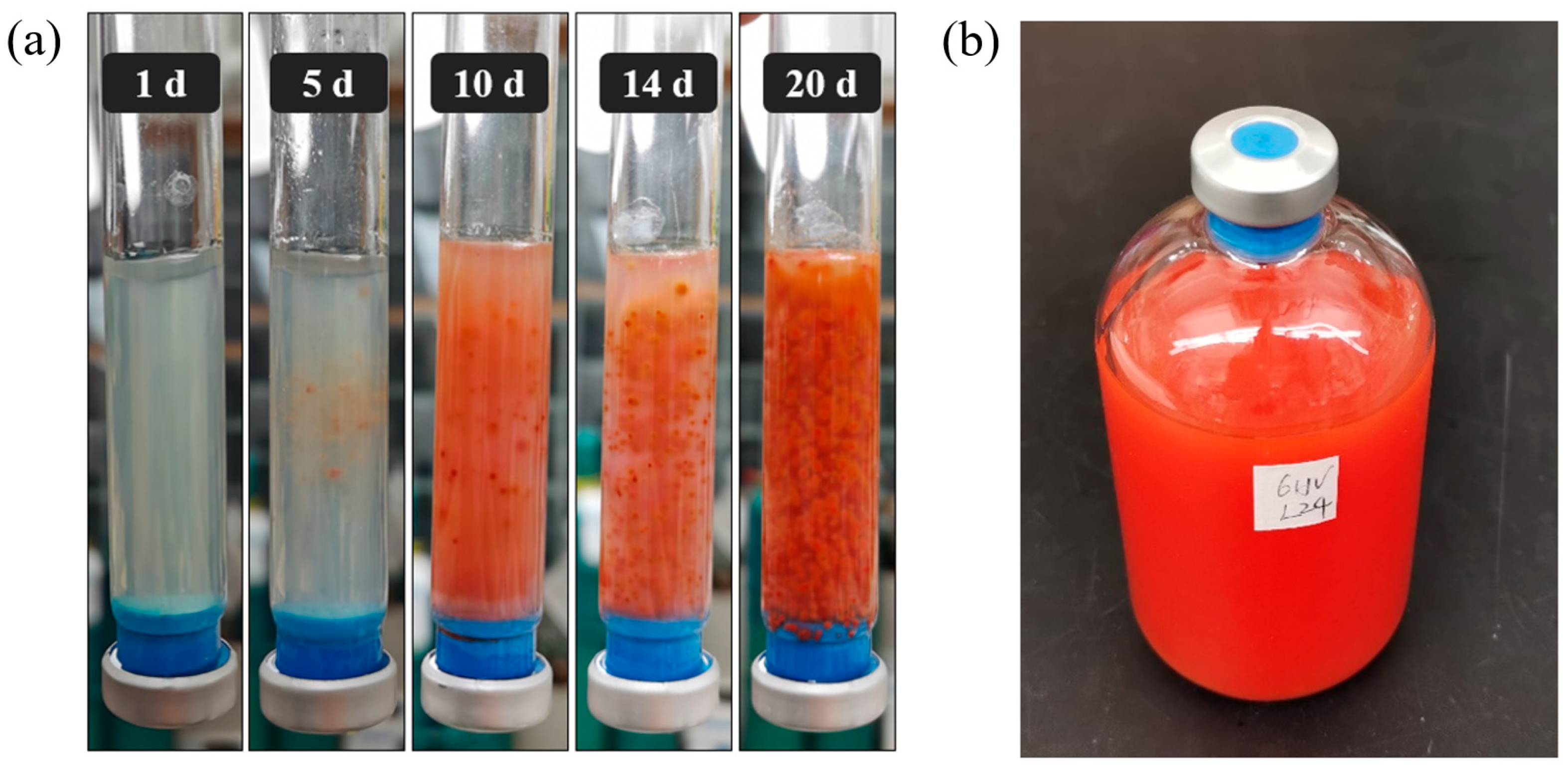
3.1. Growth Characteristic and Metabolic Diversity

Microbial-induced selenate reduction refers to the process by which specific microorganisms convert SeO42−, a highly soluble and mobile form of Se, into SeO32− and other less soluble forms, such as Se0 or Se2−. As demonstrated in Figure 1a, the isolate strain exhibits robust growth in anaerobic soft agar shake tubes, forming bright red colonies through the reduction of SeO42− to Se0. The produced Se0 is not further reduced to Se2− by the isolate, as Se2− does not display a uniform color and varies based on the specific metal or other elements it complexes with [46]. Se0 is a stable endpoint in many microbial reduction processes, and further reduction to Se2− is uncommon, requiring specific conditions and potentially different microbial pathways [47]. In the microbial-mediated process, SeO42− acts as the electron acceptor, while acetate serves as the electron donor (Equation (1)). Upon transfer to aqueous media, the isolate maintained similar metabolic activity, demonstrating consistent selenate reduction (Figure 1b). This biochemical process is critical not only in the biogeochemical cycling of Se but also for environmental remediation. Microbial-induced selenate reduction can be applied to treat Se-contaminated mining runoff or industrial wastewater, mitigating selenium’s toxic effects on aquatic ecosystems when discharged into natural water bodies. Microbial Se0 has several distinct advantages that make it an environmentally friendly process over chemical approaches. Microbial routes typically generate nanoparticles with a well-defined size and morphology, which is advantageous in nanotechnology [48]. Moreover, microbial Se0 are usually biocompatible and are suited for medical application [49].
SeO42−(aq) + 2CH3COO(aq) + 4H2O(aq) → Se0(s) + 4HCO3(aq) + 10H+(aq)
The growth characteristics of Anaerobacillus sp. strain HL2 and eight other Anaerobacillus species were compared, focusing on their fermentation capabilities and utilization of various electron donors and acceptors (Table 1). Acetate was used by all strains except A. beveridgei MLTeJB, underscoring its general role as a key electron donor in this genus. Furthermore, it may boost the metabolic activity of the TCA cycle and fermentation pathways, potentially increasing the production of reducing equivalents that could enhance selenium reduction efficiency. In contrast, the utilization of starch and pyruvate varied across species, indicating their carbon metabolic diversity. Most Anaerobacillus species did not utilize oxygen, reflecting their predominantly anaerobic nature. However, A. beveridgei MLTeJB exhibited the unique ability to use oxygen, suggesting an evolutionary adaptation to both aerobic and anaerobic environments, which could be ecologically and biotechnologically significant. The metabolic versatility of the Anaerobacillus genus is further demonstrated by their ability to use nitrate, selenate, selenite, and arsenate as electron acceptors. Notably, A. selenitireducens MLS10 and A. alkalidiazotrophicus MS 6 can reduce nitrate, facilitating survival in nitrogen-rich environments and contributing to denitrification, a key process for mitigating nitrate pollution. Five species, including Anaerobacillus sp. strain HL2, A. selenitireducens MLS10, A. beveridgei MLTeJB, A. alkalilacustris Z-05211, and A. arseniciselenatis E1H, have demonstrated the ability to reduce selenate, while A. macyae JMM-4 and A. alkalidiazotrophicus MS 6 do not. Importantly, all selenate-reducing species can also reduce selenite. The ability to utilize arsenate varied among the strains, reflecting specific environmental adaptations. These differences in electron acceptor usage highlight the metabolic flexibility of Anaerobacillus species, enabling them to survive and function in diverse and often challenging environmental conditions. Furthermore, metabolomic analyses could further elucidate how different electron donors influence metabolic pathways driving selenium reduction, offering a promising direction for future research.

3.2. Comprehensive Genome Profiling of Anaerobacillus Species

3.2.1. Overall Genome Characteristic

The genome of Anaerobacillus sp. strain HL2 comprises 3.6 Mb with a GC content of 35.0%. Within the genome, 10,686 coding sequences are present, including 81 tRNA and 21 rRNA genes. Specifically, there are six copies of 5S rRNA, nine copies of 16S rRNA, and six copies of 23S rRNA. The sequence similarity of the nine copies of 16S rRNA exceeds 99%. The genome also contains 7503 hypothetical proteins and 3183 proteins with functional annotations (Table S1, Figure S1). Figure 2 presents a pairwise comparison of the average nucleotide identity (ANI) values among different Anaerobacillus species. ANI values serve as a measure of genomic similarity, with a threshold of 95% commonly used to classify organisms as the same species [55]. Notably, only one comparison, between A. alkalilacustris Z-05211 and A. alkalidiazotrophicus MS 6, exhibits an ANI value above 95% (95.3%), indicating these two isolates belong to the same species. All other comparisons show ANI values below 95%, suggesting that the respective species are genetically distinct. For instance, all ANI comparisons involving Anaerobacillus sp. strain HL2 and other Anaerobacillus species fall below the 95% threshold, supporting the identification of Anaerobacillus sp. strain HL2 as a novel species within the genus. Overall, the consistently low ANI values across most species pairs highlight the genetic diversity within the Anaerobacillus genus.

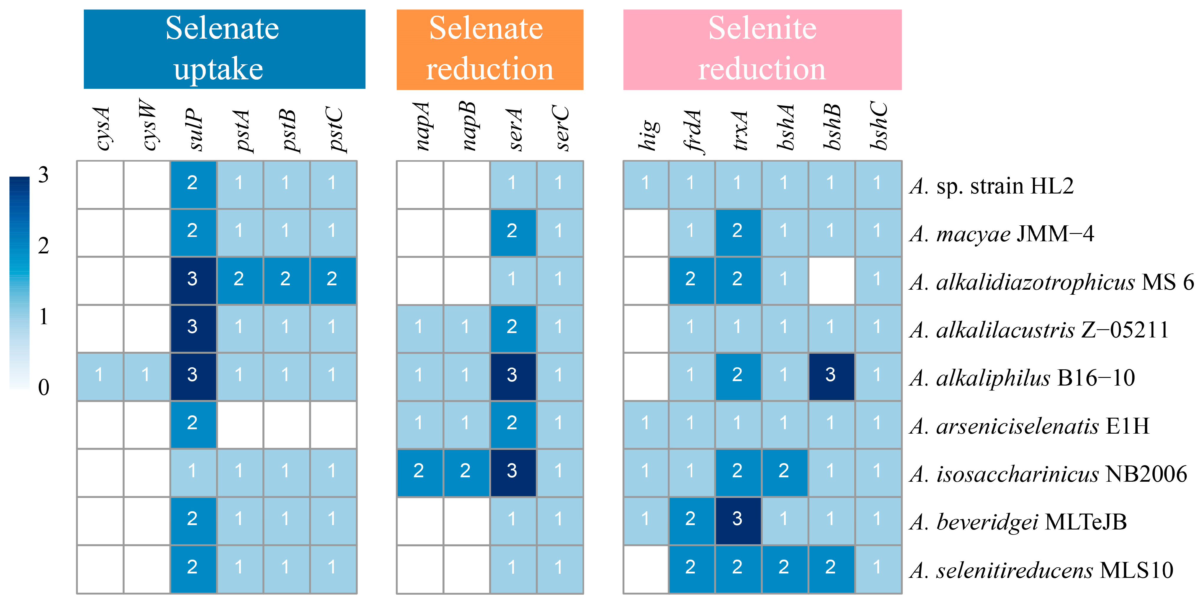
3.2.2. Identification of Genes Related to Se Transport and Transformation

A summary of the identified genes involved in Se transport and transformation across various Anaerobacillus species is provided, with a focus on the presence and copy number of genes related to selenate uptake, selenate reduction, and selenite reduction (Figure 3). Key genes, such as cysAW, sulP, and pstABC, are highlighted in the context of selenate uptake. The first set of genes, the cys gene cluster, is primarily responsible for sulfur compound transport, yet it also facilitates the uptake of selenate due to the structural similarity between sulfate and selenate. Specifically, cysW encodes a membrane protein involved in the sulfate-thiosulfate transporter system, while cysA encodes an ATP-binding protein that powers selenate uptake via ATP hydrolysis [56]. This transport mechanism, driven by the cys gene cluster, is observed in A. alkaliphilus B16-10, suggesting its importance in selenate uptake within this species. The second gene involved is sulP, which encodes sulfate permease (SulP), a protein that mediates the transport of sulfate and similar anions, including selenate [57]. Competitive inhibition studies in Desulfovibrio desulfuricans CSN indicate that sulfate and selenate share a SulP-mediated transport pathway [58]. Further evidence from Arabidopsis thaliana mutants, which show reduced selenate uptake when the sulP gene is disrupted, underscores the gene’s role in selenate transport [59]. The ubiquitous presence of sulP in all Anaerobacillus species suggests that this gene plays a critical role in selenate uptake across the genus. Finally, the pst gene cluster encodes the high-affinity phosphate transport system (Pst system), which includes pstA, pstB, and pstC genes. Due to the structural similarity between phosphate and selenate, the Pst system also facilitates selenate uptake, particularly via PstA [36]. However, in phosphate-rich environments, phosphate transport is prioritized, limiting selenate uptake. Interestingly, pstA, pstB, and pstC genes are present in most Anaerobacillus species, except for A. arseniciselenatis E1H, suggesting that the pst gene cluster may have an alternative or additional role in selenate transport in the majority of Anaerobacillus species.
Selenate reduction-related genes in the nap and ser clusters were identified in the genomes of Anaerobacillus species (Figure 3). Although srdA, ynfE/ynfF, and narG are recognized as functional genes in the selenate reduction process, these genes are not present in all Anaerobacillus species [21,24,60]. The napA and napB genes encode the subunits of the periplasmic nitrate reductase (Nap) enzyme, with NapA serving as the molybdenum-containing catalytic subunit and NapB as a diheme cytochrome c involved in electron transfer. Predominantly found in denitrifies like Rhodobacter sphaeroides, Nap plays a key role in the initial step of denitrification. While NapA and NapB can reduce selenate and tellurite in vitro, their activities toward these oxyanions are significantly less efficient compared to nitrate reduction [22]. The presence of the napA and napB genes in A. alkalilacustris Z-05211, A. alkaliphilus B16-10, A. arseniciselenatis E1H, and A. isosaccharinicus NB2006 suggests that the Nap enzyme may play a role in selenate reduction in these species. On the other hand, the ser genes, including serA, serB, and serC, encode the subunits of the periplasmic selenate reductase in Thauera selenatis [61]. serA encodes the molybdenum-containing catalytic subunit, which is proposed to host the active site for selenate reduction [62]. serB encodes an iron-sulfur protein, which likely participates in electron transfer within the enzyme complex. Its structure includes cysteine residues typical of iron-sulfur clusters, suggesting its role in mediating electron flow to the catalytic SerA subunit. Finally, SerC, the smallest subunit, is hypothesized to contain a heme b component, similar to cytochromes, potentially facilitating electron transfer from SerB to SerA during the reduction process [63]. The ser gene cluster is present in all Anaerobacillus species, underscoring its significant role in selenate reduction.
Selenite reduction occurs as a subsequent process following the reduction in selenate to selenite. The related genes, including hig, frdA, traA, and bshABC, were identified in the genomes of Anaerobacillus species (Figure 3). The gene hig encodes Hydrogenase I, which functions as a selenite reductase in Clostridium pasteurianum. The purified enzyme demonstrated the ability to reduce SeO32− to Se0 through the oxidation of H2. The observed stoichiometry of 2.3:1 (H2:Se0) closely approximates the theoretical ratio of 2:1, confirming the high efficiency of the reduction process [31]. Hydrogenase I has not been reported to catalyze selenite reduction using organic electron donors, despite the identification of the hig gene in certain Anaerobacillus species. The second gene of interest is frdA, which encodes fumarate reductase, an enzyme essential for selenite reduction via a membrane-bound mechanism. Proteomic and genetic analyses reveal that selenite exposure significantly upregulates fumarate reductase expression in Enterobacter cloacae Z0206. Knockout experiments further confirm the enzyme’s crucial role, as deletion of frdA markedly impairs the bacterium’s ability to reduce selenite [34]. The third gene involved is trxA, which encodes thioredoxin. Thioredoxin is a key component of the thioredoxin system and mediates redox reactions. These reactions include the reduction in selenite. In Pseudomonas stutzeri, trxA plays a critical role in the initial step of selenite reduction. Both frdA and trxA genes are found in all Anaerobacillus species, suggesting their potential role in selenite reduction. The bsh gene cluster is another set of genes associated with selenite reduction [35]. This cluster, which includes bshA, bshB, and bshC, is involved in the biosynthesis of bacillithiol, an important low-molecular-weight thiol found in Gram-positive bacteria, particularly in Bacillus species. Specifically, bshA encodes N-acetyl-alpha-D-glucosaminyl L-malate synthase, bshB encodes a deacetylase, and bshC encodes a cysteine-adding enzyme. Together, these genes facilitate the production of bacillithiol, which plays a key role in selenite reduction by forming selenodibacillithiol intermediates. These intermediates are further reduced to Se0 through enzymatic processes, including the thioredoxin and thioredoxin reductase systems, contributing to microbial selenite reduction. As is consistent with the presence of frdA and trxA genes, the bsh gene clusters are found in all Anaerobacillus species, highlighting the metabolic versatility of the selenite reduction process.

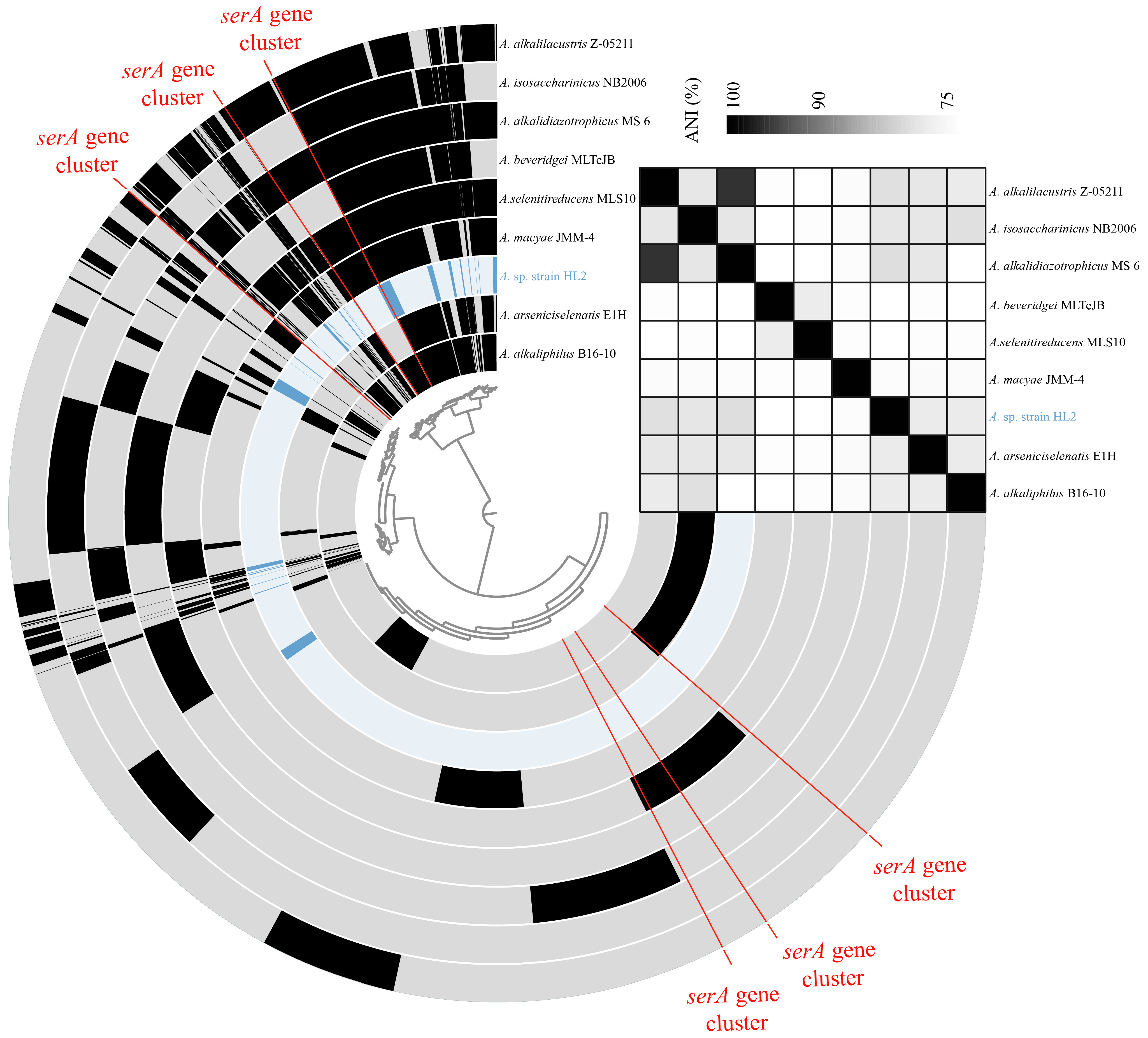
3.2.3. Comparative Analysis of serA Gene Cluster in Anaerobacillus Species

Although gene clusters related to Se reduction were identified, certain Anaerobacillus species can reduce Se, while others cannot. This highlights the need for a deeper investigation into their molecular mechanisms. As the reduction in selenate is a critical initial step in Se reduction, genes associated with selenate reductase have become a central focus of this study. The ser gene cluster, specifically linked to selenate reduction, includes serA. This gene is reported to contain the active site responsible for the reaction [62]. Therefore, further analysis of the serA gene cluster in Anaerobacillus species is necessary to elucidate its distinct role and functionality.
A comparison of the serA gene clusters in the Anaerobacillus species reveals both conserved and divergent patterns in gene organization (Figure 4). Among anaerobic Anaerobacillus species, the serA gene remains highly conserved, exhibiting only slight differences in amino acid length. In contrast, the regulatory elements and coenzyme-related genes adjacent to serA display considerable variability in both their presence and size. This variation likely reflects species-specific adaptations within Anaerobacillus, indicating distinct regulatory or metabolic mechanisms associated with serA. Specifically, two regulatory genes are located upstream of the serA gene in A. selenitireducens MLS10, A. beveridgei MLTeJB, and A. alkaliphilus B16-10. One set of these genes belongs to the Crp/Fnr and GntR families, which are key bacterial transcriptional regulators involved in metabolic and environmental response pathways. Fnr, specifically, controls anaerobic respiration by modulating reductase enzymes. This enzyme is critical in electron transport, enabling bacterial adaptation to oxygen-limited environments [64]. The second regulatory gene belongs to the GntR family, which primarily regulates carbohydrate metabolism, often functioning as a repressor [65]. Moreover, three other downstream genes encode enzymes: histidinol-phosphatase, ATP(I)alamin adenosyl transferase, and molybdopterin adenylyl transferase, respectively. Histidinol-phosphatase catalyzes the dephosphorylation of histidinol phosphate to histidinol, a key step in histidine biosynthesis [66]. ATP(I)alamin adenosyl transferase is crucial for converting cob(I)alamin to adenosyl cobalamin (coenzyme B12), a cofactor involved in reductase reactions such as methyl malonyl-CoA mutase activity in propionate metabolism [67]. Molybdopterin adenylyl transferase is essential for the biosynthesis of molybdenum cofactors, which are required by molybdenum-containing reductases that participate in electron transport [68]. These enzymes play a vital role in supporting reductase activity by facilitating cofactor biosynthesis and metabolic processes. The fer gene, which encodes the ferredoxin protein, is present in many species of Anaerobacillus. Ferredoxin, a small iron-sulfur protein, plays a crucial role in electron transfer during various biological redox reactions. Acting as an electron carrier, ferredoxin facilitates the transfer of electrons to a variety of reductases, driving enzymatic reduction processes [69]. Numerous unannotated genes are located both upstream and downstream of the serA gene, suggesting that further investigation using an integrated approach of proteomic analysis and gene knockout experiments is needed to explore the relationship between this gene cluster and selenate reduction. Notably, some gene clusters, such as the serA clusters in Anaerobacillus macyae JMM-4 and Anaerobacillus alkalidiazotrophicus MS 6, do not appear to contribute to selenate reduction (Table 1). These findings highlight the evolutionary divergence in the mechanisms regulating coenzyme utilization and serA expression across Anaerobacillus species.
The phylogenetic relationships of serA genes from various Anaerobacillus species were analyzed (Figure S2). Notably, branch lengths in the phylogenetic tree correspond to genetic distance, with longer branches indicating greater genetic divergence. While all species harbor a common serA gene, the degree of sequence similarity varies, highlighting evolutionary divergence within the Anaerobacillus genus. For example, the serA gene similarity between A. selenitireducens MLS10 and A. beveridgei MLTeJB is 76% (Figure S3). Proteins with over 70% sequence identity have at least a 90% chance of being involved in the same biological process. The serA genes of A. alkalilacustris Z-05211 and A. alkalidiazotrophicus MS 6 exhibit the highest similarity (96%), a phenomenon consistent with the ANI of their genomes (Figure 2). The phenomenon further suggests that closely related bacterial species often share similar metabolic processes.

3.2.4. Metabolic Pathway Linking Se Reduction in Anaerobacillus sp. Strain HL2

Based on the Se transport and transformation genes identified in Anaerobacillus sp. strain HL2, the proposed metabolic pathways for Se reduction by strain HL2 are illustrated in Figure 5. Selenate is taken up by SulP transporters and reduced to selenite potentially by an enzymatic complex labeled SerABC. Further reduction occurs in the periplasm, where FrdA facilitates the conversion of selenite. The selenite can then be transported to the cytoplasm by PstABC, where it is further reduced to Se0 by enzymes such as Hydrogenase I, TrxA, or BshABC [36]. A question mark in the diagram highlights an unknown or unidentified enzyme in the Se(VI) to Se(IV) reduction pathway within the cytoplasm. The model also suggests that Se0 may be released extracellularly, possibly through cell lysis, though specific transmembrane mechanisms remain unidentified. This model provides insights into Se detoxification pathways in microbial systems, with implications for bioremediation of Se-contaminated environments.

4. Conclusions

A novel Anaerobacillus species capable of reducing selenate and selenite was isolated from a mining site. Comparative growth studies indicate the metabolic versatility of Anaerobacillus species, particularly their ability to utilize diverse electron donors and acceptors. The gene clusters associated with selenate uptake, selenate reduction, and selenite reduction are identified by genomic analysis. A high degree of conservation of the serA gene across Anaerobacillus species is observed. The regulatory and coenzyme-related genes in the surrounding regions are distinct. This divergence in coenzyme utilization and serA regulation suggests different survival strategies and evolutionary pathways of these Anaerobacillus species. The Se transformation mechanisms could be further revealed by employing proteomic analysis in combination with gene knockout experiments to characterize the expression of serA, serB, and serC and to uncover unexploited functions associated with these proteins. Although Anaerobacillus species exhibit high efficiency in selenium bio-transformation, their environmental adaptability and stability need to be further considered for practical remediation applications.

Supplementary Materials

The following supporting information can be downloaded at: https://www.mdpi.com/article/10.3390/microorganisms13030659/s1. Figure S1: Circular representation of genome characteristic of Anaerobacillus sp. strain HL2; Figure S2: Phylogenetic tree of serA genes from Anaerobacillus species was constructed using Maximum Likelihood method based on Tamura–Nei model. Analysis included 16 nucleotide sequences, with all gaps and missing data excluded, resulting in final dataset of 910 positions; Figure S3: The similarity of the identified serA genes in the Anaerobacillus species. The red numbers represent the corresponding amino acid (aa) values for each gene; Table S1: General features of annotated genome Anaerobacillus sp. strain HL2.

Author Contributions

Conceptualization, H.L. and H.Z.; methodology, Q.W.; validation, H.L., H.Z. and Q.W.; formal analysis, Q.W.; investigation, Q.W., J.Z., J.L., Y.W., C.R., X.C. and D.C.; data curation, Q.W.; writing—original draft preparation, Q.W.; writing—review and editing, H.L., H.Z., J.Z., J.L., Y.W., C.R., X.C. and D.C.; supervision, H.L., H.Z., J.Z., J.L., Y.W., C.R., X.C. and D.C.; project administration, H.L. and J.Z.; funding acquisition, H.L. All authors have read and agreed to the published version of the manuscript.

Funding

This research was financially supported by the National Natural Science Foundation of China (52430005, 52370204, 42107230), the Shandong Provincial Natural Science Foundation (ZR2023ZD51), the Shandong Provincial Taishan Scholars Youth Expert Program (2021-292), the Shandong Provincial Excellent Young Scientists Fund Program (2023HWYQ-078), and the Shandong Provincial Youth Innovation Team Development Plan of Colleges and Universities (2022KJ220).

Institutional Review Board Statement

Not applicable.

Informed Consent Statement

Not applicable.

Data Availability Statement

The genome was deposited in the JGI/IMG database under Genome ID 2929886851.

Conflicts of Interest

The authors declare no conflicts of interest.

References

  1. Santesmasses, D.; Mariotti, M.; Gladyshev, V.N. Bioinformatics of Selenoproteins. Antioxid. Redox Signal. 2020, 33, 525–536. [Google Scholar] [CrossRef] [PubMed]
  2. Reich, H.J.; Hondal, R.J. Why Nature Chose Selenium. ACS Chem. Biol. 2016, 11, 821–841. [Google Scholar] [CrossRef] [PubMed]
  3. Ekumah, J.-N.; Ma, Y.; Akpabli-Tsigbe, N.D.K.; Kwaw, E.; Ma, S.M.; Hu, J. Global soil distribution, dietary access routes, bioconversion mechanisms and the human health significance of selenium: A review. Food Biosci. 2021, 41, 100960. [Google Scholar] [CrossRef]
  4. Chen, Y.; Liu, Z.; Zeng, W.; Liu, Y.; Zhao, D.; Zhang, Y.; Jia, X. Screening and Identification of Soil Selenium-Enriched Strains and Application in Auricularia auricula. Microorganisms 2024, 12, 1136. [Google Scholar] [CrossRef]
  5. Wang, L.; Ju, J.; Xie, H.; Qiao, F.; Luo, Q.; Zhou, L. Comparative Study on Growth and Metabolomic Profiles of Six Lactobacilli Strains by Sodium Selenite. Microorganisms 2024, 12, 1937. [Google Scholar] [CrossRef] [PubMed]
  6. Ibrahim, A.M.; Ghazy, M.; El-Sayed, H.; El-Hameed, R.M.; Khalil, R.G.; Korany, S.M.; Aloufi, A.S.; Hammam, O.A.; Morad, M.Y. Histopathological, Immunohistochemical, Biochemical, and In Silico Molecular Docking Study of Fungal-Mediated Selenium Oxide Nanoparticles on Biomphalaria alexandrina (Ehrenberg, 1831) Snails. Microorganisms 2023, 11, 811. [Google Scholar] [CrossRef] [PubMed]
  7. Rauschenbach, I.; Narasingarao, P.; Häggblom, M.M. Desulfurispirillum indicum sp. nov., a selenate- and selenite-respiring bacterium isolated from an estuarine canal. Int. J. Syst. Evol. Microbiol. 2011, 61, 654–658. [Google Scholar] [CrossRef]
  8. Wadgaonkar, S.L.; Nancharaiah, Y.V.; Jacob, C.; Esposito, G.; Lens, P.N.L. Microbial transformation of Se oxyanions in cultures of Delftia lacustris grown under aerobic conditions. J. Microbiol. 2019, 57, 362–371. [Google Scholar] [CrossRef]
  9. Staicu, L.C.; Barton, L.L. Selenium respiration in anaerobic bacteria: Does energy generation pay off? J. Inorg. Biochem. 2021, 222, 111509. [Google Scholar] [CrossRef]
  10. Hageman, S.P.W.; van der Weijden, R.D.; Weijma, J.; Buisman, C.J.N. Microbiological selenate to selenite conversion for selenium removal. Water Res. 2013, 47, 2118–2128. [Google Scholar] [CrossRef]
  11. von Wintzingerode, F.; Gobel, U.B.; Siddiqui, R.A.; Rösick, U.; Schumann, P.; Frühling, A.; Rohde, M.; Pukall, R.; Stackebrandt, E. Salana multivorans gen. nov., sp. nov., a novel actinobacterium isolated from an anaerobic bioreactor and capable of selenate reduction. Int. J. Syst. Evol. Microbiol. 2001, 51, 1653–1661. [Google Scholar] [CrossRef] [PubMed]
  12. Rauschenbach, I.; Posternak, V.; Cantarella, P.; McConnell, J.; Starovoytov, V.; Häggblom, M.M. Seleniivibrio woodruffii gen. nov., sp. nov., a selenate- and arsenate-respiring bacterium in the Deferribacteraceae. Int. J. Syst. Evol. Microbiol. 2013, 63, 3659–3665. [Google Scholar] [CrossRef]
  13. Narasingarao, P.; Häggblom, M.M. Pelobacter seleniigenes sp. nov., a selenate-respiring bacterium. Int. J. Syst. Evol. Microbiol. 2007, 57, 1937–1942. [Google Scholar] [CrossRef] [PubMed]
  14. Nakagawa, T.; Iino, T.; Suzuki, K.-I.; Harayama, S. Ferrimonas futtsuensis sp. nov. and Ferrimonas kyonanensis sp. nov., selenate-reducing bacteria belonging to the Gammaproteobacteria isolated from Tokyo Bay. Int. J. Syst. Evol. Microbiol. 2006, 56, 2639–2645. [Google Scholar] [CrossRef] [PubMed]
  15. Yamamura, S.; Yamashita, M.; Fujimoto, N.; Kuroda, M.; Kashiwa, M.; Sei, K.; Fujita, M.; Ike, M. Bacillus selenatarsenatis sp. nov., a selenate- and arsenate-reducing bacterium isolated from the effluent drain of a glass-manufacturing plant. Int. J. Syst. Evol. Microbiol. 2007, 57, 1060–1064. [Google Scholar] [CrossRef]
  16. Ike, M.; Kazuaki, T.M.; Tomoko, F.M.; Kashiwa, M.; Masanori, F.M. Selenate reduction by bacteria isolated from aquatic environment free from selenium contamination. Water Res. 2000, 34, 3019–3025. [Google Scholar] [CrossRef]
  17. Hunter, W.J.; Manter, D.K. Reduction of Selenite to Elemental Red Selenium by Rhizobium sp. Strain B1. Curr. Microbiol. 2007, 55, 344–349. [Google Scholar] [CrossRef]
  18. Kagami, T.; Narita, T.; Kuroda, M.; Notaguchi, E.; Yamashita, M.; Sei, K.; Soda, S.; Ike, M. Effective selenium volatilization under aerobic conditions and recovery from the aqueous phase by NT-I. Water Res. 2013, 47, 1361–1368. [Google Scholar] [CrossRef]
  19. Shi, L.-D.; Lv, P.-L.; McIlroy, S.J.; Wang, Z.; Dong, X.-L.; Kouris, A.; Lai, C.-Y.; Tyson, G.W.; Strous, M.; Zhao, H.-P. Methane-dependent selenate reduction by a bacterial consortium. ISME J. 2021, 15, 3683–3692. [Google Scholar] [CrossRef]
  20. Wen, L.-L.; Lai, C.-Y.; Yang, Q.; Chen, J.-X.; Zhang, Y.; Ontiveros-Valencia, A.; Zhao, H.-P. Quantitative detection of selenate-reducing bacteria by real-time PCR targeting the selenate reductase gene. Enzym. Microb. Technol. 2016, 85, 19–24. [Google Scholar] [CrossRef]
  21. Kuroda, M.; Yamashita, M.; Miwa, E.; Imao, K.; Fujimoto, N.; Ono, H.; Nagano, K.; Sei, K.; Ike, M. Molecular Cloning and Characterization of the srdBCA Operon, Encoding the Respiratory Selenate Reductase Complex, from the Selenate-Reducing Bacterium Bacillus selenatarsenatis SF-1. J. Bacteriol. 2011, 193, 2141–2148. [Google Scholar] [CrossRef] [PubMed]
  22. Sabaty, M.; Avazeri, C.; Pignol, D.; Vermeglio, A. Characterization of the Reduction of Selenate and Tellurite by Nitrate Reductases. Appl. Environ. Microbiol. 2001, 67, 5122–5126. [Google Scholar] [CrossRef] [PubMed]
  23. Theisen, J.; Yee, N. The Molecular Basis for Selenate Reduction in Citrobacter freundii. Geomicrobiol. J. 2014, 31, 875–883. [Google Scholar] [CrossRef]
  24. Ridley, H.; Watts, C.A.; Richardson, D.J.; Butler, C.S. Resolution of Distinct Membrane-Bound Enzymes from Enterobacter cloacae SLD1a-1 That Are Responsible for Selective Reduction of Nitrate and Selenate Oxyanions. Appl. Environ. Microbiol. 2006, 72, 5173–5180. [Google Scholar] [CrossRef]
  25. Wells, M.; McGarry, J.; Gaye Maissa, M.; Basu, P.; Oremland Ronald, S.; Stolz, J.F. Respiratory Selenite Reductase from Bacillus selenitireducens Strain MLS10. J. Bacteriol. 2019, 201, e00614-18. [Google Scholar] [CrossRef]
  26. Li, D.-B.; Cheng, Y.-Y.; Wu, C.; Li, W.-W.; Li, N.; Yang, Z.-C.; Tong, Z.-H.; Yu, H.-Q. Selenite reduction by Shewanella oneidensis MR-1 is mediated by fumarate reductase in periplasm. Sci. Rep. 2014, 4, 3735. [Google Scholar] [CrossRef]
  27. Basaglia, M.; Toffanin, A.; Baldan, E.; Bottegal, M.; Shapleigh, J.P.; Casella, S. Selenite-reducing capacity of the copper-containing nitrite reductase of Rhizobium sullae. FEMS Microbiol. Lett. 2010, 269, 124–130. [Google Scholar] [CrossRef]
  28. Wang, D.; Xia, X.; Wu, S.; Zheng, S.; Wang, G. The essentialness of glutathione reductase GorA for biosynthesis of Se(0)-nanoparticles and GSH for CdSe quantum dot formation in Pseudomonas stutzeri TS44. J. Hazard. Mater. 2019, 366, 301–310. [Google Scholar] [CrossRef]
  29. Yasir, M.; Zhang, Y.; Xu, Z.; Luo, M.; Wang, G. NAD(P)H-dependent thioredoxin-disulfide reductase TrxR is essential for tellurite and selenite reduction and resistance in Bacillus sp. Y3. FEMS Microbiol. Ecol. 2020, 96, fiaa126. [Google Scholar] [CrossRef]
  30. Xia, X.; Wu, S.J.; Li, N.H.; Wang, D.; Zheng, S.X.; Wang, G. Novel bacterial selenite reductase CsrF responsible for Se(IV) and Cr(VI) reduction that produces nanoparticles in Alishewanella sp. WH16-1. J. Hazard. Mater. 2018, 342, 499–509. [Google Scholar] [CrossRef]
  31. Yanke, L.J.; Bryant, R.D.; Laishley, E.J. Hydrogenase I of Clostridium pasteurianum functions as a novel selenite reductase. Anaerobe 1995, 1, 61–67. [Google Scholar] [CrossRef] [PubMed]
  32. Huang, S.; Wang, Y.; Tang, C.; Jia, H.; Wu, L. Speeding up selenite bioremediation using the highly selenite-tolerant strain Providencia rettgeri HF16-A novel mechanism of selenite reduction based on proteomic analysis. J. Hazard. Mater. 2021, 406, 124690. [Google Scholar] [CrossRef]
  33. Tan, Y.; Wang, Y.; Wang, Y.; Xu, D.; Huang, Y.; Wang, D.; Wang, G.; Rensing, C.; Zheng, S. Novel mechanisms of selenate and selenite reduction in the obligate aerobic bacterium Comamonas testosteroni S44. J. Hazard. Mater. 2018, 359, 129–138. [Google Scholar] [CrossRef] [PubMed]
  34. Song, D.G.; Li, X.X.; Cheng, Y.Z.; Xiao, X.; Lu, Z.Q.; Wang, Y.; Wang, F. Aerobic biogenesis of selenium nanoparticles by Enterobacter cloacae Z0206 as a consequence of fumarate reductase mediated selenite reduction. Sci. Rep. 2017, 7, 3239. [Google Scholar] [CrossRef] [PubMed]
  35. Ge, M.; Zhou, S.F.; Li, D.B.; Song, D.; Yang, S.; Xu, M. Reduction of selenite to selenium nanoparticles by highly selenite-tolerant bacteria isolated from seleniferous soil. J. Hazard. Mater. 2024, 472, 134491. [Google Scholar] [CrossRef]
  36. Zhu, T.-T.; Tian, L.-J.; Yu, H.-Q. Phosphate-Suppressed Selenite Biotransformation by Escherichia coli. Environ. Sci. Technol. 2020, 54, 10703–10711. [Google Scholar] [CrossRef]
  37. Mataigne, V.; Vannier, N.; Vandenkoornhuyse, P.; Hacquard, S. Multi-genome metabolic modeling predicts functional inter-dependencies in the Arabidopsis root microbiome. Microbiome 2022, 10, 217. [Google Scholar] [CrossRef]
  38. Joshi, A.; Thite, S.; Karodi, P.; Joseph, N.; Lodha, T. Alkalihalobacterium elongatum gen. nov. sp. nov.: An Antibiotic-Producing Bacterium Isolated From Lonar Lake and Reclassification of the Genus Alkalihalobacillus Into Seven Novel Genera. Front. Microbiol. 2022, 13, 722369, Erratum in Front. Microbiol. 2022, 13, 871596. [Google Scholar] [CrossRef]
  39. Fisher, J.C.; Hollibaugh, J.T. Selenate-Dependent Anaerobic Arsenite Oxidation by a Bacterium from Mono Lake, California. Appl. Environ. Microbiol. 2008, 74, 2588–2594. [Google Scholar] [CrossRef]
  40. Baesman, S.M.; Stolz, J.F.; Kulp, T.R.; Oremland, R.S. Enrichment and isolation of Bacillus beveridgei sp. nov., a facultative anaerobic haloalkaliphile from Mono Lake, California, that respires oxyanions of tellurium, selenium, and arsenic. Extremophiles 2009, 13, 695–705. [Google Scholar] [CrossRef]
  41. Blum, J.S.; Bindi, A.B.; Buzzelli, J.; Stolz, J.F.; Oremland, R.S. Bacillus arsenicoselenatis, sp. nov., and Bacillus selenitireducens, sp. nov.: Two haloalkaliphiles from Mono Lake, California that respire oxyanions of selenium and arsenic. Arch. Microbiol. 1998, 171, 19–30. [Google Scholar] [CrossRef] [PubMed]
  42. Santini, J.M. Bacillus macyae sp. nov., an arsenate-respiring bacterium isolated from an Australian gold mine. Int. J. Syst. Evol. Microbiol. 2004, 54, 2241–2244. [Google Scholar] [CrossRef] [PubMed]
  43. Knight, V.K.; Nijenhuis, I.; Kerkhof, L.J.; Häggblom, M.M. Degradation of aromatic compounds coupled to selenate reduction. Geomicrobiol. J. 2002, 19, 77–86. [Google Scholar] [CrossRef]
  44. Lewis, W.H.; Tahon, G.; Geesink, P.; Sousa, D.Z.; Ettema, T.J.G. Innovations to culturing the uncultured microbial majority. Nat. Rev. Microbiol. 2020, 19, 225–240. [Google Scholar] [CrossRef]
  45. Janabi, A.H.D.; Kerkhof, L.J.; McGuinness, L.R.; Biddle, A.S.; McKeever, K.H. Comparison of a modified phenol/chloroform and commercial-kit methods for extracting DNA from horse fecal material. J. Microbiol. Methods 2016, 129, 14–19. [Google Scholar] [CrossRef]
  46. Chen, W.; Karton, A.; Hussian, T.; Javaid, S.; Wang, F.; Pang, Y.; Jia, G. Spontaneous shape and phase control of colloidal ZnSe nanocrystals by tailoring Se precursor reactivity. CrystEngComm 2019, 21, 2955–2961. [Google Scholar] [CrossRef]
  47. Nie, X.L.; Yang, X.R.; He, J.Y.; Liu, P.; Shi, H.; Wang, T.; Zhang, D. Bioconversion of inorganic selenium to less toxic selenium forms by microbes: A review. Front. Bioeng. Biotechnol. 2023, 11, 1167123. [Google Scholar] [CrossRef]
  48. Fesharaki, P.J.; Nazari, P.; Shakibaie, M.; Rezaie, S.; Banoee, M.; Abdollahi, M.; Shahverdi, A.R. Biosynthesis of selenium nanoparticles using Klebsiella pneumoniae and their recovery by a simple sterilization process. Braz. J. Microbiol. 2010, 41, 461–466. [Google Scholar] [CrossRef] [PubMed]
  49. Nayak, V.; Singh, K.R.; Singh, A.K.; Singh, R.P. Potentialities of selenium nanoparticles in biomedical science. New J. Chem. 2021, 45, 2849–2878. [Google Scholar] [CrossRef]
  50. Sorokin, I.D.; Kravchenko, I.K.; Tourova, T.P.; Kolganova, T.V.; Boulygina, E.S.; Sorokin, D.Y. Bacillus alkalidiazotrophicus sp. nov., a diazotrophic, low salt-tolerant alkaliphile isolated from Mongolian soda soil. Int. J. Syst. Evol. Microbiol. 2008, 58, 2459–2464. [Google Scholar] [CrossRef]
  51. Zavarzina, D.G.; Tourova, T.P.; Kolganova, T.V.; Boulygina, E.S.; Zhilina, T.N. Description of Anaerobacillus alkalilacustre gen. nov., sp. nov.—Strictly anaerobic diazotrophic bacillus isolated from soda lake and transfer of Bacillus arseniciselenatis, Bacillus macyae, and Bacillus alkalidiazotrophicus to Anaerobacillus as the new combinations A. arseniciselenatis comb. nov., A. macyae comb. nov., and A. alkalidiazotrophicus comb. nov. Microbiology 2009, 78, 723–731. [Google Scholar] [CrossRef]
  52. Sorokin, D.Y.; Tourova, T.P.; Sukhacheva, M.V.; Muyzer, G. Desulfuribacillus alkaliarsenatis gen. nov. sp. nov., a deep-lineage, obligately anaerobic, dissimilatory sulfur and arsenate-reducing, haloalkaliphilic representative of the order Bacillales from soda lakes. Extremophiles 2012, 16, 597–605. [Google Scholar] [CrossRef] [PubMed]
  53. Borsodi, A.K.; Aszalós, J.M.; Bihari, P.; Nagy, I.; Schumann, P.; Spröer, C.; Kovács, A.L.; Bóka, K.; Dobosy, P.; Óvári, M.; et al. Anaerobacillus alkaliphilus sp. nov., a novel alkaliphilic and moderately halophilic bacterium. Int. J. Syst. Evol. Microbiol. 2019, 69, 631–637. [Google Scholar] [CrossRef] [PubMed]
  54. Bassil, N.M.; Lloyd, J.R. Anaerobacillus isosaccharinicus sp. nov., an alkaliphilic bacterium which degrades isosaccharinic acid. Int. J. Syst. Evol. Microbiol. 2018, 69, 631–637. [Google Scholar] [CrossRef]
  55. Rodriguez-R, L.M.; Gunturu, S.; Harvey, W.T.; Rosselló-Mora, R.; Tiedje, J.M.; Cole, J.R.; Konstantinidis, K.T. The Microbial Genomes Atlas (MiGA) webserver: Taxonomic and gene diversity analysis of Archaea and Bacteria at the whole genome level. Nucleic Acids Res. 2018, 46, W282–W288. [Google Scholar] [CrossRef] [PubMed]
  56. Aguilar-Barajas, E.; Díaz-Pérez, C.; Ramírez-Díaz, M.I.; Riveros-Rosas, H.; Cervantes, C. Bacterial transport of sulfate, molybdate, and related oxyanions. BioMetals 2011, 24, 687–707. [Google Scholar] [CrossRef]
  57. Stoeva, M.K.; Coates, J.D. Specific inhibitors of respiratory sulfate reduction: Towards a mechanistic understanding. Microbiology 2018, 165, 254–269. [Google Scholar] [CrossRef]
  58. Cypionka, H. Characterization of sulfate transport in Desulfovibrio desulfuricans. Arch. Microbiol. 1989, 152, 237–243. [Google Scholar] [CrossRef]
  59. Shibagaki, N.; Rose, A.; McDermott, J.; Fujiwara, T.; Hayashi, H.; Yoneyama, T.; Davies, J.P. Selenate-resistant mutants of Arabidopsis thaliana identify Sultr1;2, a sulfate transporter required for efficient transport of sulfate into roots. Plant J. Cell Mol. Biol. 2002, 29, 475–486. [Google Scholar] [CrossRef]
  60. Guymer, D.; Maillard, J.; Sargent, F. A genetic analysis of in vivo selenate reduction by Salmonella enterica serovar Typhimurium LT2 and Escherichia coli K12. Arch. Microbiol. 2009, 191, 519–528. [Google Scholar] [CrossRef]
  61. Xu, R.; Kolton, M.; Tao, W.; Sun, X.X.; Su, P.; Huang, D.; Zhang, M.; Yang, Z.; Guo, Z.; Gao, H.; et al. Anaerobic selenite-reducing bacteria and their metabolic potentials in Se-rich sediment revealed by the combination of DNA-stable isotope probing, metagenomic binning, and metatranscriptomics. J. Hazard. Mater. 2023, 457, 131834. [Google Scholar] [CrossRef] [PubMed]
  62. McEwan, A.G.; Ridge, J.P.; McDevitt, C.A.; Hugenholtz, P. The DMSO Reductase Family of Microbial Molybdenum Enzymes; Molecular Properties and Role in the Dissimilatory Reduction of Toxic Elements. Geomicrobiol. J. 2002, 19, 3–21. [Google Scholar] [CrossRef]
  63. Krafft, T.; Bowen, A.; Theis, F.; Macy, J.M. Cloning and Sequencing of the Genes Encoding the Periplasmic-Cytochrome B-Containing Selenate Reductase of Thauera selenatis. DNA Seq. 2000, 10, 365–377. [Google Scholar] [CrossRef]
  64. Septer, A.N.; Bose, J.L.; Dunn, A.K.; Stabb, E.V. FNR-mediated regulation of bioluminescence and anaerobic respiration in the light-organ symbiont Vibrio fischeri. FEMS Microbiol. Lett. 2010, 306, 72–81. [Google Scholar] [CrossRef]
  65. Salzberg, L.I.; Luo, Y.; Hachmann, A.-B.; Mascher, T.; Helmann, J.D. The Bacillus subtilis GntR Family Repressor YtrA Responds to Cell Wall Antibiotics. J. Bacteriol. 2011, 193, 5793–5801. [Google Scholar] [CrossRef]
  66. Jha, B.; Kumar, D.; Sharma, A.; Dwivedy, A.; Singh, R.; Biswal, B.K. Identification and structural characterization of a histidinol phosphate phosphatase from Mycobacterium tuberculosis. J. Biol. Chem. 2018, 293, 10102–10118. [Google Scholar] [CrossRef] [PubMed]
  67. Zhang, J.; Dobson, C.M.; Wu, X.; Lerner-Ellis, J.; Rosenblatt, D.S.; Gravel, R.A. Impact of cblB mutations on the function of ATP:cob(I)alamin adenosyltransferase in disorders of vitamin B12 metabolism. Mol. Genet. Metab. 2006, 87, 315–322. [Google Scholar] [CrossRef]
  68. Llamas, A.; Mendel, R.R.; Schwarz, G. Synthesis of adenylated molybdopterin: An essential step for molybdenum insertion. J. Biol. Chem. 2004, 279, 55241–55246. [Google Scholar] [CrossRef]
  69. Wolfgang, B.; Thauer, R.K. Flavin-Based Electron Bifurcation, Ferredoxin, Flavodoxin, and Anaerobic Respiration With Protons (Ech) or NAD+ (Rnf) as Electron Acceptors: A Historical Review. Front. Microbiol. 2018, 9, 401. [Google Scholar] [CrossRef]
Figure 1. (a) Microbial reduction of SeO42− to Se0 over time by Anaerobacillus sp. strain HL2 isolate. (b) Transfer of culture into aqueous medium, demonstrating sustained metabolic activity in selenate reduction.
Microorganisms 13 00659 g001
Figure 2. The pangenomic analysis of different Anaerobacillus species. The darker colors indicate the presence of gene clusters in each genome. The red lines connected to the center of the graph indicate the positions of the serA gene clusters. The heat map represents the average nucleotide identity (ANI) between each pair of different Anaerobacillus species.
Microorganisms 13 00659 g002
Figure 3. Number of putative genes involved in Se transport and transformation identified in Anaerobacillus sp. strain HL2.
Microorganisms 13 00659 g003
Figure 4. Comparison of serA gene clusters in strain HL2 and related Anaerobacillus species. Numbers indicate amino acid (aa) values of respective genes.
Microorganisms 13 00659 g004
Figure 5. Proposed Se metabolic pathway in Anaerobacillus sp. strain HL2.
Microorganisms 13 00659 g005
Table 1. Comparative growth characteristics of Anaerobacillus sp. strain HL2 and related species within genus Anaerobacillus. Strains compared are as follows: 1. Anaerobacillus sp. strain HL2; 2. A. macyae JMM-4; 3. A. selenitireducens MLS10; 4. A. beveridgei MLTeJB; 5. A. alkalidiazotrophicus MS 6; 6. A. alkalilacustris Z-05211; 7. A. arseniciselenatis E1H; 8. A. alkaliphilus B16-10; and 9. A. isosaccharinicus NB2006.
Metabolism12 a3 b4 c5 d6 e,f7 a8 g9 h
Fermentation++++++
Electron Donors
Acetate+++++
Starch+++++++
Pyruvate+++n.a.++
Electron Acceptors
Oxygen++
Nitrate+++++
Selenate+++++n.a.n.a.
Selenite+++++n.a.n.a.
Arsenate+++++++
Note: a, data from [42]; b, data from [41]; c, data from [40]; d, data from [50]; e, data from [51]; f, data from [52]; g, data from [53]; h, data from [54]. +, positive; −, negative; n.a., no data available.
Disclaimer/Publisher’s Note: The statements, opinions and data contained in all publications are solely those of the individual author(s) and contributor(s) and not of MDPI and/or the editor(s). MDPI and/or the editor(s) disclaim responsibility for any injury to people or property resulting from any ideas, methods, instructions or products referred to in the content.

