Article

Isolation and Genome-Based Characterization of Bacillus velezensis AN6 for Its Biocontrol Potential Against Multiple Plant Pathogens

1
Food Crops Research Institute, Yunnan Academy of Agricultural Sciences (YAAS), Kunming 650205, China
2
Laboratory of Bacteriology, Department of Plant Pathology, Nanjing Agricultural University, Nanjing 210095, China
3
Plant Protection Institute, Jiangxi Academy of Agricultural Sciences, Nanchang 330200, China
*
Author to whom correspondence should be addressed.
Microorganisms 2025, 13(12), 2701; https://doi.org/10.3390/microorganisms13122701
Submission received: 21 October 2025 / Revised: 8 November 2025 / Accepted: 12 November 2025 / Published: 27 November 2025
(This article belongs to the Special Issue Biological Control of Microbial Pathogens in Plants)

Abstract

Biological control is an effective and environmentally friendly strategy for managing plant diseases. In this study, a broad-spectrum antagonistic bacterium, designated strain AN6, was isolated from rice plants and exhibited potent inhibitory activity against a variety of phytopathogens. In Oxford cup assays, AN6 suppressed the growth of Xanthomonas oryzae pv. oryzae (Xoo) by 73.60%, and its cell-free culture filtrate caused pronounced morphological deformation in the bacterial cells. Further in vitro assays, including dual-culture assays, volatile organic compound (VOC) assays, and cell-free supernatant (CFS) assays, demonstrated that AN6 also exerted strong antifungal effects against several pathogenic fungi. In addition, the strain was found to produce proteases and siderophores, which may contribute to its antagonistic capabilities. Taxonomic identification based on morphological traits, 16S rRNA and gyrA gene sequencing, average nucleotide identity (ANI), in silico DNA–DNA hybridization (isDDH), and phylogenetic analysis classified strain AN6 as Bacillus velezensis. Whole-genome sequencing revealed that AN6 harbors a 3,929,788 bp genome comprising 4025 protein-coding genes with a GC content of 46.50%. Thirteen biosynthetic gene clusters (BGCs) associated with the production of secondary metabolites—such as nonribosomal peptides, polyketides, and dipeptide antibiotics—were identified. The pot experiment further validated the biocontrol potential of AN6, achieving an 80.49% reduction in rice bacterial blight caused by Xanthomonas oryzae pv. oryzae. Collectively, these results indicate that B. velezensis AN6 is a promising candidate for development as a highly effective biocontrol agent for the integrated management of diverse plant diseases.

1. Introduction

Rice (Oryza sativa L.) is not only a staple food for more than half of the global population, but also a key crop in many countries, particularly in Asia, where it is essential for food security and economic stability [1,2]. However, rice production faces numerous challenges, from both biotic and abiotic stresses, which significantly affect yield and quality. Among these, bacterial leaf blight (BLB), caused by the Gram-negative bacterium Xanthomonas oryzae pv. oryzae (Xoo), stands out as one of the most destructive bacterial diseases in rice cultivation; it is also listed as one of the top ten plant pathogenic bacteria [3,4]. In addition, rice bacterial blight is also a major rice disease in Asian countries. High-yield rice varieties are highly susceptible to the disease. In severely endemic areas, rice bacterial blight can cause yield losses of up to 60%, primarily by affecting the photosynthetic capacity of the plant. Symptoms typically include the appearance of water-soaked lesions that eventually turn into necrotic streaks, leading to premature leaf senescence and reduced grain filling [5]. Conventional management strategies for BLB include the use of resistant rice cultivars, chemical pesticides, and agronomic practices such as crop rotation [6,7,8]. However, these approaches often come with significant limitations. For example, the widespread use of chemical pesticides has raised concerns about environmental pollution, human health risks, and the development of pesticide-resistant bacterial strains [9]. In addition, the variability in Xoo populations and the emergence of new pathogenic strains complicate the effectiveness of resistant cultivars. Consequently, these traditional control measures are often insufficient for long-term disease management. Sustainable and eco-friendly approaches, such as biological control, are garnering increasing amounts of attention.
Biological control can inhibit the growth of harmful organisms through the use of antagonistic microorganisms such as bacteria, fungi, and actinomycetes, providing an environmentally friendly control method for agricultural production. Bio-control agents made from biocontrol bacteria are being increasingly applied in sustainable agriculture. The most studied biocontrol bacteria are Bacillus species. Inoculants derived from Bacillus bacteria have been proven to be environmentally friendly alternatives to chemical pesticides [10]. The Bacillus species that exhibit antagonistic activity against pathogenic fungi include B. subtilis, B. amyloliquefaciens, B. cereus, B. mycoides, B. pumilus, B. pasteurii, and B. sphaericus, among others [11]. It has been reported that these bacteria defend against pathogens by producing antibiotic metabolites, antimicrobial compounds, and by inducing or stimulating systemic resistance in plants [12,13,14,15,16]. Among these, B. amyloliquefaciens can produce lipopeptides (LPs) and has shown significant inhibitory activity against pathogens such as Alternaria panax, Botrytis cinerea, Colletotrichum orbiculare, Penicillium digitatum, Pyricularia grisea, Sclerotinia sclerotiorum, and Acidovorax oryzae [17,18]. Furthermore, B. amyloliquefaciens SQR9 inhibits the growth of Fusarium oxysporum by producing antibiotics, protecting cucumber plants from invasion [19]. B. subtilis ME488 produces antibiotics that strongly inhibit F. oxysporum f. sp. cucumerinum, the pathogen that causes wilt in cucumbers [20]. B. subtilis B006 produces surfactants that inhibit the pathogen in soil by 42.9% [21]. The lipopeptide extract from B. amyloliquefaciens SS-12.6 has shown significant potential for biological control against Pseudomonas syringae pv. aptata, the pathogen that causes leaf spot in beets [22]. Additionally, B. pumilus T4 induces plant resistance to P. syringae through a novel or mutated pathway [23]. Moreover, systemic resistance induced by Bacillus biocontrol bacteria can protect plants from damage caused by leaf spot diseases, viral infections, crown rot, root knot nematodes, and stem rot diseases [11].
B. velezensis is a potential biocontrol agent that was first isolated by Ruiz-García et al. from brackish water samples collected from the Vélez River [24]. B. velezensis can produce a variety of antimicrobial metabolites, including lipopeptide antibiotics (surfactin, fengycin, and bacillomycin D), polyketides (macrolactin, bacillaene, difficidin, and oxydifficidin), and peptides (plantazolicin, amylocyclicin, and bacilysin) [25]. Studies have shown that B. velezensis can significantly inhibit several pathogenic microorganisms, such as Botrytis cinerea [26], Colletotrichum gloeosporioides [27,28,29,30], Phytophthora infestans [31], Agrobacterium tumefaciens [32,33], Ralstonia solanacearum [34], F. oxysporum [16,34,35], Xanthomonas euvesicatoria [36] (which causes bacterial spot in peppers), Xanthomonas oryzae pv. oryzae [37] (which causes bacterial blight in rice), and Phytophthora nicotianae [38] (which causes black shank in tobacco). These pathogens are responsible for diseases affecting various crops, ornamental plants, and fruit trees. Additionally, B. velezensis can induce plant tolerance to abiotic stress and promote plant growth [39]. Whether used as a biocontrol agent or a plant growth regulator, B. velezensis holds great potential for widespread application in agriculture.
This study aimed to investigate the biocontrol potential of B. velezensis AN6 and to determine its genome sequence. Bioassays indicated that AN6 could serve as a biocontrol agent against rice bacterial blight (Xanthomonas oryzae pv. oryzae) and that it also has broad-spectrum antifungal activity. Phenotypic assays and genomic analysis combined with secondary metabolite prediction further indicated that the disease-suppressive capacity of AN6 may be associated with its ability to produce proteases, siderophores, volatile organic compounds (VOCs), and antimicrobial secondary metabolites. Overall, these results indicate that B. velezensis AN6 represents a valuable biocontrol resource with promising potential for environmentally friendly management of plant diseases.

2. Materials and Methods

2.1. Bacterium and Pathogens Used in This Study

The strain AN6 was isolated from the rice leaves collected at a base (100.13° E, 25.85° N) in Dali City, Yunnan Province, China. It is currently preserved at the China Center for Type Culture Collection (CCTCC) under accession number CCTCC M 20231886. The Xanthomonas oryzae pv. oryzae strains (X1, X2, X3, X12, LC2, and LC10) and other phytopathogenic fungi used in this study are summarized in Table S1.

2.2. Isolation and Morphological Observation of Endophytic Bacteria

The isolation and culture of endophytic bacteria from rice leaves were performed according to previously published methods with slight modifications [40]. The pure culture strain was then numbered sequentially. Single colonies of the pure culture strain were inoculated into NB liquid medium and incubated at 28 °C with shaking at 180 rpm for 12 h. After incubation, 1 mL of the bacterial suspension was mixed with 1 mL of 50% sterile glycerol and stored at −80 °C for long-term preservation.

2.3. Screening of Biocontrol Strains with Resistance to Xanthomonas oryzae pv. oryzae

The Oxford cup method, previously described in [41], was used to evaluate the inhibitory activity of the endophytic bacterial isolates (hereafter referred to as “test strains”) against Xoo. Briefly, the test strains and Xoo isolates (X1, X2, X3, X12, LC2, and LC10) were cultured in NB at 28 °C and 180 rpm until OD600 ≈ 1.0. Different Xoo cells were mixed with NA medium (1:100, v/v) to prepare indicator plates. A sterile 6 mm Oxford cup was placed in the center of each plate and 50 µL of the test strain suspension was added. Plates were incubated at 28 °C for 48 h. The diameter of the inhibition zone was measured using the cross method, and the inhibition rate was calculated using the following formula: Inhibition rate (%) = [(Inhibition zone diameter − 6)/Inhibition zone diameter] × 100%. We screened for the strain with the strongest antibacterial activity against Xoo.

2.4. Morphological and Biological Characteristics of the Strain AN6

Morphological and biological analyses of strain AN6 were conducted. The morphological characteristics of the AN6 strain were observed using a Biological Microscope (Olympus BX53, Shinjuku, Tokyo, Japan) and scanning electron microscope (HITACHI Regulus 8100; Hitachi Production Co., Ltd., Tokyo, Japan). Gram staining was performed using the G1060 Gram Stain Kit (Solarbio Science & Technology Co., Ltd., Beijing, China). Bacterial growth was assessed using the method described by Fan [42]. The strains were cultured overnight in NB, and then the culture was adjusted to achieve OD600 = 1.0. They were subsequently diluted 1:100 in NB medium, and absorbance at 600 nm was measured every 1 h. Each experiment was repeated at least three times with 3–5 replicates.

2.5. The Effect of Sterile AN6 Cell-Free Supernatant on the Cell Growth of Xoo

The AN6 and Xoo LC2 strains were independently inoculated into NB liquid medium and incubated at 28 °C with shaking at 180 rpm until the OD600 reached approximately 1.0. A total of 1 mL of AN6 suspension was then centrifuged at 8000 rpm for 5 min to collect the supernatant, which was filtered twice using a 0.22 μm bacterial filter to obtain sterile AN6 cell-free supernatant. In the experimental group, sterile AN6 cell-free supernatant diluted 1:100 was added to fresh NB liquid medium, along with 100 μL of the Xoo LC2 suspension. In the control group, 100 μL of the Xoo LC2 suspension was added to fresh NB liquid medium only. Both groups were incubated at 28 °C and shaken at 180 rpm until the OD600 of the control group reached approximately 1.0. A total of 1 mL of the bacterial culture from each group was then collected, and the cell morphology of LC2 was observed using a scanning electron microscope (HITACHI Regulus 8100; Hitachi Production Co., Ltd., Tokyo, Japan) and Transmission Electron Microscope (HITACHI SU8600; Hitachi Production Co., Ltd., Tokyo, Japan).

2.6. Detection of Protease Production and Iron Chelation Ability of AN6

The protease activity of AN6 was detected using the agar dilution method. The AN6 strain was cultured in NB medium for 10–12 h, and then transferred to fresh NB medium in a 1:100 dilution and cultured until the OD600 reached approximately 1.0. A total of 1 mL of the culture was collected, and the cells were suspended in sterile water, adjusting the OD600 to 1.0, and 2 µL of the suspension was dropped onto NA plates containing 10% skim milk. After the bacterial suspension dried, the plates were inverted and incubated at 28 °C for 24–36 h. The size of the clear hydrolysis zone around the colonies was then observed.
The iron chelation ability of AN6 was detected using CAS medium. CAS Solution A consisted of 1 mmol L−1 CAS (chrome azurol S), 4 mmol L−1 CTMAB (cetyltrimethylammonium bromide), and 0.1 mmol L−1 FeCl3·6H2O. CAS Solution B consisted of 0.1 mol L−1 phosphate-buffered solution, pH 7.0. CAS Solution C consisted of 2 g of sucrose, 3 g of casein hydrolysate, 20 mL of 1 mmol L−1 MgSO4, 1 mL of 1 mmol L−1 CaCl2, and 18–20 g of agar, pH 7.0. CAS Solutions A, B, and C were autoclaved at 115 °C for 15 min. Then, 5 mL of CAS Solution A and 0.5 mL of CAS Solution B were added to 100 mL of CAS Solution C, mixed well, and poured to form CAS plates. An Oxford cup was placed in the center of each CAS plate, and 50 μL of the AN6 suspension (OD600 = 1.0) was added. The plates were incubated at 28 °C for 2–4 days, and the transparent zone around the cup was observed.

2.7. Evaluation of Biocontrol Efficacy in a Light Incubator

The pot experiment was conducted in a light incubator at 28 °C and a relative humidity of 70%. Sixty-day-old rice plants of the variety “YX65” were selected for the inoculation test. The Xoo and AN6 cells were separately inoculated into NB medium and incubated at 28 °C with shaking at 180 rpm until the logarithmic growth phase was reached. The cultures were then centrifuged at 8000 rpm for 5 min to collect the cells, and the Xoo and AN6 suspensions were diluted with sterile water to an OD600 of approximately 1.0. Xoo was inoculated using the leaf clipping method, as previously described [43]. For the antagonistic bacterium AN6, it was sprayed with the diluted AN6 suspension before or after the pathogenic bacterial cells were inoculated, as described previously. The effectiveness of the treatment was tested with four different treatment groups, as detailed in Table 1. Each treatment was set up with three biological replicates, with three rice plants per replicate, and five leaves inoculated per plant. Fourteen days post-inoculation, lesion length was measured along the leaf vein from the tip of the leaf to the end of the necrotic lesion. The relative control efficacy (RCE) was calculated using the following formula: RCE (%) = [(Average lesion length in control − Lesion length in treatment)/Average lesion length in control] × 100%.

2.8. Evaluation of the Antifungal Spectrum of Antagonistic Strain AN6

The antifungal spectrum of strain AN6 was evaluated against a panel of pathogenic fungi, which were selected as indicator species: M. oryzae xg, P. oryzae YX, A. tenuissima F6, A. alternata F15, F. proliferatum A2, F. graminearum A5, N. oryzae YZJ1, L. theobromae F1, N. oryzae 46-1, F. fujikuroi 37-1, E. latusicollum A4, N. sphaerica YF2, E. sorghinum YKG1, B. cynodontis YSD7, B. oryzae YSD4, and F. oxysporum YM1 (Table S1). Three complementary assays were employed to assess the antagonistic activity.
Dual-Culture Assay: Mycelial plugs (5 mm in diameter) of the indicator fungi were placed at the center of PDA plates. Five microliters of the AN6 bacterial suspensions (OD600 = 1.0) was spotted on each side of the fungal plug, approximately 3 cm away from the center.
VOC Assay: The inhibitory effect of volatile metabolites was tested using the double-plate inverted method. Two sterile 9 cm Petri dishes (lids removed) were placed face-to-face. The bottom plate contained NB agar spread evenly with 200 μL of a freshly prepared bacterial suspension (OD600 = 1.0), while the top plate contained PDA agar inoculated in the center with a 5 mm mycelial plug of the indicator fungus. The plates were sealed together bottom-to-bottom to allow volatile exchange without physical contact between the microorganisms.
CFS Assay: Bacterial suspensions (OD600 = 1.0) of AN6 were centrifuged, and the supernatant was collected and passed through a 0.22 μm sterile filter to obtain cell-free supernatant. The cell-free supernatant of AN6 was diluted 1:100 (v/v) with PDA medium to prepare assay plates. A 5 mm mycelial plug of the indicator fungus was placed at the center of each plate. For all assays, the plates inoculated with only indicator fungal blocks were used as the control group. Each treatment was repeated three times and cultured at 28 °C for 5 days. The diameter of the indicator fungal colonies in the control group and the diameter of the indicator fungal colonies in the treatment group extending toward the antagonistic bacteria were measured, and the inhibition rate was calculated according to the following formula: Inhibition rate = (diameter of the indicator fungal colony in the control group − diameter of the indicator fungal colony in the treatment group)/diameter of the indicator fungal colony in the control group × 100%.

2.9. Genome Sequencing and Analysis

Genomic DNA of AN6 was extracted using the SDS method [44]. The harvested DNA was detected by agarose gel electrophoresis and quantified using a Qubit® 2.0 Fluorometer (Thermo Scientific, Waltham, MA, USA). Libraries for single-molecule real-time (SMRT) sequencing was constructed with an insert size of 10 kb using the SMRT bell TM Template kit, version 1.0 (Pacific Biosciences, Menlo Park, CA, USA). Finally, the library quality was assessed on the Qubit® 2.0 Fluorometer (Thermo Scientific) and the insert fragment size was determined using an Agilent 2100 instrument (Agilent Technologies, Santa Clara, CA, USA). A total amount of 1 μg DNA per sample was used as input material for the DNA sample preparation. Sequencing libraries were generated using NEBNext® Ultra™ DNA Library Prep Kit for Illumina (NEB, Ipswich, MA, USA) following the manufacturer’s recommendations and index codes were added to attribute sequences to each sample. Finally, the PCR products were purified (AMPure XP system, Beckman Coulter Life Sciences, Brea, CA, USA) and the size distribution of the libraries were determined using an Agilent 2100 Bioanalyzer and quantified using real-time PCR.
The whole genome of AN6 was sequenced using the PacBio Sequel platform and Illumina NovaSeq PE150 at the Beijing Novogene Bioinformatics Technology Co., Ltd. (Beijing, China). Preliminary assembly was performed using SMRT Link v5.0.1 and the variant Caller module of the SMRT Link software to correct and count the results of the preliminary assembly. The corrected assembly result, which was used as the reference sequence, was blasted against Illumina data using bwa. Based on the overlap between the head and the tail, which confirmed whether the chromosomal sequence formed a circle or not, the initial site was corrected using blast and the DNAa database.
The genome of AN6 was annotated using Prodigal (v2.6.3) [45]. Transfer RNA (tRNA) genes were predicted using tRNAscan-SE 2.0.12 [46]. Ribosome RNA (rRNA) genes were analyzed using the rRNAmmer 1.2 [47]. SMRT Link v5.0.1 [48] was used to identify methylation sites across the genome and to predict nucleotide motifs that could potentially be recognized by the associated methyltransferases. Multiple databases were used to predict gene functions including the GO (Gene Ontology), KEGG (Kyoto Encyclopedia of Genes and Genomes), COG (Clusters of Orthologous Groups), NR (Non-Redundant Protein Database), TCDB (Transporter Classification Database) and Swiss-Prot databases. The secondary metabolism gene clusters were analyzed using antiSMASH v8.0.4 [49].

2.10. Comparative Genomic Analysis

The 16S rRNA gene was amplified using primers 27f (5′-GAGAGTTTGATCCTGGCTCAG-3′) and 1492r (5′-ACGGATACCTTGTTACGACT-3′), while the gyrA gene was amplified using primers 1066r (5′-CAAGGTAATGCTCCAGGCATTGCT-3′) and 42f (5′-CAGTCAGGAAATGCGTACGTCCTT-3′). The PCR reaction system (25 μL) included 1 μL of template DNA, 0.5 μL of each primer (forward/reverse), 12 μL of PCR Buffer Mix, and 11 μL of ddH2O. The PCR conditions were as follows: initial denaturation at 95 °C for 5 min; followed by 30 cycles of denaturation at 94 °C for 30 s, annealing at 60 °C for 30 s, and extension at 72 °C for 90 s; and a final extension at 72 °C for 4 min. The PCR products were subjected to 1% (w/v) agarose gel electrophoresis, purified, and sequenced by Qingke Biotechnology Co., Ltd. (Beijing, China). Homology analysis of the obtained 16S rRNA and gyrA sequences was performed using the BLAST tool 2.15.0 on the NCBI (National Center for Biotechnology Information) website. Sequence similarity analysis was conducted using Geneious 8.0 software. A maximum likelihood phylogenetic tree for the 16S rRNA and gyrA genes was constructed using MEGA 11 software based on the General Time Reversible model with gamma distribution and invariant sites (GTR + G + I) and 1000 bootstrap replicates.
Average nucleotide identity (ANI) and in silico DNA–DNA hybridization (isDDH) analyses were conducted for strain AN6 and 16 species within the genus Bacillus. ANI values were calculated using JSpeciesWS (http://jspecies.ribohost.com/jspeciesws/, accessed on 15 December 2024) [50] with BLAST+ 2.2.29 (ANIb) as the alignment method. isDDH values were estimated using the Genome-to-Genome Distance Calculator 3.0 (https://ggdc.dsmz.de/distcalc2.php, accessed on 15 December 2024) [51], employing the recommended BLAST+ alignment and Formula 2 (identities/HSP length) for computation. Species delineation thresholds were defined as ANI ≥ 95% and isDDH ≥ 70% [50,52].
Based on BLAST comparisons, the protein sequences of strain AN6 were compared with those of three other strains: B. velezensis FZB42, B. amyloliquefaciens DSM7, and B. subtilis 168. Protein clustering was performed using Hcluster-sg (version 0.5.0) based on protein sequence similarity, with thresholds set at 50% identity and a 0.7 amino acid length difference cutoff to identify gene family clusters. A Venn diagram of homologous gene families was generated using an online tool (http://bioinformatics.psb.ugent.be/webtools/Venn/, accessed on 1 January 2024) to visualize the distribution of core and unique gene families among the strains. Phylogenetic analysis was conducted using PhyloSuite v1.2.3 software [53] based on the aligned protein sequences, and a maximum likelihood tree was constructed. Bootstrap analysis was performed with 1000 replicates to assess the robustness of the phylogenetic tree. Genomic alignment between the AN6 genome and reference genome (B. velezensis FZB42, B. amyloliquefaciens DSM7, and B. subtilis 168) were performed using Geneious 8.0.2 [54] and Mauve software v2.4.0 [55] with default parameters. Genomic synteny was analyzed based on the alignment results.

2.11. Statistical Analysis

Statistical analysis was performed using GraphPad software (version 6.0, GraphPad Software, San Diego, CA, USA), evaluated through one-way analysis of variance (ANOVA), followed by Dunnett multiple comparison post hoc tests. Significance levels are indicated as * p < 0.05, ** p < 0.01, *** p < 0.001, and ns (not statistically significant).

3. Results

3.1. Isolation and Screening of Bacillus

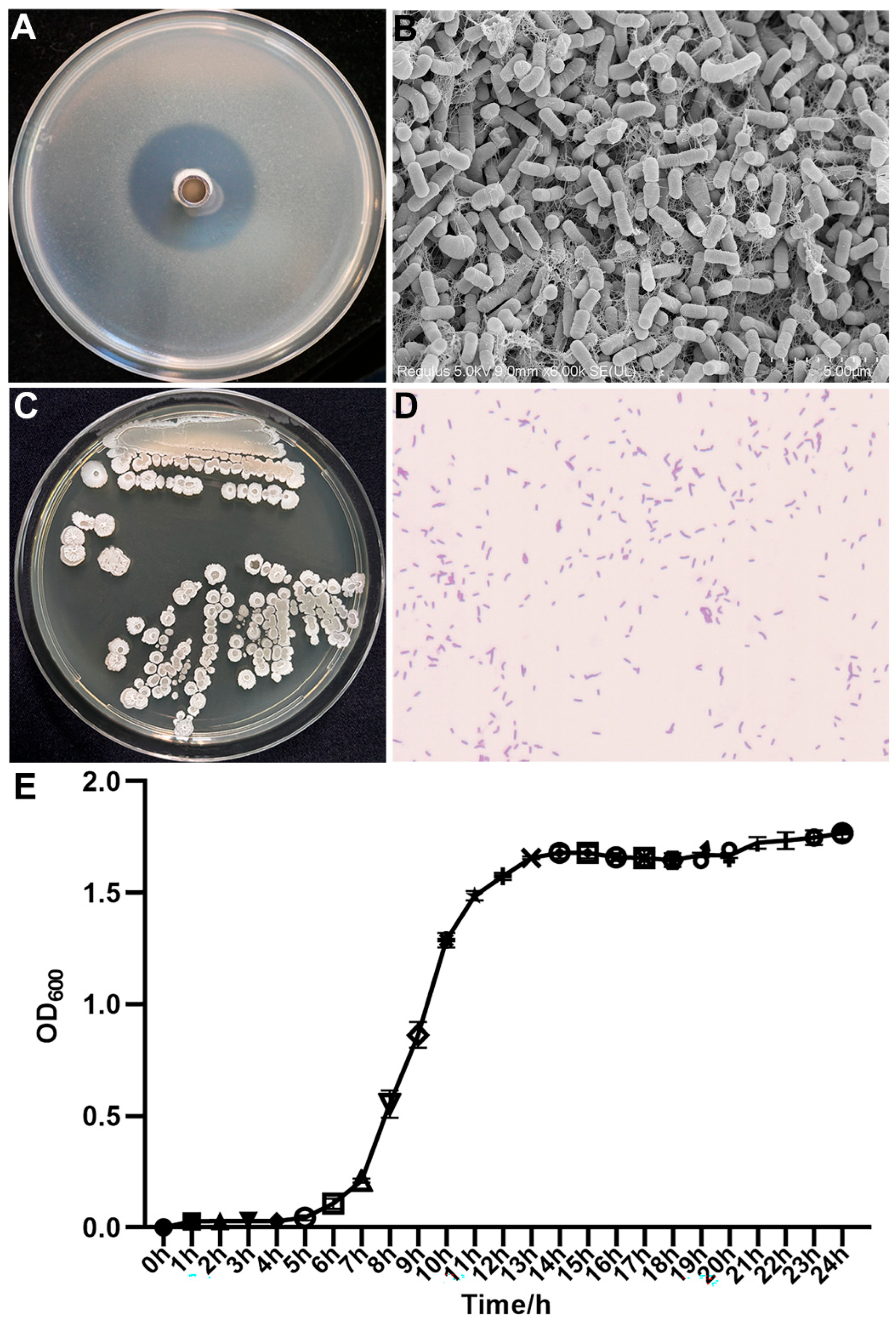
A collection of endophytic bacteria was isolated from rice leaves, and strain AN6 exhibited the strongest antagonistic activity against Xanthomonas oryzae pv. oryzae, the pathogen responsible for bacterial leaf blight (BLB) of rice, as determined by plate-based antagonism screening (Figure 1A). The inhibition rates of AN6 against six strains of X. oryzae pv. oryzae (X1, X2, X3, X12, LC2, and LC10) were 73.60%, 70.71%, 70.28%, 71.71%, 73.02%, and 72.86%, respectively (Figure 1B). Furthermore, assays related to plant probiotic traits demonstrated that isolated strain AN6 can secrete proteases and produce siderophores (Figure 1C). However, it lacks cellulase activity and the ability to degrade inorganic phosphorus and organic phosphorus (Supplementary Figure S1). This strain is currently preserved at the China Center for Type Culture Collection (CCTCC) under accession number CCTCC M 20231886.

3.2. Morphological and Biological Characteristics of the Isolated Strain AN6

Strain AN6 exhibited the strongest inhibitory effect against X. oryzae pv. oryzae (Xoo) (Figure 2A) and was therefore selected for morphological characterization. The single colony of AN6 on the NA medium showed round, white colonies with a rough surface (Figure 2C). The observation using scanning electron microscopy revealed that AN6 cells were rod-shaped with blunt ends, measuring approximately 1.55 μm × 0.62 μm on average (Figure 2B). Gram staining indicated that AN6 is a Gram-positive bacterium (Figure 2D). Growth curve analysis showed that AN6 exhibited its highest growth rate after 9–10 h of incubation at 30 °C with shaking at 160 rpm. After 13 h, the growth began to plateau, reaching a maximum optical density of approximately OD600 ≈ 1.80 (Figure 2E).

3.3. Antagonistic Activity of AN6 Strain Cell-Free Supernatant

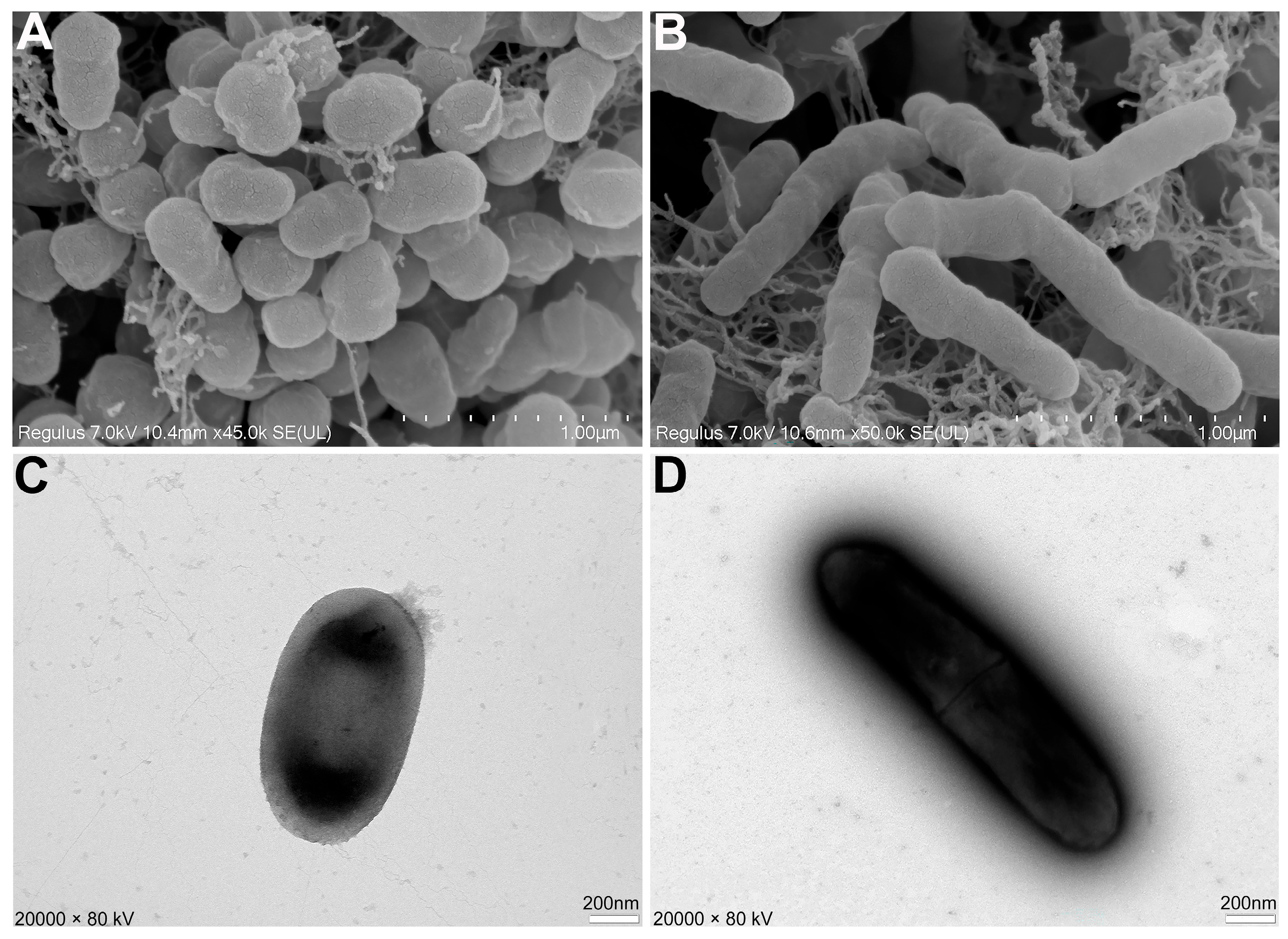
To further investigate the effect of AN6 on the morphology of X. oryzae pv. oryzae LC2, scanning electron microscopy (SEM) and transmission electron microscopy (TEM) were used to observe the morphological changes of Xoo LC2 cells under both normal culture conditions and stress induced by the sterile cell-free supernatant of Bacillus velezensis AN6. SEM analysis revealed that Xoo LC2 cells under normal conditions had smooth surfaces and maintained a typical rod-shaped morphology (Figure 3A). In contrast, cells cultured in the presence of AN6 sterile cell-free supernatant exhibited clear morphological abnormalities, including elongation and deformation, indicating membrane stress and structural damage (Figure 3B). The TEM observations supported these findings. Xoo LC2 cells grown under normal conditions displayed an intact ultrastructure with clearly defined cell walls and no observable abnormalities (Figure 3C). However, cells treated with the AN6 supernatant showed significant morphological alterations, including indistinct or disrupted cell wall structure and plasmolysis-like separation, suggesting severe physiological stress and, in some cases, cell lysis (Figure 3D). Together, these SEM and TEM results demonstrate that the sterile cell-free supernatant of Bacillus velezensis AN6 can significantly damage the cellular structure of Xoo LC2, resulting in pronounced morphological abnormalities and physiological dysfunction. These findings provide direct morphological evidence of the antagonistic activity of AN6.

3.4. Biocontrol Effect of AN6 Against Xoo

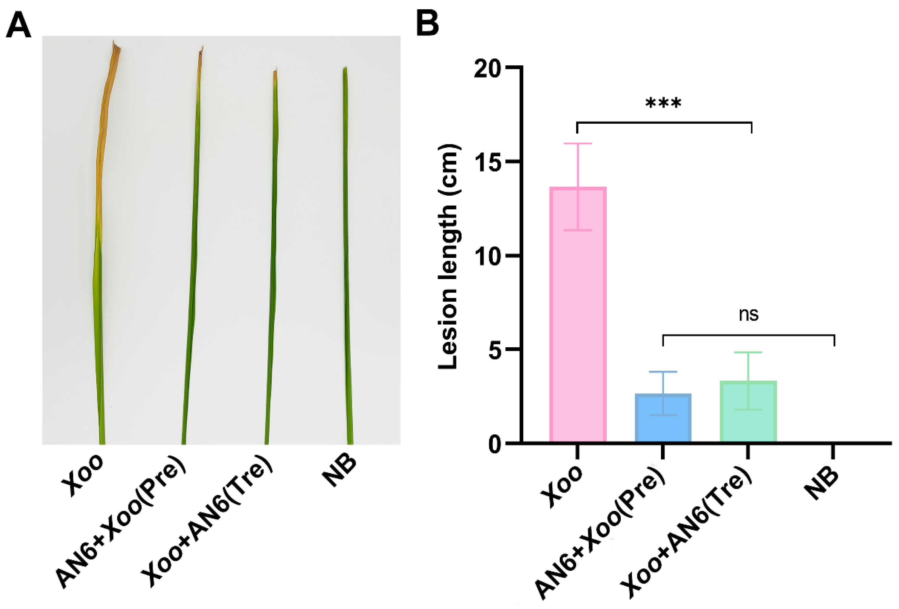
To assess the biocontrol potential of B. velezensis AN6 against X. oryzae pv. oryzae (Xoo), a leaf-clipping inoculation assay was conducted on potted rice plants in a light incubator. As shown in Figure 4, leaves in the control group, which were inoculated with Xoo alone, developed typical bacterial blight symptoms; the leaves appeared withered and had visible chlorosis, with an average lesion length of 13.67 cm. In the preventive treatment group, the leaves were pretreated with an AN6 bacterial suspension prior to Xoo inoculation; the severity of disease was significantly reduced, resulting in an average lesion length of only 2.67 cm. In the curative treatment group, where the AN6 suspension was applied after Xoo inoculation, lesion development was also substantially mitigated, with an average lesion length of 3.33 cm. Compared with the control, the AN6-Pre and AN6-Tre treatments dramatically reduced the severity of BB on YX650, with relative control efficiencies of 80.49% and 75.61%, respectively. These findings indicate that B. velezensis AN6 exhibits strong in vitro biocontrol efficacy against Xoo, and there was no significant difference between the AN6-Pre and AN6-Tre groups, which further highlights the practical flexibility of AN6, supporting its potential application as a biological agent for the management of rice bacterial blight.

3.5. Antifungal Activity of Strain AN6 In Vitro

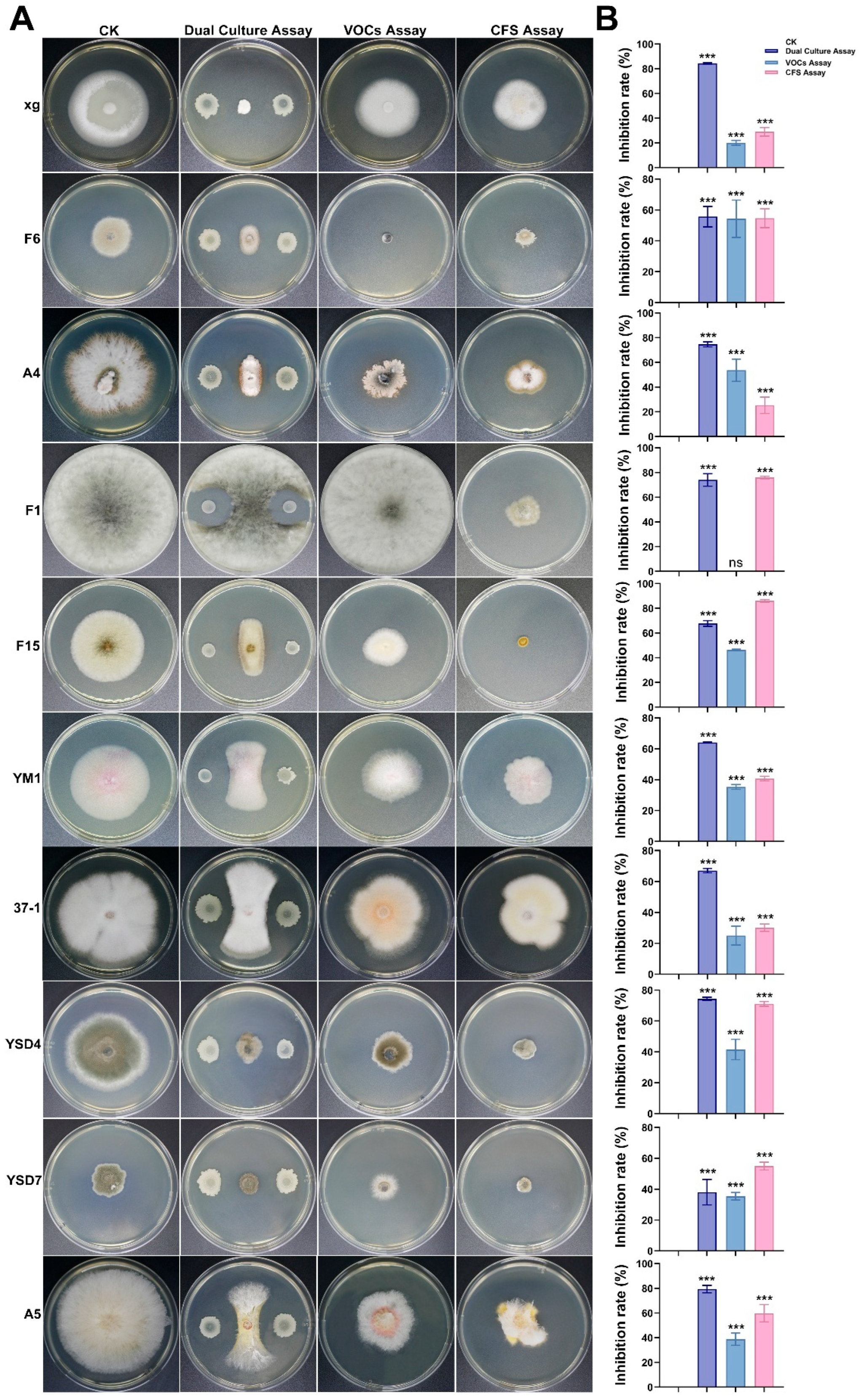
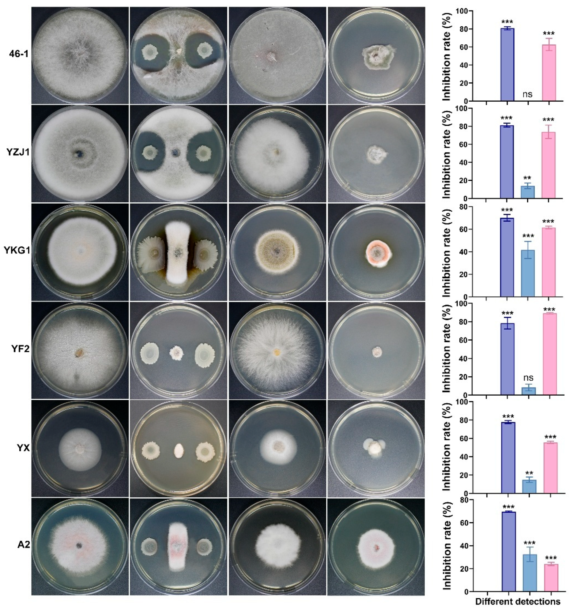
To evaluate the broad-spectrum antifungal activity of strain AN6, 16 phytopathogenic fungi were tested (Supplementary Table S1) and three distinct detection methods were employed. The results are shown in Figure 5. The dual-culture assay revealed that AN6 inhibited mycelial growth of these pathogens by 38.09% to 84.24%. Compared with the control group, AN6 exhibited an inhibitory activity of over 80% against M. oryzae xg, N. oryzae 46-1, and N. oryzae YZJ1. The maximum mycelial growth inhibition percentage was against M. oryzae xg (84.24 ± 1.05%), followed by N. oryzae YZJ1 (81.24 ± 2.10%). The minimum inhibition percentage was against B. cynodontis YSD7 (38.09 ± 2.82%), followed by A. tenuissima F6 (55.79 ± 2.27%) and F. oxysporum YM1 (64.07 ± 0.37%). The VOC assay showed that the inhibition rate of the VOCs produced by AN6 on the mycelial growth of pathogens ranged from 0 to 54.44%. The maximum mycelial growth inhibition percentage was against A. tenuissima F6 (54.44 ± 2.71%), followed by E. latusicollum A4 (53.21 ± 6.00%). The inhibition rate of the VOCs produced by AN6 against L. theobromae F1 and N. oryzae 46-1 mycelia was almost zero. The CFS assay showed that the inhibition rate of AN6 and its aseptic fermentation filtrate on the mycelial growth of the tested pathogens ranged from 25.46% to 89.17%. Compared with the control group, AN6 had an inhibitory activity of more than 80% against A. alternata F15 and N. sphaerica YF2. The maximum mycelial growth inhibition percentage was against N. sphaerica YF2 (89.17 ± 0.72%), followed by A. alternata F15 (86.27 ± 0.98%). The minimum inhibition percentage was against E. latusicollum A4 (25.26 ± 6.52%), followed by M. oryzae xg (29.09 ± 3.15%) and F. fujikuroi 37-1 (30.18 ± 2.34%). The results of the dual-culture, VOC, and CFS assays showed that AN6 had inhibitory effects on the 16 pathogenic fungi to varying degrees. Notably, AN6 displayed relatively strong inhibitory effects in the dual-culture and CFS assays, with maximum inhibition rates of 84.24% and 89.17%, respectively. These findings suggest that strain AN6 possesses substantial inhibitory potential against the 16 tested pathogenic fungi, highlighting its promise as a biocontrol candidate.

3.6. Complete Genome Sequence and Biosynthesis Gene Clusters of Strain AN6

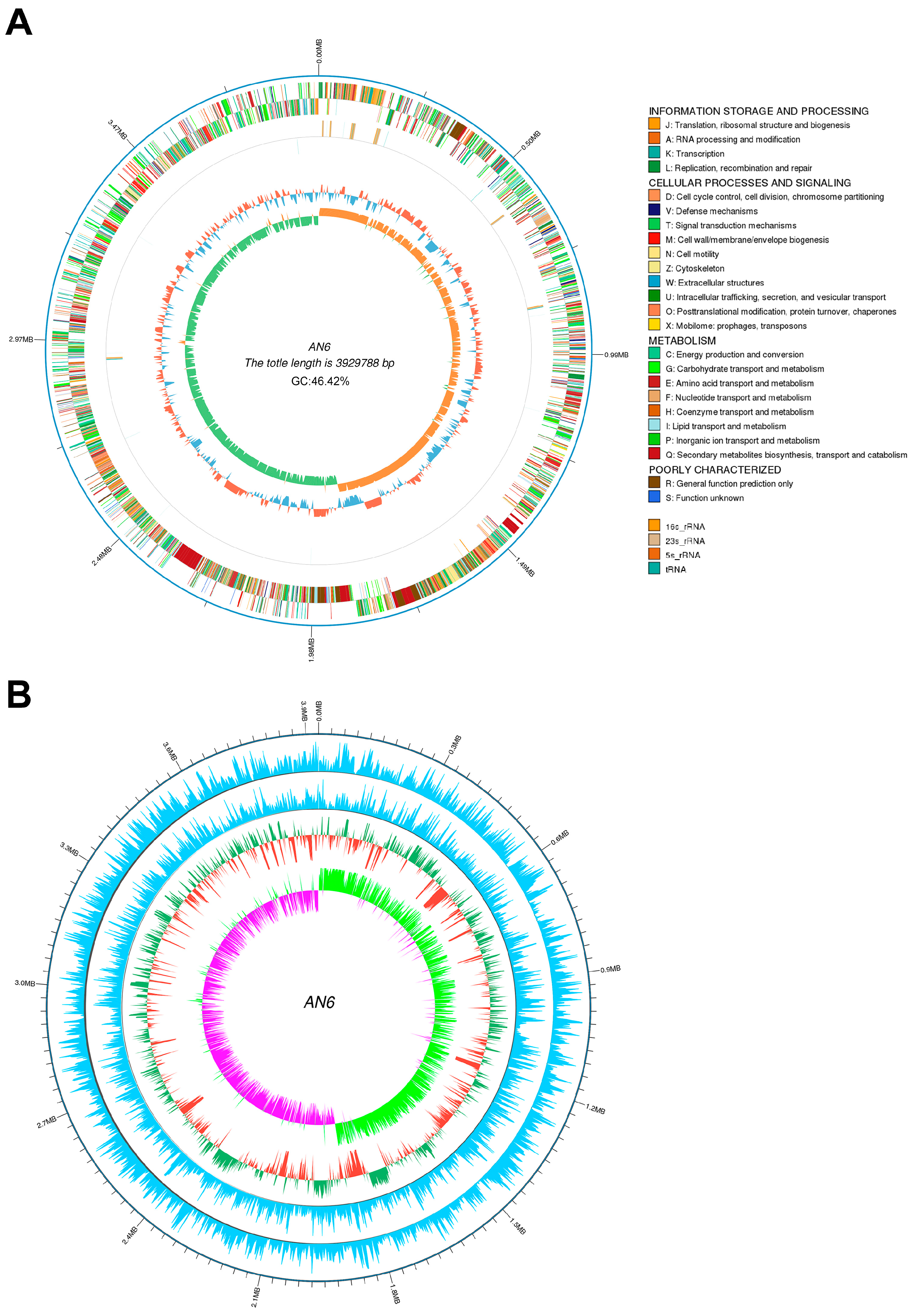
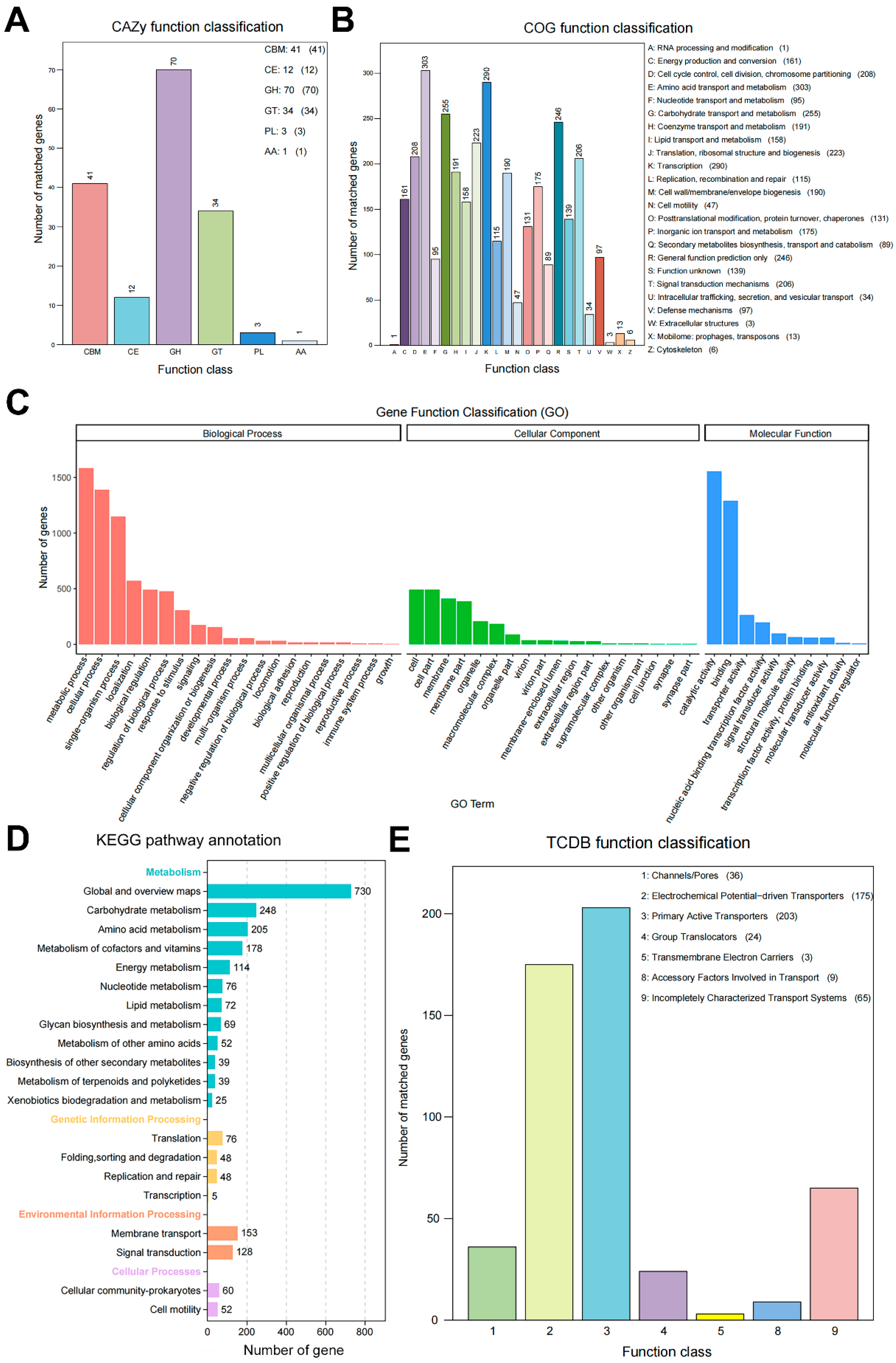
A total of 901,111 long reads were generated using PacBio sequencing, yielding 6,130,597,881 bases with an average read length of 6803 bp and an N50 of 9742 bp. These high-quality reads were used to assemble the genome of strain AN6. The final assembly resulted in a complete circular chromosome with 3,929,788 bp and a GC content of 46.50%. The circular chromosome map of AN6 is presented in Figure 6A. The assembled genome has been deposited in the NCBI database under accession number JBKKFY000000000. A genome-wide DNA methylation analysis of AN6 was performed and visualized as a circular map (Figure 6B). The AN6 genome was found to contain 2344 m4C modification sites, 1154 m5C sites, and 116 m6A sites. Genome annotation predicted 4025 protein-coding genes, 86 tRNAs, 27 rRNAs, 9 CRISPR arrays, 9 genomic islands, and 13 prophage regions. All the predicted protein sequences of AN6 were compared to those in the Nr, COG, GO, KEGG, TCDB, CAZy, Swiss-Prot and Pfam databases; the annotation summary is presented in Table 2. There are 3939 proteins in AN6 were classified into 20 Nr species (Table S2.1). A total of 2993 proteins in AN6 were classified into 24 COG families (Figure 7B, Table S2.2) and a total of 2714 genes were annotated with a GO function, which are categorized as biological process, cellular component, or molecular function (Figure 7C, Table S2.3). A total of 3875 genes were annotated using the KEGG database, which were distributed across 20 pathways in four major functional categories (Figure 7D, Table S2.4). Protein domains were identified in 2714 proteins (Table S2.5). A total of 3302 proteins were annotated using the Swiss-Prot database (Table S2.6). A total of 3875 genes were annotated using the TCDB database, which were distributed across seven major functional categories (Figure 7E, Table S2.7). A total of 156 genes had CAZy-annotated functions covering six major functional categories (Figure 7A, Table S2.8).

3.7. Comparative Genomics Analysis

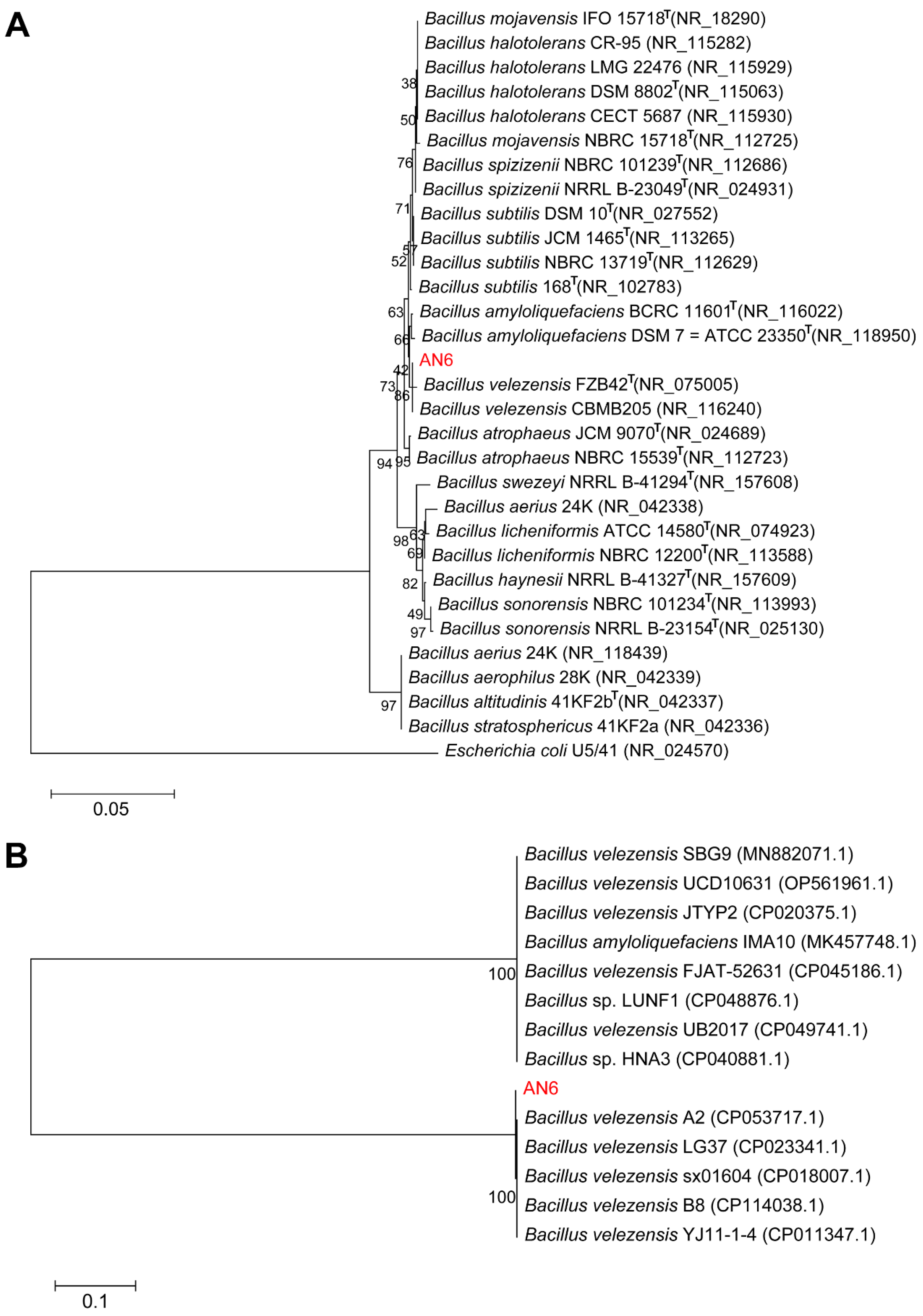
The 16S rRNA and gyrA genes of strain AN6 were amplified by PCR and subsequently sequenced. BLAST analysis revealed that the 16S rRNA gene sequence of AN6 shared the highest similarity with that of B. velezensis CBMB205 (NR_116240) and B. velezensis FZB42 (NR_075005), with sequence identities of 99.93% and 99.86%, respectively. Based on these results, a phylogenetic tree was constructed using the maximum likelihood method. The resulting tree showed that AN6 clustered within the same evolutionary branch as B. velezensis FZB42 and CBMB205 (Figure 8A). To further resolve the phylogenetic placement of AN6 among Bacillus species, phylogenetic analysis was performed based on the gyrA gene. As shown in Figure 8B, AN6 was most closely related to B. velezensis strains A2, LG37, sx01604, B8, and YJ11-1-4. These combined results clearly support the classification of strain AN6 as a member of Bacillus velezensis.
Furthermore, average nucleotide identity (ANI) and in silico DNA–DNA hybridization (isDDH) analyses were conducted between strain AN6 and 16 publicly available Bacillus strain genomes. Based on the established species delineation thresholds of ANI ≥ 96% and isDDH ≥ 70%, the results (Figure 9) were consistent with the phylogenetic relationships observed in Figure 7. Strain AN6 exhibited ANI values of 98.34%, 98.32%, 98.33%, 99.99%, 99.99%, 98.31%, and 97.77% with B. velezensis strains FZB42T (type strain), MEP218, 9D-6, LG37, YB-130, CACC 316, and B1, respectively. The corresponding isDDH values were 85.40%, 84.90%, 84.70%, 100%, 100%, 84.60%, and 80.10%. All values exceeded the established thresholds for species-level classification, further confirming that strain AN6 is a strain of Bacillus velezensis.
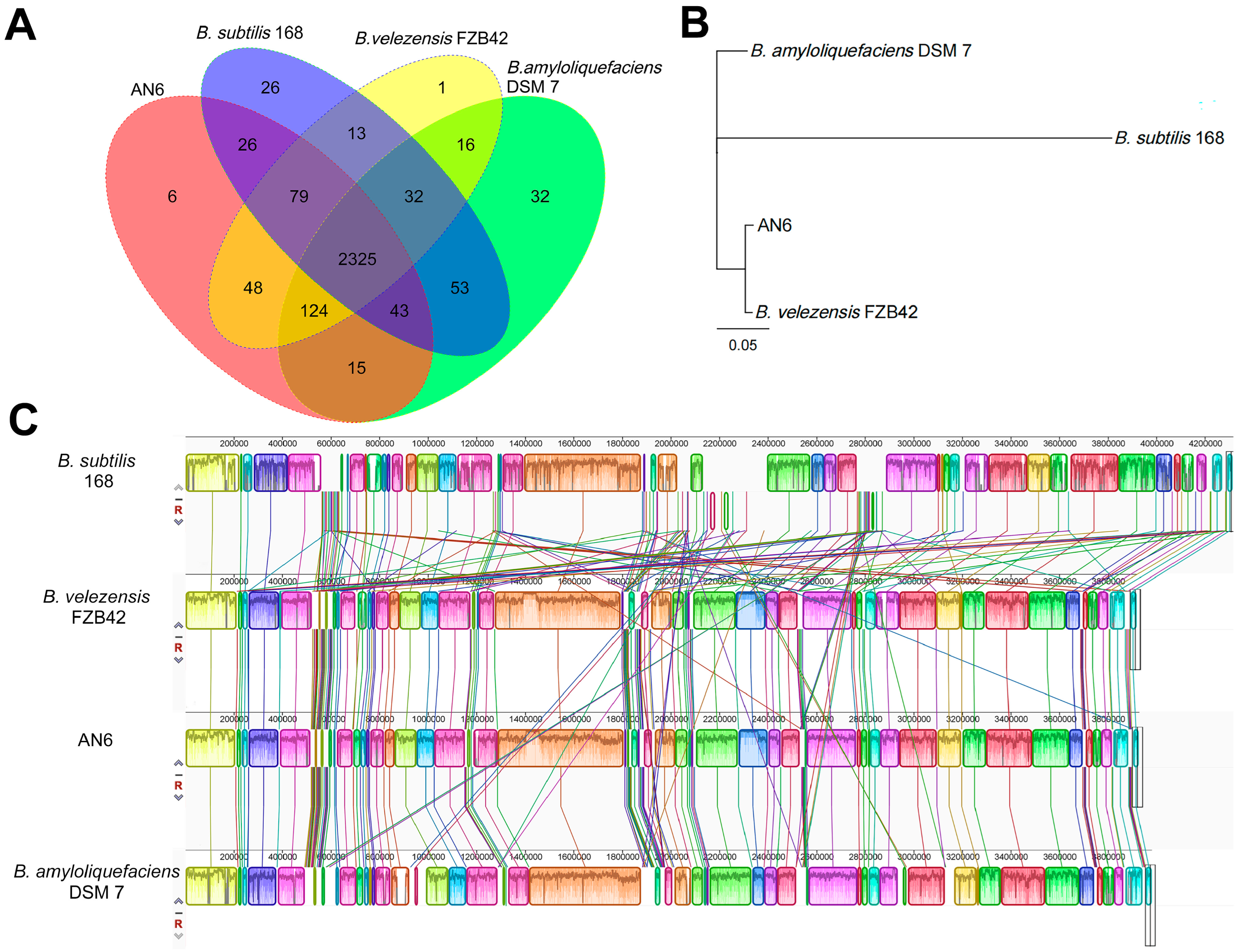
Gene family clustering analysis was performed on B. velezensis AN6, B. velezensis FZB42, B. subtilis 168, and B. amyloliquefaciens DSM 7. The results showed that strain AN6 contained 2666 gene families, including 6 unique gene families. B. velezensis FZB42, B. subtilis 168, and B. amyloliquefaciens DSM 7 possessed 2638, 2597, and 2640 gene families, with 1, 26, and 31 unique gene families, respectively. A total of 2325 gene families were conserved across all four strains, accounting for 87.21%, 88.13%, 89.53%, and 88.07% of the total gene families in AN6, FZB42, 168, and DSM 7, respectively (Figure 10A). Based on the single-copy orthologous genes identified through gene family clustering, multiple-protein sequence alignment was performed using MUSCLE, and a phylogenetic tree was constructed using the maximum likelihood method in PHYML. The phylogenetic tree showed that AN6 clustered closely with B. velezensis FZB42, indicating a close evolutionary relationship between the two strains (Figure 10B). Whole-genome synteny analysis between AN6 and B. velezensis FZB42, B. subtilis 168, and B. amyloliquefaciens DSM 7 revealed a high degree of gene homology and overall collinearity, particularly between AN6 and B. velezensis FZB42. Nevertheless, several genomic rearrangements were observed, including gene inversions, transpositions, translocations, and deletions (Figure 10C). These findings suggest that while the core genome is largely conserved across these Bacillus species, structural variations have contributed to their genomic divergence.
Secondary metabolite biosynthetic gene clusters in the AN6 genome were predicted using antiSMASH v8.0.4. The analysis revealed a total of 13 biosynthetic gene clusters (Table 3), including those responsible for the nonribosomal synthesis of lipopeptides (surfactin, fengycin, and bacillibactin), polyketides (difficidin, bacillaene, and macrolactin H), and a dipeptide antibiotic (bacilysin). In addition, six gene clusters were associated with the biosynthesis of unknown terpenes, polyketides, and class II lanthipeptides.

4. Discussion

Rice is one of the most important staple crops worldwide, and various pathogens such as X. oryzae pv. oryzae [56], M. oryzae [57], N. oryzae [58], Nigrospora spp. [59], Epicoccum spp. [60], Bipolaris spp. [61], Alternaria spp. [62], and Fusarium spp. [63] are responsible for major constraints on its cultivation and productivity. Plant diseases caused by phytopathogenic bacteria and fungi seriously threaten global rice production, leading to significant yield and economic losses. Biocontrol strategies provide environmentally friendly solutions to manage plant diseases, decreasing the dependence on harmful chemical treatments. In recent decades, biological control has emerged as a promising and eco-friendly strategy for plant disease management due to its high efficiency, broad-spectrum antagonistic effects, and sustainability compared to chemical pesticides [64,65]. Previous studies have demonstrated that beneficial microorganisms such as Bacillus, Pseudomonas, and Streptomyces can suppress diverse plant diseases through mechanisms including antibiotic production, secretion of lytic enzymes, and induction of systemic resistance [66]. In the present study, we successfully isolated a novel Bacillus velezensis strain AN6 from rice plants, which exhibited strong antagonistic activity against X. oryzae pv. oryzae as well as 16 other fungi in vitro. Therefore, the complete genome of B. velezensis AN6 was sequenced for detailed analysis.
Numerous studies have reported that B. velezensis strains, as novel biocontrol agents, exhibit significant potential in the management of various plant diseases [67,68]. In addition, the use of B. velezensis in biocontrol research has become increasingly widespread. In particular, B. velezensis strain CC09 has been demonstrated to effectively control wheat powdery mildew [69], B. velezensis AL7 possesses biocontrol potential against cotton Verticillium wilt [70], B. velezensis SF334 exhibits significant antagonistic activity against rubber tree leaf anthracnose [71], B. velezensis BRI3 can effectively antagonize Sclerotinia sclerotiorum (Lib.) [72], and B. velezensis Y6 has been shown to suppress rice sheath blight caused by R. solani [73]. These findings offer a more environmentally friendly method for managing diseases. In our study, strain AN6 exhibited strong inhibitory activity against X. oryzae pv. oryzae (Figure 1), significantly suppressing Xoo growth (Figure 3) and demonstrating effective biocontrol in pot assays (Figure 4). Its CFS caused severe morphological damage to Xoo, likely mediated by lipopeptides, polyketides, or other extracellular metabolites, as supported by genome-encoded secondary metabolite biosynthetic gene clusters (Table 2). This is consistent with previous studies [41], where the CFS of strain Bv-303 significantly inhibited Xoo growth in vitro, and in vivo applications of the cell-culture broth (CCB), CFS, or cell-suspension water (CSW) enhanced the resistance of rice plants to bacterial blight (BB). To further elucidate the key active compounds, future work could combine LC–MS/MS metabolite profiling, bioassay-guided fractionation, and targeted gene-cluster validation. The dual-culture assay can assess direct antagonism, the volatile organic compound assay can assess indirect antagonism, and the sterile fermentation filtrate assay can assess the ability of the strain to produce antimicrobial metabolites. The results showed that AN6 exhibited antimicrobial activity in all three assays, but the inhibitory activity against different fungal pathogens varied between the assays. The dual-culture assay and the sterile fermentation filtrate assay demonstrated the strongest inhibitory activity, with the highest inhibition rates reaching 84.24 ± 1.05% and 89.17 ± 0.72%, respectively. Comparing these two methods, we found that for most fungal pathogens, AN6 exhibited the strongest antifungal activity in the dual-culture assay. For fast-growing fungal pathogens (L. theobromae F1, N. oryzae 46-1, and N. oryzae YZJ1), the dual-culture assay showed only moderate inhibition, whereas the sterile fermentation filtrate assay demonstrated stronger inhibition (Figure 5). This result supports the possibility that, for certain fungal pathogens, direct cell-to-cell interaction and contact-dependent induction of antimicrobial metabolites may be more important than constitutively secreted soluble compounds. Similar “interaction-dependent induction” of lipopeptides and other antifungal secondary metabolites has been reported in antagonistic Bacillus strains. The presence of Fusarium oxysporum f. sp. strigae (Fos) in dual-plate cultures led to an increase in bacillomycin D production by Bacillus strains [74]. This suggests that, for different fungal pathogens, targeted control strategies may be implemented based on the specific antagonistic mechanisms of the biocontrol agent. Compared to some microbial strains that have shown potential in inhibiting M. oryzae in many studies in dual-culture assays, the efficacy of AN6 against M. oryzae is equivalent to or slightly higher. For example, Wang et al. reported that B. velezensis LJBV19 showed significant inhibitory effects on the growth of M. oryzae with an inhibition ratio of 75.55%, while Wockenfuss et al. reported that B. velezensis S4 significantly inhibited the growth of M. oryzae by 38.53% [75,76]. Similarly, compared with the inhibitory effect of B. velezensis B31 on N. oryzae [77], the inhibitory effect of AN6 was relatively higher. It has been reported that different Bacillus strains have inhibitory effects on different Fusarium species [78,79,80]. This study found that AN6 effectively inhibits at least five Fusarium species. B. oryzae is the main pathogen that causes brown spot disease of rice (BSR) [81]. Strain AN6 exhibited similar in vitro inhibitory activity against B. oryzae as B. velezensis LS123N [82]. N. oryzae is the pathogen that causes rice stem rot [58] and B. cynodontis is the fungus responsible for leaf blight [83,84]. To our knowledge, this is the first report of the antagonistic activity of B. velezensis against B. cynodontis and N. oryzae.
Numerous studies have shown that B. velezensis produces VOCs with strong inhibitory effects on bacterial and plant pathogen growth, such as the VOCs from B. velezensis ZSY-1, which significantly suppressed six types of plant pathogenic fungi [85], and the VOCs released by B. velezensis T9, which inhibited the growth of Apiospora mold on sugarcane caused by A. arundinis [86]. The CFS of B. velezensis exhibited significant antifungal effects by inhibiting spore germination. The CFS of B. velezensis exhibited potent antifungal activity against B. cinerea and P. olsonii in vitro. It hindered spore germination, germ tube elongation, and hyphal growth, and damaged mycelial cells by triggering excessive ROS accumulation [87,88].
The phylogenetic tree analysis using 16S rRNA gene sequences showed that the AN6 strain clusters closely with B. velezensis FZB42 and B. velezensis CBMB205 (Figure 8A). The phylogenetic tree analysis using gyrA gene sequences showed that the AN6 strain clusters closely with B. velezensis A2, B. velezensis LG37, B. velezensis sx01604, B. velezensis B8, and B. velezensis YJ11-1-4 (Figure 8B), indicating a close relationship between AN6 and these strains. The ANI and isDDH analyses revealed that AN6 could be classified as B. velezensis due to the high degree of genomic homogeneity between AN6 and B. velezensis (Figure 9). Gene family clustering revealed that AN6 shares 2325 gene families with B. velezensis FZB42, B. subtilis 168, and B. amyloliquefaciens DSM 7, and contains 6 unique gene families (Figure 10A). The phylogenetic analysis based on single-copy orthologous genes indicated that strain AN6 and B. velezensis FZB42 belong to the same evolutionary lineage (Figure 10B). The whole-genome collinearity analysis of AN6, B. velezensis FZB42, B. subtilis 168, and B. amyloliquefaciens DSM 7 demonstrated a high degree of gene homology and overall collinearity, with the similarity between AN6 and B. velezensis FZB42 being particularly pronounced (Figure 10C). However, several genomic rearrangements were also observed, including inversions, transpositions, translocations, and deletions.
Several studies indicated that the effectiveness of Bacillus strains as biocontrol agents relies on both their ability to colonize target tissues and the presence of a complete, functional biosynthetic gene cluster for producing and secreting secondary metabolites such as iturin, bacillomycin, fengycin, and surfactin [89,90,91,92]. Genome mining identified a total of thirteen secondary metabolite biosynthesis gene clusters in the genome of strain AN6. Seven clusters showed significant similarity (similarity ≥ 75%) to known BGCs, including those for the synthesis of surfactin, macrolactin H, bacillaene, fengycin, difficidin, bacillibactin, and bacilysin (Table 3). Surfactin is a natural surfactant that is primarily produced by Bacillus species and exhibits antibacterial properties. Macrolactin H is a member of a class of macrolide natural products that generally exhibits multiple biological activities, including antibacterial, antifungal, antiviral, antitumor, and anti-inflammatory activities [93]. Bacillaene is a linear polyketide/nonribosomal peptide produced by Bacillus species through the activity of trans-acyltransferase polyketide synthetase [94]. Fengycin is a cyclic lipopeptide antibiotic that is primarily produced by Bacillus species and has demonstrated significant antifungal, antitumor, and antiadhesion properties; the antifungal mechanisms of fengycin mainly involve disruption of cell wall integrity, damage to cell membranes, interference with intracellular metabolism, induction of programmed cell death and autophagy, and activation of plant defense responses [95]. Fengycin is likely a major contributor to the antifungal activity of AN6. Difficidin is a polyketide antibiotic produced by Bacillus species via the trans-acyltransferase polyketide synthase pathway [96]. Its mode of action involves inhibition of bacterial protein synthesis and disruption of key cellular metabolic processes. Previous studies have shown that difficidin exhibits potent bactericidal activity against both Gram-positive and Gram-negative pathogens, including plant-pathogenic Xanthomonas spp. and Pseudomonas spp., and can cause cell-wall and membrane destruction and suppress the expression of bacterial virulence genes [97]. Therefore, we preliminarily infer that difficidin is likely one of the major contributors to the antibacterial activity of AN6. Bacillibactin is a microbial siderophore produced by Bacillus species [98]. Bacilysin is a dipeptide antibiotic made up of L-alanine and L-anticapsin and is produced by certain strains of Bacillus species. It is attracting growing interest in industrial agriculture and the pharmaceutical industry due to its strong antagonistic effects on various bacterial, fungal, and algal pathogens [99]. Notably, six of AN6’s BGCs showed minimal similarity to known clusters, indicating that strain AN6 contains genes with potentially novel functions, underscoring its significant research potential.
Rice diseases such as bacterial blight caused by X. oryzae pv. oryzae and rice blast caused by M. oryzae remain major threats to rice production. Although chemical fungicides are widely used, sustainable agriculture requires alternative strategies. In this study, strain AN6 exhibited strong antibacterial activity against X. oryzae pv. oryzae, causing severe cell deformation in vitro and significantly reducing disease severity in planta. Furthermore, AN6 displayed antagonistic activity against M. oryzae and other fungal pathogens in plate confrontation, VOC, and soluble metabolite assays. This work highlights a methodological advance by integrating three evaluation approaches within one framework, enabling a more comprehensive assessment of biocontrol potential. Such an integrative strategy overcomes the limitations of single-method studies and better reflects natural microbial interactions. Mechanistically, AN6 suppresses pathogens through multiple complementary strategies, including direct cellular contact, secreted extracellular metabolites, and VOCs, with the inhibitory effects varying depending on the pathogen and assay. Genome analysis revealed 13 secondary metabolite biosynthetic gene clusters, including high-confidence clusters for difficidin, fengycin, and surfactin, which likely underlie the observed antibacterial and antifungal activities. Together, the genomic and phenotypic analyses highlight the multifaceted antagonistic potential of AN6. To our knowledge, this study is the first to demonstrate the biocontrol potential of B. velezensis against Bipolaris cynodontis and Nakataea oryzae, expanding the known spectrum of target pathogens. The AN6 strain shows significant potential as a biocontrol agent for the prevention and control of plant diseases.

5. Conclusions

In summary, B. velezensis strain AN6 isolated from rice plants exhibited strong biocontrol activity against X. oryzae pv. oryzae and inhibited multiple phytopathogenic fungi. AN6 can produce proteases, siderophores, antimicrobial secondary metabolites, and VOCs. The whole-genome analysis and secondary metabolite gene cluster prediction provided important clues for future studies on the regulatory mechanisms of its antimicrobial metabolites. To our knowledge, this is the first report demonstrating the biocontrol potential of B. velezensis against Bipolaris cynodontis and Nakataea oryzae. Overall, AN6 shows considerable promise as a biocontrol agent for plant disease management and represents a valuable microbial resource for discovering and exploiting novel antimicrobial compounds.

Supplementary Materials

The following supporting information can be downloaded at: https://www.mdpi.com/article/10.3390/microorganisms13122701/s1, Figure S1: Experimental results for AN6 on the degradation of cellulose, organic phosphorus solubilization, and inorganic phosphorus solubilization; Table S1: Phytopathogenic fungi used in this study (for the in vitro antifungal activity analyses of strain AN6); Table S2: The annotation results of the coding genes in the genome of strain AN6.

Author Contributions

Conceptualization: L.Y. and X.L.; Methodology: L.Y. and X.L.; Software: L.Y., A.G. and W.D. (Wei Deng); Validation: L.Y. and W.D. (Wei Dong); Formal Analysis: L.Y., S.C. and J.Z. (Jinwen Zhang); Investigation: L.Y., J.G. and L.K.; Resources: H.A., J.T., J.F. and X.L.; Data Curation: L.Y.; Writing—Original Manuscript Preparation: L.Y.; Writing—Review and Editing: L.Y., J.F. and X.L.; Visualization: L.Y.; Supervision: X.L.; Project Administration: J.Z. (Jianhua Zhang), X.L. and X.S.; Funding Acquisition: X.L. All authors have read and agreed to the published version of the manuscript.

Funding

This work was supported by the Yunnan Academy of Agricultural Sciences (Grant No. 2024KYZX-06), the Yunnan Provincial Science and Technology Department, China (Grant Nos. 202402AE090036, 202402AE090010, and 202301BD070001-224), the National Key Research and Development Program of China (Grant Nos. 2024YFD1200904 and 2024YFD1200902), the Yunnan Provincial Government (530000210000000013809).

Institutional Review Board Statement

Not applicable.

Informed Consent Statement

Not applicable.

Data Availability Statement

Sequence data presented in this study are available in GenBank at https://www.ncbi.nlm.nih.gov/datasets/genome/ (accessed on 30 November 2024) with under the accession numbers listed in the Results Section. Other relevant data are included in the article/Supplementary Materials.

Conflicts of Interest

The authors declare no conflicts of interest.

References

  1. Zeng, D.; Tian, Z.; Rao, Y.; Dong, G.; Yang, Y.; Huang, L.; Leng, Y.; Xu, J.; Sun, C.; Zhang, G.; et al. Rational design of high-yield and superior-quality rice. Nat. Plants 2017, 3, 17031. [Google Scholar] [CrossRef] [PubMed]
  2. Zeigler, R.S.; Barclay, A. The Relevance of Rice. Rice 2008, 1, 3–10. [Google Scholar] [CrossRef]
  3. Niño-Liu, D.O.; Ronald, P.C.; Bogdanove, A.J. Xanthomonas oryzae pathovars: Model pathogens of a model crop. Mol. Plant Pathol. 2006, 7, 303–324. [Google Scholar] [CrossRef] [PubMed]
  4. Mansfield, J.; Genin, S.; Magori, S.; Citovsky, V.; Sriariyanum, M.; Ronald, P.; Dow, M.; Verdier, V.; Beer, S.V.; Machado, M.A.; et al. Top 10 plant pathogenic bacteria in molecular plant pathology. Mol. Plant Pathol. 2012, 13, 614–629. [Google Scholar] [CrossRef]
  5. Kou, Y.; Wang, S. Broad-spectrum and durability: Understanding of quantitative disease resistance. Curr. Opin. Plant Biol. 2010, 13, 181–185. [Google Scholar] [CrossRef]
  6. Ramalingam, J.; Savitha, P.; Alagarasan, G.; Saraswathi, R.; Chandrababu, R. Functional Marker Assisted Improvement of Stable Cytoplasmic Male Sterile Lines of Rice for Bacterial Blight Resistance. Front. Plant Sci. 2017, 8, 1131. [Google Scholar] [CrossRef]
  7. Tian, Y.; Zhao, Y.; Xu, R.; Liu, F.; Hu, B.; Walcott, R.R. Simultaneous Detection of Xanthomonas oryzae pv. oryzae and X. oryzae pv. oryzicola in Rice Seed Using a Padlock Probe-Based Assay. Phytopathology 2014, 104, 1130–1137. [Google Scholar] [CrossRef]
  8. Namburi, K.R.; Kora, A.J.; Chetukuri, A.; Kota, V. Biogenic silver nanoparticles as an antibacterial agent against bacterial leaf blight causing rice phytopathogen Xanthomonas oryzae pv. oryzae. Bioprocess. Biosyst. Eng. 2021, 44, 1975–1988. [Google Scholar] [CrossRef]
  9. Gerhardson, B. Biological substitutes for pesticides. Trends Biotechnol. 2002, 20, 338–343. [Google Scholar] [CrossRef]
  10. Wu, L.; Wu, H.J.; Qiao, J.; Gao, X.; Borriss, R. Novel Routes for Improving Biocontrol Activity of Bacillus Based Bioinoculants. Front. Microbiol. 2015, 6, 1395. [Google Scholar] [CrossRef]
  11. Choudhary, D.K.; Johri, B.N. Interactions of Bacillus spp. and plants—With special reference to induced systemic resistance (ISR). Microbiol. Res. 2009, 164, 493–513. [Google Scholar] [CrossRef]
  12. de Boer, M.; Bom, P.; Kindt, F.; Keurentjes, J.J.; van der Sluis, I.; van Loon, L.C.; Bakker, P.A. Control of Fusarium Wilt of Radish by Combining Pseudomonas putida Strains that have Different Disease-Suppressive Mechanisms. Phytopathology 2003, 93, 626–632. [Google Scholar] [CrossRef]
  13. McSpadden Gardener, B.B. Ecology of Bacillus and Paenibacillus spp. in Agricultural Systems. Phytopathology 2004, 94, 1252–1258. [Google Scholar] [CrossRef]
  14. Kloepper, J.W.; Ryu, C.M.; Zhang, S. Induced Systemic Resistance and Promotion of Plant Growth by Bacillus spp. Phytopathology 2004, 94, 1259–1266. [Google Scholar] [CrossRef] [PubMed]
  15. Koumoutsi, A.; Chen, X.H.; Henne, A.; Liesegang, H.; Hitzeroth, G.; Franke, P.; Vater, J.; Borriss, R. Structural and functional characterization of gene clusters directing nonribosomal synthesis of bioactive cyclic lipopeptides in Bacillus amyloliquefaciens strain FZB42. J. Bacteriol. 2004, 186, 1084–1096. [Google Scholar] [CrossRef] [PubMed]
  16. Wang, G.F.; Meng, J.F.; Tian, T.; Xiao, X.Q.; Zhang, B.; Xiao, Y.N. Endophytic Bacillus velezensis strain B-36 is a potential biocontrol agent against lotus rot caused by Fusarium oxysporum. J. Appl. Microbiol. 2020, 128, 1153–1162. [Google Scholar] [CrossRef] [PubMed]
  17. Ji, S.H.; Paul, N.C.; Deng, J.X.; Kim, Y.S.; Yun, B.S.; Yu, S.H. Biocontrol Activity of Bacillus amyloliquefaciens CNU114001 against Fungal Plant Diseases. Mycobiology 2013, 41, 234–242. [Google Scholar] [CrossRef] [PubMed]
  18. Masum, M.M.I.; Liu, L.; Yang, M.; Hossain, M.M.; Siddiqa, M.M.; Supty, M.E.; Ogunyemi, S.O.; Hossain, A.; An, Q.; Li, B. Halotolerant bacteria belonging to operational group Bacillus amyloliquefaciens in biocontrol of the rice brown stripe pathogen Acidovorax oryzae. J. Appl. Microbiol. 2018, 125, 1852–1867. [Google Scholar] [CrossRef]
  19. Xu, Z.; Zhang, R.; Wang, D.; Qiu, M.; Feng, H.; Zhang, N.; Shen, Q. Enhanced control of cucumber wilt disease by Bacillus amyloliquefaciens SQR9 by altering the regulation of Its DegU phosphorylation. Appl. Environ. Microbiol. 2014, 80, 2941–2950. [Google Scholar] [CrossRef]
  20. Chung, S.; Kong, H.; Buyer, J.S.; Lakshman, D.K.; Lydon, J.; Kim, S.D.; Roberts, D.P. Isolation and partial characterization of Bacillus subtilis ME488 for suppression of soilborne pathogens of cucumber and pepper. Appl. Microbiol. Biotechnol. 2008, 80, 115–123. [Google Scholar] [CrossRef]
  21. Jia, K.; Gao, Y.-H.; Huang, X.-Q.; Guo, R.-J.; Li, S.-D. Rhizosphere Inhibition of Cucumber Fusarium Wilt by Different Surfactin- excreting Strains of Bacillus subtilis. Plant Pathol. J. 2015, 31, 140–151. [Google Scholar] [CrossRef] [PubMed]
  22. Nikolić, I.; Berić, T.; Dimkić, I.; Popović, T.; Lozo, J.; Fira, D.; Stanković, S. Biological control of Pseudomonas syringae pv. aptata on sugar beet with Bacillus pumilus SS-10.7 and Bacillus amyloliquefaciens (SS-12.6 and SS-38.4) strains. J. Appl. Microbiol. 2019, 126, 165–176. [Google Scholar] [CrossRef] [PubMed]
  23. Ryu, C.M.; Hu, C.H.; Reddy, M.S.; Kloepper, J.W. Different signaling pathways of induced resistance by rhizobacteria in Arabidopsis thaliana against two pathovars of Pseudomonas syringae. New Phytol. 2003, 160, 413–420. [Google Scholar] [CrossRef] [PubMed]
  24. Ruiz-García, C.; Béjar, V.; Martínez-Checa, F.; Llamas, I.; Quesada, E. Bacillus velezensis sp. nov., a surfactant-producing bacterium isolated from the river Vélez in Málaga, southern Spain. Int. J. Syst. Evol. Microbiol. 2005, 55, 191–195. [Google Scholar] [CrossRef]
  25. Chen, X.H.; Koumoutsi, A.; Scholz, R.; Borriss, R. More than anticipated—Production of antibiotics and other secondary metabolites by Bacillus amyloliquefaciens FZB42. J. Mol. Microbiol. Biotechnol. 2009, 16, 14–24. [Google Scholar] [CrossRef]
  26. Toral, L.; Rodríguez, M.; Béjar, V.; Sampedro, I. Crop Protection against Botrytis cinerea by Rhizhosphere Biological Control Agent Bacillus velezensis XT1. Microorganisms 2020, 8, 992. [Google Scholar] [CrossRef]
  27. Balderas-Ruíz, K.A.; Bustos, P.; Santamaria, R.I.; González, V.; Cristiano-Fajardo, S.A.; Barrera-Ortíz, S.; Mezo-Villalobos, M.; Aranda-Ocampo, S.; Guevara-García, Á.A.; Galindo, E.; et al. Bacillus velezensis 83 a bacterial strain from mango phyllosphere, useful for biological control and plant growth promotion. AMB Express 2020, 10, 163. [Google Scholar] [CrossRef]
  28. Choub, V.; Ajuna, H.B.; Won, S.J.; Moon, J.H.; Choi, S.I.; Maung, C.E.H.; Kim, C.W.; Ahn, Y.S. Antifungal Activity of Bacillus velezensis CE 100 against Anthracnose Disease (Colletotrichum gloeosporioides) and Growth Promotion of Walnut (Juglans regia L.) Trees. Int. J. Mol. Sci. 2021, 22, 10438. [Google Scholar] [CrossRef]
  29. Choub, V.; Maung, C.E.H.; Won, S.J.; Moon, J.H.; Kim, K.Y.; Han, Y.S.; Cho, J.Y.; Ahn, Y.S. Antifungal Activity of Cyclic Tetrapeptide from Bacillus velezensis CE 100 against Plant Pathogen Colletotrichum gloeosporioides. Pathogens 2021, 10, 209. [Google Scholar] [CrossRef]
  30. Kim, Y.S.; Lee, Y.; Cheon, W.; Park, J.; Kwon, H.T.; Balaraju, K.; Kim, J.; Yoon, Y.J.; Jeon, Y. Characterization of Bacillus velezensis AK-0 as a biocontrol agent against apple bitter rot caused by Colletotrichum gloeosporioides. Sci. Rep. 2021, 11, 626. [Google Scholar] [CrossRef]
  31. Yan, H.; Qiu, Y.; Yang, S.; Wang, Y.; Wang, K.; Jiang, L.; Wang, H. Antagonistic Activity of Bacillus velezensis SDTB038 against Phytophthora infestans in Potato. Plant Dis. 2021, 105, 1738–1747. [Google Scholar] [CrossRef]
  32. Chen, L.; Wang, X.; Ma, Q.; Bian, L.; Liu, X.; Xu, Y.; Zhang, H.; Shao, J.; Liu, Y. Bacillus velezensis CLA178-Induced Systemic Resistance of Rosa multiflora Against Crown Gall Disease. Front. Microbiol. 2020, 11, 587667. [Google Scholar] [CrossRef] [PubMed]
  33. Ben Gharsa, H.; Bouri, M.; Mougou Hamdane, A.; Schuster, C.; Leclerque, A.; Rhouma, A. Bacillus velezensis strain MBY2, a potential agent for the management of crown gall disease. PLoS ONE 2021, 16, e0252823. [Google Scholar] [CrossRef] [PubMed]
  34. Cao, Y.; Pi, H.; Chandrangsu, P.; Li, Y.; Wang, Y.; Zhou, H.; Xiong, H.; Helmann, J.D.; Cai, Y. Antagonism of Two Plant-Growth Promoting Bacillus velezensis Isolates Against Ralstonia solanacearum and Fusarium oxysporum. Sci. Rep. 2018, 8, 4360. [Google Scholar] [CrossRef]
  35. Luo, W.; Liu, L.; Qi, G.; Yang, F.; Shi, X.; Zhao, X. Embedding Bacillus velezensis NH-1 in Microcapsules for Biocontrol of Cucumber Fusarium Wilt. Appl. Environ. Microbiol. 2019, 85, e03128-18. [Google Scholar] [CrossRef]
  36. Pajčin, I.; Vlajkov, V.; Frohme, M.; Grebinyk, S.; Grahovac, M.; Mojićević, M.; Grahovac, J. Pepper Bacterial Spot Control by Bacillus velezensis: Bioprocess Solution. Microorganisms 2020, 8, 1463. [Google Scholar] [CrossRef]
  37. Jin, P.; Wang, Y.; Tan, Z.; Liu, W.; Miao, W. Antibacterial activity and rice-induced resistance, mediated by C(15)surfactin A, in controlling rice disease caused by Xanthomonas oryzae pv. oryzae. Pestic. Biochem. Physiol. 2020, 169, 104669. [Google Scholar] [CrossRef]
  38. Ding, H.; Mo, W.; Yu, S.; Cheng, H.; Peng, L.; Liu, Z. Whole Genome Sequence of Bacillus velezensis Strain GUMT319: A Potential Biocontrol Agent Against Tobacco Black Shank Disease. Front. Microbiol. 2021, 12, 658113. [Google Scholar] [CrossRef]
  39. Abd El-Daim, I.A.; Bejai, S.; Meijer, J. Bacillus velezensis 5113 Induced Metabolic and Molecular Reprogramming during Abiotic Stress Tolerance in Wheat. Sci. Rep. 2019, 9, 16282. [Google Scholar] [CrossRef]
  40. Njoloma, J.P.B. Isolation of Endophytic Bacteria from Leaves, Stems, or Roots. In Endophytic Microbes: Isolation, Identification, and Bioactive Potentials; Sankaranarayanan, A., Amaresan, N., Dwivedi, M.K., Eds.; Springer: New York, NY, USA, 2023; pp. 3–6. [Google Scholar] [CrossRef]
  41. Liu, X.; Lu, Z.; Ma, Z.; Yu, T.; Chen, H.; Wang, L.; Chen, X. Antagonistic activity and application of Bacillus velezensis strain Bv-303 against rice bacterial-blight disease caused by Xanthomonas oryzae pv. oryzae. Sheng Wu Gong Cheng Xue Bao 2023, 39, 741–754. [Google Scholar] [CrossRef]
  42. Fan, J.; Qian, G.; Chen, T.; Zhao, Y.; Liu, F.; Hu, W.B. The acyl-homoserine lactone (AHL)-type quorum sensing system affects growth rate, swimming motility and virulence in Acidovorax avenae subsp. citrulli. World J. Microbiol. Biotechnol. 2011, 27, 1155–1166. [Google Scholar] [CrossRef]
  43. Ke, Y.; Hui, S.; Yuan, M. Xanthomonas oryzae pv. oryzae Inoculation and Growth Rate on Rice by Leaf Clipping Method. Bio-Protoc. 2017, 7, e2568. [Google Scholar] [CrossRef] [PubMed]
  44. Lim, H.J.; Lee, E.H.; Yoon, Y.; Chua, B.; Son, A. Portable lysis apparatus for rapid single-step DNA extraction of Bacillus subtilis. J. Appl. Microbiol. 2016, 120, 379–387. [Google Scholar] [CrossRef] [PubMed]
  45. Hyatt, D.; Chen, G.L.; Locascio, P.F.; Land, M.L.; Larimer, F.W.; Hauser, L.J. Prodigal: Prokaryotic gene recognition and translation initiation site identification. BMC Bioinform. 2010, 11, 119. [Google Scholar] [CrossRef] [PubMed]
  46. Chan, P.P.; Lin, B.Y.; Mak, A.J.; Lowe, T.M. tRNAscan-SE 2.0: Improved detection and functional classification of transfer RNA genes. Nucleic Acids Res. 2021, 49, 9077–9096. [Google Scholar] [CrossRef]
  47. Lagesen, K.; Hallin, P.; Rødland, E.A.; Staerfeldt, H.H.; Rognes, T.; Ussery, D.W. RNAmmer: Consistent and rapid annotation of ribosomal RNA genes. Nucleic Acids Res. 2007, 35, 3100–3108. [Google Scholar] [CrossRef]
  48. Reiner, J.; Pisani, L.; Qiao, W.; Singh, R.; Yang, Y.; Shi, L.; Khan, W.A.; Sebra, R.; Cohen, N.; Babu, A.; et al. Cytogenomic identification and long-read single molecule real-time (SMRT) sequencing of a Bardet-Biedl Syndrome 9 (BBS9) deletion. NPJ Genom. Med. 2018, 3, 3. [Google Scholar] [CrossRef]
  49. Blin, K.; Shaw, S.; Vader, L.; Szenei, J.; Reitz Zachary, L.; Augustijn Hannah, E.; Cediel-Becerra José, D.D.; de Crécy-Lagard, V.; Koetsier Robert, A.; Williams Sam, E.; et al. antiSMASH 8.0: Extended gene cluster detection capabilities and analyses of chemistry, enzymology, and regulation. Nucleic Acids Res. 2025, 53, W32–W38. [Google Scholar] [CrossRef]
  50. Richter, M.; Rosselló-Móra, R.; Oliver Glöckner, F.; Peplies, J. JSpeciesWS: A web server for prokaryotic species circumscription based on pairwise genome comparison. Bioinformatics 2016, 32, 929–931. [Google Scholar] [CrossRef]
  51. Meier-Kolthoff, J.P.; Carbasse, J.S.; Peinado-Olarte, R.L.; Göker, M. TYGS and LPSN: A database tandem for fast and reliable genome-based classification and nomenclature of prokaryotes. Nucleic Acids Res. 2022, 50, D801–D807. [Google Scholar] [CrossRef]
  52. Konstantinidis, K.T.; Tiedje, J.M. Genomic insights that advance the species definition for prokaryotes. Proc. Natl. Acad. Sci. USA 2005, 102, 2567–2572. [Google Scholar] [CrossRef]
  53. Zhang, D.; Gao, F.; Jakovlić, I.; Zou, H.; Zhang, J.; Li, W.X.; Wang, G.T. PhyloSuite: An integrated and scalable desktop platform for streamlined molecular sequence data management and evolutionary phylogenetics studies. Mol. Ecol. Resour. 2020, 20, 348–355. [Google Scholar] [CrossRef]
  54. Kearse, M.; Moir, R.; Wilson, A.; Stones-Havas, S.; Cheung, M.; Sturrock, S.; Buxton, S.; Cooper, A.; Markowitz, S.; Duran, C.; et al. Geneious Basic: An integrated and extendable desktop software platform for the organization and analysis of sequence data. Bioinformatics 2012, 28, 1647–1649. [Google Scholar] [CrossRef]
  55. Darling, A.C.; Mau, B.; Blattner, F.R.; Perna, N.T. Mauve: Multiple alignment of conserved genomic sequence with rearrangements. Genome Res. 2004, 14, 1394–1403. [Google Scholar] [CrossRef] [PubMed]
  56. Qi, Y.; Rao, Q.; Lu, C.; Gong, J.; Hou, Y. Recent Progress in Rice–Xanthomonas oryzae Interactions. Biology 2025, 14, 471, Correction in Biology 2025, 14, 837. [Google Scholar] [CrossRef] [PubMed]
  57. Tan, J.; Zhao, H.; Li, J.; Gong, Y.; Li, X. The Devastating Rice Blast Airborne Pathogen Magnaporthe oryzae—A Review on Genes Studied with Mutant Analysis. Pathogens 2023, 12, 379. [Google Scholar] [CrossRef] [PubMed]
  58. Luo, J.; Zhang, N. Magnaporthiopsis, a new genus in Magnaporthaceae (Ascomycota). Mycologia 2013, 105, 1019–1029. [Google Scholar] [CrossRef]
  59. Liu, Y.; An, J.; Safdar, A.; Shen, Y.; Sun, Y.; Shu, W.; Tan, X.; Zhu, B.; Xiao, J.; Schirawski, J.; et al. Identification and Characterization of Nigrospora Species and a Novel Species, Nigrospora anhuiensis, Causing Black Leaf Spot on Rice and Wild Rice in the Anhui Province of China. J. Fungi 2024, 10, 156. [Google Scholar] [CrossRef]
  60. Liu, L.M.; Zhao, Y.; Zhang, Y.L.; Wang, L.; Hou, Y.X.; Huang, S.W. First Report of Leaf Spot Disease on Rice Caused by Epicoccum sorghinum in China. Plant Dis. 2020, 104, 2735. [Google Scholar] [CrossRef]
  61. Kaboré, K.H.; Kassankogno, A.I.; Tharreau, D. Brown Spot of Rice: Worldwide Disease Impact, Phenotypic and Genetic Diversity of the Causal Pathogen Bipolaris oryzae, and Management of the Disease. Plant Pathol. 2025, 74, 908–922. [Google Scholar] [CrossRef]
  62. Chen, J.; Cao, X.; Fu, X.; Zhou, M.; Chen, C.; Song, X. Pathogen Composition and Fungicide Sensitivity in Rice Spikelet Rot Disease. Plant Dis. 2025, 109, 1075–1082. [Google Scholar] [CrossRef]
  63. Qiu, J.; Gu, H.; Wang, S.; Ji, F.; He, C.; Jiang, C.; Shi, J.; Liu, X.; Shen, G.; Lee, Y.-W.; et al. A diverse Fusarium community is responsible for contamination of rice with a variety of Fusarium toxins. Food Res. Int. 2024, 195, 114987. [Google Scholar] [CrossRef]
  64. Ayaz, M.; Li, C.-H.; Ali, Q.; Zhao, W.; Chi, Y.-K.; Shafiq, M.; Ali, F.; Yu, X.-Y.; Yu, Q.; Zhao, J.-T.; et al. Bacterial and Fungal Biocontrol Agents for Plant Disease Protection: Journey from Lab to Field, Current Status, Challenges, and Global Perspectives. Molecules 2023, 28, 6735. [Google Scholar] [CrossRef]
  65. Haq, I.U.; Rahim, K.; Yahya, G.; Ijaz, B.; Maryam, S.; Paker, N.P. Eco-smart biocontrol strategies utilizing potent microbes for sustainable management of phytopathogenic diseases. Biotechnol. Rep. 2024, 44, e00859. [Google Scholar] [CrossRef]
  66. Dehbi, I.; Achemrk, O.; Ezzouggari, R.; El Jarroudi, M.; Mokrini, F.; Legrifi, I.; Belabess, Z.; Laasli, S.-E.; Mazouz, H.; Lahlali, R. Beneficial Microorganisms as Bioprotectants against Foliar Diseases of Cereals: A Review. Plants 2023, 12, 4162. [Google Scholar] [CrossRef]
  67. Zhong, X.; Jin, Y.; Ren, H.; Hong, T.; Zheng, J.; Fan, W.; Hong, J.; Chen, Z.; Wang, A.; Lu, H.; et al. Research progress of Bacillus velezensis in plant disease resistance and growth promotion. Front. Ind. Microbiol. 2024, 2, 1442980. [Google Scholar] [CrossRef]
  68. Jin, P.; Chu, L.; Xuan, Z.; Lin, Z.; Fang, Y.; Pan, X.; Wang, J.; Liu, W.; Miao, W. Bacillus velezensis, a new valuable source of bioactive molecules within plant microbiomes and natural weapons for the biocontrol of plant pathogens. Trop. Plants 2025, 4, e001. [Google Scholar] [CrossRef]
  69. Cai, X.C.; Liu, C.H.; Wang, B.T.; Xue, Y.R. Genomic and metabolic traits endow Bacillus velezensis CC09 with a potential biocontrol agent in control of wheat powdery mildew disease. Microbiol. Res. 2017, 196, 89–94. [Google Scholar] [CrossRef] [PubMed]
  70. Liu, H.; Zeng, Q.; Yalimaimaiti, N.; Wang, W.; Zhang, R.; Yao, J. Comprehensive genomic analysis of Bacillus velezensis AL7 reveals its biocontrol potential against Verticillium wilt of cotton. Mol. Genet. Genom. 2021, 296, 1287–1298. [Google Scholar] [CrossRef]
  71. Wang, M.; Zhang, Y.; Cai, H.; Zhao, X.; Zhu, Z.; Yan, Y.; Yin, K.; Cheng, G.; Li, Y.; Chen, G.; et al. A New Biocontrol Agent Bacillus velezensis SF334 against Rubber Tree Fungal Leaf Anthracnose and Its Genome Analysis of Versatile Plant Probiotic Traits. J. Fungi 2024, 10, 158. [Google Scholar] [CrossRef]
  72. Liu, Y.; Yin, C.; Zhu, M.; Zhan, Y.; Lin, M.; Yan, Y. Comparative Genomic Analysis of Bacillus velezensis BRI3 Reveals Genes Potentially Associated with Efficient Antagonism of Sclerotinia sclerotiorum (Lib.) de Bary. Genes 2024, 15, 1588. [Google Scholar] [CrossRef]
  73. Tao, H.; Li, X.; Huo, H.; Cai, Y.; Cai, A. Bacillus velezensis Y6, a Potential and Efficient Biocontrol Agent in Control of Rice Sheath Blight Caused by Rhizoctonia solani. Microorganisms 2024, 12, 1694. [Google Scholar] [CrossRef] [PubMed]
  74. Assena, M.W.; Pfannstiel, J.; Rasche, F. Inhibitory activity of bacterial lipopeptides against Fusarium oxysporum f.sp. Strigae. BMC Microbiol. 2024, 24, 227. [Google Scholar] [CrossRef] [PubMed]
  75. Wang, B.; Yang, B.; Peng, H.; Lu, J.; Fu, P. Genome sequence and comparative analysis of fungal antagonistic strain Bacillus velezensis LJBV19. Folia Microbiol. 2023, 68, 73–86. [Google Scholar] [CrossRef] [PubMed]
  76. Wockenfuss, A.; Chan, K.; Cooper, J.G.; Chaya, T.; Mauriello, M.A.; Yannarell, S.M.; Maresca, J.A.; Donofrio, N.M. A Bacillus velezensis strain shows antimicrobial activity against soilborne and foliar fungi and oomycetes. Front. Fungal Biol. 2024, 5, 1332755. [Google Scholar] [CrossRef]
  77. Zhang, J.; Yang, F.; Zhang, A.; Guo, Q.; Sun, X.; Zhang, S.; Di, D. Identification of Nigrospora oryzae Causing Leaf Spot Disease in Tomato and Screening of Its Potential Antagonistic Bacteria. Microorganisms 2025, 13, 1128. [Google Scholar] [CrossRef]
  78. Li, X.J.; Yao, C.X.; Qiu, R.; Bai, J.K.; Liu, C.; Chen, Y.G.; Li, S.J. Isolation, identification, and evaluation of the biocontrol potential of a Bacillus velezensis strain against tobacco root rot caused by Fusarium oxysporum. J. Appl. Microbiol. 2023, 134, lxac049. [Google Scholar] [CrossRef]
  79. Khan, M.S.; Gao, J.; Chen, X.; Zhang, M.; Yang, F.; Du, Y.; Moe, T.S.; Munir, I.; Xue, J.; Zhang, X. The Endophytic Bacteria Bacillus velezensis Lle-9, Isolated from Lilium leucanthum, Harbors Antifungal Activity and Plant Growth-Promoting Effects. J. Microbiol. Biotechnol. 2020, 30, 668–680. [Google Scholar] [CrossRef]
  80. Wang, S.; Jin, P.; Zheng, Y.; Kangkang, W.; Chen, J.; Liu, J.; Li, Y. Bacillus velezensis B105-8, a potential and efficient biocontrol agent in control of maize stalk rot caused by Fusarium graminearum. Front. Microbiol. 2024, 15, 1462992. [Google Scholar] [CrossRef]
  81. Kaboré, K.H.; Kassankogno, A.I.; Adreit, H.; Milazzo, J.; Guillou, S.; Blondin, L.; Chopin, L.; Ravel, S.; Charriat, F.; Barro, M.; et al. Genetic diversity and structure of Bipolaris oryzae and Exserohilum rostratum populations causing brown spot of rice in Burkina Faso based on genotyping-by-sequencing. Front. Plant Sci. 2022, 13, 1022348. [Google Scholar] [CrossRef]
  82. Tu, C.-K.; Huang, W.-D.; Wang, P.-H.; Lin, W.-L.; Chen, H.-Y.; Rau, S.-T.; Chang, T.-C.; Young, L.-S.; Wang, C.-L.; Lee, M.-H. The rice endophytic bacterium Bacillus velezensis LS123N provides protection against multiple pathogens and enhances rice resistance to wind with increase in yield. Biol. Control 2024, 192, 105507. [Google Scholar] [CrossRef]
  83. Sun, X.; Qi, X.; Wang, W.; Liu, X.; Zhao, H.; Wu, C.; Chang, X.; Zhang, M.; Chen, H.; Gong, G. Etiology and Symptoms of Maize Leaf Spot Caused by Bipolaris spp. in Sichuan, China. Pathogens 2020, 9, 229. [Google Scholar] [CrossRef] [PubMed]
  84. Sugawara, F.; Strobel, G.; Fisher, L.E.; Van Duyne, G.D.; Clardy, J. Bipolaroxin, a selective phytotoxin produced by Bipolaris cynodontis. Proc. Natl. Acad. Sci. USA 1985, 82, 8291–8294. [Google Scholar] [CrossRef] [PubMed]
  85. Gao, Z.; Zhang, B.; Liu, H.; Han, J.; Zhang, Y. Identification of endophytic Bacillus velezensis ZSY-1 strain and antifungal activity of its volatile compounds against Alternaria solani and Botrytis cinerea. Biol. Control 2017, 105, 27–39. [Google Scholar] [CrossRef]
  86. Liao, J.; Liang, X.; Li, H.; Mo, L.; Mo, R.; Chen, W.; Wei, Y.; Wang, T.; Jiang, W. Biocontrol ability of Bacillus velezensis T9 against Apiospora arundinis causing Apiospora mold on sugarcane. Front. Microbiol. 2023, 14, 1314887. [Google Scholar] [CrossRef]
  87. Zhao, H.; Liu, K.; Fan, Y.; Cao, J.; Li, H.; Song, W.; Liu, Y.; Miao, M. Cell-free supernatant of Bacillus velezensis suppresses mycelial growth and reduces virulence of Botrytis cinerea by inducing oxidative stress. Front. Microbiol. 2022, 13, 980022. [Google Scholar] [CrossRef]
  88. Fu, C.; Wan, S.; Yang, P.; Zhao, X.; Yan, Y.; Jiang, S.; Ali, H. Identification of the Ilex macrocarpa anthracnose pathogen and the antifungal potential of the cell-free supernatant of Bacillus velezensis against Colletotrichum fioriniae. Front. Microbiol. 2024, 15, 1419436. [Google Scholar] [CrossRef]
  89. Arrebola, E.; Jacobs, R.; Korsten, L. Iturin A is the principal inhibitor in the biocontrol activity of Bacillus amyloliquefaciens PPCB004 against postharvest fungal pathogens. J. Appl. Microbiol. 2010, 108, 386–395. [Google Scholar] [CrossRef]
  90. Li, Y.; Héloir, M.C.; Zhang, X.; Geissler, M.; Trouvelot, S.; Jacquens, L.; Henkel, M.; Su, X.; Fang, X.; Wang, Q.; et al. Surfactin and fengycin contribute to the protection of a Bacillus subtilis strain against grape downy mildew by both direct effect and defence stimulation. Mol. Plant Pathol. 2019, 20, 1037–1050. [Google Scholar] [CrossRef]
  91. Köhl, J.; Kolnaar, R.; Ravensberg, W.J. Mode of Action of Microbial Biological Control Agents Against Plant Diseases: Relevance Beyond Efficacy. Front. Plant Sci. 2019, 10, 845. [Google Scholar] [CrossRef]
  92. Fan, H.; Zhang, Z.; Li, Y.; Zhang, X.; Duan, Y.; Wang, Q. Biocontrol of Bacterial Fruit Blotch by Bacillus subtilis 9407 via Surfactin-Mediated Antibacterial Activity and Colonization. Front. Microbiol. 2017, 8, 1973. [Google Scholar] [CrossRef]
  93. Wu, T.; Xiao, F.; Li, W. Macrolactins: Biological activity and biosynthesis. Mar. Life Sci. Technol. 2021, 3, 62–68. [Google Scholar] [CrossRef] [PubMed]
  94. Miao, S.; Liang, J.; Xu, Y.; Yu, G.; Shao, M. Bacillaene, sharp objects consist in the arsenal of antibiotics produced by Bacillus. J. Cell. Physiol. 2024, 239, e30974, Correction in J. Cell. Physiol. 2024, 239, e31228 https://doi.org/10.1002/jcp.31228. [Google Scholar] [CrossRef]
  95. Sun, J.; Qi, X.; Du, C. Biosynthesis and yield improvement strategies of fengycin. Arch. Microbiol. 2025, 207, 90. [Google Scholar] [CrossRef] [PubMed]
  96. Zimmerman, S.B.; Schwartz, C.D.; Monaghan, R.L.; Pelak, B.A.; Weissberger, B.; Gilfillan, E.C.; Mochales, S.; Hernandez, S.; Currie, S.A.; Tejera, E.; et al. Difficidin and oxydifficidin: Novel broad spectrum antibacterial antibiotics produced by Bacillus subtilis. I. Production, taxonomy and antibacterial activity. J. Antibiot. 1987, 40, 1677–1681. [Google Scholar] [CrossRef] [PubMed]
  97. Wu, L.; Wu, H.; Chen, L.; Yu, X.; Borriss, R.; Gao, X. Difficidin and bacilysin from Bacillus amyloliquefaciens FZB42 have antibacterial activity against Xanthomonas oryzae rice pathogens. Sci. Rep. 2015, 5, 12975. [Google Scholar] [CrossRef]
  98. Izumi, M.; Tomita, H.; Miyazaki, K.; Otsuka, R.; Honda, K. Biosynthetic Characterization of Bacillibactin in Thermophilic Bacillaceae and its Potential for In Vitro Mutational Synthesis. Chembiochem 2025, 26, e202400836. [Google Scholar] [CrossRef]
  99. Abdulmalek, H.W.; Yazgan-Karataş, A. Improvement of Bacilysin Production in Bacillus subtilis by CRISPR/Cas9-Mediated Editing of the 5′-Untranslated Region of the bac Operon. J. Microbiol. Biotechnol. 2023, 33, 410–418. [Google Scholar] [CrossRef]
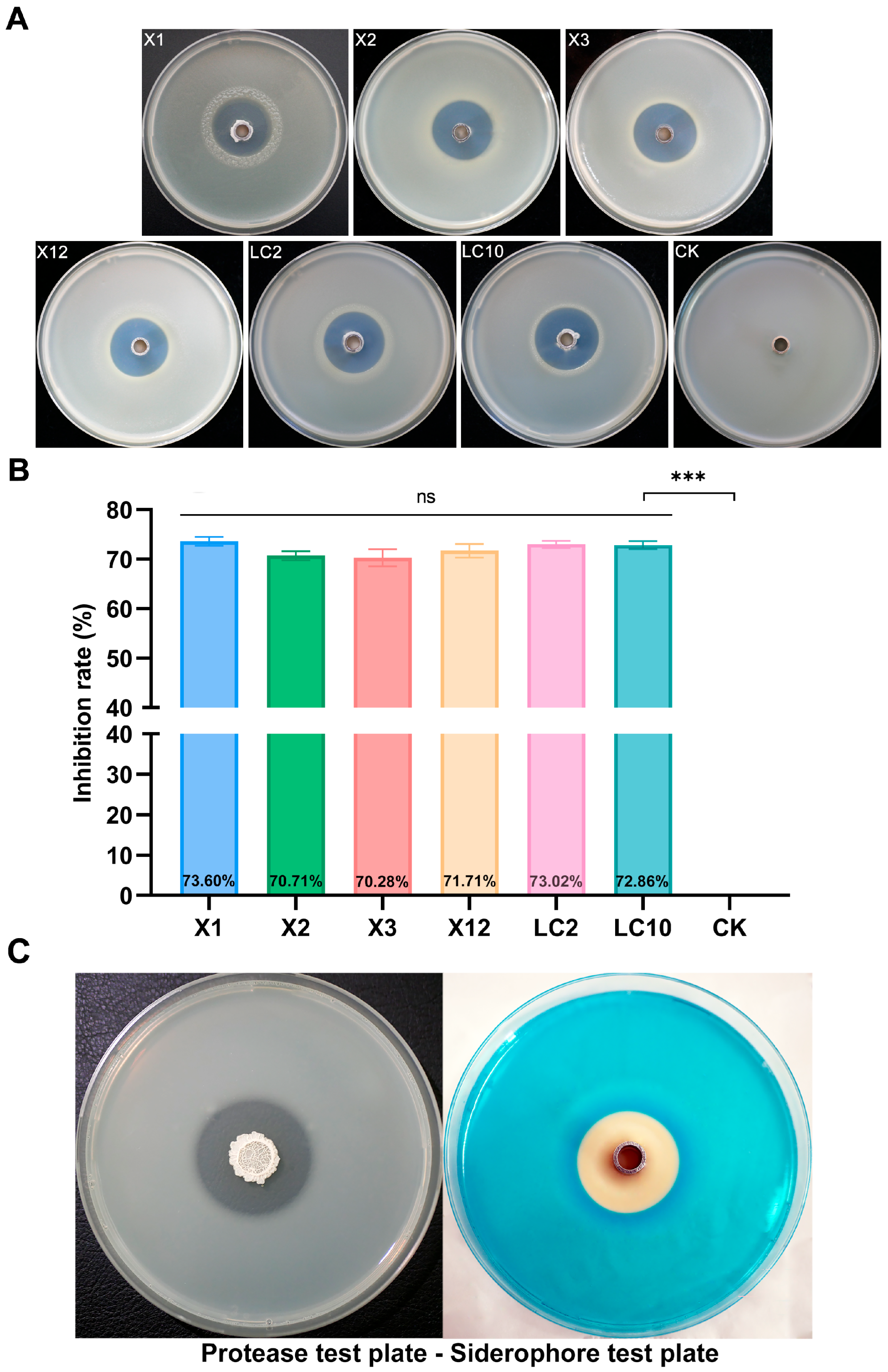
Figure 1. Isolation antimicrobial assay of AN6 against Xanthomonas oryzae pv. oryzae (Xoo). (A) In vitro antagonistic activity of AN6 against Xoo strains. (B) Inhibitory rate of AN6 against Xoo growth. *** p < 0.001; ns, not statistically significant. Statistical analysis was performed using GraphPad software, evaluated through one-way analysis of variance (ANOVA), followed by Dunnett multiple comparison post hoc tests. (C) Protease and siderophore production of AN6.
Microorganisms 13 02701 g001
Figure 2. Biological characteristics of AN6 strain. (A) Inhibitory effect of AN6 against Xanthomonas oryzae pv. oryzae (Xoo). (B) Morphology of the AN6 strain observed under a scanning electron microscope. Scale: 5.00 µm. (C) Colony morphology of the AN6 strain on LA medium. (D) Morphology of Gram-stained AN6 strain. (E) Growth curves of the AN6 strain cultured in LB at 30 °C and 160 rpm. The experiments were repeated three times with 3–5 internal replicates each.
Microorganisms 13 02701 g002
Figure 3. SEM and TEM images of cell morphology of Xanthomonas oryzae pv. oryzae LC2. (A,B) SEM image of Xanthomonas oryzae pv. oryzae LC2 cell morphology under normal conditions and when exposed to AN6 cell-free supernatant, respectively. Scale: 1.00 µm. (C,D) TEM image of Xanthomonas oryzae pv. oryzae LC2 cell morphology under normal conditions and when exposed to AN6 cell-free supernatant, respectively. Scale: 200.00 nm.
Microorganisms 13 02701 g003
Figure 4. Efficacy of B. velezensis AN6 for controlling Xoo LC2 in a light incubator. The indoor trials were performed using cultivar YX650. The groups were as follows: Xoo LC2 only (Control), NB only, rice leaves sprayed with B. velezensis AN6 12 h before inoculation with Xoo LC2 suspension (AN6-Pre, prevention strategy), and 12 h after inoculation with Xoo LC2 suspension (AN6-Tre, treatment strategy). (A) The severity of rice bacterial blight disease (BB) 15 days after Xoo LC2 inoculation. (B) Lesion length of Xoo LC2 growth. *** p < 0.001; ns, not statistically significant compared to NB only. Statistical analysis was performed using GraphPad software, evaluated through one-way analysis of variance (ANOVA), followed by Dunnett multiple comparison post hoc tests.
Microorganisms 13 02701 g004
Figure 5. Antifungal activity of AN6 against different fungi. (A) In vitro inhibition activity of strain AN6, its volatile organic compounds, and cell-free supernatant on mycelial growth of different fungi. CK: Control samples consisting of the fungal plate that was not inoculated with bacteria. Dual-culture assay measures inhibitory effect of AN6 on fungi. VOC assay measures inhibition of fungi by strain AN6 and its VOCs. CFS assay measures inhibition of fungi by aseptic fermentation filtrate of AN6. (B) Statistical analysis of AN6’s inhibitory impact on different pathogenic fungi. ** p < 0.01; *** p < 0.001; ns, not statistically significant vs. CK. Statistical analysis was conducted using GraphPad software: one-way ANOVA followed by Dunnett’s multiple comparison post hoc tests.
Microorganisms 13 02701 g005aMicroorganisms 13 02701 g005b
Figure 6. The complete genome map and epigenetic modification distribution map of Bacillus velezensis AN6. (A) Circular genome map of Bacillus velezensis AN6 visualized using Circos 0.69-9 software. From the outermost to the innermost ring: ring 1 displays the genome coordinates and physical positions along the chromosome (black line); ring 2 shows protein-coding genes located on the forward and reverse strands; ring 3 indicates non-coding RNAs (ncRNAs); ring 4 presents the GC content; ring 5 shows the GC skew, with positive skew shown in orange and negative skew shown in green. (B) Whole-genome methylation map of Bacillus velezensis AN6. The concentric rings in the figure represent, from the outside to the inside, genomic coordinates; the distribution of modification sites on the forward (sense) strand, calculated using a 2000 bp window and a 2000 bp step size; the distribution of modification sites on the reverse (antisense) strand using the same parameters; the outer two blue rings represent the distribution of modification sites on the sense strand (using a 2000 bp window with a 2000 bp step) and on the antisense strand (using a 2000 bp window with a 2000 bp step), respectively; the GC content, also calculated with a 2000 bp window and step size (red segments pointing inward indicate regions with a GC content lower than the genome-wide average, while green segments pointing outward indicate regions with a higher GC content and the taller the peak, the greater the deviation from the average); and the innermost ring represents the GC skew, calculated using the formula (G − C)/(G + C), with a 2000 bp window and step size (pink segments pointing inward indicate regions where the G content is lower than the C content, while light green segments pointing outward indicate the opposite).
Microorganisms 13 02701 g006
Figure 7. The functional annotation and gene prediction results for the strain AN6. (A) CAZy (Carbohydrate-Active enZYmes Database) functional classification. (B) COG (clusters of orthologous groups) functional classification. (C) Gene distribution based on Gene Ontology. (D) KEGG (Kyoto Encyclopedia of Genes and Genomes) metabolic pathways annotation with the gene functions. (E) TCDB (Transporter Classification Database) functional classification.
Microorganisms 13 02701 g007
Figure 8. Phylogenetic analysis of AN6 and Bacillus strains. (A) Neighbor-joining phylogenetic tree showing the phylogenetic relationship of strain AN6 with other members of the Bacillus genus based on 16S rRNA gene sequences. The gene sequences of E. coli U5/41 (NR_024570) were utilized as the root for constructing the phylogenetic tree. The bootstrap consensus was inferred from 1000 replicates. Scale bar: 0.01 substitutions per nucleotide position. (B) Neighbor-joining phylogenetic tree showing the phylogenetic relationship of strain AN6 with other members of the Bacillus genus based on gyrA gene sequences. The bootstrap consensus was inferred from 1000 replicates. Scale bar: 0.01 substitutions per nucleotide position. The strain obtained in this study is highlighted in red.
Microorganisms 13 02701 g008
Figure 9. Pairwise average nucleotide identity (ANI) and in silico DNA–DNA hybridization (isDDH) values derived from the analysis of the whole genomes of AN6 and 16 other Bacillus strains. The left lower triangle illustrates the ANI values, whereas the right upper triangle displays the isDDH values. Squares highlighted in red indicate an ANI ≥ 96% or isDDH ≥ 70%. The superscript T on the right denotes the standard strain. The asterisk (*) marks self-vs-self comparisons, which are omitted as they are non-informative.
Microorganisms 13 02701 g009
Figure 10. Comparative analysis of the genomes of AN6, B. velezensis FZB42, B. amyloliquefaciens DSM7, and B. subtilis 168. (A) Venn diagram showing ortholog gene families between AN6 and B. velezensis FZB42, B. amyloliquefaciens DSM7, and B. subtilis 168. (B) Analysis of the evolutionary relationship. The bootstrap consensus was inferred from 1000 replicates. Scale bar: 0.01 substitutions per nucleotide position. (C) Collinearity analysis. Boxes of similar colors represent syntenic regions, boxes below the horizontal line represent inversion regions, and rearrangements are represented by colored lines.
Microorganisms 13 02701 g010
Table 1. Methods for assessing the biological control effect of AN6 on Xoo.
TreatmentExperimental VariablesTiming of Biocontrol Agent Addition
PathogenBiocontrol Agent
ControlXooNB12 h after pathogen inoculation
Pre-treatedXooAN612 h before pathogen inoculation
Co-treatedXooAN612 h after pathogen inoculation
NBNBNB12 h after pathogen inoculation
Table 2. Genome characteristics of strain AN6.
DatabaseNumber of Annotated
Functional Proteins
Proportion/%
Nr393997.86
GO271467.43
KEGG387596.27
COG299374.36
Pfam271467.43
Swiss-Prot330282.04
TCDB51512.80
CAZy1563.88
Table 3. Secondary metabolite clusters in strain AN6 predicted by antiSMASH v8.0.4.
Gene ClusterSecondary Metabolite(s)Genome LocationMost Similar
Known Cluster
Similarity Confidence
Cluster 1NRPS322,200–387,607SurfactinHigh
Cluster 2PKS-like924,057–965,301
Cluster 3Terpene1,047,341–1,068,081
Cluster 4Lanthipeptide class ii1,188,574–1,217,462
Cluster 5TransAT-PKS1,384,025–1,472,258Macrolactin HHigh
Cluster 6TransAT-PKS, T3PKS, and NRPS1,690,949–1,801,042BacillaeneHigh
Cluster 7NRPS, transAT-PKS, and betalactone1,865,673–2,003,474FengycinHigh
Cluster 8Terpene2,028,701–2,050,584
Cluster 9T3PKS2,113,902–2,155,002
Cluster 10TransAT-PKS2,269,988–2,376,170DifficidinHigh
Cluster 11Terpene precursor2,399,464–2,420,354
Cluster 12NRP metallophore, NRPS, and RiPP-like3,000,874–3,052,665BacillibactinHigh
Cluster 13Other3,588,975–3,630,393BacilysinHigh
Disclaimer/Publisher’s Note: The statements, opinions and data contained in all publications are solely those of the individual author(s) and contributor(s) and not of MDPI and/or the editor(s). MDPI and/or the editor(s) disclaim responsibility for any injury to people or property resulting from any ideas, methods, instructions or products referred to in the content.