


Abstract
Black pepper (Piper nigrum L.) has historically been among the most consumed spices globally. Brazil is one of the world’s largest producers and exporters, and is the largest in the Western Hemisphere. This study describes two new fungal species associated with black pepper commercialized in Brazil. The first, Penicillium pipericola sp. nov., belongs to the subgenus Penicillium, section Paradoxa, series Atramentosa. The second, Syncephalastrum brasiliense sp. nov., belongs to the order Mucorales, family Syncephalastraceae. The taxonomic classification of these species was supported by a pluralistic approach, based on multilocus phylogenetic analyses, morphological analyses, and metabolomics. Furthermore, the metabolomic analysis revealed considerable biosynthetic versatility of the new species under different cultivation conditions, producing metabolites with therapeutic and biotechnological potential. The identification of these species increases the understanding of fungal diversity in the black pepper production chain and may have important implications for the microbiological quality of the product, for the understanding of ecological interactions within the agroecosystem and for potential industrial applications.
1. Introduction
Black pepper (Piper nigrum L.), often referred to as the “queen of spices,” has significant economic and cultural importance as the most widely consumed spice in the world. Native to India and belonging to the Piperaceae family, this tropical plant is cultivated in several regions of the world, with Brazil standing out as the second largest producer and the main exporter in the Western Hemisphere [1,2,3]. It contains several bioactive compounds, including alkaloids, terpenoids, phenolics, and flavonoids, with piperine being a key component [4,5]. Brazilian production is concentrated in the states of Espírito Santo and Pará, which collectively account for over 90% of national output [6]. The fruits of P. nigrum are processed into black or white pepper, serving diverse purposes in the culinary, pharmaceutical, cosmetic, and food industries due to their essential oils, resins, and piperine content [2].
Despite its global prominence, the production and processing of black pepper face significant challenges regarding microbial contamination. During post-harvest handling, including drying, storage, and packaging, the product is exposed to environmental and handling conditions conducive to microbial growth. Fungi, particularly storage-associated genera such as Aspergillus and Penicillium, are frequent contaminants capable of altering organoleptic properties, reducing market value and, most alarmingly, producing toxic secondary metabolites known as mycotoxins [7,8]. Among these, aflatoxins and ochratoxin A (OTA) are of particular concern due to their potential carcinogenicity, immunosuppressive effects, and hepatotoxicity [9,10]. The initial diversity of fungal contamination plays a crucial role in the final quality of pepper products [11].
Recent studies have shown that spices harbor complex microbial communities dominated by Aspergillus and Penicillium species, many of which are toxigenic or opportunistic pathogens. Garcia et al. [12] found high fungal contamination in several spices, with black and white peppers exhibiting particularly high frequencies of toxigenic Aspergillus species, including A. flavus (up to 32%) and the A. niger complex (up to 52%), with 14.2–66.7% of A. flavus isolates producing aflatoxins. Jeswal & Kumar [13] identified A. flavus and A. niger as the predominant species in Indian spices, with red pepper exhibiting 85.4% aflatoxin contamination and the highest aflatoxin levels (219.6 ng/g). Mandeel [14] reported similar results in imported spices, with red pepper and black pepper exhibiting the highest contamination (1580 and 1120 CFU/g, respectively), predominantly by the genera Aspergillus and Penicillium. Black pepper is generally reported to have high fungal loads; however, mycotoxin levels are generally below regulatory limits. However, high consumption and the risk of cumulative exposure justify closer monitoring of the fungal biodiversity of this spice.
The Brazilian regulatory framework is on par with international standards by establishing strict microbiological and mycotoxin limits to ensure food safety [15,16]. However, compliance remains an ongoing challenge, as contamination is frequently reported in the production chain. This highlights the importance of interventions in agronomic practices and advanced microbial monitoring and control strategies.
In addition to contamination, the microbial diversity associated with black pepper may offer a distinct perspective for examining fungal ecology and taxonomy. Spices are an underexplored niche that contains a wide diversity of fungal taxa, including previously undescribed species.
Polyphasic taxonomy combines genotypic and phenotypic data and has contributed significantly to the delimitation of fungal species and the understanding of biodiversity. This approach integrates diverse data types, such as molecular phylogeny, morphology, physiology, and chemotaxonomic markers, into consensus classification systems [17]. In addition to taxonomy, omics approaches such as genomics, transcriptomics, proteomics, and metabolomics enable more accurate fungal identification and taxonomy, beyond traditional morphological approaches [18]. These multiomics approaches are particularly useful for mycotoxin research, as they help identify and characterize the gene clusters responsible for the production of these metabolites [19]. On the other hand, omics approaches have also facilitated the discovery of beneficial fungal secondary metabolites, including antibiotics, drugs, and other bioactive compounds, highlighting the great versatility of fungi [20].
In this study, we report two new species of fungi associated with black pepper commercialized in Brazil. The first species is named Penicillium pipericola sp. nov. and belongs to the subgenus Penicillium sect. Paradoxa Ser. Atramentosa. The genus Penicillium is highly diverse and globally distributed, with many species frequently isolated from soil, organic waste, and a variety of food products. Section Paradoxa accommodates species that are primarily isolated from plant material and are notable for their rich secondary metabolism, which may include the production of bioactive compounds [21,22,23].
The second species, Syncephalastrum brasiliense sp. nov., is classified within the order Mucorales, family Syncephalastraceae, genus Syncephalastrum. Members of the genus Syncephalastrum are distinguished by the formation of spores in characteristic clusters, arranged in structures resembling bunches or masses, and are commonly found in soil and decaying organic matter [24]. The occurrence of S. brasiliense sp. nov. in black pepper is therefore an intriguing discovery, as this genus does not belong to the typical groups of microorganisms associated with spice crops. Discovering members of this group within the black pepper production chain raises questions about their ecological role and potential interactions with other microorganisms or even with the pepper plant itself.
This study aimed to describe, based on a polyphasic approach, two new fungal species obtained from black pepper commercialized in Brazil.
2. Materials and Methods
2.1. Samples
The strains corresponding to the two candidate species (ITAL-3JA, ITAL-3JB, and ITAL-3JC of P. pipericola sp. nov., and ITAL-36NA, ITAL-36NB, and ITAL-36NC of S. brasiliense sp. nov.) were isolated from black pepper samples obtained from retail establishments in the city of Campinas, São Paulo State, Brazil. The isolation procedure followed the methodology of direct plating of grains described in Pitt and Hocking [25]. The surfaces were disinfected in a 0.4% sodium hypochlorite solution for 2 min, then 50 grains were plated on Dichloran Glycerol Agar 18% (DG18). Each sample weighed approximately 150 g.
2.2. Morphological Analysis
Initially, the fungi were evaluated based on their macroscopic characteristics. All fungal isolates were purified and plated at three equidistant points on Czapek Yeast Extract Agar (CYA) and Malt Extract Agar (MEA) at 25 °C and incubated for 5 days. They were subsequently morphologically characterized according to the taxonomic keys of [25,26,27], supplemented by other sources when necessary. Morphological identification was limited to the fungal genus/group (morphogroup) level and subsequently combined with molecular analysis.
For P. pipericola sp. nov. analysis, morphological studies were performed according to the methodology proposed by Visagie et al. [23]. To evaluate the macroscopic characteristics, strains of candidate species were plated on different media: CYA, MEA, DG18, Creatine Sucrose Agar (CREA), and Yeast Extract Sucrose Agar (YESA). Incubation was performed in the dark at 25 °C for 7 days. Additionally, strains were tested on CYA medium at 30 °C and 37 °C for 7 days. While for S. brasiliense sp. nov., morphological analyses were performed on MEA, Potato Dextrose Agar (PDA), and Sucrose Dextrose Chloramphenicol Agar supplemented with Gentamicin (SDCA+G) media at temperatures (5 °C, 25 °C, 40 °C, and 45 °C) over 7 days. All experiments were performed in triplicate.
For microscopic analysis, slides were prepared using lactic acid from colonies grown on MEA medium for 7 days at 25 °C. Micromorphological characteristics, including the size of conidiophores, stipes, vesicles, conidia, metula, and phialides, were measured using Carl Zeiss™ AxioVision Release 4.8.2 software. Observations were performed with a Zeiss Axio Imager. A2 (Carl Zeiss Microscopy GmbH, Jena, Germany), an upright optical microscope). Measurements and imaging were conducted under brightfield contrast at magnifications of 400× and 1000×.
2.3. Molecular Analysis
For genomic DNA extraction, purified strains were grown in liquid Yeast Sucrose medium (YES) at 25 °C for 3 days until mycelial membranes were formed, and then were macerated with liquid nitrogen. This material was used to obtain genomic DNA according to the manufacturer’s protocol (PureLink Mini Kit, Thermo Fisher Scientific, MA, USA). DNA was quantified using spectrophotometry (NanoDrop®, Thermo Fisher Scientific, MA, USA).
Different loci were used for molecular identification of fungal isolates. The best loci for each group were selected according to the literature and current taxonomic context. Amplification of the universal fungal barcode, ITS region (rRNA), was performed using the primer pair its1/its4 described in White et al. [28]; for amplification of Large subunit ribosomal ribonucleic acid (LSU rRNA), we used the primer pair D1/D2 described in Hoog et al. [29]. Part of the calmodulin gene was amplified using the primer pairs CF1/CF4 [30]. The beta-tubulin gene (BenA) was amplified using the primers Bt2a and Bt2b [31]. The locus of the second largest subunit of RNA polymerase II (RPB2) was amplified using the primers 5Feur/7CReur, described in Houbraken et al. [32]. The amplification conditions were the same as those described in Silva et al. [33].
After amplification, the PCR products were separated by agarose gel electrophoresis (1% w/v), stained with ethidium bromide, and visualized under ultraviolet light. After amplification, PCR product purification was performed using ExoSAP-IT™ PCR Product Cleanup (Thermo Fisher Scientific, Santa Clara, CA, USA). PCR fragments were then subjected to direct sequencing using the method described by Sanger et al. [34]. Sequencing was performed bidirectionally (forward and reverse) using a BigDye® Terminator v3.1 Cycle Sequencing Kit (Applied Biosystems, Foster City, CA, USA) on a SeqStudio Genetic Analyzer® (Applied Biosystems, Waltham, MA, USA).
Sequence alignment was performed using ClustalW in BioEdit Sequence Alignment Editor v.7.1.3.0. Concatenation of the loci was performed using DnaSP v.6 DNA Sequence Polymorphism.
Maximum likelihood (ML) trees were constructed separately for each locus and for the combined data set. Sequence alignment was performed with the type or neotype strain sequences of species formally accepted within each taxonomic group (Penicillium or Syncephalastrum) using ClustalW.
For ML tree construction, the optimal nucleotide substitution model was determined using jModelTest2 based on the Akaike information criterion (AIC). ML trees were constructed using MEGA 11, using 1000 bootstrap replicates, its final visualization took place through the ITOL platform (Interactive Tree of Life, https://itol.embl.de/itol.cgi, accessed on 11 October 2025). The GenBank accession numbers corresponding to the sequences used in the phylogenetic analysis are provided in Supplementary Material S1.
2.4. Secondary Metabolites Analysis
Secondary metabolites were extracted following adapted protocols from Smedsgaard [35] and Houbraken et al. [36]. Fungal strains were grown for 7 days on solid YESA, MEA, and CYA media under two temperature regimes designed to assess physiological plasticity: a condition supporting robust growth (25 °C for P. pipericola sp. nov. and 28 °C for S. brasiliense sp. nov.) and a condition imposing thermal stress, where growth was notably reduced (30 °C for P. pipericola sp. nov. and 40 °C for S. brasiliense sp. nov.). For each medium and condition tested, three agar plugs (5–7 mm2) were collected from each colony (edges and center), and homogenized in 3 mL of ethyl acetate/dichloromethane/methanol (3:2:1, v/v/v) containing 1% formic acid. Homogenates were sonicated for 50 min, filtered through 0.22 μm membranes, evaporated to dryness, and stored at −18 °C until further analysis.
The samples were received at the Compounds Management Laboratory (LGC/LNBio-CNPEM), where they were solubilized in DMSO (Sigma-Aldrich, Darmstadt, Germany, 99.7%), using the automated liquid handler Janus G3 (Revvity®, Waltham, MA, USA) to a final concentration of 10 mg/mL. Aliquots of 12 µL of each sample were plated in 384-well plates (Greiner Bio-One GmbH, Kremsmünster, Austria) for UPLC-MS/MS analysis.
A liquid chromatography (LC) method was utilized in an Acquity UPLC HClass system (Waters, MA, USA) coupled to a uHR-ESI-QqTOF Impact II (Bruker Daltonics, Billerica, MA, USA) tandem mass spectrometer, operating in the positive mode. The LC system was equipped with a C18 BEH Acquity 1.7 µm column 2.1 × 100 mm (Waters, MA, USA), operating at a flow rate of 0.5 mL/min and a column temperature of 40 °C. The elution gradient involved an initial mobile phase composed by 90% water, 5% acetonitrile (CH3CN) (Merck, Darmstadt, Germany), and 5% of 2% formic acid solution. At 10 min, a CH3CN gradient was performed (gradient curve 6 in the Acquity system) to increase the CH3CN concentration to 95%, while maintaining 5% of the 2% formic acid solution. At 12 min, the mobile phase was changed to 100% CH3CN (gradient curve 1 in the Acquity system). This was followed by 3 min of recalibration to the initial mobile phase, totaling 15 min elution time per analysis.
For mass spectrometry the electrospray source was set to the range of 30–2000 Da, a detection speed of 8 Hz 500 V end plate offset, a capillarity of 4500 V, nebulizer at 4.0 bar, and drying gas flow (nitrogen) at 10 L/min with a drying temperature of 200 °C. For MS/MS, the collision cell was 5.0 eV, with collision energy in the range of 20–70 V and an absolute fragmentation cutoff of 1000. Ions below 200 Da were excluded, and the “active exclusion” function was enabled. For internal calibration, 10 mM sodium formate solution was used. The raw data were converted to mzXML using the instrument’s software Bruker Compass Data Analysis v. 4.3.
Data obtained from LC-MS/MS (mzXML) were processed using NP3 MS Workflow software [37] with default parameters. The software utilizes the UNPD-ISDB [38] library as its built-in database for analysis. The consensus spectra generated from the NP3 MS Workflow (*.mgf files) were further searched against GNPS spectral libraries (https://gnps.ucsd.edu/ProteoSAFe/status.jsp?task=24ed5f80d0e74215800cdbed01080bb5 and https://gnps.ucsd.edu/ProteoSAFe/status.jsp?task=c8b8d3bf7ea74d24a19513ccdbe062b4, accessed on 16 June 2025) [39]. After processing in GNPS, the chemical annotations were grouped into the clean variable table, which resulted from NP3 MS Workflow softwmare processing. In the post-processing step, protonated ions (M+H+) were selected, along with a chemical curation procedure to determine the most appropriate chemical annotation (GNPS or UNPD), based on the parameters: MQScore, mzErrorPPM, and NumPeakShared. For GNPS, the following thresholds were applied: MQScore > 0.8, mzErrorPPM < 20, and NumPeakShared > 6; for UNPD-ISDB, the thresholds were: MQScore > 0.4, mzErrorPPM < 20, and NumPeakShared > 6.
3. Results and Discussion
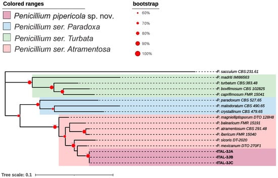
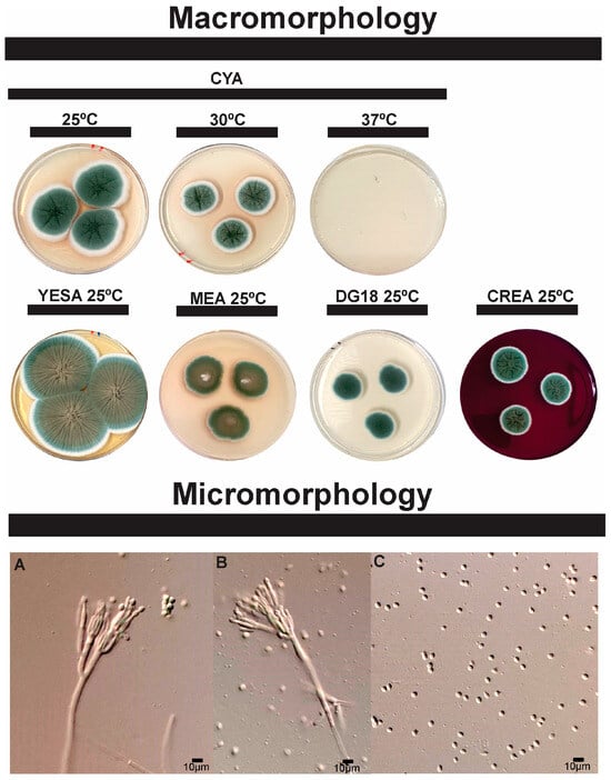
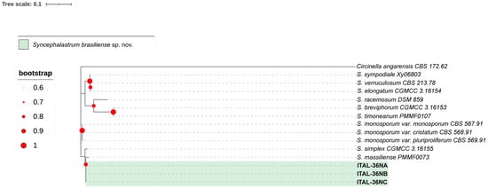
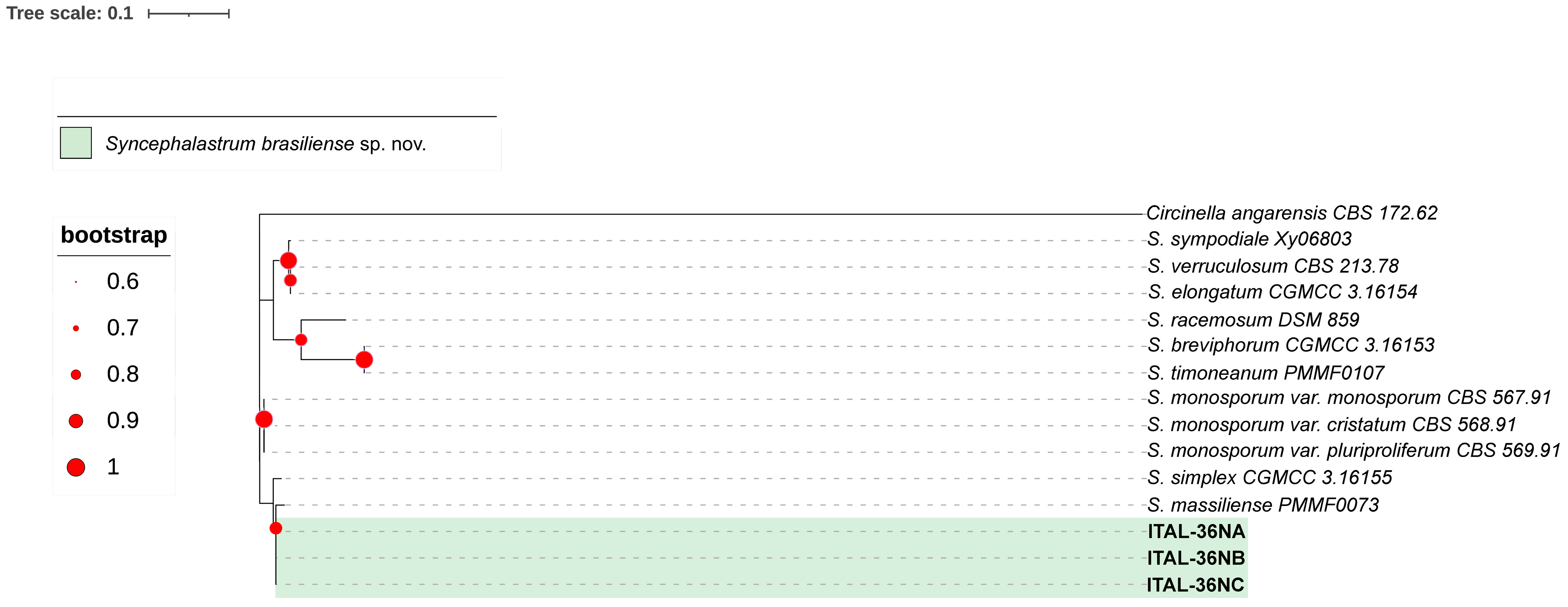
The candidate species Penicillium pipericola sp. nov. (Figure 1 and Figure 2) and Syncephalastrum brasiliense sp. nov. (Figure 3 and Figure 4) have been identified as independent evolutionary lineages, each forming robust monophyletic groups as indicated by genealogical concordance. Individual phylogenetic trees for each analyzed locus are provided in Supplementary Material S2.
Figure 1.
Maximum-likelihood tree (GTR+G+I) of Penicillium series Paradoxa, Atramentosa and Turbata based on combined dataset sequences (CaM+BenA+RPB2+ITS). Only bootstraps ≥ 60% are shown. Penicillium sacculum is the outgroup.
Figure 2.
Morphological characters of Penicillium pipericola sp. nov. (ex-type ITAL-3JA). A colonies from left to right (top row) CYA (25 °C, 30 °C and 37 °C); (bottom row) YESA, MEA, DG18, and CREA (25 °C). Conidiophores (A,B), conidia (C).
Figure 3.
Maximum-likelihood tree (T93+G) of Syncephalastrum genus based on combined dataset sequences (LSU+ITS). Only bootstraps ≥ 60% are shown. Circinella angarensis is the outgroup.
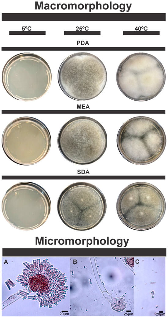
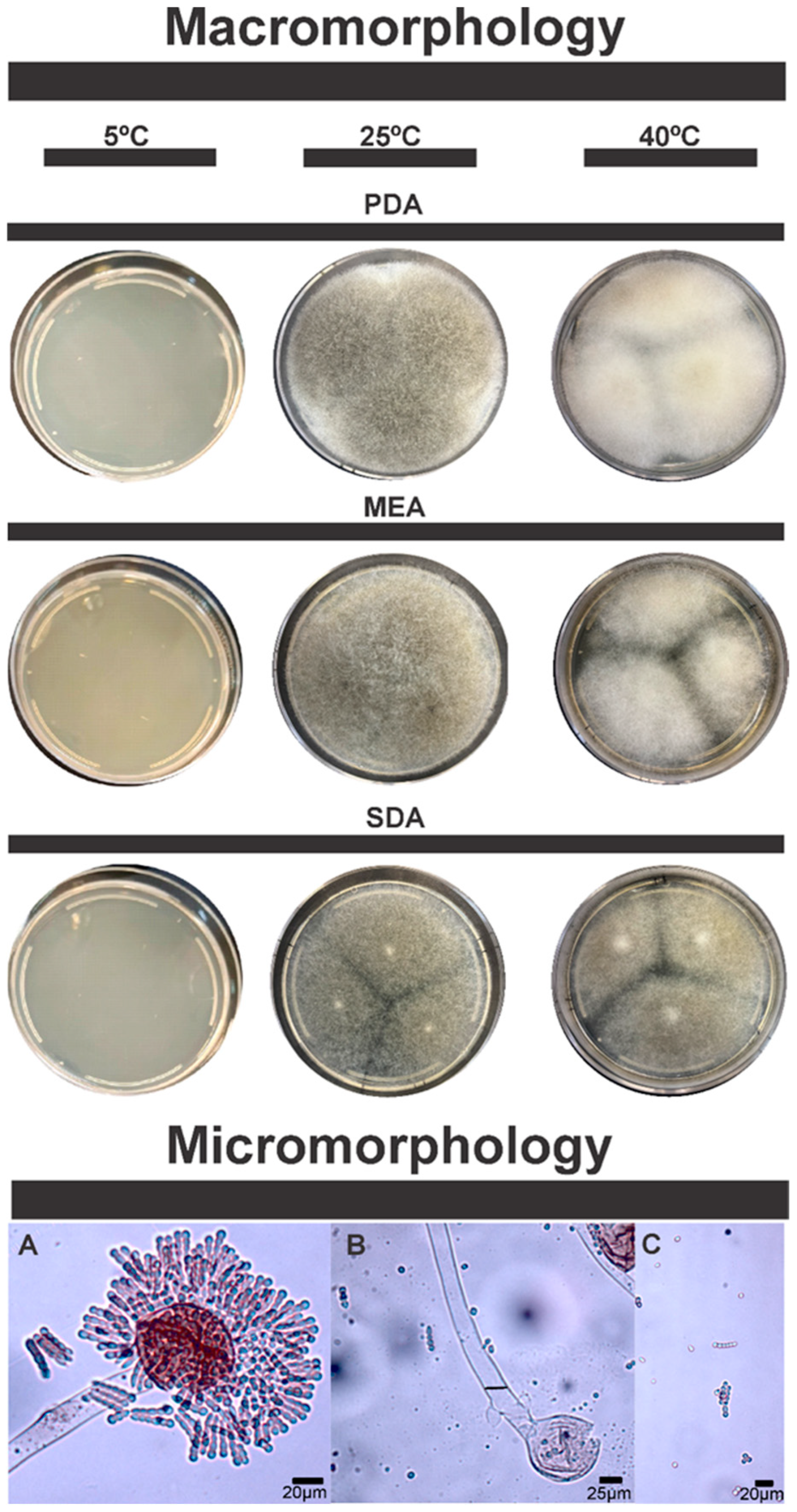
Figure 4.
Morphological characters of Syncephalastrum brasiliense sp. nov. (ex-type ITAL-36NA). Columns from left to right represent different temperatures: 5 °C, 25 °C and 40 °C; Rows from top to bottom represent different media: PDA, MEA and SDCA+G. Sporangiophores and Merosporangium (A), Columella (B) and Sporangiospores (C).
Penicillium is a large and diverse fungal genus with over 350 species distributed throughout the world in different environmental matrices, among which soils, plants, and food products stand out [40,41,42]. These fungi are important decomposers but are also capable of food spoilage and producing mycotoxins such as ochratoxin A and patulin [40,42], which are of concern for food safety. However, industrial applications for Penicillium are noted in cheese and sausage manufacturing and production of pharmaceutical important metabolites, including penicillin [40,41]. Recent genomic studies are providing some insight into the mechanisms of adaptation and horizontal gene transfer in Penicillium species [41]. Moreover, these fungi are promising in their biotechnological applications such as bioremediation, biofuel production, and food fermentation [43]. The versatility of Penicillium species makes them valuable objects for research into adaptation mechanisms and prospecting for metabolites of interest.
Some species of Penicillium have been shown to increase plant growth as well as resistance to pathogens. Penicillium culture filtrates increased seed germination and improved root and shoot growth in tomato and wheat [44,45]. Improvement in chlorophyll content, protein, and amino acid levels has been associated with some Penicillium strains in sesame plants [46]. These fungi have been shown to have protective effects against salt stress, as well to fungal pathogens such as Fusarium spp. [46]. Penicillium sp. strain GP15-1 has been shown to improve cucumber plants growth, and to protect them from damping off caused by Rhizoctonia solani, as well as anthracnose caused by Colletotrichum orbiculare [47].
The plant growth-promoting abilities of Penicillium spp. have been attributed to the production of beneficial metabolites, efficient root colonization, and induction of systemic resistance in host plants [44,47]. The isolation of P. pipericola sp. nov. from black pepper raises interest in investigating its ecological role in the black pepper production chain, its microbial interactions with other microorganisms, or with the plant host itself, and the production of mycotoxins by this species.
Analysis of the metabolomic profile of P. pipericola sp. nov. revealed a set of specialized metabolites whose production was modulated by variations in temperature and culture medium (Table 1). These compounds belong to different chemical classes, such as diketopiperazines, indole alkaloids, perylenequinones, and modified sterols, and reflect the biosynthetic plasticity of the studied strain when faced with different stimuli. Below we discuss some of the most relevant compounds that could be annotated in our analysis.
Table 1.
Metabolomic Characterization of Penicillium pipericola sp. nov. and Syncephalastrum brasiliense sp. nov. under Variable Growth Conditions. Annotated metabolites were considered according to the spectra annotation parameters and thresholds described in the Material and Methods section.
The altertoxin group of mycotoxins are perylene quinones, known to be produced by fungi of the genus Alternaria [48,49]. This group of mycotoxins can induce DNA strand breaks as they are highly mutagenic [48]. Altertoxins contaminate food and feed, especially after long storage periods, with altertoxins I, II, and III considered in the risk assessment of EFSA Panel on Contaminants in the Food Chain [50,51]. They are synthesized through the polyketide pathway, with the altertoxin I biosynthesis mechanism being proposed by Stinson [52]. Altertoxins can be produced in large quantities when Alternaria spp. are grown in modified Czapek-Dox medium with low glucose and ammonium sulfate [53]. Altertoxin II suppresses the immune response in the gastrointestinal tract and alters cell architecture in the colon [54], acting through the Nrf2-ARE pathway. Interestingly, the Nrf2-ARE pathway in mammalian cells is activated by altertoxin II (epoxyde), but not by its alcohol analog, altertoxin I [49]. Commercial food samples analyzed by Liu & Rychlik [53] contain alterotoxin I and II, but did not contain altertoxin III, although the latter is also an EFSA concern. However, some organic whole grains and paprika powder were contaminated with altertoxin I and another perylene derivative, alterperylenol. The highest concentrations of altertoxins were found in sorghum feed samples [53]. The finding that Penicillium pipericola sp. nov. occurs in Brazilian black pepper, and that this strain produces altertoxin III, highlights the importance to monitor altertoxins in this food ingredient.
Besides mycotoxins with potential concerns on food safety, we could also detect metabolites in Penicillium pipericola sp. nov. that are of interest in pharmaceutical development.
Myriberin A, which we found in P. pipericola sp. nov. (Table 1), is an alkaloid isolated from the plant Myrioneuron faberi, possessing an unprecedented heteropentacyclic skeleton [55]. It demonstrates inhibitory effects against the hepatitis C virus (HCV) life cycle in vitro [55,56]. To our knowledge, this is the first report of the production of this metabolite by a fungus.
Dehydrohistidyl-tryptophanyl-diketopiperazine (Table 1) is a fungal metabolite of the diketopiperazine (DKP) class, which in turn can be found in several microorganisms, particularly fungi and bacteria [57,58,59]. These compounds have varied chemical structures and biological activities, making them promising drug candidates [57,60]. The wide structural and bioactivity variation in indole DKPs, isolated mainly from Aspergillus and Penicillium species, has been the subject of extensive studies [57]. Some DKPs show potential in pest control, as observed in compounds from Aspergillus ochraceus [58]. Novel DKP derivatives, such as maremycins, have been found in Streptomyces sp., possessing unusual structural features such as spiro-indole groups. These compounds showed slight cytotoxicity against the mouse fibroblastoma cell line L-929, the human leukemia cell line K562, and the Hela human cervix carcinoma cell line [59]. Notably, 2,5-DKPs (such as those found in P. pipericola (Table 1)) have attracted considerable attention for their anticancer properties, and both natural and synthetic derivatives are being explored, particularly for use as anticarcinogenic agents [60].
Galangin (Table 1) is a flavonoid extracted from galangal and propolis. It has anti-inflammatory, antibacterial, and antioxidant activities [61]. Galangin inhibits cancer cell metastasis by blocking the PKC/ERK signaling pathways and reducing MMP-2/MMP-9 activity [62]. Furthermore, it stimulates the degradation of β-catenin, inhibiting the growth of colorectal and liver cancer cells [63]. In ovarian cancer, galangin can induce apoptosis through p53-dependent pathways, thus being selective for cancer cells, while sparing normal cells [64]. The anticarcinogenic effects are associated with the upregulation of pro-apoptotic proteins and the downregulation of the Akt/p70S6K pathways [64]. Although galangin shows promise as a potential therapeutic agent, further clinical and toxicity studies are needed to support its application [61].
Morphologically, P. pipericola sp. nov. exhibits distinct characteristics compared to its closest relative, Penicillium mexicanum. These differences include faster and broader growth patterns and a smoother colony appearance (Figure 2), while P. mexicanum has a grooved colony, which is not the case for P. pipericola sp. nov. The main comparable morphological characteristics between these two species are summarized in Table 2. The combined analysis of phenotypic and genotypic data justifies the classification of P. pipericola as a new species, placed in the subgenus Penicillium, section Paradoxa, series Atramentosa.
Table 2.
Main morphological characteristics comparable between Penicillium pipericola sp. nov. and Penicillium mexicanum.
The genus Syncephalastrum is included in the group of zygomycetes, which play different ecological roles. First described by Thaxter in 1897 [65], Syncephalastrum species are generally known as environmental fungi and laboratory contaminants [66]. The genus is characterized by irregular, branched and aseptate hyphae with terminal vesicles surrounded by cylindrical merosporangia [66].
The genus Syncephalastrum belongs to the order Mucorales and has been increasingly reported in human infections, especially in immunocompromised individuals [66,67]. Two recently described species, S. massiliense and S. timoneanum, were isolated from clinical samples and presented unique phenotypic and genotypic characteristics [66]. Syncephalastrum racemosum is another member of this genus implicated in infections, known to cause highly invasive subcutaneous mucormycosis [67]. S. brasiliense sp. nov. is phylogenetically close to S. massiliense, which raises the need for further investigation into its potential as a pathogen in humans.
Similarly, S. brasiliense sp. nov. also shows clear morphological differences from its closest relatives, Syncephalastrum massiliense and Syncephalastrum simplex. Syncephalastrum brasiliense sp. nov. differs from related taxa by its ability to grow up to 45 °C. Colonies expand rapidly on MEA, PDA, and SDCA+G (40–70 mm/5 days), cottony to velvety, white to grayish, with pale reverse (Table 3).
Table 3.
Main morphological characteristics of Syncephalastrum brasiliense sp. nov. and the closest species.
Complementary metabolomic analyses showed that S. brasiliense sp. nov. exhibited considerable biosynthetic plasticity, producing diverse metabolites from different classes, many of which have biotechnological potential.
The compound Ac,Et ester-mandelic acid, detected in five of the six conditions (Table 1), is a derivative of mandelic acid, which in turn is a lipophilic α-hydroxy acid, which has antibacterial activity, in addition to the ability to inhibit sebocyte lipid synthesis, therefore being useful in the care of oily skin [68]. Some mandelic acid derivatives organize into chiral gels by self-assembly and can be used as templates for circularly polarized luminescent materials, applicable in the biomedical field [69]. Results of DPPH, FRAP, CUPRAC, and ABTS assays showed that mandelic acid and its derivatives have antioxidant activities [70]. Products containing mandelic acid have been found to improve parameters related to oiliness, shine, and signs of skin aging [68]. This profile makes mandelic acid suitable for cosmetic and pharmaceutical applications.
The metabolite 6-[3-[(3,4-dimethoxyphenyl)methyl]-4-methoxy-2-(methoxymethyl)butyl]-4-methoxy-1,3-benzodioxole NCGC00385811-01, a benzodioxol derivative, was detected in three of the six growth conditions tested (Table 1). Studies show that benzodioxole derivatives form stable metabolic complexes that can induce and modulate cytochrome P450 enzymes. Murray et al. [71] found a strong correlation between the enzyme-inducing ability of these compounds and aryl hydrocarbon hydroxylase activity (r = 0.980). Later, Kumagai et al. [72] confirmed that these complexes form and have characteristic absorption properties at 455 nm. They also found that these complexes inhibit the catalytic cycle of enzymes, which helped to better understand how these mechanisms work. The metabolism of benzodioxoles includes oxidation to catechols and the generation of carbon monoxide derived from the methylene carbon atom [73]. Some benzodioxole derivatives exhibit anticancer potential, particularly against hepatocellular carcinoma cells, as they reduce alpha-fetoprotein secretion and induce cell cycle arrest [74]. Furthermore, some benzodioxole-derived compounds exhibit antioxidant properties [74]. The unique structure of benzodioxoles leads to pharmacokinetic interactions and potential toxicity through a quinone-reactive oxygen mechanism [72].
The flavone 7,8-dihydroxyflavone (DHF) (Table 1) is a natural flavonoid with diverse therapeutic potential. As a high-affinity TrkB agonist, DHF exhibits neuroprotective effects against several central nervous system diseases, including Alzheimer’s disease and Parkinson’s disease [75]. In the ICV-STZ mouse model of sporadic Alzheimer’s disease, DHF improved cognitive function by alleviating oxidative stress, mitochondrial dysfunction, and insulin resistance [76]. In addition to its neuroprotective effects, DHF also exhibits anti-aging effects in human dermal fibroblasts by inducing collagen synthesis, inhibiting MMP-1 expression, and reducing intracellular reactive oxygen species. It also upregulates antioxidant enzymes and modulates the MAPK/Akt signaling pathway, which is involved in skin aging [77]. Overall, these studies highlight the potential of DHF as a nutraceutical for the treatment of neurological diseases and as an anti-aging skin agent.
The analysis of phenotypic and genotypic data confirms that S. brasiliense sp. nov. is a new species within the order Mucorales, family Syncephalastraceae, and genus Syncephalastrum.
Therefore, the detailed descriptions of these new species, P. pipericola sp. nov. and S. brasiliense sp. nov., are based on a robust phylogenetic analysis and the observation of unique morphological and physiological characteristics that definitively differentiate them from related taxa.
4. Taxonomy
Penicillium pipericola sp. nov. Rosa, V.S.; Taniwaki, M.H.; Silva, J.J.
Mycobank: MB861249
Etymology: The specific epithet refers to the substrate from which it was isolated, black pepper, Piper nigrum.
In Penicillium subgen. Penicillium sect. Paradoxa ser. Atramentosa.
Typification: BRAZIL. São Paulo State, Campinas City, 22°54′06.7″ S 47°03′37.2″ W, in black pepper, 10 April 2024, isolated by Rosa, V.S. Holotype: CIMFI 3JA, preserved as a metabolically inactive culture. Ex-type culture: ITAL-3JA.
DNA barcodes: BenA (PV022467), CaM (PV022465), RPB2 (PV022466), ITS (PV007905).
Colony diam.: 7 days, 25 °C: CYA 38.3–43.0 mm, MEA 30.0–33.0 mm, YESA 47.0–51.0 mm; DG18 19.0-26.0 mm; CREA 19.6-21.0 mm; CYA 30 °C 28.0–33.0 mm.
Diagnosis: Morphologically, P. pipericola sp. nov. can be differentiated from P. mexicanum by its significantly faster growth on several media: CYA, YESA and MEA. Its colony morphology is also notably distinct; the colony surface of P. pipericola is velvetier with a radial structure, while P. mexicanum tends to have deep and highly sulcate colonies (see description of P. mexicanum Visagie et al. [78]. Phylogenetically, P. pipericola sp. nov. is easily distinguished from P. mexicanum and other species in ser. Atramentosa. The main distinguishing features compared to the type strain of P. mexicanum (CBS H-21805) are: 9 single-nucleotide polymorphisms (SNPs) in the BenA locus, 16 SNPs in RPB2, 24 SNPs in CaM, and 2 SNPs in ITS. When the CaM+ITS+RPB2+BenA genealogies are combined, P. pipericola sp. nov. forms a well-supported independent evolutionary lineage (IEL) with a bootstrap value of 90% (Figure 1).
Colony characters: On CYA 25 °C, green colonies, with white edges, abundant sporulation, velvety surface with radial juices, exudates present; soluble pigments and sclerotia absent. In MEA 25 °C, 7 days: Small colonies with a white center and a gradient from brown to green towards the edges. Irregular and thin edges, smooth surface. Pigments, exudates and sclerotia absent. In YESA 25 °C, 7 days: Large colonies with brownish center with gradient of green tones towards the edges; velvety appearance; abundant and ordered radial grooves with wrinkled center. Soluble pigments, exudates and sclerotia absent. In DG18 25 °C, 7 days, Small, flat colonies with velvety appearance, white edges; uniform green color throughout the colony. Soluble pigments, exudates and sclerotia absent. In CREA 25 °C, 7 days, small colonies, green in color, pronounced center with cerebrospinal appearance, smooth rounded edges, without acid production.
Micromorphology: Conidiophores triverticillate; Stipes smooth-walled, 31.2 × 3.1 μm; Metulae, 10.6 × 3.0 μm (7.7 − 16.5 × 2.9 − 3.5); Phialides ampulliform, 10.0 × 2.4 μm (9.9 − 12.5 × 2.2 − 3.5); Conidia smooth and ellipsoidal 3.3 × 2.9 μm (2.9 − 4.7 × 2.7 − 3.0).
Syncephalastrum brasiliense sp. nov. Rosa, V.S.; Iamanaka, B.T.; Silva, J.J.
Mycobank: MB861250.
Etymology: Named after its place of isolation, Brazil.
In gen. Syncephalastrum fam. Syncephalastraceae ordo. Mucorales cl. Mucoromycetes subdiv. Mucoromycotina div. Mucoromycota subregn. Mucoromyceta.
Typification: BRAZIL. São Paulo State, Campinas City, 22°54′06.7″ S 47°03′37.2″ W, in black pepper, 10 April 2024, isolated by Rosa, V.S. Holotype: CIMFI 36NA, preserved as a metabolically inactive culture. Ex-type culture: ITAL-36NA.
DNA barcodes: ITS (PV007760); LSU (PV015155).
Colony diam.: 7 days, 25 °C: MEA 40.0–68.0 mm, PDA 44.0–67.0 mm, SDCA+G 41.0–70.0 mm.
Diagnosis: Morphologically, S. brasiliense can be distinguished from S. massilense primarily by its robust growth at 40 °C (with growth observed at 45 °C), sporangiospore size, and colony coloration (see the description of S. massilense by Kabtani et al. [66]). It differs from S. simplex in colony texture and coloration, as well as vesicle shape (see the description of S. simplex by Zhao et al. [79]). Phylogenetically, the main distinguishing features compared to the type strain of S. massilense (CBS H-21805) are: 6 single-nucleotide polymorphisms (SNPs) and 1 INDEL (insertion/deletion) in the ITS locus, as well as 5 INDELs in the LSU locus. When the ITS+LSU genealogies are combined, S. brasiliense sp. nov. forms a well-supported independent evolutionary lineage (IEL) with a bootstrap value of 82% (Figure 3).
Colony characters: On MEA 25 °C, rapid and expansive growth, covering the entire plate in 5 days; homogeneous white to cream coloration, some strains may develop grayish tones from the fifth day onwards; cottony and aerial texture, typical of Mucorales fungi, with long and diffuse hyphae, indistinct margin, diffuse growth without clear delimitation. reverse of the plate pale, colonies between 40–68 mm. Grows well at 40 °C, but there is no growth at 5 °C. In PDA 25 °C, rapid and expansive growth, covering the entire plate in 5 days, white to light gray coloration, with a slightly darker center, suggesting the beginning of sporulation. Cottony and densely aerial texture, with diffuse and filamentous mycelium; diffuse and irregular margin, with homogeneous radial growth; reverse of the plate pale to yellowish brown. Colonies between 44–67 mm. Grows well at 40 °C, but no growth at 5 °C; exudates are produced. On SDCA+G 25 °C, rapidly growing colony covering nearly the entire surface of the plate; predominantly white to grayish in color, with a slight darkening in the center. Flaky, densely filamentous texture, with a velvety or cottony appearance. Well-defined margins, which may be slightly diffuse due to the rapid expansion of the mycelium. Reverse of the plate pale to light, colonies 41–70 mm, with aerial hyphae. Grows well at 40 °C, but no growth at 5 °C.
Micromorphology: Sporangiophores erect, branched or simple, sporangia of variable shape, mainly subglobose, born from aerial, coenocytic, smooth and hyaline hyphae. Merosporangium present with globose to subglobose vesicle covered radially by cylindrical merosporangia lined up on the surface of the vesicle. Sporangiospores with smooth surface, elliptical, globose and subglobose, individual or in chains of up to 7 sporangiospores. Zygospores and chlamydospores not observed. Sporangia, 41–70 μm; hyphae 11–24 μm wide; merosporangia 6–11 μm; sporangiospores, 4–7 μm.
5. Conclusions
This study describes two new species, Penicillium pipericola sp. nov. and Syncephalastrum brasiliense sp. nov., isolated from Brazilian black pepper. The integration of morphological, phylogenetic, and metabolomic data supports their proposal as new species. The presence of these fungi in black pepper highlights the importance of microbiological monitoring, both for food safety purposes and for expanding our understanding of ecological interactions and biotechnological applications of these fungi. These results prompt further investigations to clarify the impact of these species on product quality and their role in the microbial dynamics of the agroecosystem. Furthermore, the metabolomic profile provides a promising perspective for future investigations into the biotechnological potential of these two new species.
Supplementary Materials
The following supporting information can be downloaded at: https://www.mdpi.com/article/10.3390/microorganisms13122691/s1, Supplementary Material S1: Table showing the GenBank accession numbers used in the phylogenetic analyses conducted in this study. Table (A) Accessions included in the phylogenetic analysis presented in Figure 1. Table (B) Accessions included in the phylogenetic analysis presented in Figure 3; Supplementary Material S2: Phylogenetic trees of maximum likelihood for each locus individually. Figures S2A–S2D (Penicillium pipericola sp. nov.); Figures S2E–S2G (Syncephalastrum brasiliense sp. nov.).
Author Contributions
Conceptualization, J.J.S.; methodology, J.J.S.; formal analysis, J.J.S., R.d.F. and D.B.B.T.; investigation, J.J.S., A.R.P.d.S., M.C.d.S., L.D.S., P.F., R.d.F., D.B.B.T. and V.S.R.; data curation, A.R.P.d.S. and V.S.R.; writing—original draft preparation, J.J.S. and V.S.R.; writing—review and editing, D.B.B.T., B.T.I., M.H.T., L.D.S., R.d.F. and J.J.S.; visualization, V.S.R., R.d.F. and J.J.S.; supervision, J.J.S., B.T.I. and M.H.T.; project administration, J.J.S.; funding acquisition, J.J.S., L.D.S. and D.B.B.T. All authors have read and agreed to the published version of the manuscript.
Funding
This research was funded by Fundação de Amparo à Pesquisa do Estado de São Paulo (FAPESP) (Processes 2023/04445-9; 2021/12590-3; A.R.P. da Silva grant 2023/17037-6 and PF fellowship 2024-22909-5). V.S.R. received a Grant (Process 130871/2023-0) from Conselho Nacional de Desenvolvimento Científico e Tecnológico (CNPq).
Institutional Review Board Statement
Not applicable.
Informed Consent Statement
Not applicable.
Data Availability Statement
The sequences newly generated in this study have been submitted to the GenBank database. Additional data are available upon request from the corresponding authors.
Acknowledgments
The authors would like to thank the technical and administrative staff of the Instituto de Tecnologia de Alimentos (ITAL) and of the Brazilian Biosciences National Laboratory of the Brazilian Center for Research in Energy and Material (LNBio-CNPEM) for the infrastructure provided to carry out the study.
Conflicts of Interest
The authors declare no conflicts of interest.
References
- Srinivasan, K. Black Pepper and Its Pungent Principle-Piperine: A Review of Diverse Physiological Effects. Crit. Rev. Food Sci. Nutr. 2007, 47, 735–748. [Google Scholar] [CrossRef] [Scilit] [PubMed]
- Nair, K.P. The Geography of Black Pepper (Piper Nigrum); Springer: Berlin/Heidelberg, Germany, 2020. [Google Scholar]
- FAOSTAT. Available online: https://www.fao.org/faostat/en/#data/QCL/visualize (accessed on 10 October 2025).
- Pop, A.S.; Marc, A.R.; Mureșan, C.C. Therapeutic Insights into Black Pepper (Piper Nigrum): Phytochemical Composition, Bioactive Properties, and Health Benefits. Hop Med. Plants 2024, 32, 139–152. [Google Scholar] [CrossRef] [Scilit]
- Yadav, S.; Tyagi, A.; Kumari, R.; Srivastava, H.; Rangan, P.; Wankhede, D.; Gaikwad, A.B. Comparative Transcriptome Profiling of Fruit Tissue Provides Novel Insights into Piperine Biosynthesis in Black Pepper (Piper nigrum L.). Sci. Hortic. 2023, 322, 112451. [Google Scholar] [CrossRef] [Scilit]
- Produção de Pimenta-Do-Reino No Brasil|IBGE. Available online: https://www.ibge.gov.br/explica/producao-agropecuaria/pimenta-do-reino/br (accessed on 10 October 2025).
- Yogendrarajah, P.; Deschuyffeleer, N.; Jacxsens, L.; Sneyers, P.-J.; Maene, P.; De Saeger, S.; Devlieghere, F.; De Meulenaer, B. Mycological Quality and Mycotoxin Contamination of Sri Lankan Peppers (Piper nigrum L.) and Subsequent Exposure Assessment. Food Control 2014, 41, 219–230. [Google Scholar] [CrossRef] [Scilit]
- Pickova, D.; Ostry, V.; Malir, J.; Toman, J.; Malir, F. A Review on Mycotoxins and Microfungi in Spices in the Light of the Last Five Years. Toxins 2020, 12, 789. [Google Scholar] [CrossRef] [Scilit]
- IARC Working Group on the Evaluation of Carcinogenic Risks to Humans. Chemical Agents and Related Occupations; International Agency for Research on Cancer: Lyon, France, 2012; (IARC Monographs on the Evaluation of Carcinogenic Risks to Humans, No. 100F). Available online: https://www.ncbi.nlm.nih.gov/books/NBK304416/ (accessed on 10 October 2025).
- Mohsenzadeh, M.S.; Hedayati, N.; Riahi-Zanjani, B.; Karimi, G. Immunosuppression Following Dietary Aflatoxin B1 Exposure: A Review of the Existing Evidence. Toxin Rev. 2016, 35, 121–127. [Google Scholar] [CrossRef] [Scilit]
- Weil, M.; Remize, F.; Durand, N.; Alter, P.; Hoarau, M.; Meile, J.C. Effect of Processing on Microbial Safety of Wild Pepper (Piper Borbonense) from Reunion Island. Food Control 2020, 111, 107061. [Google Scholar] [CrossRef] [Scilit]
- Garcia, M.V.; Parussolo, G.; Moro, C.B.; Bernardi, A.O.; Copetti, M.V. Fungi in Spices and Mycotoxigenic Potential of Some Aspergilli Isolated. Food Microbiol. 2018, 73, 93–98. [Google Scholar] [CrossRef] [Scilit]
- Jeswal, P.; Kumar, D. Mycobiota and Natural Incidence of Aflatoxins, Ochratoxin A, and Citrinin in Indian Spices Confirmed by LC-MS/MS. Int. J. Microbiol. 2015, 2015, 242486. [Google Scholar] [CrossRef] [Scilit]
- Mandeel, Q.A. Fungal Contamination of Some Imported Spices. Mycopathologia 2005, 159, 291–298. [Google Scholar] [CrossRef] [Scilit]
- Nacional, I. INSTRUÇÃO NORMATIVA-IN N° 161, DE 1° DE JULHO DE 2022-INSTRUÇÃO NORMATIVA-IN N° 161, DE 1° DE JULHO DE 2022-DOU-Imprensa Nacional. Available online: https://www.cidasc.sc.gov.br/inspecao/files/2024/10/IN-No-161-DE-1o-DE-JULHO-DE-2022-Alterada-pela-IN-N%C2%B0-313-DE-4-DE-SETEMBRO-DE-2024.pdf (accessed on 10 October 2025).
- Food and Drug Administration. Mycotoxins. FDA. 2024. Available online: https://www.fda.gov/food/natural-toxins-food/mycotoxins (accessed on 19 September 2025).
- Kim, J. Fungal Identification Based on the Polyphasic Approach: A Clinical Practice Guideline. Ann. Clin. Microbiol. 2024, 27, 221–230. [Google Scholar] [CrossRef] [Scilit]
- Wijayawardene, N.N.; Boonyuen, N.; Ranaweera, C.B.; de Zoysa, H.K.; Padmathilake, R.E.; Nifla, F.; Dai, D.-Q.; Liu, Y.; Suwannarach, N.; Kumla, J. OMICS and Other Advanced Technologies in Mycological Applications. J. Fungi 2023, 9, 688. [Google Scholar] [CrossRef] [Scilit]
- Eshelli, M.; Qader, M.M.; Jambi, E.J.; Hursthouse, A.S.; Rateb, M.E. Current Status and Future Opportunities of Omics Tools in Mycotoxin Research. Toxins 2018, 10, 433. [Google Scholar] [CrossRef] [Scilit] [PubMed]
- Zeilinger, S.; García-Estrada, C.; Martín, J.-F. Fungal Secondary Metabolites in the “OMICS” Era. In Biosynthesis and Molecular Genetics of Fungal Secondary Metabolites, Volume 2; Zeilinger, S., Martín, J.-F., García-Estrada, C., Eds.; Springer: New York, NY, USA, 2015; pp. 1–12. ISBN 978-1-4939-2531-5. [Google Scholar]
- Nicoletti, R.; Fiorentino, A.; Scognamiglio, M. Endophytism of Penicillium Species in Woody Plants. Open Mycol. J. 2014, 8, 1–26. [Google Scholar] [CrossRef] [Scilit]
- Nielsen, J.C.; Grijseels, S.; Prigent, S.; Ji, B.; Dainat, J.; Nielsen, K.F.; Frisvad, J.C.; Workman, M.; Nielsen, J. Global Analysis of Biosynthetic Gene Clusters Reveals Vast Potential of Secondary Metabolite Production in Penicillium Species. Nat. Microbiol. 2017, 2, 17044. [Google Scholar] [CrossRef] [Scilit] [PubMed]
- Visagie, C.M.; Houbraken, J.; Frisvad, J.C.; Hong, S.-B.; Klaassen, C.H.W.; Perrone, G.; Seifert, K.A.; Varga, J.; Yaguchi, T.; Samson, R.A. Identification and Nomenclature of the Genus Penicillium. Stud. Mycol. 2014, 78, 343–371. [Google Scholar] [CrossRef] [Scilit]
- Pavlovic, M.D.; Bulajic, N. Great Toenail Onychomycosis Caused by Syncephalastrum Racemosum. Dermatol. Online J. 2006, 12, 7. [Google Scholar] [CrossRef] [Scilit] [PubMed]
- Pitt, J.I.; Hocking, A.D. Fungi and Food Spoilage; Springer: Berlin/Heidelberg, Germany, 2009; Volume 519. [Google Scholar]
- Samson, R.A.; Pitt, J.I. (Eds.) Integration of Modern Taxonomic Methods For Penicillium and Aspergillus Classification; CRC Press: London, UK, 2003; ISBN 978-0-429-18160-3. [Google Scholar]
- Klich, M.A. Identification of Common Aspergillus Species; Centraalbureau Voor Schimmelcultures: Utrecht, The Netherlands, 2002. [Google Scholar]
- White, T.J.; Bruns, T.; Lee, S.; Taylor, J. Amplification and Direct Sequencing of Fungal Ribosomal RNA Genes for Phylogenetics. In PCR Protocols: A Guide to Methods and Applications; Academic Press: Cambridge, MA, USA, 1990; Volume 18, pp. 315–322. [Google Scholar]
- de Hoog, G.; Guarro, J.; Gene, J.; Figueras, M.J. Atlas of Clinical Fungi; Amer Society for Microbiology: Washington, DC, USA, 2000; ISBN 90-70351-43-9. [Google Scholar]
- Peterson, S.W.; Vega, F.E.; Posada, F.; Nagai, C. Penicillium Coffeae, a New Endophytic Species Isolated from a Coffee Plant and Its Phylogenetic Relationship to P. Fellutanum, P. Thiersii and P. Brocae Based on Parsimony Analysis of Multilocus DNA Sequences. Mycologia 2005, 97, 659–666. [Google Scholar] [CrossRef]
- Glass, N.L.; Donaldson, G.C. Development of Primer Sets Designed for Use with the PCR to Amplify Conserved Genes from Filamentous Ascomycetes. Appl. Environ. Microbiol. 1995, 61, 1323–1330. [Google Scholar] [CrossRef] [Scilit]
- Houbraken, J.; Frisvad, J.C.; Seifert, K.A.; Overy, D.P.; Tuthill, D.M.; Valdez, J.G.; Samson, R.A. New Penicillin-Producing Penicillium Species and an Overview of Section Chrysogena. Persoonia-Mol. Phylogeny Evol. Fungi 2012, 29, 78–100. [Google Scholar] [CrossRef] [Scilit]
- da Silva, J.J.; Iamanaka, B.T.; Ferranti, L.S.; Massi, F.P.; Taniwaki, M.H.; Puel, O.; Lorber, S.; Frisvad, J.C.; Fungaro, M.H.P. Diversity within Aspergillus Niger Clade and Description of a New Species: Aspergillus vinaceus sp. nov. J. Fungi 2020, 6, 371. [Google Scholar] [CrossRef] [Scilit]
- Sanger, F.; Nicklen, S.; Coulson, A.R. DNA Sequencing with Chain-Terminating Inhibitors. Proc. Natl. Acad. Sci. USA 1977, 74, 5463–5467. [Google Scholar] [CrossRef] [Scilit]
- Smedsgaard, J. Micro-Scale Extraction Procedure for Standardized Screening of Fungal Metabolite Production in Cultures. J. Chromatogr. A 1997, 760, 264–270. [Google Scholar] [CrossRef] [Scilit]
- Houbraken, J.; Spierenburg, H.; Frisvad, J.C. Rasamsonia, a New Genus Comprising Thermotolerant and Thermophilic Talaromyces and Geosmithia Species. Antonie Van Leeuwenhoek 2012, 101, 403–421. [Google Scholar] [CrossRef] [Scilit] [PubMed]
- Bazzano, C.F.; de Felicio, R.; Alves, L.F.G.; Costa, J.H.; Ortega, R.; Vieira, B.D.; Morais-Urano, R.P.; Furtado, L.C.; Ferreira, E.L.; Gubiani, J.R. NP3 MS Workflow: An Open-Source Software System to Empower Natural Product-Based Drug Discovery Using Untargeted Metabolomics. Anal. Chem. 2024, 96, 7460–7469. [Google Scholar] [CrossRef] [Scilit] [PubMed]
- Allard, P.-M.; Péresse, T.; Bisson, J.; Gindro, K.; Marcourt, L.; Pham, V.C.; Roussi, F.; Litaudon, M.; Wolfender, J.-L. Integration of Molecular Networking and In-Silico MS/MS Fragmentation for Natural Products Dereplication. Anal. Chem. 2016, 88, 3317–3323. [Google Scholar] [CrossRef] [Scilit] [PubMed]
- Wang, M.; Carver, J.; Phelan, V.; Sanchez, L.M.; Garg, N.; Peng, Y.; Nguyen, D.D.; Watrous, J.; Kapono, C.A.; Luzzatto-Knaan, T.; et al. Sharing and Community Curation of Mass Spectrometry Data with Global Natural Products Social Molecular Networking. Nat. Biotechnol. 2016, 34, 828–837. [Google Scholar] [CrossRef] [Scilit]
- Fernandez-Bunster, G. Diversity, Phylogenetic Profiling of Genus Penicillium, and Their Potential Applications. In Industrially Important Fungi for Sustainable Development; Springer: Berlin/Heidelberg, Germany, 2021; Volume 1: Biodiversity and Ecological Perspectives, pp. 335–361. [Google Scholar]
- Ropars, J.; de La Vega, R.R.; López-Villavicencio, M.; Gouzy, J.; Dupont, J.; Swennen, D.; Dumas, E.; Giraud, T.; Branca, A. Diversity and Mechanisms of Genomic Adaptation in Penicillium. In Aspergillus Penicillium Post-Genomic Era; Caister Academic Press: Poole, UK, 2016. [Google Scholar]
- Perrone, G.; Susca, A. Penicillium Species and Their Associated Mycotoxins. In Mycotoxigenic Fungi: Methods and Protocols; Humana: New York, NY, USA, 2016; pp. 107–119. [Google Scholar]
- Akaniro, I.R.; Chibuike, I.V.; Onwujekwe, E.C.; Gbadamosi, F.A.; Enyi, D.O.; Onwe, O.N. Penicillium Species as Chassis for Biomanufacturing and Environmental Sustainability in the Modern Era: Progress, Challenges, and Future Perspective. Fungal Biol. Rev. 2023, 46, 100326. [Google Scholar] [CrossRef] [Scilit]
- Mushtaq, S.; Nasim, G.; Khokhar, I.; Mukhtar, I. Effects of Penicillium Extracts on Germination Vigour in Subsequent Seedling Growth of Tomato (Solanum lycopersicum L.). Arch. Phytopathol. Plant Prot. 2012, 45, 932–937. [Google Scholar] [CrossRef] [Scilit]
- Ibatsam Khokhar, I.K.; Haider, M.S.; Irum Mukhtar, I.M.; Amna Ali, A.A.; Sobia Mushtaq, S.M.; Muhammad Ashfaq, M.A. Effect of Penicillium Species Culture Filtrate on Seedling Growth of Wheat. Int. Res. J. Agric. Sci. Soil Sci. 2013, 3, 24–29. [Google Scholar]
- Radhakrishnan, R.; Kang, S.-M.; Baek, I.-Y.; Lee, I.-J. Characterization of Plant Growth-Promoting Traits of Penicillium Species against the Effects of High Soil Salinity and Root Disease. J. Plant Interact. 2014, 9, 754–762. [Google Scholar] [CrossRef] [Scilit]
- Hossain, M.M.; Sultana, F.; Miyazawa, M.; Hyakumachi, M. The Plant Growth-Promoting Fungus Penicillium Spp. GP15-1 Enhances Growth and Confers Protection against Damping-off and Anthracnose in the Cucumber. J. Oleo Sci. 2014, 63, 391–400. [Google Scholar] [CrossRef] [Scilit]
- Fleck, S.C.; Burkhardt, B.; Pfeiffer, E.; Metzler, M. Alternaria Toxins: Altertoxin II Is a Much Stronger Mutagen and DNA Strand Breaking Mycotoxin than Alternariol and Its Methyl Ether in Cultured Mammalian Cells. Toxicol. Lett. 2012, 214, 27–32. [Google Scholar] [CrossRef] [Scilit]
- Jarolim, K.; Del Favero, G.; Pahlke, G.; Dostal, V.; Zimmermann, K.; Heiss, E.; Ellmer, D.; Stark, T.D.; Hofmann, T.; Marko, D. Activation of the Nrf2-ARE Pathway by the Alternaria Alternata Mycotoxins Altertoxin I and II. Arch. Toxicol. 2017, 91, 203–216. [Google Scholar] [CrossRef] [Scilit]
- Authority, E.F.S. Evaluation of the FoodEx, the Food Classification System Applied to the Development of the EFSA Comprehensive European Food Consumption Database. EFSA J. 2011, 9, 1970. [Google Scholar] [CrossRef] [Scilit]
- European Food Safety Authority; Arcella, D.; Eskola, M.; Ruiz, J.A.G. Dietary Exposure Assessment to Alternaria Toxins in the European Population. EFSA J. 2016, 14, e04654. [Google Scholar] [CrossRef] [Scilit]
- Stinson, E.E. Mycotoxins-Their Biosynthesis in Alternaria. J. Food Prot. 1985, 48, 80–91. [Google Scholar] [CrossRef] [Scilit]
- Liu, Y.; Rychlik, M. Biosynthesis of Seven Carbon-13 Labeled Alternaria Toxins Including Altertoxins, Alternariol, and Alternariol Methyl Ether, and Their Application to a Multiple Stable Isotope Dilution Assay. Anal. Bioanal. Chem. 2015, 407, 1357–1369. [Google Scholar] [CrossRef] [Scilit] [PubMed]
- Groestlinger, J.; Spindler, V.; Pahlke, G.; Rychlik, M.; Del Favero, G.; Marko, D. Alternaria Alternata Mycotoxins Activate the Aryl Hydrocarbon Receptor and Nrf2-ARE Pathway to Alter the Structure and Immune Response of Colon Epithelial Cells. Chem. Res. Toxicol. 2022, 35, 731–749. [Google Scholar] [CrossRef] [Scilit] [PubMed]
- Huang, S.-D.; Zhang, Y.; Cao, M.-M.; Di, Y.-T.; Tang, G.-H.; Peng, Z.-G.; Jiang, J.-D.; He, H.-P.; Hao, X.-J. Myriberine A, a New Alkaloid with an Unprecedented Heteropentacyclic Skeleton from Myrioneuron Faberi. Org. Lett. 2013, 15, 590–593. [Google Scholar] [CrossRef] [Scilit]
- Cao, M.-M.; Zhang, Y.; Li, X.-H.; Peng, Z.-G.; Jiang, J.-D.; Gu, Y.-C.; Di, Y.-T.; Li, X.-N.; Chen, D.-Z.; Xia, C.-F. Cyclohexane-Fused Octahydroquinolizine Alkaloids from Myrioneuron Faberi with Activity against Hepatitis C Virus. J. Org. Chem. 2014, 79, 7945–7950. [Google Scholar] [CrossRef] [Scilit]
- Ma, Y.-M.; Liang, X.-A.; Kong, Y.; Jia, B. Structural Diversity and Biological Activities of Indole Diketopiperazine Alkaloids from Fungi. J. Agric. Food Chem. 2016, 64, 6659–6671. [Google Scholar] [CrossRef] [Scilit]
- De Guzman, F.S.; Gloer, J.B.; Wicklow, D.T.; Dowd, P.F. New Diketopiperazine Metabolites from the Sclerotia of Aspergillus Ochraceus. J. Nat. Prod. 1992, 55, 931–939. [Google Scholar] [CrossRef] [Scilit]
- Tang, Y.-Q.; Sattler, I.; Thiericke, R.; Grabley, S.; Feng, X.-Z. Maremycins C and D, New Diketopiperazines, and Maremycins E and F, Novel Polycyclic spiro-Indole Metabolites Isolated from Streptomyces sp. Eur. J. Org. Chem. 2001, 2001, 261–267. [Google Scholar]
- Goher, S.S.; Abdrabo, W.S.; Veerakanellore, G.B.; Elgendy, B. 2,5-Diketopiperazines (DKPs): Promising Scaffolds for Anticancer Agents. Curr. Pharm. Des. 2024, 30, 597–623. [Google Scholar] [CrossRef] [Scilit]
- Wang, D.; Chen, J.; Pu, L.; Yu, L.; Xiong, F.; Sun, L.; Yu, Q.; Cao, X.; Chen, Y.; Peng, F.; et al. Galangin: A Food-Derived Flavonoid with Therapeutic Potential against a Wide Spectrum of Diseases. Phytother. Res. 2023, 37, 5700–5723. [Google Scholar] [CrossRef] [Scilit]
- Chien, S.-T.; Shi, M.-D.; Lee, Y.-C.; Te, C.-C.; Shih, Y.-W. Galangin, a Novel Dietary Flavonoid, Attenuates Metastatic Feature via PKC/ERK Signaling Pathway in TPA-Treated Liver Cancer HepG2 Cells. Cancer Cell Int. 2015, 15, 15. [Google Scholar] [CrossRef] [Scilit]
- Gwak, J.; Oh, J.; Cho, M.; Bae, S.K.; Song, I.-S.; Liu, K.-H.; Jeong, Y.; Kim, D.-E.; Chung, Y.-H.; Oh, S. Galangin Suppresses the Proliferation of β-Catenin Response Transcription-Positive Cancer Cells by Promoting Adenomatous Polyposis Coli/Axin/Glycogen Synthase Kinase-3β-Independent β-Catenin Degradation. Mol. Pharmacol. 2011, 79, 1014–1022. [Google Scholar] [CrossRef] [Scilit] [PubMed]
- Huang, H.; Chen, A.Y.; Ye, X.; Guan, R.; Rankin, G.O.; Chen, Y.C. Galangin, a Flavonoid from Lesser Galangal, Induced Apoptosis via P53-Dependent Pathway in Ovarian Cancer Cells. Molecules 2020, 25, 1579. [Google Scholar] [CrossRef] [Scilit] [PubMed]
- Thaxter, R. New or Peculiar Zygomycetes. 2. Syncephalastrum and Syncephalis. Bot. Gaz. 1897, 24, 1–15. [Google Scholar] [CrossRef] [Scilit]
- Kabtani, J.; Boulanouar, F.; Gaye, P.M.; Militello, M.; Ranque, S. Syncephalastrum massiliense sp. nov. and Syncephalastrum timoneanum sp. nov. Isolated from Clinical Samples. J. Fungi 2024, 10, 64. [Google Scholar] [CrossRef] [Scilit] [PubMed]
- Mangaraj, S.; Sethy, G.; Patro, M.K.; Padhi, S. A Rare Case of Subcutaneous Mucormycosis Due to Syncephalastrum Racemosum: Case Report and Review of Literature. Indian J. Med. Microbiol. 2014, 32, 448–451. [Google Scholar] [CrossRef] [Scilit]
- Edison, B.L.; Smith, H.A.; Li, W.-H.; Parsa, R.; Green, B.A.; Konish, P.; Dufort, M.; Tierney, N.K. 18295 Mandelic Acid, a Lipophilic Alpha Hydroxy Acid, Reduces Lipid Production, Enhances Exfoliation and Provides Clinical and Patient Perceivable Benefits to Oily and Photodamaged Skin. J. Am. Acad. Dermatol. 2020, 83, AB97. [Google Scholar] [CrossRef] [Scilit]
- Reddy, K.L.; Mathew, J.P.; Maniappan, S.; Tom, C.; Shiby, E.; Pujala, R.K.; Kumar, J. Mandelic Acid Appended Chiral Gels as Efficient Templates for Multicolour Circularly Polarized Luminescence. Nanoscale 2022, 14, 4946–4956. [Google Scholar] [CrossRef] [Scilit]
- Parcheta, M.; Świsłocka, R.; Świderski, G.; Matejczyk, M.; Lewandowski, W. Spectroscopic Characterization and Antioxidant Properties of Mandelic Acid and Its Derivatives in a Theoretical and Experimental Approach. Materials 2022, 15, 5413. [Google Scholar] [CrossRef] [Scilit]
- Murray, M.; Wilkinso, C.F.; Dube, C.E. Induction of Rat-Hepatic Microsomal Cytochrome P-450 and Aryl Hydrocarbon Hydroxylase by 1,3-Benzodioxole Derivatives. Xenobiotica 1985, 15, 361–368. [Google Scholar] [CrossRef] [Scilit]
- Kumagait, Y.; Fukuto, J.M.; Cho, A.K. The Biochemical Disposition of Methylenedioxyphenyl Compounds. Curr. Med. Chem. 1994, 1, 254–261. [Google Scholar] [CrossRef] [Scilit]
- Anders, M.W.; Sunram, J.M.; Wilkinson, C.F. Mechanism of the Metabolism of 1,3-Benzodioxoles to Carbon Monoxide. Biochem. Pharmacol. 1984, 33, 577–580. [Google Scholar] [CrossRef] [Scilit]
- Hawash, M.; Eid, A.M.; Jaradat, N.; Abualhasan, M.; Amer, J.; Naser Zaid, A.; Draghmeh, S.; Daraghmeh, D.; Daraghmeh, H.; Shtayeh, T. Synthesis and Biological Evaluation of Benzodioxole Derivatives as Potential Anticancer and Antioxidant Agents. Heterocycl. Commun. 2020, 26, 157–167. [Google Scholar] [CrossRef] [Scilit]
- Paul, R.; Nath, J.; Paul, S.; Mazumder, M.K.; Phukan, B.C.; Roy, R.; Bhattacharya, P.; Borah, A. Suggesting 7,8-Dihydroxyflavone as a Promising Nutraceutical against CNS Disorders. Neurochem. Int. 2021, 148, 105068. [Google Scholar] [CrossRef] [Scilit] [PubMed]
- Akhtar, A.; Dhaliwal, J.; Sah, S.P. 7,8-Dihydroxyflavone Improves Cognitive Functions in ICV-STZ Rat Model of Sporadic Alzheimer’s Disease by Reversing Oxidative Stress, Mitochondrial Dysfunction, and Insulin Resistance. Psychopharmacology 2021, 238, 1991–2009. [Google Scholar] [CrossRef] [Scilit]
- Choi, J.W.; Lee, J.; Park, Y.I. 7,8-Dihydroxyflavone Attenuates TNF-α-Induced Skin Aging in Hs68 Human Dermal Fibroblast Cells via down-Regulation of the MAPKs/Akt Signaling Pathways. Biomed. Pharmacother. 2017, 95, 1580–1587. [Google Scholar] [CrossRef] [Scilit] [PubMed]
- Visagie, C.M.; Hirooka, Y.; Tanney, J.B.; Whitfield, E.; Mwange, K.; Meijer, M.; Amend, A.S.; Seifert, K.A.; Samson, R.A. Aspergillus, Penicillium and Talaromyces Isolated from House Dust Samples Collected around the World. Stud. Mycol. 2014, 78, 63–139. [Google Scholar] [CrossRef] [Scilit] [PubMed]
- Zhao, H.; Nie, Y.; Zong, T.-K.; Wang, K.; Lv, M.-L.; Cui, Y.-J.; Tohtirjap, A.; Chen, J.-J.; Zhao, C.-L.; Wu, F. Species Diversity, Updated Classification and Divergence Times of the Phylum Mucoromycota. Fungal Divers. 2023, 123, 49–157. [Google Scholar] [CrossRef] [Scilit]

Disclaimer/Publisher’s Note: The statements, opinions and data contained in all publications are solely those of the individual author(s) and contributor(s) and not of MDPI and/or the editor(s). MDPI and/or the editor(s) disclaim responsibility for any injury to people or property resulting from any ideas, methods, instructions or products referred to in the content. |
© 2025 by the authors. Licensee MDPI, Basel, Switzerland. This article is an open access article distributed under the terms and conditions of the Creative Commons Attribution (CC BY) license (https://creativecommons.org/licenses/by/4.0/).