Article

Unveiling Species Diversity Within Early-Diverging Fungi from China XI: Eight New Species of Cunninghamella (Mucoromycota)

1
College of Life Sciences, Shandong Normal University, Changqinghu Campus, Jinan 250358, China
2
CAS Key Laboratory of Forest Ecology and Silviculture, Institute of Applied Ecology, Chinese Academy of Sciences, Shenyang 110016, China
3
Institute of Microbiology, Chinese Academy of Sciences, Beijing 100101, China
*
Author to whom correspondence should be addressed.
These authors contributed equally to this work.
Microorganisms 2025, 13(11), 2508; https://doi.org/10.3390/microorganisms13112508
Submission received: 24 September 2025 / Revised: 28 October 2025 / Accepted: 30 October 2025 / Published: 31 October 2025
(This article belongs to the Section Environmental Microbiology)

Abstract

The genus Cunninghamella is widely distributed, primarily saprotrophic, occasionally endophytic and phytopathogenic. Analysis based on the internal transcribed spacer (ITS), the large subunit (LSU) of ribosomal DNA, and translation elongation factor 1 alpha (TEF1α), along with morphological comparisons, resulted in a discovery of eight new species. Molecular phylogenetic analyses placed each of these new species within well-supported clades. Cunninghamella crassior sp. nov., with short and thick spines, and C. fusca sp. nov. with brown sporangiola, are sister clades to each other. C. diffundens sp. nov., containing dispersed granules in sporangiola, is closely related to C. irregularis Zhao. C. tuberculata sp. nov., producing sporangiola with nodule-like protrusions. C. fulvicolor sp. nov., sister to C. irregularis, forms yellowish-brown pigmented colonies. C. guttulata sp. nov., with teardrop-shaped sporangiola, and C. inaequalis sp. nov., with uneven sporangiola, are both closely related to C. regularis Zhao. C. monosporangiola sp. nov., characterized by only one sporangiolum on some vesicles, is sister to C. verrucosa Zhao. This study represents the eleventh installment in a series investigating early-diverging fungal diversity in China and expands the number of accepted species in Cunninghamella to 39.

1. Introduction

Cunninghamella Matruchot belongs to Cunninghamellaceae, Mucorales, Mucoromycetes, Mucoromycota (https://indexfungorum.org/, accessed on 21 April 2025). The genus exhibits a significant metabolic capacity, characterized by the biosynthesis of a diverse spectrum of compounds including fatty acids, terpenes, sugars, and nickel-iron carriers [1]. Members are distributed worldwide, with a rich record in Australia, Brazil, Cameroon, Canada, China, Czech Republic, India, Latvia, Nigeria, and Thailand (GBIF Backbone Taxonomy. https://doi.org/10.15468/39omei, accessed via https://www.gbif.org/species/2551965, accessed on 17 April 2025) (https://www.gbif.org/, accessed on April 2025) [2,3,4,5,6,7,8,9,10,11,12,13,14].
As the type genus of the family Cunninghamellaceae, it was established by Matruchot in 1903 [15,16]. Nearly a century later, Zheng and Chen [2] conducted a monographic study based on morphology and molecular systematics. Since then, many new species of Cunninghamella have been introduced based on phylogenetic data and morphological characteristics [2,3,4,5,17,18]. Fungi of the genus Cunninghamella exhibit well-developed, broad hyphae that are generally aseptate but may occasionally form septa [6,16,19]. Their colonies are cottony or fluffy, appearing white initially before maturing to gray or dark gray [20]. The sporangiophores end in vesicles that bear single-spored, spinulose (short-spined) sporangiola [7,8,9,10,21].
The genus Cunninghamella, within the order Mucorales, is primarily differentiated by a combination of morphological characteristics of its asexual stage and molecular markers. Morphologically, key diagnostic features include the development of erect sporangiophores that typically exhibit verticillate branching. These branches terminate in swollen, spherical to pyriform vesicles, which are not true sporangia as they bear denticles (small projections) on their surface. The asexual spores, known as globose to subglobose sporangiospores, are produced directly on these denticles. Species identification relies on specific traits such as the branching pattern of the sporangiophores (e.g., presence of apical vesicles, number and arrangement of branches), vesicle shape (e.g., spherical, polyhedral, or irregular) and size, and spore morphology.
The classification of the genus Cunninghamella has long been challenged by the limitations of morphology-based approaches. Although the integration of morphological observations with molecular phylogenies (e.g., using ITS rDNA and TEF1α sequences) by Liu and Zhao [17,22] established a robust consensus framework, the true diversity within this genus, particularly in under-explored regions like southern China, remains poorly understood. It is plausible that several undiscovered species, potentially forming species complexes, constitute a hidden diversity awaiting revelation under this modern framework. To address this gap, we applied this integrated approach to the investigation of soil fungi from southern China. This study, which is the eleventh installment in our series on early-diverging fungi in China [23,24,25,26,27,28,29,30,31,32], aims to delineate and formally describe the novel Cunninghamella species discovered, thereby enriching our comprehension of the genus’s diversity and phylogeny.

2. Materials and Methods

2.1. Isolation and Morphology

Soil samples were obtained from Hainan Province in April and October 2023, and from Yunnan Province in June 2023. Fungal strains were subsequently isolated from these samples using a combination of the soil dilution and single spore methods. A soil suspension with a concentration of 10−1 was prepared by shaking approximately 1 g of soil sample with 10 mL of sterile water. Subsequently, 1 mL of this diluted suspension was introduced into 9 mL of fresh sterile water to generate a 10−2 concentration suspension. Soil suspensions with 10−3 and 10−4 concentrations were prepared using the same serial dilution method as above [23,24,25,26,27,28,29,30,33]. The soil suspension (200 μL) was evenly spread on Rose Bengal Chloramphenicol (RBC: peptone 5.00 g/L, glucose 10.00 g/L, KH2PO4 1.00 g/L, MgSO4·7H2O 0.50 g/L, rose red 0.05 g/L, chloramphenicol 0.10 g/L, agar 15.00 g/L) plates using a sterile L-shaped spreader, and incubated in darkness at 25 °C for 2–5 d. The mycelium was transferred from the colonial margin to a fresh Potato Dextrose Agar (PDA: glucose 20 g/L, potato 200 g/L, agar 20 g/L) medium. Photographs of both the obverse and reverse of the colony were captured using a digital camera (Canon PowerShot G7X, Canon, Tokyo, Japan). All strains were stored with 10% sterilized glycerol at 4 °C. Microscopic morphology was observed using an optical microscope (Olympus BX53 Manufacturer: Olympus Corporation Tokyo, Japan) and captured with a high-definition digital camera (Olympus DP80 Manufacturer: Olympus Corporation Tokyo, Japan) [34,35,36,37]. Morphological traits were measured using Digimizer software (v5.6.0), each with a minimum of 30 individuals. The living cultures were preserved at the China General Microbiological Culture Collection Center, Beijing, China (CGMCC). Equivalent strains were stored at Shandong Normal University (XG). Dry cultures of types were kept at the Herbarium Mycologicum Academiae Sinicae, Beijing, China (Fungarium; HMAS). The taxonomic information was deposited at the Fungal Names repository (https://nmdc.cn/fungalnames/ (accessed on 24 October 2025)).

2.2. DNA Extraction and Amplification

DNA was extracted using an extraction kit (Changchun GeneOn BioTech Co., Ltd., Changchun, China). The ITS, LSU, and TEF1α regions were amplified using the primers and programs listed in Table 1. The PCR mixture (25 μL final volume) was prepared using 12.5 μL of 2 × Hieff Canace Plus PCR Master Mix with dye (Yeasen Biotechnology, Cat No. 10154ES03. Shanghai, China), to which 9.5 μL of ddH2O, 1 μL of each primer (10 μM), and 1 μL of genomic DNA template (1 ng/μL) were added. PCR products were subjected to 1% agarose gel electrophoresis and observed under UV light (Shenhua Science Technology Co., Ltd., Hangzhou, China) at 254 nm [38,39].

2.3. Phylogenetic Analyses

Sequences were obtained from a nucleotide database from NCBI (see Table 2) [4]. Mucor janssenii Lendn was chosen as the outgroup for its close phylogenetic affinity to Cunninghamella. This single, closely-related outgroup served as an unambiguous root for the ingroup analysis. Newly acquired sequences were processed with MEGA v.7.0 to ensure alignment consistency [44]. Phylogenetic analyses on single markers and the ITS-LSU-TEF1α combined matrix were conducted to reconstruct the phylogeny, employing both ML and BI approaches integrated into the CIPRES Science Gateway V.3.3 (https://www.phylo.org/, accessed on 15 February 2025). Maximum Likelihood (ML) analyses were carried out with 1000 bootstrap replicates using RAxML 8.2.4 (https://www.phylo.org/, accessed on 15 February 2025), and Bayesian Inference (BI) was performed under the GTR + I + G model with a sampling frequency of 1000 generations. The substitute model is calculated using PAPU software (version 4.0b10), and the relevant parameters are set to DSet distance = JC objective = ME base = equal rates = equal pinv = 0subst = all negbrlen = setzero; NJ showtree = no breakties = random. After running MCMC analyses on concatenated genes for 835,000 generations, we computed posterior probabilities from the sampled trees to construct majority-rule consensus trees. Eight cold Markov chains were executed simultaneously across two million generations, ensuring MCMC convergence criteria [45,46]. The resulting phylogenetic trees underwent iTOL based optimization [47], followed by visual refinement in Adobe Illustrator CC 2019.

3. Result

3.1. Molecular Phylogeny

We performed phylogenetic analyses of Cunninghamella using a dataset composing 73 strains of 40 species, with Mucor janssenii (CBS 205.68) designated as outgroup. The concatenated sequence matrix contains 3157 aligned characters, partitioned as 1–1514 positions (ITS), 1515–2591 (LSU), and 2592–3157 (TEF1α). Three categories of sites were identified: 1152 parsimony-informative, 551 parsimony-uninformative, and 1454 constants. Bayesian analyses employed with the GTR+G model for ITS and the GTR+I+G model for LSU and TEF1α loci. The Bayesian phylogeny exhibits topological congruence with the ML tree, supported by concordant nodal bootstrap as well as BI posterior probability values. The phylogenetic analysis showed eight monographic clades (Figure 1), and eight novel species were consequently identified within Cunninghamella. They are C. diffundens sp. nov., C. crassior sp. nov., C. fulvicolor sp. nov., C. fusca sp. nov., C. guttulata sp. nov., C. inaequalis sp. nov., C. monosporangiola sp. nov., and C. tuberculata sp. nov.

3.2. Taxonomy

3.2.1. Cunninghamella Crassior Y. Jiang, H. Zhao, Z. Meng & X.Y. Liu. sp. nov. Figure 2

Fungal Name Number: FN 571687
Etymology—The epithet “crassior” refers to thicker spines.
Type—China, Yunnan Province (27.4375858° N, 99.809103° E), soil sample, collected by H. Zhao, holotype HMAS 353832, ex-holotype CGMCC3.28882
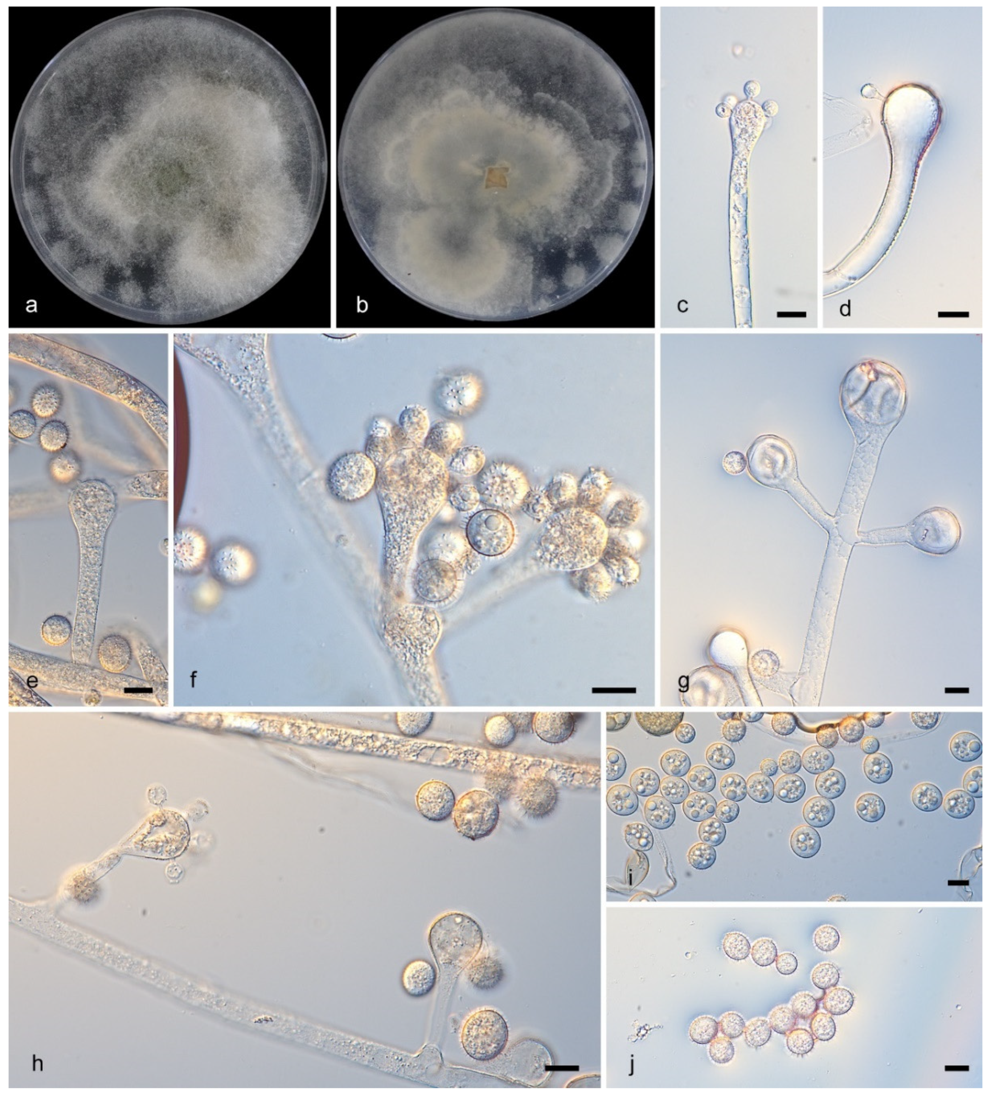
Description—Colonies on PDA, initially white becoming Drab Gray, attained dimensions of 70 mm in diameter and 10 mm in height when cultured at 27 °C for 6 days, exhibiting a floccose texture and a regular reverse margin. The hyphae were branched, mostly aseptate when young (4.5–20.5 μm in diam.), with rhizoids abundant and stolons present. Sporangiophores developing from these were erect, straight or bent, and unbranched or verticillate, broadening upwards and lacking septa. Their vesicles were subglobose to clavate, hyaline, and rough, measuring 7.0–36.5 μm long by 5.0–29.5 μm wide, and carried spiny, globose and brownish sporangiola (7.5–13.0 μm) on pedicels of 3.0–4.5 μm. No chlamydospores or zygospores were detected.
Figure 2. Cunninghamella crassior (CGMCC3.28882). (a,b): Colonies on PDA after 7 days of cultivation ((a), obverse, (b), reverse); (cg): Sporangiophores showing branching patterns; (h): Rhizoids; (i,j): Sporangiola. Scale bars: 10 µm (cj).
Microorganisms 13 02508 g002
Notes—Cunninghamella crassior (CGMCC3.28882) was isolated from a soil sample in Yunnan Province, China. In terms of phylogeny, C. crassior (CGMCC3.28882) clusters alone on a separate branch. Compared with the ITS sequence of C. clavata Zheng (CBS 362.95), the similarity is 96.86% (693/728 identical, 45/591 gaps). The similarity of the LSU sequence is 100% (776/777 identical, 0/777 gaps), and the similarity of the TEF1α sequence is 99.8% (560/567 identical, 0/567 gaps). Morphologically, C. crassior (CGMCC3.28882) releases sporangiospores with short thick spines, and no similar morphology has been found.

3.2.2. Cunninghamella diffundens Y. Jiang, H. Zhao, Z. Meng & X.Y. Liu. sp. nov. Figure 3
Fungal Name: FN573054
Etymology—The epithet “diffundens” refers to dispersed granules in sporangiola.
Type—China, Yunnan Province (26.5642168° N, 101.268054° E), soil sample, collected by H. Zhao, holotype HMAS 353829, ex-holotype CGMCC 3.28881.
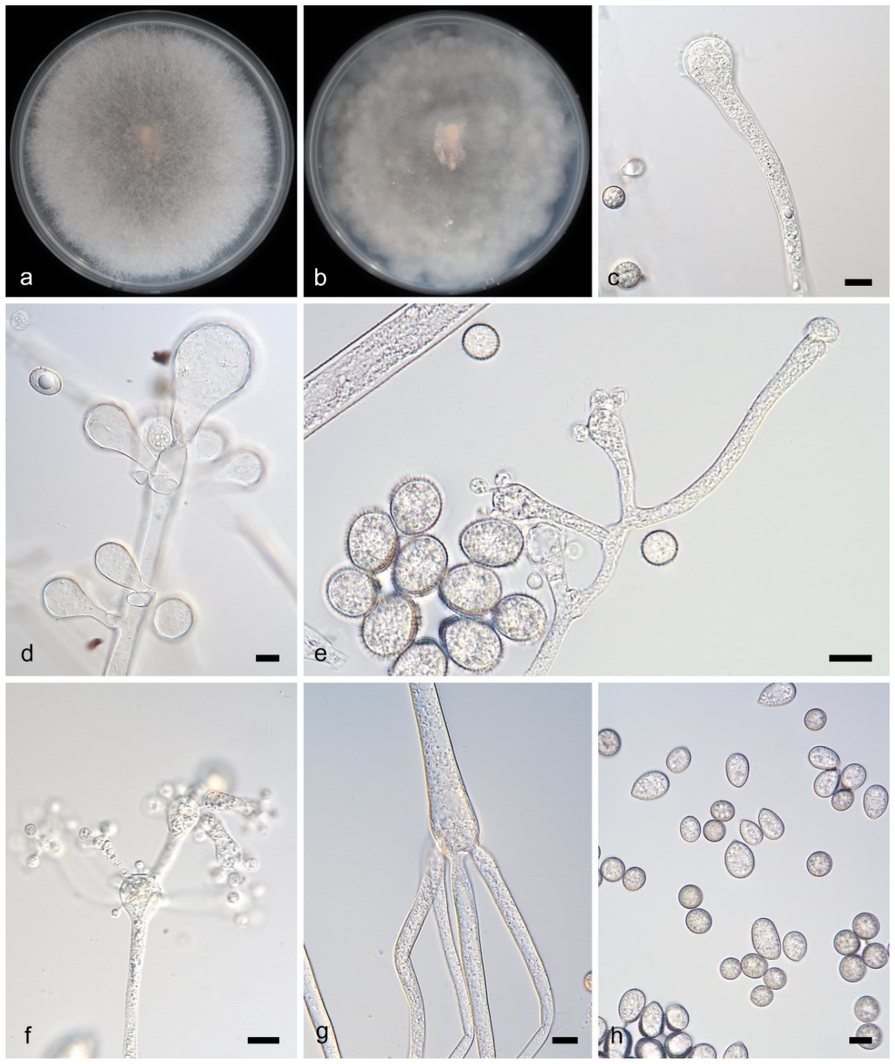
Figure 3. Cunninghamella diffundens (ex-holotype CGMCC 3.28881).(a,b): Colonies on PDA after 7 days of cultivation ((a), obverse; (b), reverse); (ch): Sporangiophores showing branching patterns; (ij): Sporangiola, showing spines and dispersed granules. Scale bars: 10 µm (cj).
Microorganisms 13 02508 g003
Description—Colonies on PDA at 27 °C for 7 days, reaching 90 mm in diameter, 10–15 μm high, initially white, soon becoming Drab Gray, floccose, in reverse irregular at margin. Hyphae branched, aseptate when young, septate when old, 5.4–14.5 μm in diameter. Rhizoids present. Stolons present. Sporangiophores arising from stolons or aerial hyphae, erect, straight or recumbent, sometimes slightly broadening upwards in main axes, solitary or branched in pairs, sympodially or 2–5 verticillate, 21.5–171.5 μm long. Sporangiola containing dispersed granules, borne on pedicels on vesicles, spiny, mostly ovoid, 8.8–15.1 μm in length and 8.0–12.8 μm in width. Chlamydospores absent. Zygospores unknown.
Notes—The holotype strain Cunninghamella diffundens (CGMCC3.28881), collected from a soil sample in Yunnan Province, China, forms a distinct monophyletic clade in phylogenetic analyses. It shows close genetic affinity to C. irregularis (CGMCC 3.16113), with sequence similarities of 95.8% in the ITS region (765/790 identical sites, 8 gaps) and 99.8% in the LSU region (940/942 identical sites, 1 gap). Notably, that no TEFα sequence of C. irregularis (CGMCC 3.16113) was available for comparison. Morphologically, C. diffundens (CGMCC3.28881) produces two distinct types of sporangiola, a feature unobserved in related species. C. diffundens (CGMCC3.28881) was described as a new species.

3.2.3. Cunninghamella fulvicolor Y. Jiang, H. Zhao, Z. Meng & X.Y. Liu. sp. nov. Figure 4

Fungal name: FN573067
Etymology—The epithet “fulvicolor” refers to the yellowish-brown pigmentation of colonies.
Type—China, Yunnan Province (26.9523432° N, 99.9502813° E), soil sample, collected by H. Zhao. Holotype HMAS 353825, ex-holotype CGMCC3.28884.
Description—Colonies on PDA at 25 °C for 5 days, reaching 90 mm in diameter, more than 20 mm high, initially white, gradually becoming light gray, floccose, produce dark or brown precipitate, irregular at margin. Hyphae flourishing, branched, aseptate when young, septate when old, 5.7–9.5 μm in diameter. Rhizoids infrequent, root-like, always branched. Stolons present. Sporangiophores developed erectly and straight from aerial hyphae, consistently broadening upwards; they were monopodial, appearing either unbranched or simply branched in pairs but never verticillate; septa were frequently present in the upper parts. The vesicles were subglobose to globose, generally hyaline or subhyaline (10.0–19.2 μm long, 6.9–17.4 μm wide), though occasionally brownish. Attached via pedicels, the sporangiola were ovoid, hyaline, and measured 9.0–12.5 μm by 4.7–7.4 μm, often exhibiting droplets in youth. Chlamydospores absent. Zygospores unknown.
Notes—The holotype strain of Cunninghamella fulvicolor was isolated from soil in Yunnan Province, China. Phylogenetic analyses revealed that C. fulvicolor (CGMCC3.28884) and C. fulvicolor (HZ012-2) represent the same strain, with maximum likelihood (ML) and Bayesian inference (BI) support values of 100/1. Both C. fulvicolor (CGMCC3.28884) and C. fulvicolor (HZ012-2) clustered closely with C. irregularis (CGMCC 3.16113) on the same branch, indicating a close genetic relationship. However, morphological differences were observed: C. irregularis (CGMCC 3.16113) produces irregular spores: (with thick spines, ovoid, 10.0–12.5 μm long and 8.0–10.5 μm wide, and globose, 9.0–16.5 μm in diameter.) C. fulvicolor produces only one morphotype of sporangiola (length: 9.0–12.5 μm, width: 4.7–7.4 μm). Conclusion: based on morphological distinctiveness and molecular phylogenetic divergence, C. fulvicolor and C. irregularis (CGMCC 3.16113) are recognized as two distinct species.
Figure 4. Cunninghamella fulvicolor (CGMCC3.28884). (a,b): Colonies on PDA after 5 days of cultivation ((a) obverse, (b) reverse); (cg): Sporangiophores showing branching patterns; (h): Rhizoids; (i,j): Sporangiola. Scale bars: 10 µm (cj).
Microorganisms 13 02508 g004

3.2.4. Cunninghamella fusca Y. Jiang, H. Zhao, Z. Meng & X.Y. Liu. sp. nov. Figure 5
Fungal name: FN573068
Etymology—The epithet “fusca” describes the brown pigmentation of sporangiola.
Type—China, Yunnan Province (26.9523432° N, 99.9502813° E), soil sample, collected by H. Zhao. Holotype HMAS 353826, ex-holotype CGMCC3.28885.
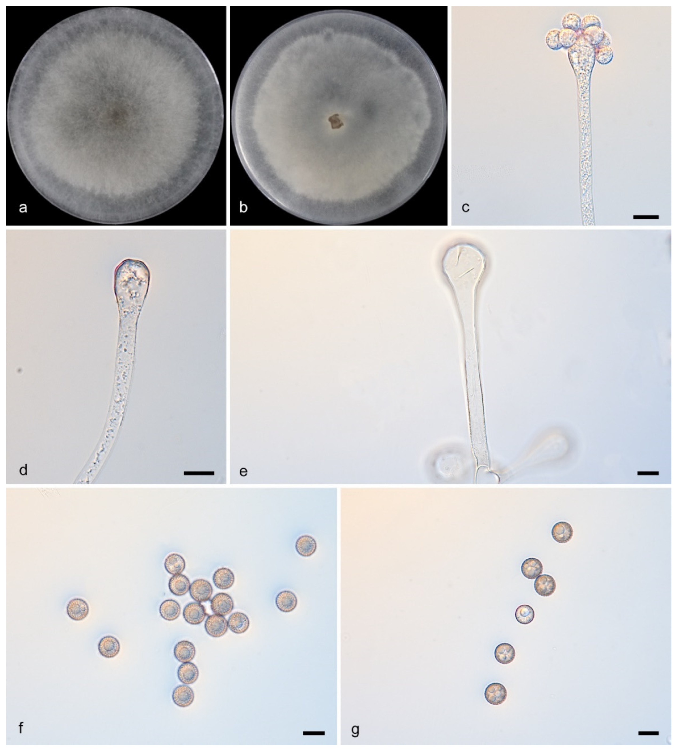
Description—Colonies on PDA incubated at 27 °C for 7 days attained a diameter of 80 mm and a height of 10 mm; they were floccose, light grayish-white in color, and exhibited an irregular margin. The mycelial system consisted of flourishing, branched hyphae (2.0–9.6 μm in diam.) that developed septa with age, and produced abundant, often swollen, root-like rhizoids along with stolons. The reproductive structures featured sympodially branched sporangiophores (37.0–107.5 μm long) that arose aerially, were typically straight, and always broadened upwards; these bore vesicles supporting large, globose, and thick-spined sporangiola (14.1–35.5 μm in diam.). Chlamydospores absent. Zygospores unknown.
Figure 5. Cunninghamella fusca (CGMCC3.28885). (a,b): Colonies on PDA after 7 days of cultivation ((a), obverse; (b), reverse); (ce): Sporangiophores showing branching patterns; (f,g): Sporangiola. Scale bars: 10 µm (cg).
Microorganisms 13 02508 g005
Notes—Strains CGMCC 3.28885 and HZ108-2 form a robust clade (MLBS/BIPP = 100/1), sister to Cunninghamella crassior (Figure 1). Morphologically, these two strains produce brown sporangiola, while C. crassior produce brownish ones.

3.2.5. Cunninghamella guttulata Y. Jiang, H. Zhao, Z. Meng & X.Y. Liu. sp. nov. Figure 6
Fungal name: FN573069
Etymology—The epithet “guttulata” refers to teardrop-shaped sporangiola.
Type—China, Hainan Province, Lingshui Li Autonomous County (18.668825° N, 109.919678° E), soil sample, collected by Y.X. Wang. Holotype HMAS 353833, ex-holotype CGMCC3.28886.
Figure 6. Cunninghamella guttulata (CGMCC3.28886). (a,b): Colonies on PDA after 5 days of cultivation ((a), obverse; (b), reverse); (cf): Sporangiophores showing branching patterns; (g): Rhizoids; (h): sporangiola. Scale bars: 10 µm (ch).
Microorganisms 13 02508 g006
Description—Colonies on PDA at 27 °C for 5 days, reaching 90 mm in diameter, 15 mm high, initially white, becoming Drab Gray with age, floccose, in reverse regular at margin. Hyphae branching, aseptate when young, rarely septate with age, 4.5–20.5 μm in diameter. Rhizoids abundant, root-like, branched. Stolons present. Sporangiophores developed from stolons or aerial hyphae. They were erect, either straight or bent, and exhibited an unbranched or verticillate (2–6 branches) architecture, consistently broadening upwards with frequent septation in the upper regions. The terminal vesicles were subglobose to globose, generally hyaline or subhyaline, and measured 13.5–40.5 μm in length and 12.5–40.5 μm in width. Attached via pedicels, the sporangiola were ovoid and eardrop-shaped (9.0–12.5 × 7.5–9.0 μm), hyaline to subhyaline, often bearing droplets in their early stages, and characterized by a distinctive ornamentation of ovate or drop-shaped spines. Chlamydospores absent. Zygospores unknown.
Notes—The holotype strain of Cunninghamella guttulata (CGMCC3.28886) was isolated from a soil sample collected in Lingshui Li Autonomous County, Hainan Province, China. Phylogenetically, C. guttulata (CGMCC3.28886) and C. guttulata (XG04011-1-2) form a distinct clade with strong statistical support (BI/ML = 94/1). Comparative sequence analysis with C. regularis (CGMCC 3.16114) revealed: ITS similarity: 97.4% (607/618 identical sites, 18/618 gaps); LSU similarity: 99.7% (925/925 identical sites, 0/925 gaps); TEF1α sequence: Not available. Morphologically, C. guttulata (CGMCC3.28886) produces teardrop-shaped sporangiola (length: 9.0–12.5 μm, width: 7.5–9.0 μm), a feature not observed in C. regularis (CGMCC 3.16114), which produces spherical sporangiola (diameter: 6.5–10.0 μm).

3.2.6. Cunninghamella inaequalis Y. Jiang, H. Zhao, Z. Meng & X.Y. Liu. sp. nov. Figure 7

Fungal name: FN573070
Etymology—The epithet “inaequalis” refers to the size variation of sporangiola.
Type—China, Yunnan Province (26.5642168° N, 101.268054° E), soil sample, collected by H. Zhao. Holotype HMAS 353831, ex-holotype CGMCC3.28888.
Figure 7. Cunninghamella inaequalis (CGMCC3.28887). (a,b): Colonies on PDA after 7 days of cultivation ((a), obverse; (b), reverse); (ce): Sporangiophores showing branching patterns; (g): Rhizoids; (f,h): Sporangiola. Scale bars: 10 µm (ch).
Microorganisms 13 02508 g007
Description—Colonies on PDA at 27 °C for 7 days, fast growing, reaching 90 mm in diameter, 20 mm high, at first white, finally Light Gray. Hyphae flourishing, branched, aseptate when young, septate with age, 4.5–17.0 μm in diameter. Stolons present. Sporangiophores were aseptate and developed erectly and straight from aerial hyphae, consistently broadening upwards; they exhibited an unbranched or simply paired, sympodial/dichotomous branching habit, with ovoid or globose vesicles present on the main stalks. Pedicels 1.5–4.0 μm long. Sporangiola borne on pedicels on vesicles, globose, 10.0–17.0 μm in diameter, hyaline when young, Light Brown with age, with many thin spines on the surface. Chlamydospores absent. Zygospores unknown.
Notes—The holotype strain Cunninghamella inaequalis (CGMCC3.28887) was isolated from a soil sample in Yunnan Province, China. Phylogenetically, C. inaequalis (CGMCC3.28887) forms a distinct clade. Comparative analysis with C. regularis (CGMCC 3.16114) revealed 97.5% ITS sequence similarity (578/591 identical, 14/591 gaps) and 100% LSU sequence similarity (1439/1439 identical, 0/1439 gaps); the TEF1α sequence of C. regularis was not available. Morphologically, C. inaequalis produces smaller sporangiola (4.4–12.6 μm in diameter) compared to C. regularis (6.5–10.0 μm in diameter). Additionally, C. regularis sporangiola exhibit slight spines, whereas C. inaequalis sporangiola are smooth with no spines observed.

3.2.7. Cunninghamella monosporangiola Y. Jiang, H. Zhao, Z. Meng & X.Y. Liu. sp. nov. Figure 8

Fungal name: FN573071
Etymology—The epithet “monosporangiola” refers to single sporangiola.
Type—China, Yunnan Province (26.5642168° N, 101.268054° E), soil sample, collected by H. Zhao, Holotype HMAS 353830, ex-holotype CGMCC 3.28888.
Description—Colonies on PDA at 27 °C for 5 days reached 90 mm in diameter and 10–15 mm in height, were floccose, transitioned from white to Drab Gray, and had an irregular reverse margin. Hyphae were branched and 2.5–15.0 μm wide, aseptate when young but septate when mature. Sporangiophores arose from stolons or aerial hyphae, were erect, straight or recumbent, 1.5–251.5 μm long, sometimes broadening upwards, and branched singly, in pairs, sympodially, or verticillately (2–5), often septate below vesicles. Vesicles were ovoid/globose, mostly hyaline (6.0–44.0 × 5.0–40.0 μm). Sporangiola on pedicels (1.0–1.5 μm) were ovoid/globose (7.0–14.5 × 6.5–12.5 μm), thick-spined and produce single sporangiola (Figure 8e,h). Chlamydospores absent. Zygospores unknown.
Notes—The type strain Cunninghamella monosporangiola (CGMCC3.28888) was isolated from a soil specimen collected in Yunnan Province, China. Phylogenetically, C. monosporangiola (CGMCC3.28888) formed a distinct clade in the phylogenetic tree and exhibited 54 nucleotide differences compared to C. verrucosa (CGMCC 3.16260) (ITS: 49/637, LSU: 4/1442, TEF1α: 1/567). Morphologically, C. monosporangiola (CGMCC3.28888) was observed to release a single sporangiospore from its vesicles, a feature not documented in other strains.
Figure 8. Cunninghamella monosporangiola (CGMCC3.28888). (a,b): Colonies on PDA after 5 days of cultivation ((a), obverse; (b), reverse); (ch): Sporangiophores showing characteristic branching patterns; (i,j): Sporangiola. Scale bars: 10 µm (cj).
Microorganisms 13 02508 g008

3.2.8. Cunninghamella tuberculata Y. Jiang, H. Zhao, Z. Meng & X.Y. Liu. sp. nov. Figure 9

Fungal name: FN573072
Etymology—The epithet “tuberculata” describes the tuberculate, nodular, wart-like ort knotted protrusions on sporangiola.
Type—China, Yunnan Province (26.5642168° N, 101.268054° E), soil sample, collected by Zhao Heng, Holotype HMAS 353828, ex-holotype CGMCC3.28889.
Description—Colonies on PDA floccose, reaching 9 cm at 25 °C for 5 days, initially white, soon gray, finally gray-brownish, in reverse gray. Hyphae branched, 3.5–19.5 μm diameter, septate when old. Rhizoids and stolons present. Sporangiophores erect or recumbent, arising from stolons or aerial hyphae, main axes usually equivalent and 3.5–20.4 μm in diameter, rarely gradually thickening upwards, primary branches 61.5–287.7 μm long and 4.6–13.4 μm wide, rebranching or third-branching into branchlets. Sporangiola on pedicels on vesicles, spherical, spiny, covered with tuberculate, nodular, wart-like ort knotted protrusions, 4.1–10.7 μm in diameter. Chlamydospores absent. Zygospores unknown.
Notes—The type strain Cunninghamella tuberculata (CGMCC3.28889) was isolated from a soil specimen collected in Yunnan Province, China (N: 26.5642168°, E: 101.268054°). Phylogenetically, C. tuberculata (CGMCC3.28889) formed a distinct clade in the phylogenetic tree and showed close affinity to C. guttata (CGMCC 3.16112), which also occupied an independent clade. Morphologically, C. tuberculata produces smooth-walled sporangiola with tuberculate (nodule-like) protrusions, a trait not observed in any known related species.
Figure 9. Cunninghamella tuberculata (CGMCC3.28889). (a,b): Colonies on PDA after 5 days of cultivation ((a) obverse, (b) reverse); (cg): Sporangiophores showing branching patterns; (h,i): Sporangiola. Scale bars: 10 µm (ci).
Microorganisms 13 02508 g009

4. Discussion

The integrative approach, which combines molecular evidence with morphological, ecological, and other relevant data, forms the foundation for defining taxonomic novelties and reconstructing phylogenies for species delimitation [47,48,49,50,51,52]. Molecular sequences derived from type specimens provide indispensable data for constructing robust phylogenetic frameworks; these frameworks underpin evolutionarily meaningful classification systems [53]. Despite the primacy of molecular data, traditional morphological methods remain indispensable taxonomic tools [54]. Consequently, we describe novel species from China through integrated phylogenetic-morphological analyses [48,49,55].
This study resolves the evolutionary relationships among Cunninghamella species, including the eight new species reported herein via multilocus phylogenetics. We provide detailed morphological illustrations of key taxonomic features, including microscopic structures and colonial characteristics observed in pure cultures. We formally describe eight novel species: C. crassior sp. nov., Cunninghamella diffundens sp. nov., C. fulvicolor sp. nov., C. fusca sp. nov., C. guttulata sp. nov., C. inaequalis sp. nov., C. monosporangiola sp. nov., and C. tuberculata sp. nov. These species share the diagnostic morphological traits in Cunninghamella, echinulate sporangiola borne on verticillately branched vesicles. With these eight new species, this study expands the number of accepted species in Cunninghamella to 39.

Author Contributions

Y.J. and H.Z. were responsible for DNA sequencing, photo editing and paper drafting; Z.-Y.D. and X.-Y.J. were responsible for data analyses; W.-X.L. and F.L. collected soil samples; S.W. revised the manuscript and provided funding. X.-Y.L. and Z.M. took charge of naming the new species, conceiving and revising this paper, and providing funding. All authors have read and agreed to the published version of the manuscript.

Funding

This study was supported by the National Natural Science Foundation of China (Nos. 32170012, 32470004 and 32300011), Key Technological Innovation Program of Shandong Province, China (2022CXGC020710), Ji’nan City’s ‘New University 20 Policies’ Initiative for Innovative Research Teams Project (202228028), Innovative Agricultural Application Technology Project of Jinan City (CX202210).

Institutional Review Board Statement

Not applicable.

Data Availability Statement

No new data were created or analyzed in this study. Data sharing is not applicable to this article.

Conflicts of Interest

The authors declare no conflicts of interest.

References

  1. Huelsenbeck, J.P.; Ronquist, F. MRBAYES: Bayesian inference of phylogenetic trees. Bioinformatics 2001, 17, 754–755. [Google Scholar] [CrossRef]
  2. Zheng, R.Y.; Chen, G.Q. A monograph of Cunninghamella. Mycotaxon 2001, 80, 1–57. [Google Scholar]
  3. Hyde, K.D.; Hongsanan, S.; Jeewon, R.; Bhat, D.J.; McKenzie, E.H.C.; Jones, E.B.G.; Phookamsak, R.; Ariyawansa, H.A.; Boonmee, S.; Zhao, Q.; et al. Fungal diversity notes 367–490: Taxonomic and phylogenetic contributions to fungal taxa. Fungal Divers. 2016, 80, 1–270. [Google Scholar] [CrossRef]
  4. Zhao, H.; Zhu, J.; Zong, T.K.; Liu, X.L.; Ren, L.Y.; Lin, Q.; Qiao, M.; Nie, Y.; Zhang, Z.D.; Liu, X.Y. Two new species in the family Cunninghamellaceae from China. Mycobiology 2021, 49, 142–150. [Google Scholar] [CrossRef] [PubMed]
  5. Zhang, Z.Y.; Zhao, Y.X.; Shen, X.; Chen, W.H.; Han, Y.F.; Huang, J.Z.; Liang, Z.Q. Molecular phylogeny and morphology of Cunninghamella guizhouensis sp. nov. (Cunninghamellaceae, Mucorales), from soil in Guizhou, China. Phytotaxa 2020, 455, 31–39. [Google Scholar] [CrossRef]
  6. Nguyen, T.T.T.; Choi, Y.J.; Lee, H.B. Zygomycete fungi in Korea: Cunninghamella bertholletiae, Cunninghamella echinulata, and Cunninghamella elegans. Mycobiology 2017, 45, 318–326. [Google Scholar] [CrossRef]
  7. Liu, C.W.; Liou, G.Y.; Chien, C.Y. New records of the genus Cunninghamella (Mucorales) in Taiwan. Fungal Sci. 2005, 20, 1–9. [Google Scholar]
  8. Alves, A.L.; de Souza, C.A.; de Oliveira, R.J.; Cordeiro, T.R.; de Santiago, A.L. Cunninghamella clavata from Brazil: A new record for the western hemisphere. Mycotaxon 2017, 132, 381–389. [Google Scholar] [CrossRef]
  9. Bragulat, M.R.; Castella, G.; Isidoro-Ayza, M.; Domingo, M.; Cabanes, F.J. Characterization and phylogenetic analysis of a Cunninghamella bertholletiae isolate from a bottlenose dolphin (Tursiops truncatus). Rev. Iberoam. Micol. 2017, 34, 215–219. [Google Scholar] [CrossRef]
  10. Suwannarach, N.; Kumla, J.; Supo, C.; Honda, Y.; Nakazawa, T.; Khanongnuch, C.; Wongputtisin, P. Cunninghamella saisamornae (Cunninghamellaceae, Mucorales), a new soil fungus from northern Thailand. Phytotaxa 2021, 509, 291–300. [Google Scholar] [CrossRef]
  11. Yu, J.; Walther, G.; Van Diepeningen, A.D.; Gerrits Van Den Ende, A.H.G.; Li, R.Y.; Moussa, T.A.A.; Almaghrabi, O.A.; De Hoog, G.S. DNA barcoding of clinically relevant Cunninghamella species. Med. Mycol. 2015, 53, 99–106. [Google Scholar] [CrossRef]
  12. Wang, Y.J.; Zhao, T.; Wu, W.Y.; Wang, M.; Liu, X.Y. Cunninghamella verrucosa sp. nov. (Mucorales, Mucoromycota) from Guangdong province in China. Phytotaxa 2022, 560, 274–284. [Google Scholar] [CrossRef]
  13. Zhao, H.; Nie, Y.; Zong, T.; Wang, K.; Lv, M.; Cui, Y.; Tohtirjap, A.; Chen, J.; Zhao, C.; Wu, F.; et al. Species diversity, updated classification and divergence times of the phylum Mucoromycota. Fungal Divers. 2023, 123, 49–157. [Google Scholar] [CrossRef]
  14. Kwon-Chung, K.J. Taxonomy of Fungi Causing Mucormycosis and Entomophthoramycosis (Zygomycosis) and Nomenclature of the Disease: Molecular Mycologic Perspectives. Clin. Infect. Dis. 2012, 54, S8–S15. [Google Scholar] [CrossRef]
  15. Matruchot, L. Une Mucorinée purement conidienne, Cunninghamella africana. Ann. Mycologici. 1903, 1, 45–60. [Google Scholar]
  16. Waren, L.; Freitas, S.D.; Souza-Motta, C.M.; Barbosa, R.D.N.; Melo, R.F.R.; Santiago, A.L.C.M.D.A. A new homothallic species of Cunninghamella (Mucorales, Mucoromycota) with a synoptic key for the genus in the Neotropics. Sydowia 2024, 77, 119–127. [Google Scholar] [CrossRef]
  17. Spatafora, J.W.; Chang, Y.; Benny, G.L.; Lazarus, K.; Smith, M.E.; Berbee, M.L.; Bonito, G.; Corradi, N.; Grigoriev, I.; Gryganskyi, A.; et al. A phylum-level phylogenetic classification of zygomycete fungi based on genome-scale data. Mycologia 2017, 108, 1028–1046. [Google Scholar] [CrossRef] [PubMed]
  18. Zhao, Y.H.; Wang, H.; Liu, T.X.; Xin, Z.H. The individual lipid compositions produced by Cunninghamella sp. Salicorn 5, an endophytic oleaginous fungus from Salicornia bigelovii Torr. Eur. Food Res. Technol. 2014, 238, 621–633. [Google Scholar] [CrossRef]
  19. Fakas, S.; Papanikolaou, S.; Galiotou-Panayotou, M.; Komaitis, M.; Aggelis, G. Organic nitrogen of tomato waste hydrolysate enhances glucose uptake and lipid accumulation in Cunninghamella echinulata. J. Appl. Microbiol. 2008, 105, 1062–1070. [Google Scholar] [CrossRef]
  20. Alakhras, R.; Bellou, S.; Fotaki, G.; Stephanou, G.; Demopoulos, N.A.; Papanikolaou, S.; Aggelis, G. Fatty acid lithium salts from Cunninghamella echinulata have cytotoxic and genotoxic effects on HL-60 human leukemia cells. Eng. Life. Sci. 2015, 15, 243–253. [Google Scholar] [CrossRef]
  21. Guo, J.; Wang, H.; Liu, D.; Zhang, J.N.; Zhao, Y.H.; Liu, T.X.; Xin, Z.H. Isolation of Cunninghamella bigelovii sp. nov. CGMCC 8094 as a new endophytic oleaginous fungus from Salicornia bigelovii. Mycol. Prog. 2015, 14, 11. [Google Scholar] [CrossRef]
  22. Liu, X.Y.; Huang, H.; Zheng, R.Y. Relationships within Cunninghamella based on sequence analysis of ITS rDNA. Mycotaxon 2001, 80, 77–95. [Google Scholar]
  23. Zhao, H.; Nie, Y.; Huang, B.; Liu, X.Y. Unveiling species diversity within early-diverging fungi from China I: Three new species of Backusella (Backusellaceae, Mucoromycota). MycoKeys 2024, 109, 285–304. [Google Scholar] [CrossRef]
  24. Tao, M.F.; Ding, Z.Y.; Wang, Y.X.; Zhang, Z.X.; Zhao, H.; Meng, Z.; Liu, X.Y. Unveiling species diversity within early-diverging fungi from China II: Three new species of Absidia (Cunninghamellaceae, Mucoromycota) from Hainan Province. MycoKeys 2024, 110, 255–272. [Google Scholar] [CrossRef] [PubMed]
  25. Wang, Y.X.; Zhao, H.; Jiang, Y.; Liu, X.Y.; Tao, M.F.; Liu, X.Y. Unveiling species diversity within early-diverging fungi from China III: Six new species and a new record of Gongronella (Cunninghamellaceae, Mucoromycota). MycoKeys 2024, 110, 287–317. [Google Scholar] [CrossRef]
  26. Ding, Z.Y.; Ji, X.Y.; Tao, M.F.; Jiang, Y.; Liu, W.X.; Wang, Y.X.; Meng, Z.; Liu, X.Y. Unveiling species diversity within early-diverging fungi from China IV: Four new species of Absidia (Cunninghamellaceae, Mucoromycota). MycoKeys 2025, 117, 267–288. [Google Scholar] [CrossRef] [PubMed]
  27. Ji, X.Y.; Ding, Z.Y.; Nie, Y.; Zhao, H.; Wang, S.; Huang, B.; Liu, X.Y. Unveiling species diversity: Within early-diverging fungi. From China V: Five new species of Absidia (Cunninghamellaceae, Mucoromycota). MycoKeys 2025, 117, 267–288. [Google Scholar] [CrossRef]
  28. Wang, Y.X.; Ding, Z.Y.; Ji, X.Y.; Meng, Z.; Liu, X.Y. Unveiling species diversity within early-diverging fungi from China VI: Four Absidia sp. nov. (Mucorales) in Guizhou and Hainan. Microorganisms 2025, 13, 1315. [Google Scholar] [CrossRef]
  29. Ding, Z.Y.; Tao, M.F.; Ji, X.Y.; Jiang, Y.; Liu, W.X.; Wang, S.; Liu, X.Y. Unveiling species diversity within early-diverging fungi from China VII: Seven new species of Cunninghamella (Cunninghamellaceae, Mucoromycota). J. Fungi 2025, 11, 417. [Google Scholar] [CrossRef]
  30. Ji, X.Y.; Jiang, Y.; Li, F.; Ding, Z.Y.; Meng, Z.; Liu, X.Y. Unveiling Species Diversity Within Early-Diverging Fungi from China VIII: Four New Species in Mortierellaceae (Mortierellomycota). Microorganisms 2025, 13, 1330. [Google Scholar] [CrossRef]
  31. Ding, Z.Y.; Ji, X.Y.; Li, F.; Liu, W.X.; Wang, S.; Zhao, H.; Liu, X.Y. Unveiling species diversity within early-diverging fungi from China IX: Four new species of Mucor (Mucoromycota). J. Fungi 2025, 11, 682. [Google Scholar] [CrossRef]
  32. Corry, J.E.L. Rose bengal chloramphenicol (RBC) agar. In Progress in Industrial Microbiology; Elsevier: Amsterdam, The Netherlands, 1995; pp. 431–433. [Google Scholar]
  33. Zhao, H.; Nie, Y.; Zong, T.K.; Dai, Y.C.; Liu, X.Y. Three New Species of Absidia (Mucoromycota) from China Based on Phylogeny, Morphology and Physiology. Diversity 2022, 14, 132. [Google Scholar] [CrossRef]
  34. Zheng, R.Y.; Chen, G.Q.; Huang, H.; Liu, X.Y. A monograph of Rhizopus. Sydowia 2007, 59, 273–372. [Google Scholar]
  35. Zheng, R.Y.; Liu, X.Y.; Li, R.Y. More Rhizomucor causing human mucormycosis from China: R. chlamydosporus sp. nov. Sydowia 2009, 61, 135–147. [Google Scholar]
  36. Zheng, R.Y.; Liu, X.Y. Taxa of Pilaira (Mucorales, Zygomycota) from China. Nova Hedwig. 2009, 88, 255–267. [Google Scholar] [CrossRef]
  37. Zong, T.K.; Zha, H.; Liu, X.L.; Ren, L.Y.; Zhao, C.L.; Liu, X.Y. Taxonomy and phylogeny of four new species in Absidia (Cunninghamellaceae, Mucorales) from China. Front. Microbiol. 2021, 12, 677836. [Google Scholar] [CrossRef] [PubMed]
  38. Doyle, J.J.; Doyle, J.L. Isolation of plant DNA from fresh tissue. Focus 1990, 12, 13–15. [Google Scholar] [CrossRef]
  39. Guo, L.D.; Hyde, K.D.; Liew, E.C.Y. Identification of endophytic fungi from Livistona chinensis based on morphology and rDNA sequences. New Phytol. 2000, 147, 617–630. [Google Scholar] [CrossRef]
  40. White, T.J.; Bruns, T.; Lee, S.; Taylor, J. Amplification and direct sequencing of fungal ribosomal RNA genes for phylogenetics. In PCR Protocols: A Guide to Methods and Applications; Academic Press: New York, NY, USA, 1990; pp. 315–322. [Google Scholar]
  41. Vilgalys, R.; Hester, M. Rapid genetic identification and mapping of enzymatically amplified ribosomal DNA from several Cryptococcus species. J. Bacteriol. 1990, 172, 4238–4246. [Google Scholar] [CrossRef] [PubMed]
  42. O’Donnell, K.; Lutzoni, F.M.; Ward, T.J.; Benny, G.L. Evolutionary relationships among mucoralean fungi Zygomycota: Evidence for family polyphyly on a large scale. Mycologia 2001, 93, 286–297. [Google Scholar] [CrossRef]
  43. Carbone, I.; Kohn, L.M. A method for designing primer sets for speciation studies in filamentous ascomycetes. Mycologia 1999, 91, 553–556. [Google Scholar] [CrossRef]
  44. Kumar, S.; Stecher, G.; Tamura, K. MEGA7: Molecular evolutionary genetics analysis version 7.0 for bigger datasets. Mol. Biol. Evol. 2016, 33, 1870–1874. [Google Scholar] [CrossRef]
  45. Stamatakis, A. RAxML version 8: A tool for phylogenetic analysis and post-analysis of large phylogenies. Bioinformatics 2014, 30, 1312–1313. [Google Scholar] [CrossRef]
  46. Nylander, J. MrModeltest V2: Program distributed by the author. Bioinformatics 2004, 24, 581–583. [Google Scholar] [CrossRef]
  47. Letunic, I.; Bork, P. Interactive Tree of Life (iTOL) v6: Recent updates to the phylogenetic tree display and annotation tool. Nucleic Acids Res. 2024, 52, 78–82. [Google Scholar] [CrossRef] [PubMed]
  48. Jeewon, R.; Hyde, K.D. Establishing species boundaries and new taxa among fungi: Recommendations to resolve taxonomic ambiguities. Mycosphere 2016, 7, 1669–1677. [Google Scholar] [CrossRef]
  49. Hu, D.M.; Wang, M.; Cai, L. Phylogenetic assessment and taxonomic revision of Mariannaea. Mycol. Prog. 2016, 16, 271–283. [Google Scholar] [CrossRef]
  50. Zou, Y.; Hou, J.; Guo, S.; Li, C.; Li, Z.; Stephenson, S.L.; Pavlov, I.N.; Liu, P.; Li, Y. Diversity of Dictyostelid Cellular Slime Molds, Including Two Species New to Science, in Forest Soils of Changbai Mountain, China. Microbiol. Spectr. 2022, 10, e02402-22. [Google Scholar] [CrossRef]
  51. Hurdeal, V.G.; Gentekaki, E.; Lee, H.B.; Jeewon, R.; Hyde, K.D.; Tibpromma, S.; Mortimer, P.E.; Xu, J.C. Mucoralean fungi in Thailand: Novel species of Absidia from tropical forest soil cryptogamie. Mycologie 2021, 42, 39–61. [Google Scholar] [CrossRef]
  52. Zhao, H.; Nie, Y.; Zong, T.K.; Wang, Y.J.; Wang, M.; Dai, Y.C.; Liu, X.Y. Species Diversity and Ecological Habitat of Absidia (Cunninghamellaceae, Mucorales) with Emphasis on Five New Species from Forest and Grassland Soil in China. J. Fungi 2022, 8, 471. [Google Scholar] [CrossRef]
  53. Zhao, H.; Lv, M.L.; Liu, Z.; Zhang, M.Z.; Wang, Y.N.; Ju, X.; Song, Z.; Ren, L.Y.; Jia, B.S.; Qiao, M.; et al. High-yield oleaginous fungi and high-value microbial lipid resources from Mucoromycota. BioEnergy Res. 2021, 14, 1196–1206. [Google Scholar] [CrossRef]
  54. Weitzman, I.; Crist, M.Y. Studies with clinical isolates of Cunninghamella II. 654 Physiological and morphological studies. Mycologia 1980, 72, 661–669. [Google Scholar] [CrossRef] [PubMed]
  55. Alors, D.; Lumbsch, H.T.; Divakar, P.K.; Leavitt, S.D.; Crespo, A. An integrative approach for understanding diversity in the Punctelia rudecta Species Complex (Parmeliaceae, Ascomycota). PLoS ONE 2016, 11, e0146537. [Google Scholar] [CrossRef] [PubMed]
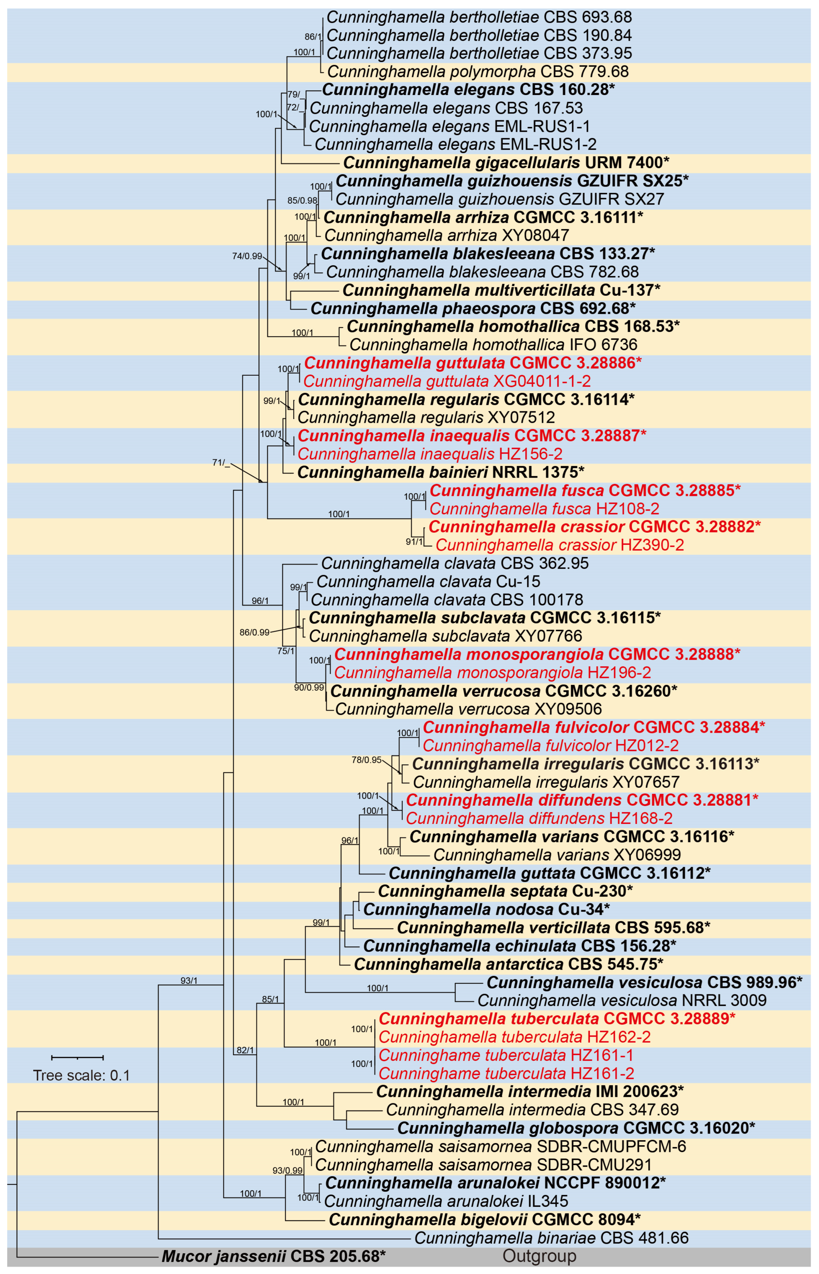
Figure 1. The Maximum Likelihood (ML) phylogenetic tree of the genus Cunninghamella based on ITS-LSU-TEF1α sequences. The ML Bootstrap Support (MLBS no less than 70%)/Bayesian Inference Posterior Probability (BIPP no less than 0.90) values are shown on branches. Strains newly isolated in this study are indicated in red. Ex-types are bold, plus an asterisk “*”. The scale bar above the tree roots represents 0.1 substitutions per site.
Microorganisms 13 02508 g001
Table 1. PCR information used in this study.
LociPCR PrimersPrimer Sequences (5′–3′)PCR CyclesReferences
ITSITS5GGA AGT AAA AGT CGT AAC AAG G94 °C 5 min; (94 °C 30 s, 55 °C 30 s, 72 °C 1 min) × 35 cycles; 72 °C 10 min[40]
ITS4TCC TCC GCT TAT TGA TAT GC
LSULR0RGTA CCC GCT GAA CTT AAG C94 °C 5 min; (94 °C 30 s, 51 °C 30 s, 72 °C 1 min) × 35 cycles; 72 °C 10 min[41]
LR7TAC TAC CAC CAA GAT CT
TEF1αEF1-728FATG TGC AAG GCC GGT TTC GC94 °C 5 min; (94 °C 30 s, 48 °C 30 s, 72 °C 1 min) × 35 cycles; 72 °C 10 min[42,43]
EF-2GGA RGT ACC AGT SAT CAT GTT
Table 2. GenBank accession numbers of Cunninghamella and Mucor sequences used in this study.
SpeciesStrainsGenBank Accession Numbers
ITSLSUTEF1α
C. antarcticaCBS 545.75 TJN205893JN206597KJ156492
C. arrhizaCGMCC 3.16111 TOL678142PQ399916NA
XY08047OL678143NANA
C. arunalokeiIL3459MN431159MN431158NA
NCCPF 890012 TNR_177485NG153887NA
C. bainieriCBS 481.66MH858865MH870507KJ156495
CGMCC 8094 TKJ013403KJ013405KJ395944
NRRL 1375 TAF254935NANA
C. bertholletiaeCBS 190.84JN205878HM849701NA
CBS 373.95JN205873NAKJ156497
CBS 693.68AF254931MH870924KJ156490
C. bigeloviiCGMCC 8094 TKJ013403KJ013405KJ395944
C. binariaeCBS 481.66MH858865MH870507KJ156495
C. blakesleeanaCBS 133.27 TNR119974MH866397KJ156479
CBS 782.68JN205869MH870950KJ156478
C. clavataCBS 100178JN205890JN206604KJ156477
Cu-15AF254942NANA
CBS 362.95JN205891NANA
C. crassiorCGMCC3.28882 TPV235924PV239680PV254891
HZ390-2PV235925PV239681PV254892
C. diffundensCGMCC3.28881 TPV235920PV239676PV254889
HZ168-2PV235921PV239677PV254890
C. echinulataCBS 156.28 TJN205895JN939199.1KJ156500
C. elegansCBS 160.28 TAF254928NR_154747KJ156470
CBS 167.53JN205882HM849700KJ156494
EML-RUS1-1MF806023MF806027NA
EML-RUS1-2MF806021MF806028NA
C. fulvicolorCGMCC3.28884 TPV235910PV239666PV254879
HZ012-2PV235911PV239667PV254880
C. fuscaCGMCC3.28885 TPV235908PV239664PV254877
HZ108-2PV235909PV239665PV254878
C. globosporaCGMCC 3.16020 TMW264073MW264132NA
C. gigacellularisURM 7400 TNR_168760NG_068773NA
C. guizhouensisGZUIFR-SX25 TMN908596MN908599MN912633
GZUIFR-SX27MN908598MN908601MN912635
C. guttataCGMCC 3.16112 TOL678144PQ399917NA
C. guttulataCGMCC3.28886 TPV235930PV239686PV254893
XG04011-1-2PV235931PV239687PV254894
C. homothallicaCBS 168.53 TJN205863JN206605KJ156498
IFO 6736AF254941NANA
C. inaequalisCGMCC3.28887 TPV235914PV239670PV254883
HZ156-2PV235915PV239671PV254884
C. intermediaCBS 347.69JN205892JN206606NA
IMI 200623 TAF254939NANA
C. irregularisCGMCC 3.16113 TOL678145PQ399918NA
XY07657OL678146NANA
C. monosporangiolaCGMCC3.28888 TPV235922PV239678PV785980
HZ196-2PV235923PV239679PV785981
C. multiverticillataCBS 989.96 TJN205897HM849693KJ156474
Cu-137 TAF254933NANA
NRRL 3009AF254943NANA
C. nodosaCu-34 TAF346407NANA
C. phaeosporaCBS 692.68 TJN205864HM849697NA
C. polymorphaCBS 779.68JN205874JN206599NA
C. regularisCGMCC 3.16114 TOL678148PQ399919NA
XY07512OL678150NANA
C. saisamornaeSDBR-CMUPFCM-6MW709394MW699571MW715866
SDBR-CMU291MG571234MW699591MW715865
C. septataCu-230 TAF346408NANA
C. subclavataCGMCC 3.16115 TOL678152NANA
XY07766OL678153NANA
C. tuberculataCGMCC3.28889 TPV235918PV239674PV254887
HZ162-2PV235919PV239675PV254888
C. variansCGMCC 3.16116 TOL678154PQ399920NA
XY06999OL678155NANA
C. vesiculosaCBS 989.96 TJN205897HM849693KJ156474
NRRL 3009AF254943NANA
C. verrucosaCGMCC 3.16260ON262555ON261192NA
XY09506ON262556ON261193NA
C. verticillataCBS595.68 TAF254937NANA
M. jansseniiCBS 205.68 TMH859119MH870832NA
Notes: New species discovered herein are shown in bold. The “T” indicates ex-type or ex-holotype strains. The “NA” stands for “not available”.
Disclaimer/Publisher’s Note: The statements, opinions and data contained in all publications are solely those of the individual author(s) and contributor(s) and not of MDPI and/or the editor(s). MDPI and/or the editor(s) disclaim responsibility for any injury to people or property resulting from any ideas, methods, instructions or products referred to in the content.