Article

Genomic Drivers of Biofilm Formation in Salmonella Enteritidis and S. Kentucky from Poultry Production

1
Department of Food and Human Nutritional Sciences, University of Manitoba, Winnipeg, MB R3T 2S2, Canada
2
Guelph Research and Development Center, Agriculture and Agri-Food Canada (AAFC), Guelph, ON N1G 5C9, Canada
3
National Microbiology Laboratory, Public Health Agency of Canada, Winnipeg, MB R3E 3R2, Canada
4
Department of Medical Microbiology and Infectious Diseases, University of Manitoba, Winnipeg, MB R3E 0J9, Canada
5
Lethbridge Research and Development Centre, Agriculture and Agri-Food Canada, Lethbridge, AB T1J 4B1, Canada
6
Department of Microbiology, University of Manitoba, Winnipeg, MB R3T 2N2, Canada
*
Author to whom correspondence should be addressed.
Microorganisms 2025, 13(11), 2473; https://doi.org/10.3390/microorganisms13112473
Submission received: 19 September 2025 / Revised: 26 October 2025 / Accepted: 27 October 2025 / Published: 29 October 2025
(This article belongs to the Section Biofilm)

Abstract

Salmonella Enteritidis (SE) remains a leading cause of human illness worldwide, and its persistence in poultry environments might be partially attributed to their ability to form biofilm. This study compared the biofilm capacity of 15 SE and 24 Salmonella Kentucky (SK) isolates from poultry products and processing facilities to uncover genetic factors driving biofilm heterogeneity. Biofilm formation and curli/cellulose production were evaluated at 20–22 °C. Genomic analyses included phylogenetic reconstruction, comparative system profiling, SNP variation, and BLASTp v2.17.0 comparisons. Phenotypic assays showed that most SE isolates (73%) were strong biofilm formers, while the majority of SK isolates (62%) failed to form biofilms, despite many carrying the complete curli–cellulose gene set and other biofilm-associated genes. Genomic analysis identified 124 biofilm-related genes, 108 of which were conserved across all isolates, and revealed 24 variants with potential functional impact. Mutations in cellulose biosynthesis (bcs) genes were linked to weaker biofilms, whereas nonsynonymous variants in tol family genes may impair flagellar biosynthesis and matrix stability. These findings demonstrate that genetic variation, not just gene presence, shapes biofilm phenotypes and highlight key molecular targets that may explain why SE persists in poultry production while SK is less successful.

1. Introduction

Despite extensive efforts to implement food safety measures and reduce salmonellosis, outbreaks continue to occur, posing a persistent threat to food safety and public health worldwide. Notably, current food safety programs in the United States fell short of achieving the 2020 national public health goal for reducing Salmonella-related illnesses [1]. Non-typhoidal Salmonella serotypes are one of the leading causes of foodborne illness [2]. Salmonella enterica serovar Enteritidis (SE) has been linked to numerous poultry, egg, and egg product-associated outbreaks [3,4,5,6,7]. SE persistence in poultry barns, egg layer barns, and poultry processing facilities remains a persistent problem. In 2023, Salmonella Enteritidis is considered the predominant serotype in Canada, accounting for 37.02% of all human salmonellosis, followed by Salmonella Typhimurium (6.15%), and Salmonella ssp. I 4,[5], 12:I:- (5.46%) [8]. Salmonella enterica serovar Kentucky (SK) is another serovar where poultry is the primary carrier, transmitting the bacteria to humans through the consumption of contaminated foods [9]. SK is a polyphyletic non-typhoidal Salmonella serovar that comprises two major sequence types (STs), ST152 and ST198, representing distinct genetic lineages [10]. The ST 152 is more prevalent in the U.S. poultry and poultry products. The ST 152 is sporadically linked with human illness; however, it has not been associated with major foodborne Salmonella outbreaks in the U.S. [11]. In contrast, ST 198 is more prevalent in international poultry (poultry outside the U.S.) and is more frequently associated with human disease [10]. In the EU, confirmed and reported human salmonellosis cases caused by Salmonella Kentucky accounted for only 0.3–0.4% of cases between 2010 and 2016, making it far less prevalent than Salmonella Enteritidis, which accounted for 48.0–57.1% during the same period [12]. In 2023, cases of Salmonella Kentucky were reported across five Canadian provinces: British Columbia (n = 8), Alberta (n = 3), Saskatchewan (n = 1), Ontario (n = 19), and Quebec (n = 7) [8].
Similarly to salmonellosis caused by S. Enteritidis, human illness from S. Kentucky is likely underreported, which may be attributed to its self-limiting nature. However, it is still considered one of the new five target serovars for breeding hens in the EU after 2014, and it had the highest proportion of travel-associated cases (53.2%) in the EU in 2016 [12]. Despite the high prevalence of Salmonella Kentucky in poultry and the poultry processing environment, it causes a very low estimated burden of illness; this has been a longstanding conundrum for the food safety and public health communities, and it has been thought to be attributed to low virulence [13].
Salmonella continues to evade food safety controls, a persistence that may be linked to its remarkable adaptability and resilience. One mechanism that bacteria use to withstand environmental stress is the formation of biofilms. Biofilms have also been associated with bacterial persistence, primarily due to the extracellular polymeric substance (EPS) matrix, which impedes the penetration of antibiotics and disinfectants. The EPS matrix also provides humidity, which helps bacteria to survive longer [14,15,16]. Biofilm refers to a group of surface-associated microorganisms that can attach to the surface and produce an amorphous extracellular polymeric substance (EPS) matrix. EPS serves as a structural framework for the biofilm’s three-dimensional layout, allowing cells to adhere to surfaces and interact with one another within the biofilm [17]. Bacteria within biofilms possess strong capabilities to survive in hostile environments, as the EPS can protect them against antimicrobial compounds, desiccation, and other environmental stresses [18,19]. The structure of EPS varies based on the ecological conditions in which biofilms are formed. Biofilm EPS matrix consists predominantly of polysaccharides, proteins, phospholipids, and other biomolecules [14]. Biofilms can be composed of single species or multispecies, with either single or multi-layered [20]. Bacteria can form biofilms on numerous surfaces, both biotic and abiotic, and at different temperatures [14]. The ability of Salmonella to form resilient biofilms in facilities like barns and processing plants creates a constant source of potential contamination. These persistent isolates can be difficult to eradicate, leading to ongoing food safety hazards and recurring outbreaks. In broiler houses, key risk factors for Salmonella contamination include feces, dust, and feed. Day-old chicks and transport vehicles also introduce and recirculate the pathogen. In contrast, the most significant risk factors in laying hen houses are dust, surfaces, and feces [21]. Microbial biofilms readily establish themselves across food-production environments, within water distribution systems and poultry drinkers, as well as on facility surfaces such as floors, walls, ceilings, and equipment (e.g., conveyor belts, plastic, and rubber components) [22]. Bacterial biofilm production can be classified as strong, moderate (or intermediate), weak, or non-biofilm formers [23]. In layer barns in Manitoba, the ambient temperature is carefully controlled and typically maintained within the 20–22 °C range to optimize hen welfare and egg production. However, these temperatures could facilitate Salmonella biofilm formation in poultry production environments [24]. The ability of Salmonella to establish robust biofilms on diverse surfaces is closely associated with bacterial surface structures, including flagella, fimbriae, and cellulose [25]. The specific type of fimbria called curli, as well as cellulose, are key phenotypic markers commonly associated with the biofilm-forming ability of bacteria. The RDAR morphotype (red, dry, and rough) is a characteristic colony morphology observed in some Enterobacteriaceae family members, particularly S. enterica and E. coli, when grown on Congo Red agar. It is commonly associated with biofilm-forming ability and environmental persistence. The RDAR phenotype-expressing colonies consist of proteinaceous compounds, such as adhesive curli fimbriae and exopolysaccharides, including cellulose, as well as an O-antigenic capsule, capsular polysaccharide, and lipopolysaccharide [14]. Curli are amyloid-like cell-surface proteins that are responsible for host colonization, persistence, motility, and invasion in Salmonella spp. and have been reported to be essential for biofilm formation since they promote cell-surface and cell-to-cell interactions [14]. By comparing genomic sequences with observed biofilm phenotypes, critical links between a strain’s genetic makeup and its resilience can be revealed. This genomic insight not only enhances our ability to predict the behavior of emerging Salmonella strains but also provides the poultry industry with tools for risk assessment and management. In addition, elucidating the genetic basis of strong biofilm formation provides valuable targets for innovative control measures, ultimately supporting the development of more effective strategies to mitigate pathogen persistence and enhance food safety. Accordingly, this research investigated the relationship between isolates of Salmonella Enteritidis (SE) and Salmonella Kentucky (SK) serotypes exhibiting different biofilm formation phenotypes (strong, intermediate, weak, and non-biofilm formers) and their corresponding genomic profiles. The objective was to identify marker genes linked to biofilm formation that explain variations in bacterial resilience and offer insights for developing targeted control strategies to reduce Salmonella contamination and enhance risk assessment.

2. Materials and Methods

2.1. Salmonella Biofilm Formation

2.1.1. Salmonella Bacterial Isolates Selection and Culture Conditions

Fifteen Salmonella Enteritidis (SE) isolates were included in this study, selected based on their source within the poultry environment, facilities, and processing steps (Table 1). Several SE isolates were recovered from end-products, including chicken nuggets, chicken breast, and chicken feed, indicating their persistence through multiple processing stages. One additional SE isolate (#107) was included as a control, as it was the only intermediate biofilm former that tested negative for curli and cellulose production. In total, all SE isolates were evaluated for their ability to form biofilms. To broaden the range of biofilm phenotypes, 24 Salmonella Kentucky (SK) isolates, also isolated from a poultry processing facility in Agassiz, Canada, were included (Table 1). All isolates were obtained from the University of Manitoba culture collection.

2.1.2. Culturing Procedures

All bacterial isolate cultures used in this research were kept in a −80 °C freezer with Trypticase Soy Broth (TSB; Becton, Becton and Dickinson, Sparks, NV, USA) and 15% glycerol. To activate the cultures, a loopful of the frozen culture was streaked onto Xylose Lysine Deoxycholate (XLD) agar plates (Hardy Diagnostics Inc., Santa Maria, CA, USA) and incubated at 37 °C overnight to obtain isolated colonies. A single colony of each bacterial isolate was transferred and inoculated from the overnight agar plate into a 10 mL sterile Lennox Broth no-salt (LN-NS (10 g tryptone (VWR Chemicals LLC, Solon, OH, USA) +5 g yeast extract (Thermo Fisher Scientific, Sasingstoke, UK) in 1 L distilled water and incubated at 37 °C overnight. The overnight culture was adjusted to a concentration of 108 colony-forming units (CFU/mL) by diluting it with LB-NS using a 0.5 McFarland Standard (108 CFU), for culture concentration confirmation. OD (optical density) was measured (0.30 nm to 0.35 nm) on the spectrophotometer (Thermo Scientific™, Genesys 20) for further use.

2.1.3. Biofilm Assessment Using the Crystal Violet Method

For Salmonella biofilm assessment, the CV method was followed [26]. For this purpose, Salmonella cultures (108 CFU) were diluted to achieve a 107 CFU/mL. In a 96-well microplate, 180 µL of sterile LB-NS was dispensed into each of the 96 wells. Using a multi-channel pipette, 20 µL of each individual Salmonella culture was dispensed into the wells to achieve a 106 CFU/mL cells in the microplates. Each plate included one positive control (E. coli R508) and one sterility control (200 µL LB-NS with no bacteria). Each experiment was repeated four times. The microplates were incubated at 20–22 °C for 5 days. After the 5-day incubation, biofilm development was assessed. Microplate wells were washed tree times with 300 µL 1.25% Butterfield’s Phosphate Buffer (KH2PO4) (BPB) (Hardy Diagnostics Inc., Santa Maria, CA, USA) using a microplate washer (405 LS, BioTek, Winooski, VT, USA), then plates were air dried for 10 min, following by adding 200 µL of absolute methanol to each well, for 15 min to fix the attached biofilm. After 15 min, the methanol was removed and air-dried for 15 min. The biofilm was then stained with 200 µL of 0.1% crystal violet (CV) (Sigma Aldrich, St. Louis, MO, USA) [26] for 20 min. Next, the fixed biofilm was washed three times with 200 µL BPB and air-dried for 15 min. Finally, 200 µL of 85% ethanol was added to each well, microplates were agitated for 10 s to dissolve the crystal violet attached to the biofilm. The optical density (OD) was measured at 630 nm using a microplate reader (BioTek ELx800; BioTek Instruments Inc., Winooski, VT, USA).
The data obtained from the microplate reader were analyzed using the method described by Stepanović et al. and Adator et al. [23,26]. The biofilm classification cutoff line was based on the value of the negative controls, where ODc (optical density cut-offs) = mean of negative control+3*SD of the negative control. The biofilm-forming ability, as measured by the OD value, was then adjusted to fit within the range. When OD ≤ ODc, it is a non-biofilm former; when ODc < OD ≤ 2ODc, it is a weak biofilm former; when 2ODc > OD ≤ 4ODc, it is an intermediate biofilm former; when 4ODc < OD, it is a strong biofilm former.
For assessing the biofilm-forming ability of the Salmonella isolates, interactions between isolates and biofilm categories were analyzed using the Chi-Square method. For all statistical analyses, significance was declared at p < 0.0001.

2.1.4. Curli and Cellulose Detection

Salmonella Enteritidis and Kentucky isolates were assessed for their capacity to produce curli and cellulose at 20–22 °C. For curli production, the overnight cultures of Salmonella isolates were steaked on Congo red agar (10 g/L casamino acids (VWR, Solon, OH, USA), 1 g/L yeast extract (Thermo Fisher Scientific, Basingstoke, UK), and 20 g/L agar (Becton, Dickinson and Company, Sparks, MD, USA), supplemented with 20 µg/mL Coomassie brilliant blue dye (Sigma Aldrich, St. Louis, MO, USA) and 40 µg/mL Congo red dye (Sigma Aldrich, St. Louis, MO, USA) [26]. For cellulose production, the overnight cultures of Salmonella isolates were streaked on Luria–Bertani (LB) agar (Hardy Diagnostics CulGenexTM, Santa Maria, CA, USA) supplemented with 200 mg/L Calcofluor dye (Sigma Aldrich, St. Louis, MO, USA). The plates were incubated at 22 °C for 1–2 days. A positive result for curli production is shown as red, dry, and rough (RDAR) morphotype, which indicates the presence of curli in the extracellular matrix; other morphotypes indicate negative results. The presence of fluorescence colonies on LB agar with Calcofluor dye at 366 nm UV light indicates a positive result for cellulose, and colonies that do not fluoresce indicate a negative result.

2.2. Genomic Comparison of S. Enteritidis and S. Kentucky with Regard to Their Biofilm Formation Gene Information

2.2.1. Whole Genome Sequence Generation

Genomic DNA from Salmonella isolates was extracted using the DNeasy Blood and Tissue Kit-Gram-Negative protocol (Qiagen, Inc., Toronto, ON, Canada) according to the manufacturer’s recommendations. DNA concentration and quality were tested using NanoDrop (NanoDrop Technologies, Wilmington, DE, USA). The whole genome sequence for the fifteen Salmonella Enteritidis (SE) isolates and twenty-four Salmonella Kentucky (SK) isolates was carried out by the National Microbiological Laboratory, Public Health Agency of Canada, located in Winnipeg.
Quantification of the DNA was performed using the Qubit HS kit. The genomic DNA (gDNA) was first normalized using the Biomek i7 robot. The library samples were prepared using the Illumina DNA prep kit, followed by post-PCR purification with AmpPure XP beads. Size selection was performed using the Blue Pippin system, and the average base pair size was determined using the Agilent TapeStation automated electrophoresis instrument. Sequencing was performed on the Illumina MiSeq platform with the 600 v3 kit. The National Microbiology Laboratory, Winnipeg, Genomics Core, conducted all sequencing and library preparation procedures. The whole genome sequences can be found on the NCBI website (bio project PRJNA1285942 and PRJNA1290672) and Table A1.
Whole genome sequence raw data were assembled [27] and annotated [28] using the Bacterial and Viral Bioinformatics Resource Center (BV-BRC v3.54.6) platform SPAdes v4.2.0 and RAST/SEED systems (https://rast.nmpdr.org/rast.cgi), respectively, using the Illumina platform with a unicycler v0.5.1. In total, all 15 SE and 24 SK whole genomes showed good quality (completeness at 100%, contamination at 0%) [29].
Summary of sequencing reads, assembly metrics, and genome Features for isolates included in This Study can be found in S1.

2.2.2. Phylogenetic Tree and Heatmapping

A phylogenetic tree was generated for both SE and SK isolates together using 500 single-copy gene parameters in the mafft alignment program and RAxML Fast Bootstrapping [30]. The genes in the phylogenetic tree were selected randomly from a defined number of the BV-BRC Global Protein Families to build an alignment and develop a phylogenetic tree based on the sequence differences. The number of genes ranges from 10 to 1000 for selections based on the genomes of interest [31]. A comparative system [29] was used to compare up to 500 genes and protein families using the whole genome sequences of all SE and SK isolates. Using the protein family sorter, genes of interest were searched, filtered, and grouped to examine the distribution of protein families across the genomes. A pan-genome of 5396 genes was gathered under the comparative system tool with a focus on the intra-genus comparison (PLfam) to examine the presence and absence of 124 genes and proteins of interest which play essential roles in biofilm formation, curli and cellulose production, flagella, fimbriae and pili expression, and type IV secretion system since these components were reported to be associated with biofilm development. Since all the isolates belong to the same genus (Salmonella), the PLfam uses only proteins within a genus, and more stringent criteria [32]. More than 100 genes were filtered and selected as they were genes associated with biofilm formation, the production of curli and cellulose, the functions of flagella, fimbriae, and pili [33]. Filtered genes were examined for their functions and were illustrated with the iTol v7 service [34].

2.2.3. SNP Variation Analysis

A single nucleotide polymorphism (SNP) analysis was performed using BWA-MEM for alignment and FreeBayes as the SNP caller to identify genomic variations at specific nucleotide positions where a single base pair differs among individuals or populations [35]. SNP impacts were categorized using SnpEff, a variant annotation tool integrated into the BV-BRC pipeline. SnpEff predicts the functional consequences of each SNP based on its genomic context and classifies them into four categories, high, moderate, low, and modifier, according to the expected effect on gene or protein function. This analysis provided a comprehensive overview of potential mutations across samples. Since almost all essential protein-coding genes were present regardless of biofilm phenotype, it was vital to examine functional mutations, particularly in coding regions and upstream/downstream regulatory elements, as genes may be present but only partially functional. SNPs were examined among all studied 39 SE and SK isolates studied, using the SE isolate 53932 and SK isolate PW9-3 as the reference genomes due to their strong biofilm-forming abilities.

2.2.4. BLASTp (Basic Local Alignment Search Tool for Protein) Comparison

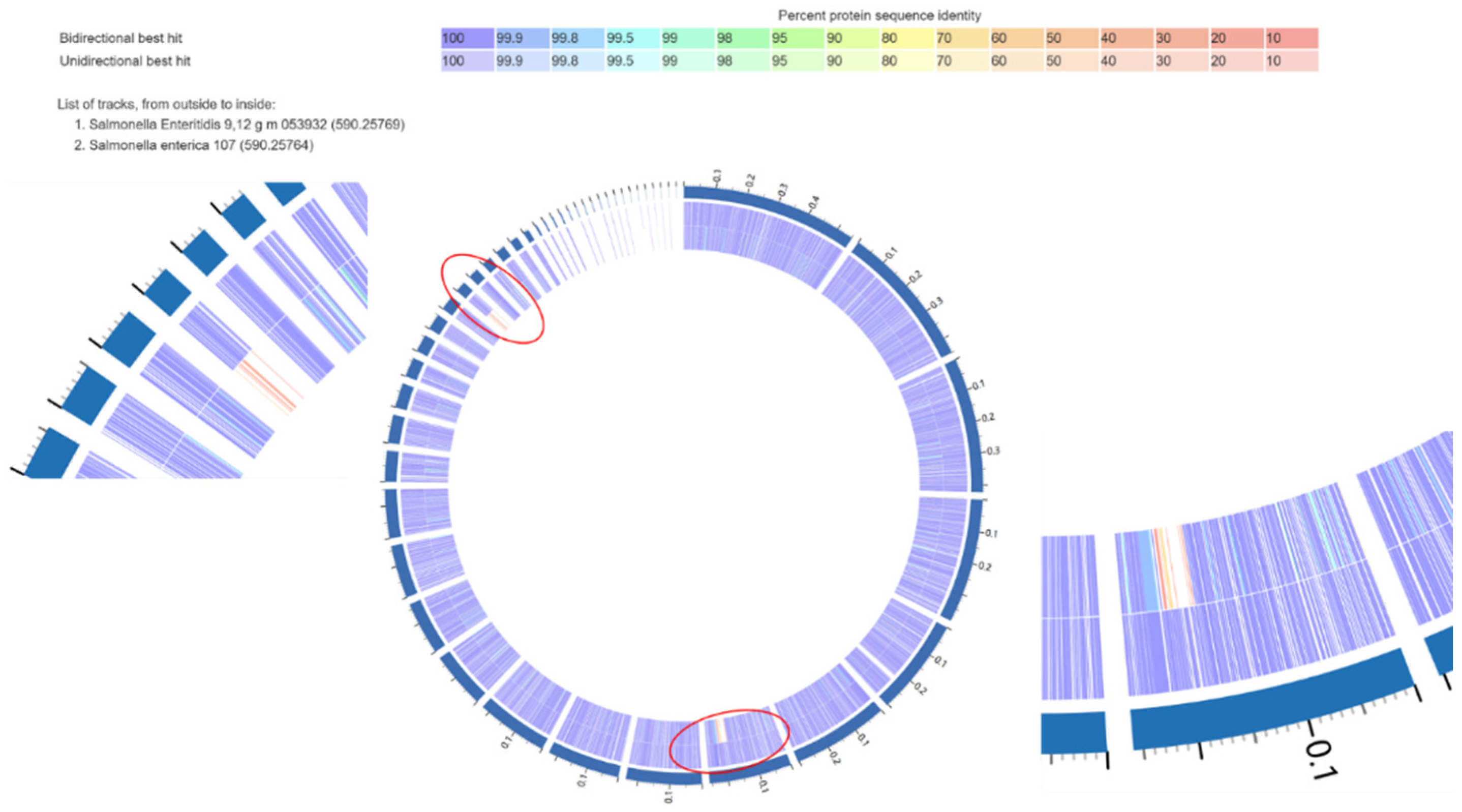
A BLASTp comparison was used in this study to identify similarities between protein sequences and to find the best unique/unidirectional/bidirectional hit between the two trains used in the current study [36]. This sequence-based method, part of the RAST annotation pipeline [37], helped determine whether specific proteins found in strong biofilm-forming isolates were missing in intermediate, weak, or non-biofilm-forming isolates. Specifically, for the Salmonella Enteritidis (SE) isolates, a strong biofilm-forming poultry isolate (isolate 53932) was compared against the intermediate biofilm-forming SE isolate 107.

2.2.5. BLASTp (Basic Local Alignment Search Tool for Protein) Comparison

Associations between phenotypic traits, including biofilm formation category (strong, moderate, weak, and non-producer), and the production of curli and cellulose, and genomic characteristics (SNP type and predicted SNP impact) were analyzed using the Chi-square test. Specifically, the analysis evaluated whether the distribution of isolates across biofilm formation categories was independent of genomic traits, such as the presence or absence of specific SNPs or their functional impact. Statistical significance was determined at p < 0.0001.

3. Results

3.1. Biofilm Formation

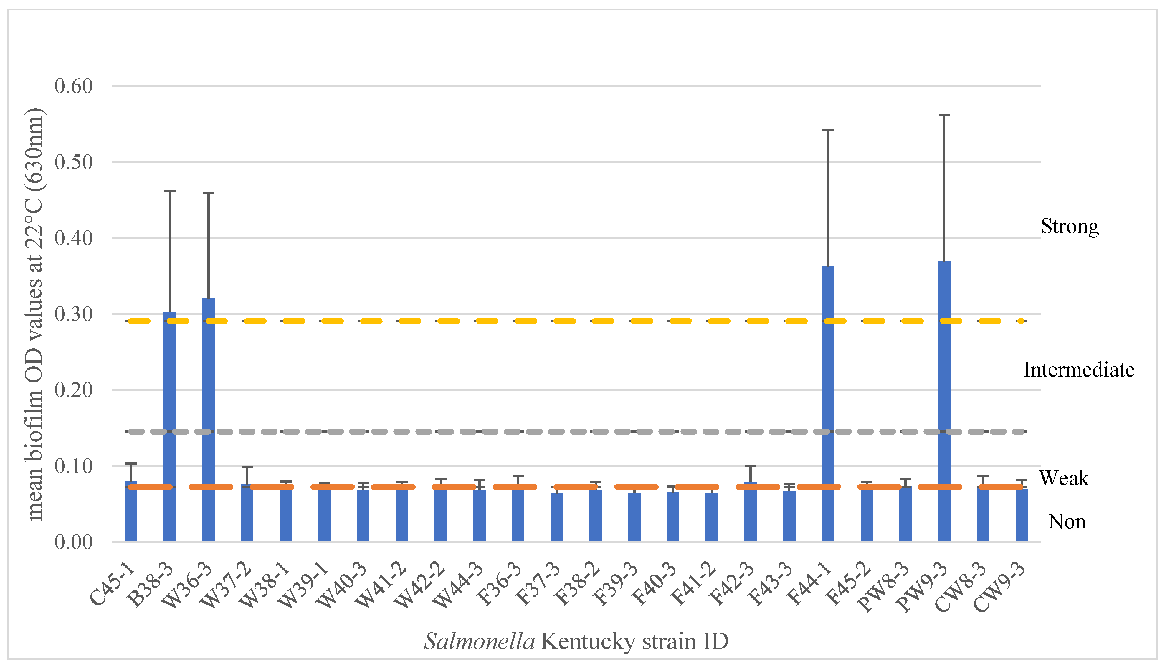
In this study, 15 SE and 24 SK isolates were tested for biofilm formation using the crystal violet 96-well microplate method (Appendix A.2) at 20–22 °C, a temperature range that reflects conditions commonly found in Canadian poultry barns, typically ranging from 19 to 25 °C [38]. Within the 15 SE isolates, results showed that 11 isolates (73.3%) produced strong biofilms, while 4 isolates (26.7%) produced intermediate biofilms (p < 0.0001) (Figure 1). The three intermediate biofilm producers were isolated from chicken nuggets (n = 2) and chicken feed (n = 1), indicating that SE isolated from various poultry-related sources can form biofilms at 22 °C under typical egg-layer barn temperatures. A bovine isolate also yielded an intermediate biofilm under the same conditions. Among the 24 SK isolates tested at 22 °C, only four isolates (16.7%) were strong biofilm formers, while five isolates (20.8%) were weak, and fifteen isolates (62.5%) were non-biofilm formers (p < 0.0001) (Figure 2).

3.2. Curli and Cellulose Phenotypic Results for SE and SK Isolates

Curli fimbriae and cellulose are two components typically associated with biofilm produced by Salmonella isolates [39]. We analyzed the production of curli and cellulose in 15 Salmonella Enteritidis isolates at 20 °C. Results showed that 14/15 SE isolates (93.3%) were positive for both curli and cellulose (Appendix A.3), with one exception, isolate #107. Interestingly, SE #107 was categorized as having intermediate biofilm formation, which could be attributed to the lack of curli and cellulose production. Conversely, not all isolates that produce curli and cellulose formed strong biofilm. Three intermediate SE isolates have had positive results for curli and cellulose. For the 24 SK isolates, 4/24 (16.7%) showed RDAR morphology for curli production and fluorescence for cellulose expression, corresponding to only four strong biofilm-forming isolates. The remaining 20 SK isolates exhibited a distinct morphology for curli expression, characterized by smooth, red, and dry colonies (SRAD) [40,41].

3.3. Phylogenetic Tree of the SE and SK Isolates

The phylogenetic tree, based on 500 randomly selected core genes (Figure 3), was constructed to assess the genetic relationships among SE and SK isolates with varying biofilm-forming abilities and different phenotypic results. The tree revealed two distinct major clades corresponding to SE and SK serovars, confirming their genetic divergence (branch length = 3.7 × 10⁻⁵), with greater similarity observed within each serovar than between them.
Within the SE isolates, two major clades emerged, referred to as the red and pink clades (Figure 3). Isolate 107 (a bovine isolate) did not cluster with either clade, forming a distinct outgroup, consistent with its bovine origin. The pink clade consisted exclusively of strong biofilm-formers, positive for curli and cellulose, whereas the red clades contained a mix, with only half of the isolates (n = 3) showing strong biofilm and positive curli and cellulose results. Among the 14 poultry-associated SE isolates, three of the four intermediate biofilm-formers clustered within the same red clade, suggesting some genetic basis for biofilm variation. In contrast, SK isolates formed two tightly grouped clades (green, blue + orange), with minimal genetic divergence (branch length 1.0 × 10⁻⁶) between members of each clade, suggesting possible clonal relationships in some of the isolates.

3.4. Genomic Comparison Between Different Biofilm Formation Categories and Between Serotypes

3.4.1. Comparative Genomic Analysis Results with SE and SK Isolates

Using the BV-BRC Comparative Systems service and heatmap visualization, a pan-genome consisting of 5396 protein-coding genes was constructed. Among these, 4043 genes were classified as part of the core genome, shared across all analyzed isolates, while 1353 genes were designated as accessory, present in a subset of the genomes. A total of 122 protein-coding genes associated with biofilm formation, including those related to curli, cellulose, fimbriae, pili, flagella, and type IV secretion systems, were examined in these genomes, as listed in Table 2. In addition, by filtering protein-coded genes that were in the strong SE and SK isolates but absent in the other isolates, a total of 67 protein-coding genes were generated (Figure 4). Of those, 12 (CspF/H, DNA topoisomerase III, Modification methylase NgoFVII, mRNA interferase RelE, Plasmid encoded restriction endonuclease Per, Resolvase, VirB1, VirB4, VirB10, VirB6, VirB11, VirD4) had specific functions, while the remaining 55 were hypothetical proteins.

3.4.2. SNP Variation Analysis

The variation analysis provides a comprehensive report of the potential mutations of the samples using SNPs. Since almost all the essential protein-coding genes linked to biofilm formation were present in nearly all samples regardless of their biofilm phenotypes, it is important to check their genomic variation, mutations, and the products of their upstream and downstream regulatory regions, since genes could be present but non-functional or partially functional.
Types of SNP variants may be found in regulatory regions (RNA-polymerase and ribosomal binding regions), coding regions, and intergenic regions (regions between the genes). Non-coding DNA is DNA that does not encode protein sequences. Some non-coding DNA can be transcribed into RNA, and even though they do not code for proteins, they are still responsible for some regulations in gene expression. Coding region SNPs consist of synonymous and non-synonymous substitutions. Synonymous substitutions change one nucleotide in a codon while still having the same amino acids after translation, and the mutations are usually silent. Nonsynonymous substitutions include missense and nonsense. A missense mutation involves changing a single base, which in turn changes the amino acids, possibly causing a malfunctioning protein. A nonsense mutation is a mutation that results in a stop codon, causing a premature stop in the translation of a protein, which could lead to a nonfunctional protein product.

3.4.3. SNP Variation Analysis of 15 SE Isolates

The SNP variation analysis report (Table 3) showed a comparison of the 14 multiple-read samples of our Salmonella Enteritidis genomes and a closely related reference genome. SE 53932 was selected to be the reference genome due to its strong biofilm-forming ability. The report was categorized into four different SNP impacts (high, moderate, low, and modifier) and 12 specific SNP types based on the computer program SnpEff used in the BV-BRC for SNP variation analysis [42].
The results have generated a total of 62 high-impact variants, 417 moderate-impact variants, 243 low-impact variants, and 179 modifier-impact variants in all 14 SE isolates. A high-impact variant is described as having a disruptive impact on the protein, probably causing protein truncation and loss of function. In addition, the high impact could result in one or more of the following consequences: start lost, frameshift variant, missense variant, stop gained, and splice region variant.
The functions of the genes, upstream and downstream features, were found to be associated with biofilm formation development, or the major components needed for biofilm formation, such as cellulose. The association between biofilm category (strong, intermediate, weak, and non-biofilm producers) and the production of curli and cellulose was analyzed using a chi-square test, which showed a highly significant relationship across Salmonella Enteritidis isolates (p < 0.0001). However, the biofilm categories were not statistically significant or strongly associated with the different SNP types (p > 0.0001), whereas the phenotypic results in the production of curli and cellulose were strongly associated with the different SNP types (p < 0.0001). Neither the biofilm categories nor the production of curli and cellulose were yet strongly associated with the SNPs impact types (high, moderate, low, or modifier) (p > 0.0001).

3.4.4. SNP Variation Analysis of 24 SK Isolates

The variation analysis report showed a comparison of 23 multiple-read samples of our SK isolates genomes and a closely related reference genome. The SK isolate PW9-3 isolated from plucking water was selected to be the reference genome due to its strong biofilm-forming ability. The results have generated a total of 19 moderate variants, 53 low-impact variants, and 39 modifier impacts. From the total of 111 output of genetic variations and mutations, 43 possible genes, proteins, and enzymes were revealed. Of those 43 potential candidates, 3 of them were selected upon screening after comprehensive reviews of the literature, and these include tolA, tolB, and tolR (Table 3).

3.5. SE Isolates Functional Genomic Comparison (Strong vs. Intermediate Biofilm-Forming Isolates)

To examine how the intermediate bovine isolate was different from the other strong poultry isolates, a proteome comparison was conducted. A strong SE biofilm isolate 53932, as a reference, was blasted against an intermediate SE isolate 107. A graph is provided in Figure 5, and a detailed table is provided in Table 4. Out of 4765 protein-coding gene sequences that were compared against each other, 148 of them had 0% identity, indicating that a specific protein was present in the strong reference genome but absent in the intermediate comparison genome. Excluding hypothetical proteins and proteins with unknown functions, 65 proteins remained. Within those 65 proteins, a group of T4SS proteins was especially associated with biofilm formation and virulence factors. VirB1, VirB3, VirB4, VirB5, VirB6, VirB9, VirB10, VirB11, VirD2 homolog, and VirD4 were present in the strong biofilm SE isolate genome but absent in the intermediate one. According to our results, VirB1, VirB3, VirB4, VirB5, VirB6, VirB10, VirB11, and VirD4 were present in 4 strong SE isolates, and absent in the rest of the SE isolates and all SK isolates. The detailed mechanisms underlying biofilm formation and gene expression involve multiple regulatory pathways that are complex and need more investigation. As a result, the broader implication of these findings remains elusive.
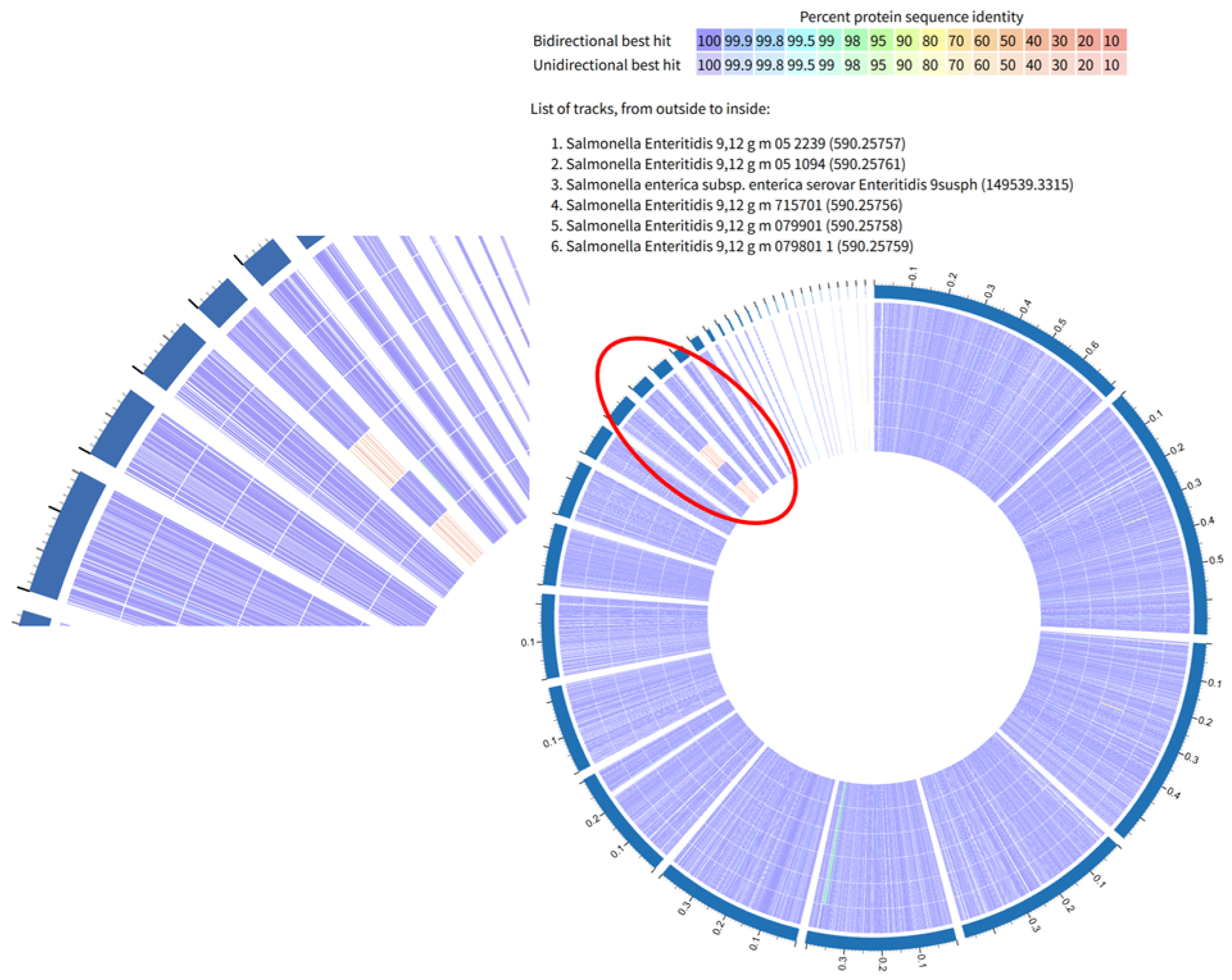
A group of six SE isolates (three strong isolates: 52239, 51094, and 51095 (9susph), and three intermediate isolates 715701, 79901, and 79801), which belonged to the same clade in red color in Figure 3 were compared by their functional genomic information due to their clustered SNPs results (Figure 6). One of the strong biofilm-forming isolates 52239, served as the reference isolate. Comparing the strong reference isolate 52239 to the intermediate isolate 79901, excluding the hypothetical proteins and proteins with unknown functions, only two proteins were absent in 79901 that were present in 52239: secreted effector protein and Thiol: disulfide interchange protein DsbA precursor, which indicated that this intermediate isolate was not too different from the strong isolate. However, when comparing 715701 and 79801 to the reference isolate, many proteins appeared to be absent. Table 5 revealed the proteins present in the reference isolate 52239, but lacking in either the intermediate isolate 715701 (a) or 79801 (b).

4. Discussion

4.1. Biofilm Formation

Among the 15 Salmonella Enteritidis (SE) isolates, 11 (73.3%) produced strong biofilms, while 4 (26.7%) produced intermediate biofilms (p < 0.0001). Biofilm results were in alignment with the curli and cellulose results, which were 93% positive for both traits. These findings clearly demonstrate that SE isolates from poultry sources can form strong biofilms at 20–22 °C, a trait that may contribute to the persistence of SE strains in the environment. Studies from Brazil present contrasting results. De Oliveira et al. found that SE isolates incubated for 96 h, at 20 °C on various surfaces (stainless steel, glass, and PVC) were predominantly non-biofilm formers [43]. Similarly, Rodrigues et al. reported that among 171 SE isolates from foodborne outbreaks (foods and stool samples) and poultry products, most were weak or non-biofilm formers after 24 h at 25 °C using the crystal violet method at 550 nm in the biofilm reader [19]. Both studies highlight that isolates with strong biofilm-forming capacities were a minority. These results suggest that SE isolates from Brazil possess different biofilm formation abilities compared to those isolated in North America, possibly associated with distinct genomic traits or methodological differences, such as incubation times (24/48/120 h) and broth compositions (TSB vs. LB-NS).
Regarding Salmonella Kentucky (SK), of the 24 isolates tested at 22 °C, only four (16.7%) were strong biofilm formers, five (20.8%) were weak formers, and the majority, fifteen (62.5%), were non-biofilm formers (p < 0.0001). Notably, among the four strong biofilm formers, only curli and cellulose production were positive for both traits, which may help explain the overall lower biofilm-forming capacity observed in this group. These results demonstrate that the biofilm-forming ability of SK isolates was markedly lower compared to SE; perhaps this could limit SK’s ability to persist in poultry barn environments. SK results on biofilm formation are similar to those reported by Obe et al., where biofilm formation of SK (n = 12) isolated from commercial processing equipment ranged in biofilm production from strong (n = 3), to moderate (n = 2), to weak biofilm producers (n = 7) after incubation in TSB for 48 h at 25 °C [44]. In our study, SK isolates from similar processing environments exhibited a wide range of biofilm abilities under the specific conditions tested. For example, of the eight SK isolates isolated from the carcass wash/pre-chill step, seven were non-biofilm formers, while one was a strong biofilm former. Similarly, of the ten SK isolates isolated from the final wash/post chill, nine were non-biofilm formers and one was a strong biofilm former. Biofilm formation is also strongly influenced by incubation time and nutrient availability. Stepanović et al. demonstrated that biofilm production peaked at different times depending on temperatures, with stronger biofilm formation at 22 °C after 48 h [45]. Piras et al. found similar temperature-dependent variability in Salmonella isolates from pig slaughterhouses [46]. Ziech et al. also reported that most Salmonella spp. isolated from poultry cutting rooms were weak or intermediate adherents after 96 h at 35 °C [47]. Another key factor affecting biofilm formation is the type of growth medium. Our study utilized Lennox Broth No-Salt (LB-NS), whereas most other studies employed tryptic soy broth (TSB), which may yield different results in biofilm formation.
The strong biofilm formation observed in SE and some SK isolates suggests that these strains have an enhanced capacity for persistence. Thus, thorough cleaning and sanitation in poultry environments and other processing environments are critical. Effective scrubbing, correct use of detergents, and ensuring proper sanitizer concentration and contact time are essential to prevent surface conditioning, bacterial attachment, and subsequent biofilm development. SE and SK results on biofilm formation abilities raises a critical question: can genomic features be used to differentiate Salmonella strains that are capable of form biofilms and persist in the environment from those that are more easily eliminated. Genomic analysis may help address this gap by identifying genetic determinants associated with biofilm formation, thereby providing a clearer picture of the factors driving strain survival.

4.2. Genomic Analysis

To further explore the genetic relationships among the isolates, a phylogenetic tree was constructed including SE and SK (Figure 3). In the phylogenetic tree, the absence of clustering by source indicates that isolates from different geographic locations are not confined to specific environments. Instead, they are genetically intermixed, which suggests that these Salmonella strains possess broad fitness traits rather than niche adaptations. This pattern reflects a high degree of adaptability, allowing them to persist and survive under the diverse and often harsh conditions encountered throughout poultry production and processing environments [48]. The bootstrap values, represented as percentages in the tree, indicate the frequency with which the same branch is observed when the phylogenetic tree is generated 100 times [49]. A bootstrap value of 100% indicates that this arrangement was observed every time, confirming that the result is reliable and not due to random chance, while 0% indicates a random ordering of the isolates within clades [49]. Within the SE isolates, three groupings were confidently supported: 79801 and 71501 (100%), 619102 and 618901 (91%), 53932, 53936, and 61401 (98%), while SK isolates consistently grouped with 100% support, though individual branch supports were low (0%), further suggesting low divergence. Horizontal branch lines in the tree represent the number of genetic changes and evolutionary lineages change over time, with the branch length representing the number of substitutions per 100 nucleotide sites [50]. The shorter the horizontal lines, the fewer genetic base differences along the sequences, suggesting closer relatedness between the isolates. Indeed, all of the SK isolates in blue and yellow diverge by only 1 base substitution per million bases, using the 500 selected genes in the analysis. The phylogenetic analysis offered limited resolution of phenotypic and genomic differences among isolates. While it supports the hypothesis that genetic variation underlies differences in biofilm-forming capacity in SE isolates, no clear clustering aligned with phenotypic traits, underscoring the need to examine functional mutations in core genes and the regulatory region. Although Salmonella Kentucky serovar is said to be polyphyletic globally [51], all isolates in our study clustered within a single well-supported clade, indicating a monophyletic origin. This suggests a shared ancestry and possibly a common source or limited diversity among these poultry-associated isolates.
The comparative genomic analysis showed that all 39 Salmonella isolates (SE and SK) carried nearly all genes previously reported to encode proteins involved in biofilm formation, including those related to curli and cellulose production, flagella, fimbriae, pili, and type IV secretion systems, regardless of their phenotypic biofilm formation category, curli/cellulose production, or isolation source. The presence or absence of these genes did not correlate with the observed biofilm formation phenotypes. Several key genes associated with biofilm development and regulation, such as bapA, crl, and ddhC, were absent in all 15 SE and 24 SK isolates. In contrast, some proteins, including YadK, YhcA, and EcpD, were lacking in SE isolates but present in SK isolates. Notably, genes csgDEF were missing in the non-biofilm-forming SK isolate F41-2, and type IV secretion system proteins (VirB1, VirB3, VirB4, VirB6, VirB10, VirB11, and VirD4) were absent from all 24 SK isolates but present in four strong biofilm-forming SE isolates. Interestingly, isolate #107, the only bovine isolate and the only isolate with negative results for both curli and cellulose production, carries all the screened genes except those type IV secretion system genes (Figure 4).
All 39 isolates carried four protein-coding genes linked to biofilm regulation: bssS, bssR, yjgK, and the bdcA (cyclic-di-GMP-binding biofilm dispersal mediator protein) (Figure 4). The genes bssS and bssR act as negative regulators of biofilm formation, reducing biofilm mass, surface coverage, and mean thickness in Salmonella and E. coli [52,53]. It has been reported that overexpression of yjgK has been shown to repress fimbrial genes, reducing biofilm formation at 8 h but enhancing it after 24 h. The bdcA gene facilitates biofilm dispersal using c-di-GMP [54]. Cyclic-di-GMP further illustrates the complexity of biofilm regulation, acting antagonistically by suppressing motility, dispersal, and virulence gene expression in single-cell states, while promoting sessility, adhesion, and biofilm development in multicellular states [55]. Interestingly, bapA, which encodes a cell-surface protein critical for biofilm formation, was absent in all 39 isolates. Previous studies have shown that deletion of bapA abolishes biofilm formation, while overexpression enhances it. The absence of this gene in our isolates suggests that alternative pathways or compensatory mechanisms are driving biofilm development, highlighting the genetic diversity and regulatory complexity underlying this phenotype in Salmonella.
Regarding the csg (curli specific gene) family, essential for curli and cellulose production [33,56], it was found that genes csgA-csgG were present in all 15 SE isolates and 23 out of 24 SK isolates. In contrast, genes csgDEF were absent in the SK F41-2 isolate, a non-biofilm former. Two key operons regulate curli production: csgBAC and csgDEFG. Among them, csgA, csgB, and csgD are particularly crucial. Gene csgA directly contributes to curli fiber formation, a key factor in biofilm adhesion and stability [51], while csgB stabilizes csgA expression and promotes the initial aggregation of csgA to form curli fibers [57]. Gene csgC and genes csgEFG function as accessory genes involved in csgA polymerization [58] and in the assembly, secretion and structural integrity of curli fibers [59]. The absence of csgD in SK F41-2 explains its non-biofilm-forming phenotype. Several proteins that regulate csgD expression were found in our Salmonella genomes: RpoS, OmpR, Crl, and MlrA. It has been reported that the transcription of csgD was utterly dependent on the expression of RpoS and OmpR proteins [60,61,62], both of which were present in all 39 Salmonella isolates. However, Crl, an RopS-binding factor, was absent in all 39 isolates. Crl protein interacts with RpoS and it is required for the maximal expression of the csgD, csgB, adrA, and bcsA genes, which are essential for the curli and cellulose synthesis [63]. MlrA, present in all strains, is a novel transcriptional regulator of csgD that acts as a positive regulator of RpoS-dependent curli production in Salmonella Typhi and E. coli [64]. These findings suggest that csgD regulation in these strains may rely exclusively on RpoS and OmpR, without the modulatory influence of Crl. Interestingly, while all non-biofilm formers (except SK F41-2) harbored all seven csg genes, only 4 out of 24 SK isolates exhibited the red, dry, and rough (RDAR) curli morphotype [60]. Gene ddhc (formerly rfbH), which codes for CDP-4-keto-6-deoxy-D-glucose-3-dehydrase, is responsible for synthesizing abequose, the last sugar component of the O antigen in LPS. Gene waaG (formerly rfaG), encodes UDP-glucose-LPS α1,3-glucosyltransferase, an enzyme that adds a glucose molecule to heptose II of the core polysaccharide of LPS [40]. Both mutations result in decreased levels of curli while increasing cellulose production. The gene waaG was present in all 15 SE and 24 SK isolates, while the gene ddhC was absent in all isolates. Although all non-biofilm formers (except SK F41-2) harbored the complete set of csg genes (csgAcsgG), most of them did not exhibit the RDAR morphotype. This disconnect between gene presence and phenotype demonstrates that biofilm formation is not determined solely by the presence of the csg gene cluster, but instead depends on regulatory dynamics and additional factors.
All isolates carried bcsABCEFGQ, with three intermediate SE isolates showing a duplicated bcsC, which may be linked to reduced biofilm formation. In contrast, bcsZ and yhjR were absent in all isolates. The diguanylate cyclase gene adrA, a positive regulator of csgD-dependent cellulose synthesis, was present in all strains. SNP analysis revealed mutations in several bcs genes, suggesting possible functional alterations that could affect cellulose production; these will be discussed in later sections. Overall, while cellulose biosynthetic operons are broadly conserved, genetic variations such as bcsC duplication, absence of accessory genes, and SNPs in key loci may influence the balance between curli and cellulose production, contributing to observed biofilm phenotypes.
We also looked at fimbriae and pili genes. Of the 35 fimbriae protein-coding genes analyzed, 32 of them were present in all Salmonella isolates. However, YadK, YhcA, and EcpD were absent in all SE isolates but present in all SK isolates (Figure 4). The adhesion operon Yad has several genes, including yadK. In E. coli, the fimbria Yad contributes to biofilm formation and bladder epithelial cell adherence [65]. YhcA is an uncharacterized fimbrial chaperone protein, while the ecp operon encodes ecpD, which is necessary for pili assembly and functions as a polymerized tip adhesin [66]. The absence of these three genes in SE isolates remains unexplained. YehA protein was found in both serovars, but had different amino acid lengths and was present in two copies in 6 SK isolates. Additionally, a total of 8 Type IV pilin genes were present in all isolates, including pilABCMNOPQ, which encode bacterial appendages involved in adhesion and motility.
The flagellar filament and its motility in Salmonella play an important role in the early development of biofilms, which is the initial attachment to the surface, rather than contributing to the biofilm maturation [14]. In our study, a total of 45 flagellar-related protein-coding genes were found, of which, 18 belong to the Fli family (FliC-FliT), 14 belong to the Fla family (FlgA-FlgN), 5 Flh proteins (FlhA-FlhE), motor rotation proteins (MotA and MotB), flagellar regulator proteins (flk, RtsA, and RtsB), chemotaxis regulator, and RNA polymerase sigma factor for flagellar operon. All of which were present in all isolates (Figure 4). In the SNP variation analysis, several proteins had mutations in the SE isolates, including MotA, MotB, FlgA, FlgB, FlgM, FlhC, FliT, and RtsB, which will be discussed in later sections.
Virulence factors such as the type III secretion system (T3SS) have been studied extensively regarding biofilm formation in Salmonella isolates. However, all of the protein-coding genes associated with T3SS were present in all isolates in our study. Interestingly, type IV secretion system (T4SS) proteins have shown different results. T4SS plays an important role in mediating the translocation of macromolecules across bacterial cell envelopes [67]. Out of 8 proteins found, 1 of them (NAD(P)H dehydrogenase) was present in all isolates. Seven of them were only found in 4 strong-biofilm SE isolates but were absent in the rest of the 11 SE and all 24 SK isolates. Those proteins include peptidoglycan hydrolase (VirB1), inner membrane protein (VirB3, VirB6, and VirB10), ATPase (VirB4 and VirB11), and coupling protein VirD4. This result suggests that these groups of T4SS proteins may influence biofilm formation or its related mechanisms. Similarly to the Agrobacterium tumefaciens VirB/D T4S system, the T4S system in Gram-negative bacteria typically consists of 12 proteins: VirB1-11 and VirD [67]. Of those 12 core proteins, 7 of them (VirB1, VirB3, VirB4, VirB6, VirB10, VirB11, and VirD4) were detected in 4 of the 15 SE isolates, but none were detected in the SK isolates (Figure 4). To date, only a few Salmonella serotypes, such as Salmonella Enteritidis phage type 34, have been reported to carry T4SS [68]. The functions of T4SS genes and their relationship with biofilm formation are still elusive and need further investigation.
For the SNPs variation analysis of the SE isolates, three curli genes were found to have a moderate-impact nonsynonymous missense mutation in the curli gene csgF, its upstream feature csgG, and its downstream feature csgE in SE 52239 (a strong biofilm-former), changing the codon TCA into CCA. Gene csgEFG are all components for curli production assembly and transportation. These are considered accessory genes, which might explain why SE 52239 remained a strong biofilm producer despite the presence of a missense mutation.
This research found genetic mutations in SE isolates in essential proteins responsible for cellulose and curli biosynthesis (Table 3). As discussed in the cellulose section, bcs operon yhjR-bcsQABZC encodes genes yhjR, bcsQ, bcsA, bcsB, bcsZ, and bcsC, and operon bcsEFG encodes gene bcsE, bcsF, and bcsG. Out of 5 mutations associated with cellulose biosynthesis, one was stop-gained, one was frameshift deletion mutation, and three were synonymous variants. Gene bcsA, its upstream feature bcsQ, and its downstream feature bcsB had a high-impact stop-gained nonsynonymous mutation with a codon changed from TGG to TAG. Stop codons signal the end of the translation, when it occurs, it can result in a truncated (shortened) protein and disrupt the normal function of this protein. The stop-gained mutation was found in 6 SE isolates (intermediate formers: 715701, 79801, 79901, strong formers: 52239, 51094, and 51095). This group corresponded to the red clade in Figure 3. Having both intermediate and strong isolates present indicates that this mutation might not be associated with loss or reduction in biofilm functions, or that only three intermediate isolates were impacted by this mutation. Gene bcsC had a high-impact frameshift deletion mutation in the 3 intermediate isolates mentioned above and a low-impact synonymous variation in isolate 107. Frameshift deletion mutation is missing nucleotides “cg”. Interestingly, bcs mutations were not seen in SK isolates, where non-biofilm formers were predominant. These bcs genes are essential in synthesizing cellulose in the matrix. The fact that all three isolates associated with this frameshift deletion mutation were intermediate biofilm formers indicates that this mutation might be associated with a reduction in the biofilm functions. Gene bcsG and its downstream feature bcsF had a low-impact synonymous variation in isolate 107, indicating that isolate 107, being synonymous, was most likely not associated with a reduction in biofilm formation. However, it is worth noticing that isolate 107 is a quite distant relative of the other SE isolates. Gene bcsQ and its upstream feature YhjR also had a low-impact synonymous variation in isolate 107. The deletion of gene bcsQ and yhjR could result in no or reduced cellulose production in E. coli [69]. However, synonymous variation should not alter the amino acid sequence.
Yeh, Yad, and Yfc are all core-associated chaperon-usher fimbriae-related proteins that are conserved in E. coli and Salmonella isolates [70]. A high-impact frameshift insertion in yehA and its upstream feature yehB was detected in six isolates (three intermediate and three strong biofilm formers), suggesting the mutation is not directly linked to reduced biofilm capacity. In isolate 107, yadE and its upstream feature yadI carried synonymous variants with no expected functional effect, while yadS harbored a moderate-impact missense mutation that may contribute to reduced biofilm formation. Similarly, yadU showed moderate-impact missense variants in isolate 107 and three intermediate isolates, suggesting a possible role in biofilm modulation. In contrast, synonymous variants in yfcL and yfcC in isolate 107 are unlikely to affect protein function.
Flagella are another critical component in biofilm formation. Several flagellar-associated genes were detected with genetic variations and mutations. Gene flgA, its upstream feature, flgM, and its downstream feature, flgB, had moderate-impact missense mutations in all six isolates mentioned above. motA, its upstream feature motB, and its downstream feature flhC had low-impact synonymous variations in isolate 107. fliT had a missense mutation and rtsB had a synonymous variation, both in isolate 107.
For the SNP variation analysis of SK isolates, gene tolA and tolR are both part of the Tol-Pal system of the E. coli envelope, which consists of tolQ, tolR, tolA, tolB, and pal [71] and this system is involved in maintaining the integrity of the outer membrane [72]. More importantly, tolA can promote survival, biofilm formation, and virulence of avian pathogenic E. coli [73]. According to Su et al., a tolA mutation with a motility defect results in significantly less biofilm biomass, a reduced amount of fliR expression, and weakened resistance to environmental The regulation of the fnr gene controls the expression of TolA [74]. Therefore, even though tolA, tolB, and tolR genes are present in all isolates with 1 copy in each isolate except for tolA gene in SK F43-3 and SK W39-1 (non-biofilm producers), which have two copies, it is possible that the changes in the amino acids in the nonsynonymous mutation (1 out of 5 tolA mutations found in SK isolates, 3 out of 4 in SE isolates) missense variations altered the protein’s structure and function, and lead to reduced biofilm formation and the flagellar biosynthesis (Table 3). Both serovars had synonymous and nonsynonymous mutations. This could explain the strong biofilm formation in SK B38-3, since it did not have this mutation, and intermediate biofilm formation in SE 715701 and SE 79801, since they have this mutation. In summary, the three SE intermediate isolates exhibited more high-impact variations than the others, and none of the SK stains showed any high-impact variation. These findings suggest that while both serovars carry tolA, tolB, and tolR, nonsynonymous mutations, especially in tolA, may compromise protein function and reduce biofilm capacity, with SE isolates showing more high-impact variations than SK. This highlights tolA as a potential key determinant of biofilm formation differences between isolates.

5. Conclusions

The results revealed distinct genetic patterns differentiating strong from weak and non-biofilm-producing isolates. Although most isolates carried the core genes for biofilm formation (curli, cellulose, fimbriae, pili, and flagella), the SK isolates were predominantly non-biofilm producers despite possessing nearly all essential genes. No major SNPs were detected in core biofilm genes, except mutations in bcs (isolate#107) and csg (strong SE 52239). Notably, SE isolates showed the highest variation, with three high-impact mutations: a nonsynonymous stop-gain in bcsA (six isolates: three intermediate, three strong), a frameshift deletion in bcsC (three intermediate), and a frameshift deletion in yehA (the same six SE isolates). These variations suggest that SE, more than SK, may rely on genetic changes to modulate biofilm capacity.
Furthermore, strong SE isolates carried a cluster of type IV secretion system proteins (VirB1, VirB3, VirB4, VirB5, VirB6, VirB9, VirB11, VirD2 homolog, and VirD4) on a lysogenic phage, absent in intermediate strains. Additional biofilm-associated genes were linked to a conjugative plasmid. Together, these findings imply that beyond SNPs in core genes, horizontal gene transfer through phages and plasmids may play a crucial role in enhancing SE biofilm formation. This highlights the need for future studies to clarify how genetic variations and mobile elements interact to drive biofilm development.
Variations in tolA, tolB, and tolR genes, particularly nonsynonymous mutations in tolA, may influence protein function and reduce biofilm-forming capacity, with Salmonella Enteritidis isolates showing more high-impact mutations than S. Kentucky. These findings identify tolA as a potential determinant of biofilm variability; however, further functional studies are needed to verify its specific role in regulating biofilm formation across different S. Enteritidis isolates.
A key limitation of this study is that the isolates of Salmonella Enteritidis (SE) and Salmonella Kentucky (SK) were not selected to capture the full genetic diversity within each serovar. It is possible that the isolates tested were clonally related, which could explain the similarity in biofilm formation and underlying genetic traits observed within each group. As a result, the findings may not fully represent the broader variability in biofilm-forming capacity across genetically diverse isolates of SE and SK. Future studies should therefore include a wider range of isolates to better account for intra-serovar diversity and provide more generalizable insights.

Supplementary Materials

The following supporting information can be downloaded at: https://www.mdpi.com/article/10.3390/microorganisms13112473/s1. S1 Summary of sequencing reads, assembly metrics, and genome Features for isolates included in This Study was provided as supplementary material in excel format.

Author Contributions

J.Z. contributed to the lab work, data curation, writing, and preparation of the original draft. O.E. was involved in the lab work. M.D. provided resources and contributed to writing, review, and editing. C.N. contributed resources, data curation, and writing, review, and editing. T.M. contributed to writing, reviewing, and editing. R.S. contributed to methodology, supervision, writing, review, and editing. C.N.-B. was responsible for conceptualization, methodology, funding acquisition, supervision, project administration, and writing, review, and editing. All authors have read and agreed to the published version of the manuscript.

Funding

This research was primarily funded by Mitacs grant number IT29351 and the Canadian Poultry Research Council, grant number FSQ139. Additionally, Salmonella Kentucky strains used in this study were obtained through a project funded by Agriculture and Agri-Food Canada (AAFC) under the Genomics Research and Development Initiative—Antimicrobial Resistance (GRDI-AMR 2) One Health program, leveraging Growing Forward II (Grant 1299).

Institutional Review Board Statement

Not applicable.

Informed Consent Statement

Not applicable.

Data Availability Statement

The raw data supporting the conclusions of this article will be made available by the authors on request.

Acknowledgments

During the preparation of this manuscript/study, the authors used Grammarly for the purposes of improving English grammar. The authors have reviewed and edited the output and take full responsibility for the content of this publication.

Conflicts of Interest

The authors declare no conflicts of interest.

Abbreviations

The following abbreviations are used in this manuscript:
SNPSingle Nucleotide Polymorphism
BLASTpBasic Local Alignment Search Tool for proteins
EPSExtra polymeric substance
LB-NSLuria Broth no-salt
ODOptical density
CVCrystal violet

Appendix A

Appendix A.1

Table A1. Salmonella Enteritidis and Salmonella Kentucky samples Bioproject and BioSample IDs.
Strains SerotypeCountryIsolation SourceBioProject BioSample
Salmonella Enteritidis 0618901Enteritidis Canada Chicken nuggetsPRJNA1285942SAMN49784150
Salmonella Enteritidis 0619102Enteritidis Canada Chicken nuggetsPRJNA1285942SAMN49784151
Salmonella Enteritidis 061401Enteritidis Canada FeedPRJNA1285942SAMN49784152
Salmonella Enteritidis 053932 Enteritidis Canada Chicken breastPRJNA1285942SAMN49784153
Salmonella Enteritidis 053936Enteritidis Canada Chicken breastPRJNA1285942SAMN49784154
Salmonella Enteritidis 0710901Enteritidis Canada Chicken nuggetsPRJNA1285942SAMN49784155
Salmonella Enteritidis 052239Enteritidis Canada Chicken nuggetsPRJNA1285942SAMN49784156
Salmonella Enteritidis 051094Enteritidis Canada Chicken nuggetsPRJNA1285942SAMN49784157
Salmonella Enteritidis 051095Enteritidis Canada Chicken nuggetsPRJNA1285942SAMN49784158
Salmonella Enteritidis 078501Enteritidis Canada Chicken nuggetsPRJNA1285942SAMN49784159
Salmonella Enteritidis 079901Enteritidis Canada Chicken nuggetsPRJNA1285942SAMN49784160
Salmonella Enteritidis 079801Enteritidis Canada Chicken nuggetsPRJNA1285942SAMN49784161
Salmonella Enteritidis 0715701Enteritidis Canada FeedPRJNA1285942SAMN49784162
Salmonella Enteritidis 719001Enteritidis Canada Chicken nuggetsPRJNA1285942SAMN49784163
Salmonella Enteritidis 107Enteritidis Canada BovinePRJNA1285942SAMN49784164
Salmonella Kentucky C45-1KentuckyCanadaCratePRJNA1290672SAMN49932175
Salmonella Kentucky B38-3KentuckyCanadaPlucking beltPRJNA1290672SAMN49932176
Salmonella Kentucky W36-3KentuckyCanadaCarcass wash/Pre-chillPRJNA1290672SAMN49932177
Salmonella Kentucky W37-2KentuckyCanadaCarcass wash/Pre-chillPRJNA1290672SAMN49932178
Salmonella Kentucky W38-1KentuckyCanadaCarcass wash/Pre-chillPRJNA1290672SAMN49932179
Salmonella Kentucky W39-1KentuckyCanadaCarcass wash/Pre-chillPRJNA1290672SAMN49932180
Salmonella Kentucky W40-3KentuckyCanadaCarcass wash/Pre-chillPRJNA1290672SAMN49932181
Salmonella Kentucky W41-2KentuckyCanadaCarcass wash/Pre-chillPRJNA1290672SAMN49932182
Salmonella Kentucky W42-2KentuckyCanadaCarcass wash/Pre-chillPRJNA1290672SAMN49932183
Salmonella Kentucky W44-3KentuckyCanadaCarcass wash/Pre-chillPRJNA1290672SAMN49932184
Salmonella Kentucky F36-3KentuckyCanadaFinal wash/post chillPRJNA1290672SAMN49932185
Salmonella Kentucky F37-3KentuckyCanadaFinal wash/post chillPRJNA1290672SAMN49932186
Salmonella Kentucky F38-2KentuckyCanadaFinal wash/post chillPRJNA1290672SAMN49932187
Salmonella Kentucky F39-3KentuckyCanadaFinal wash/post chillPRJNA1290672SAMN49932188
Salmonella Kentucky F40-3KentuckyCanadaFinal wash/post chillPRJNA1290672SAMN49932189
Salmonella Kentucky F41-2KentuckyCanadaFinal wash/post chillPRJNA1290672SAMN49932190
Salmonella Kentucky F42-3KentuckyCanadaFinal wash/post chillPRJNA1290672SAMN49932191
Salmonella Kentucky F43-3KentuckyCanadaFinal wash/post chillPRJNA1290672SAMN49932192
Salmonella Kentucky F44-1KentuckyCanadaFinal wash/post chillPRJNA1290672SAMN49932193
Salmonella Kentucky F45-2KentuckyCanadaFinal wash/post chillPRJNA1290672SAMN49932194
Salmonella Kentucky PW8-3KentuckyCanadaPlucking waterPRJNA1290672SAMN49932195
Salmonella Kentucky PW9-3KentuckyCanadaPlucking waterPRJNA1290672SAMN49932196
Salmonella Kentucky CW8-3KentuckyCanadaChill waterPRJNA1290672SAMN49932197
Salmonella Kentucky CW9-3KentuckyCanadaChill waterPRJNA1290672SAMN49932198

Appendix A.2

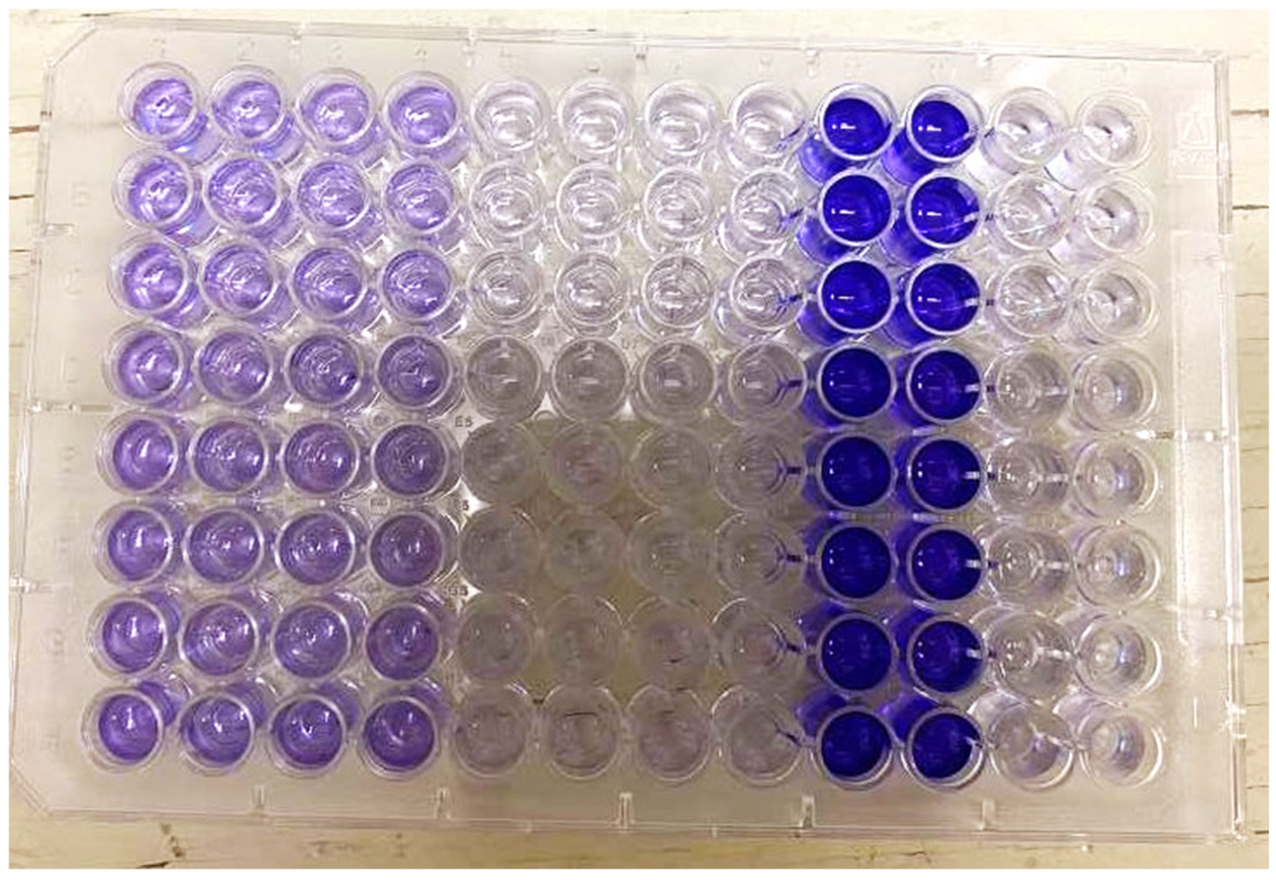
Figure A1. Crystal violet assay for biofilm development microplates example, demonstrating different biofilm density from clear, to light purple and dark purple. The layout is as followed: strain 1 (column 1 & 2), strain 2 (column 3 & 4), strain 3 (column 5 & 6), strain 4 (column 7 & 8), positive control (column 9 & 10), and negative/sterility control (column 11 & 12).
Microorganisms 13 02473 g0a1

Appendix A.3

Figure A2. Curli and cellulose morphotypes in Salmonella Enteritidis (SE) and Salmonella Kentucky (SK) strains (Representative morphotypes showing curli and cellulose phenotypes. (A) Panel A shows representative morphotypes of Salmonella strains exhibiting both curli and cellulose expression (“+/– morphotype”). For curli, the positive morphotype (left) exhibits a red, dry, and rough (RDAR) appearance, while the negative morphotype (right) appears smooth and white (SAW). For cellulose, positive colonies show strong fluorescence (bright blue) on LB agar containing Calcofluor dye, whereas cellulose-negative colonies show weak or no fluorescence. (B) SE strain 107 was the only isolate exhibiting negative results for both curli and cellulose (highlighted in red). (C) The image shows four SK strains (B38-3, W36-3, F44-1, and PW9-3) positive for both curli and cellulose production. The curli phenotype is consistent with previous research [40,41,61]. and the cellulose phenotype aligns with other research [75] showing that Salmonella strains lacking cellulose biosynthesis genes exhibit reduced or no fluorescence under UV light.
Microorganisms 13 02473 g0a2

References

  1. FSIS. Roadmap to Reducing Salmonella; United States Department of Agriculture: Washington, DC, USA, 2020. [Google Scholar]
  2. Sher, A.A.; Mustafa, B.E.; Grady, S.C.; Gardiner, J.C.; Saeed, A.M. Outbreaks of foodborne Salmonella enteritidis in the United States between 1990 and 2015: An analysis of epidemiological and spatial-temporal trends. Int. J. Infect. Dis. 2021, 105, 54–61. [Google Scholar] [CrossRef]
  3. CDC. 2021 Salmonella Outbreak Linked to Raw Frozen Breaded Stuffed Chicken Products. 2012. Available online: https://archive.cdc.gov/#/details?url=https://www.cdc.gov/salmonella/enteritidis-06-21/index.html (accessed on 26 February 2025).
  4. CDC. 2018 Salmonella Infections Linked to Gravel Ridge Farms Shell Eggs—Final Update. 2018. Available online: https://archive.cdc.gov/www_cdc_gov/salmonella/enteritidis-09-18/index.html (accessed on 26 February 2025).
  5. CDC. 2018 Salmonella Braenderup Infections Linked to Rose Acre Farms Shell Eggs (Final Update). 2018. Available online: https://archive.cdc.gov/#/details?url=https://www.cdc.gov/salmonella/braenderup-04-18/index.html (accessed on 26 February 2025).
  6. CDC. 2018 Salmonella Infections Linked to Raw Chicken Products. 2019. Available online: https://archive.cdc.gov/#/details?url=https://www.cdc.gov/salmonella/infantis-10-18/index.html (accessed on 26 February 2025).
  7. CDC. Salmonella Outbreak Linked to Eggs. 2024. Available online: https://www.cdc.gov/salmonella/outbreaks/eggs-09-24/investigation.html (accessed on 26 February 2025).
  8. Public Health Agency of Canada. National Enteric Surveillance Program (NESP) Annual Summary 2023; Public Health Agency of Canada: Ottawa, ON, Canada, 2025. [Google Scholar]
  9. Ferrari, R.G.; Rosario, D.K.A.; Cunha-Neto, A.; Mano, S.B.; Figueiredo, E.E.S.; Conte-Junior, C.A. Worldwide Epidemiology of Salmonella Serovars in Animal-Based Foods: A Meta-analysis. Appl. Environ. Microbiol. 2019, 85, e00591-19. [Google Scholar] [CrossRef]
  10. Soltys, R.C.; Sakomoto, C.K.; Oltean, H.N.; Guard, J.; Haley, B.J.; Shah, D.H. High-resolution comparative genomics of Salmonella Kentucky aids source tracing and detection of ST198 and ST152 lineage-specific mutations. Front. Sustain. Food Syst. 2021, 5, 695368. [Google Scholar] [CrossRef]
  11. Shah, D.H.; Paul, N.C.; Sischo, W.C.; Crespo, R.; Guard, J. Population dynamics and antimicrobial resistance of the most prevalent poultry-associated Salmonella serotypes. Poult. Sci. 2017, 96, 687–702. [Google Scholar] [CrossRef] [PubMed]
  12. Koutsoumanis, K.; Allende, A.; Alvarez-Ordóñez, A.; Bolton, D.; Bover-Cid, S.; Chemaly, M.; De Cesare, A.; Herman, L.; Hilbert, F.; Lindqvist, R.; et al. Salmonella control in poultry flocks and its public health impact. EFSA J. 2019, 17, e05596. [Google Scholar] [CrossRef] [PubMed]
  13. Kim, M.; Barnett-Neefs, C.; Chavez, R.A.; Kealey, E.; Wiedmann, M.; Stasiewicz, M.J. Risk Assessment Predicts Most of the Salmonellosis Risk in Raw Chicken Parts is Concentrated in Those Few Products with High Levels of High-Virulence Serotypes of Salmonella. J. Food Prot. 2024, 87, 100304. [Google Scholar] [CrossRef]
  14. Steenackers, H.; Hermans, K.; Vanderleyden, J.; De Keersmaecker, S.C.J. Salmonella biofilms: An overview on occurrence, structure, regulation and eradication. Food Res. Int. 2012, 45, 502–531. [Google Scholar] [CrossRef]
  15. Vestby, L.K.; Møretrø, T.; Langsrud, S.; Heir, E.; Nesse, L.L. Biofilm forming abilities of Salmonella are correlated with persistence in fish meal- and feed factories. BMC Vet. Res. 2009, 5, 20. [Google Scholar] [CrossRef]
  16. Shree, P.; Singh, C.K.; Sodhi, K.K.; Surya, J.N.; Singh, D.K. Biofilms: Understanding the structure and contribution towards bacterial resistance in antibiotics. Med. Microecol. 2023, 16, 100084. [Google Scholar] [CrossRef]
  17. Flemming, H.-C.; Wingender, J. The biofilm matrix. Nat. Rev. Microbiol. 2010, 8, 623–633. [Google Scholar] [CrossRef]
  18. Morita, Y.; Komoda, E.; Ono, K.; Kumagai, S. Survival of Biofilm-Forming Salmonella on Stainless Steel Bolt Threads under Dry Conditions. Food Hyg. Saf. Sci. (Shokuhin Eiseigaku Zasshi) 2011, 52, 299–303. [Google Scholar] [CrossRef]
  19. Rodrigues, L.B.; Webber, B.; Levandowski, R.; Gehlen, S.S.; Santos, L.R.d.; Pilotto, F.; Tondo, E.C.; Nascimento, V.P.d. Biofilm formation by Salmonella enteritidis at different incubation temperatures. Acta Sci. Vet. 2019, 47, 1654. [Google Scholar] [CrossRef]
  20. Schulze, A.; Mitterer, F.; Pombo, J.P.; Schild, S. Biofilms by bacterial human pathogens: Clinical relevance-development, composition and regulation-therapeutical strategies. Microb. Cell 2021, 8, 28. [Google Scholar] [CrossRef] [PubMed]
  21. Marin, C.; Hernandiz, A.; Lainez, M. Biofilm development capacity of Salmonella strains isolated in poultry risk factors and their resistance against disinfectants. Poult. Sci. 2009, 88, 424–431. [Google Scholar] [CrossRef] [PubMed]
  22. Carrascosa, C.; Raheem, D.; Ramos, F.; Saraiva, A.; Raposo, A. Microbial Biofilms in the Food Industry—A Comprehensive Review. Int. J. Environ. Res. Public. Health 2021, 18, 2014. [Google Scholar] [CrossRef] [PubMed]
  23. Stepanović, S.; Ćirković, I.; Ranin, L.; Svabić-Vlahović, M. Biofilm formation by Salmonella spp. and Listeria monocytogenes on plastic surface. Lett. Appl. Microbiol. 2004, 38, 428–432. [Google Scholar] [CrossRef]
  24. Musa, L.; Toppi, V.; Stefanetti, V.; Spata, N.; Rapi, M.C.; Grilli, G.; Addis, M.F.; Di Giacinto, G.; Franciosini, M.P.; Casagrande Proietti, P. High Biofilm-Forming Multidrug-Resistant Salmonella Infantis Strains from the Poultry Production Chain. Antibiotics 2024, 13, 595. [Google Scholar] [CrossRef]
  25. Kroupitski, Y.; Golberg, D.; Belausov, E.; Pinto, R.; Swartzberg, D.; Granot, D.; Sela, S. Internalization of Salmonella enterica in Leaves Is Induced by Light and Involves Chemotaxis and Penetration through Open Stomata. Appl. Environ. Microbiol. 2009, 75, 6076–6086. [Google Scholar] [CrossRef]
  26. Adator, E.H.; Cheng, M.; Holley, R.; McAllister, T.; Narvaez-Bravo, C. Ability of Shiga toxigenic Escherichia coli to survive within dry-surface biofilms and transfer to fresh lettuce. Int. J. Food Microbiol. 2018, 269, 52–59. [Google Scholar] [CrossRef]
  27. Wick, R.R.; Judd, L.M.; Gorrie, C.L.; Holt, K.E. Unicycler: Resolving bacterial genome assemblies from short and long sequencing reads. PLoS Comput. Biol. 2017, 13, e1005595. [Google Scholar] [CrossRef]
  28. Brettin, T.; Davis, J.J.; Disz, T.; Edwards, R.A.; Gerdes, S.; Olsen, G.J.; Olson, R.; Overbeek, R.; Parrello, B.; Pusch, G.D.; et al. RASTtk: A modular and extensible implementation of the RAST algorithm for building custom annotation pipelines and annotating batches of genomes. Sci. Rep. 2015, 5, 8365. [Google Scholar] [CrossRef] [PubMed]
  29. Davis, J.J.; Gerdes, S.; Olsen, G.J.; Olson, R.; Pusch, G.D.; Shukla, M.; Vonstein, V.; Wattam, A.R.; Yoo, H. PATtyFams: Protein families for the microbial genomes in the PATRIC database. Front. Microbiol. 2016, 7, 118. [Google Scholar] [CrossRef] [PubMed]
  30. Stamatakis, A. RAxML version 8: A tool for phylogenetic analysis and post-analysis of large phylogenies. Bioinformatics 2014, 30, 1312–1313. [Google Scholar] [CrossRef] [PubMed]
  31. Olson, R.D.; Assaf, R.; Brettin, T.; Conrad, N.; Cucinell, C.; Davis, J.J.; Dempsey, D.M.; Dickerman, A.; Dietrich, E.M.; Kenyon, R.W. Introducing the bacterial and viral bioinformatics resource center (BV-BRC): A resource combining PATRIC, IRD and ViPR. Nucleic Acids Res. 2023, 51, D678–D689. [Google Scholar] [CrossRef]
  32. BV-BRC. Protein Families. 2023. Available online: https://www.bv-brc.org/docs/quick_references/organisms_taxon/protein_families.html (accessed on 26 February 2025).
  33. Brombacher, E.; Baratto, A.; Dorel, C.; Landini, P. Gene Expression Regulation by the Curli Activator CsgD Protein: Modulation of Cellulose Biosynthesis and Control of Negative Determinants for Microbial Adhesion. J. Bacteriol. 2006, 188, 2027–2037. [Google Scholar] [CrossRef]
  34. Letunic, I.; Bork, P. Interactive Tree Of Life (iTOL) v5: An online tool for phylogenetic tree display and annotation. Nucleic Acids Res. 2021, 49, W293–W296. [Google Scholar] [CrossRef]
  35. NIH. Single Nucleotide Polymorphisms (SNPs). 2024. Available online: https://www.genome.gov/genetics-glossary/Single-Nucleotide-Polymorphisms-SNPs (accessed on 26 February 2025).
  36. Boratyn, G.M.; Camacho, C.; Cooper, P.S.; Coulouris, G.; Fong, A.; Ma, N.; Madden, T.L.; Matten, W.T.; McGinnis, S.D.; Merezhuk, Y. BLAST: A more efficient report with usability improvements. Nucleic Acids Res. 2013, 41, W29–W33. [Google Scholar] [CrossRef]
  37. Overbeek, R.; Olson, R.; Pusch, G.D.; Olsen, G.J.; Davis, J.J.; Disz, T.; Edwards, R.A.; Gerdes, S.; Parrello, B.; Shukla, M.; et al. The SEED and the Rapid Annotation of microbial genomes using Subsystems Technology (RAST). Nucleic Acids Res 2014, 42, D206–D214. [Google Scholar] [CrossRef]
  38. Van Staaveren, N.; Decina, C.; Baes, C.F.; Widowski, T.M.; Berke, O.; Harlander-Matauschek, A. A description of laying hen husbandry and management practices in Canada. Animals 2018, 8, 114. [Google Scholar] [CrossRef]
  39. Jonas, K.; Tomenius, H.; Kader, A.; Normark, S.; Römling, U.; Belova, L.M.; Melefors, Ö. Roles of curli, cellulose and BapA in Salmonella biofilm morphology studied by atomic force microscopy. BMC Microbiol. 2007, 7, 70. [Google Scholar] [CrossRef]
  40. Anriany, Y.; Sahu, S.N.; Wessels, K.R.; McCann, L.M.; Joseph, S.W. Alteration of the rugose phenotype in waaG and ddhC mutants of Salmonella enterica serovar Typhimurium DT104 is associated with inverse production of curli and cellulose. Appl. Environ. Microbiol. 2006, 72, 5002–5012. [Google Scholar] [CrossRef]
  41. Turki, Y.; Ouzari, H.; Mehri, I.; Ben Aissa, R.; Hassen, A. Biofilm formation, virulence gene and multi-drug resistance in Salmonella Kentucky isolated in Tunisia. Food Res. Int. 2012, 45, 940–946. [Google Scholar] [CrossRef]
  42. Cingolani, P.; Platts, A.; Wang, L.L.; Coon, M.; Nguyen, T.; Wang, L.; Land, S.J.; Lu, X.; Ruden, D.M. A A program for annotating and predicting the effects of single nucleotide polymorphisms, SnpEff: SNPs in the genome of Drosophila melanogaster strain w1118; iso-2; iso-3. Fly 2012, 6, 80–92. [Google Scholar] [CrossRef] [PubMed]
  43. De Oliveira, D.C.V.; Fernandes Junior, A.; Kaneno, R.; Silva, M.G.; Araujo Junior, J.P.; Silva, N.C.C.; Rall, V.L.M. Ability of Salmonella spp. to produce biofilm is dependent on temperature and surface material. Foodborne Pathog. Dis. 2014, 11, 478–483. [Google Scholar] [CrossRef] [PubMed]
  44. Obe, T.; Nannapaneni, R.; Schilling, W.; Zhang, L.; Kiess, A. Antimicrobial tolerance, biofilm formation, and molecular characterization of Salmonella isolates from poultry processing equipment. J. Appl. Poult. Res. 2021, 30, 100195. [Google Scholar] [CrossRef]
  45. Stepanović, S.; Ćirković, I.; Mijač, V.; Švabić-Vlahović, M. Influence of the incubation temperature, atmosphere and dynamic conditions on biofilm formation by Salmonella spp. Food Microbiol. 2003, 20, 339–343. [Google Scholar] [CrossRef]
  46. Piras, F.; Fois, F.; Consolati, S.G.; Mazza, R.; Mazzette, R. Influence of Temperature, Source, and Serotype on Biofilm Formation of Salmonella enterica Isolates from Pig Slaughterhouses. J. Food Prot. 2015, 78, 1875–1878. [Google Scholar] [CrossRef]
  47. Ziech, R.E.; Perin, A.P.; Lampugnani, C.; Sereno, M.J.; Viana, C.; Soares, V.M.; Pereira, J.G.; Pinto, J.P.d.A.N.; Bersot, L.d.S. Biofilm-producing ability and tolerance to industrial sanitizers in Salmonella spp. isolated from Brazilian poultry processing plants. LWT—Food Sci. Technol. 2016, 68, 85–90. [Google Scholar] [CrossRef]
  48. Podolak, R.; Enache, E.; Stone, W.; Black, D.G.; Elliott, P.H. Sources and Risk Factors for Contamination, Survival, Persistence, and Heat Resistance of Salmonella in Low-Moisture Foods. J. Food Prot. 2010, 73, 1919–1936. [Google Scholar] [CrossRef]
  49. Ojha, K.K.; Mishra, S.; Singh, V.K. Chapter 5—Computational molecular phylogeny: Concepts and applications. In Bioinformatics; Singh, D.B., Pathak, R.K., Eds.; Academic Press: Cambridge, MA, USA, 2022; pp. 67–89. [Google Scholar]
  50. Holmes, S. Bootstrapping phylogenetic trees: Theory and methods. Stat. Sci. 2003, 18, 241–255. [Google Scholar] [CrossRef]
  51. Haley, B.J.; Kim, S.W.; Pettengill, J.; Luo, Y.; Karns, J.S.; Van Kessel, J.A.S. Genomic and evolutionary analysis of two salmonella enterica serovar Kentucky sequence types isolated from bovine and poultry sources in North America. PLoS ONE 2016, 11, e0161225. [Google Scholar] [CrossRef]
  52. Chin, K.C.J.; Taylor, T.D.; Hebrard, M.; Anbalagan, K.; Dashti, M.G.; Phua, K.K. Transcriptomic study of Salmonella enterica subspecies enterica serovar Typhi biofilm. BMC Genom. 2017, 18, 836. [Google Scholar] [CrossRef]
  53. Domka, J.; Lee, J.; Wood Thomas, K. YliH (BssR) and YceP (BssS) Regulate Escherichia coli K-12 Biofilm Formation by Influencing Cell Signaling. Appl. Environ. Microbiol. 2006, 72, 2449–2459. [Google Scholar] [CrossRef] [PubMed]
  54. Ma, Q.; Yang, Z.; Pu, M.; Peti, W.; Wood, T.K. Engineering a novel c-di-GMP-binding protein for biofilm dispersal. Environ. Microbiol. 2011, 13, 631–642. [Google Scholar] [CrossRef] [PubMed]
  55. Jenal, U.; Malone, J. Mechanisms of cyclic-di-GMP signaling in bacteria. Annu. Rev. Genet. 2006, 40, 385–407. [Google Scholar] [CrossRef] [PubMed]
  56. Gerstel, U.; Römling, U. The csgD promoter, a control unit for biofilm formation in Salmonella typhimurium. Res. Microbiol. 2003, 154, 659–667. [Google Scholar] [CrossRef]
  57. Hammar, M.; Arnqvist, A.; Bian, Z.; Olsén, A.; Normark, S. Expression of two csg operons is required for production of fibronectin- and congo red-binding curli polymers in Escherichia coli K-12. Mol. Microbiol. 1995, 18, 661–670. [Google Scholar] [CrossRef]
  58. Bhoite, S.; van Gerven, N.; Chapman, M.R.; Remaut, H. Curli Biogenesis: Bacterial Amyloid Assembly by the Type VIII Secretion Pathway. EcoSal Plus 2019, 8. [Google Scholar] [CrossRef]
  59. Chapman, M.R.; Robinson, L.S.; Pinkner, J.S.; Roth, R.; Heuser, J.; Hammar, M.; Normark, S.; Hultgren, S.J. Role of Escherichia coli curli operons in directing amyloid fiber formation. Science 2002, 295, 851–855. [Google Scholar] [CrossRef]
  60. Römling, U.; Rohde, M.; Olsén, A.; Normark, S.; Reinköster, J. AgfD, the checkpoint of multicellular and aggregative behaviour in Salmonella typhimurium regulates at least two independent pathways. Mol. Microbiol. 2000, 36, 10–23. [Google Scholar] [CrossRef]
  61. Römling, U.; Bian, Z.; Hammar, M.; Sierralta, W.D.; Normark, S. Curli fibers are highly conserved between Salmonella typhimurium and Escherichia coli with respect to operon structure and regulation. J. Bacteriol. 1998, 180, 722–731. [Google Scholar] [CrossRef]
  62. Römling, U.; Sierralta, W.D.; Eriksson, K.; Normark, S. Multicellular and aggregative behaviour of Salmonella typhimurium strains is controlled by mutations in the agfD promoter. Mol. Microbiol. 1998, 28, 249–264. [Google Scholar] [CrossRef] [PubMed]
  63. Robbe-Saule, V.; Jaumouillé, V.; Prévost, M.-C.; Guadagnini, S.; Talhouarne, C.; Mathout, H.; Kolb, A.; Norel, F. Crl activates transcription initiation of RpoS-regulated genes involved in the multicellular behavior of Salmonella enterica serovar Typhimurium. J. Bacteriol. 2006, 188, 3983–3994. [Google Scholar] [CrossRef] [PubMed]
  64. Brown, P.K.; Dozois, C.M.; Nickerson, C.A.; Zuppardo, A.; Terlonge, J.; Curtiss, R., III. MlrA, a novel regulator of curli (AgF) and extracellular matrix synthesis by Escherichia coli and Salmonella enterica serovar Typhimurium. Mol. Microbiol. 2001, 41, 349–363. [Google Scholar] [CrossRef] [PubMed]
  65. Spurbeck, R.R.; Stapleton, A.E.; Johnson, J.R.; Walk, S.T.; Hooton, T.M.; Mobley, H.L. Fimbrial profiles predict virulence of uropathogenic Escherichia coli strains: Contribution of ygi and yad fimbriae. Infect. Immun. 2011, 79, 4753–4763. [Google Scholar] [CrossRef]
  66. Garnett, J.A.; Martínez-Santos, V.I.; Saldaña, Z.; Pape, T.; Hawthorne, W.; Chan, J.; Simpson, P.J.; Cota, E.; Puente, J.L.; Girón, J.A. Structural insights into the biogenesis and biofilm formation by the Escherichia coli common pilus. Proc. Natl. Acad. Sci. USA 2012, 109, 3950–3955. [Google Scholar] [CrossRef]
  67. Christie, P.J.; Atmakuri, K.; Krishnamoorthy, V.; Jakubowski, S.; Cascales, E. Biogenesis, architecture, and function of bacterial type IV secretion systems. Annu. Rev. Microbiol. 2005, 59, 451–485. [Google Scholar] [CrossRef]
  68. Chen, C.-L.; Wang, C.-Y.; Chu, C.; Su, L.-H.; Chiu, C.-H. Functional and molecular characterization of pSE34 encoding a type IV secretion system in Salmonella enterica serotype Enteritidis phage type 34. FEMS Immunol. Med. Microbiol. 2009, 57, 274–283. [Google Scholar] [CrossRef]
  69. Serra, D.O.; Richter, A.M.; Hengge, R. Cellulose as an architectural element in spatially structured Escherichia coli biofilms. J. Bacteriol. 2013, 195, 5540–5554. [Google Scholar] [CrossRef]
  70. Wurpel, D.J.; Beatson, S.A.; Totsika, M.; Petty, N.K.; Schembri, M.A. Chaperone-Usher Fimbriae of Escherichia coli. PLoS ONE 2013, 8, e52835. [Google Scholar] [CrossRef]
  71. Cascales, E.; Gavioli, M.; Sturgis, J.N.; Lloubès, R. Proton motive force drives the interaction of the inner membrane tolA and outer membrane Pal proteins in Escherichia coli. Mol. Microbiol. 2000, 38, 904–915. [Google Scholar] [CrossRef]
  72. Walburger, A.; Lazdunski, C.; Corda, Y. The Tol/Pal system function requires an interaction between the C-terminal domain of tolA and the N-terminal domain of tolB. Mol. Microbiol. 2002, 44, 695–708. [Google Scholar] [CrossRef]
  73. Su, S.; Li, Z.; Sun, Y.; Gao, S.; Gao, Q. The multifaceted role of tolA protein in promoting survival, biofilm formation and virulence of avian pathogenic Escherichia coli. Poult. Sci. 2024, 103, 104142. [Google Scholar] [CrossRef]
  74. Nikhil, K.C.; Priyadarsini, S.; Pashupathi, M.; Ratta, B.; Saxena, M.; Ramakrishnan, S.; Behera, P.; Kumar, A. Regulatory Role of fnr Gene in Growth and tolA Gene Expression in Salmonella Typhimurium. Indian J. Anim. Res. 2021, 55, 774–779. [Google Scholar] [CrossRef]
  75. Solano, C.; García, B.; Valle, J.; Berasain, C.; Ghigo, J.M.; Gamazo, C.; Lasa, I. Genetic analysis of Salmonella enteritidis biofilm formation: Critical role of cellulose. Mol. Microbiol. 2002, 43, 793–808. [Google Scholar] [CrossRef]
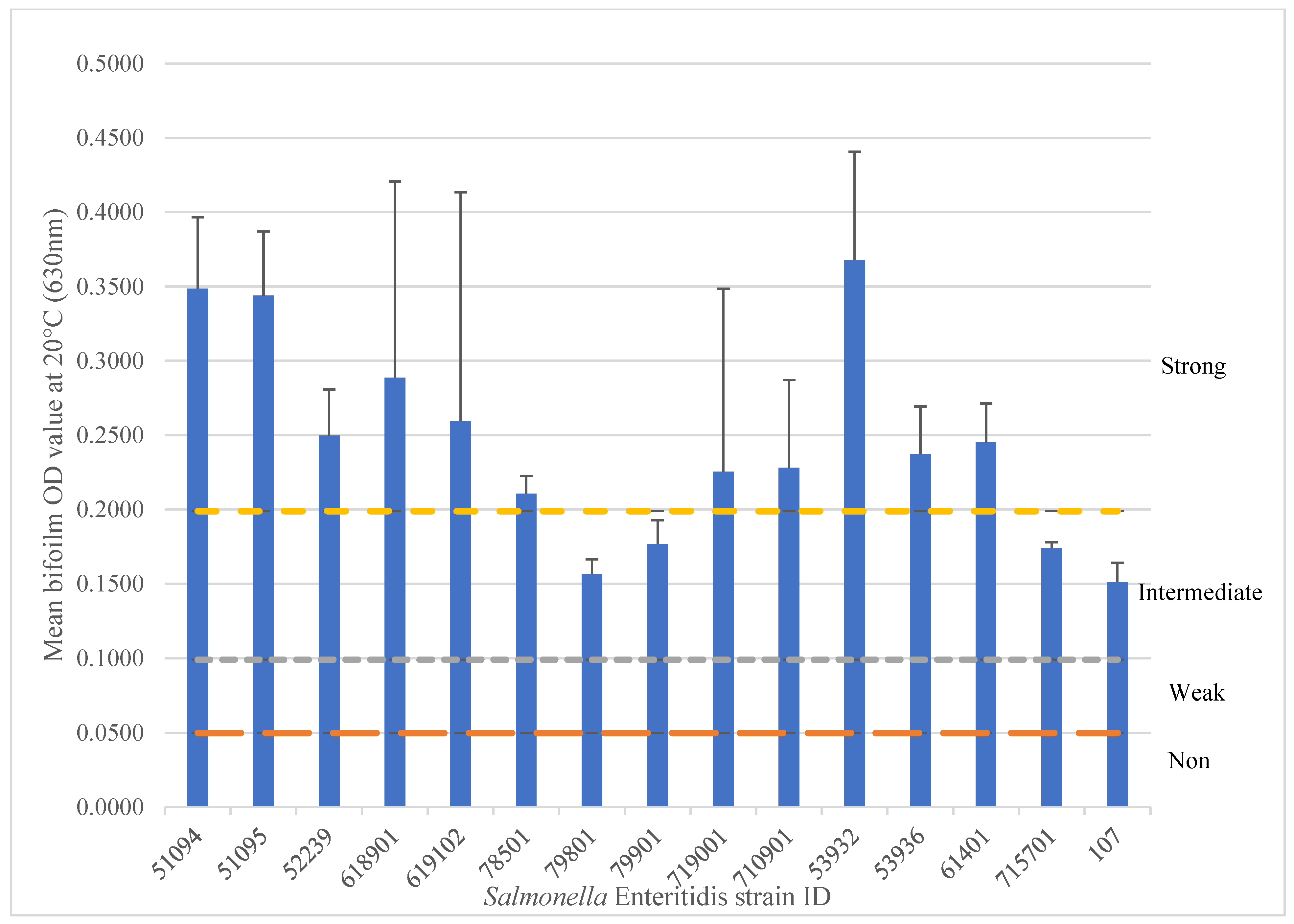
Figure 1. Salmonella Enteritidis isolates form biofilms at 22 °C after 5 days. The biofilm formation ability is categorized by strong (above the yellow line), intermediate (above the gray line), weak (above the orange line), and non-biofilm formers (below the orange line). The experiment was repeated three times.
Microorganisms 13 02473 g001
Figure 2. Salmonella Kentucky isolates biofilm formation at 22 °C after 5 days. The biofilm formation ability is categorized by strong (above the yellow line), intermediate (above the gray line), weak (above the orange line), and non-biofilm formers (below the orange line). The experiment was repeated four times.
Microorganisms 13 02473 g002
Figure 3. Phylogenetic tree of 15 SE isolates and 24 SK isolates using 500 single-copy genes parameter with the mafft alignment program and RAxML Fast Bootstrapping branch supporting method. The tree was divided into two big clades. The upper part belongs to the 15 SE isolates (further divided into the red clade, pink clade, and outgroup isolate #107), and the lower part belongs to the 24 SK isolates (further divided into the green, blue, and orange clades). The numbers in percentages indicate the level of confidence in this clustering method, and the number in the horizontal line represents the number of nucleotide changes or substitutions per site, indicating the closeness of those isolates. The isolates IDs were color-coded to show their sources of isolation. Filled (solid) symbols indicate positive results for biofilm formation, curli and cellulose production. The black-to-white color scale represents the biofilm formation category (strong, intermediate, or weak). Empty (hollow) symbols indicate negative results (no significant biofilm formation).
Microorganisms 13 02473 g003
Figure 4. Biofilm formation results and binary data of the biofilm-related genes in all 39 isolates. For the biofilm development assay, the intensity of the shaded squares (black-white scale) represents biofilm-forming ability: black = strong, medium dark gray = intermediate, light-gray = weak, and white = non-biofilm former. For the production of curli and cellulose, a filled shape indicates a positive result and an empty shape indicates a negative result. For the genotypic results (heat map on the right side), the genes associated with biofilm formation, curli, cellulose, fimbriae, flagella, pili, and T4SS are shown with the gene names at the top of the image. Solid figures represent gene presence and empty ones, gene absence. The * in the gene name represents the 7 cluster groups in the flagella family. The left side shows a dendrogram that illustrates the genetic relatedness among all 39 Salmonella isolates. Each branch point (node) represents a common ancestor between the isolates or groups connected by that node. The branch length reflects the degree of genetic difference; shorter branches indicate closely related isolates, while longer branches indicate greater genetic divergence. Clusters of isolates that share both short branch lengths and the same color suggest high genetic similarity among isolates from the same environment or sample type. The bootstrap values displayed at the nodes represent the level of confidence in each branch grouping (higher values indicate stronger support for that relationship).
Microorganisms 13 02473 g004
Figure 5. Protein coding gene comparison graph showing differences in gene sequence identity between a strong SE biofilm isolate isolated from poultry product (53932) on the outside ring, and an intermediate SE isolate isolated from bovine (107) on the inside ring. The image shows the plot of the bi-directional BLAST hits and a legend that shows the list of tracks and the strength of the BLAST hits, where purple is the strongest and red is the weakest. Two regions of the cluster of differences in the red circles were enlarged for better visualization.
Microorganisms 13 02473 g005
Figure 6. Protein coding gene comparison graph showing differences in gene sequence identity between three strong SE biofilm isolates (52239, 51094 and 51095 (another name 9susph)) on the outside ring, and three intermediate SE isolates (715701, 79901, and 79801), with 52239 being in the most outside ring and 79801 being in the most inside ring. The image shows the plot of the bi-directional BLAST hits and a legend that shows the list of tracks and the strength of the BLAST hits, where purple is the strongest and red is the weakest. One region of cluster of differences in the red circles was enlarged for better visualization. The red circle indicated an obvious difference among those isolates.
Microorganisms 13 02473 g006
Table 1. List of Salmonella enterica serovars Enteritidis and Kentucky used in this study.
SerovarIsolate IDSourceSerovarIsolate IDSource
Enteritidis51094Chicken nuggetsKentuckyC45-1Crate
Enteritidis51095Chicken nuggetsKentuckyB38-3Plucking belt
Enteritidis52239Chicken nuggetsKentuckyW36-3Carcass wash/Pre-chill
Enteritidis618901Chicken nuggetsKentuckyW37-2Carcass wash/Pre-chill
Enteritidis619102Chicken nuggetsKentuckyW38-1Carcass wash/Pre-chill
Enteritidis78501Chicken nuggetsKentuckyW39-1Carcass wash/Pre-chill
Enteritidis79801Chicken nuggetsKentuckyW40-3Carcass wash/Pre-chill
Enteritidis79901Chicken nuggetsKentuckyW41-2Carcass wash/Pre-chill
Enteritidis719001Chicken nuggetsKentuckyW42-2Carcass wash/Pre-chill
Enteritidis710901Chicken nuggetsKentuckyW44-3Carcass wash/Pre-chill
Enteritidis53932Chicken breastKentuckyF36-3Final wash/post chill
Enteritidis53936Chicken breastKentuckyF37-3Final wash/post chill
Enteritidis61401FeedKentuckyF38-2Final wash/post chill
Enteritidis715701FeedKentuckyF39-3Final wash/post chill
Enteritidis107BovineKentuckyF40-3Final wash/post chill
KentuckyF41-2Final wash/post chill
KentuckyF42-3Final wash/post chill
KentuckyF43-3Final wash/post chill
KentuckyF44-1Final wash/post chill
KentuckyF45-2Final wash/post chill
KentuckyPW8-3Plucking water
KentuckyPW9-3Plucking water
KentuckyCW8-3Chill water
KentuckyCW9-3Chill water
Table 2. Screened genes, protein groups associated with biofilm formation, curli and cellulose production, pili, flagella, and fimbriae, and their functions (functions were provided by the BV-BRC website).
Gene CategoryProtein NameFunction
Biofilm regulation protein-coding genesBdcA (Yjgl) Cyclic-di-GMP-binding biofilm dispersal mediator protein
BssS/BssRBiofilm regulator
YjgKLinked to biofilm formation
Biofilm structural proteinBapAA large cell-surface protein required for biofilm formation by S. Enteritidis
Curli productionCsgAMajor curlin subunit precursor
CsgBMinor curlin subunit, nucleation component of curlin monomers
CsgCPutative curli production protein
CsgDTranscriptional regulator for the 2nd curli operon
CsgE/CsgF, CsgGCurli production assembly/transport component
OmpRTwo-component system response regulator
RpoSRNA polymerase sigma factor
Regulator of RpoS
CrlAn RpoS-binding factor, binding to RpoS, facilitates RNA polymerase holoenzyme formation (EσS)
Curlin gene transcriptional activator
MlrAHTH-type transcriptional regulator; Positive regulator of CsgD expression
DdhCInvolved in the synthesis of abequose
WaaGUDP-glucose:(heptosyl) LPS alpha1,3-glucosyltransferase
Cellulose productionBcsACellulose synthase catalytic subunit
BcsBCyclic di-GMP-binding protein
BcsCCellulose synthase operon protein C
BcsE/BcsF/BcsG/BcsQCellulose biosynthesis protein
AdrA (DgcC)Diguanylate cyclase positively regulates cellulose synthesis via production of the secondary messenger signaling molecule (3′-5′)-cyclic diguanosine monophosphate (c-di-GMP)
Fimbriae productionStbC */StdB */SthB */StiC *Fimbriae usher protein
SthA */StiB *Putative fimbrial chaperone
StiA *Putative fimbrial subunit
SafB *Salmonella atypical fimbria periplasmic chaperone
FimI *Fimbriae-like adhesin
FimW *Fimbriae W protein
FimY */FimZ *Transcriptional regulator of fimbriae expression
BcfA */BcfD */BcfE */BcfF *Fimbrial subunit
BcfB */BcfG *Fimbrial chaperone
StfAMajor fimbrial subunit
StfCFimbriae usher protein
StfDPeriplasmic fimbrial chaperone
StfE/StfF/StfGMinor fimbrial subunit
SfmA/SfmH/YehD/SfmFUncharacterized fimbrial-like protein
SfmC/YehCProbable fimbrial chaperone
YehA/YadKUncharacterized fimbrial-like protein
YadU Uncharacterized protein in the stf fimbrial cluster
YhcAUncharacterized fimbrial chaperone
EcpDFimbria adhesin
PiliPilAType IV pilin
PilBType IV pilus assembly, ATPase
PilCType IV pilus assembly protein
PilM/PilN/PilO/PilP/PilQType IV pilus biogenesis protein
FlagellaFliCFlagellin
FliDFlagellar cap protein
FliEFlagellar hook-basal body complex protein
FliFFlagellar M-ring protein
FliGFlagellar motor switch protein
FliHFlagellar assembly protein
FliIFlagellum-specific ATP synthase
FliJFlagellar protein
FliKFlagellar hook-length control protein
FliLFlagellar basal body-associated protein
FliM/FliNFlagellar motor switch protein
FliO/FliP/FliQ/FliR/FliS/FliTFlagellar biosynthesis protein
FlgAFlagellar basal-body P-ring formation protein
FlgB/FlgC/FlgF/FlgGFlagellar basal-body rod protein
FlgDFlagellar basal-body rod modification protein
FlgEFlagellar hook protein
FlgHFlagellar L-ring protein
FlgIFlagellar P-ring protein
FlgJ/FlhEFlagellar protein
FlgK/FlgLFlagellar hook-associated protein
FlgM Negative regulator of flagellin synthesis
FlgN/FlhA/FlhBFlagellar biosynthesis protein
FlhC/FlhDFlagellar transcriptional activator
MotA/MotBFlagellar motor rotation protein
YcgRFlagellar brake protein
flkFlagellar regulator
RtsAType III secretion and flagellar regulator
RtsBFlagellar regulon repressor
Chemotaxis regulatorTransmits chemoreceptor signals to flagellar motor components CheY
RNA polymerase sigma factor for the flagellar operon
Type IV secretion systemNAD(P)H dehydrogenase (quinone), VirB4, VirB11ATPase is required for both the assembly of the type IV secretion complex and the secretion of the T-DNA complex
VirB1Peptidoglycan hydrolase, involved in T-DNA transfer
VirB3Inner membrane protein forms a channel for the type IV secretion of the T-DNA complex
VirB6Inner membrane protein of the type IV secretion of the T-DNA complex
VirB10Inner membrane protein of type IV secretion of T-DNA complex, TonB-like
VirD4Coupling protein, ATPase required for T-DNA transfer
*: Gene names that belong to the seven cluster groups in the flagella family.
Table 3. Gene sequence variation analysis study of the SE and SK isolates that could affect the function and performance of some important genes that are associated with biofilm formation, and related components against isolate SE 53932 and SK PW9-3 as reference, respectively.
SerovarIsolatesTypeFunctionUpstream
Feature
Downstream
Feature
snpEff
Type
snpEff
Impact
SE715701 (I), 79801 (I), 79901 (I), 52239 (S), 51094 (S), 51095 (S)NonsynCellulose synthase catalytic subunit [UDP-forming] (EC 2.4.1.12)Cellulose biosynthesis protein BcsQCyclic di-GMP-binding protein BcsBstop gainedHigh
SE715701, 79801, 79901 (I)DeletionCellulose synthase operon protein Cbeta-1,4-glucanase (cellulase) (EC 3.2.1.4)C-di-GMP phosphodiesterase (EC 3.1.4.52)frameshiftHigh
SE715701 (I), 79801 (I), 79901 (I), 52239 (S), 51094 (S), 51095 (S)InsertionUncharacterized fimbrial-like protein YehAOuter membrane usher protein YehBNickel/cobalt homeostasis protein RcnBframeshiftHigh
SKF43-3 (N)NonsynTolA proteinTol biopolymer transport system, TolR proteinTol-Pal system beta propeller repeat protein TolBmissenseModerate
SE53936 (S)NonsynTolA proteinTol biopolymer transport system, TolR proteinTol-Pal system beta propeller repeat protein TolBmissenseModerate
SE52239, 51094, 53936 (S)NonsynTolA proteinTol biopolymer transport system, TolR proteinTol-Pal system beta propeller repeat protein TolBmissenseModerate
SE719001 (S), 715701 (I), 52239 (S), 51094 (S), 710901 (S), 53936 (S), 618901 (S), 619102 (S)NonsynTolA proteinTol biopolymer transport system, TolR proteinTol-Pal system beta propeller repeat protein TolBmissenseModerate
SE107 (I)NonsynUPF0126 inner membrane protein YadSVitamin B12 ABC transporter, substrate-binding protein BtuFIron-sulfur cluster insertion protein ErpAmissenseModerate
SE107 (I)NonsynGlutamate-1-semialdehyde 2,1-aminomutase (EC 5.4.3.8)FIG01048481: hypothetical proteinUncharacterized protein YadU in the stf fimbrial clustermissenseModerate
SE715701, 79801, 79901 (I)NonsynGlutamate-1-semialdehyde 2,1-aminomutase (EC 5.4.3.8)FIG01048481: hypothetical proteinUncharacterized protein YadU in the stf fimbrial clustermissenseModerate
SE715701 (I), 79801 (I), 79901 (I) 52239 (S), 51094 (S), 51095 (S)NonsynFlagellar basal-body P-ring formation protein FlgANegative regulator of flagellin synthesis, FlgM (anti-sigma28)Flagellar basal-body rod protein FlgBmissenseModerate
SE107 (I)NonsynCytoplasmic alpha-amylase (EC 3.2.1.1)Flagellar biosynthesis protein FliTUncharacterized lipoprotein YedDmissenseModerate
SE52239 (S)NonsynCurli production assembly/transport component CsgFCurli production assembly/transport component CsgGCurli production assembly/transport component CsgEmissenseModerate
SKCW8-3 (W), F38-2 (N), F39-3 (N), F41-2 (N), F42-3 (N), F43-3 (N), F44-1 (S), F45-2 (N), W36-3 (S), W37-2 (N), W38-1 (N), W39-1 (N), W41-2 (N), W42-2 (N), W44-3 (N), F36-3 (N)SynonTolA proteinTol biopolymer transport system, TolR proteinTol-Pal system beta propeller repeat protein TolBsynonymousLow
SKC45-1 (N), CW8-3 (W), F38-2 (N), F39-3 (N), F41-2 (N), F42-3 (N), F43-3 (N), F44-1 (S), F45-2, W36-3 (S), W37-2 (S), W38-1 (N), W39-1 (N), W41-2 (N), W44-3 (N), F36-3 (N)SynonTolA proteinTol biopolymer transport system, TolR proteinTol-Pal system beta propeller repeat protein TolBsynonymousLow
SKCW8-3 (W), W38-1 (N), W39-1 (N), W41-2 (N), F36-3 (N)SynonTolA proteinTol biopolymer transport system, TolR proteinTol-Pal system beta propeller repeat protein TolBsynonymousLow
SKW38-1, W39-1 (N)SynonTolA proteinTol biopolymer transport system, TolR proteinTol-Pal system beta propeller repeat protein TolBsynonymousLow
SE715701 (I), 79801 (I), 52239 (S), 51094 (S), 710901 (S), 53936 (S)SynonTolA proteinTol biopolymer transport system, TolR proteinTol-Pal system beta propeller repeat protein TolBsynonymousLow
SE107 (I)SynonCellulose biosynthesis protein BcsGSmall inner membrane protein, YmgF familyCellulose biosynthesis protein BcsFsynonymousLow
SE107 (I)SynonCellulose biosynthesis protein BcsQPutative cytoplasmic protein YhjRCellulose synthase catalytic subunit [UDP-forming] (EC 2.4.1.12)synonymousLow
SE107 (I)SynonCellulose synthase operon protein Cbeta-1,4-glucanase (cellulase) (EC 3.2.1.4)c-di-GMP phosphodiesterase (EC 3.1.4.52)synonymousLow
SE107 (I)SynonUncharacterized protein YadEPutative PTS system IIA component YadIAspartate 1-decarboxylase (EC 4.1.1.11)synonymousLow
SE107 (I)SynontRNA (5-methylaminomethyl-2-thiouridylate)-methyltransferase (EC 2.1.1.61)/FAD-dependent cmnm(5)s(2)U34 oxidoreductaseUncharacterized protein YfcL3-oxoacyl-[acyl-carrier-protein] synthase, KASI (EC 2.3.1.41)synonymousLow
SE107 (I)SynonUncharacterized protein YfcCTransketolase, C-terminal section (EC 2.2.1.1)BioD-like N-terminal domain/Phosphate acetyltransferase (EC 2.3.1.8)synonymousLOW
SE107 (I)SynonFlagellar motor rotation protein MotAFlagellar motor rotation protein MotBFlagellar transcriptional activator FlhCsynonymousLOW
SE107 (I)Synonhypothetical proteinFlagellar regulon repressor RtsBhypothetical proteinsynonymousLOW
Synon = synonymous, Nonsyn = nonsynonymous. S = strong, I = intermediate, W = weak, N = non-biofilm producers. snpEFF type = snpEFF variant type, snpEFF impact = snpEFF variant impact.
Table 4. Protein encoding gene comparison between SE 53932 and SE 107.
Proteins Present in Strong SE 53932, but Absent in Intermediate SE 107
Excluding Hypothetical Proteins and Proteins with Unknown Functions
Exoenzymes regulatory protein AepA precursor
putative e6 protein
ail and ompX Homolog
Phage repressor protein cI
Phage Cox (control of excision) protein
Phage activator protein cII
FIL protein
Phage replication protein GpB *
Phage Orf80 protein *
Phage replication protein GpA, endonuclease *
Phage protein *
DNA-damage-inducible protein I *
Phage protein *
Phage portal vertex protein GpQ *
Phage terminase, ATPase subunit GpP *
Phage capsid scaffolding protein GpO *
Phage major capsid protein GpN *
Phage terminase, endonuclease subunit GpM *
Phage head completion-stabilization protein GpL *
Phage tail protein GpX *
Phage holin *
Phage lysis regulatory protein, LysB *
Phage tail completion protein GpR *, GpS *
Phage baseplate assembly protein GpV *, GpW *, GpJ *
Phage tail formation protein GpI *
putative inner membrane protein *
Phage tail sheath monomer GpFI *
Phage major tail tube protein GpFII *
Phage tail protein GpE *
Phage P2 GpE family protein *
Phage tail protein GpU *
Phage tail formation protein GpD *
Oxaloacetate decarboxylase Na(+) pump, alpha chain (EC 4.1.1.3)
Anaerobic sulfite reductase subunit A
SdiA-regulated putative outer membrane protein SrgB
Putative cytoplasmic protein
Oxaloacetate decarboxylase Na(+) pump, alpha chain (EC 4.1.1.3)
RelB/StbD replicon stabilization protein (antitoxin to RelE/StbE) **
PI protein **
DNA distortion protein 3 **
Cell division protein FtsH (EC 3.4.24.-) **
IncN plasmid KikA protein **
Coupling protein VirD4, ATPase required for T-DNA transfer **
ATPase required for both assembly of type IV secretion complex and secretion of T-DNA complex, VirB11 **
Inner membrane protein of type IV secretion of T-DNA complex, TonB-like, VirB10 **
Forms the bulk of type IV secretion complex that spans outer membrane and periplasm (VirB9) **
putative conjugal transfer protein
Inner membrane protein of type IV secretion of T-DNA complex, VirB6 **
IncQ plasmid conjugative transfer protein TraG **
Minor pilin of type IV secretion complex (VirB5) **
Inner membrane protein forms channel for type IV secretion of T-DNA complex, VirB3/ATPase required for both assembly of type IV secretion complex and secretion of T-DNA complex, VirB4 **
Pilx2 protein **
Peptidoglycan hydrolase VirB1, involved in T-DNA transfer **
IncQ plasmid conjugative transfer DNA nicking endonuclease TraR (pTi VirD2 homolog) **
DNA distortion protein 1 **
putative membrane protein
Phage integrase
E3 ubiquitin-protein ligase SspH2
* = genes associated with the cluster on the bottom of Figure 5. ** = genes associated with the cluster on the top left of Figure 5.
Table 5. Protein encoding gene comparison.
Proteins Present in Strong SE 53932, but Absent in Intermediate SE 107
Excluding Hypothetical Proteins and Proteins with Unknown Functions
Phage head, head-DNA stabilization protein D *
Pahe protein b
Ail and ompX Homolog b
Protein KdpF a
Oxaloacetate decarboxylase Na(+) pump, alpha chain (EC 4.1.1.3) *
Kappa-fimbriae probable subunit *
Kappa-fimbriae major subunit *
Kappa-fimbriae regulatory protein *
RepFIB replication protein A *
Putative periplasmic protein *
Putative inner membrane protein *
putative invasin *
Transposase *
27.5 kDa virulence protein *
Actin-ADP-ribosyltransferase, toxin SpvB *
Outer membrane protein *
Virulence genes transcriptional activator *
putative phosphoribulokinase/uridine kinase protein *
putative integrase protein *
RlgA *
Putative cytoplasmic protein *
Alpha-helical coiled coil protein *
Chromosome (plasmid) partitioning protein ParA *, ParB
putative ParB-like nuclease *
Plasmid SOS inhibition protein PsiA *, PsiB *
UPF0380 proteins YafZ and homologs *
IncF plasmid conjugative transfer mating signal transduction protein TraM *
IncF plasmid conjugative transfer regulator TraJ *, TraY *
IncF plasmid conjugative transfer pilin protein TraA *
IncF plasmid conjugative transfer pilus assembly protein TraB *, TraE *, TraK *, TraL *
Oxaloacetate decarboxylase Na(+) pump, alpha chain (EC 4.1.1.3)
secreted effector protein *
a = genes only absent in 715701. b = genes only absent in 79801. * = genes associated with the cluster on the top left circle in Figure 6.
Disclaimer/Publisher’s Note: The statements, opinions and data contained in all publications are solely those of the individual author(s) and contributor(s) and not of MDPI and/or the editor(s). MDPI and/or the editor(s) disclaim responsibility for any injury to people or property resulting from any ideas, methods, instructions or products referred to in the content.