Article

Comprehensive Analysis of the Proteome of S. cerevisiae Wild-Type and pdr5Δ Cells in Response to Bisphenol A (BPA) Exposure

Department of Cell Biology, Harvard Medical School, Boston, MA 02115, USA
*
Authors to whom correspondence should be addressed.
Microorganisms 2025, 13(1), 114; https://doi.org/10.3390/microorganisms13010114
Submission received: 17 December 2024 / Revised: 2 January 2025 / Accepted: 6 January 2025 / Published: 8 January 2025
(This article belongs to the Section Molecular Microbiology and Immunology)

Abstract

:
Bisphenol A, an endocrine-disrupting compound, is widely used in the industrial production of plastic products. Despite increasing concerns about its harmful effects on human health, animals, and the environment, the use of BPA has been banned only in infant products, and its effects on cellular processes are not fully understood. To investigate the impact of BPA on eukaryotic cells, we analyzed the proteome changes of wild-type and PDR5-deleted S. cerevisiae strains exposed to different doses of BPA using sample multiplexing-based proteomics. We found that the ABC multidrug transporter Pdr5 plays an important role in protecting yeast cells from BPA toxicity, with its absence significantly sensitizing cells to BPA. BPA inhibited yeast growth in a dose-dependent manner, with a more pronounced effect in PDR5-deleted cells. Proteomic analysis revealed that BPA induces widespread dose-dependent changes in protein abundance, including the upregulation of metabolic pathways such as arginine biosynthesis and the downregulation of mitochondrial proteins. Additionally, we observed markers of cellular stress induced by BPA by identifying multiple stress-induced proteins that were upregulated by this compound. As cellular processes affected by BPA have been shown to be evolutionarily conserved, these insights can advance our understanding of BPA’s cellular impact and its broader effects on human health.

1. Introduction

First synthesized in 1891, bisphenol A (BPA), or 4,40-dihydroxy-2,2-diphenylpropane, has become a central component of modern industrial chemistry. BPA is widely used, particularly in the production of plastics, and it is present in everyday items such as food packaging, baby bottles, toys, and electronics [1,2]. However, its safety has been a subject of controversy as an unexpected discovery altered its perception in the early 1990s, providing evidence that BPA could leach from consumer products. Researchers discovered an estrogenic compound leaching from autoclaved plastic flasks into S. cerevisiae yeast growth media, which was subsequently identified as BPA [3]. This finding raised the first alarm about BPA’s potential to leach from plastic containers into food and beverages [4,5,6,7], posing a threat to human health. The risks associated with BPA leaching were confirmed in another study, where a sudden increase in meiotic aneuploidy was observed in oocytes from wild-type female mice [8]. The cause of this phenomenon was accidental exposure of the mice to BPA, which had leached from their cages and feeding bottles [8]. The ubiquity of BPA exposure in the world’s population is well documented, with studies detecting BPA in the urine of 95% of tested individuals, with children under six exhibiting higher concentrations [9,10,11,12,13,14].
Concerns about human health arose from the fact that BPA, due to its structural similarity to estrogen, can mimic its hormonal activity, disrupting endocrine signaling even at low doses [15,16,17]. Numerous studies have explored the association between BPA and hormone-related cancers, such as breast, prostate, and ovarian cancers [18]. BPA is suspected to act as a cancer-promoting agent in thyroid and breast cancers [19,20,21,22] and has been linked to the increasing incidence of lung cancer in non-smoking women [23]. BPA has been classified by the International Agency for Research on Cancer as a Group 2A agent, indicating it as a probable human carcinogen. Additionally, BPA exposure has been implicated in brain development defects [24,25], impaired immune function [26] and has been linked to an increased risk of diabetes [27].
BPA is also a prevalent component of microplastics, which are increasingly recognized for their impact on the environment and human health [28,29]. Microplastics have been detected in multiple tissues, including the liver, lungs, spleen, and placenta, as well as in bodily fluids like blood and breast milk [30]. While the full impact of microplastics on human health remains under investigation, multiple studies have suggested significant health risks [29,30]. For instance, microplastics have been implicated in cardiovascular diseases, such as stroke and heart attack [31], and are speculated to contribute to the rising incidence of early-onset colorectal cancer [32]. Despite all the above evidence of the harmful effects of BPA, BPA continues to be used widely, albeit with restrictions, and its use has been banned only in infant products. To address consumer concerns, many manufacturers market products as “BPA-free”. However, these often contain BPA analogs, which may be equally harmful [33,34].
Understanding BPA’s mechanisms of action is a pressing scientific challenge. Beyond its ability to mimic estrogen by binding to estrogen receptors (ESrs), BPA has been associated with oxidative stress [35,36], mitochondrial damage [37,38], and other cellular dysfunctions [39,40]. These effects have been observed in both Saccharomyces cerevisiae [41] and mammalian cells, highlighting the evolutionary conservation of BPA’s impact on cellular pathways in eukaryotes. The yeast model system is particularly advantageous for discerning ESr-dependent from ESr-independent BPA effects, as S. cerevisiae lacks ESrs [41,42,43,44]. Transcriptional profiling of yeast cells exposed to BPA has revealed extensive gene regulation changes in a dose-dependent manner [44]. However, a significant gap exists in understanding how BPA affects the yeast proteome.
Here, we sought to address this gap by profiling the proteomic response of wild-type and PDR5-deleted S. cerevisiae strains to BPA exposure. We found that the absence of the ABC multidrug transporter Pdr5, which plays a crucial role in exporting toxic compounds, sensitizes cells to BPA. Our findings show that BPA inhibits yeast growth in a dose-dependent manner, with a more pronounced effect in PDR5-deleted cells. Proteomic analysis reveals that BPA induces widespread changes in protein abundance in a dose-dependent manner, highlighting pathways, such as arginine metabolism and mitochondrial protein import. Furthermore, our study underscores both the oxidative stress induced by BPA and the expression of multiple stress-induced proteins upregulated by this compound. These insights advance our understanding of BPA’s cellular impacts, providing a framework for assessing its broader implications for human health.

2. Materials and Methods

2.1. Materials

The reagents used in this work are commercially available. The S. cerevisiae strains used (wild-type and pdr5Δ) were purchased from Horizon Scientific (Cambridge, UK). YPD media was from SunriseScientific (Knoxville, TN, USA), and BPA was from MilliporeSigma (Burlington, MA, USA). The protease inhibitors, Y-PER yeast protein extraction reagent, the BCA kit, trypsin protease, and the tandem mass tag (TMTpro) isobaric reagents were from ThermoFisher Scientific (Rockford, IL, USA). Lys-C protease was purchased from Fujifilm Wako (Richmond, VA, USA). Mass spectrometry-grade water and organic solvents were from J.T. Baker (Center Valley, PA, USA). Empore-C18 disks used to make StageTips were from CDSanalytical (Oxford, PA, USA), while Sep-Pak cartridges used in sample preparation (50 mg) were from Waters (Milford, MA, USA).

2.2. S. cerevisiae Strains, Growth Conditions, Cell Lysis, and Protein Extraction

The yeast strains used in this work belong to the BY4742 genetic background (his3Δ1, leu2Δ0, lys2Δ0, ura3Δ0) [45]. S. cerevisiae cultures were grown overnight in YPD medium at 24 °C (1% yeast extract, 2% bactopeptone, 2% glucose). The next day, cultures were divided in triplicate and diluted with fresh YPD medium to OD600 = 0.15 (ethanol treatment) or OD600 = 0.3 (drug treatment). After 6 h, we collected yeast cells by centrifugation at 2000× g (2 min), rinsed the cell pellet with 1 mL water, and flash-frozen it in liquid nitrogen. We stored the frozen pellets at −80 °C until sample processing. BPA stock solutions were prepared in ethanol, and the concentration of ethanol in YPD medium was 1% in all the conditions (no treatment, 50 mg/mL, 150 mg/mL, and 300 mg/mL). The growth curve was generated by measuring the optical density of the cell cultures, treated as indicated, at OD = 600 nm every two hours. Cell lysis and protein extraction were performed as previously [46]. Briefly, cell pellets were lysed with the Y-PER yeast protein extraction reagent following the manufacturer’s instructions. Protein concentration was determined using a BCA assay performed according to the manufacturer’s instructions. Proteins were reduced with 5 mM tris(2-carboxyethyl)phosphine (TCEP) for 20 min, alkylated with 10 mM NEM for 20 min (in the dark), and finally quenched with 10 mM dithiothreitol (DTT) for 20 min (in the dark). All reactions were incubated at room temperature [47]. A total of 100 µg of protein from each sample was precipitated by chloroform–methanol precipitation [48].

2.3. Protein Digestion, TMT Labeling, and Sample Processing

The samples were digested using Lys-C (overnight at 24 °C) and trypsin (6 h at 37 °C). A total of 1 µg of each enzyme was used per 100 µg of protein. A final volume of 30% acetonitrile was added to each digest, followed by the addition of specified tandem mass tag (TMTpro) labeling reagents [47]. An aliquot of 50 µg of peptide for each sample was labeled with 100 µg of the appropriate TMTpro reagent as follows: wild-type EtOH 126,127n; wild-type 50mg/mL 127c 128n; wild-type 150 mg/mL 128c 129n; wild-type 300 mg/mL 129c 130n; pdr5Δ EtOH 130c 131; pdr5Δ 50 mg/mL 131c 132n; pdr5Δ 150 mg/mL 132c 133n; pdr5Δ 300 mg/mL 133c 134n. The samples underwent a one-hour incubation at room temperature. Labeling efficiently was verified before finishing the sample preparation. Briefly, ~1 µg of peptide from each sample was combined and analyzed to verify labeling efficiency (ensuring that it was >97%) [49]. Each sample was treated with hydroxylamine (final concentration of ~0.3%), to quench the labeling reaction, for 15 min. The labeled samples were pooled together 1:1 and desalted with a 50 mg Sep-Pak solid-phase extraction column. Peptide fractionation was executed using basic pH reversed-phase (BPRP) high-performance liquid chromatography (HPLC). A 1260 pump with a 300 Extend C18 column (3.5 μm particles, 2.1 mm ID, and 250 mm in length) from Agilent was used (Lexington, MA, USA). A linear gradient from 5% to 35% acetonitrile in 10 mM ammonium bicarbonate pH 8 with a flow rate of 0.25 mL/min was used to fractionate peptides, and 96 fractions were collected. These 96 fractions were then concatenated and condensed into 24 superfractions [50]. Formic acid 1% was added to the superfractions to acidify them. Each superfraction was vacuum-centrifugated to near dryness and desalted via StageTip. After desalting, each superfraction was dried by vacuum centrifugation and resuspended in 5% acetonitrile and 5% formic acid.

2.4. Mass Spectrometry Data Acquisition and Processing

Mass spectrometry data were collected using an Orbitrap Astral mass spectrometer (Thermo Fisher Scientific, San Jose, CA, USA) coupled with an nLC-1200 liquid chromatograph. Peptides were separated on a 100 μm inner diameter microcapillary column packed with ~35 cm of Accucore C18 resin (2.6 μm, 150 Å, Thermo Fisher Scientific). We loaded ~1 μg onto the column. Peptides were separated using a 75 min gradient of 5 to 30% acetonitrile in 0.125% formic acid with a flow rate of 320 nL/min.
The scan sequence began with an Orbitrap MS1 spectrum with the following parameters: resolution 120,000, scan range 350−1350 Th, automatic gain control (AGC) target 200%, maximum injection time 50 ms, RF lens setting 50%, and centroid spectrum data type. FAIMS was enabled with compensation voltages (CVs): −35 V, −45 V, −55 V, −65 V, and −75 V. We selected the top 35 precursors for MS2 analysis, which consisted of HCD high-energy collision dissociation with the following parameters: Astral data acquisition (TMT on), AGC 100%, maximum injection time 30 ms, isolation window 0.4 Th, normalized collision energy (NCE) 35%, and centroid spectrum data type. In addition, unassigned and singly charged species were excluded from MS2 analysis and the dynamic exclusion was set to 15 s.
Spectra were converted to mzXML using MSconvert (v. 3.0.24094) [51], after which database searching included all S. cerevisiae entries from UniProt and all protein sequences in that database in reverse order. Searches were performed using a 50 ppm precursor ion tolerance and a product ion tolerance of 0.02 Da to maximize sensitivity, in conjunction with Comet database searching and linear discriminant analysis (LDA) [52,53]. TMTpro tags on lysine residues and peptide N-termini (+304.207 Da) and carbamidomethylation of cysteines (+57.021 Da) were set as static, whereas oxidation of methionine residues (+15.995 Da) was set as a variable modification. Peptide-spectrum matches (PSMs) were adjusted to a 1% false discovery rate (FDR), and filtering thereof was performed using LDA to assemble the dataset further to achieve a final protein-level FDR of 1% [54]. Once completed, the proteins were quantified by summing reporter ion counts across matching PSMs [55]. PSMs with a signal-to-noise value < 1000 or resolving power < 40,000 for reporter ions were omitted from further analysis. Reporter ion intensities were corrected for the isotopic impurities of the TMT reagents according to the manufacturer’s specifications. The signal-to-noise (S/N) measurements of peptides assigned to each protein were summed and normalized such that the sum of the signal for all proteins in each channel was the same, thereby accounting for unequal protein loading (i.e., column normalization was performed). Finally, each protein abundance measurement was represented as a percentage of the total, in that the summed S/N for that protein across all channels was 100, thus providing a relative abundance (RA) measurement. Proteins were considered significantly changing if they met the fold change threshold of|log2 ratio| > 0.5 and a p-value of less than 0.05.

3. Results

3.1. BPA Inhibits S. cerevisiae Cell Growth

To elucidate the cellular pathways affected by bisphenol A (BPA) (Figure 1A), we profiled the proteome of wild-type (wt) and pdr5Δ S. cerevisiae cells following six hours of treatment with low (50 mg/mL), medium (150 mg/mL), and high (300 mg/mL) concentrations of BPA using ethanol (EtOH) as a control. We employed isobaric-tag-based quantitative proteomics (Figure 1B), leveraging a single TMTpro18-plex experiment with duplicates for each condition. The experimental setup involved exposing both yeast strains to four different conditions: a control (EtOH) and three increasing concentrations of BPA (50, 150, and 300 mg/mL). Following exposure, the cells undergo a series of processing steps for mass spectrometry analysis, including harvesting, lysis, reduction, alkylation, protein precipitation, and digestion [45]. The resulting peptides are then labeled using Tandem Mass Tag (TMT) isobaric labels, with each condition assigned a specific label, enabling multiplexed analysis [56]. TMT labeling is a crucial aspect of this methodology, allowing for simultaneous analysis of multiple samples. In this experiment, different isotopic variants of the TMT reagents were used to label peptides from each experimental condition. These isotopic variants have identical chemical properties, but differ slightly in mass due to the incorporation of different combinations of 13C and 15N isotopes [57]. When peptides from different samples are labeled with these isotopically distinct tags and then mixed, they behave identically during chromatographic separation and ionization. But, upon fragmentation in the mass spectrometer, the tags release reporter ions of different masses, each corresponding to a specific experimental condition. The relative intensities of these reporter ions reflect the relative abundances of the peptides (and thus proteins) across the different samples. This allows for precise relative quantification of proteins between the wild-type and pdr5Δ yeast strains under various BPA concentrations. The labeled samples were subsequently pooled and fractionated using basic pH reversed-phase (BPRP) chromatography to reduce sample complexity [58]. Finally, the fractionated samples were analyzed using liquid chromatography–field asymmetric waveform ion mobility spectrometry–tandem mass spectrometry (LC-FAIMS-MS/MS). This experimental design allows for a comparative analysis of the wild-type and PDR5 deleted yeast proteomes under varying BPA concentrations. The use of isobaric tagging and multiplexing techniques enhances experimental efficiency and reduces variability, while the fractionation step increases proteome coverage, altogether providing a powerful approach to understanding the molecular mechanisms underlying BPA toxicity and defense in yeast. This analysis quantified over 75% of the annotated yeast proteome, specifically 4687 proteins (at a 1% false discovery rate), from 219,243 unique peptides (Figure 1D, Table S1).
The BPA concentrations of 50 mg/mL and 300 mg/mL were selected based on a prior transcriptomic study in the wild-type strain belonging to the genetic background BY4742 [44], the same which we used. Additionally, we included an intermediate concentration of 150 mg/mL as we also analyzed the proteome of a pdr5Δ strain, which lacked the ABC multidrug transporter Pdr5, a key player against xenobiotic compounds [59]. In parallel to the proteome analysis, we collected samples to measure the growth rates of wild-type and pdr5Δ cells under these conditions (Figure S1A,B). At 50 and 300 mg/mL BPA, wild-type cell growth was inhibited by approximately 10% and 30%, respectively, after 6 h (Figure 1C). In contrast, pdr5Δ cells showed growth inhibition of 10% at 50 mg/mL and a dramatic 90% at 300 mg/mL (Figure 1C). The intermediate concentration (150 mg/mL) inhibited wild-type growth by 15% and pdr5Δ growth by 30% (Figure 1C and Figure S1A,B). These findings confirm that BPA inhibited S. cerevisiae growth in a dose-dependent manner, consistent with previous studies [41,44]. Furthermore, we observed that pdr5Δ cells showed greater sensitivity to BPA, suggesting that Pdr5 plays a protective role in exporting BPA from yeast cells.

3.2. BPA Induces Proteome Changes in a Dose-Dependent Manner

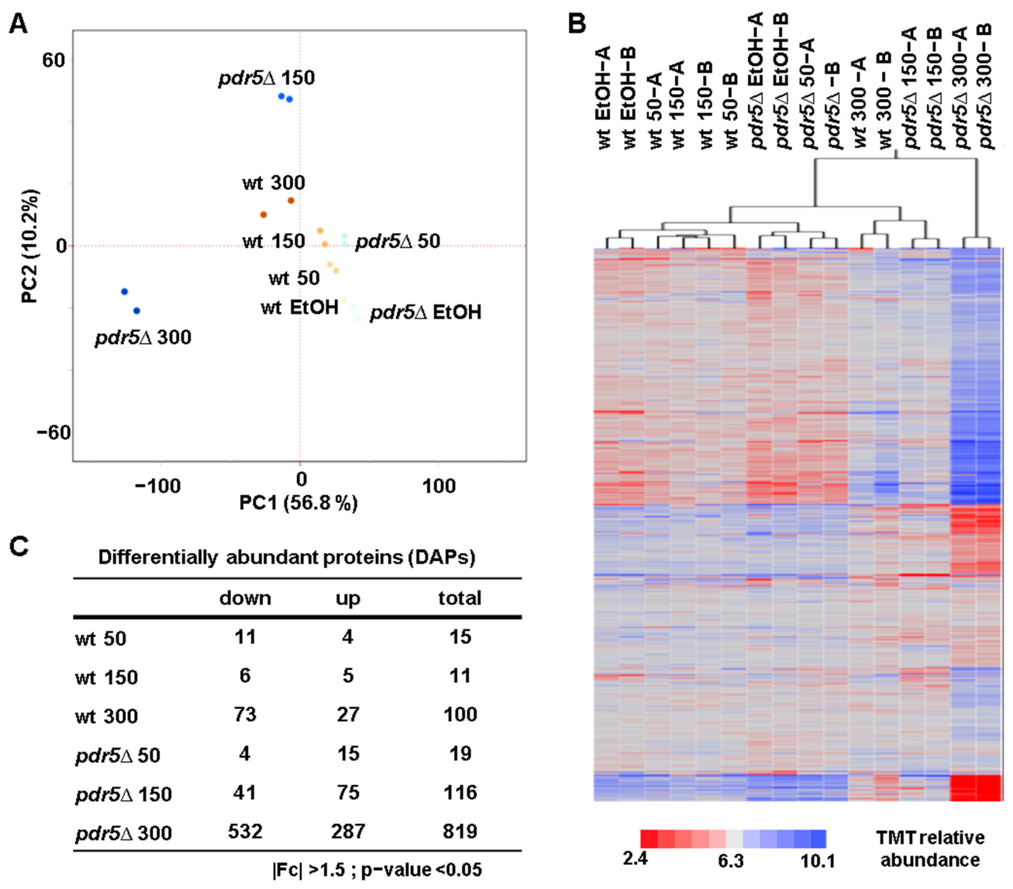
To assess the overall proteome changes, we performed principal component analysis (PCA) and hierarchical clustering analysis (HCA). PCA revealed tight clustering of replicates, with PC1 explaining over 56% of the variance and PC2 accounting for 10% (Figure 2A). Notably, pdr5Δ cells treated with 150 mg/mL and 300 mg/mL of BPA displayed distinct clustering, likely due to higher intracellular BPA accumulation. HCA, conducted using Euclidean distance and Ward linkage, showed similar clustering patterns (Figure 2B). Interestingly, wild-type cells treated with 50 mg/mL of BPA clustered with those treated with 150 mg/mL, suggesting similar proteomic responses at these lower BPA concentrations. However, wild-type cells treated with 300 mg/mL of BPA clustered near pdr5Δ cells treated with 150 mg/mL reflecting more pronounced proteome alterations. Lastly, pdr5Δ cells treated with 300 mg/mL of BPA formed a distinct cluster, exhibiting even more significant alterations in protein abundance.
Next, we identified differentially abundant proteins (DAPs) as those with a |log2 fold change| > 0.5 and an unadjusted p-value < 0.05 (Figure 2C). The number of DAPs increased with BPA concentration in both wild-type and pdr5Δ cells. Consistent with HCA, the magnitudes of proteomic changes induced by 50 mg/mL and 150 mg/mL BPA in wild-type cells were similar. As expected, pdr5Δ cells exhibited a greater number of DAPs across all concentrations, supporting the hypothesis of elevated intracellular BPA levels compared to wild-type cells due to the absence of Pdr5.

3.3. BPA Exposure Affects the Levels of the Pleiotropic Drug Resistance (PDR) Proteins

We confirmed the absence of Pdr5 in pdr5Δ cells by measuring its protein abundance in our proteomic experiment. We observed some residual signal in pdr5Δ cells likely due to interference (Figure S2A) [60]. Interestingly, wild-type cells exposed to BPA exhibited a dose-dependent increase in Pdr5, supporting its role in mediating BPA resistance. These data align with the higher growth inhibition and the stronger proteome changes observed in pdr5Δ cells (Figure 1C and Figure 2C).
The PDR family of proteins is responsible for multidrug resistance and includes several members [61], in addition to Pdr5, which may contribute to BPA resistance. We quantified seven PDR members in our proteomic experiment (Figure S2B): the transcription factors Pdr1, Pdr3, and Pdr8; the transporters Pdr12 and Pdr15; and the phosphatidylinositol transfer proteins Pdr16 and Pdr17. Notably, Pdr16 abundance increased in both wild-type and pdr5Δ cells in a dose-dependent manner. In contrast, Pdr15 abundance increased only in pdr5Δ cells, suggesting that it may compensate for the absence of its paralog Pdr5. However, Pdr12 abundance decreased in pdr5Δ cells with an increasing BPA concentration. These results highlight the dose-dependent regulation of specific PDR proteins by BPA and the intricate regulatory network among them in response to this compound.

3.4. BPA Induces Arginine Biosynthesis and Glucose Transporters While Downregulating Mitochondrial Proteins in Wild-Type Cells

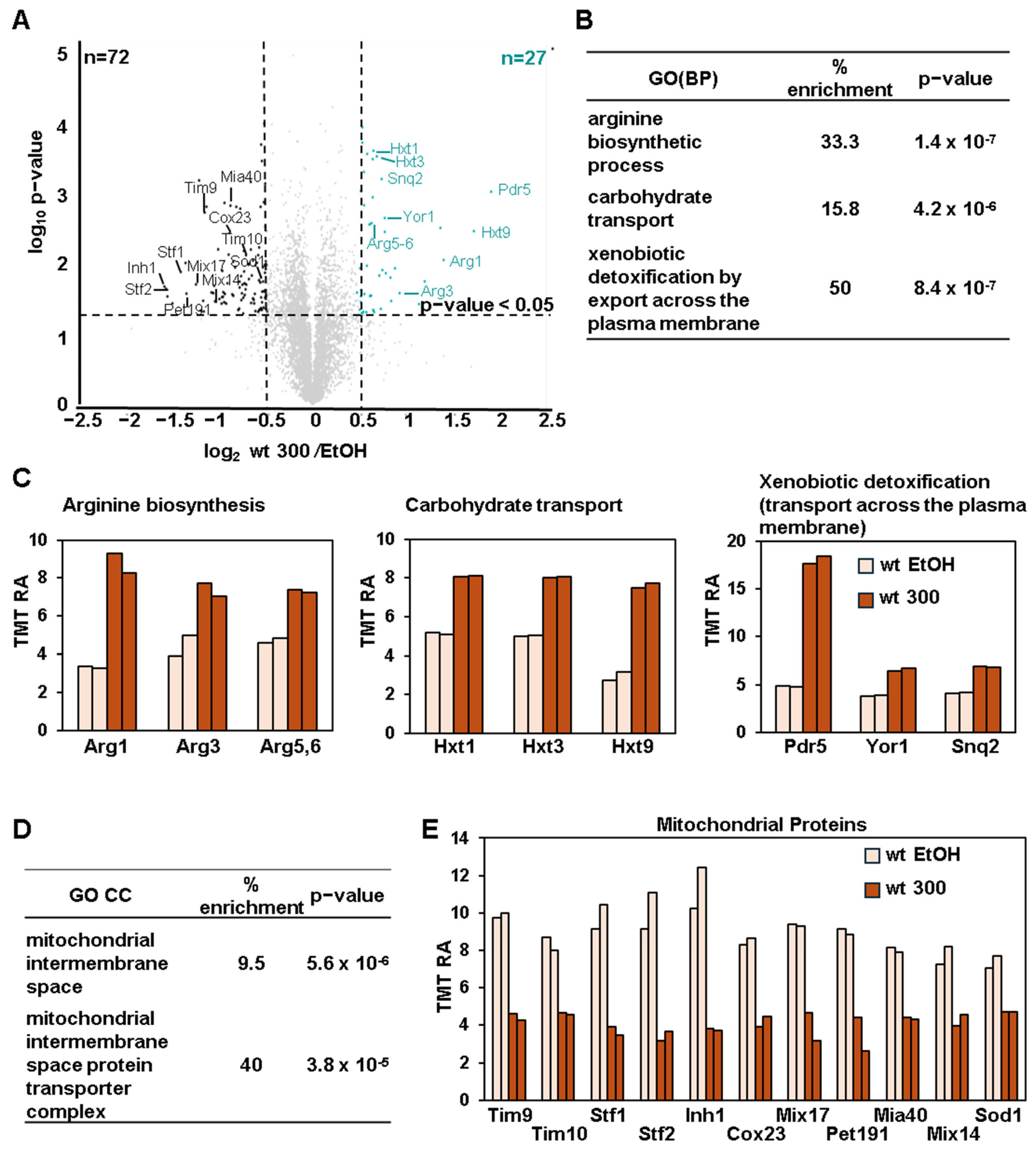
As only a small number of DAPs were identified at lower doses of BPA (50 and 150 mg/mL) in wild-type cells (Figure 2C). We focused our analysis on wild-type cells treated with 300 mg/mL BPA. We identified 27 significantly upregulated and 72 downregulated proteins compared to EtOH-treated cells (Figure 3A). Gene ontology (GO) analysis of the upregulated proteins revealed enrichment of pathways related to arginine biosynthesis, carbohydrate transport, and xenobiotic detoxification (Figure 3B). Specifically, the arginine biosynthesis enzymes Arg1, Arg3, and Arg5,6 showed increased abundance (Figure 3C, left). Additionally, low-affinity glucose transporters Hxt1, Hxt3, and Hxt9 were upregulated (Figure 3C, middle). Low-affinity glucose transporters are usually upregulated when glucose levels in the medium are high, suggesting that BPA altered glucose uptake or metabolism. BPA also increased the abundance of ABC transporters Snq2 and Yor1, alongside Pdr5, indicating their potential roles in BPA resistance (Figure 3C, right). As the upregulation of Pdr5 induced by BPA is much higher compared to that of Snq2 and Yor1, it is likely that the deletion of PDR5 would have a strong effect on intracellular BPA accumulation. These data indicated that deleting these two genes, along with PDR5, could further enhance the effects of BPA on cellular functions and on the proteome compared to cells in which only PDR5 has been deleted.
Conversely, BPA downregulated a set of mitochondrial proteins, specifically those in the mitochondrial intermembrane space and those constituting the mitochondrial intermembrane space protein transporter complex (Figure 3D). These proteins included the transporters Tim9, Tim10, and the assembly factors Mia40 and Cox23 (Figure 3E). Reduced levels of these proteins suggest that BPA could disrupt mitochondrial protein import, a process critical for mitochondrial function.

3.5. Proteomic Changes in pdr5Δ Cells Indicate Oxidative Stress

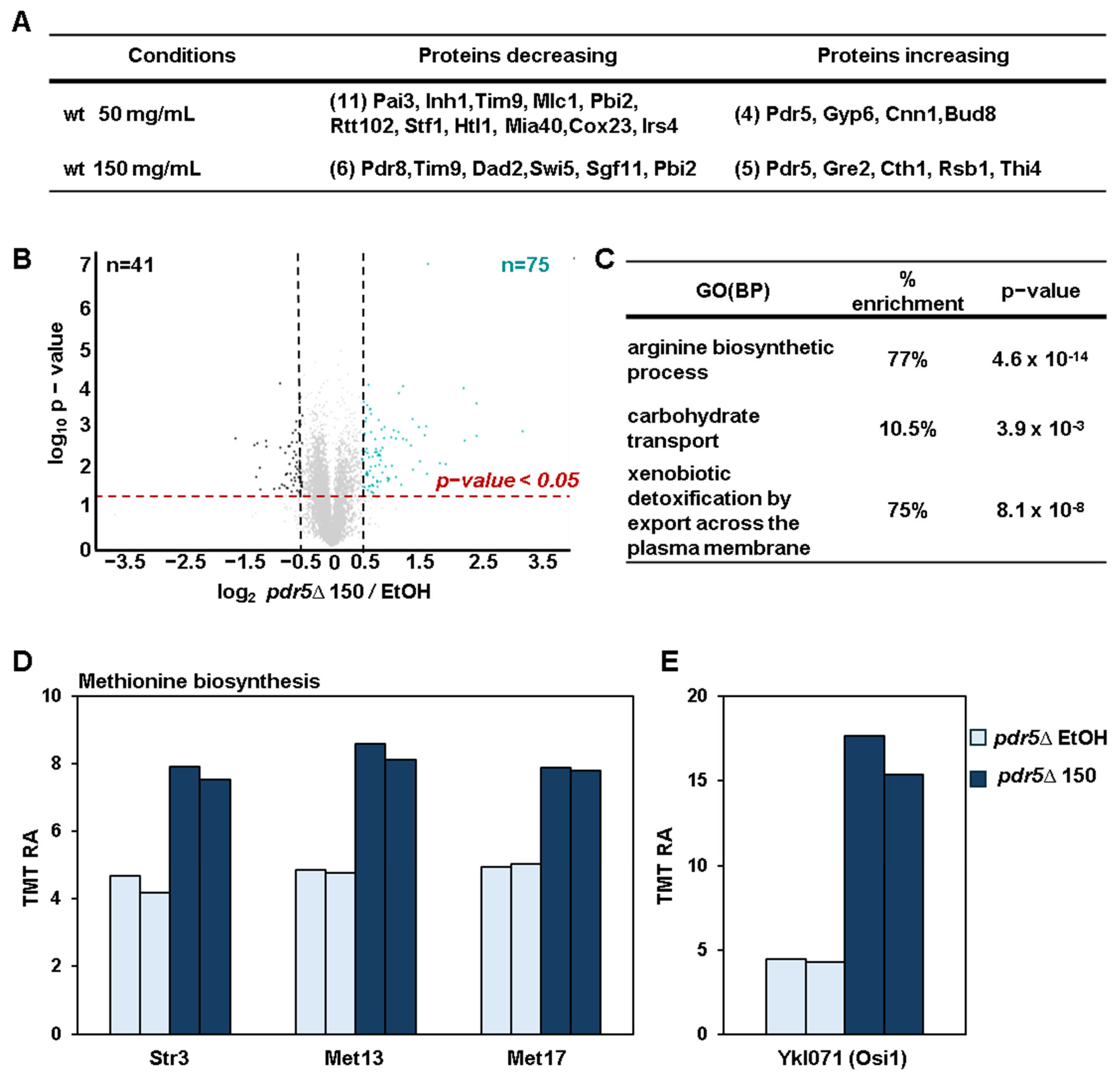
As in wild-type cells, only a small number of DAPs were identified at the lowest dose of BPA (50 mg/mL) in wild-type cells (Figure 4A). We identified 75 and 41 proteins with higher and lower abundance, respectively (Figure 4B), from our analysis of pdr5Δ cells treated with 150 mg/mL BPA compared to the EtOH-treated control cells. Here, we have omitted the comparison with the 300 mg/mL dose as it caused near-complete growth inhibition of pdr5Δ cells (Figure 1C). GO analysis highlighted similar enrichment patterns observed for wild-type cells treated with 300 mg/mL BPA, including arginine biosynthesis, carbohydrate transport, and detoxification pathways (Figure 4C). Additionally, we observed upregulation of methionine biosynthesis enzymes (e.g., Str3, Met13, and Met17) and of Osi1 (oxidative stress-induced protein 1), which are both markers of oxidative stress (Figure 4D,E). These findings suggest that BPA induces oxidative stress, which is consistent with previous studies in yeast and mammalian cells [35,41].

3.6. BPA-Induced Stress and Cellular Damage to S. cerevisiae Cells

In agreement with the observed dose-dependent cell growth inhibition by BPA, several BPA-responsive proteins provided insights into the stresses and cellular damage induced by this compound. For instance, Rsb1, a transporter activated by membrane stress and by glycerophospholipid asymmetry [62], was upregulated in a dose-dependent manner (Figure S3). Similarly, stress-response proteins Gre2 [63] and Tsv1 [64] increased with BPA exposure, indicating the presence of a cellular response to stress (Figure S3). Dld3 [65], associated with mitochondrial dysfunction, was also of relatively higher abundance (Figure S3), corroborating our earlier observations of disrupted mitochondrial protein import. Yhi9 [66] and Iml2 [67], for which upregulation was particularly evident in pdr5Δ cells treated with 150 mg/mL, were involved, respectively, in the endoplasmic reticulum unfolded protein response and in the clearance of inclusion bodies that form during protein misfolding stress (Figure S3). These findings demonstrated that BPA exposure triggered a wide range of cellular stress responses and proteome changes, which can serve as a valuable resource and starting point for uncovering BPA’s mechanisms of action.

4. Discussion

Here, we investigated the impact of different doses of BPA on cell growth and on the proteome of S. cerevisiae wild-type and pdr5Δ strains. Yeast cells, which naturally lack estrogen receptors (ESr), offer a distinct advantage for studying BPA. By analyzing their response to BPA, we can identify BPA targets that operate independently of the ESr pathway. As the effects of BPA on eukaryotic cells appear to be conserved [42], our data could help distinguish between ESr-mediated and non-ESr-mediated BPA-dependent effects in higher eukaryotes.
Consistent with previous studies in S. cerevisiae [41,44], we observed that BPA inhibited cell growth in a dose-dependent manner. Additionally, we demonstrated that the deletion of PDR5 sensitizes yeast cells to BPA, suggesting that this ABC transporter is required to export BPA out from yeast cells. In agreement with this, BPA increased the protein abundance of Pdr5 in a dose-dependent manner in the wild-type strain. Furthermore, the proteome-level changes were more pronounced in pdr5Δ cells compared to wild-type cells, likely reflecting the higher BPA uptake in pdr5Δ cells.
In addition to Pdr5, we observed that other ABC transporters, such as Snq2 and Yor1, were also upregulated in a BPA-dependent manner. These data suggest that these transporters may contribute to BPA resistance in yeast cells. To comprehensively assess the impact of BPA on yeast cells, it may be advantageous to investigate further cells with combined deletions of PDR5 and SNQ2 or YOR1. Such multiple deletions are likely to result in higher intracellular concentrations of BPA, thereby amplifying its effects compared to wild-type cells. We speculate that at lower doses of BPA, these transporters’ actions do not significantly alter the proteome or the growth of wild-type cells. Our data suggest that the role of ABC transporters in exporting BPA outside the cells is conserved, as this class of transporter has been also implicated in removing BPA from human cells [68].
One unexpected finding from our proteome analysis was that BPA induced an increase in the protein abundance of glucose transporters in both wild-type and pdr5Δ cells. Interestingly, the glucose transporters that were upregulated by BPA were specifically those with low affinity, such as Hxt1, Hxt3, and Hxt9. These transporters are usually upregulated when yeast cells sense more glucose in the medium [69]. An analysis of the literature revealed that BPA increases the abundance of the glucose transporter GLUT1 (Hxt1 homolog) in rat’s placenta and in human trophoblasts [70], as well as GLUT4 in mouse adipocytes [71]. These data suggest that BPA may influence glucose uptake with a mechanism that is likely evolutionarily conserved. As an increase in glucose transporters is observed in S. cerevisiae, which lacks estrogen receptors (ESRs), this function of BPA is likely ESr-independent. The increase in glucose transporters could contribute to the observed link between BPA and diabetes development [27]. However, the mechanism underlying the upregulation of glucose transporters remains unclear.
We also observed an increase in enzymes involved in arginine biosynthesis in both wild-type and pdr5Δ cells and an increase in enzymes involved in methionine biosynthesis in pdr5Δ cells. Both arginine and methionine biosynthesis have been shown to be upregulated in S. cerevisiae in response to oxidative stress [72,73], suggesting that S. cerevisiae cells treated with BPA may be experiencing this type of stress. Consistent with this finding, Osi1 or oxidative stress-induced protein 1, a known marker of oxidative stress [74], was one of the proteins with the highest fold change in pdr5Δ cells treated with BPA. We hypothesized that the increase in methionine biosynthesis enzymes was not evident in wild-type cells, as significantly less BPA accumulated in these cells compared to pdr5Δ cells. Our proteome analysis suggests that BPA induces oxidative stress in S. cerevisiae. To validate this observation, it is essential to use standard methods to measure oxidative stress, such as assessing ROS levels or detecting oxidatively modified proteins. It is worth noting that these methods have been employed in previously published studies. Indeed, several studies have reported that BPA can induce oxidative stress in both S. cerevisiae and mammalian cells [35,36,41]; however again the mechanisms underlying this effect remain uncharacterized.
Proteomic analysis has been performed in multiple model systems, such as human, mice, rat, and zebrafish cells. Exposure to BPA has led to the alteration of proteins involved in multiple signaling pathways, such as pathways associated with tumor progression [75], development and metabolism [76], and endocrine and reproductive signaling [77,78]. Furthermore, multiple studies have identified alterations that indicate the presence of toxicity and stresses, in particular oxidative stress [35,79,80].
One of the most interesting findings from our analysis was the downregulation of a specific set of mitochondrial proteins, particularly those located in the intermembrane space. Mitochondrial dysfunction has been shown to be induced by BPA in multiple model systems [37,38,41], but the underlying mechanisms thereof remain undiscovered. Our data suggest that mitochondrial dysfunction in S. cerevisiae cells may be a result of defects in the process of mitochondria protein import.
A previous transcriptomic analysis examining the effects of BPA on the yeast transcriptome, conducted using the same yeast background as in our study [44], revealed findings that align with our proteomic analysis. Specifically, it reported the upregulation of Pdr5 and Pdr16 and the downregulation of mitochondrial proteins. Additionally, we observed a downregulation of glucose transporters induced by low glucose levels, which is consistent with our observation of low affinity glucose transporter upregulation.
Overall, our proteome analysis, the first on the effects of BPA on the proteome of S. cerevisiae, suggests the cellular pathways that can be affected by this compound. Additionally, we identified hundreds of differentially abundant proteins regulated at the protein level by BPA. Among these, we highlighted a few, such as Yhi9, which is involved in the unfolded protein response, and Dld3, which is upregulated in response to mitochondrial dysfunction. We hypothesize that many of these proteins regulated in a BPA-dependent manner will help to eventually fully elucidate the mechanism of action of this compound and will guide future studies to better comprehend its impact on human health.

Supplementary Materials

The following supporting information can be downloaded at: https://www.mdpi.com/article/10.3390/microorganisms13010114/s1, Figure S1: Yeast growth curves; Figure S2: Protein abundance profile of Pdr5 and of other members of the PDR protein network; Figure S3: Example proteins that change in a BPA dose-dependent manner; Table S1: Proteins quantified in the experiment.

Author Contributions

Conceptualization, V.R. and J.A.P.; methodology, V.R. and J.A.P.; software, J.A.P.; validation, V.R. and J.A.P.; formal analysis, V.R. and J.A.P.; investigation, V.R. and J.A.P.; resources, J.A.P.; data curation, V.R. and J.A.P.; writing—original draft preparation, V.R. and J.A.P.; writing—review and editing, V.R. and J.A.P.; visualization, V.R. and J.A.P.; supervision, V.R. and J.A.P.; project administration, J.A.P.; funding acquisition, J.A.P. All authors have read and agreed to the published version of the manuscript.

Funding

This work was supported by NIH grant R01 GM132129 (JAP).

Institutional Review Board Statement

Not applicable.

Data Availability Statement

RAW files will be made available upon request, in addition to the data that have been deposited to the ProteomeXchange Consortium via the PRIDE [81] partner repository with the dataset identifier. Project accession: PXD058913.

Acknowledgments

We acknowledge S. P. Gygi and the Taplin Mass Spectrometry Facility at Harvard Medical School for the use of their mass spectrometers. We thank R. W. King for his support.

Conflicts of Interest

The authors declare no conflicts of interest.

References

  1. Geens, T.; Aerts, D.; Berthot, C.; Bourguignon, J.-P.; Goeyens, L.; Lecomte, P.; Maghuin-Rogister, G.; Pironnet, A.-M.; Pussemier, L.; Scippo, M.-L.; et al. A Review of Dietary and Non-Dietary Exposure to Bisphenol-A. Food Chem. Toxicol. 2012, 50, 3725–3740. [Google Scholar] [CrossRef]
  2. Michałowicz, J. Bisphenol A—Sources, Toxicity and Biotransformation. Environ. Toxicol. Pharmacol. 2014, 37, 738–758. [Google Scholar] [CrossRef] [PubMed]
  3. Miller, S.C.; Bottema, C.D.K.; Stathis, P.A.; Tokés, L.G.; Feldman, D. Unexpected Presence of Estrogens in Culture Medium Supplements: Subsequent Metabolism by the Yeast Sacchromyces Cerevisiae *. Endocrinology 1986, 119, 1362–1369. [Google Scholar] [CrossRef]
  4. Kubwabo, C.; Kosarac, I.; Stewart, B.; Gauthier, B.R.; Lalonde, K.; Lalonde, P.J. Migration of Bisphenol A from Plastic Baby Bottles, Baby Bottle Liners and Reusable Polycarbonate Drinking Bottles. Food Addit. Contam. 2009, 26, 928–937. [Google Scholar] [CrossRef]
  5. Chailurkit, L.; Srijaruskul, K.; Ongphiphadhanakul, B. Bisphenol A in Canned Carbonated Drinks and Plastic-Bottled Water from Supermarkets. Expo. Health 2017, 9, 243–248. [Google Scholar] [CrossRef]
  6. Rudel, R.A.; Gray, J.M.; Engel, C.L.; Rawsthorne, T.W.; Dodson, R.E.; Ackerman, J.M.; Rizzo, J.; Nudelman, J.L.; Brody, J.G. Food Packaging and Bisphenol A and Bis(2-Ethyhexyl) Phthalate Exposure: Findings from a Dietary Intervention. Environ. Health Perspect. 2011, 119, 914–920. [Google Scholar] [CrossRef]
  7. Brede, C.; Fjeldal, P.; Skjevrak, I.; Herikstad, H. Increased Migration Levels of Bisphenol A from Polycarbonate Baby Bottles after Dishwashing, Boiling and Brushing. Food Addit. Contam. 2003, 20, 684–689. [Google Scholar] [CrossRef]
  8. Hunt, P.A.; Koehler, K.E.; Susiarjo, M.; Hodges, C.A.; Ilagan, A.; Voigt, R.C.; Thomas, S.; Thomas, B.F.; Hassold, T.J. Bisphenol a Exposure Causes Meiotic Aneuploidy in the Female Mouse. Curr. Biol. 2003, 13, 546–553. [Google Scholar] [CrossRef]
  9. Vandenberg, L.N.; Hauser, R.; Marcus, M.; Olea, N.; Welshons, W.V. Human Exposure to Bisphenol A (BPA). Reprod. Toxicol. 2007, 24, 139–177. [Google Scholar] [CrossRef]
  10. Lehmler, H.-J.; Liu, B.; Gadogbe, M.; Bao, W. Exposure to Bisphenol A, Bisphenol F, and Bisphenol S in U.S. Adults and Children: The National Health and Nutrition Examination Survey 2013–2014. ACS Omega 2018, 3, 6523–6532. [Google Scholar] [CrossRef]
  11. Govarts, E.; Gilles, L.; Rodriguez Martin, L.; Santonen, T.; Apel, P.; Alvito, P.; Anastasi, E.; Andersen, H.R.; Andersson, A.-M.; Andryskova, L.; et al. Harmonized Human Biomonitoring in European Children, Teenagers and Adults: EU-Wide Exposure Data of 11 Chemical Substance Groups from the HBM4EU Aligned Studies (2014–2021). Int. J. Hyg. Environ. Health 2023, 249, 114119. [Google Scholar] [CrossRef] [PubMed]
  12. Covaci, A.; Den Hond, E.; Geens, T.; Govarts, E.; Koppen, G.; Frederiksen, H.; Knudsen, L.E.; Mørck, T.A.; Gutleb, A.C.; Guignard, C.; et al. Urinary BPA Measurements in Children and Mothers from Six European Member States: Overall Results and Determinants of Exposure. Environ. Res. 2015, 141, 77–85. [Google Scholar] [CrossRef] [PubMed]
  13. Calafat, A.M.; Ye, X.; Wong, L.-Y.; Reidy, J.A.; Needham, L.L. Exposure of the U.S. Population to Bisphenol A and 4-Tertiary-Octylphenol: 2003–2004. Environ. Health Perspect. 2008, 116, 39–44. [Google Scholar] [CrossRef]
  14. Huang, R.-P.; Liu, Z.-H.; Yin, H.; Dang, Z.; Wu, P.-X.; Zhu, N.-W.; Lin, Z. Bisphenol A Concentrations in Human Urine, Human Intakes across Six Continents, and Annual Trends of Average Intakes in Adult and Child Populations Worldwide: A Thorough Literature Review. Sci. Total Environ. 2018, 626, 971–981. [Google Scholar] [CrossRef] [PubMed]
  15. Routledge, E.J.; White, R.; Parker, M.G.; Sumpter, J.P. Differential Effects of Xenoestrogens on Coactivator Recruitment by Estrogen Receptor (ER) Alpha and ERbeta. J. Biol. Chem. 2000, 275, 35986–35993. [Google Scholar] [CrossRef]
  16. Matthews, J.B.; Twomey, K.; Zacharewski, T.R. In Vitro and in Vivo Interactions of Bisphenol A and Its Metabolite, Bisphenol A Glucuronide, with Estrogen Receptors Alpha and Beta. Chem. Res. Toxicol. 2001, 14, 149–157. [Google Scholar] [CrossRef]
  17. Husain, Q.; Qayyum, S. Biological and Enzymatic Treatment of Bisphenol A and Other Endocrine Disrupting Compounds: A Review. Crit. Rev. Biotechnol. 2013, 33, 260–292. [Google Scholar] [CrossRef]
  18. Gao, H.; Yang, B.-J.; Li, N.; Feng, L.-M.; Shi, X.-Y.; Zhao, W.-H.; Liu, S.-J. Bisphenol A and Hormone-Associated Cancers: Current Progress and Perspectives. Medicine 2015, 94, e211. [Google Scholar] [CrossRef]
  19. Keshavarz-Maleki, R.; Kaviani, A.; Omranipour, R.; Gholami, M.; Khoshayand, M.R.; Ostad, S.N.; Sabzevari, O. Bisphenol-A in Biological Samples of Breast Cancer Mastectomy and Mammoplasty Patients and Correlation with Levels Measured in Urine and Tissue. Sci. Rep. 2021, 11, 18411. [Google Scholar] [CrossRef]
  20. Molina, L.; Figueroa, C.D.; Ehrenfeld, P.; Molina, L.; Figueroa, C.D.; Ehrenfeld, P. Interaction of Bisphenol A with G Protein: Coupled Receptors—New Paradigms in Breast Cancer. In Bisphenols; IntechOpen: London, UK, 2021. [Google Scholar] [CrossRef]
  21. Marotta, V.; Grumetto, L.; Neri, I.; Russo, G.; Tortora, A.; Izzo, G.; Panariello, I.; Rocco, D.; Pezzullo, L.; Vitale, M. Exposure to Bisphenol A Increases Malignancy Risk of Thyroid Nodules in Overweight/Obese Patients. Environ. Pollut. 2023, 316, 120478. [Google Scholar] [CrossRef]
  22. Dhimolea, E.; Wadia, P.R.; Murray, T.J.; Settles, M.L.; Treitman, J.D.; Sonnenschein, C.; Shioda, T.; Soto, A.M. Prenatal Exposure to BPA Alters the Epigenome of the Rat Mammary Gland and Increases the Propensity to Neoplastic Development. PLoS ONE 2014, 9, e99800. [Google Scholar] [CrossRef] [PubMed]
  23. Xiao, M.; Zhang, Y.; Zhang, X.; Zhang, G.; Jin, C.; Yang, J.; Wu, S.; Lu, X. Bisphenol A and Di(2-Ethylhexyl) Phthalate Promote Pulmonary Carcinoma in Female Rats via Estrogen Receptor Beta: In Vivo and in Silico Analysis. Ecotoxicol. Environ. Saf. 2023, 250, 114496. [Google Scholar] [CrossRef] [PubMed]
  24. Malloy, M.A.; Kochmanski, J.J.; Jones, T.R.; Colacino, J.A.; Goodrich, J.M.; Dolinoy, D.C.; Svoboda, L.K. Perinatal Bisphenol A Exposure and Reprogramming of Imprinted Gene Expression in the Adult Mouse Brain. Front. Genet. 2019, 10, 951. [Google Scholar] [CrossRef] [PubMed]
  25. Kundakovic, M.; Gudsnuk, K.; Franks, B.; Madrid, J.; Miller, R.L.; Perera, F.P.; Champagne, F.A. Sex-Specific Epigenetic Disruption and Behavioral Changes Following Low-Dose in Utero Bisphenol A Exposure. Proc. Natl. Acad. Sci. USA 2013, 110, 9956–9961. [Google Scholar] [CrossRef]
  26. Kodila, A.; Franko, N.; Sollner Dolenc, M. A Review on Immunomodulatory Effects of BPA Analogues. Arch. Toxicol. 2023, 97, 1831–1846. [Google Scholar] [CrossRef]
  27. Farrugia, F.; Aquilina, A.; Vassallo, J.; Pace, N.P. Bisphenol A and Type 2 Diabetes Mellitus: A Review of Epidemiologic, Functional, and Early Life Factors. Int. J. Environ. Res. Public Health 2021, 18, 716. [Google Scholar] [CrossRef]
  28. Thompson, R.C.; Courtene-Jones, W.; Boucher, J.; Pahl, S.; Raubenheimer, K.; Koelmans, A.A. Twenty Years of Microplastic Pollution Research-What Have We Learned? Science 2024, 386, eadl2746. [Google Scholar] [CrossRef]
  29. Li, Y.; Tao, L.; Wang, Q.; Wang, F.; Li, G.; Song, M. Potential Health Impact of Microplastics: A Review of Environmental Distribution, Human Exposure, and Toxic Effects. Environ. Health 2023, 1, 249–257. [Google Scholar] [CrossRef]
  30. Dzierżyński, E.; Gawlik, P.J.; Puźniak, D.; Flieger, W.; Jóźwik, K.; Teresiński, G.; Forma, A.; Wdowiak, P.; Baj, J.; Flieger, J. Microplastics in the Human Body: Exposure, Detection, and Risk of Carcinogenesis: A State-of-the-Art Review. Cancers 2024, 16, 3703. [Google Scholar] [CrossRef]
  31. Marfella, R.; Prattichizzo, F.; Sardu, C.; Fulgenzi, G.; Graciotti, L.; Spadoni, T.; D’Onofrio, N.; Scisciola, L.; Grotta, R.L.; Frigé, C.; et al. Microplastics and Nanoplastics in Atheromas and Cardiovascular Events. N. Engl. J. Med. 2024, 390, 900–910. [Google Scholar] [CrossRef]
  32. Li, S.; Keenan, J.I.; Shaw, I.C.; Frizelle, F.A. Could Microplastics Be a Driver for Early Onset Colorectal Cancer? Cancers 2023, 15, 3323. [Google Scholar] [CrossRef]
  33. Beal, M.A.; Coughlan, M.C.; Nunnikhoven, A.; Gagné, M.; Barton-Maclaren, T.S.; Bradford, L.M.; Rowan-Carroll, A.; Williams, A.; Meier, M.J. High-Throughput Transcriptomics Toxicity Assessment of Eleven Data-Poor Bisphenol A Alternatives. Environ. Pollut. 2024, 361, 124827. [Google Scholar] [CrossRef] [PubMed]
  34. Mhaouty-Kodja, S.; Zalko, D.; Tait, S.; Testai, E.; Viguié, C.; Corsini, E.; Grova, N.; Buratti, F.M.; Cabaton, N.J.; Coppola, L.; et al. A Critical Review to Identify Data Gaps and Improve Risk Assessment of Bisphenol A Alternatives for Human Health. Crit. Rev. Toxicol. 2024, 54, 696–753. [Google Scholar] [CrossRef] [PubMed]
  35. Deng, Y.; He, H.; Wan, H.; Shen, N.; Li, J.; Zhang, S.; Zeng, Q.; Chang, J.; Lu, Q.; Zhong, R.; et al. Bisphenol A Exposure, Interaction with Genetic Variants and Colorectal Cancer via Mediating Oxidative Stress Biomarkers. Environ. Pollut. 2021, 287, 117630. [Google Scholar] [CrossRef] [PubMed]
  36. Meli, R.; Monnolo, A.; Annunziata, C.; Pirozzi, C.; Ferrante, M.C. Oxidative Stress and BPA Toxicity: An Antioxidant Approach for Male and Female Reproductive Dysfunction. Antioxidants 2020, 9, 405. [Google Scholar] [CrossRef]
  37. Goyal, S.; Tiwari, S.; Seth, B.; Tandon, A.; Shankar, J.; Sinha, M.; Singh, S.J.; Priya, S.; Chaturvedi, R.K. Bisphenol-A Inhibits Mitochondrial Biogenesis via Impairment of GFER Mediated Mitochondrial Protein Import in the Rat Brain Hippocampus. Neurotoxicology 2021, 85, 18–32. [Google Scholar] [CrossRef]
  38. Nayak, D.; Adiga, D.; Khan, N.G.; Rai, P.S.; Dsouza, H.S.; Chakrabarty, S.; Gassman, N.R.; Kabekkodu, S.P. Impact of Bisphenol A on Structure and Function of Mitochondria: A Critical Review. Rev. Env. Contam. (Former. Residue Rev.) 2022, 260, 10. [Google Scholar] [CrossRef]
  39. Khan, N.G.; Tungekar, B.; Adiga, D.; Chakrabarty, S.; Rai, P.S.; Kabekkodu, S.P. Alterations Induced by Bisphenol A on Cellular Organelles and Potential Relevance on Human Health. Biochim. Biophys. Acta (BBA)—Mol. Cell Res. 2023, 1870, 119505. [Google Scholar] [CrossRef]
  40. Kim, S.; Gwon, D.; Kim, J.A.; Choi, H.; Jang, C.-Y. Bisphenol A Disrupts Mitotic Progression via Disturbing Spindle Attachment to Kinetochore and Centriole Duplication in Cancer Cell Lines. Toxicol. In Vitro 2019, 59, 115–125. [Google Scholar] [CrossRef]
  41. Ďurovcová, I.; Goffa, E.; Šestáková, Z.; Mániková, D.; Gaplovská-Kyselá, K.; Chovanec, M.; Ševčovičová, A. Acute Exposure to Bisphenol A Causes Oxidative Stress Induction with Mitochondrial Origin in Saccharomyces Cerevisiae Cells. J. Fungi 2021, 7, 543. [Google Scholar] [CrossRef]
  42. Kumar, R.; Oke, A.; Rockmill, B.; de Cruz, M.; Verduzco, R.; Shodhan, A.; Woodruff-Madeira, X.; Abrahamsson, D.P.; Varshavsky, J.; Lam, J.; et al. Rapid Identification of Reproductive Toxicants among Environmental Chemicals Using an in Vivo Evaluation of Gametogenesis in Budding Yeast Saccharomyces Cerevisiae. Reprod. Toxicol. 2024, 128, 108630. [Google Scholar] [CrossRef] [PubMed]
  43. Alhoch, B.; Chen, A.; Chan, E.; Elkabti, A.; Fariña, S.; Gilbert, C.; Kang, J.; King, B.; Leung, K.; Levy, J.; et al. Comparative Genomic Screen in Two Yeasts Reveals Conserved Pathways in the Response Network to Phenol Stress. G3 2019, 9, 639–650. [Google Scholar] [CrossRef] [PubMed]
  44. Bereketoglu, C.; Arga, K.Y.; Eraslan, S.; Mertoglu, B. Analysis of Transcriptional Profiles of Saccharomyces Cerevisiae Exposed to Bisphenol A. Curr. Genet. 2017, 63, 253–274. [Google Scholar] [CrossRef] [PubMed]
  45. Rossio, V.; Liu, X.; Paulo, J.A. Comparative Proteomic Analysis of Two Commonly Used Laboratory Yeast Strains: W303 and BY4742. Proteomes 2023, 11, 30. [Google Scholar] [CrossRef]
  46. Rossio, V.; Paulo, J.A. Comparison of the Proteomes and Phosphoproteomes of S. Cerevisiae Cells Harvested with Different Strategies. Proteomes 2023, 11, 28. [Google Scholar] [CrossRef]
  47. Liu, X.; Rossio, V.; Paulo, J.A. Spin Column-Based Peptide Fractionation Alternatives for Streamlined Tandem Mass Tag (SL-TMT) Sample Processing. J. Proteom. 2023, 276, 104839. [Google Scholar] [CrossRef]
  48. Liu, X.; Rossio, V.; Gygi, S.P.; Paulo, J.A. Enriching Cysteine-Containing Peptides Using a Sulfhydryl-Reactive Alkylating Reagent with a Phosphonic Acid Group and Immobilized Metal Affinity Chromatography. J. Proteome Res. 2023, 22, 1270–1279. [Google Scholar] [CrossRef]
  49. Rossio, V.; Paulo, J.A.; Liu, X.; Gygi, S.P.; King, R.W. Specificity Profiling of Deubiquitylases against Endogenously Generated Ubiquitin-Protein Conjugates. Cell Chem. Biol. 2024, 31, 1349–1362.e5. [Google Scholar] [CrossRef]
  50. Zhang, T.; Liu, X.; Rossio, V.; Dawson, S.L.; Gygi, S.P.; Paulo, J.A. Enhancing Proteome Coverage by Using Strong Anion-Exchange in Tandem with Basic-pH Reversed-Phase Chromatography for Sample Multiplexing-Based Proteomics. J. Proteome Res. 2023, 23, 2870–2881. [Google Scholar] [CrossRef]
  51. Chambers, M.C.; Maclean, B.; Burke, R.; Amodei, D.; Ruderman, D.L.; Neumann, S.; Gatto, L.; Fischer, B.; Pratt, B.; Egertson, J.; et al. A Cross-Platform Toolkit for Mass Spectrometry and Proteomics. Nat. Biotechnol. 2012, 30, 918–920. [Google Scholar] [CrossRef]
  52. Beausoleil, S.A.; Villén, J.; Gerber, S.A.; Rush, J.; Gygi, S.P. A Probability-Based Approach for High-Throughput Protein Phosphorylation Analysis and Site Localization. Nat. Biotechnol. 2006, 24, 1285–1292. [Google Scholar] [CrossRef] [PubMed]
  53. Huttlin, E.L.; Jedrychowski, M.P.; Elias, J.E.; Goswami, T.; Rad, R.; Beausoleil, S.A.; Villén, J.; Haas, W.; Sowa, M.E.; Gygi, S.P. A Tissue-Specific Atlas of Mouse Protein Phosphorylation and Expression. Cell 2010, 143, 1174–1189. [Google Scholar] [CrossRef] [PubMed]
  54. Elias, J.E.; Gygi, S.P. Target-Decoy Search Strategy for Mass Spectrometry-Based Proteomics. In Proteome Bioinformatics; Hubbard, S.J., Jones, A.R., Eds.; Humana Press: Totowa, NJ, USA, 2010; pp. 55–71. [Google Scholar] [CrossRef]
  55. McAlister, G.C.; Huttlin, E.L.; Haas, W.; Ting, L.; Jedrychowski, M.P.; Rogers, J.C.; Kuhn, K.; Pike, I.; Grothe, R.A.; Blethrow, J.D.; et al. Increasing the Multiplexing Capacity of TMTs Using Reporter Ion Isotopologues with Isobaric Masses. Anal. Chem. 2012, 84, 7469–7478. [Google Scholar] [CrossRef] [PubMed]
  56. Thompson, A.; Schäfer, J.; Kuhn, K.; Kienle, S.; Schwarz, J.; Schmidt, G.; Neumann, T.; Johnstone, R.; Mohammed, A.K.A.; Hamon, C. Tandem Mass Tags: A Novel Quantification Strategy for Comparative Analysis of Complex Protein Mixtures by MS/MS. Anal. Chem. 2003, 75, 1895–1904. [Google Scholar] [CrossRef] [PubMed]
  57. Navarrete-Perea, J.; Liu, X.; Rad, R.; Gygi, J.P.; Gygi, S.P.; Paulo, J.A. Assessing Interference in Isobaric Tag-Based Sample Multiplexing Using an 18-Plex Interference Standard. Proteomics 2022, 22, e2100317. [Google Scholar] [CrossRef]
  58. Liu, X.; Rossio, V.; Thakurta, S.G.; Flora, A.; Foster, L.; Bomgarden, R.D.; Gygi, S.P.; Paulo, J.A. Fe3+-NTA Magnetic Beads as an Alternative to Spin Column-Based Phosphopeptide Enrichment. J. Proteom. 2022, 260, 104561. [Google Scholar] [CrossRef]
  59. Golin, J.; Ambudkar, S.V.; May, L. The Yeast Pdr5p Multidrug Transporter: How Does It Recognize so Many Substrates? Biochem. Biophys. Res. Commun. 2007, 356, 1–5. [Google Scholar] [CrossRef]
  60. Paulo, J.A.; O’Connell, J.D.; Gygi, S.P. A Triple Knockout (TKO) Proteomics Standard for Diagnosing Ion Interference in Isobaric Labeling Experiments. J. Am. Soc. Mass. Spectrom. 2016, 27, 1620–1625. [Google Scholar] [CrossRef]
  61. Balzi, E.; Goffeau, A. Yeast Multidrug Resistance: The PDR Network. J. Bioenerg. Biomembr. 1995, 27, 71–76. [Google Scholar] [CrossRef]
  62. Kihara, A.; Igarashi, Y. Cross Talk between Sphingolipids and Glycerophospholipids in the Establishment of Plasma Membrane Asymmetry. Mol. Biol. Cell 2004, 15, 4949–4959. [Google Scholar] [CrossRef]
  63. Garay-Arroyo, A.; Covarrubias, A.A. Three Genes Whose Expression Is Induced by Stress in Saccharomyces Cerevisiae. Yeast 1999, 15, 879–892. [Google Scholar] [CrossRef]
  64. Ayers, M.C.; Sherman, Z.N.; Gallagher, J.E.G. Oxidative Stress Responses and Nutrient Starvation in MCHM Treated Saccharomyces Cerevisiae. G3 2020, 10, 4665–4678. [Google Scholar] [CrossRef] [PubMed]
  65. Chelstowska, A.; Liu, Z.; Jia, Y.; Amberg, D.; Butow, R.A. Signalling between Mitochondria and the Nucleus Regulates the Expression of a New D-Lactate Dehydrogenase Activity in Yeast. Yeast 1999, 15, 1377–1391. [Google Scholar] [CrossRef]
  66. Giaever, G.; Chu, A.M.; Ni, L.; Connelly, C.; Riles, L.; Véronneau, S.; Dow, S.; Lucau-Danila, A.; Anderson, K.; André, B.; et al. Functional Profiling of the Saccharomyces Cerevisiae Genome. Nature 2002, 418, 387–391. [Google Scholar] [CrossRef]
  67. Moldavski, O.; Amen, T.; Levin-Zaidman, S.; Eisenstein, M.; Rogachev, I.; Brandis, A.; Kaganovich, D.; Schuldiner, M. Lipid Droplets Are Essential for Efficient Clearance of Cytosolic Inclusion Bodies. Dev. Cell 2015, 33, 603–610. [Google Scholar] [CrossRef]
  68. Mazur, C.S.; Marchitti, S.A.; Dimova, M.; Kenneke, J.F.; Lumen, A.; Fisher, J. Human and Rat ABC Transporter Efflux of Bisphenol A and Bisphenol A Glucuronide: Interspecies Comparison and Implications for Pharmacokinetic Assessment. Toxicol. Sci. 2012, 128, 317–325. [Google Scholar] [CrossRef]
  69. Reifenberger, E.; Boles, E.; Ciriacy, M. Kinetic Characterization of Individual Hexose Transporters of Saccharomyces Cerevisiae and Their Relation to the Triggering Mechanisms of Glucose Repression. Eur. J. Biochem. 1997, 245, 324–333. [Google Scholar] [CrossRef]
  70. Ermini, L.; Nuzzo, A.M.; Ietta, F.; Romagnoli, R.; Moretti, L.; Masturzo, B.; Paulesu, L.; Rolfo, A. Placental Glucose Transporters and Response to Bisphenol A in Pregnancies from of Normal and Overweight Mothers. Int. J. Mol. Sci. 2021, 22, 6625. [Google Scholar] [CrossRef]
  71. Mullainadhan, V.; Viswanathan, M.P.; Karundevi, B. Effect of Bisphenol-A (BPA) on Insulin Signal Transduction and GLUT4 Translocation in Gastrocnemius Muscle of Adult Male Albino Rat. Int. J. Biochem. Cell Biol. 2017, 90, 38–47. [Google Scholar] [CrossRef]
  72. Campbell, K.; Vowinckel, J.; Keller, M.A.; Ralser, M. Methionine Metabolism Alters Oxidative Stress Resistance via the Pentose Phosphate Pathway. Antioxid. Redox Signal. 2016, 24, 543. [Google Scholar] [CrossRef]
  73. Cheng, Y.; Du, Z.; Zhu, H.; Guo, X.; He, X. Protective Effects of Arginine on Saccharomyces Cerevisiae Against Ethanol Stress. Sci. Rep. 2016, 6, 31311. [Google Scholar] [CrossRef] [PubMed]
  74. Gruhlke, M.C.H.; Schlembach, I.; Leontiev, R.; Uebachs, A.; Gollwitzer, P.U.G.; Weiss, A.; Delaunay, A.; Toledano, M.; Slusarenko, A.J. Yap1p, the Central Regulator of the S. Cerevisiae Oxidative Stress Response, Is Activated by Allicin, a Natural Oxidant and Defence Substance of Garlic. Free Radic. Biol. Med. 2017, 108, 793–802. [Google Scholar] [CrossRef] [PubMed]
  75. Segovia-Mendoza, M.; Gómez de León, C.T.; García-Becerra, R.; Ambrosio, J.; Nava-Castro, K.E.; Morales-Montor, J. The Chemical Environmental Pollutants BPA and BPS Induce Alterations of the Proteomic Profile of Different Phenotypes of Human Breast Cancer Cells: A Proposed Interactome. Environ. Res. 2020, 191, 109960. [Google Scholar] [CrossRef] [PubMed]
  76. Zhao, X.; Liu, Z.; Zhang, Y.; Pan, Y.; Wang, T.; Wang, Z.; Li, Z.; Zeng, Q.; Qian, Y.; Qiu, J.; et al. Developmental Effects and Lipid Disturbances of Zebrafish Embryos Exposed to Three Newly Recognized Bisphenol A Analogues. Environ. Int. 2024, 189, 108795. [Google Scholar] [CrossRef]
  77. Yadav, S.K.; Kumar, A.; Yadav, B.G.; Bijalwan, V.; Yadav, S.; Patil, G.P.; Sarkar, K.; Palkhade, R.; Das, S.; Singh, D.P. Sub-acute bisphenol A exposure induces proteomic alterations and impairs male reproductive health in mice-Yadav-2024. J. Biochem. Mol. Toxicol. 2024, 38, e23862. [Google Scholar] [CrossRef] [PubMed]
  78. Jia, S.; Marques Dos Santos, M.; Li, C.; Fang, M.; Sureshkumar, M.; Snyder, S.A. Analogy or Fallacy, Unsafe Chemical Alternatives: Mechanistic Insights into Energy Metabolism Dysfunction Induced by Bisphenol Analogs in HepG2 Cells. Environ. Int. 2023, 175, 107942. [Google Scholar] [CrossRef]
  79. Wu, X.; Lu, C.; Dong, X.; Zhang, Z.; Yang, M.; Xu, H. Proteomics Analysis of Zebrafish Brain Following Chronically Exposed to Bisphenol A. Toxicol. Environ. Chem. 2017, 99, 469–481. [Google Scholar] [CrossRef]
  80. Zhang, H.; Yang, R.; Shi, W.; Zhou, X.; Sun, S. The Association between Bisphenol A Exposure and Oxidative Damage in Rats/Mice: A Systematic Review and Meta-Analysis. Environ. Pollut. 2022, 292, 118444. [Google Scholar] [CrossRef]
  81. Perez-Riverol, Y.; Csordas, A.; Bai, J.; Bernal-Llinares, M.; Hewapathirana, S.; Kundu, D.J.; Inuganti, A.; Griss, J.; Mayer, G.; Eisenacher, M.; et al. The PRIDE Database and Related Tools and Resources in 2019: Improving Support for Quantification Data. Nucleic Acids Res. 2019, 47, D442–D450. [Google Scholar] [CrossRef]
Figure 1. Experimental workflow, dataset summary, and the effect of bisphenol A (BPA) on cellular growth after six-hour treatment. (A) Wild-type and pdr5S. cerevisiae cells were grown in duplicate to exponential phase (24 °C) and treated with the indicated BPA concentrations or ethanol (EtOH) as a control for six hours. (B) Cells were harvested and processed for mass spectrometry analysis. In brief, yeast cells were lysed, and total protein was extracted and digested. The subsequent peptides were labeled with tandem mass tag (TMTpro) reagents, as indicated, pooled 1:1, and fractionated by basic pH reversed-phase (BPRP) HPLC prior to mass spectrometry analysis. This panel was assembled, in part, using Biorender.com. (C) Percentage of cells at 6 h treated with the indicated BPA concentration compared to EtOH-treated cells. (D) Dataset summary.
Microorganisms 13 00114 g001
Figure 2. Principal component analysis (PCA), hierarchical clustering analysis (HCA), and differentially abundant proteins (DAPs) in wild-type and pdr5Δ cells treated with BPA. (A) PCA of the dataset illustrates the clustering of the replicates. (B) HCA of the TMT relative abundance (TMT RA) for the 4687 proteins quantified across the 16 TMT channels. Duplicates of each condition are indicated as A and B. (C) The table summarizes the differentially abundant proteins (DAPs) in the two yeast strains, wt and pdr5Δ, treated with the indicated BPA concentrations compared to the control (EtOH-treated) strains.
Microorganisms 13 00114 g002
Figure 3. Proteome-wide profiling of differentially abundant proteins in wild-type cells after treatment with 300 mg/mL BPA. (A) The volcano plot illustrates differentially abundant proteins (i.e., |log2 ratio| > 0.5, and p-value < 0.05) in wild-type cells after treatment with 300 mg/mL BPA. Proteins highlighted in (C,E) are labeled. (B) The top gene ontology (GO) biological processes (BP) terms associated with the proteins that are increasing in (A). (C) Bar graphs illustrate the TMT relative abundance (RA) of the classes of proteins in (B). (D) The top GO cellular component (CC) terms associated with proteins with decreased abundance after BPA treatment. (E) TMT relative abundance measurements of mitochondrial proteins with decreased abundance in (A).
Microorganisms 13 00114 g003
Figure 4. Proteins changing with lower doses of BPA in wild-type cells and proteome-wide profiling of differentially abundant proteins in pdr5∆ cells treated with 150 mg/mL BPA. (A) The table summarizes the differentially abundant proteins in wild-type cells treated with 50 and 150 mg/mL of BPA compared to the EtOH condition. (B) The volcano plot illustrates differentially abundant proteins (i.e., |log2 ratio| > 0.5, and p-value < 0.05) in pdr5Δ cells after treatment with 150 mg/mL BPA compared to pdr5Δ cells treated with EtOH. (C) The top gene ontology (GO) biological processes (BP) terms associated with the proteins increasing after BPA treatment in pdr5Δ cells. Bar graphs illustrate the TMT relative abundance (RA) of proteins increasing in pdr5Δ cells involved in (D) methionine metabolism and of (E) the protein Ykl071 (Osi1, oxidative stress-induced protein 1).
Microorganisms 13 00114 g004
Disclaimer/Publisher’s Note: The statements, opinions and data contained in all publications are solely those of the individual author(s) and contributor(s) and not of MDPI and/or the editor(s). MDPI and/or the editor(s) disclaim responsibility for any injury to people or property resulting from any ideas, methods, instructions or products referred to in the content.