Article

Elizabethkingia anophelis MSU001 Isolated from Anopheles stephensi: Molecular Characterization and Comparative Genome Analysis

1
Medical Laboratory Sciences Program, College of Health and Human Sciences, Northern Illinois University, DeKalb, IL 60115, USA
2
Corewell Health William Beaumont University Hospital, Royal Oak, MI 48073, USA
3
Laboratoire Architecture et Fonction des Macromolécules Biologiques (AFMB), UMR7257 CNRS AMU, USC 1408 INRAE, 13009 Marseille, France
4
Bioinformatics and Systems Biology, Justus-Liebig University Giessen, 35392 Giessen, Germany
5
Department of Microbiology, Genetics, and Immunology, Michigan State University, East Lansing, MI 48824, USA
*
Author to whom correspondence should be addressed.
Microorganisms 2024, 12(6), 1079; https://doi.org/10.3390/microorganisms12061079
Submission received: 27 April 2024 / Revised: 17 May 2024 / Accepted: 22 May 2024 / Published: 27 May 2024
(This article belongs to the Section Molecular Microbiology and Immunology)

Abstract

:
Elizabethkingia anophelis MSU001, isolated from Anopheles stephensi in the laboratory, was characterized by matrix-assisted laser desorption/ionization time of flight mass spectrometry (MALDI-ToF/MS), biochemical testing, and genome sequencing. Average nucleotide identity analysis revealed 99% identity with the type species E. anophelis R26. Phylogenetic placement showed that it formed a clade with other mosquito-associated strains and departed from a clade of clinical isolates. Comparative genome analyses further showed that it shared at least 98.6% of genes with mosquito-associated isolates (except E. anophelis As1), while it shared at most 88.8% of common genes with clinical isolates. Metabolites from MSU001 significantly inhibited growth of E. coli but not the mosquito gut symbionts Serratia marcescens and Asaia sp. W12. Insect-associated E. anophelis carried unique glycoside hydrolase (GH) and auxiliary activities (AAs) encoding genes distinct from those of clinical isolates, indicating their potential role in reshaping chitin structure and other components involved in larval development or formation of the peritrophic matrix. Like other Elizabethkingia, MSU001 also carried abundant genes encoding two-component system proteins (51), transcription factor proteins (188), and DNA-binding proteins (13). E. anophelis MSU001 contains a repertoire of antibiotic resistance genes and several virulence factors. Its potential for opportunistic infections in humans should be further evaluated prior to implementation as a paratransgenesis agent (by transgenesis of a symbiont of the vector).

1. Introduction

Elizabethkingia anophelis is an aerobic, non-fermenting, non-motile, and non-spore-forming Gram-negative rod [1,2,3]. It belongs to the class Weeksellaceae, within the family Flavobacteriales [1,2,3]. Although it commonly thrives in aquatic environments, E. anophelis has been isolated from both field-caught and laboratory-reared mosquitoes across diverse geographic regions [4,5]. Bacterial transmission between mosquitoes may occur vertically or horizontally [6,7,8,9]. Elizabethkingia significantly influenced host physiology including larval development, survival, and adult size in various vector mosquitoes [5,10,11]. E. anophelis has great potential to be utilized as a paratransgenesis agent [12]. For example, a recent study showed that E. anophelis exhibited broad-spectrum antiviral activity, inhibiting the replication of ZIKV, DENV, and CHIKV in vitro [13]. Furthermore, when introduced at a low bacterial dose, E. anophelis yielded a significant deleterious effect on Plasmodium parasite development, reducing the oocyst load [10]. It also demonstrated antibacterial properties, likely providing a competitive advantage in the mosquito midgut [9,14,15,16,17]. Therefore, E. anophelis imparts a “Swiss Army Knife” protective function against the viruses, parasites, and other pathogens that mosquitoes acquire and transmit [10,12,13].
Recent studies have shown that clinical human specimens including wound swabs, sputum, urine, body fluids, and blood frequently reveal the presence of E. anophelis [18,19]. Infections with E. anophelis pose a significant risk to individuals who are already ill, immunocompromised, or at age extremes [4,18,20]. Its causative diseases include neonatal meningitis, catheter-related bacteremia, and many others, leading to high mortality rates, ranging from 18% to 70% [6,20]. Moreover, a recent outbreak in the Upper Midwest region of the United States, specifically in Wisconsin, Illinois, and Michigan between 2015 and 2016, was attributed to E. anophelis [21]. In the Chicago metropolitan area, 14 people were sickened by Elizabethkingia in a ventilator-capable skilled nursing facility between 2021 and 2023 [22]. Several outbreaks have also been documented in Asia (Singapore, Taiwan, Hong Kong, and Mainland China), Europe, and Africa [11,20,21,23]. Elizabethkingia infections can apparently be acquired through both community and nosocomial settings, via exposure to contaminated surfaces of medical devices and equipment (such as hemodialysis and mechanical ventilation), water bodies and faucets, and the contaminated hands of healthcare workers [6]. Multiple transmission routes of Elizabethkingia to humans have been proposed [1,6]. An outbreak of Elizabethkingia infections has been linked to mosquitoes in the Central African Republic, while E. anophelis was further demonstrated to be transmitted from mosquitoes to mammalian hosts through mosquito bites [24,25]. However, the occurrence of several winter outbreaks may diminish the significance of this transmission route [21,22]. The above observations suggest that clinically important E. anophelis may have emerged from different lineages compared to mosquito-associated ones.
Several genomes of mosquito-associated E. anophelis strains have been sequenced, yet comprehensive genome analyses and systematic comparisons with clinically important strains have rarely been reported [11,26,27,28]. E. anophelis MSU001, a predominant bacterial member in the mosquito midgut, infected multiple mosquito species and was present in larval and adult life stages [9,17]. Therefore, it has great potential for the biocontrol of mosquito-borne disease. Moreover, it can be used as a model organism for studying microbe–mosquito interactions, due to its amenability for genetic manipulation [9,17]. In this study, we characterized a newly isolated strain and sequenced its genome to better understand its symbiotic traits. Furthermore, comparative genome analyses permitted investigation of its virulence factors and drug resistance, antecedent to applications as a paratransgenesis agent.

2. Materials and Methods

2.1. Culture

E. anophelis strain MSU001, the primary strain of focus in this study, was isolated from the dissected midguts of adult, female Anopheles stephensi Liston mosquitoes (Johns Hopkins strain) fed with 10% sucrose on the 7th day after adult emergence. It was held at a colony in an insectary at Michigan State University, using mosquito colonization methods and sterile techniques, as described elsewhere [9,17]. E. anophelis strain MSU001, E. coli JM109, and Serratia marcescens strain ano1 were grown in Luria–Bertani (LB) broth while shaking at 200 rpm at 30 °C [15]. Trypticase soy broth (TSB) medium was used for the culture of Asaia sp. W12 under the same conditions [15]. After MSU001 was cultured for 48 h, the spent broth was centrifuged at 4000 rpm for 15 min, filtered through a 2 µm filter, and heated at 80 °C for 10 min. To assess the effects of the spent medium on the growth of the tested bacteria including E. coli, Serratia marcescens ano1, and Asaia sp. W12, we added 100 µL of spent broth (prepared above) to 1.9 mL of bacterial suspension. After being cultured at 28 °C without shaking for 24 h, cell formation units (CFUs) were assayed by plating 100 µL of the above culture on their respective solid agars. For solid LB medium, Bacto agar (Difco, Detroit, MI, USA) was added at a final concentration of 20 g/liter and supplemented with erythromycin (Em) (100 µg/mL) for transposon selection. Previous studies showed that arginine is a critical amino acid that supports E. anophelis growth in M9 medium [9]. An arginine utilization-deficient mutant (strain SCH873) was obtained by transposon-directed (pHimarEm1) mutagenesis (Chen, unpublished). Strain SCH814 (as the wild-type control) had been previously created by conjugatively transferring a transposon carrying expression cassette PompA + nluc [9]. Both strains were used for metabolism experiments. For biochemical characterization of E. anophelis MSU001, we inoculated 150 μL of the bacterial suspension into a Biolog GEN III microplate and then incubated it at 30 °C. The color change was determined by following the manufacturer’s recommendation.

2.2. MALDI-ToF MS Analyses

E. anophelis strains were streaked onto separate sheep blood agar plates and incubated at a temperature of 35.5 °C. Individual colonies were chosen for identification through VITEK MS, a MALDI-TOF/MS system manufactured by BioMérieux in the USA. A small portion of a colony was applied to a target plate and then immediately covered with 1 μL of α-cyano-4-hydroxycinnamic acid matrix solution. After drying, the target plate was inserted into a VITEK mass spectrometer instrument. The resulting spectra were recorded in linear mode within a mass range of 2 to 20 kDa. The subsequent spectra were analyzed by comparing the characteristics of the obtained spectrum with the typical spectrum of each known species. The primary spectrum for MSU001 was compared to the VITEK MS MS-ID database (version 2.0) for identification.

2.3. Antibiotic Susceptibility Testing

A drug susceptibility panel was used to study the minimal inhibitory concentrations (MIC) of the selected isolates against antibiotics and antibacterial agents using a VITEK 2 system (BioMérieux, Durham, NC, USA). Then, 0.5 McFarland of bacterial inoculation was prepared, and the suspension was transferred into VITEK-2 AST-GN69 card. The antimicrobials included piperacillin/tazobactam, ticarcillin/clavulanic acid, trimethoprim/sulfamethoxazole, ampicillin/sulbactam, imipenem, ampicillin, piperacillin, meropenem, ceftazidime, aztreonam, cefepime, ceftriaxone, doripenem, ertapenem, cefazolin, amikacin, gentamicin, tobramycin, tetracycline, minocycline, tigecycline, levofloxacin, ciprofloxacin, and nitrofurantoin. The results were interpreted according to standards recommended by the Clinical and Laboratory Standards Institute (CLSI) for non-Enterobacteriaceae.

2.4. Genome Sequencing, Assembly, and Annotation

Next generation sequencing (NGS) libraries were prepared using an Illumina TruSeq Nano DNA Library Preparation Kit. Completed libraries were evaluated using a combination of Qubit dsDNA HS, Caliper LabChipGX HS DNA, and Kapa Illumina Library Quantification qPCR assays. Libraries were combined in a single pool for multiplexed sequencing, loaded on one standard MiSeq flow cell (v2), and sequencing was performed in a 2 × 250 bp paired-end format using a v2, 500 cycle reagent cartridge. NGS libraries were sequenced by Illumina Miseq paired-end sequencing technology at the Research Technology Support Facility (RTSF) at Michigan State University. The reads were assembled using CLC Genomics Workbench (version 10). Gene annotation was carried out using National Center for Biotechnology Information (NCBI) Prokaryotic Genome Automatic Annotation Pipeline (PGAAP 3.3) [29]. Initial prediction and annotation of coding sequences (CDS) and tRNA/rRNA gene prediction were carried out via Glimmer 3 through the Rapid Annotation using Subsystem Technology server (RAST) [30].

2.5. Bioinformatics

The selected genome sequences (Table 1) were downloaded from NCBI and annotated using Prokaryotic Genome Annotation Pipeline (PGAP) (version 6.5). The average GC contents, coding sequences, predicted genes, and genome size were predicted by PGAP. The functional categorization and classification of predicted CDS of MSU001 were performed on the RAST server-based SEED viewer [31]. The multi-drug resistance genes were predicted in the CARD database [31]. Prophages and clustered regularly interspaced short palindromic repeats (CRISPR) were predicted using CRISPRfinder [32]. For genomic similarity assessment, average nucleotide identity (ANI) and digital DNA-DNA hybridization (dDDH) values were computed using the web tools OrthoANIu and GGDC 2.0, respectively [33,34]. For quantification and classification of regulatory system proteins, the web tool P2RP was used [35]. The pan genome, core genome, and specific genes of MSU001 were analyzed by comparison with 16 representative Elizabethkingia genomes using EDGAR 3.2 [36]. Sizes of pan genomes and core genomes were estimated using the core/pan development feature [37].
Carbohydrate active enzyme families, including enzymes of glycan assembly (glycosyltransferases, GT) and deconstruction (glycoside hydrolases, GH, polysaccharide lyases, PL, carbohydrate esterases, CE), were semi-manually annotated using the Carbohydrate Active Enzyme (CAZy) database curation pipelines [38]. The metabolism pathways were predicted using antiSMASH (https://antismash.secondarymetabolites.org, accessed on 23 October 2023), RAST, gutSMASH (https://gutsmash.bioinformatics.nl, accessed on 23 October 2023), and previous metabolomics data. A phylogenetic tree of the 18 Elizabethkingia genomes was constructed based on the complete core genome. For all 2307 gene sets of the core genome, a multiple alignment was constructed using MUSCLE [37]. Subsequently, all alignments were concatenated and used as input for the neighbor joining method, as implemented in PHYLIP [39] and the approximate maximum likelihood method of Fasttree 2.1 [40]. The resulting phylogenies were basically identical. In total, 41,526 CDS were used, with 783,693 amino acid residues per genome, and 14,106,474 in total.

3. Results

3.1. Biochemical Characterization and Identification by MALDI-ToF/MS

E. anophelis MSU001 recovered from A. stephensi grew well in 5% sheep blood agar, without obvious hemolytic activity (Figure 1A) after 24 h incubation. It was nonmotile when cultured on motility test media (Figure 1B). It was oxidase positive and catalase positive. MSU001 cells were straight rods (Figure 1C,D) and had a diameter of 0.3 μm and length of 13.0 μm (Figure 1C). Carbon source (see Table S1), nitrogen source utilization, and osmotic tolerance were characterized by incubating cells in Biolog GEN III microplates at 37 °C overnight (Table S1). Our results showed that E. anophelis MSU001 tolerated up to 4% NaCl, but growth was inhibited at 8% NaCl. It metabolized several carbon sources, including the carbohydrates d-maltose, d-trehalose, d-cellobiose, d-gentibiose, d-sucrose, d-turanose, d-melibiose, d-glucose, d-mannose, d-fructose, d-fucose, d-mannitol, and d-glycerol. Moreover, it utilized d-serine, l-alanine, l-aspartic acid, l-glutamic acid, and l-histidine. The above observations indicated that E. anophelis MSU001 was capable of surviving in diverse environments.
The MALDI-TOF/MS system initially identified the strain as Elizabethkingia meningosepticum (Figure S1). However, analysis of the 16s rDNA sequence revealed a striking 99.93% similarity with E. anophelis Ag1 and E. anophelis R26, while only sharing an 80.37% similarity with E. meningosepticum strain NCTC10016 (ATCC 13253). This discrepancy can be attributed to the limitations of the default MALDI-ToF MS databases inaccurately classifying various members of the Flavobacteriaceae, particularly closely related strains within the Chryseobacterium and Elizabethkingia genera [41].

3.2. Genomic Features of E. anophelis MSU001

E. anophelis MSU001 had a genome size of 4.05 Mb and an average GC content of 35.4% (Table 1). The MSU001 genome encompassed 3857 coding sequences and 3753 genes. MSU001 possessed the second highest number of coding sequences (3857). The 17 selected Elizabethkingia genomes (comprising fourteen E. anophelis, two E. meningoseptica, and one E. miricola) exhibited similar general features (Table 1). These strains were isolated from diverse sources, such as mosquitoes, aquatic animals, plants, and humans in clinical settings. The genome sizes ranged from 3.59 to 4.42 Mb, with the GC content ranging between 35% and 36%. Among the mosquito-isolated E. anophelis strains (n = 6), the average genome size was 4.00 Mb. The genome size of E. anophelis MSU001 closely resembled those isolated from A. gambiae and A. sinensis, except for being slightly larger than E. anophelis As1. However, there was no statistically significant difference (p > 0.05, Student’s t-test) compared to the average genome size of 4.2 Mb (n = 5) observed in E. anophelis strains isolated from human clinical samples. The distribution of coding sequences among specific subsystems was predicted using SEED subsystems by RAST analysis (Supplemental Figure S2). This revealed 27 subsystems consisting of 87 categories. The major subsystems included “Amino acids and derivatives” (265 coding sequences), “Carbohydrates” (133 coding sequences), “Cofactors, vitamins, prosthetic groups, pigments” (131 coding sequences), and “Protein metabolism” (124 coding sequences). Notable subsystems also encompassed “Virulence, disease, and defense” (32 coding sequences) and several invasive genetic elements such as “Phages, prophages, transposable elements, plasmids” (24 coding sequences) (Figure S2). CRISPRs may alter the genome and modulate gene functions to serve as an adaptive immune system. MSU001 showed the presence of one CRISPR, while the other mosquito-associated isolates lacked any. Of the remaining E. anophelis isolates, CRISPRs were only seen in LDVH-AR107, 296-96, and SUE (each of which showed the presence of two CRISPRs). CRISPRs were otherwise only seen in E. meningoseptica strains (Table 1).

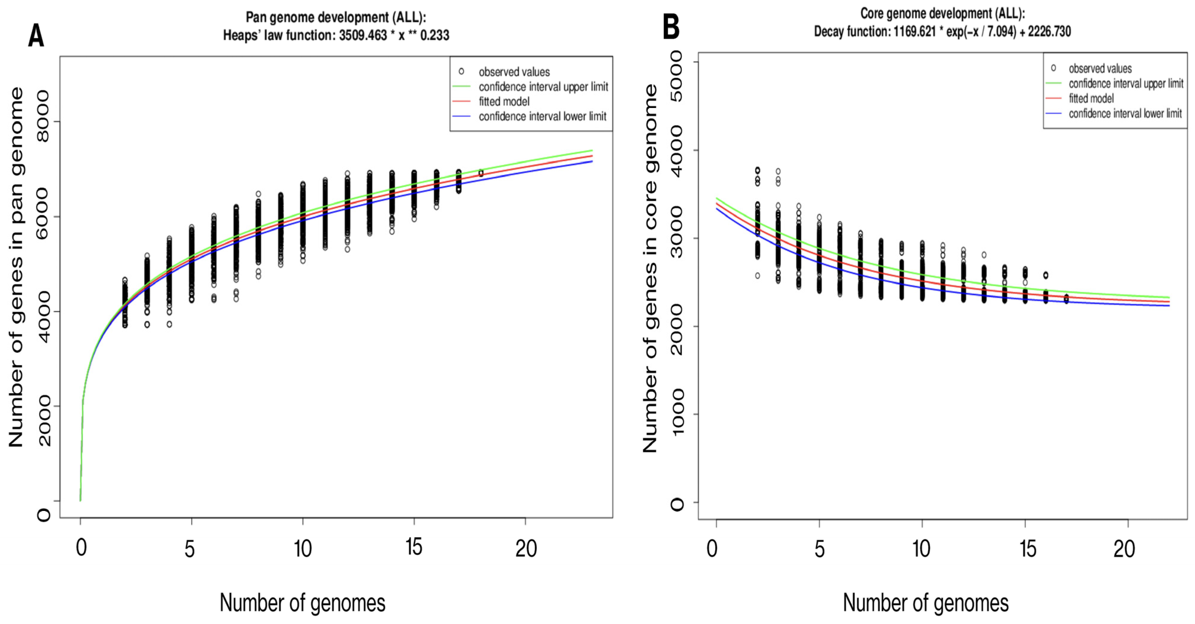
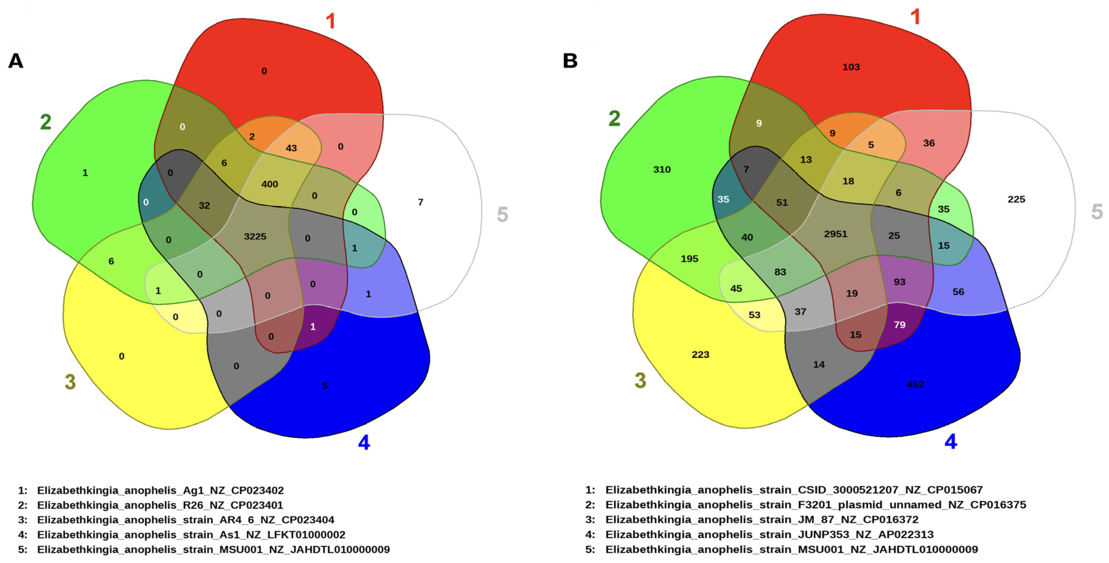
3.3. Gene Repertoire and Phylogenetic Interference of E. anophelis MSU001

MSU001 showed a high ANI (>99%) with other strains of E. anophelis including R26 (type species), Ag1, AR4_6, AR6_8, and As1 (Table S2). The ANI value was greater than 97% for all other selected E. anophelis strains, indicating that MSU001 is indeed a strain of E. anophelis. However, ANI values were lower in comparison with E. meningoseptica (<81%) and E. miricola (<93%). Additionally, DDH values were calculated and were consistent with the analysis by ANI (Table S2). The phylogeny of selected E. anophelis strains is shown in Figure 2. E. anophelis MSU001 from A. stephensi was phylogenetically close to isolates from other mosquitoes (strain Ag1, R26, AR4-6, AR4-8 and As-1). The clinical strains were divided into three clusters and separated from the clade formed by mosquito isolates (Figure 2).
The genomic elements encompassing the core and pan-genomes were organized and utilized to conduct an examination of the gene repertoire within selected genomes of E. anophelis (Figure 3A,B). Analysis of the core genome revealed a reduction in the shared gene count as more genomes were included in the analysis (Figure 3A). In general, E. anophelis exhibited characteristics of an open pan-genome, as evidenced by the appearance of new genes upon the addition of more sequenced genomes to the analysis (Figure 3B). Furthermore, the strain MSU001 (3678) shared 3668, 3627, 3669, and 3669 genes in common with the mosquito isolates Ag1, R26, AR4, and AR6, respectively (Figure 4A). These commonly shared genes accounted for approximately 99.7%, 98.6%, 99.8%, and 99.8% of the encoding genes of MSU001, respectively. It shared 3225 common genes with As1, which is ~87.7% of the common encoding genes of MSU001, due to the small genome size of As1. However, MSU001 shared far fewer genes with clinical E. anophelis strains (Figure 4B) including CSID_3000521207 (3153), JUNP 353 (3257), F3201 (3165), 296-96 (3266), and SUE (3264). These accounted for less than 85.7%, 88.6%, 86.1%, 88.8%, and 88.7% of the MSU001 encoding genes, respectively. Even fewer genes were shared between isolates found in other hosts such as LDVH-AR107 (3193), OSUVM 2 (3117), and JM-87 (3195). These accounted for less than 84.7%, 84.7%, and 86.9% of MSU001 encoding genes (Figure S3), respectively.

3.4. Metabolites Involved in Symbiosis

Several important metabolites such as sphingolipids (SLs) and inositol were detected in the extracts from the midguts of mosquitoes which were fed with both sugar and blood meals in a previous study [42]. Genes involved in the biosynthesis of SLs and inositol were detected in E. anophelis genomes, highlighting that E. anophelis may contribute to the above process. Although SLs are not commonly found as components of bacterial membranes, they have been uniquely identified in certain groups of microbes such as Bacteroides and Sphingomonads [43]. Interestingly, the putative sphingolipid synthesis genes were identified in all selected Elizabethkingia genomes, suggesting their potential involvement in symbiotic relationships, affecting cytotoxicity, colonization of the host, biofilm formation, and modulation of host inflammation [44]. Furthermore, inositol, an important nutritional and signaling factor, was found to be involved in metabolic pathways [45]. These pathways may participate in regulating the stress response, such as cold tolerance, in the hosts.
The growth of SCH873 in M9 medium was impaired, compared to the WT (SCH814) (Figure 5A, left panel). When a 20-diluted LB broth was added into M9 medium, the growth of SCH873 was promoted, while the cell density was much lower than that in SCH814 (Figure 5A, right panel). At 7 days post-infection in adult mosquitos, the cell density of WT Elizabethkingia cells was around 15.8-fold higher than that of arginine utilization mutants in A. stephensi, indicating that Elizabethkingia cells might need to interact with either mosquito host or other microbes to obtain arginine for growth (Figure 5B). To assess the effects of E. anophelis metabolites on the growth of other common mosquito gut symbionts (Asaia sp. W12 and Serratia marcescens), the number of colonies that grew from cultures with added metabolites was compared to control groups (Figure 5). In cultures of E. coli (a representative for non-symbionts), the metabolites significantly hindered colony formation, resulting in less than half the number of viable colonies compared to the control group and indicating a reduction in growth by approximately 58%. The growth inhibition of Asaia sp. W12 and Serratia marcescens with metabolites was less pronounced, with approximately 26% and 17% reductions in growth (Figure 5C), respectively. These findings suggest that E. anophelis metabolites have inhibitory effects on the growth of common mosquito gut symbionts, highlighting the potential role of E. anophelis in modulating the microbial community within the mosquito gut.

3.5. Regulatory System Proteins

The genome of E. anophelis MSU001 possessed genes encoding 51 two-component system proteins, 188 transcription factor proteins, and 13 other DNA-binding proteins, resulting in a total count of 252 regulatory proteins (Table 2). This count was the highest among the mosquito-associated E. anophelis isolates, except for As1, which displayed reduced protein counts in all categories, totaling 215 proteins (Table 2). The other mosquito-associated isolates shared similar counts of two-component system proteins and transcription factor proteins. The main variation among these isolates was observed in the number of DNA-binding proteins, with Ag1, AR4-6, and AR6-8 lacking only one fewer ODP (another DNA-binding protein), and R26 lacking two (Table 2).

3.6. Carbohydrate Active Enzymes

A total of 124 CAZyme-encoding genes were predicted in E. anophelis MSU001, consisting of approximately 3% of the bacterial genome (Tables S3 and S4). Notably, CBM12 (carbohydrate-binding module family 12) and AA10 (auxiliary activity family 10, lytic polysaccharide monooxygenases) were exclusive to mosquito-associated E. anophelis strains, highlighting their importance in establishing a symbiotic relationship with insects. The overall predicted CAZyme repertoires in mosquito-associated E. anophelis were comparable, featuring 61 glycoside hydrolases (GHs). In contrast, E. anophelis As1 exhibited a slightly lower count of 56 GHs (Table S3). This collective decrease in GHs among mosquito isolates, ranging from 61 to 67, contrasted with clinical species, suggesting a distinct evolutionary route. Compared to the clinically important strains, decreased copy numbers of GH3, GH29, and GT4 were detected in insect-associated Elizabethkingia strains (Table S3), showing that while these specific CAZyme genes may be involved in pathogenesis in humans, they may not be relevant for insect symbiosis. Both E. anophelis and E. miricola species harbored single copies of GH1 (β-glycosidase), which is absent in E. meningoseptica. Conversely, GH30, present in E. meningoseptica, was only detected in selected clinical E. anophelis strains and was absent in E. miricola. Additionally, E. anophelis lacked GH33 (sialidase), a characteristic found in E. meningoseptica and some E. miricola strains. Genes encoding GH5 (subfamily 46) and CBM6 (β-glucan binding), consistently observed in E. anophelis, were not found in E. meningoseptica.

3.7. Pathogenesis Potential Revealed by Virulence Factors and MDR Analysis

Using the VFDB protein Set B database, a comparative analysis of selected Elizabethkingia isolates was conducted to identify homologs of virulence factors (VFs) (Table 3). Ten VFs of interest were discovered, namely C8J 1080, DnaK, EF-Tu, eno, htpB, katG, mps1-1, mps1-2, pgIC, and RmIA. These VFs play diverse roles in cellular functions such as mitotic regulation, capsule formation, stress response (involving heat shock proteins, catalase, and hydratase), ion transport proteins, secretion systems, and defense or invasion mechanisms during pathogenesis. Among the selected VFs, genes encoding DnaK, EF-Tu, mps1-1, mps1-2, and RmIA were present in all E. anophelis isolates. Eno and htpB were found in all mosquito-associated isolates, while their presence in clinically isolated human samples varied. PgIC was observed in all mosquito-associated isolates but was completely absent in human Elizabethkingia strains. Both mosquito- and human-associated E. anophelis strains shared the presence of C8J 1080 and katG, which were not identified in other animal-associated strains (Table 3).
The antimicrobial resistance profile of E. anophelis was determined using the broth microdilution method. The strain exhibited resistance to 13 out of the 16 tested antibiotics, including aminoglycosides, tetracycline, nitrofuran, and all β-lactam antibiotics, such as cephalosporins, monobactams, and extended-spectrum penams/β-lactamase inhibitors. However, it showed susceptibility to trimethoprim/sulfamethoxazole (sulfonamide) and ciprofloxacin (quinolone), and intermediate susceptibility to tigecycline (Table 4). In addition, the prediction of antibiotic resistance genes in E. anophelis MSU001 revealed its multidrug resistance traits (Table S4). Notably, Elizabethkingia species are known for their high resistance to β-lactam drugs, due to the production of β-lactamases (Table S4), which hydrolyze these antibiotics. In the case of MSU001, it carried at least five different β-lactamase genes (BlaB, CME-1, GOB-9, IND-7, and TLA-1) that may confer broad resistance to penams, cephalosporins, and carbapenems. It is interesting that the presence of IND-7, which encodes for a class B carbapenem-hydrolyzing β-lactamase, was unique to the MSU001 strain. Mosquito-associated E. anophelis strains carried GOB-9 (encoding a class B β-lactamase) and TLA-1, which were only found in a few clinical Elizabethkingia isolates. Furthermore, it is noteworthy that GOB-9 was absent in E. miricola and E. meningoseptica. Genes encoding BlaB (inducible class C cephalosporinase) and CME-1 (class A β-lactamase) were present in most selected Elizabethkingia species (Table S4). However, mosquito-associated E. anophelis lacked several β-lactamase genes found in other selected Elizabethkingia strains, indicating unique evolutionary routes for these mosquito-associated strains.

4. Discussion

Studies have shown that a substantial portion of the colonizing bacteria found within adult mosquito hosts are acquired in aquatic habitats during larval life stages [9,16,17]. Elizabethkingia species are common mosquito symbionts dispersed in natural water bodies (dams, wetlands, and rivers), but do not normally predominate in these environments (composing 6.25 × 10⁻⁶ to 8.21 × 10⁻⁶ of the total bacterial community) [46,47]. However, Elizabethkingia species populate mosquito midguts and can spread to other organs and tissues, including the salivary glands, reproductive organs (ovary or testicles), crop, and alimentary canal of mosquitoes at various development stages [47]. The complex interactions between arthropod hosts and their associated microbes warrant a holistic analysis of these communities and the environments that foster them [47]. Bacteria need to overcome digestion, microbial competition, and a multitude of other stress factors (e.g., iron and oxidative stress, larval metamorphosis, temperature, pH) associated with mosquito physiology [9,17]. The ability to thrive in dynamic environments within a host emphasizes the importance of bacterial adaptability and likely highlights a deeper symbiotic relationship underlying microbial persistence [47]. By conducting an analysis of the genomic and molecular mechanisms behind Elizabethkingia colonization, we hoped to enhance our understanding of microbe–host interactions.
Correctly identifying Elizabethkingia species has proven to be a challenge with varying success, further complicated by prior nomenclature changes and various method limitations [41]. Current classification of Flavobacteriaceae members relies heavily on MALDI-ToF mass spectrometry, but despite its wide utility in bacterial identification, it struggles to accurately classify members from Chryseobacterium and Elizabethkingia genera [19,41,46]. Furthermore, standard databases are limited to only a few Elizabethkingia isolates, often falsely defaulting to E. meningoseptica or E. miricola [41]. This was evidenced by our own study, as well as others, where MALDI-ToF frequently misidentified E. anophelis as E. meningosepticum [41,46,48]. The use of 16S rRNA sequences has been shown to be limited in its taxonomic utility as well [48]. The fact that misidentification via conventional methodologies is so prevalent in the literature may indicate E. anophelis is an underrepresented pathogen responsible for more disease in humans than previously attributed [46]. These limitations highlight the need for updating standard MALDI-ToF databases, as well as for thorough, enhanced identification methodologies that utilize a combination of widely adopted bacterial identification methods like 16s rDNA sequencing in conjunction with biochemical testing [41,46,48]. Moreover, whole genomic sequence analysis and average nucleotide identity as a complementary method may be used to correctly identify E. anophelis [46,49].
Genome size and GC content were similar among most E. anophelis strains. MSU001 exhibited characteristics of an open pan-genome, likely relating to its diverse habitats, spanning both aquatic and terrestrial environments, as well as the many different human, animal, and plant hosts that it may colonize [46]. However, the core genome analysis demonstrated that strains from mosquitoes shared more conserved genes than those from clinical specimens. Furthermore, the phylogenetic placement of mosquito-associated E. anophelis species formed different clades from clinical isolates. They were also distinct from E. meningoseptica and E. miricola clades. Collectively, these results indicate that E. anophelis MSU001 and other mosquito isolates likely evolved in different routes to adapt to mosquito hosts compared to clinical strains.
Another notable finding was the presence of Elizabethkingia genes involved in sphingolipid biosynthesis. Sphingolipids are a ubiquitous component in eukaryotic cell membranes that have been shown to play critical roles in cell signal transduction, regulation of apoptosis, adhesion and uptake, and inflammation in the host [50]. Several pathogens can actively synthesize or hydrolyze these molecules to hijack host cell responses and orchestrate favorable immune responses [50]. Furthermore, certain sphingolipids like sphingosine have also been shown to possess a possible antibacterial effect [50]. Bacteria employ diverse mechanisms to facilitate host interactions and survival in their environments. The production of various secondary metabolites by Elizabethkingia likely conferred advantages over other members of the microbial community, allowing it to disturb the bacterial consortium and outcompete or even inhibit its competitors [50].
Chitin is one of the most abundant polysaccharides, forming important structures in the insect exoskeleton and gut linings [51]. Due to the vital role of chitin in development and defense against pathogen invasion, insects need to frequently reshape its structure and components [51]. Microbial symbionts may be involved in chitin degradation and its synthesis [52]. In this study, we observed that the modules of CBM12 associated with chitinase and AA10 were uniquely found in mosquito-associated E. anophelis (except As1). These CAZymes possibly contribute to the binding and lysing of chitin [52]. For example, upon a mosquito’s bite, the ingested blood meal triggers the midgut epithelium to release various factors including chitin microfibrils (3–13%) and protein complexes, which form a peritrophic matrix (PM) [53]. The PM effectively creates a barrier between the blood bolus and the midgut epithelial cells, serving as a protective shield against abrasive particles and microbial infections [53]. After the red blood cells have been thoroughly digested, the PM needs to be dismantled to release the nutrients. Microbial chitinase secreted by gut microbiota may facilitate this process [52,53,54]. Moreover, microbial chitinases may contribute to the reshaping of chitin components during mosquito molting, supported by the presence of E. anophelis in various mosquito body sites [51,52]. The majority of predicted CAZymes in Elizabethkingia species appear to be involved in utilizing simple sugars rather than degrading complex plant polysaccharides, which is consistent with their living niches (e.g., within mosquitoes or humans) [46,47,48]. Our results also indicated that pathogenic E. anophelis possibly requires additional copies of GH3, GH29, and GT4 to participate in pathogenesis. Furthermore, E. anophelis and E. miricola have different sets of CAZymes involved in sugar metabolism. Therefore, future characterization of their physiological functions is warranted.
Despite their different sources, Elizabethkingia bacteria exhibited comparable numbers of response regulators, phosphotransferase proteins, histidine kinases, one-component systems, transcriptional regulators, sigma factors, and other DNA-binding proteins (Table 2). These regulatory proteins play critical roles in maintaining bacterial metabolism and function, explaining their consistent presence across Elizabethkingia species (Table 2). The numbers of regulatory protein genes between mosquito-associated and clinical E. anophelis genomes varied and were not statistically different. The retainment of similar complicated regulatory systems may indicate an adaptability of this organism to diverse host environments [46]. E. anophelis living in the adult female mosquito midgut may experience similar stress conditions to those where bacteria invade the bloodstream of mammalian hosts [9,16,17]. For example, mosquito-associated bacteria are exposed to iron-depleting conditions and relatively lower temperatures prior to blood meals [13,17]; conversely, they encounter iron-rich environments during and after blood meals [13]. Similar processes may occur prior to entry into the bloodstream or after the lysis of the erythrocytes during a bacteremia event [25,28]. Furthermore, the evasion of immune cells and resistance to temperature variations during the above processes are expected to be similar [55].
The emerging pathogenicity of Elizabethkingia is likely attributed to its large genome, ecological and metabolic plasticity, a multitude of virulence factor genes present in its genetic repertoire, and broad antibiotic resistance [46,48]. Among the diverse virulence factors, we discovered that PgIC was only present in mosquito-associated isolates. PglC plays a vital role in the N-linked protein glycosylation pathway in Campylobacter jejuni [56]. This pathway primes proteins for nucleophilic attack by the polyprenol acceptor within the cellular membranes, which may play important roles in epithelial cell adherence, invasion, and colonization of the host during the infection course [56,57]. Antimicrobial susceptibility patterns vary across strains and in the case of clinical isolates, provide an additional layer of difficulty in the selection of appropriate therapeutics [23,58,59]. While β-lactamase synthesis remains the most employed defense among Gram-negative bacteria to withstand antibiotics, other resistance mechanisms include the alteration of target drug sites and the implementation of efflux pumps to eliminate the drug from the cell [59]. The presence of specific β-lactamase genes varies across different host-associated strains, suggesting that these genes confer certain advantages within Elizabethkingia and their respective evolutionary routes [20,23]. Those virulence factors that aid in transmission promote adhesion, motility, and biofilm formation, while other factors mediate host interactions and allow for extended persistence within hostile environments [58,60]. Further research into variations in genomic features between mosquito-associated and clinically significant strains of Elizabethkingia is warranted.

Supplementary Materials

The following supporting information can be downloaded at https://www.mdpi.com/article/10.3390/microorganisms12061079/s1. Table S1: Biolog tests of E. anophelis MSU001; Table S2: Average nucleotide identity values (up, black font) and Digital DNA-DNA Hybridization values (low, red font) amongst different Elizabethkingia species; Table S3: CAZyase analyses in the selected Elizabethkinigia; Table S4: Resistome analysis of Elizabethkingia spp.; Figure S1: mass spectrum; Figure S2: 27 subsystems consisting of 87 categories; Figure S3: Agininine prediction by gutSMASH.

Author Contributions

Conceptualization, S.C. and E.D.W.; methodology, S.C.; formal analysis, S.C., J.B., S.P. and N.T.; investigation, S.C.; resources, S.C. and E.D.W.; data curation, S.C.; writing—original draft preparation, S.C.; writing—review and editing, S.C., E.D.W., J.B., S.P. and N.T; supervision, S.C.; funding acquisition, S.C. and E.D.W. All authors have read and agreed to the published version of the manuscript.

Funding

This project was funded by the Seed Grant of College of Health and Human Sciences at Northern Illinois University (awarded to S.C.) and NIH grant R37AI21884 (awarded to E.D.W).

Data Availability Statement

Data from these whole-genome shotgun projects have been deposited at DDBJ/ENA/GenBank under accession number GCA_024357565.1. The BioProject designation for this project is PRJNA731841, and the BioSample accession number is SAMN19296199.

Acknowledgments

The authors expressed their gratitude to the Department of Microbiology at Henry Ford Health System (Jackson, Michigan) for their assistance in antimicrobial susceptibility determination and identification by MALDI-ToF.

Conflicts of Interest

The authors declare no conflicts of interest.

References

  1. Coyle, A.L. Elizabethkingia anophelis: Exploring the outbreak of disease in the Midwest. Nursing 2017, 47, 61–63. [Google Scholar] [CrossRef] [PubMed]
  2. Janda, J.M.; Lopez, D.L. Mini Review: New Pathogen Profiles: Elizabethkingia anophelis. Diagn. Microbiol. Infect. Dis. 2017, 88, 201–205. [Google Scholar] [CrossRef] [PubMed]
  3. Kämpfer, P.; Matthews, H.; Glaeser, S.P.; Martin, K.; Lodders, N.; Faye, I. Elizabethkingia anophelis Sp. Nov., Isolated from the Midgut of the Mosquito Anopheles gambiae. Int. J. Syst. Evol. Microbiol. 2011, 61, 2670–2675. [Google Scholar] [CrossRef] [PubMed]
  4. Breurec, S.; Criscuolo, A.; Diancourt, L.; Rendueles, O.; Vandenbogaert, M.; Passet, V.; Caro, V.; Rocha, E.P.C.; Touchon, M.; Brisse, S. Genomic Epidemiology and Global Diversity of the Emerging Bacterial Pathogen Elizabethkingia anophelis. Sci. Rep. 2016, 6, 30379. [Google Scholar] [CrossRef] [PubMed]
  5. Ganley, J.G.; D’Ambrosio, H.K.; Shieh, M.; Derbyshire, E.R. Coculturing of Mosquito-Microbiome Bacteria Promotes Heme Degradation in Elizabethkingia anophelis. ChemBioChem 2020, 21, 1279–1284. [Google Scholar] [CrossRef]
  6. Mallinckrodt, L.; Huis in ’t Veld, R.; Rosema, S.; Voss, A.; Bathoorn, E. Review on Infection Control Strategies to Minimize Outbreaks of the Emerging Pathogen Elizabethkingia anophelis. Antimicrob. Resist. Infect. Control 2023, 12, 97. [Google Scholar] [CrossRef]
  7. Lee, Y.-L.; Hsueh, P.-R. Emerging Infections in Vulnerable Hosts: Stenotrophomonas maltophilia and Elizabethkingia anophelis. Curr. Opin. Infect. Dis. 2023, 36, 481–494. [Google Scholar] [CrossRef] [PubMed]
  8. Lee, Y.-L.; Liu, K.-M.; Chang, H.-L.; Lin, J.-S.; Kung, F.-Y.; Ho, C.-M.; Lin, K.-H.; Chen, Y.-T. A Dominant Strain of Elizabethkingia anophelis Emerged from a Hospital Water System to Cause a Three-Year Outbreak in a Respiratory Care Center. J. Hosp. Infect. 2021, 108, 43–51. [Google Scholar] [CrossRef]
  9. Chen, S.; Bagdasarian, M.; Walker, E.D. Elizabethkingia anophelis: Molecular Manipulation and Interactions with Mosquito Hosts. Appl. Environ. Microbiol. 2015, 81, 2233–2243. [Google Scholar] [CrossRef]
  10. Akhouayri, I.G.; Habtewold, T.; Christophides, G.K. Melanotic Pathology and Vertical Transmission of the Gut Commensal Elizabethkingia meningoseptica in the Major Malaria Vector Anopheles gambiae. PLoS ONE 2013, 8, e77619. [Google Scholar] [CrossRef]
  11. Teo, J.; Tan, S.Y.-Y.; Liu, Y.; Tay, M.; Ding, Y.; Li, Y.; Kjelleberg, S.; Givskov, M.; Lin, R.T.P.; Yang, L. Comparative Genomic Analysis of Malaria Mosquito Vector-Associated Novel Pathogen Elizabethkingia anophelis. Genome Biol. Evol. 2014, 6, 1158–1165. [Google Scholar] [CrossRef] [PubMed]
  12. Steven, B.; Hyde, J.; LaReau, J.C.; Brackney, D.E. The Axenic and Gnotobiotic Mosquito: Emerging Models for Microbiome Host Interactions. Front. Microbiol. 2021, 12, 714222. [Google Scholar] [CrossRef] [PubMed]
  13. Onyango, M.G.; Lange, R.; Bialosuknia, S.; Payne, A.; Mathias, N.; Kuo, L.; Vigneron, A.; Nag, D.; Kramer, L.D.; Ciota, A.T. Zika Virus and Temperature Modulate Elizabethkingia anophelis in Aedes albopictus. Parasit. Vectors 2021, 14, 573. [Google Scholar] [CrossRef] [PubMed]
  14. Chen, S.; Blom, J.; Walker, E.D. Genomic, Physiologic, and Symbiotic Characterization of Serratia marcescens Strains Isolated from the Mosquito Anopheles stephensi. Front. Microbiol. 2017, 8, 283169. [Google Scholar] [CrossRef]
  15. Chen, S.; Yu, T.; Terrapon, N.; Henrissat, B.; Walker, E.D. Genome Features of Asaia Sp. W12 Isolated from the Mosquito Anopheles stephensi Reveal Symbiotic Traits. Genes 2021, 12, 752. [Google Scholar] [CrossRef] [PubMed]
  16. Chen, S.; Zhao, J.; Joshi, D.; Xi, Z.; Norman, B.; Walker, E.D. Persistent Infection by Wolbachia WAlbB Has No Effect on Composition of the Gut Microbiota in Adult Female Anopheles stephensi. Front. Microbiol. 2016, 7, 1485. [Google Scholar] [CrossRef] [PubMed]
  17. Chen, S.; Johnson, B.K.; Yu, T.; Nelson, B.N.; Walker, E.D. Elizabethkingia anophelis: Physiologic and Transcriptomic Responses to Iron Stress. Front. Microbiol. 2020, 11, 804. [Google Scholar] [CrossRef] [PubMed]
  18. Mirza, H.C.; Tuncer, Ö.; Ölmez, S.; Şener, B.; Tuğcu, G.D.; Özçelik, U.; Gürsoy, N.C.; Otlu, B.; Büyükçam, A.; Kara, A.; et al. Clinical Strains of Chryseobacterium and Elizabethkingia Spp. Isolated from Pediatric Patients in a University Hospital: Performance of MALDI-TOF MS-Based Identification, Antimicrobial Susceptibilities, and Baseline Patient Characteristics. Microb. Drug Resist. 2018, 24, 816–821. [Google Scholar] [CrossRef] [PubMed]
  19. Comba, I.Y.; Schuetz, A.N.; Misra, A.; Friedman, D.Z.P.; Stevens, R.; Patel, R.; Lancaster, Z.D.; Shah, A. Antimicrobial Susceptibility of Elizabethkingia Species: Report from a Reference Laboratory. J. Clin. Microbiol. 2022, 60, e02541-21. [Google Scholar] [CrossRef]
  20. Lin, J.-N.; Lai, C.-H.; Yang, C.-H.; Huang, Y.-H. Elizabethkingia Infections in Humans: From Genomics to Clinics. Microorganisms 2019, 7, 295. [Google Scholar] [CrossRef]
  21. Perrin, A.; Larsonneur, E.; Nicholson, A.C.; Edwards, D.J.; Gundlach, K.M.; Whitney, A.M.; Gulvik, C.A.; Bell, M.E.; Rendueles, O.; Cury, J.; et al. Evolutionary Dynamics and Genomic Features of the Elizabethkingia anophelis 2015 to 2016 Wisconsin Outbreak Strain. Nat. Commun. 2017, 8, 15483. [Google Scholar] [CrossRef] [PubMed]
  22. Thigpen, S.; Walblay, K.; Adil, H.; Zelencik, S.; Zelinski, C.; Nelson, K.; Cox, B.; McQuiston, J.R.; Turner, J.; Toews, K.-A.; et al. 1451. Elizabethkingia spp. Outbreak in a Ventilator-Capable Skilled Nursing Facility, Chicago 2023. Open Forum. Infect. Dis. 2023, 10, ofad500.1288. [Google Scholar] [CrossRef]
  23. Hu, S.; Xu, H.; Meng, X.; Bai, X.; Xu, J.; Ji, J.; Ying, C.; Chen, Y.; Shen, P.; Zhou, Y.; et al. Population Genomics of Emerging Elizabethkingia anophelis Pathogens Reveals Potential Outbreak and Rapid Global Dissemination. Emerg. Microbes Infect. 2022, 11, 2590–2599. [Google Scholar] [CrossRef] [PubMed]
  24. Frank, T.; Gody, J.C.; Nguyen, L.B.L.; Berthet, N.; Fleche-Mateos, A.L.; Bata, P.; Rafaï, C.; Kazanji, M.; Breurec, S. First Case of Elizabethkingia anophelis Meningitis in the Central African Republic. Lancet 2013, 381, 1876. [Google Scholar] [CrossRef] [PubMed]
  25. Accoti, A.; Damiani, C.; Nunzi, E.; Cappelli, A.; Iacomelli, G.; Monacchia, G.; Turco, A.; D’Alò, F.; Peirce, M.J.; Favia, G.; et al. Anopheline Mosquito Saliva Contains Bacteria That Are Transferred to a Mammalian Host through Blood Feeding. Front. Microbiol. 2023, 14, 1157613. [Google Scholar] [CrossRef] [PubMed]
  26. Kukutla, P.; Lindberg, B.G.; Pei, D.; Rayl, M.; Yu, W.; Steritz, M.; Faye, I.; Xu, J. Insights from the Genome Annotation of Elizabethkingia anophelis from the Malaria Vector Anopheles gambiae. PLoS ONE 2014, 9, e97715. [Google Scholar] [CrossRef] [PubMed]
  27. Raygoza Garay, J.A.; Hughes, G.L.; Koundal, V.; Rasgon, J.L.; Mwangi, M.M. Genome Sequence of Elizabethkingia anophelis Strain EaAs1, Isolated from the Asian Malaria Mosquito Anopheles stephensi. Genome Announc. 2016, 4, 10-1128. [Google Scholar] [CrossRef] [PubMed]
  28. Li, Y.; Liu, Y.; Chew, S.C.; Tay, M.; Salido, M.M.S.; Teo, J.; Lauro, F.M.; Givskov, M.; Yang, L. Complete Genome Sequence and Transcriptomic Analysis of the Novel Pathogen Elizabethkingia anophelis in Response to Oxidative Stress. Genome Biol. Evol. 2015, 7, 1676–1685. [Google Scholar] [CrossRef]
  29. Tatusova, T.; DiCuccio, M.; Badretdin, A.; Chetvernin, V.; Nawrocki, E.P.; Zaslavsky, L.; Lomsadze, A.; Pruitt, K.D.; Borodovsky, M.; Ostell, J. NCBI Prokaryotic Genome Annotation Pipeline. Nucleic Acids Res. 2016, 44, 6614–6624. [Google Scholar] [CrossRef]
  30. Overbeek, R.; Olson, R.; Pusch, G.D.; Olsen, G.J.; Davis, J.J.; Disz, T.; Edwards, R.A.; Gerdes, S.; Parrello, B.; Shukla, M.; et al. The SEED and the Rapid Annotation of Microbial Genomes Using Subsystems Technology (RAST). Nucleic Acids Res. 2014, 42, D206–D214. [Google Scholar] [CrossRef]
  31. Alcock, B.P.; Huynh, W.; Chalil, R.; Smith, K.W.; Raphenya, A.R.; Wlodarski, M.A.; Edalatmand, A.; Petkau, A.; Syed, S.A.; Tsang, K.K.; et al. CARD 2023: Expanded Curation, Support for Machine Learning, and Resistome Prediction at the Comprehensive Antibiotic Resistance Database. Nucleic. Acids Res. 2023, 51, D690–D699. [Google Scholar] [CrossRef] [PubMed]
  32. Grissa, I.; Vergnaud, G.; Pourcel, C. CRISPRFinder: A Web Tool to Identify Clustered Regularly Interspaced Short Palindromic Repeats. Nucleic Acids Res. 2007, 35, W52-7. [Google Scholar] [CrossRef] [PubMed]
  33. Yoon, S.-H.; Ha, S.; Lim, J.; Kwon, S.; Chun, J. A Large-Scale Evaluation of Algorithms to Calculate Average Nucleotide Identity. Antonie Van Leeuwenhoek 2017, 110, 1281–1286. [Google Scholar] [CrossRef] [PubMed]
  34. Meier-Kolthoff, J.P.; Carbasse, J.S.; Peinado-Olarte, R.L.; Göker, M. TYGS and LPSN: A Database Tandem for Fast and Reliable Genome-Based Classification and Nomenclature of Prokaryotes. Nucleic Acids Res. 2022, 50, D801–D807. [Google Scholar] [CrossRef] [PubMed]
  35. Barakat, M.; Ortet, P.; Whitworth, D.E. P2RP: A Web-Based Framework for the Identification and Analysis of Regulatory Proteins in Prokaryotic Genomes. BMC Genom. 2013, 14, 269. [Google Scholar] [CrossRef] [PubMed]
  36. Dieckmann, M.A.; Beyvers, S.; Nkouamedjo-Fankep, R.C.; Hanel, P.H.G.; Jelonek, L.; Blom, J.; Goesmann, A. EDGAR3.0: Comparative Genomics and Phylogenomics on a Scalable Infrastructure. Nucleic Acids Res. 2021, 49, W185–W192. [Google Scholar] [CrossRef] [PubMed]
  37. Blom, J.; Kreis, J.; Spänig, S.; Juhre, T.; Bertelli, C.; Ernst, C.; Goesmann, A. EDGAR 2.0: An Enhanced Software Platform for Comparative Gene Content Analyses. Nucleic Acids Res. 2016, 44, W22–W28. [Google Scholar] [CrossRef] [PubMed]
  38. Drula, E.; Garron, M.-L.; Dogan, S.; Lombard, V.; Henrissat, B.; Terrapon, N. The Carbohydrate-Active Enzyme Database: Functions and Literature. Nucleic Acids Res. 2022, 50, D571–D577. [Google Scholar] [CrossRef]
  39. Baum, B.R. PHYLIP: Phylogeny Inference Package. Version 3.2. Joel Felsenstein. Q Rev. Biol. 1989, 64, 539–541. [Google Scholar] [CrossRef]
  40. Price, M.N.; Dehal, P.S.; Arkin, A.P. FastTree 2—Approximately Maximum-Likelihood Trees for Large Alignments. PLoS ONE 2010, 5, e9490. [Google Scholar] [CrossRef]
  41. Eriksen, H.B.; Gumpert, H.; Faurholt, C.H.; Westh, H. Determination of Elizabethkingia Diversity by MALDI-TOF Mass Spectrometry and Whole-Genome Sequencing. Emerg. Infect. Dis. 2017, 23, 320–323. [Google Scholar] [CrossRef] [PubMed]
  42. Champion, C.J.; Kukutla, P.; Glennon, E.K.K.; Wang, B.; Luckhart, S.; Xu, J. Anopheles gambiae: Metabolomic Profiles in Sugar-Fed, Blood-Fed, and Plasmodium falciparum-Infected Midgut. Dataset Pap. Sci. 2017, 2017, 8091749. [Google Scholar] [CrossRef]
  43. Brown, E.M.; Ke, X.; Hitchcock, D.; Jeanfavre, S.; Avila-Pacheco, J.; Nakata, T.; Arthur, T.D.; Fornelos, N.; Heim, C.; Franzosa, E.A.; et al. Bacteroides-Derived Sphingolipids Are Critical for Maintaining Intestinal Homeostasis and Symbiosis. Cell Host Microbe. 2019, 25, 668–680.e7. [Google Scholar] [CrossRef] [PubMed]
  44. Hannun, Y.A.; Obeid, L.M. Sphingolipids and Their Metabolism in Physiology and Disease. Nat. Rev. Mol. Cell Biol. 2018, 19, 175–191. [Google Scholar] [CrossRef] [PubMed]
  45. Heaver, S.L.; Le, H.H.; Tang, P.; Baslé, A.; Mirretta Barone, C.; Vu, D.L.; Waters, J.L.; Marles-Wright, J.; Johnson, E.L.; Campopiano, D.J.; et al. Characterization of Inositol Lipid Metabolism in Gut-Associated Bacteroidetes. Nat. Microbiol. 2022, 7, 986–1000. [Google Scholar] [CrossRef] [PubMed]
  46. Hem, S.; Jarocki, V.M.; Baker, D.J.; Charles, I.G.; Drigo, B.; Aucote, S.; Donner, E.; Burnard, D.; Bauer, M.J.; Harris, P.N.A.; et al. Genomic Analysis of Elizabethkingia Species from Aquatic Environments: Evidence for Potential Clinical Transmission. Curr. Res. Microb. Sci. 2022, 3, 100083. [Google Scholar] [CrossRef] [PubMed]
  47. Villegas, L.E.M.; Radl, J.; Dimopoulos, G.; Short, S.M. Bacterial Communities of Aedes aegypti Mosquitoes Differ between Crop and Midgut Tissues. PLoS Negl. Trop. Dis. 2023, 17, e0011218. [Google Scholar] [CrossRef] [PubMed]
  48. Kadi, H.; Tanriverdi Cayci, Y.; Yener, N.; Gur Vural, D.; Bilgin, K.; Birinci, A. 16s RRNA-Based Phylogenetic Analyses of Elizabethkingia anophelis: Detection of Elizabethkingia anophelis, a Rare Infectious Agent from Blood and Determination of Antibiotic Susceptibility in Turkey. Indian J. Med. Microbiol. 2022, 40, 557–559. [Google Scholar] [CrossRef]
  49. McTaggart, L.R.; Stapleton, P.J.; Eshaghi, A.; Soares, D.; Brisse, S.; Patel, S.N.; Kus, J.V. Application of Whole Genome Sequencing to Query a Potential Outbreak of Elizabethkingia anophelis in Ontario, Canada. Access Microbiol. 2019, 1, e000017. [Google Scholar] [CrossRef]
  50. Rolando, M.; Buchrieser, C. A Comprehensive Review on the Manipulation of the Sphingolipid Pathway by Pathogenic Bacteria. Front. Cell Dev. Biol. 2019, 7, 168. [Google Scholar] [CrossRef]
  51. Merzendorfer, H.; Zimoch, L. Chitin Metabolism in Insects: Structure, Function and Regulation of Chitin Synthases and Chitinases. J. Exp. Biol. 2003, 206, 4393–4412. [Google Scholar] [CrossRef] [PubMed]
  52. Beier, S.; Bertilsson, S. Bacterial Chitin Degradation—Mechanisms and Ecophysiological Strategies. Front. Microbiol. 2013, 4, 149. [Google Scholar] [CrossRef] [PubMed]
  53. Rodgers, F.H.; Gendrin, M.; Wyer, C.A.S.; Christophides, G.K. Microbiota-Induced Peritrophic Matrix Regulates Midgut Homeostasis and Prevents Systemic Infection of Malaria Vector Mosquitoes. PLoS Pathog. 2017, 13, e1006391. [Google Scholar] [CrossRef] [PubMed]
  54. Kuraishi, T.; Binggeli, O.; Opota, O.; Buchon, N.; Lemaitre, B. Genetic Evidence for a Protective Role of the Peritrophic Matrix against Intestinal Bacterial Infection in Drosophila melanogaster. Proc. Natl. Acad. Sci. USA 2011, 108, 15966–15971. [Google Scholar] [CrossRef] [PubMed]
  55. Skaar, E.P. The Battle for Iron between Bacterial Pathogens and Their Vertebrate Hosts. PLoS Pathog. 2010, 6, e1000949. [Google Scholar] [CrossRef] [PubMed]
  56. Chen, M.M.; Weerapana, E.; Ciepichal, E.; Stupak, J.; Reid, C.W.; Swiezewska, E.; Imperiali, B. Polyisoprenol Specificity in the Campylobacter jejuni N-Linked Glycosylation Pathway. Biochemistry 2007, 46, 14342–14348. [Google Scholar] [CrossRef] [PubMed]
  57. Lukose, V.; Walvoort, M.T.; Imperiali, B. Bacterial Phosphoglycosyl Transferases: Initiators of Glycan Biosynthesis at the Membrane Interface. Glycobiology 2017, 27, 820–833. [Google Scholar] [CrossRef] [PubMed]
  58. Hu, S.; Lv, Y.; Xu, H.; Zheng, B.; Xiao, Y. Biofilm Formation and Antibiotic Sensitivity in Elizabethkingia anophelis. Front. Cell Infect. Microbiol. 2022, 12, 953780. [Google Scholar] [CrossRef]
  59. Wang, M.; Gao, H.; Lin, N.; Zhang, Y.; Huang, N.; Walker, E.D.; Ming, D.; Chen, S.; Hu, S. The Antibiotic Resistance and Pathogenicity of a Multidrug-resistant Elizabethkingia anophelis Isolate. Microbiologyopen 2019, 8, e804. [Google Scholar] [CrossRef]
  60. Puah, S.M.; Fong, S.P.; Kee, B.P.; Puthucheary, S.D.; Chua, K.H. Molecular Identification and Biofilm-Forming Ability of Elizabethkingia Species. Microb. Pathog. 2022, 162, 105345. [Google Scholar] [CrossRef]
Figure 1. Growth features and microscopic observation of E. anophelis MSU001. (A) Hemolytic activity on sheep blood agar; (B) motility test; (C) scan electron microscopy; (D) demonstration of bacterial morphology by electron microscopy with negative stain.
Microorganisms 12 01079 g001
Figure 2. Phylogenetic placement of E. anophelis MSU001. The tree was constructed with 18 genomes with a core of 2307 genes per genome, 41,526 in total. The core had 783,693 amino acid residues/bp per genome, 14,106,474 in total. The horizontal bar represents 0.05 substitutions per site.
Microorganisms 12 01079 g002
Figure 3. Pan and core genome evolution according to the number of selected Elizabethkingia genomes. (A) Number of genes (pan-genome) for a given number of sequentially added genomes. A pan development plot was generated for the following genomes: E. anophelis Ag1 (NZ_CP023402), E. anophelis R26 (NZ_CP023401), E. anophelis 2_62 (NZ_CP071551), E. anophelis 296_96 (NZ_CP046080), E. anophelis AR4_6 (NZ_CP023404), E. anophelis AR6_8 (NZ_CP023403), E. anophelis As1 (NZ_LFKT01000002), E. anophelis CSID_3000521207 (NZ_CP015067), E. anophelis F3201 (NZ_CP016375), E. anophelis JM_87 (NZ_CP016372), E. anophelis MSU001 (NZ_JAHDTL010000009), E. anophelis SUE (NZ_CP034247), E. anophelis LDVH-AR107 (NZ CP023403), E. anophelis JUNP 353 (NZ_ AP022313). (B) Number of shared genes (core genome) as a function of the number of genomes sequentially added. The genomes used for generating the core genome development plot were the same as listed in (A).
Microorganisms 12 01079 g003
Figure 4. Venn diagram illustrating the distribution of shared and specific clusters of orthologous groups in the selected Elizabethkingia genomes. (A) Venn diagram of shared and unique genes in the selected mosquito-associated Elizabethkiniga. (B) Venn diagram of shared and unique genes in MSU001 and the clinically important Elizabethkiniga. The unique and shared genomes among the compared genomes were determined using the BLAST score ratio approach of EDGAR 3.2 with a cutoff of 30%.
Microorganisms 12 01079 g004
Figure 5. Inhibitory effects of Elizabethkingia metabolites on selected bacteria. * Statistically significant difference (p < 0.05). (A) Growth comparison between wild type strain for arginine utilization (SCH814) and arginine metabolism mutant (SCH873) in the M9 medium and M9 medium supplemented with 20-fold diluted LB medium. (B) Comparison between growth of SCH814 and SCH873 in mosquitoes. (C) The effects of spent media on the growth of Asaia sp. W12, Serratia marcescens and E. coli. The spent broth from E. anophelis MSU001 (48-h incubation) was added E. coli, Serratia marcescens ano1 and Asaia sp. W12, statically cultured at 28 °C for 24 h and plated on their respective solid agar media for CFU calculation.
Microorganisms 12 01079 g005
Table 1. Genomic features in selected Elizabethkingia species.
StrainOriginal Region *aIsolation Source *bSize (Mb)GC%CDSGeneCRISPR Count
E. anophelis
As1USAA. gambiae3.5935.5323733150
Ag1USAA. gambiae4.0935.5368637880
R26SwedenA. gambiae4.0635.5363537410
AR4-6ChinaA. sinensis4.0935.5367837850
AR6-8ChinaA. sinensis4.0935.5367837850
MSU001USAA. stephensi4.0535.4385737531
LDVH-AR107FranceC. carpio3.9935.7355536672
OSUVM 2USAE. caballus4.135.4364437540
CSID_3000521207USAH. sapiens3.8535.7341235130
JUNP 353NepalH. sapiens4.3235.8389740490
F3201KuwaitH. sapiens4.2835.46379739270
296-96TaiwanH. sapiens4.235.8377938982
SUETaiwanH sapiens4.235.8377138912
JM-87USAZ. mays4.1835.5369538370
E. meningoseptica
NCTC10016UKH. sapiens3.8736.5339734801
G4120FranceH. sapiens436.4351936281
E. miricola
FL160902ChinaFrog4.2535.7376038920
*a,b The information about specimen and sources used for these selected isolates was obtained from BioSample (https://www.ncbi.nlm.nih.gov/biosample).
Table 2. Predicted regulatory proteins in the selected Elizabethkingia species *.
ElizabethkingiaPredicted Regulatory Proteins
TOCTFODP
RRPPHKOCSRRTRSF
E. anophelis
Ag12691631231181612
As12381422201031510
R262691631231181611
AR4-62691631231181612
AR6-82691631231181612
MSU0012691631231181613
LDVH-AR1072681726231191712
OSUVM 2299213226128188
CSID_30005212072781727231131610
JUNP 3532781830231171711
F32011892030251331612
296-962671929221191810
SUE2771829231181811
JM-87309212827124189
E. meningoseptica
NCTC100161929102725117156
G41202810181615121166
E. miricola
FL16090235112531311312010
* The regulatory proteins were predicted by the web tool P2RP [35]. TOC, two-component systems; TF, transcription factors; ODP, other DNA-binding proteins; RR, response regulators; PP, phosphotransferase proteins; HK, histidine kinases; OCS, one-component systems; TR, transcriptional regulators; SF, sigma factors. The numbers in this table are the gene copies encoding the regulatory proteins.
Table 3. Selected virulence factors in Elizabethkingia species *.
C8J 1080DnaKEF-TuenohtpBkatGmps1-1mps1-2pglCRmlA
E. anophelis
As1++++++++++
Ag1++++++++++
R26++++++++++
AR4-6++++++++++
R6-8++++++++++
MSU001++++++++++
LDVH-AR107+++---++-+
OSUVM 2-+++++++-+
CSID_3000521207++++++++-+
JUNP 353+++--+++-+
F3201++++++++-+
296-96+++--+++-+
SUE+++--+++-+
JM-87+++--+++-+
E. meningoseptica
NCTC10016-++--+---+
G4120-++-------
E. miricola
FL160902--+---++--
* + indicates the presence; - indicates the absence.
Table 4. Antimicrobial susceptibility test.
Antibiotic ClassAntimicrobialMIC (µg/mL) *SIR
Aminoglycosides
Amikacin≥64R
Gentamicin≥16R
β-lactams and β-lactamase inhibitors
Meropenem≥16R
Cefazolin≥64R
Cefotaxime≥32R
Tobramycin≥16R
Aztreonam≥64R
Ampicillin≥32R
Ampicillin/Sulbactam≥32R
Piperacillin≥64R
Ceftriaxone≥64R
Piperacillin/Tazobactam≥128R
SulfonamideTrimethoprim/Sulfamethoxazole40S
QuinoloneCiprofloxacin0.5S
TetracyclineTigecycline4I
NitrofuranNitrofurantoin128R
* Minimum inhibitory concentration (μg/mL) was determined by the VITEK. S, I, and R stand for sensitive (S), intermediately sensitive (I), and resistant (R), respectively. The results were interpreted using the Clinical and Laboratory Standards Institute (CLSI) for non-Enterobacteriaceae.
Disclaimer/Publisher’s Note: The statements, opinions and data contained in all publications are solely those of the individual author(s) and contributor(s) and not of MDPI and/or the editor(s). MDPI and/or the editor(s) disclaim responsibility for any injury to people or property resulting from any ideas, methods, instructions or products referred to in the content.

