Article

Histological Dissection of Fusarium-Banana Interaction Using a GFP-Tagged Subtropical Race 4 Strain of Fusarium oxysporum f. sp. cubense on Banana Cultivars with Differing Levels of Resistance

1
School of Agriculture and Food Sustainability, The University of Queensland, St. Lucia, QLD 4072, Australia
2
Integrative Legume Research Group, School of Agriculture and Food Sustainability, The University of Queensland, St. Lucia, QLD 4072, Australia
3
Queensland Alliance for Agriculture and Food Innovation, The University of Queensland, St. Lucia, QLD 4072, Australia
*
Authors to whom correspondence should be addressed.
These authors contributed equally to this work.
Current address: Faculty of Science and Engineering, Southern Cross University, Lismore, NSW 2480, Australia.
§
Current address: School of Life Science, Jiaying University, Meizhou 514015, China.
Current address: Department of Agriculture and Fisheries, Ecosciences Precinct, Dutton Park, QLD 4102, Australia.
Microorganisms 2024, 12(12), 2472; https://doi.org/10.3390/microorganisms12122472
Submission received: 25 October 2024 / Revised: 20 November 2024 / Accepted: 29 November 2024 / Published: 1 December 2024
(This article belongs to the Special Issue Fungal Biology and Interactions, 2nd Edition)

Abstract

:
Fusarium wilt, caused by Fusarium oxysporum f. sp. cubense (Foc), poses a significant threat to global banana production. This study used a GFP-tagged subtropical race 4 strain of Foc (GFP-Foc-STR4) to trace the pathogen’s movement in different banana cultivars. These include a race 4 resistant cultivar FHIA25 and the Cavendish somaclone ‘GCTCV119’, as well as susceptible cultivars including ‘Lady Finger’, ‘FHIA02’, and ‘Williams’ Cavendish. GFP localization revealed that GFP-Foc-STR4 was able to infect all tested cultivars, moving from the roots to the rhizome and aerial parts of the plant. Tyloses formation in root and rhizome vasculature, visualised with GFP autofluorescence and confirmed by scanning electron microscopy, was found to restrict Foc within the xylem vessels, slowing its spread but not fully preventing infection. This containment mechanism contributes to the host tolerance of ‘FHIA25’ and ‘GCTCV119’, though it does not confer complete immunity. The use of the fluorescently tagged Foc strain provides valuable insight into the infection process, and supports efforts in the integrated management of Fusarium wilt of banana.

Graphical Abstract

1. Introduction

The banana (Musa spp.), an herbaceous plant native to tropical regions of Indomalaya and Australia, is a key global food crop, valued for its flavour and nutritional content. Widely cultivated across the tropics, bananas serve as a staple food in many countries. In 2022, global banana production reached 135 million tons, with exports totalling 24 million tons, valued at approximately $13 billion USD [1].
The Cavendish subgroup, belonging to the triploid banana cultivar group (genome AAA) of Musa acuminata, includes the cultivar Williams, which is the most dominant dessert banana in the market, accounting for 47% of global banana production [1]. The over-reliance on a few cultivars and limited genetic diversity makes banana plantations highly vulnerable to pests and diseases, resulting in major outbreaks that constrain production at both commercial and small-scale levels [2]. One such disease is Fusarium wilt of banana (FWB), caused by the soil-borne fungus Fusarium oxysporum f. sp. cubense (Foc). The Fusarium oxysporum species complex encompasses numerous plant pathogens affecting economically significant crops. These pathogens are categorised into specialised forms (formae speciales), each defined by their specific plant hosts. Within the specialised form infecting banana (Foc), isolates are classified into races based on their pathogenicity to different banana cultivars [3,4,5]. The evolutionary relationships amongst formae speciales of F. oxysporum are complex, characterised by a polyphyletic origin and evidence of horizontal gene transfer [5,6].
Foc produces three types of asexual spores: short-lived microconidia, macroconidia, and long-lived chlamydospores. All three can infect susceptible banana plants [7]. Foc can persist in the soil for decades, surviving as chlamydospores or saprophytes on dead plant tissue [8]. Spores germinate and adhere to banana roots, entering through the epidermis or wounds, then moving into the xylem vessels of roots and rhizomes [9,10]. As Foc colonises the plant, it forms extensive mycelial networks that block xylem vessels, impeding nutrient and water transport, causing wilting, and eventually leading to plant death [11]. External symptoms of FWB include pseudostem splitting, leaf yellowing, necrosis, and stunted growth. Although the fungus does not infect banana fruit directly, infected plants yield less fruit [12]. A recent study has shown that Foc race 4 can colonise the fruit peduncle of infected banana plants [13]. This highlights that despite the infection, improvements in agronomic practices have allowed affected plants to continue producing bunches of bananas. This ability to produce fruit, even in the presence of Foc tropical race 4 (TR4), suggests potential management strategies for maintaining banana production in regions affected by FWB caused by TR4.
Foc, particularly Foc race 1 and Foc race 4, has had a significant impact on global banana production. Foc race 1 caused the collapse of the ‘Gros Michel’ banana (AAA)-dominated banana trade in the 1950s, leading to the widespread replacement of ‘Gros Michel’ with its successor, the Cavendish subgroup of the triploid (AAA) banana cultivar group, which were resistant to Foc race 1 [12]. Foc race 1 affects banana cultivars such as ‘Gros Michel’ (AAA), ‘Maqueño’ (AAB), ‘Silk’ (AAB), ‘Pome’ (AAB), and ‘Pisang Awak’ (ABB) [14]. The emergence of Foc race 4 from Southeast Asia in the 1960s led to the decline of Cavendish bananas and other cultivated forms once again [15]. Foc race 4 is further divided into two groups: subtropical race 4 (STR4), which infects Cavendish under a cooler subtropical climate, and TR4, which causes infection in both tropical and subtropical climates [16,17].
Foc TR4 has spread globally, severely affecting banana plantations, and reached Latin America’s major banana-producing regions in 2019 [18]. It was first recorded in Southeast Asia in the early 1990s, and later in Australia’s Northern Territory in 1997 [19]. However, it was not until 2015 that Foc TR4 was detected in North Queensland, particularly in Tully, prompting a major increase in biosecurity measures and quarantine controls in the region [20].
FWB is a challenging disease to manage, making it crucial to minimise the spread of infection through quarantine measures and the use of disease-free propagative materials [21]. In regions where the disease has already spread, there are no effective chemical control options, so efforts have shifted towards developing banana cultivars with genetic resistance to Foc [22,23,24]. Somaclonal variants derived from Cavendish subgroup bananas have shown enhanced resistance to both Foc TR4 and Foc STR4 [15,25]. The resistance of various banana genotypes to these races has been evaluated in both field and pot trials [26,27,28,29]. Research suggests that rhizomes play a crucial role in a plant’s response to Foc [26]. Certain somaclonal variants, particularly the Giant Cavendish tissue culture variants (GCTCV), have demonstrated promising results in field trials in the Philippines [30], Australia [27], Africa [31], and China [28], as well as in pot trials [26,28].
Conventional crossing has been employed to develop Foc resistant banana cultivars. The Honduran Foundation for Agricultural Research (FHIA) initiated a banana breeding program aimed at creating resistant cultivars as a sustainable approach to combat various diseases, offering alternatives to the predominantly susceptible Cavendish bananas [32]. FHIA cultivars include synthetic hybrids (SH) of dwarf dessert bananas, plantains, and cooking bananas, some of which exhibit resistance to Foc race 4. Field evaluations have shown that some FHIA cultivars can be as productive and robust as natural hybrids [33]. Despite their high yields and resistance to FWB, these hybrids have not fully met market preferences [34]. The FHIA hybrids were developed from a set of diverse progenitor hybrids, which have been widely used in breeding programs at institutions such as the International Institute for Tropical Agriculture (IITA) in East and West Africa, the National Agricultural Research Organisation (NARO) in Uganda, the Brazilian Agricultural Research Corporation (EMBRAPA), and the Centre for International Cooperation in Agricultural Research (CIRAD) in France. FHIA lines have also been used to study disease responses against Foc TR4 in both field and pot trials [26,27].
Previous studies in many different host species have demonstrated a series of plant defence reactions against Fusarium pathogens, including papilla formation, production of antimicrobial substances, cell wall lignification, occlusion by gums, gels, or tyloses within xylem vessels, and vessel crushing [35,36]. The roles of each of these reactions in plant resistance are unclear, but they all contribute to the overall resistance capacity in plants. For instance, tyloses formation is considered a protective response of trees and herbaceous plants to vascular damage whether from mechanical injury or fungal or bacterial infections [37]. Tyloses formation occurs in the vessel lumens of root xylem in infected plants. They are formed from the extension of parenchyma cells through the pit membrane of the inner xylem wall and can fill up the entire xylem lumen. The amount of tyloses accumulation can vary depending on the resistance level of a cultivar. VanderMolen et al. (1987) showed that tyloses formation occurred in both susceptible and resistant cultivars, where rapid occlusion with tyloses occurred in infected root xylem vessels of resistant cultivars, whereas susceptible cultivars showed a similar tyloses initiation with plant growth reduced at later stages [38]. Tyloses formation has been shown to have a significant role in plant defence mechanisms in relation to the susceptibility of banana plants.
Green fluorescent protein (GFP)-tagged Foc strains have been used to track the movement of Foc in banana hosts resistant and susceptible to Foc TR4 [39,40] and Foc STR4 [11]. In the study presented here, the histological process of Foc infection was investigated using five banana cultivars: ‘Williams’ Cavendish (AAA), its somaclone ‘GCTCV119’ (AAA), as well as the Pome type cultivar known in Australia as ‘Lady Finger’ (AAB), ‘FHIA02’ (AAAA), and ‘FHIA25’ (AAB) hybrids. We observed that host susceptibility was linked to the active proliferation of the fungus within the xylem vessels of the rhizomes. In contrast, the fungus was restricted in the rhizomes of the Foc resistant cultivars like ‘FHIA25’ and ‘GCTCV119’. However, Foc was still able to enter the roots of these resistant cultivars, suggesting that resistance may not completely prevent initial fungal entry, but rather limit its spread. As Foc TR4-resistant cultivars are already being deployed in regions affected by the disease, understanding the true level of resistance is crucial. This includes examining how resistant cultivars respond to Foc infection, whether fungal presence is still detectable, and the nature of their resistance mechanisms. Addressing these questions is vital for assessing the effectiveness of deploying these resistant genotypes in regions affected by Foc TR4.

2. Materials and Methods

2.1. Plant Material

Tissue-culture banana plantlets of the Foc STR4 susceptible cultivars ‘FHIA02’, ‘Lady Finger’, ‘Williams’, and the Foc STR4 tolerant/resistant cultivars ‘GCTCV119’ (somaclonal variant of ‘Williams’) and ‘FHIA25’ were de-flasked into 30 cell potting trays (individual cells 5.8 cm × 5.8 cm × 5.5 cm). The plantlets were incubated on a lab bench at 20–22 °C under a 16/8 h day/night cycle of fluorescent light. The soil mix, UQ23, was steam-pasteurised, and contained 70% composted pine bark 0–5 mm in size and 30% coco peat, and had a pH range of 5.5–6.5. After 4 weeks of hardening off post-tissue culture, plants were repotted into 140 mm diameter pots (1.4 L in volume). A teaspoon of a balanced fertiliser (Osmocote) was added to each pot. The plants were moved to a temperature-controlled glasshouse with day and night temperatures maintained at 22 °C and 26 °C, respectively. They were grown for 6 weeks under a 16/8 h day/night photoperiod, supplemented with high-pressure sodium lamps. Watering was conducted to field capacity once every 2–3 days.

2.2. Fungal Strain and Inoculum Preparation

The GFP-tagged Foc STR4 (GFP-Foc-STR4) strain UQ6817 was derived from the strain BRIP40389 (Queensland Plant Pathology Herbarium) and has been described in previous studies with respect to its pathogenicity on Musa spp. [26,41]. The strain was single-spored and stored in the form of water-agar plugs in sterile water at 4 °C.
The GFP-Foc-STR4 strain was grown on full strength potato dextrose agar (PDA, Merck, Darmstadt, Germany), supplemented with 100 mg/L hygromycin B, and was incubated at 25 °C for 4 days. Four 5 mm3 mycelial blocks of GFP-Foc-STR4 were cut from a fully colonised PDA plate and were used to inoculate 500 mL of sterile potato dextrose broth (PDB, Merck, Darmstadt, Germany), supplemented with 50 mg/L hygromycin B. After four days of incubation at 28 °C, and on an orbital shaker at 180 rpm, the culture was filtered through four layers of sterile Miracloth (Merck, Rahway, NJ, USA). The spores were collected and washed with sterile distilled water (SDW).

2.3. Inoculations and Growth Conditions

For each selected cultivar, 30 plants with five to six healthy leaves and a 30 cm stem height were selected. The roots were washed with SDW and dipped for two hours in the GFP-Foc-STR4 spore suspension (2 × 10⁶ spores per mL). Soil from each pot was transferred into a clean disposable bag, and additional spore suspension (approximately 50,000 spores per gram of soil) was added and hand mixed. For the non-inoculated control, roots were dipped in SDW. All plants, GFP-Foc-STR4 treated and controls, were transplanted into 200 mm diameter (4 L volume) pots containing the UQ23 soil mix.
Plants subjected to scanning electron microscopy were inoculated with 45 g of GFP-Foc-STR4 infested Japanese millet (Echinochloa esculenta) variety ‘Shirohie’, as previously described [26]. A spore suspension of the same concentration and application method as above was also directly poured onto the root zone of the plants. Non-inoculated plants served as controls.

2.4. Symptoms Assessment and Reisolation

After inoculation with GFP-Foc-STR4, plants were harvested at weekly intervals for a total of 70 days during the experiment. At each collection time point, external symptoms were visually assessed on plant leaves and pseudostems. Internal symptoms, including discolouration within the roots, rhizome, and stems were assessed at 5–70 dpi when confocal microscopy was performed to detect the localisation of GFP-Foc-STR4 inside the host.
GFP-Foc-STR4 re-isolation was performed on ‘FHIA02’ and ‘FHIA25’. Plants were surface sterilised with 0.5% bleach (30 s), washed twice in SDW, and blotted dry on sterile paper towel under a laminar flow hood. Sections (2–5 mm) of leaf, petiole, and stem just above the rhizome, the central cylinder of the rhizome, and the rhizome node connecting roots were cut and embedded into water agar. After 10 days of incubation at 25 °C in the dark, the Fusarium-like colonies (white mycelia with pink to mauve staining of medium producing macro- and microconidia) were sub-cultured onto half strength PDA supplemented with 100 mg/L hygromycin B at 25 °C in the dark for 10 days and examined under a confocal microscope to confirm GFP fluorescence.

2.5. Investigation of Fungal Colonisation

The colonisation of inoculated banana plants by GFP-Foc-STR4 was monitored at 5–70 dpi using laser scanning and scanning electron microscopies.

2.5.1. Laser Scanning Microscopy

For visualisation under a confocal microscope, plant tissues, including roots, stems, rhizomes, and leaf parts, were sectioned transversely and longitudinally using a double-edged razor blade. The tissue sections were mounted in sterile deionised water for imaging.
GFP-Foc-STR4 was detected using a Zeiss 700 laser scanning microscope (Zeiss, Oberkochen, Germany) and a laser at an excitation wavelength of 488 nm. The Z-stack function was used to capture 3D images consisting of 10–30 optical slices taken at intervals of 1–5 μm. The T-PMT (transmission detector setting) was also used to view the sectioned plant tissues in an overlay of brightfield.

2.5.2. Scanning Electron Microscopy

At 2- and 6-weeks post-inoculation, primary roots and rhizomes were collected and prepared for observation under a HitachiTM4000Plus Bench top (Hitachi High-Tech, Tokyo, Japan) scanning electron microscope (SEM) in a high-vacuum mode operating at 15 kV and a working distance of 15–18 mm.
The sample preparation protocol was adapted from Ratnayake et al. [42]. In brief, samples (0.5–1 cm) were immediately fixed in 2.5% (v/v) glutaraldehyde in 0.1 M sodium phosphate buffer (pH 6.8). The samples, embedded in agarose (50%, w/v), were sliced at 100 μm with a vibratome (VT1000 S, Leica Biosystems, Wetzlar, Germany). Sections were stored in 6% sodium azide buffer.
Excised samples were washed twice in 0.1 M sodium phosphate buffer (pH 6.8) to remove the glutaraldehyde, and processed in a Biowave microwave (Ted Pella, Redding, CA, USA) operating at 150 W per 1 min, following 1 min off and further 1 min treatment. Then, the samples were dehydrated in a graded (60, 70, 80, 90, and two 100%) series of ethanol, and dried in hexamethyldisilazane (HMDS)/absolute ethanol solution (1:1, v/v), and twice in absolute ethanol. Samples were left overnight to evaporate HMDS.
Each dried section was mounted onto aluminium stub using double-sided sticky carbon tabs (ProSciTech, Kirwan, Australia), and sputter coated with platinum (EIKO IB-5 Sputter Coater, EIKO Engineering Co., Ltd., Hitachinaka, Japan), ensuring a complete and uniform film (∼15 nm thick) over the surfaces. Approximately 400 sections were imaged. The presence/absence of tyloses within the tissue due to GFP-Foc-STR4 inoculation was recorded and compared with findings from similar studies [38,43].

3. Results

3.1. Symptoms Assessment and Reisolation

Under the confocal microscope, the GFP-Foc-STR4 suspension showed strong fluorescence in both microconidia and macroconidia, attributed to the constitutive expression of GFP (Figure 1A).
Symptoms assessment at 32–35 dpi revealed varying degrees of internal discoloration in the rhizomes of all GFP-Foc-STR4 inoculated plants, except ‘GCTCV119’ (Figure S1). Extensive discoloration was also observed in the pseudostems of all tested cultivars. F. oxysporum-like colonies were reisolated from the rhizomes and lower stems of ‘FHIA02’ and ‘FHIA25’ plants at 35 dpi, and the presence of GFP-Foc-STR4 was confirmed (Table S1).

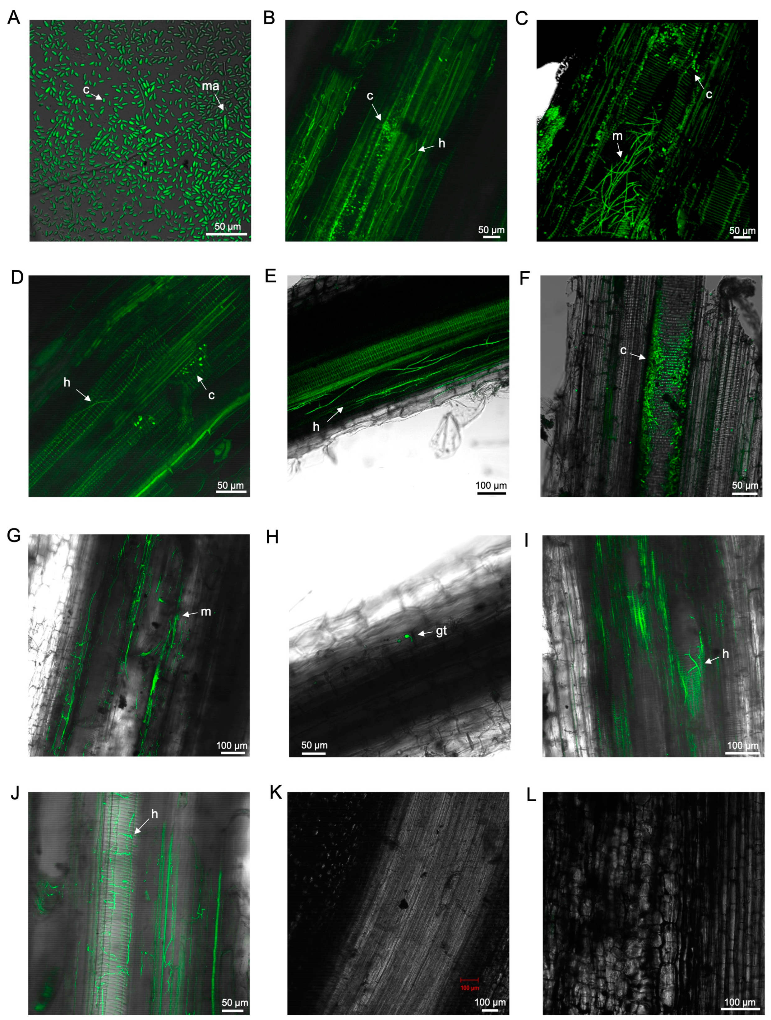
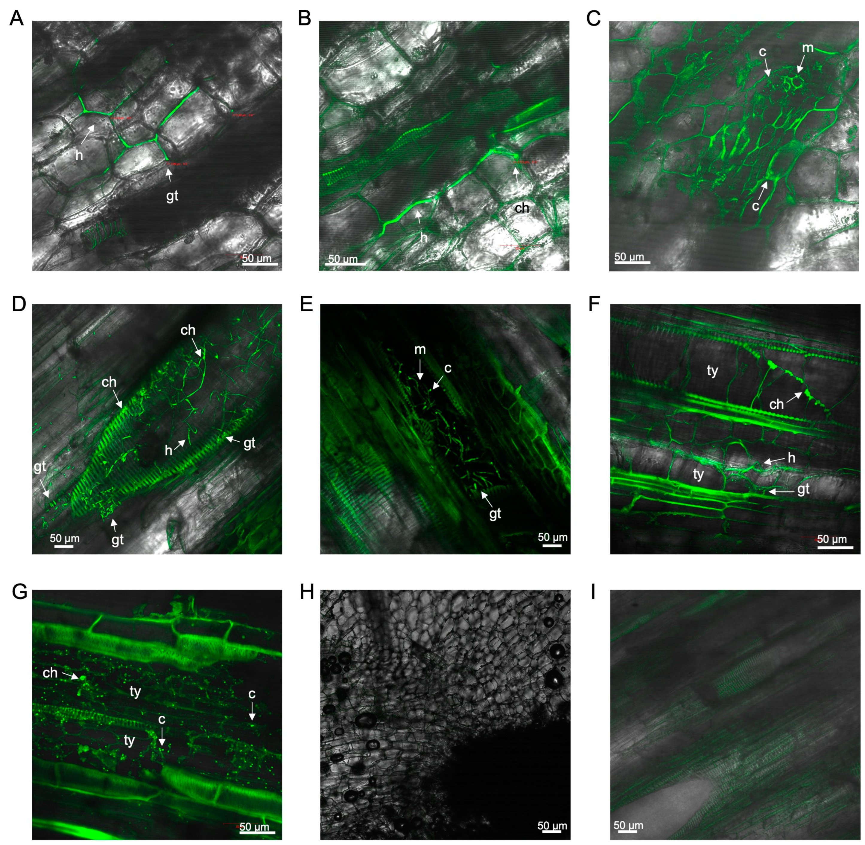
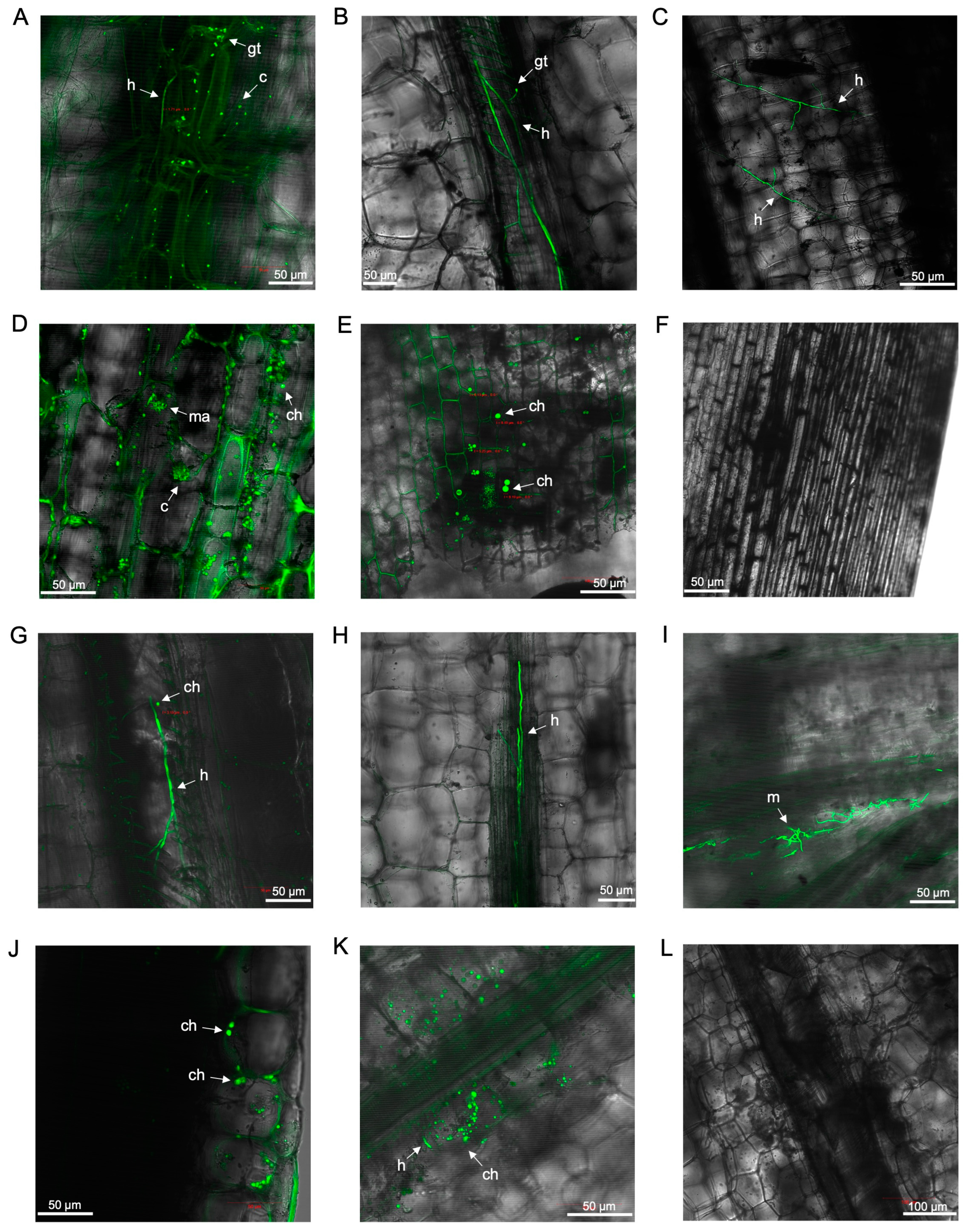
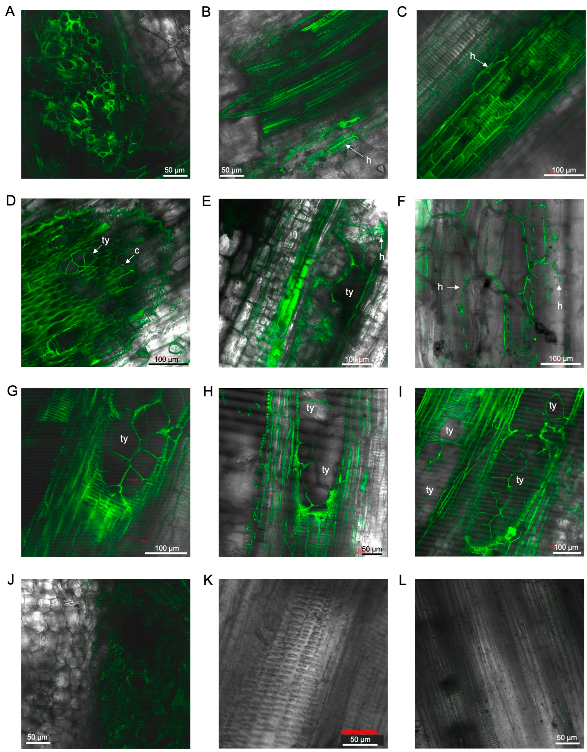
3.2. Laser Scanning Microscopy

3.2.1. Observations on Susceptible Cultivar FHIA02

At 5 dpi, highly abundant microconidia and mycelial networks were detected under a confocal microscope in the xylems of both the lateral and main roots of ‘FHIA02’ plants (Figure 1B,C).
Between 12 and 70 dpi, it was evident that the fungus moved uninhibited through the plant vasculature, affecting both the roots and rhizomes of ‘FHIA02’ (Figure 2). At 12 dpi, extensive mycelial networks were detected in the fine roots (Figure 2A). By 14 dpi, chlamydospores and a spore germ tube were visible on the epidermis of lateral roots (Figure 2B), and intercellular hyphal movement was observed (Figure 2C). At 19 dpi, the fungus appeared to establish itself through a wound site in a root hair (Figure 2D). From 26 to 42 dpi, extensive mycelial networks and germinated spores were found not only in the fine roots but also in the main roots and root nodes, which connected to the rhizome (Figure 2E–J).
In the rhizome of ‘FHIA02’, the intercellular presence of the fungus through the rhizome cortex was observed as early as 14 days dpi (Figure 3A–C). An intercellular hypha with a single terminal chlamydospore was also visualised (Figure 3B). Strong vascular autofluorescence, with microconidia forming in clusters, was detected in the rhizome at 14 dpi (Figure 3C). By 36 dpi, abundant germinated microconidia were detected in the xylem vessels (Figure 3D,E). Tyloses were noted as fine angular intra-xylem walled networks, resembling those described by VanderMolen et al. [38], which auto-fluoresced along with the plant cell wall structures of the xylem (Figure 3D,E). These tyloses appeared to occlude vessels in the rhizome at 68–70 dpi (Figure 3F,G). Microconidia and chlamydospores were co-localised in and around the xylem vessels (Figure 3D,E,G).
The presence of GFP-Foc-STR4 was detected in the outer leaf sheath and petiole edge of ‘FHIA02’ plants (Figure 4). Microconidia and hyphae were detected as early as 14 dpi (Figure 4A). The fungus was confined to the xylem vessels in the midrib of the leaf sheath at 26 dpi (Figure 4B), with mycelial networks observed on the epidermis of leaf sheaths at 29 dpi (Figure 4C). At 54–62 dpi, macroconidia and chlamydospores were abundantly visible along the petiole edge of a senescing leaf in ‘FHIA02’ (Figure 4D,E).

3.2.2. Observations on Resistant Cultivar FHIA25

The presence of GFP-Foc-STR4 in the tolerant/resistant cultivar ‘FHIA25’ was detected less frequently than in ‘FHIA02’. This was especially evident at 5 dpi, where only a single hyphal strand and a few conidia were observed (Figure 1D).
The presence of GFP-Foc-STR4 in the fine roots was clearly visualised from 12 to 49 dpi (Figure 5). The extent of root colonisation by the fungus appeared similar to that in ‘FHIA02’. At 12 dpi, microconidia on false heads in monophialides were observed in the fine roots (Figure 5A), and germinated spores were detected in the xylem vessels at 14 dpi (Figure 5B). The infection continued to spread, with clear fungal colonisation of xylem vessels by 21 dpi (Figure 5C) and extension through lateral root nodes connecting to the rhizome by 26 dpi (Figure 5D). This consistent germination of spores persisted at later time points up to 29 dpi, with germinated microconidia and hyphae visible in the root cap and elongation zone (Figure 5E,F).
Plants often respond to xylem-invading vascular wilt pathogens by depositing vascular coatings of lignin and suberin in colonised vessels [44]. Vascular coatings, such as lignin and suberin, are visualised as GFP autofluorescence from phenolic deposits in vessels colonised by vascular wilt pathogens [44]. Strong GFP fluorescence was observed in colonised compartments from 36 to 49 dpi, suggesting the presence of vascular coatings (Figure 5G–I). The GFP autofluorescence appeared co-localised with the fungus in these regions, indicating an active defence response.
In the rhizome of ‘FHIA25’, the presence of Foc STR4 appeared more restricted (Figure 6). Strong GFP autofluorescence marked infected regions in the rhizome and the main root nodes connecting to the rhizome (Figure 6A,D,E,G–I). The GFP-Foc-STR4 presence was primarily confined to the xylem vessels (Figure 6B,C). At 36–41 dpi, tyloses were observed in the vasculature of the rhizome (Figure 6D,E), with hyphal movement restricted between individual tyloses (Figure 6F). Tyloses formation continued to be detected in the rhizome from 42 to 70 dpi (Figure 6G–I), and fungal presence in these regions remained minimal, suggesting a more effective containment in ‘FHIA25’.
The presence of GFP-Foc-STR4 was detected in the outer leaf sheath and petiole edge of ‘FHIA25’ plants (Figure 4). Microconidia and hyphae were detected as early as 16 dpi (Figure 4G). By 21 dpi, the fungus was confined to the xylem vessels in the midrib of the leaf sheath (Figure 4H), with mycelial networks observed on the epidermis of leaf sheaths at 29 dpi (Figure 4I). At 62–70 dpi, chlamydospores and hyphae were abundantly observed in the petiole of ‘FHIA25’ (Figure 4J,K).

3.2.3. Observations on Cultivars Williams, GCTCV119 and Lady Finger

In ‘Williams’, extensive microconidia and hyphae were detected in the lateral roots at 5 dpi (Figure 1E) and appeared to occlude the xylem in these roots by 9 dpi (Figure 1F). Similarly, abundant mycelia were observed in the xylem vessels of the lateral roots in ‘Lady Finger’ plants at 8 dpi (Figure 1J). In ‘GCTCV119’ plants, GFP-Foc-STR4 hyphae were present in the lateral roots at 5–7 dpi (Figure 1G,I) but to a lesser extent than in ‘Williams’, ‘Lady Finger’, or ‘FHIA02’.
GFP-Foc-STR4 chlamydospores and hyphae were visualized in the vasculature of primary roots of ‘GCTCV119’, ‘Williams’, and ‘Lady Finger’ from 15 to 45 dpi (Figure S2).
To assess the movement from the roots into the rhizome, the presence of GFP-Foc-STR4 was checked in the connection between root nodes and corms at 32–60 dpi. GFP-Foc-STR4 hyphae and mycelia were present in the rhizome nodes of all three cultivars, indicating movement through the vasculature from the roots into the rhizome (Figure S3).
In the rhizomes, however, GFP-Foc-STR4 was not detected in ‘GCTCV119’ 26–42 dpi (Figure S4A–C). In contrast, chlamydospores and hyphae were clearly present in the vasculature of the rhizomes in ‘Lady Finger’ and ‘Williams’ at 27–40 dpi (Figure S4D–I), with strong GFP fluorescence observed in the infected regions.
Tyloses were observed within the vascular bundles of primary roots and the rhizome in ‘GCTCV119’ and ‘Williams’ plants at 55–59 dpi (Figure 7). In ‘GCTCV119’, tyloses were also formed in the non-inoculated controls, suggesting that tyloses formation can occur naturally in plants (Figure 7A). In both ‘Williams’ and its somaclonal derivative ‘GCTCV119’, GFP-Foc-STR4 appeared confined within occluded xylem vessels (Figure 7B,E,F) or located between two adjacent vessels (Figure 7D).
Above ground, GFP-Foc-STR4 was detected in the pseudostem of ‘Lady Finger’ and ‘Williams’ at 18–20 dpi (Figure S5A–C), but was not in ‘GCTCV119’ at 28 dpi (Figure S5E). At 51–59 dpi, GFP-Foc-STR4 mycelia were found in the pseudostem of all three cultivars (Figure S5B,D–F). In the leaves of all three cultivars presented in this section, mycelial networks and sporodochia were consistently present at both early (13–40 dpi) and late (60–65 dpi) stages (Figure S5G–L).

3.3. Scanning Electron Microscopy

SEM analysis of the primary roots of both ‘FHIA02‘ and ‘FHIA25‘ detected the presence of tyloses and fungal structures within xylem vessels (Figures 8 and Figure 9). Tyloses were clearly visible in the vascular bundles of both cultivars at 14 and 42 dpi (Figure 8C,E,F,H,I and Figure 9C,F,H,I). Various sizes of tyloses, including those in the process of forming, were observed in the xylem vessels of plants inoculated with GFP-Foc-STR4 (Figure 8F and Figure 9F,H). These tyloses were in close proximity to GFP-Foc-STR4 (Figure 8C) and, in some instances, were forming within the same vascular bundle as the fungal structures (Figure 9C). Additionally, tyloses were observed in the vascular bundles of control ‘FHIA02’ plants not treated with Foc, suggesting that tyloses can form during regular plant growth (Figure 8A).
Fungal growth was observed in the parenchyma cells and vascular bundles of both ‘FHIA02’ and ‘FHIA25’ at 14 and 42 dpi, aligning with GFP study findings that confirmed the presence of the fungus within the host at these time points. GFP-Foc-STR4 mycelia were clearly detected within individual vascular bundles and the pits of the main roots (Figure 8C,E and Figure 9C,G). In terms of root response, there were no significant differences between ‘FHIA02’ and ‘FHIA25’ in the number of tyloses formed or the extent of fungal colonization within the vasculature.

4. Discussion

Green fluorescent proteins have proven to be an invaluable tool for studying pathogen–host interactions in plants, allowing for real-time visualisation of fungal movement and colonisation within plant tissues [26,45,46]. In F. oxysporum, the production of microconidia and macroconidia is essential for the fungus to successfully proliferate within host plants. By tracking the accumulation of spores in specific plant compartments, researchers can gain insights into the dynamics of infection, which is crucial for managing and containing Foc in agricultural settings. However, the plant defence mechanisms in resistant or tolerant cultivars, such as ‘FHIA25’ and ‘GCTCV119’, are not yet fully understood, and these cultivars offer an important opportunity to explore how the host responds to pathogen attack.
The movement of Foc through the root systems was observed across all five banana cultivars, ‘Williams’, ‘Lady Finger’, ‘FHIA02’, ‘FHIA25’, and ‘GCTCV119’. Despite ‘FHIA25’ showing good resistance to Foc STR4, chlamydospores and microconidia from the GFP-Foc-STR4 isolate were found to attach to and germinate on the root tips and fine root hairs of this cultivar. Following this, the fungus was able to penetrate the root surface and spread through the root vascular system. This suggests that, while resistance mechanisms are present in certain cultivars, Foc can still actively enter banana hosts through their root systems.
In both ‘FHIA02’ and ‘FHIA25’, the fungus exhibited intercellular movement, initially in the epidermis and then through the elongation zones of the roots. This pattern aligns with the behaviour of biotrophic pathogens, similar to other Fusarium species, which rely on living plant cells to progress through the host [47,48]. The consistent detection of the fungus in the xylem vessels of both the roots and root nodes connecting to the rhizomes further suggests that vascular tissues play a critical role in facilitating the movement of the pathogen within the plant, regardless of the cultivar’s resistance level.
In the current study, a differential plant response to GFP-Foc-STR4 infection was observed in the rhizome tissues of the ‘FHIA02’ and ‘FHIA25’ banana cultivars. In ‘FHIA02’, a susceptible cultivar, proliferation of spores and mycelia was detected extensively in the xylem vessels of the symptomatic rhizomes. This indicates that the fungus is able to spread freely in the rhizome tissue, which likely contributes to the continued infection of the plant. In contrast, fewer hyphae were observed in the rhizome of the resistant cultivar ‘FHIA25’, and those present appeared to be confined to the xylem vessels, suggesting that the plant was mounting a defence response to restrict fungal spread. The formation of tyloses—cellular obstructions that block fungal movement—was detected in both cultivars, but was more pronounced in the resistant ‘FHIA25’, where it seemed to play a role in restricting the pathogen. Additionally, in the highly susceptible cultivars ‘Lady Finger’ and ‘Williams’, the rhizomes were heavily colonized by the fungus, further supporting their susceptibility to Foc. Meanwhile, the rhizomes of ‘GCTCV119’, a more tolerant cultivar, displayed limited fungal movement in the xylem vessels. This suggests that while the fungus can initially enter the plant, its spread in the rhizome is limited by the host plant’s defence mechanisms. Taken together, these observations emphasize the critical role of the rhizome in the banana–Fusarium interaction [26]. The plant’s ability to contain the fungus within the rhizome, possibly through mechanisms like tyloses formation, appears to be a key factor in determining resistance or susceptibility to Foc.
A similar pattern of restricted colonisation in Dianthus caryophyllus by F. oxysporum f. sp. dianthi has been reported, and further characterisation revealed that the infected regions of the xylem became compartmentalised by cell wall thickening, hyperplasia of parenchyma cells, and the built-up of vascular occluded materials [36]. In this study, vascular occlusion in the rhizome of ‘FHIA25’ was detected at 36 dpi and in ‘FHIA2’ at 68 dpi. SEM revealed that vascular occlusion in the main roots of both ‘FHIA2’ and ‘FHIA25’ occurred as early as 14 dpi following fungal inoculation. Similar occlusion was observed in the ‘GCTCV119’ and ‘Williams’ cultivars at 56–59 dpi. These results collectively suggest that vascular occlusion is an inducible plant defence mechanism to prevent the spread of an invading pathogen. While it may contribute to deterring the movement of the pathogen inside the host, tyloses formation [38] do not fully explain the host resistances observed in ‘FHIA25’ and ‘GCTCV119’. Formation of tyloses is typically triggered by infections [49], wounding [50], heartwood formation [51], and abscission [52]. The ability to form tyloses was found to be an important factor in resistance to F. oxysporum in cotton [53] and has been shown to be upregulated by an exogenous chemical application in banana [54]. Therefore, the presence of tyloses observed in this study is consistent with their roles in growth and development, as well as in the regulation of stresses including pathogen attacks and the activation of oxidative stress.
Another aspect of pathogen deterrence is the formation of vascular coatings in or around infected regions. These regions have been identified as plant physico-chemical barriers induced against xylem vascular wilt pathogens [44]. Particularly, gel and lignin depositions have been found to be associated with vertical and horizontal restrictions, respectively, of Foc in banana hosts [38,55]. These mechanisms, involving the formation of gels, gums, or mucilage in and around the vascular systems, have been shown to limit fungal growth in banana [43], tomato [56], carnation [57], pea [35,58], cotton [45,53], and bean plants [59]. These barriers are mostly composed of carbohydrates like pectin, polyphenols, and sometimes phytoalexins, lignin-like compounds, or lipoidal substances [35]. Upon a pathogen attack, it was observed in peas that the production of carbohydrates and polyphenolics by vascular parenchyma cells progressively accumulated in the lumen of xylem cells, highlighting important functions of these compounds in defence against F. oxysporum f. sp. pisi [35]. In this study, the movement of Foc through the aerial parts of banana plants was observed. Chlamydospores and hyphae were detected on the petiole and outer leaf sheath of both ‘FHIA02’ and ‘FHIA25’ plants, indicating that the fungus can penetrate and travel through the leaf sheaths. Although ‘FHIA25’ is resistant to Foc STR4, the fungus was still able to move through these tissues and reach the aerial parts of the plant. This observation highlights that while resistance mechanisms in the roots and rhizomes may limit the spread of the fungus, the pathogen can still access and potentially colonise the upper parts of the plant.
Additionally, sporodochia, which are fungal structures that produce conidia, were observed around the stomata in the leaves of both ‘Williams’ and ‘Lady Finger’. This suggests that the fungus can not only invade through the vascular tissue but may also spread externally, likely through the stomatal pores in the leaves, a known pathway for fungal pathogens. These findings align with previous studies showing the movement of Foc through the leaf sheaths [40] and its potential entry via stomata [11]. However, this study adds novel insight by reporting the transmission of Foc through the aerial parts of a resistant cultivar, FHIA25, which has not been documented before. This new observation underscores the complex nature of host–pathogen interactions and suggests that even resistant plants may not be fully immune to infection in all tissues.
This study highlights an important aspect of the ongoing battle against Foc TR4, as it shows that even cultivars considered resistant or tolerant to Foc TR4, such as ‘GCTCV119’ and ‘FHIA25’, can still be colonized by the fungus, though at varying levels of severity. While ‘GCTCV119’ and its improved variant, ‘GCTCV218’ (also known as ‘Formosana’), are acknowledged for their tolerance to Foc TR4, they are not fully immune [18,27,31], and their resistance can be compromised depending on the inoculum threshold and environmental conditions. The resistance of ’FHIA25’ to both Foc STR4 and Foc TR4 is well-documented [26,27], but this study suggests that the pathogen can still invade the plant tissues, albeit at lower frequencies compared to more susceptible cultivars like ‘FHIA02’ and ‘Williams’. This raises the question of how much inoculum these cultivars can tolerate before they succumb to the disease.
To make informed decisions about deploying these cultivars in regions affected by Foc TR4, further research is needed to establish the inoculum threshold levels that resistant or tolerant cultivars can withstand. Stress testing these cultivars under various field conditions will be crucial to understanding the limits of their resistance and optimizing their use in affected regions. This research will also guide the development of effective management strategies for controlling Foc TR4, ensuring that resistant cultivars can be effectively utilised in the field without prematurely succumbing to the disease.

5. Conclusions

This study examined the movement of a GFP-tagged Foc STR4 strain across five banana cultivars. Findings showed that in resistant cultivars, Foc was contained within the rhizome, while it spread further in susceptible cultivars. This suggests that the rhizome plays a critical role in limiting fungal spread within resistant plants. However, Foc was still detected in the leaves and outer leaf sheaths of resistant cultivars, indicating a potential risk of pathogen spread even in resistant cultivars. These insights have important implications for monitoring and containment protocols in Foc TR4-affected regions. Notably, Foc TR4 was not used in this study due to biosecurity restrictions.

Supplementary Materials

The following supporting information can be downloaded at: https://www.mdpi.com/article/10.3390/microorganisms12122472/s1, Figure S1: Evaluation of internal symptoms in the rhizomes of the banana cultivars tested in this study; Figure S2: Laser scanning microscopy showing the localisation of GFP-Foc-STR4 in the main roots of ‘GCTCV119’, ‘Williams’, and ‘Lady Finger’ at 15—45 dpi; Figure S3: Laser scanning microscopy showing the localisation of GFP-Foc-STR4 in the corm nodes of ‘Williams’, ‘GCTCV119’, and ‘Lady Finger’ at 26—60 dpi. (A) Mycelia in the xylem of a rhizome node in ‘GCTCV119’ at 32 dpi; Figure S4: Laser scanning microscopy showing the localisation of GFP-Foc-STR4 in the rhizomes of ‘Williams’, ‘GCTCV119’, and ‘Lady Finger’ at 26—51 dpi. (A) vascular coating in the rhizome of ‘GCTVC119’ at 26 dpi; Figure S5: Laser scanning microscopy showing the localisation of GFP-Foc-STR4 in the pseudostems and leaves of ‘Lady Finger’, ‘Williams’, and ‘GCTCV119’ at 18—65 dpi; Table S1: Reisolation of Fusarium oxysporum-like colonies from the leaves, petioles, lower sems and rhizomes of ‘FHIA02’ and ‘FHIA25’ plants inoculated with GFP-Foc-STR4.

Author Contributions

Conceptualization, A.C. and E.A.B.A.; methodology, A.C., T.-Y.C., Y.C. and S.M.A.F.; software, A.C., T.-Y.C., Y.C. and S.M.A.F.; validation, A.C., J.A., J.S., H.C., S.G., B.J.F., A.S., D.M.G. and E.A.B.A.; formal analysis, A.C., T.-Y.C., Y.C. and S.M.A.F.; investigation, T.-Y.C., Y.C. and S.M.A.F.; resources, A.C. and E.A.B.A.; data curation, A.C.; writing—original draft preparation, A.C.; writing—review and editing, A.C., T.-Y.C., Y.C., S.M.A.F., J.A., J.S., H.C., S.G., B.J.F., A.S., D.M.G. and E.A.B.A.; visualization, A.C.; supervision, A.C. and E.A.B.A.; project administration, A.C. and E.A.B.A.; funding acquisition, A.C. and E.A.B.A. All authors have read and agreed to the published version of the manuscript.

Funding

This project was funded by the Bill and Melinda Gates Foundation (Grant ID number: OPP1093845) through its grant to the International Institute of Tropical Agriculture (IITA) under the project Accelerated Breeding of Better Bananas, grant number IITA 20600.15/0008-8—Phase II. Research funding support for this project was also provided by Hort Innovation Australia under grant BA17006. Jay Anderson was supported by Hort Innovation Australia grant BA14014. Jiaman Sun was supported by Guangxi Natural Science Foundation grant 2021GXNSFAA196014.

Data Availability Statement

The original contributions presented in the study are included in the article/Supplementary Materials, further inquiries can be directed to the corresponding authors.

Acknowledgments

We would like to kindly thank Sharon Hamill for the supply of tissue culture derived plants of the banana cultivars used in this study. Plants used in this study were generated from the Australian in vitro banana cultivar collection that is maintained in the Quality Banana Approved Nursery (QBAN) scheme accredited Plant Biotechnology Laboratory located at the Maroochy Research Facility, Department of Agriculture and Fisheries, Nambour, Queensland, Australia. All cultivars were sourced under agreements allowing them to be used for research purposes. No ownership is claimed for those cultivars sourced from owners or other institutions. The plants were destructively sampled for the purposes of this study and were not further propagated. We would also like to thank Ning Chen for the in vitro multiplication and maintenance of these plantlets in tissue culture. We also graciously thank Richard I. Webb for providing technical support in scanning electron microscopy. The graphic abstract was created using bioRender and its graphic assets (https://www.biorender.com; accessed on 1 December 2024). We would like to thank the two reviewers for their comprehensive and thoughtful reviews of this manuscript. Their constructive feedback has greatly contributed to improving the quality and clarity of this work.

Conflicts of Interest

The authors declare no conflicts of interest.

References

  1. FAO. 2024. Available online: https://www.fao.org/statistics/en (accessed on 24 October 2024).
  2. Ortiz, R.; Ferris, R.; Vuylsteke, D. Banana and plantain breeding. In Bananas and Plantains; Springer: Berlin/Heidelberg, Germany, 1995; pp. 110–146. [Google Scholar]
  3. Maryani, N.; Lombard, L.; Poerba, Y.; Subandiyah, S.; Crous, P.; Kema, G. Phylogeny and genetic diversity of the banana Fusarium wilt pathogen Fusarium oxysporum f. sp. cubense in the Indonesian centre of origin. Stud. Mycol. 2019, 92, 155–194. [Google Scholar] [CrossRef]
  4. Lombard, L.; Sandoval-Denis, M.; Lamprecht, S.; Crous, P.W. Epitypification of Fusarium oxysporum—Clearing the taxonomic chaos. Persoonia Mol. Phylogeny Evol. Fungi 2019, 43, 1–47. [Google Scholar] [CrossRef]
  5. Czislowski, E.; Fraser-Smith, S.; Zander, M.; O’Neill, W.T.; Meldrum, R.A.; Tran-Nguyen, L.T.T.; Batley, J.; Aitken, E.A.B. Investigation of the diversity of effector genes in the banana pathogen, Fusarium oxysporum f. sp. cubense, reveals evidence of horizontal gene transfer. Mol. Plant Pathol. 2018, 19, 1155–1171. [Google Scholar] [CrossRef] [PubMed]
  6. O’Donnell, K.; Kistler, H.C.; Cigelnik, E.; Ploetz, R.C. Multiple evolutionary origins of the fungus causing Panama disease of banana: Concordant evidence from nuclear and mitochondrial gene genealogies. Proc. Natl. Acad. Sci. USA 1998, 95, 2044–2049. [Google Scholar] [CrossRef] [PubMed]
  7. Ploetz, R.C. Fusarium wilt of banana is caused by several pathogens referred to as Fusarium oxysporum f. sp. cubense. Phytopathology 2006, 96, 653–656. [Google Scholar] [CrossRef]
  8. Gordon, T.R. Fusarium oxysporum and the Fusarium Wilt Syndrome. Annu. Rev. Phytopathol. 2017, 55, 23–39. [Google Scholar] [CrossRef]
  9. Stover, R.H. Fusarial Wilt (Panama Disease) of Bananas and Other Musa Species; Commonwealth Mycological Institute: Kew, UK, 1962; p. 117. [Google Scholar]
  10. Zhang, L.; Yuan, T.; Wang, Y.; Zhang, D.; Bai, T.; Xu, S.; Wang, Y.; Tang, W.; Zheng, S.J. Identification and evaluation of resistance to Fusarium oxysporum f. sp. cubense tropical race 4 in Musa acuminata Pahang. Euphytica 2018, 214, 106. [Google Scholar] [CrossRef]
  11. Warman, N.M.; Aitken, E.A.B. The Movement of Fusarium oxysporum f.sp. cubense (Sub-Tropical Race 4) in susceptible cultivars of banana. Front. Plant Sci. 2018, 9, 1748. [Google Scholar] [CrossRef] [PubMed]
  12. Pegg, K.G.; Coates, L.M.; O’Neill, W.T.; Turner, D.W. The epidemiology of Fusarium wilt of banana. Front. Plant Sci. 2019, 10, 1395. [Google Scholar] [CrossRef]
  13. Bai, T.; Qin, M.; Li, X.; Fan, H.; Xu, S.; Zeng, L.; Zheng, S.J. An additional threat to ‘Cavendish’ banana growers and traders: The infection of banana peduncles by Fusarium oxysporum f. sp. cubense Tropical Race 4 (Foc TR4). Plant Health Prog. 2020, 21, 312–316. [Google Scholar] [CrossRef]
  14. Ploetz, R.C. Management of Fusarium wilt of banana: A review with special reference to tropical race 4. Crop Prot. 2015, 73, 7–15. [Google Scholar] [CrossRef]
  15. Hwang, S.C.; Ko, W.H. Cavendish banana cultivars resistant to Fusarium wilt acquired through somaclonal variation in Taiwan. Plant Dis. 2004, 88, 580–588. [Google Scholar] [CrossRef] [PubMed]
  16. Buddenhagen, I. Understanding strain diversity in Fusarium oxysporum f. sp. cubense and history of introduction of ‘Tropical Race 4’ to better manage banana production. Acta Hortic. 2009, 828, 193–204. [Google Scholar] [CrossRef]
  17. Mostert, D.; Molina, A.B.; Daniells, J.; Fourie, G.; Hermanto, C.; Chao, C.P.; Fabregar, E.; Sinohin, V.G.; Masdek, N.; Thangavelu, R.; et al. The distribution and host range of the banana Fusarium wilt fungus, Fusarium oxysporum f. sp. cubense, in Asia. PLoS ONE 2017, 12, e0181630. [Google Scholar] [CrossRef]
  18. Munhoz, T.; Vargas, J.; Teixeira, L.; Staver, C.; Dita, M. Fusarium Tropical Race 4 in Latin America and the Caribbean: Status and global research advances towards disease management. Front. Plant Sci. 2024, 15, 1397617. [Google Scholar] [CrossRef] [PubMed]
  19. Ploetz, R.C. Fusarium wilt of banana. Phytopathology 2015, 105, 1512–1521. [Google Scholar] [CrossRef]
  20. O’Neill, W.T.; Henderson, J.; Pattemore, J.A.; O’Dwyer, C.; Perry, S.; Beasley, D.R.; Tan, Y.P.; Smyth, A.; Goosem, C.; Thomson, K.; et al. Detection of Fusarium oxysporum f. sp. cubense tropical race 4 strain in northern Queensland. Australas. Plant Dis. Notes 2016, 11, 33. [Google Scholar] [CrossRef]
  21. Dita, M.; Barquero, M.; Heck, D.; Mizubuti, E.S.G.; Staver, C.P. Fusarium wilt of banana: Current knowledge on epidemiology and research needs toward sustainable disease management. Front. Plant Sci. 2018, 9, 1468. [Google Scholar] [CrossRef]
  22. Dale, J.; James, A.; Paul, J.Y.; Khanna, H.; Smith, M.; Peraza-Echeverria, S.; Garcia-Bastidas, F.; Kema, G.; Waterhouse, P.; Mengersen, K.; et al. Transgenic Cavendish bananas with resistance to Fusarium wilt tropical race 4. Nat. Commun. 2017, 8, 1496. [Google Scholar] [CrossRef]
  23. Chen, A.; Sun, J.; Martin, G.; Gray, L.-A.; Hřibová, E.; Christelová, P.; Yahiaoui, N.; Rounsley, S.; Lyons, R.; Batley, J.; et al. Identification of a major QTL-controlling resistance to the Subtropical Race 4 of Fusarium oxysporum f. sp. cubense in Musa acuminata ssp. malaccensis. Pathogens 2023, 12, 289. [Google Scholar] [CrossRef]
  24. Chen, A.; Sun, J.; Viljoen, A.; Mostert, D.; Xie, Y.; Mangila, L.; Bothma, S.; Lyons, R.; Hřibová, E.; Christelová, P.; et al. Genetic mapping, candidate gene identification and marker validation for host plant resistance to the race 4 of Fusarium oxysporum f. sp. cubense using Musa acuminata ssp. malaccensis. Pathogens 2023, 12, 820. [Google Scholar] [CrossRef]
  25. Rebouças, T.A.; de Jesus Rocha, A.; Cerqueira, T.S.; Adorno, P.R.; Barreto, R.Q.; dos Santos Ferreira, M.; Lino, L.S.M.; de Oliveira Amorim, V.B.; dos Santos-Serejo, J.A.; Haddad, F.; et al. Pre-selection of banana somaclones resistant to Fusarium oxysporum f. sp. cubense, subtropical race 4. Crop Prot. 2021, 147, 105692. [Google Scholar] [CrossRef] [PubMed]
  26. Chen, A.; Sun, J.; Matthews, A.; Armas-Egas, L.; Chen, N.; Hamill, S.; Mintoff, S.; Tran-Nguyen, L.T.; Batley, J.; Aitken, E.A. Assessing variations in host resistance to Fusarium oxysporum f. sp. cubense race 4 in Musa species, with a focus on the subtropical race 4. Front. Microbiol. 2019, 10, 1062. [Google Scholar] [CrossRef]
  27. Mintoff, S.J.; Nguyen, T.V.; Kelly, C.; Cullen, S.; Hearnden, M.; Williams, R.; Daniells, J.W.; Tran-Nguyen, L.T. Banana cultivar field screening for resistance to Fusarium oxysporum f. sp. cubense tropical race 4 in the Northern Territory. J. Fungi 2021, 7, 627. [Google Scholar] [CrossRef]
  28. Zuo, C.; Deng, G.; Li, B.; Huo, H.; Li, C.; Hu, C.; Kuang, R.; Yang, Q.; Dong, T.; Sheng, O.; et al. Germplasm screening of Musa spp. for resistance to Fusarium oxysporum f. sp. cubense tropical race 4 (Foc TR4). Eur. J. Plant Pathol. 2018, 151, 723–734. [Google Scholar] [CrossRef]
  29. Zhan, N.; Kuang, M.; He, W.; Deng, G.; Liu, S.; Li, C.; Roux, N.; Dita, M.; Yi, G.; Sheng, O. Evaluation of resistance of banana genotypes with AAB genome to Fusarium wilt Tropical Race 4 in China. J. Fungi 2022, 8, 1274. [Google Scholar] [CrossRef]
  30. Molina, A.; Razo, S.; Sinohin, V.G.; Herradura, L. DA-BAR Terminal Report: Mitigating Banana Fusarium Wilt Tropical Race 4 Through a Farmer-Participatory Approach of Developing Disease Management Strategies; Bioversity International: Rome, Italy, 2018. [Google Scholar] [CrossRef]
  31. Viljoen, A.; Mostert, D.; Chiconela, T.; Beukes, I.; Fraser, C.; Dwyer, J.; Murray, H.; Amisse, J.; Matabuana, E.L.; Tazan, G.; et al. Occurrence and spread of the banana fungus Fusarium oxysporum f. sp. cubense TR4 in Mozambique. S. Afr. J. Sci. 2020, 116, 1–11. [Google Scholar] [CrossRef]
  32. Daly, A.; Walduck, G. Fusarium wilt of Bananas (Panama Disease):(Fusarium oxysporum f. sp. cubense); Agnote—Northern Territory of Australia; No.I51; Northern Territory Government: Darwin, NT, Australia, 2006; pp. 1–5.
  33. Smith, M.; Langdon, P.; Pegg, K.; Daniells, J. Growth, yield and Fusarium wilt resistance of six FHIA tetraploid bananas (Musa spp.) grown in the Australian subtropics. Sci. Hortic. 2014, 170, 176–181. [Google Scholar] [CrossRef]
  34. Sanya, L.N.; Sseguya, H.; Kyazze, F.B.; Diiro, G.M.; Nakazi, F. The role of variety attributes in the uptake of new hybrid bananas among smallholder rural farmers in central Uganda. Agric. Food Secur. 2020, 9, 1. [Google Scholar] [CrossRef]
  35. Bani, M.; Pérez-De-Luque, A.; Rubiales, D.; Rispail, N. Physical and chemical barriers in root tissues contribute to quantitative resistance to Fusarium oxysporum f. sp. pisi in pea. Front. Plant Sci. 2018, 9, 199. [Google Scholar] [CrossRef]
  36. Ouellette, G.; Baayen, R.; Simard, M.; Rioux, D. Ultrastructural and cytochemical study of colonization of xylem vessel elements of susceptible and resistant Dianthus caryophyllus by Fusarium oxysporum f. sp. dianthi. Can. J. Bot. 1999, 77, 644–663. [Google Scholar] [CrossRef]
  37. De Micco, V.; Balzano, A.; Wheeler, E.A.; Baas, P. Tyloses and gums: A review of structure, function and occurrence of vessel occlusions. IAWA J. 2016, 37, 186–205. [Google Scholar] [CrossRef]
  38. VanderMolen, G.E.; Beckman, C.H.; Rodehorst, E. The ultrastructure of tylose formation in resistant banana following inoculation with Fusarium oxysporum f. sp. cubense. Physiol. Mol. Plant Pathol. 1987, 31, 185–200. [Google Scholar] [CrossRef]
  39. Li, C.; Chen, S.; Zuo, C.; Sun, Q.; Ye, Q.; Yi, G.; Huang, B. The use of GFP-transformed isolates to study infection of banana with Fusarium oxysporum f. sp. cubense race 4. Eur. J. Plant Pathol. 2011, 131, 327–340. [Google Scholar] [CrossRef]
  40. Xiao, R.F.; Zhu, Y.J.; Li, Y.D.; Liu, B. Studies on vascular infection of Fusarium oxysporum f. sp. cubense race 4 in banana by field survey and green fluorescent protein reporter. Int. J. Plant Pathol. 2013, 2, 44–51. [Google Scholar] [CrossRef]
  41. Anderson, J.; Aitken, E. Effect of in planta treatment of ‘Cavendish’ Banana with herbicides and fungicides on the colonisation and sporulation by Fusarium oxysporum f. sp. cubense Subtropical Race 4. J. Fungi 2021, 7, 184. [Google Scholar] [CrossRef]
  42. Ratnayake, K.; Joyce, D.C.; Webb, R.I. A convenient sample preparation protocol for scanning electron microscope examination of xylem-occluding bacterial biofilm on cut flowers and foliage. Sci. Hortic. 2012, 140, 12–18. [Google Scholar] [CrossRef]
  43. Rocha, A.d.J.; Soares, J.M.d.S.; Nascimento, F.d.S.; Rocha, A.d.S.; Amorim, V.B.O.d.; Ramos, A.P.d.S.; Ferreira, C.F.; Haddad, F.; Amorim, E.P. Molecular, histological and histochemical responses of banana cultivars challenged with Fusarium oxysporum f. sp. cubense with different levels of virulence. Plants 2022, 11, 2339. [Google Scholar] [CrossRef]
  44. Kashyap, A.; Planas-Marquès, M.; Capellades, M.; Valls, M.; Coll, N.S. Blocking intruders: Inducible physico-chemical barriers against plant vascular wilt pathogens. J. Exp. Bot. 2020, 72, 184–198. [Google Scholar] [CrossRef]
  45. Chen, A.; Morrison, S.; Gregson, A.; Le, D.P.; Urquhart, A.S.; Smith, L.J.; Aitken, E.A.; Gardiner, D.M. Fluorescently Tagged Verticillium dahliae to understand the infection process on cotton (Gossypium hirsutum) and weed plant species. Pathogens 2024, 13, 442. [Google Scholar] [CrossRef]
  46. Vallad, G.; Subbarao, K. Colonization of resistant and susceptible lettuce cultivars by a green fluorescent protein-tagged isolate of Verticillium dahliae. Phytopathology 2008, 98, 871–885. [Google Scholar] [CrossRef] [PubMed]
  47. Lagopodi, A.L.; Ram, A.F.; Lamers, G.E.; Punt, P.J.; Van den Hondel, C.A.; Lugtenberg, B.J.; Bloemberg, G.V. Novel aspects of tomato root colonization and infection by Fusarium oxysporum f. sp. radicis-lycopersici revealed by confocal laser scanning microscopic analysis using the green fluorescent protein as a marker. Mol. Plant Microbe Interact. 2002, 15, 172–179. [Google Scholar] [CrossRef] [PubMed]
  48. Yuan, H.; Ling, X.; Liu, T.; Chen, T.; Yang, Y.; Yao, S.; Zhang, B. Microscopic observations of strawberry plant colonization by a GFP-labelled strain of Fusarium oxysporum f. sp. fragariae. Can. J. Plant Pathol. 2014, 36, 501–508. [Google Scholar] [CrossRef]
  49. Pérez-Donoso, A.G.; Greve, L.C.; Walton, J.H.; Shackel, K.A.; Labavitch, J.M. Xylella fastidiosa infection and ethylene exposure result in xylem and water movement disruption in grapevine shoots. Plant Physiol. 2007, 143, 1024–1036. [Google Scholar] [CrossRef] [PubMed]
  50. Sun, Q.; Rost, T.L.; Matthews, M.A. Pruning-induced tylose development in stems of current-year shoots of Vitis vinifera (Vitaceae). Am. J. Bot. 2006, 93, 1567–1576. [Google Scholar] [CrossRef]
  51. Taylor, A.M.; Gartner, B.L.; Morrell, J.J. Heartwood formation and natural durability—A review. Wood Fiber Sci. 2002, 34, 587–611. [Google Scholar]
  52. Dute, R.R.; Duncan, K.M.; Duke, B. Tyloses in abscission scars of loblolly pine. IAWA J. 1999, 20, 67–74. [Google Scholar] [CrossRef]
  53. Shi, J.; Mueller, W.; Beckman, C. Vessel occlusion and secretory activities of vessel contact cells in resistant or susceptible cotton plants infected with Fusarium oxysporum f. sp. vasinfectum. Physiol. Mol. Plant Pathol. 1992, 40, 133–147. [Google Scholar] [CrossRef]
  54. Zhou, G.-D.; He, P.; Tian, L.; Xu, S.; Yang, B.; Liu, L.; Wang, Y.; Bai, T.; Li, X.; Li, S.; et al. Disentangling the resistant mechanism of Fusarium wilt TR4 interactions with different cultivars and its elicitor application. Front. Plant Sci. 2023, 14, 1145837. [Google Scholar] [CrossRef]
  55. De Ascensao, A.R.; Dubery, I.A. Panama disease: Cell wall reinforcement in banana roots in response to elicitors from Fusarium oxysporum f. sp. cubense Race Four. Phytopathology 2000, 90, 1173–1180. [Google Scholar] [CrossRef]
  56. Beckman, C.; Elgersma, D.; MacHardy, W. The localization of fusarial infections in the vascular tissue of single-dominant-gene resistant tomatoes. Phytopathology 1972, 62, 1256–1260. [Google Scholar] [CrossRef]
  57. Baayen, R.; Van Eijk, C.; Elgersma, D. Histology of roots of resistant and susceptible carnation cultivars from soil infested with Fusarium oxysporum f. sp. dianthi. Neth. J. Plant Pathol. 1989, 95, 3–13. [Google Scholar] [CrossRef]
  58. Tessier, B.; Mueller, W.; Morgham, A. Histopathology and ultrastructure of vascular responses in peas resistant or susceptible to Fusarium oxysporum f. sp. pisi. Phytopathology 1990, 80, 756–764. [Google Scholar] [CrossRef]
  59. Pereira, A.C.; Cruz, M.F.A.; Paula Júnior, T.J.; Rodrigues, F.A.; Carneiro, J.E.S.; Vieira, R.F.; Carneiro, P.C.S. Infection process of Fusarium oxysporum f. sp. phaseoli on resistant, intermediate and susceptible bean cultivars. Trop. Plant Pathol. 2013, 38, 323–328. [Google Scholar] [CrossRef]
Figure 1. Laser scanning microscopy pictures: (A) GFP-Foc-STR4 suspension of macroconidia and microconidia. Localisation of GFP-Foc-STR4 in banana cultivars during the infection process: (B) Mycelia and microconidia within the xylem of a lateral root in ‘FHIA02’ at 5 dpi. (C) Microconidia and mycelia between the cortical cells of the elongation zone and the xylem vessels of the primary root in ‘FHIA02’ at 5dpi. (D) Microconidia and hyphae visualised in the elongation zone of a ‘FHIA25’ lateral root at 5 dpi. (E) Hyphae along the epidermis of a ‘Williams’ lateral root at 5 dpi. (F) Microconidia proliferating in the xylem vessels of a ‘Williams’ lateral root at 9 dpi. (G) Mycelial networks in the xylem vessels of a ‘GCTCV119’ lateral root at 5 dpi. (H) Germ tube on the epidermis of a ‘GCTCV119’ lateral root at 7 dpi. (I) Hyphae within the vasculature of a ‘GCTCV119’ lateral root at 7 dpi. (J) Hyphae in the xylem vessels of a ‘Lady Finger’ lateral root at 8 dpi. Longitudinal section of non-inoculated lateral root in ‘FHIA02’ at 5 dpi (K) and ‘FHIA25’ at 6 dpi (L). Abbreviations are annotated as: ma = macroconidia; c = conidia; h = hyphae; m = mycelium; gt = germ tube. Horizontal bars indicate the scale used to capture the images.
Microorganisms 12 02472 g001
Figure 2. Laser scanning microscopy showing the localisation of GFP-Foc-STR4 in ‘FHIA02’ during the infection process: (A) Microconidia and mycelia colonising the xylem vessels of a fine root at 12 dpi. (B) Germ tube, hyphae, and chlamydospores in the epidermis of a lateral root at 14 dpi. (C) Germ tube, hyphae, and chlamydospores in the cortex region of a lateral root at 14 dpi. (D) A wound site penetrated by GFP-Foc-STR4 on a lateral root at 19 dpi. (E) Germ tubes and mycelia in the xylem vessels of a fine root at 26 dpi. (F) Germ tubes and mycelia in the xylem vessels of a primary root at 29 dpi. (G) Mycelia within the xylem vessels of a primary root near a rhizome node at 36 dpi. (H) Germ tubes and hyphae in the cortex of a main root at 41 dpi. (I) Germ tubes and mycelia in the xylem vessels of the rhizome connecting the main root at 41 dpi. (J) Germ tubes and mycelia colonising the xylem vessels of the rhizome connecting the main root at 42 dpi. Non-inoculated control of a fine root at 18 (K) and 62 (L) dpi. Abbreviations are annotated as: ch = chlamydospores; c = conidia; h = hyphae; m = mycelium; gt = germ tube. Horizontal bars indicate the scale used to capture the images.
Microorganisms 12 02472 g002
Figure 3. Laser scanning microscopy showing the localisation of GFP-Foc-STR4 in the rhizome of ‘FHIA02’ during the infection process. (A) Germ tubes and hyphae in the cortical cells of a discoloured rhizome at 14 dpi. (B) Hyphae and chlamydospores in a discoloured region of a rhizome at 14 dpi. (C) Microconidia and mycelial networks in the rhizome at 14 dpi. (D) Germ tubes, hyphae, and chlamydospores in the vascular vessel of a rhizome at 36 dpi. (E) Germ tubes, microconidia, and mycelia in the xylem vessel of a rhizome at 36 dpi. (F) Tyloses, germ tubes, hyphae, and chlamydospores in the xylem vessel lumen of a rhizome node at 68 dpi. (G) Tyloses, microconidia, and chlamydospores in the xylem vessels of a rhizome at 70 dpi. Non-inoculated control of a rhizome at 14 dpi (H) and 68 dpi (I). Abbreviations are annotated as: ch = chlamydospores; c = conidia; h = hyphae; m = mycelium; gt = germ tube; ty = tyloses. Horizontal bars indicate the scale used to capture the images.
Microorganisms 12 02472 g003
Figure 4. Laser scanning microscopy showing the localisation of GFP-Foc-STR4 in the leaves of ‘FHIA02’ and ‘FHIA25’. (A) Germ tubes, microconidia, and hyphae on an outer leaf sheath of ‘FHIA02’ at 14 dpi. (B) Germ tube and hyphae visualised in the xylem vessel of an outer leaf sheath of ‘FHIA02’ at 26 dpi. (C) Hyphae at the epidermis of an outer leaf sheath of ‘FHIA02’ at 29 dpi. (D) Microconidia, hyphae, and chlamydospores at the edge of a petiole of a senescing leaf in ‘FHIA02’ at 54 dpi. (E) Chlamydospores at the edge of a petiole of a senescing leaf in ‘FHIA02’ at 62 dpi. (F) Non-inoculated control at the edge of a petiole in ‘FHIA02’ at 63 dpi. (G) Hyphae and chlamydospore confined to the xylem vessel in the outer leaf sheath of ‘FHIA25’ at 16 dpi. (H) Hyphae confined to the xylem vessel in the outer leaf sheath of ‘FHIA25’ at 21 dpi. (I) Mycelial networks in the xylem vessels of a leaf of ‘FHIA25’ at 29 dpi. (J) Chlamydospores in a discoloured region at the edge of a petiole of ‘FHIA25’ at the 62 dpi. (K) Chlamydospores and hyphae at the edge of a petiole in ‘FHIA25’ at 70 dpi. (L) Non-inoculated control in the midrib of a ‘FHIA25’ leaf at 70 dpi. Abbreviations are annotated as: ma = macroconidia; ch = chlamydospores; c = conidia; h = hyphae; m = mycelium; gt = germ tube. Horizontal bars indicate the scale used to capture the images.
Microorganisms 12 02472 g004
Figure 5. Laser scanning microscopy showing the localisation of GFP-Foc-STR4 in the roots of ‘FHIA25’ during the infection process. (A) Microconidia and hyphae visualised at the epidermis of a fine root at 12 dpi. (B) Germ tubes and hyphae in the xylem vessel of a fine root at 12 dpi. (C) Germ tubes and hyphae in the xylem vessel of a fine root at 21 dpi. (D) Germ tubes, hyphae, and microconidia in the xylem vessel of a junction between lateral root and rhizome at 26 dpi. (E) Germ tubes, microconidia, and mycelia at the epidermis of a primary root tip at 29 dpi. (F) Germ tubes, microconidia, and hyphae in the xylem vessel of a fine root at 29 dpi. (G) Hyphae and a single terminal chlamydospore in the xylem vessel of a fine root at 36 dpi. (H) Hyphae and GFP autofluorescence in the xylem of a junction between root and rhizome at 49 dpi. (I) Hyphae and GFP autofluorescence in the xylem of a junction between root and rhizome at 49 dpi. (J) Non-inoculated control in a lateral root at 21 dpi (J) and 36 dpi (K). (L) Non-inoculated control in the junction between lateral root to rhizome at 51 dpi. Abbreviations are annotated as: ch = chlamydospores; c = conidia; h = hyphae; m = mycelium; gt = germ tube. Horizontal bars indicate the scale used to capture the images.
Microorganisms 12 02472 g005
Figure 6. Laser scanning microscopy showing the localisation of GFP-Foc-STR4 in the rhizome of ‘FHIA25’ during the infection process. (A) GFP autofluorescence in the rhizome at 14 dpi. (B) Hyphae and GFP autofluorescence in the xylem vessels of a rhizome at 14 dpi. (C) Hyphae and GFP autofluorescence in the xylem of a junction between root and rhizome at 36 dpi. (D) Tyloses and microconidia in the vascular bundles of a rhizome at 36 dpi. (E) Tyloses and hyphae in the xylem vessel of a junction between root and rhizome at 41 dpi. (F) Hyphae in a junction between primary root and rhizome at 41 dpi. (G) Tyloses in the xylem of a junction between primary root and rhizome at 42 dpi. (H) Tyloses in the xylem vessel of a junction between primary root and rhizome at 62 dpi. (I) Tyloses in xylem vessels of a junction between primary root and rhizome at 70 dpi. (J) Non-inoculated control in the rhizomes at 16 dpi (J), 36 dpi (K), and 70 dpi (L). Abbreviations are annotated as: c = conidia; h = hyphae; ty = tyloses. Horizontal bars indicate the scale used to capture the images.
Microorganisms 12 02472 g006
Figure 7. Laser scanning microscopy showing tyloses formation and the localisation of GFP-Foc-STR4 in the roots and rhizome of ‘GCTCV119’ and ‘Williams’. (A) Tyloses in the xylem vessel of a non-inoculated ‘GCTCV119’ plant at 56 dpi. (B) Tyloses and hyphae in the main root of ‘GCTCV119’ at 56 dpi. (C) Tyloses and hyphae in the xylem vessel of a rhizome of ‘GCTCV119’ at 59 dpi. (D) Hyphae confined in between two occluded vessels in the primary root of ‘Williams’ at 56 dpi. (E) Tyloses and hyphae in the xylem vessel of the main root of ‘Williams’ at 56 dpi. (F) Tyloses and hyphae in the xylem vessel of a rhizome of ‘Williams’ at 55 dpi. Abbreviations are annotated as: h = hyphae; ty = tyloses. Horizontal bars indicate the scale used to capture the images.
Microorganisms 12 02472 g007
Figure 8. Scanning electron micrographs showing transverse sections of primary roots from ‘FHIA02’. A non-inoculated control of a root (A) and under magnified view (B) at 14 dpi. (C) Mycelia and vascular occlusion in the vascular cavities of the main root at 14 dpi. (D) Tyloses in the vascular cavities of the main root at 14 dpi. (E) Tyloses and mycelia in the vascular cavities of the main root at 14 dpi. (F) Tyloses and vascular occlusion in the vascular cavities of the main root at 14 dpi. (G,H) Tyloses and vascular occlusion in the xylem cavities of the main root at 42 dpi. (I) Tyloses occluding multiple xylem cavities in the main root at 42 dpi. Abbreviations are annotated as: m = mycelium; ty = tyloses; o = vascular occlusion. Horizontal bars indicate the scale used to capture the images.
Microorganisms 12 02472 g008
Figure 9. Scanning electron micrographs showing transverse sections of primary roots from ‘FHIA25’. Vascular bundles in the primary root of a non-inoculated plant (A) and under a magnified view (B) at 14 dpi. (C) Tyloses formation and mycelia in the same xylem cavity of a primary root at 14 dpi. (D) Hyphae in the xylem cavities of the primary root at 14 dpi. (E) Tyloses formation and vascular occlusion in the xylem cavities of the primary root at 14 dpi. (F) Tyloses formation, vascular occlusion, and mycelia in the xylem cavities of the primary root at 14 dpi. (G) Mycelial networks in the xylem cavities of the primary root at 42 dpi. (H,I) Tyloses formation and vascular occlusion in the xylem cavities of the primary roots at 42 dpi. Abbreviations are annotated as: m = mycelium; ty = tyloses; o = vascular occlusion. Horizontal bars indicate the scale used to capture the images.
Microorganisms 12 02472 g009
Disclaimer/Publisher’s Note: The statements, opinions and data contained in all publications are solely those of the individual author(s) and contributor(s) and not of MDPI and/or the editor(s). MDPI and/or the editor(s) disclaim responsibility for any injury to people or property resulting from any ideas, methods, instructions or products referred to in the content.

