Article

The Sweet Cherry Tree Genotype Restricts the Aggressiveness of the Wood Decay Fungi Cytospora sorbicola and Calosphaeria pulchella

1
Departamento de Sanidad Vegetal, Facultad de Ciencias Agronómicas, Universidad de Chile, Santiago 8820808, Chile
2
Centre of Molecular Biology in Plants, Departamento de Biología, Facultad de Ciencias, Universidad de Chile, Santiago 7800003, Chile
*
Authors to whom correspondence should be addressed.
Microorganisms 2024, 12(12), 2456; https://doi.org/10.3390/microorganisms12122456
Submission received: 21 August 2024 / Revised: 1 October 2024 / Accepted: 12 October 2024 / Published: 29 November 2024

Abstract

:
The wood decay fungi Cytospora sorbicola and Calosphaeria pulchella severely threaten the worldwide cultivation of sweet cherry trees (Prunus avium L.). Both fungi cause similar symptoms, including vascular necrosis, which leads to branch and twig dieback. In advanced stages of the disease, cankers are visible on tree branches and trunks. The sweet cherry is the most widely planted fruit tree in Chile, with 74,000 hectares in 2023. According to the planted surface, the predominant sweet cherry varieties are Lapins, Santina, Regina, and Bing. Variety-dependent susceptibility studies on Cyt. sorbicola and Cal. pulchella are lacking. The main entry points for wood necrosis-causing fungi are pruning wounds; therefore, we evaluated the aggressiveness of Cyt. sorbicola and Cal. pulchella in one-year-old sweet cherry plants. Santina and Lapins showed the lowest necrotic lesion caused by Cyt. sorbicola (13.6 and 14.31 mm, respectively), followed by Bing (19.51 mm) and Regina (26.14 mm). All plants infected by Cyt. sorbicola showed shoot blight regardless of the variety. In addition, there was a varying susceptibility to Cal. pulchella, with Lapins (21.6 mm), Bing (22.83 mm), Santina (27.62 mm), and Regina (30.8 mm) showing increasing levels of observed necrosis. The lesion caused by Cal. pulchella was more significant than that observed for Cyt. sorbicola, regardless of the cherry tree genotype. We identified each fungal growth from the wood necrosis progression area using two independent novel PCR-HRM strategies based on the ITS fungal region, which allowed us to differentiate each pathogen of interest individually or simultaneously. This study demonstrates different levels of susceptibility of sweet cherry tree genotypes to wood-degrading pathogens, emphasizing the need to include these factors in phytosanitary management programs.

1. Introduction

The fungal canker pathogens Cytospora sorbicola (Cyt. sorbicola) and Calosphaeria pulchella (Cal. pulchella) pose a significant phytosanitary threat to fruit trees orchards worldwide, including sweet cherry trees (Prunus avium L.). Both pathogens cause wood necrosis in sweet cherry trees, resulting in the decay of branches, twigs, and trunks, impacting orchard longevity and productivity [1,2,3,4]. To infect their hosts, Cyt. sorbicola and Cal. pulchella require a wound [5]. While pruning wounds have been considered the primary cause of fungal infection in sweet cherry trees, recent research indicates that natural and fruit harvesting wounds are also critical infection points [5]. Cyt. sorbicola and Cal. pulchella cause similar longitudinal necrosis from pruning wounds. However, different infection strategies have been proposed for these pathogens based on the symptoms observed in several woody plants. Cytospora species colonize the subcortical bark tissue and then spread toward the wood pith [2,6]. Necrosis by Cal. pulchella begins at the pith and gradually spreads to the xylem, cambium, phloem, and cortical tissues [7]. In trees where the infection has progressed severely, multiple Cyt. sorbicola pycnidia or Cal. pulchella perithecia can be observed beneath the bark. Consequently, overwintering spores of both pathogens serve as the primary source of inoculum for initiating new infections.
Chile leads sweet cherry exports in the Southern Hemisphere, with 415,394 metric tons exported in the 2023 season [8]. Sweet cherry orchards cover 74,000 hectares, making it the country’s leading fresh fruit crop [9]. Four sweet cherry tree varieties contributed 84% of the total production in 2023: Lapins (36%), Santina (27%), Regina (15%), and Bing (6%) [9]. The cultivation area is concentrated in the Chilean Central Valley, which has a Mediterranean climate, ensuring the crop’s temperature and water requirements are met. These varieties produce large fruits in colors ranging from dark red to deep purple, with Bing cherries serving as a standard for color and flavor even a century and a half after the variety was bred. The genetics of the sweet cherry tree varieties determine some agronomically relevant characteristics. Lapins and Santina are self-fertile varieties, while Regina and Bing require cross-pollination. Furthermore, in this group, Santina is an early-season variety, followed by Lapins and Bing as a mid-season variety and Regina as a late-season variety. In addition, Lapins are known for having the best compatibility with rootstocks [10,11]. However, differences in pathogen responses have not been thoroughly researched.
The area planted with sweet cherry trees increased 19 times in 20 years in Chile. High-density orchards have accompanied the expansion of this crop. However, only recently have Cytospora and Calosphaeria canker been formally reported as a distinct issue in sweet cherry trees from Chilean orchards [1,4]. Symptoms such as shoot dieback, wood decay, and gummosis have confused previous diagnoses due to their similarity with bacterial canker caused by Pseudomonas syringae pv. syringae [12]. Cyt. sorbicola and Cal. pulchella can be found in young or mature orchards, even coexisting in the same cherry tree. Managing these fungal canker pathogens is primarily preventive in cherry tree orchards. Pruning wounds are treated with pruning pastes, with or without fungicides selected by in vitro tests. Removing pruning material is also crucial to reducing inoculum sources. In addition, applying biological control microorganisms such as Trichoderma spp. to restrict inoculum sources has shown promising results in Prunus spp. [13,14]. However, the reference Trichoderma, Trichoderma atroviride, has low efficacy against Cal. pulchella in the specific case of cherry trees [15]. The only available strategy for managing infected plants is to remove visibly necrotic tissues. In addition, biological differences between Cyt. sorbicola and Cal. pulchella, including optimal growth temperatures, spore production, and spore release, prevent a universal field management system [16]. Therefore, efficient field management of these canker-causing pathogens requires developing and implementing tools that facilitate their rapid and accurate differentiation. Specific polymerase chain reaction (PCR)-based methods have been developed to detect canker pathogens in Prunus spp., including Cytospora spp. [17,18]. However, they are inefficient as they require testing each fungal microorganism with species-specific PCR. In situ strategies based on loop-mediated isothermal amplification (LAMP) have been developed to detect Cytospora species [19]. Although their contribution is significant in determining the health of asymptomatic plants, the technique is limited to one single species. In contrast, the detection of Cal. pulchella is still pending. The high-resolution melting (HRM) analysis is a specific and highly sensitive post-PCR method based on the identity of a DNA fragment. The nucleotide content of the fragment determines the energy required to break the hydrogen bonds forming the double strand. By applying a temperature gradient with a DNA-intercalating fluorophore, specific denaturation profiles can be obtained, essentially functioning as the fragment fingerprint [20]. Accordingly, a single nucleotide difference between two DNA fragments can be distinguished by PCR-HRM. PCR-HRM is successfully employed to detect food, human, animal, and plant pathogens [21,22,23,24,25]. Remarkably, PCR-HRM is versatile for detecting multiple microorganisms or genotypes in a single reaction. Hypervariable DNA regions flanked by conserved regions enable the simultaneous identification of distant organisms using a single set of primers. Therefore, the costs related to diagnosing pathogens using PCR-HRM are considerably reduced.
This study aimed to assess the susceptibility of the most agriculturally significant sweet cherry tree varieties to Cyt. sorbicola and Cal. pulchella. Additionally, a novel PCR-HRM tool was designed and implemented to quickly and precisely detect these canker-causing fungi. The successful diagnosis of Cyt. sorbicola and Cal. pulchella and the subsequent implementation of pathogen/variety-specific management programs have the potential to significantly reduce disease incidence in the field, thereby enhancing the productivity and longevity of sweet cherry tree orchards.

2. Materials and Methods

2.1. Sweet Cherry Orchards Survey and Sampling

During the 2020–2021 growing season, symptoms associated with wood decay fungi in sweet cherry tree orchards of Santina, Lapins, Bing, and Regina varieties were screened. The orchards were located in the Chilean Central Valley, including the Metropolitan, O’Higgins, and Maule regions (n = 3 per variety). Symptomatic wood was collected from Lapins sweet cherry trees with signs of Cytospora sp. (presence of pycnidia). The wood bordering the necrosis area was cut into 5 mm2 pieces. Then, they were treated in 1.5% sodium hypochlorite for 1 min for surface disinfection and flamed for 15 s. The wood pieces were plated on 60 mm Petri dishes containing 0.5 g/L streptomycin-amended potato dextrose agar (PDA) medium. After one week, the growing mycelium from the wood pieces was recovered and plated on a fresh antibiotic-amended PDA medium. Colonies were allowed to grow until sporulation (10 to 15 days). Then, single-conidia cultures were generated.

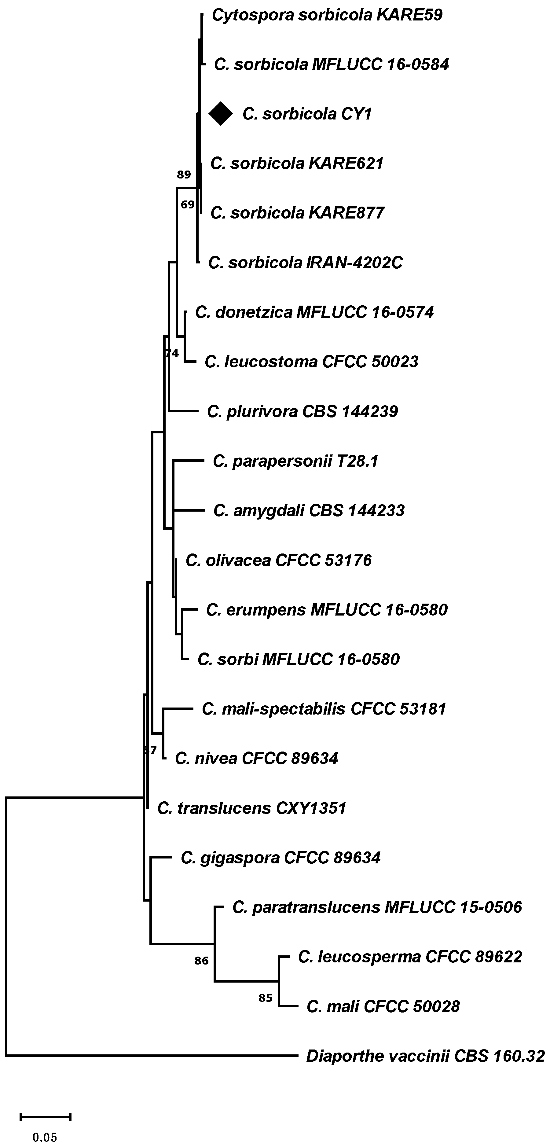
2.2. Molecular Identification of Cyt. sorbicola

Three-day-old isolates of Cytospora sp. generated from a single conidia were used for species identification. Genomic DNA was isolated using the DNeasy Plant Mini Kit following the manufacturer’s instructions (QIAGEN, Hilden, Germany). The Internal Transcribed Spacer (ITS) region and gene fragments of Actin (ACT) and Translation elongation factor 1-alpha (TEF-1α) were amplified employing the primers ITS1F/ITS4, ACT512F/ACT-783R, and EF1-728/EF1-986, respectively [26,27]. The PCR mix was composed of 12 ng genomic DNA, 1X GoTaq® Green Master Mix (Promega, Madison, WI, USA), and 100 nM of each primer; 20 μL volume was completed with nanopure water (Promega, Madison, WI, USA). The PCR conditions consisted of a 5 min denaturation step at 95 °C, followed by 40 cycles of 30 s at 95 °C, 30 s at 55 °C, and 30 s at 72 °C for ITS; 40 s at 95 °C, 35 s at 55 °C, and 1 min at 72 °C for ACT; and 40 s at 95 °C, 30 s at 52 °C, and 1 min at 72 °C for TEF-1α, with a final extension of 7 min at 72 °C. The PCR product was evaluated in an agarose gel (Lafken, Fermelo Biotec, Santiago, Chile), purified, and used for sequencing (Psomagen, Rockville, MD, USA). Sequences were analyzed in the BioEdit software version 7.2.5 [28] to obtain consensus sequences for each gene fragment. Then, each group of sequences was aligned through the ClustalW program integrated into the BioEdit software and refined independently under the G-Block algorithm, conditioning the less strict block option to establish the most informative regions for subsequent phylogenetic analysis [29]. The regions were concatenated manually. Then, phylogenetic analyses were conducted in MEGAX [30]. Specifically, the maximum likelihood method, the general time-reversible nucleotide substitution model (GTR model), gamma distribution, and a Bootstrap of 1000 repetitions were used. The analysis included sixteen species (Table 1), and the tree was rooted (out-group) to Diaporthe vaccinii (CBS 160.32) [31].

2.3. Growth and Cultivation Conditions of Cyt. sorbicola and Cal. pulchella

Cyt. sorbicola CY1 and Cal. pulchella CpL01 isolates were grown on a PDA medium at 20 °C in continuous darkness. Cultures were refreshed every 15 days by transferring 5 mm diameter sections from the active growth zone of the colony to plates with fresh PDA medium.

2.4. In Planta Infection Assays

For the Cyt. sorbicola infection assay, 20 sweet cherry trees of the Santina, Lapins, Bing, and Regina varieties were acclimatized for six months in the O’Higgins region (34°42′51.3″ S 71°15′11.2″ W). All plants used were one year old and 1.5 m tall. The trees were cut down to 80 cm and inoculated with the infection or control treatment. The plants were evaluated after six months. A volume of 10 µL at a concentration of 3 × 10⁵/mL conidia was inoculated into the wounds generated in the plant material. As a control, 10 µL of sterile distilled water was inoculated. For each variety, ten plants were used for the control treatment and ten for the pathogen infection. Vaseline was used to cover the wound after the treatments, preventing nonspecific infections and moisture loss. The wound was then sealed with parafilm.
For the Cal. pulchella infection assay, 20 cm long branches were obtained from independent plants, 20 for each variety of sweet cherry tree under study. A wound of 60 mm in diameter was produced under the bark, where the infection or control treatment was inoculated. The infection and control treatment were generated using the same methodology previously described for Cyt. sorbicola. The inoculated branches were kept in a humid chamber at 25 °C. The assay was evaluated after one month.

2.5. Design and Validation of the Cyt. sorbicola and Cal. pulchella Identification System by PCR-HRM

The ITS region of Cyt. sorbicola isolates obtained in this study and those of Cal. pulchella previously obtained by our group were aligned in the BioEdit software [28]. The ITS regions of the isolates described in the first report of Cyt. sorbicola in sweet cherry trees and Cal. pulchella in peach trees from Chile were included [4,32]. Conserved DNA segments between the ITS sequences of both species were examined manually. Primers were designed based on the ITS-conservated regions (Table 1), and two different PCR-HRM strategies were conceived. First, a universal primer system (Calo-Cytos For(ITS) and Calos-Cytos Rev(ITS) primers) was used to detect Cyt. sorbicola and Cal. pulchella simultaneously. Alternatively, a system using a universal forward primer (Calo-Cytos_For-univ1(ITS)) with pathogen-specific reverse primers (Cyto_Rev-esp(ITS) and Calo_Rev-esp(ITS)) was used. In the last strategy, the primer pairs produced DNA fragments of different sizes: 114 and 85 bp for Cyt. sorbicola and Cal. pulchella, respectively. The primer sequences used are detailed in Table 2. The annealing temperature for the designed primers was determined using temperature-gradient PCR in a T-100 model thermocycler (BioRad, Hercules, CA, USA). Then, the sensitivity and efficiency of the primers were determined using a DNA concentration gradient, where the primers reached an efficiency of 99%, defined through the LinRegPCR software version 2021.2 [33]. The PCR mix was composed of 12 ng genomic DNA, 1X SensiFast Master Mix containing the third-generation fluorescent dye EvaGreen (Bioline, Meridian Bioscience, Cincinnati, OH, USA), and 100 nM of each primer; the volume was completed with nanopure water (Promega, Madison, WI, USA). The PCR protocol was performed in a 72-well carousel in a Rotor-Gene Q 2plex (QIAGEN) PCR-HRM thermocycler (QIAGEN, Hilden, Germany). A fast PCR protocol was used with the following parameters: (i) For the universal primers system, an initial denaturing step of 95 °C for 5 min was used, followed by 25 cycles at 95 °C for 30 s and 64 °C for 30 s. HRM was performed from 80.5 to 85.5 °C, rising by 0.05 °C in each step. (ii) For pathogen-specific reverse primers, an initial denaturing step of 95 °C for 5 min was used, followed by 30 cycles at 95 °C for 30 s and 62 °C for 30 s. HRM was performed from 76 to 80 °C, rising by 0.05 °C in each step. Both protocols considered the default parameters of 2 s hold time for each step and 90 s pre-melt conditioning on the first step. The Rotor-Gene Q Series software version 2.3.5 was used for data analysis.

2.6. Recovery of Cyt. sorbicola and Cal. pulchella from Wood Pieces

Wood fragments of 5 mm2 were collected from the area adjacent to the necrosis developed in plants infected with Cyt. sorbicola and Cal. pulchella. Wood pieces were also obtained from plants under control treatment. For each variety, 60 pieces were generated in total. Pieces were disinfected in a 1.5% sodium hypochlorite solution for 1 min, flamed for 15 s, and plated on 60 mm Petri dishes containing 0.5 g/L streptomycin-amended PDA medium. After four days, fungal mycelium growing out of the wood pieces were collected. Then, the DNA was isolated, and the PCR-HRM pathogen-specific reverse primer system was utilized.

2.7. Statistical Analysis

Statistical analyses were conducted using ANOVA and the Tukey post hoc test in the InfoStat software 2017 version [34]. Differences between PCR-HRM profiles were determined using the Rotor-Gene Q Series software version 2.3.5 (QIAGEN, Hilden, Germany) with a closed threshold at 98% confidence. Data were plotted in GraphPad Prism v.9.4.0 (San Diego, CA, USA).

3. Results

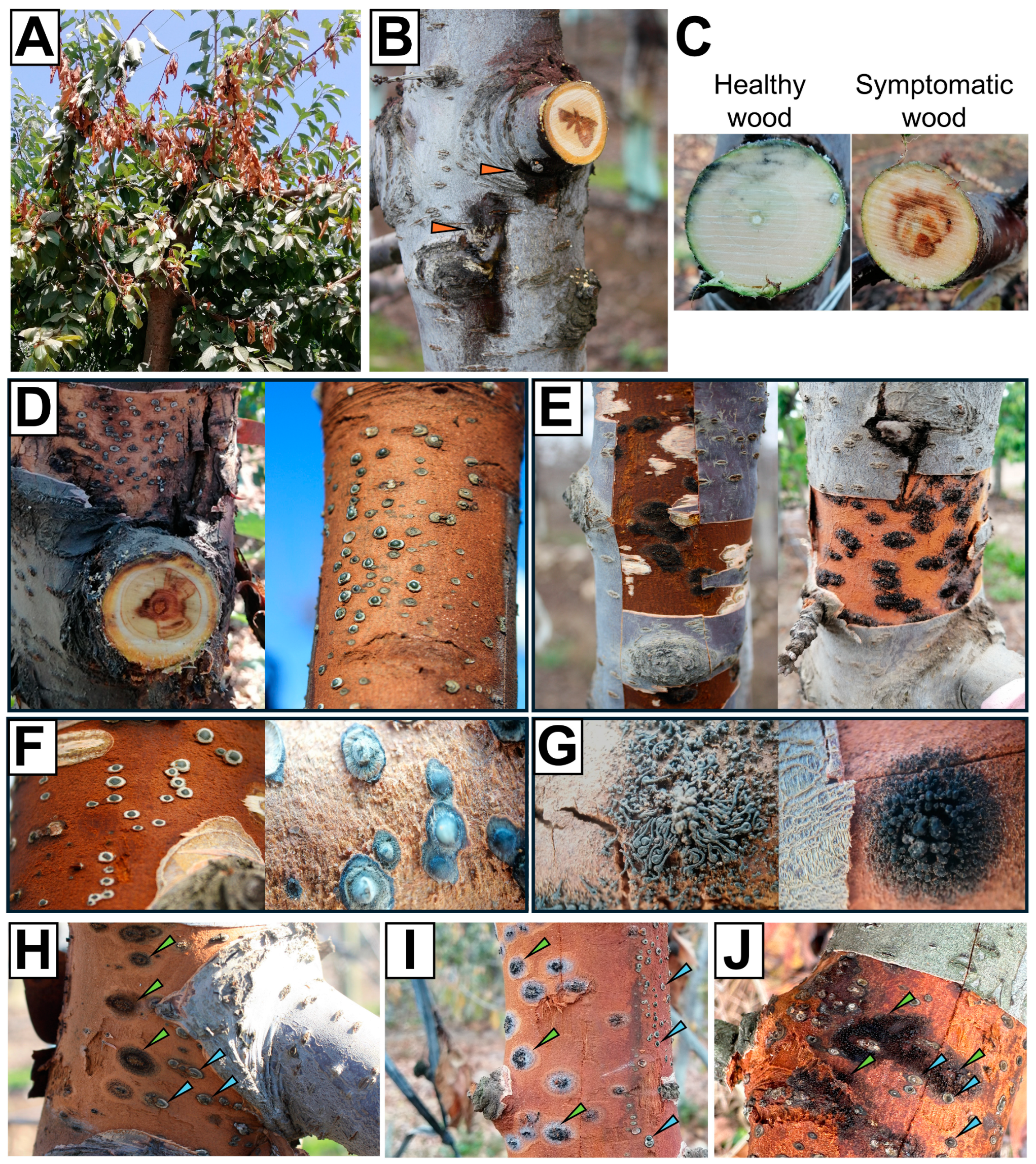
3.1. Cyt. sorbicola and Cal. pulchella Are Widespread in Sweet Cherry Orchards from the Chilean Central Valley

The sweet cherry orchards (n = 3 per variety) surveyed in the Central Valley of Chile exhibited at least one tree with symptoms associated with wood decay fungi with signs of Cytospora sp. or Calosphaeria sp. Wood necrosis, along with exudates or gummosis in some cases, was noticed during the growing season in branch dieback (Figure 1A,B). The wood showed browning and necrosis in the pith, affecting different tissues to varying degrees. Healthy wood, on the other hand, did not exhibit oxidation or browning (Figure 1C). Cytospora sp. pycnidia (Figure 1D,F) or Calosphaeria sp. perithecia (Figure 1E,G) could be observed beneath the bark in symptomatic trees. Notably, Cytospora sp. and Calosphaeria sp. could coexist within the same plant. Pycnidia and perithecia were observed occupying neighboring domains of the plant trunk (Figure 1H–J). Cytospora sp. isolated from symptomatic wood was identified as Cyt. sorbicola by multilocus molecular phylogeny (MMF) (n = 3) with a three-locus data set (ITS-ACT-TEF-1α) (Figure 2). For this study, we utilized the Cyt. sorbicola isolate called CY1, identified by MMF, and the Cal. pulchella CpL01 isolate, previously characterized by Auger et al. (2020) [1], from the same geographic location.

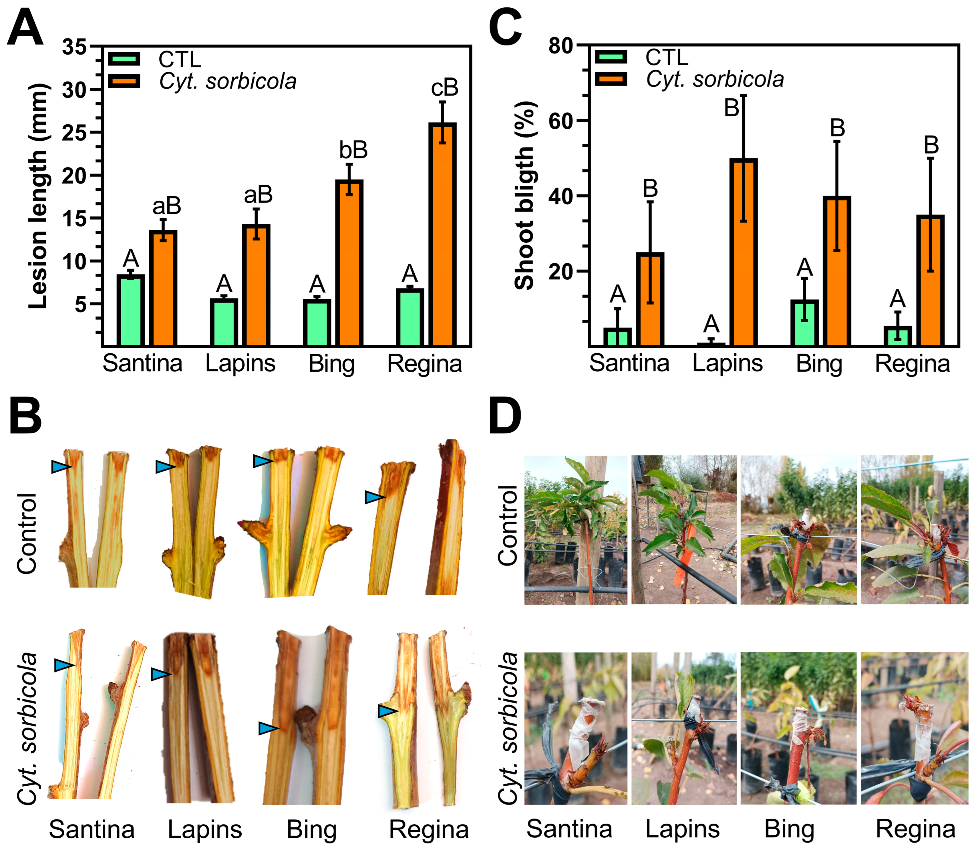
3.2. Regina Is the Sweet Cherry Variety Most Susceptible to Cyt. sorbicola and Cal. pulchella

One-year-old sweet cherry trees inoculated with Cyt. sorbicola developed wood necrosis. The pathogen-induced necrosis was significantly higher than the control condition in each sweet cherry variety evaluated (Figure 3); however, the response between sweet cherry genotypes was heterogeneous. The lesion length generated by Cyt. sorbicola was significantly greater in Regina and Bing, with average lesion sizes of 26.14 and 19.51 mm, respectively (Figure 3). The observed necrosis in Santina and Lapins averaged 13.6 and 14.1 mm, respectively, significantly lower than the remaining evaluated varieties but 39% and 61% greater than the control treatment. The evaluated sweet cherry varieties responded similarly to mechanical damage stress, as shown by the lesion length under control treatment.
Plant top remotion leads to the loss of apical dominance, prompting lateral sprouting. Shoot and twig blight is a common symptom of plants infected with wood decay fungi. Hence, the death of new shoots was assessed following the Cyt. sorbicola infection. Shoot blight was significantly higher in all infected plants compared to the control treatment, regardless of the sweet cherry variety (Figure 3). Lateral buds developed smaller leaves that brown and split early throughout the whole plant. Notably, the shoot blight percentage was statistically equivalent among the sweet cherry varieties in the study, suggesting a conserved program of lateral organ development in response to infection (Figure 3).
Necrosis caused by Cal. pulchella was significantly more extensive than the control treatment. On average, the lesion caused by the pathogen was four times bigger than the control treatment (Figure 4), exceeding the 2.8-fold average observed by Cyt. sorbicola infection. Remarkably, the mechanical wound caused by the control treatment was the same for both experimental conditions: 6.612 vs. 6.613 mm on average. The sweet cherry varieties Lapins and Bing exhibited minor necrosis caused by Cal. pulchella, measuring 21.6 and 22.8 mm, respectively. Necrosis increased significantly in the Santina variety with 27.62 mm. However, the most severe necrosis recorded within the group of sweet cherry tree varieties studied was 30.8 mm on average in the Regina variety (Figure 4).
Therefore, the Regina variety was consistently the most susceptible to fungi that cause wood cankers. The Santina variety was an exceptional case, with a lower susceptibility to Cyt. sorbicola but a wound equivalent to that of the Regina variety by Cal. pulchella, suggesting a differential response mechanism for these pathogens. Meanwhile, the Lapins variety was less susceptible to Cyt. sorbicola and Cal. pulchella. The Bing variety, in contrast, showed an intermediately high susceptibility among the studied sweet cherry genotypes.

3.3. Implementation of a System for Cyt. sorbicola and Cal. pulchella Identification by PCR-HRM

The identification of wood-colonizing fungi presents several challenges. In situ detection depends on DNA isolation, restricted by the wood’s lignin and other phenolic compounds and the underrepresentation of fungal DNA within the sample. In vitro isolation requires long periods for the fungus to grow and develop spores and/or reproductive structures that allow its morphological recognition. Furthermore, finding multiple wood-living fungi within the same host is possible. Indeed, Cyt. sorbicola and Cal. pulchella can coexist on the same sweet cherry tree (Figure 5). Hence, a PCR-HRM strategy was designed to identify Cyt. sorbicola and Cal. pulchella, reducing diagnosis time.
Cyt. sorbicola and Cal. pulchella are phylogenetically distant, belonging to Cytosporaceae (order Diaporthales) and Calosphaeriaceae (order Calosphaeriales), respectively. However, exploring the Internal Transcribed Spacer (ITS) genomic region from both pathogens allowed the design of two different PCR-HRM strategies based on conserved DNA regions (Figure 5). The Calo-Cytos For(ITS) and Calos-Cytos Rev(ITS) universal primers amplified a 137 bp fragment from the ITS region. The nucleotide content of the fragment was species-specific, with 47.45% of CG and 52.55% of AT for Cyt. sorbicola and 49.64% of CG and 50.36% of AT for Cal. pulchella (Figure 5A). The second strategy, which employed species-specific reverse primers, was developed to verify and strengthen the diagnosis results. The forward primer Calo-Cytos_For-univ1(ITS) with the Cyt. sorbicola-specific reverse primer Cyto_Rev-esp(ITS) generated a 114 bp fragment, while the Cal. pulchella-specific reverse primer Calo_Rev-esp(ITS) generated an 85 bp fragment from the ITS region. Each fragment differed in nucleotide content; the Cyt. sorbicola fragment contained 36.84% of CG and 63.16% of AT, and the Cal. pulchella fragment contained 42.35% of CG and 57.65% of AT (Figure 5B). Differences in nucleotide content are critical to the success of the PCR-HRM strategy.
The primer pairs designed for each strategy (universal or reverse-specific primers) amplified the expected fragments from Cyt. sorbicola and Cal. pulchella DNA, even from the mixture of DNA of both species (Cyt. Sorbicola + Cal. pulchella) (Figure 6A,B). None of the designed systems was able to amplify a DNA fragment from the genomic DNA of cosmopolitan microorganisms frequently found in Prunus spp. wood, such as Stereum hirsutum and Chondrostereum purpureum (Figure 6A,B). The ITS region of each microorganism tested was amplified using the ITS1F and ITS4 primers as a DNA amplification control (Figure 6A). Therefore, the amplification was specific to Cyt. sorbicola and Cal. pulchella.
The PCR-HRM analysis produced three distinct curves individualizing Cyt. sorbicola, Cal. pulchella, and Cyt. sorbicola + Cal. pulchella (Figure 6C,D). The differences between the profiles of Cyt. sorbicola and Cyt. sorbicola + Cal. pulchella were more evident under the reverse-specific primer strategy under the normalized fluorescence criteria (Figure 6D). However, the difference graph generated using the Cal. pulchella profile as the baseline in universal or reverse-specific primer strategies revealed that Cyt. sorbicola and Cyt. sorbicola + Cal. pulchella profiles deviated significantly (Figure 6E,F), indicating that the profiles were indeed different and facilitating the discrimination of Cyt. sorbicola and Cal. pulchella.

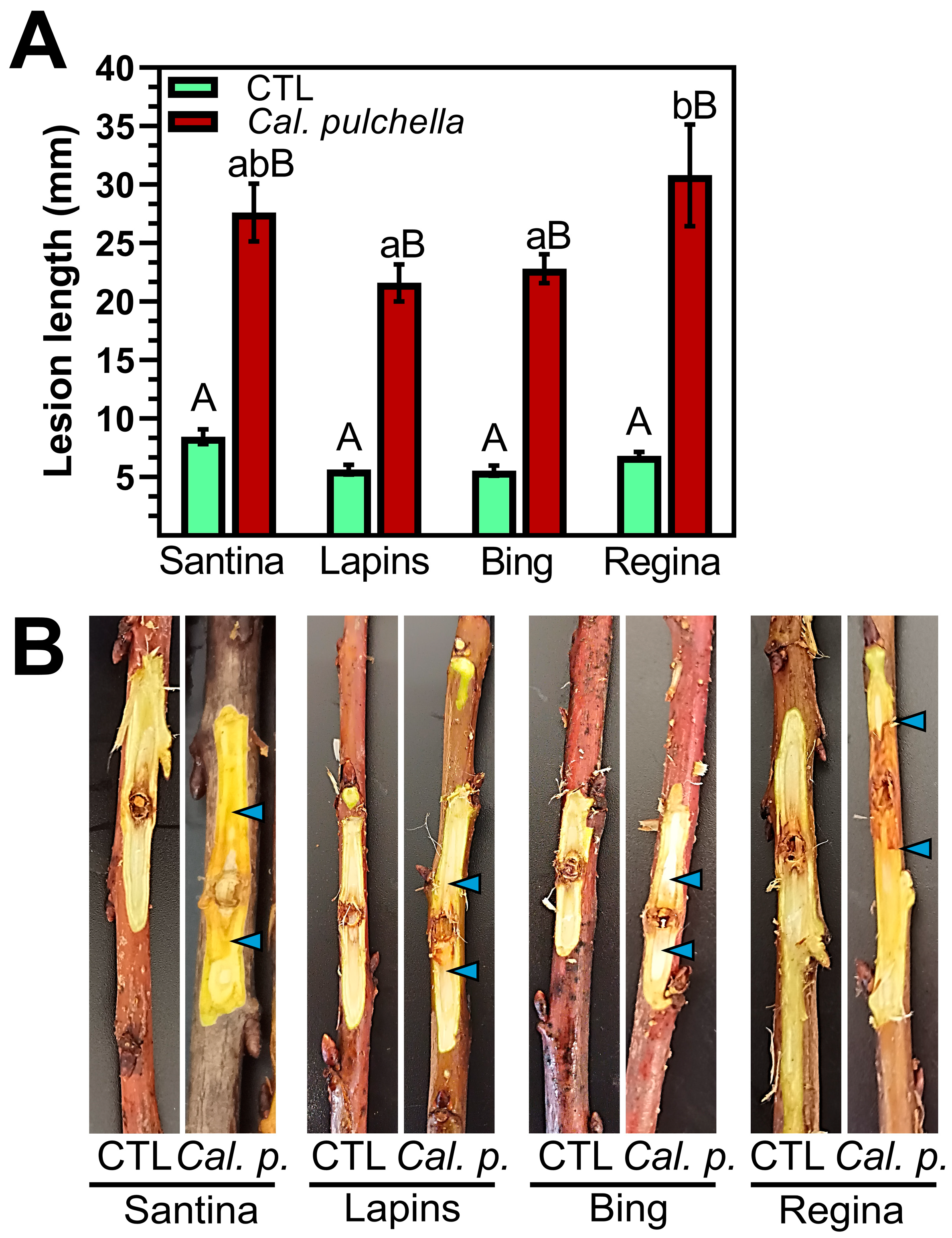
3.4. Cal. pulchella Colonizes Sweet Cherry Wood More Effectively than Cyt. sorbicola

Wood pieces (n = 60) were recovered from the area adjacent to the necrosis of each plant infected with Cyt. sorbicola or Cal. pulchella to evaluate the pathogen’s development. After four and seven days of incubation, fungal growth was recovered from the wood and analyzed by PCR-HRM with the reverse-specific primer strategy. In the studied time frame, the mycelium observed growing from the wood pieces did not have morphological characteristics allowing species identification. Cyt. sorbicola was less frequently recovered in the Santina and Regina sweet cherry varieties, with average values of 18.5% and 18.3%, respectively, on day seven (Table 3). In contrast, Cyt. sorbicola recovery from Lapins and Bing wood roughly doubled over the same period: 33.3% and 36.7% on average, respectively (Table 3). The recovery of Cal. pulchella was higher than Cyt. sorbicola in all sweet cherry varieties evaluated, with average percentages over 50% from day four of analysis (Table 3). In the Santina variety, the recovery of Cal. pulchella was lower on day seven, with 63.2% on average, followed by Regina, Bing, and Lapins, with 72.5%, 79.1%, and 89.7%, respectively (Table 3). Remarkably, the growth of Cyt. sorbicola and Cal. pulchella was slowed by 20% when comparing growth on days four to seven on wood of the Santina variety (Table 3). None of the pathogens were identified from the control wood, confirming the plant health of the material used in the experiments.

4. Discussion

The sweet cherry canker fungi Cyt. sorbicola and Cal. pulchella are a threat to the industry worldwide. Signs of Cyt. sorbicola and Cal. pulchella are widespread in sweet cherry orchards in the Chilean Central Valley, indicating that the implemented control measures are ineffective. Our work showed that Cyt. sorbicola and Cal. pulchella can be found in young or mature orchards and even coexist in the same cherry tree. This potential for coexistence adds another layer of challenge to managing these pathogens.
The genetics of sweet cherry tree varieties determine the fruit’s organoleptic characteristics and relevant agronomic traits [35,36]. In addition, the genetic background of each variety determines differential responses to abiotic and biotic stress [37,38,39]. The susceptibility of sweet cherry and other Prunus spp. has been evaluated for a limited group of economically relevant bacteria and fungi, including Pseudomonas syringae pv. syringae (Pss), Erwinia amylovora, and Diaporthe amygdali; however, these works show that the susceptibility depends on the species and the variety/genotype tested [12,40,41]. Coherently, our research showed that different sweet cherry tree varieties have variable susceptibility to Cyt. sorbicola and Cal. pulchella, and the susceptibility extent determined by wood necrosis is specific to each pathogen. It is noteworthy that the Lapins variety, which was less susceptible to wood decay fungi in our study, is highly vulnerable to the bacteria E. amylovora. Conversely, the Regina variety, the most prone to infection by Cyt. sorbicola and Cal. pulchella, is actually the most resistant to the Pss bacteria. Bacterial and fungal perception occurs through plant differential systems [42,43,44]. Our results are consistent with differential mechanisms conditioning susceptibility to pathogenic fungi and bacteria in cherry trees. Alternatively, signaling pathways relevant to bacteria and fungi may have segregated independently during the selection of sweet cherry tree varieties, favoring even the functional selection of more aggressive or pathogenic isolates. Although this hypothesis needs to be validated, it is an exciting avenue of research to be undertaken by breeders of sweet cherry varieties. This topic is crucial for cherry production in Chile, where Pss is as relevant as, or even more than, wood decay fungi from a phytosanitary perspective [45,46]. Furthermore, the climate crisis has caused changes in the optimal agroclimatic conditions for crops, leading to increased biotic stress in plants. A recent study demonstrated that cherry trees adjust their response to heat stress, negatively impacting their defense response to Pss [47]. Therefore, future works should thoroughly review the sweet cherry tree response to fungal and bacterial pathogens under stress conditions to align with the new field reality.
Morphological analysis is the most commonly used strategy to identify Cytospora spp. and Calosphaeria spp. The time needed to develop key morphological characteristics by wood microorganisms in vitro leads to diagnoses that fall outside the time required to construct effective management programs. Molecular biology methods have been developed for Cytospora spp., including PCR-pathogen-specific, ddPCR, and LAMP methods, but not for Calosphaeria spp. [17,18,19]. Our research designed and developed a PCR-HRM assay for simultaneous detection of Cyt. sorbicola and Cal. pulchella. The three genotypes tested (Cyt. sorbicola, Cal. pulchella, and Cyt. sorbicola + Cal. Pulchella) provided melting profiles with distinct and consistent shapes, enabling easy discrimination between them. Nucleotide content, particularly the GC/AT ratio, determines the PCR-HRM design’s success [20,48,49]. Our strategies using universal primers or specific reverse primers were successful. Indeed, the specific reverse primer strategy generated melting profiles with greater differences, correlating with larger GC/AT ratio variations between the designed Cyt. sorbicola and Cal. pulchella fragments. Diagnosis depends on obtaining mycelium from four-day-old wood pieces, allowing fungi identification independent of the plant species. For example, Cal. pulchella has recently been identified as a cause of wood necrosis in peach trees in Chile [30]. Contaminants (other fungi) do not cause failures in the diagnostic system. Together, these tests can be completed in four days, significantly less than the morphology-based analyses that require between two and four weeks. Finally, this PCR-HRM diagnostic system can be used as a scaffold to include other microorganisms. Other species closely related to Cytospora that have been reported as problems for other Prunus spp. can be easily identified due to the conservation in the ITS region.
The PCR-HRM technique allowed us to evaluate the frequency of each pathogen in infected sweet cherry plants. Notably, recovery of Cyt. sorbicola and Cal. pulchella from wood samples showed no positive correlation between sweet cherry tree susceptibility and pathogen growth. However, the results revealed that Cal. pulchella has inherent advantages over Cyt. sorbicola in colonizing wood, suggesting a higher permanence in pruning remains.
Our study reveals the differential susceptibility of sweet cherry tree varieties to Cyt. sorbicola and Cal. pulchella. The differential responses recorded depend on the plant genotype; therefore, specific control programs for each pathogen are required. Future studies should address the coexistence of Cyt. sorbicola and Cal. pulchella, including the plant defense response and changes in the aggressiveness of the fungi as a result of sharing the host. The PCR-HRM tool developed and validated in this study will enable decisions based on the pathogen’s presence in the field. In addition, the tool allows the orchard’s health status or nursery material to be checked.

Author Contributions

Conceptualization, J.A., M.E. and C.O.-N.; methodology, J.A., M.E. and C.O.-N.; software, C.O.-N.; validation, C.O.-N., C.S., F.D., P.R.-B., M.R., M.A., V.E., M.E. and J.A.; formal analysis, C.O.-N., M.R., M.E. and J.A.; investigation, C.O.-N., C.S., F.D., P.R.-B., M.R., M.A. and V.E.; data curation, C.O.-N.; writing—original draft preparation, C.O.-N.; writing—review and editing, J.A., M.E., M.R. and C.O.-N.; supervision, J.A., M.E. and C.O.-N.; project administration, M.E.; funding acquisition, M.E. All authors have read and agreed to the published version of the manuscript.

Funding

This research received no external funding.

Data Availability Statement

Data are the property of the Laboratory of Fruit and Molecular Phytopathology of the Faculty of Agronomic Sciences, University of Chile, Santiago, Chile. Any requests should be directed to Marcela Esterio (Principal Investigator).

Conflicts of Interest

The authors declare no conflicts of interest.

References

  1. Auger, J.; Pozo, L.; Rubilar, M.; Briceño, N.; Osorio-Navarro, C.; Esterio, M. First Report of canker and branch dieback of sweet cherry trees caused by Calosphaeria pulchella in Chile. Plant Dis. 2020, 105, 217. [Google Scholar] [CrossRef]
  2. Lawrence, D.P.; Holland, L.A.; Nouri, M.T.; Travadon, R.; Abramians, A.; Michailides, T.J.; Trouillas, F.P. Molecular phylogeny of Cytospora species associated with canker diseases of fruit and nut crops in California, with the descriptions of ten new species and one new combination. IMA Fungus 2018, 9, 333–370. [Google Scholar] [CrossRef]
  3. Li, S.; Travadon, R.; Trouillas, F.P. Seasonal Susceptibility of Sweet Cherry Pruning Wounds to Calosphaeria pulchella, Cytospora sorbicola, and Eutypa lata. Plant Dis. 2023, 107, 3517–3522. [Google Scholar] [CrossRef]
  4. Muñoz-Reyes, V.; Castro, J.F.; Cisterna-Oyarce, V.; Santelices, C.; Carrasco, J.; Guerra, M.; France, R.A.; Claret, M.; Millas, P. First report of canker caused by Cytospora sorbicola on Sweet Cherry (Prunus avium) in Chile. Plant Dis. 2023, 108, 524. [Google Scholar] [CrossRef]
  5. Li, S.; Travadon, R.; Nouri, M.T.; Trouillas, F.P. Determining the Main Infection Courts in Sweet Cherry Trees of the Canker Pathogens Calosphaeria pulchella, Cytospora sorbicola and Eutypa lata. Plant Dis. 2024, 108, 1695–1702. [Google Scholar] [CrossRef]
  6. Pan, M.; Zhu, H.; Bonthond, G.; Tian, C.; Fan, X. High Diversity of Cytospora Associated With Canker and Dieback of Rosaceae in China, With 10 New Species Described. Front. Plant Sci. 2020, 11, 690. [Google Scholar] [CrossRef]
  7. Trouillas, F.P.; Peduto, F.; Lorber, J.D.; Sosnowski, M.R.; Grant, J.; Coates, W.W.; Anderson, K.K.; Caprile, J.; Gubler, W.D. Calosphaeria Canker of Sweet Cherry Caused by Calosphaeria pulchella in California and South Australia. Plant Dis. 2012, 96, 648–658. [Google Scholar] [CrossRef]
  8. iQonsulting. Sweet Cherry Book 2024; iQonsulting: Santiago, Chile, 2024; Available online: http://www.iqonsulting.com/yb/ (accessed on 6 August 2024).
  9. Oficina de Estudios y Políticas Agrarias (ODEPA). Informe Superficie Frutícola Regional 2024. Available online: https://www.odepa.gob.cl/estadisticas-del-sector/catastros-fruticolas (accessed on 6 August 2024).
  10. De Salvador, F.R.; Pititto, A.; Giorgioni, M.; Folini, L.; Bassi, G.; Longo, L. Performance of ‘Lapins’ sweet cherry on several rootstocks in Italy. Acta Hortic. 2008, 795, 311–316. [Google Scholar] [CrossRef]
  11. Csihon, Á.; Bicskei, K.; Dremák, P.; Gonda, I. Evaluation of the growing and fruit bearing characteristics of the ‘Lapins’ sweet cherry cultivar grafted on rootstocks with different vigor. Int. J. Hortic. Sci. 2017, 23, 15–18. [Google Scholar] [CrossRef]
  12. Marroni, M.V.; Casonato, S.; Pitman, A.R.; Visnovsky, S.B.; Beresford, R.M.; Jones, E.E. Review of Pseudomonas species causing bacterial canker of Prunus species with emphasis on sweet cherry (Prunus avium) in New Zealand. Eur. J. Plant Pathol. 2024, 168, 297–314. [Google Scholar] [CrossRef]
  13. Travadon, R.; Lawrence, D.P.; Li, S.; Trouillas, F.P. Evaluation of Biological Control Agents for the Protection of Almond Pruning Wounds Against Infection by Fungal Canker Pathogens. Phytopathology 2023, 113, 1417–1427. [Google Scholar] [CrossRef]
  14. Holland, L.A.; Travadon, R.; Lawrence, D.P.; Nouri, M.T.; Trouillas, F. Evaluation of Pruning Wound Protection Products for the Management of Almond Canker Diseases in California. Plant Dis. 2021, 105, 3368–3375. [Google Scholar] [CrossRef]
  15. Travadon, R.; Lawrence, D.P.; Li, S.; Trouillas, F.P. Field evaluation of biological wound treatments for the management of almond, cherry, and grapevine fungal canker diseases. Biol. Control. 2023, 185, 105292. [Google Scholar] [CrossRef]
  16. Li, S.; Travadon, R.; Trouillas, F.P. Effects of Temperature on Spore Germination and Mycelial Growth of Calosphaeria pulchella, Cytospora sorbicola, and Eutypa lata Isolates Associated with Sweet Cherry Canker Diseases. Plant Dis. 2023, 107, 3448–3456. [Google Scholar] [CrossRef]
  17. Avenot, H.F.; Jaime-Frias, R.; Travadon, R.; Holland, L.A.; Lawrence, D.P.; Trouillas, F.P. Development of PCR-Based Assays for Rapid and Reliable Detection and Identification of Canker-Causing Pathogens from Symptomatic Almond Trees. Phytopathology 2022, 112, 1710–1722. [Google Scholar] [CrossRef]
  18. Stewart, J.E.; Miller, S.T.; Zink, F.A.; Caballero, J.I.; Tembrock, L.R. Genetic and Phenotypic Characterization of the Fungal Pathogen Cytospora plurivora from Western Colorado Peach Orchards and the Development of a ddPCR Assay for Detection and Quantification. Phytopathology 2022, 112, 917–928. [Google Scholar] [CrossRef]
  19. Xu, L.; Wang, Y.; Zhu, S.; Li, J.; Chang, Y.; Huang, L. Development and Application of a LAMP Assay for the Detection of the Latent Apple Tree Pathogen Valsa mali. Plant Dis. 2021, 105, 1065–1071. [Google Scholar] [CrossRef]
  20. Słomka, M.; Sobalska-Kwapis, M.; Wachulec, M.; Bartosz, G.; Strapagiel, D. High Resolution Melting (HRM) for High-Throughput Genotyping—Limitations and Caveats in Practical Case Studies. Int. J. Mol. Sci. 2017, 18, 2316. [Google Scholar] [CrossRef]
  21. Tong, S.Y.; Giffard, P.M. Microbiological applications of high-resolution melting analysis. J. Clin. Microbiol. 2012, 50, 3418–3421. [Google Scholar] [CrossRef]
  22. Chatzidimopoulos, M.; Ganopoulos, I.; Moraitou-Daponta, E.; Lioliopoulou, F.; Ntantali, O.; Panagiotaki, P.; Vellios, E.K. High-Resolution Melting (HRM) Analysis Reveals Genotypic Differentiation of Venturia inaequalis Populations in Greece. Front. Ecol. Evol. 2019, 7, 489. [Google Scholar] [CrossRef]
  23. Ratti, M.F.; Farrer, R.A.; Cano, L.M.; Faedda, R.; Goss, E.M. Evaluation of High-Resolution Melting for Rapid Differentiation of Phytophthora Hybrids and Their Parental Species. Plant Dis. 2019, 103, 2295–2304. [Google Scholar] [CrossRef]
  24. Wang, N.Y.; Gama, A.B.; Marin, M.V.; Peres, N.A. Development of a Multiplex High-Throughput Diagnostic Assay for the Detection of Strawberry Crown Rot Diseases Using High-Resolution Melting Analysis. Phytopathology 2021, 111, 1470–1483. [Google Scholar] [CrossRef]
  25. Druml, B.; Cichna-Markl, M. High resolution melting (HRM) analysis of DNA—its role and potential in food analysis. Food Chem. 2014, 158, 245–254. [Google Scholar] [CrossRef]
  26. White, T.J.; Bruns, T.D.; Lee, S.B.; Taylor, J.W. Amplification and Direct Sequencing of Fungal Ribosomal RNA Genes for Phylogenetics. In PCR Protocols: A Guide to Methods and Applications; Innis, M.A., Gelfand, D.H., Sninsky, J.J., White, T.J., Eds.; Academic Press: New York, NY, USA, 1990; pp. 315–322. [Google Scholar] [CrossRef]
  27. Carbone, I.; Kohn, L.M. A method for designing primer sets for speciation studies in filamentous ascomycetes. Mycologia 1999, 91, 553–556. [Google Scholar] [CrossRef]
  28. Hall, T.A. BioEdit: A user-friendly biological sequence alignment editor and analysis program for Windows 95/98/NT. Nucleic Acids Symp. Series. 1999, 41, 95–98. [Google Scholar]
  29. Talavera, G.; Castresana, J. Improvement of phylogenies after removing divergent and ambiguously aligned blocks from protein sequence alignments. Syst. Biol. 2007, 56, 564–577. [Google Scholar] [CrossRef]
  30. Kumar, S.; Stecher, G.; Li, M.; Knyaz, C.; Tamura, K. MEGA X: Molecular Evolutionary Genetics Analysis across Computing Platforms. Mol. Biol. Evol. 2018, 35, 1547–1549. [Google Scholar] [CrossRef]
  31. Hanifeh, S.; Zafari, D.; Soleimani, M.J.; Arzanlou, M. Multigene phylogeny, morphology, and pathogenicity trials reveal novel Cytospora species involved in perennial canker disease of apple trees in Iran. Fungal Biol. 2022, 126, 707–726. [Google Scholar] [CrossRef]
  32. Grinbergs, D.; Chilian, J.; Isla, M.; Otarola, J. First report of Calosphaeria pulchella causing cankers in peach (Prunus persica) in Chile. Plant Dis. 2023, 107, 3314. [Google Scholar] [CrossRef]
  33. Untergasser, A.; Ruijter, J.M.; Benes, V.; van den Hoff, M.J.B. Web-based LinRegPCR: Application for the visualization and analysis of (RT)-qPCR amplification and melting data. BMC Bioinform. 2021, 22, 398. [Google Scholar] [CrossRef]
  34. Di Rienzo, J.A.; Casanoves, F.; Balzarini, M.G.; González, L.; Tablada, M.; Robledo, C.W. InfoStat; InfoStat Group, FCA: London, UK; Universidad Nacional de Córdoba: Córdoba, Argentina, 2015. [Google Scholar]
  35. Takácsné Hájos, M.; Nyéki, J.; Veres, E.; Fieszl, C.; Szabó, Z. Organoleptic evaluation of sweet cherry varieties. Int. J. Hortic. Sci. 2011, 17, 7–13. [Google Scholar] [CrossRef]
  36. Vavoura, M.V.; Badeka, A.V.; Kontakos, S.; Kontominas, M.G. Characterization of four popular sweet cherry cultivars grown in Greece by volatile compound and physicochemical data analysis and sensory evaluation. Molecules 2015, 20, 1922–1940. [Google Scholar] [CrossRef]
  37. Measham, P.F.; Quentin, A.G.; MacNair, N. Climate, winter chill, and decision-making in sweet cherry production. HortScience 2014, 49, 254–259. [Google Scholar] [CrossRef]
  38. Mgbechi-Ezeri, J.; Porter, L.; Johnson, K.B.; Oraguzie, N. Assessment of sweet cherry (Prunus avium L.) genotypes for response to bacterial canker disease. Euphytica 2017, 213, 145. [Google Scholar] [CrossRef]
  39. Garrett, C.M.E. Screening Prunus rootstocks for resistance to bacterial canker, caused by Pseudomonas morsprunorum. J. Hortic. Sci. Biotechnol. 1979, 54, 189–193. [Google Scholar] [CrossRef]
  40. Beluzán, F.; Miarnau, X.; Torguet, L.; Zazurca, L.; Abad-Campos, P.; Luque, J.; Armengol, J. Susceptibility of Almond (Prunus dulcis) Cultivars to Twig Canker and Shoot Blight Caused by Diaporthe amygdali. Plant Dis. 2022, 106, 1890–1897. [Google Scholar] [CrossRef]
  41. Mohan, S.K.; Bijman, V.P. Susceptibility of Prunus Species to Erwinia amylovora. Acta Hortic. 1999, 489, 145–148. [Google Scholar] [CrossRef]
  42. Dodds, P.N.; Chen, J.; Outram, M.A. Pathogen perception and signaling in plant immunity. Plant cell 2024, 36, 1465–1481. [Google Scholar] [CrossRef]
  43. Trdá, L.; Boutrot, F.; Claverie, J.; Brulé, D.; Dorey, S.; Poinssot, B. Perception of pathogenic or beneficial bacteria and their evasion of host immunity: Pattern recognition receptors in the frontline. Front. Plant Sci. 2015, 6, 219. [Google Scholar] [CrossRef]
  44. Saur, I.M.L.; Hückelhoven, R. Recognition and defence of plant-infecting fungal pathogens. J. Plant Physiol. 2021, 256, 153324. [Google Scholar] [CrossRef]
  45. Correa, F.; Beltrán, M.F.; Millas, P.; Moreno, Z.; Hinrichsen, P.; Meza, P.; Sagredo, B. Genome Sequence Resources of Pseudomonas syringae Strains Isolated from Sweet Cherry Orchards in Chile. Mol. Plant Microbe Interact. 2022, 35, 933–937. [Google Scholar] [CrossRef]
  46. Beltrán, M.F.; Osorio, V.; Lemus, G.; Millas, P.; France, A.; Correa, F.; Sagredo, B. Bacterial community associated with canker disease from sweet cherry orchards of central valley of Chile presents high resistance to copper. Chil. J. Agric. Res. 2021, 81, 378–389. [Google Scholar] [CrossRef]
  47. Villalobos-González, L.; Carreras, C.; Beltrán, M.F.; Figueroa, F.; Rubilar-Hernández, C.; Opazo, I.; Toro, G.; Salvatierra, A.; Sagredo, B.; Pizarro, L.; et al. Sweet Cherry Plants Prioritize Their Response to Cope with Summer Drought, Overshadowing the Defense Response to Pseudomonas syringae pv. syringae. Plants 2024, 13, 1737. [Google Scholar] [CrossRef]
  48. Ririe, K.M.; Rasmussen, R.P.; Wittwer, C.T. Product differentiation by analysis of DNA melting curves during the polymerase chain reaction. Anal. Biochem. 1997, 245, 154–160. [Google Scholar] [CrossRef]
  49. Reed, G.H.; Kent, J.O.; Wittwer, C.T. High-resolution DNA melting analysis for simple and efficient molecular diagnostics. Pharmacogenomics 2007, 8, 597–608. [Google Scholar] [CrossRef]
Figure 1. Symptoms and signs of Cytospora sp. and Calosphaeria sp. in sweet cherry trees. (A). Branch and twig dieback in adult trees during the productive stage. (B). Wood necrosis accompanied by exudations (orange arrowheads). (C). Cross section of healthy and symptomatic branches showing changes in tissue coloration. (D,F). Cytospora sp. signs in adult trees. (E,G). Calosphaeria sp. signs in adult trees. (HJ). Cytospora sp. (light blue arrowheads) and Calosphaeria sp. (green arrowheads) can be observed coexisting on the same host.
Microorganisms 12 02456 g001
Figure 2. Cytospora sorbicola identification by molecular phylogeny. A phylogenetic tree was generated from a maximum-parsimony analysis of three Chilean isolates and 16 Cytospora species. The tree was inferred from a three-locus data set (ITS-ACT-TEF-1α). The numbers above branches represent bootstrap values from 1000 replicates. The diamond indicates a Cyt. sorbicola representative Chilean isolate. Diaporthe vaccinii strain CBS 160.32 was used as an outgroup.
Microorganisms 12 02456 g002
Figure 3. Susceptibility of sweet cherry varieties Santina, Lapins, Bing, and Regina to Cyt. sorbicola. (A,B). Lesions in one-year-old trees were evaluated after six months of control treatment (water) or infection treatment (Cyt. sorbicola conidia suspension). (C,D). The shoot blight resulting from infection treatment was measured. The lesion limit is highlighted by the blue arrowheads in (B). Error bars represent standard error. Different letters represent significant differences according to Tukey’s HSD test (p > 0.05). Upper- and lower-case letters represent independent statistical analyses.
Microorganisms 12 02456 g003
Figure 4. Susceptibility of sweet cherry varieties Santina, Lapins, Bing, and Regina to Cal. pulchella. (A,B). Lesions in branches from one-year-old trees were evaluated after one month of control treatment (water) or infection treatment (Cal. pulchella conidia suspension). The lesion is delimited by the blue arrowheads in (B). Error bars represent standard error. Different letters represent significant differences according to Tukey’s HSD test (p > 0.05). Upper- and lower-case letters represent independent statistical analyses.
Microorganisms 12 02456 g004
Figure 5. Primer design for PCR-HRM assays. (A). The universal detection system employed primers Calo-Cytos For(ITS) and Calos-Cytos Rev(ITS) to produce a 137 bp fragment in Cyt. sorbicola and Cal. pulchella. (C). The pathogen-specific reverse primer system employed the Calo-Cytos_For-univ1(ITS) (blue arrow), Cyto_Rev-esp(ITS) (yellow arrow), and Calo_Rev-esp(ITS) (magenta arrow) primers to obtain fragments of 114 and 85 bp from Cyt. sorbicola and Cal. pulchella DNA, respectively. (B,D). The fragments obtained vary in their nucleotide content.
Microorganisms 12 02456 g005
Figure 6. Cyt. sorbicola and Cal. pulchella detection by PCR-HRM. (A,B). Amplifications by the universal (A) or pathogen-specific reverse primer system (B) were specific to Cyt. sorbicola or Cal. pulchella. Neither system could use DNA from other common wood fungi, such as Stereum hirsutum (S. hirsutum) or Chondrostereum purpureum (C. purpureum). The universal system led to a 137 bp fragment being obtained (A). Amplification of the ITS region from all fungi used was amplified as a positive control (A). The pathogen-specific primer reverse system generated fragments of 114 and 85 pb for Cyt. sorbicola and Cal. pulchella, respectively (B). From agarose gels: 1 = Cyt. sorbicola, 2 = Cal. pulchella, 3 = Cyt. sorbicola + Cal. Pulchella, 4 = S. hirsutum, 5 = C. purpureum, (-) = negative control. (C,D). Normalized HRM profiles of Cyt. sorbicola, Cal. pulchella, and Cyt. sorbicola + Cal. pulchella by the universal (C) or pathogen-specific reverse primer system (D). (E,F). Difference curves of HRM using Cal. pulchella as the reference baseline curve by the universal (E) or pathogen-specific reverse primer system (F).
Microorganisms 12 02456 g006
Table 1. Sequences used in the phylogenetic analysis of Cyt. sorbicola from sweet cherry trees.
SpeciesStrainHostOrigenGenBank Accession Number
ITSACTTEF-1α
Cytospora sorbicolaKARE59Prunus dulcisCalifornia, USAMG971861MG972010MG971572
Cyt. sorbiMFLUCC 16-0631TSorbus aucupariaRusiaKY417752KY417718NA
Cyt. sorbicolaKARE621Prunus aviumCalifornia, USAMG971811MG971961MG971526
Cyt. sorbicolaKARE877Prunus aviumCalifornia, USAMG971825MG971975MG971540
Cyt. sorbicolaIRAN 4202CMalus domesticaIranMW295654MZ014514MW394148
Cyt. donetzicaMFLUCC 16-0574TRosa sp.RusiaKY417731KY417696NA
Cyt. leucostomaCFCC 50023Cornus albaChinaKR045635KU711003KU710926
Cyt. plurivoraCBS 144239TOlea europaeaCalifornia, USAMG971861MG972010NA MG971572
Cyt. parapersooniiT28.1TPrunus persicaUSAAF191181NANA
Cyt. amygdaliCBS 144233TPrunus dulcisCalifornia, USAMG971853MG972002MG971659
Cyt. olivaceaCFCC 53176TSorbus tianschanicaChinaMK673068MK673038MK672955
Cyt. erumpensMFLUCC 16-0580TSalix _ fragilisRusiaKY417733KY417699NA
Cyt. mali-spectabilisCFCC 53181TMalus spectabilisChinaMK673066MK673036MK672953
Cyt. niveaCFCC 89642TSalix psammophilaChinaKF765684KF765732NA
Cyt. translucensCXY 1351Populus davidianaChinaKM034874NANA
Cyt. gigasporaCFCC 89634TSalix psammophilaChinaKF765671KU711000KU710923
Cyt. paratranslucensMFLUCC 15-0506TPopulus alba var. bolleanaRusiaKY417741KY417707NA
Cyt. leucospermaCFCC 89622Pyrus bretschneideriChinaKR045616KU710988KU710911
Cyt. maliCFCC 50028Malus pumilaChinaMH933641MH933548MH933513
Diaporthe vacciniiCBS 160.32 Vaccinium macrocarponUSAKC343228 JQ807297KC343954
Table 2. Primer sequences used in this study.
LocusPrimer NamePrimer Sequence (5′→3′)Reference
Internal Transcribed SpacerITS1FTCCGTAGGTGAACCTGCGG[24]
ITS4TCCTCCGCTTATTGATATGC[24]
ActinACT512FATGTGCAAGGCCGGTTTCGC[25]
ACT-783RTACGAGTCCTTCTGGCCCAT[25]
Translation elongation factor 1-alphaEF1-728CATCGAGAAGTTCGAGAAGG[25]
EF1-986TACTTGAAGGAACCCTTACC[25]
Internal Transcribed SpacerCalo-Cytos_For(ITS)GGGTTGAAATGACGCTCGThis study
Calos-Cytos_Rev(ITS)CTGGCATCGATGAAGAACGThis study
Calo-Cytos_For-univ1(ITS)CATTTCGCTRCGTTCTTCATCThis study
Cyto_Rev-esp(ITS)GGCCCACTAAACTCTTGTATTThis study
Calos _Rev-esp(ITS)GTCCTCTGAGCCAACTGAAAThis study
Table 3. Cyt. sorbicola and Cal. pulchella recovery from inoculated sweet cherry wood.
Cyt. sorbicola Cal. pulchella
4 Days (%)7 Days (%)Δ 1 (%)4 Days (%)7 Days (%)Δ 1 (%)
Santina14.818.520.050.963.219.5
Lapins33.333.30.087.989.72.0
Bing35.036.74.679.179.10.0
Regina18.318.30.070.072.53.4
1 Variation between days 7 and 4.
Disclaimer/Publisher’s Note: The statements, opinions and data contained in all publications are solely those of the individual author(s) and contributor(s) and not of MDPI and/or the editor(s). MDPI and/or the editor(s) disclaim responsibility for any injury to people or property resulting from any ideas, methods, instructions or products referred to in the content.

