Article

Characterization of Host-Associated Microbiota and Isolation of Antagonistic Bacteria from Greater Amberjack (Seriola dumerili, Risso, 1810) Larvae

by
Vasiliki Paralika
1,
Fotini Kokou
2,
Stelios Karapanagiotis
3 and
Pavlos Makridis
1,*
1
Department of Biology, University of Patras, 26504 Rio, Greece
2
Aquaculture and Fisheries Group, Department of Animal Sciences, Wageningen University, 6700 AH Wageningen, The Netherlands
3
Galaxidi Marine Farm S.A., 33200 Galaxidi, Greece
*
Author to whom correspondence should be addressed.
Microorganisms 2023, 11(8), 1889; https://doi.org/10.3390/microorganisms11081889
Submission received: 12 June 2023 / Revised: 22 July 2023 / Accepted: 24 July 2023 / Published: 26 July 2023
(This article belongs to the Special Issue Host–Bacteria Interactions in Aquaculture Systems)

Abstract

:
Greater amberjack (Seriola dumerili) is a new species in marine aquaculture with high mortalities at the larval stages. The microbiota of amberjack larvae was analyzed using 16S rDNA sequencing in two groups, one added copepod nauplii (Acartia tonsa) in the diet, and one without copepods (control). In addition, antagonistic bacteria were isolated from amberjack larvae and live food cultures. Proteobacteria was the most abundant phylum followed by Bacteroidota in amberjack larvae. The composition and diversity of the microbiota were influenced by age, but not by diet. Microbial community richness and diversity significantly increased over time. Rhodobacteraceae was the most dominant family followed by Vibrionaceae, which showed the highest relative abundance in larvae from the control group 31 days after hatching. Alcaligenes and Thalassobius genera exhibited a significantly higher relative abundance in the copepod group. Sixty-two antagonistic bacterial strains were isolated and screened for their ability to inhibit four fish pathogens (Aeromonas veronii, Vibrio harveyi, V. anguillarum, V. alginolyticus) using a double-layer test. Phaeobacter gallaeciensis, Phaeobacter sp., Ruegeria sp., and Rhodobacter sp. isolated from larvae and Artemia sp. inhibited the fish pathogens. These antagonistic bacteria could be used as host-derived probiotics to improve the growth and survival of the greater amberjack larvae.

1. Introduction

During the early life stages of marine fish larvae, high mortalities are often observed. Larval mortalities could be caused by disturbances of the microbial balance in the fish, influenced by the microbiomes associated with the water, the live feed, and the rearing system, as well as the interactions between them [1,2]. Consequently, it is important to understand how the microbiome composition is shaped during the early larval stages and its impact on fish survival and growth [3,4]. The diversity of larval microbiomes has been studied using next-generation sequencing (NGS)-based analysis of commercially valuable fish species, such as gilthead seabream (Sparus aurata) [5,6], rabbitfish (Siganus guttatus) [7], yellowtail kingfish (Seriola lalandi) [8], rainbow trout (Oncorhynchus mykiss) [9], tilapia (Oreochromis niloticus) [10], Atlantic cod (Gadus morhua) [11], channel catfish (Ictalurus punctatus) [12], and Atlantic salmon (Salmo salar) [13]. However, there is still a lack of information about new species in aquaculture, such as the greater amberjack (Seriola dumerili).
After hatching, chorion-associated bacteria may become the first colonizers of the developing gastrointestinal tract. The first bacterial colonization of the fish larvae gut primarily comes from the rearing environment [14,15,16], and secondarily, as fish grow, from the diet [17] while other factors, such as ontogenetic development [9], the trophic level [18], and species-specific characteristics [19], also play an important role. After the initial colonization of the larvae gut microbiota, diet influences the gut microbial community and substantial diversification occurs from the first feeding [20,21]. The diversity of bacteria increases as fish develop and the complexity of the gut increases. While the microbial community changes with life stage and habitat, a relatively stable gut microbiota has been reported to be established within the first weeks after hatching [22]. In zebrafish, Danio rerio, a core microbial community, is formed via host-selective processes regardless of the environmental parameters [19], which could be related to the maturation of the adaptive immune system [23]. Therefore, knowledge of the larval microbiota composition and the factors that affect it such as the early life diet could be used for microbial community manipulation strategies to improve larval survival.
Selective enhancement of beneficial bacteria in the intensive rearing of the larvae may be achieved by the addition of probiotic bacteria in water or live feed [1]. The use of indigenous probiotic bacteria, isolated from the culture system or the host, could be a potential novel source for health management [24]. The modes of action of probiotic bacteria include the production of inhibitory compounds against pathogenic bacteria, competition for nutrients, or adhesion sites on the gut or other tissue surfaces, and improvement in the rearing water quality [25,26]. The selection of candidate probiotics from the host or the local environment may improve the colonization potential of the gut [27] or improve the survival and growth of the larvae [28,29]. The use of host-associated microorganisms as probiotics with immunomodulatory functions has been tested in salmonids and Atlantic cod [30]. Host-derived probiotics could be a powerful tool for the rearing of fish in critical periods with high mortalities, such as the larval stages of marine fish.
The greater amberjack (Seriola dumerili, Risso 1810) is a new species in Mediterranean aquaculture. However, high mortality during its larval and juvenile stages imposes a bottleneck for its domestication. Rotifers and Artemia are commonly used as live food for the larval rearing of this species in marine hatcheries, although none of them forms part of the natural diet of marine fish larvae. Copepods, on the other hand, are part of the natural diet of marine fish larvae, but their routine production is both expensive and unpredictable [31]. The use of copepods in the rearing of greater amberjack larvae resulted in an improvement in bone development and a decrease in the appearance of malformations [32]. Diet may modulate the gut microbiota in fish, and different feeding protocols for the rearing of greater amberjack larvae and juveniles could influence their vulnerability during the early life stages. The characterization of microbial communities during the development of greater amberjack, which is currently lacking, but also under different feeding protocols, may provide new insights into the development of better rearing protocols. Antagonistic bacteria in general comprise bacteria that can outcompete other populations of bacteria in a specific environment by higher affinity to the substrate or attachment sites, production of inhibitory compounds, disruption of intercellular communication, or strong binding to micronutrients [4,25,28]. The isolation of antagonistic bacteria from the “local” environment, with the ultimate target to use them as probiotics, was a secondary target in this work. Therefore, the aims of this study were 1) to describe the host-associated microbiota in greater amberjack larvae fed with two different feeding protocols, and 2) to isolate culturable antagonistic bacteria against fish pathogens from the larvae or the live feed with potential probiotic properties.

2. Materials and Methods

2.1. Rearing of Greater Amberjack Larvae

Fertilized eggs of greater amberjack (Seriola dumerili) were hatched in egg incubators at the facilities of Galaxidi Marine Farm S.A., Greece. Yolk-sac larvae were stocked in four cylindroconical tanks of 3000 L of volume each at about 56 larvae/L, T = 23–24 °C, and pH = 8. The dissolved oxygen saturation level was 85–95%, and water renewal was initiated three days after hatching (dah) and increased thereafter. Lights were turned on at 3 dah at 800 lux intensity, increased to 1500 lux at 15 dah, and reduced to 1000 lux at 20 dah. Two different feeding protocols were applied. Two tanks of the control group were fed with rotifers (Brachionus sp.) from 3 to 27 dah, newly hatched Artemia sp. nauplii from 10 to 23 dah, enriched Artemia sp. metanauplii from 17 dah until the end of the experiment, and formulated feed from 23 dah onwards. Two tanks in the copepod group were additionally fed with newly hatched copepod nauplii (Acartia tonsa) 3–17 dah (Figure 1).

2.2. Amberjack Larvae Microbiota Characterization

At each sampling point, two replicates (n = 2) consisting of pooled samples of three larvae were collected from each tank at 9, 15, 23, and 31 dah (Figure 1). The larvae were first anaesthetized with 3-aminobenzoic acid ethyl ester 0.2 mg/mL (MS-222, Sigma, St. Louis, MO, USA), washed with 50 mL sterile seawater using a mesh with 250 μm pore size, and thereafter homogenized in 5 mL filtered sterile seawater in glass homogenizers. Total DNA was extracted from larvae homogenate using Nucleospin tissue (Macherey-Nagel, Germany) DNA, RNA, and protein purification kit following the manufacturer΄s protocol, and analyzed via amplicon sequencing to characterize the microbiota of the homogenized larvae.
The hypervariable V4 region of the 16S ribosomal RNA gene (16S rDNA) was amplified using primers 515f (5′-CTAGTGCCAGCMGCCGCGGTAA-3′) and 806r (5′-CTAGGACTACHVGGGTWTCTAAT-3′) [33,34]. Sequencing was performed using an Illumina MiSeq Next Generation system (Illumina), following the company’s protocol (MrDNA, Shallowater, TX, USA). Sequencing data can be found in the NCBI (SRA) database under the study accession code PRJNA875121.
An open-source software package, DADA2, version 1.26 [35], was used to model and correct Illumina-sequenced amplicon errors. Data were demultiplexed into forward and reverse reads according to the barcode sequence into sample identity, and trimming was performed [36]. For the forward reads based on the quality profiles, the first 240 nucleotides were kept, and the rest were trimmed. DADA2 resolves differences at the single-nucleotide level and the end product is an amplicon sequence variant table, recording the number of times each exact sequence variant ESV) was observed in each sample (100% sequence identity). Taxonomy was assigned using the Ribosomal Database Project Classifier [37] against the 16S gene reference Silva database (138 version) [38]. Chloroplasts and Mitochondria DNA were removed from the analysis. Due to the variation in sequence depths between samples, all samples were normalized to the lowest depth by subsampling at 29,254 reads per sample.
For the alpha-diversity analysis, Shannon H′ diversity and richness (observed taxa) were calculated. Non-parametric tests (Wilcoxon test) and linear mixed-effect models (nlme R package) were used to assess alpha diversity [39]. The adonis implementation of Permanova (non-parametric permutational multivariate analysis of variance) was used for the comparison of the beta diversity between groups [40]. Cluster analysis exploring the similarities between microbial community compositions of different samples was examined using Amplicon Sequence Variants (ASVs) abundance (Bray–Curtis metric). The examination of differentially abundant ASVs between groups (sampling days and feed type) was performed using the DESeq2 tool.
We used the PICRUSt2 tool (Phylogenetic Investigation of Communities by Reconstruction of Unobserved States; PICRUSt) [41] to predict the functional content of the fish microbiome originating from the different groups. The ASVs were aligned to the reference sequences and the Nearest Sequenced Taxon Index (NSTI) score was used to evaluate the availability of reference genomes that are closely related to the most abundant microorganisms in the samples. Sequences with NSTI scores >2 were removed from the dataset (49 out of 1434 ASVs), as predictions would be of low accuracy. The functional profile results were then analyzed using the DESeq2 tool.

2.3. Bacteria Isolation from Greater Amberjack Larvae and Live Feed

Serial ten-fold dilutions of the larvae homogenate, at each sampling point, were plated on 90 mm Marine Agar plates (Difco Laboratories, Detroit, MI, USA). The remaining homogenate was stored at −80 °C for the microbiota characterization, as described above. The Petri dishes were thereafter incubated at room temperature (20–22 °C) for 10 days. Colonies that showed inhibition zones of growth were considered antagonistic and were sub-cultured to pure cultures using the streak plate method. These bacterial isolates were preserved at −80 °C in cryovials with ceramic beads (MicrobankTM Freezer kit, PRO-LAB Diagnostics, Richmond Hill, ON, Canada).
Similarly, samples were taken from live food organisms (Artemia sp. and rotifers) for microbiological analysis. The sampled live food cultures were washed with 50 mL filtered autoclaved seawater using a mesh with 50 μm pore size and thereafter homogenized in one mL of autoclaved seawater in a glass homogenizer. The homogenates were plated on Marine Agar and the same procedure was followed for the isolation of antagonistic bacteria as previously described for the fish larvae samples.

2.4. Bacterial DNA Extraction and PCR Amplification

Bacterial isolates were later regenerated in test tubes with marine broth and thereafter spread on Marine Agar to evaluate the purity of the cultures. Bacterial cells (1:20) were heated in distilled water at 98 °C for 15 min and centrifuged for 10 min according to Jensen, Bergh, Enger, and Hjeltnes [42]. The universal bacterial primers 27f and 1492r were used to amplify the 16S ribosomal genes [43]. PCR was performed in 50 μL reaction mixtures comprising 0.05U Taq polymerase (Promega, Madison, WI, USA), 2.5 mM MgCl2, 1× buffer, 200 μM dNTPs, 1 μL diluted cell suspension, and 0.5 μM of each of the 27f and 1492r primers. Reactions were carried out in 50 μL reaction mixtures with an initial denaturation step of 95 °C for 15 min followed by 30 cycles of 92 °C for 1 min, 55 °C for 1 min, and 72 °C for 45 s; the final extension step was performed at 72 °C for 5 min. PCR products were purified using a DNA purification kit (Qiagen, DNeasy Tissue Kit, Venlo, The Netherlands) were quantified using a NanoDrop spectrophotometer and visualized by 1% agarose/TAE gel electrophoresis. The samples were sent for sequencing at Macrogen Europe, The Netherlands.

2.5. Inhibition Tests

The bacteria isolated from the larvae and live feed homogenates were screened in vitro for antagonism against four fish pathogenic bacteria using a double-layer approach [44]. The pathogens were Vibrio anguillarum type strain LMG 4437, isolated from Atlantic cod (Gadus morhua L.) by Dr. J. Bagge [24]; Vibrio alginolyticus type strain V2 isolated from Dentex dentex, during outbreaks of vibriosis [45]; Vibrio harveyi type strain VH2, isolated from farmed juvenile Seriola dumerili during outbreaks of vibriosis in Crete, Greece [46]; and Aeromonas veronii biovar sobria isolated from farmed European seabass in the Mediterranean Sea [47]. The isolates were kindly provided by Dr Pantelis Katharios from the Hellenic Center for Marine Research, Heraklion, Crete, Greece. Briefly, the bacteria tested were first cultured in 5 mL of tryptic soy broth (TSB) supplemented with 2% NaCl (TSBS) at 25 °C. After 2 days, a 5 μL drop was transferred to the center of a plate with TSAS, which was thereafter incubated for 2–3 days at 20 °C. When the colonies appeared, the dishes were exposed to chloroform vapor for 20 min. A second layer of semisolid TSAS (8 gr agar/L of tryptic soy broth supplemented with 2% NaCl) was poured on each plate where the pathogenic strain had been previously inoculated. For each test, the inhibition was considered positive if a clear zone area (inhibition halo) was apparent around the colonies, and inhibition zones were measured at the biggest distance (diameter) of the inhibition zone in mm. The double-layer test against the four fish pathogenic strains was repeated using Marine Agar as a culture medium for the antagonistic bacterial strains.

3. Results

3.1. Microbiota Characterization of the Greater Amberjack Larvae

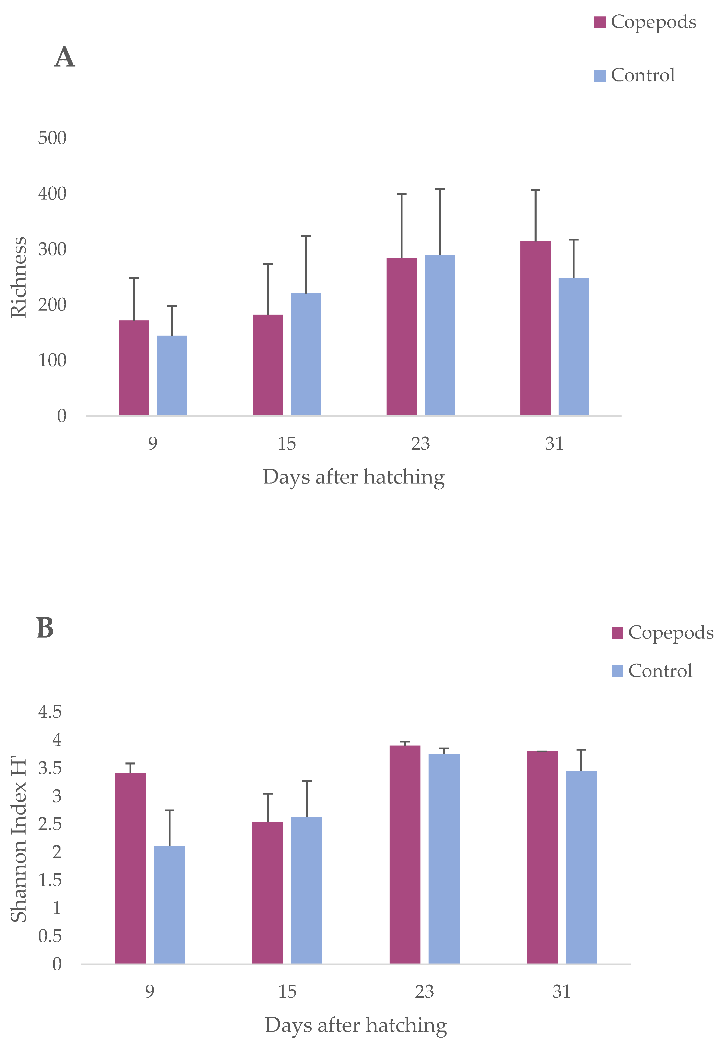
The microbial richness, as measured using linear mixed-effect model analysis, significantly increased over time (p < 0.05; Table 1), starting from a lower richness during early larval stages at 9 dah, and increasing gradually until 31 dah (Figure 2A). The lowest ASV richness occurred at 9 dah (145 ± 53) and the highest at 23 dah (290 ± 119) for the control and 31 dah (314.5 ± 92.5) for the copepods group, respectively. For the microbial diversity using Shannon H indices, which consider not only the number of bacterial phylotypes (i.e., ASVs) but also their relative abundances in each sample, significant effects were observed by the age of the fish (p < 0.05; Table 1). A gradual increase in both richness and diversity was observed during the experiment indicating an increase in microbial colonization in the larvae (Figure 2B). The dietary treatment had no significant effect on either the microbial richness or diversity.
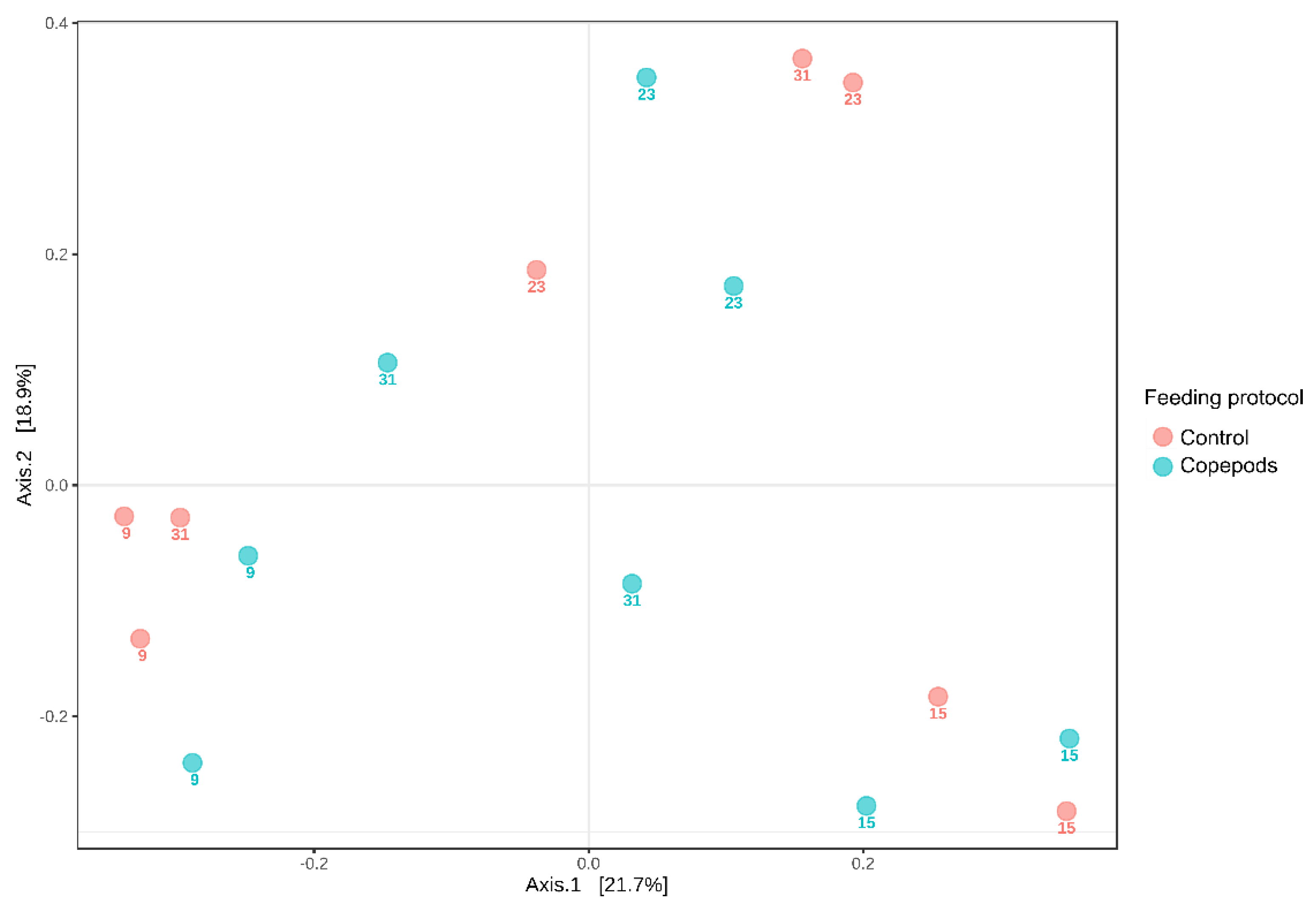
Beta-diversity (microbial composition) of the bacterial communities of amberjack larvae was significantly influenced by age (two-way Permanova analysis; p < 0.001; Table 2), while no significant effect of the feeding protocol was shown (p > 0.05; Figure 3).
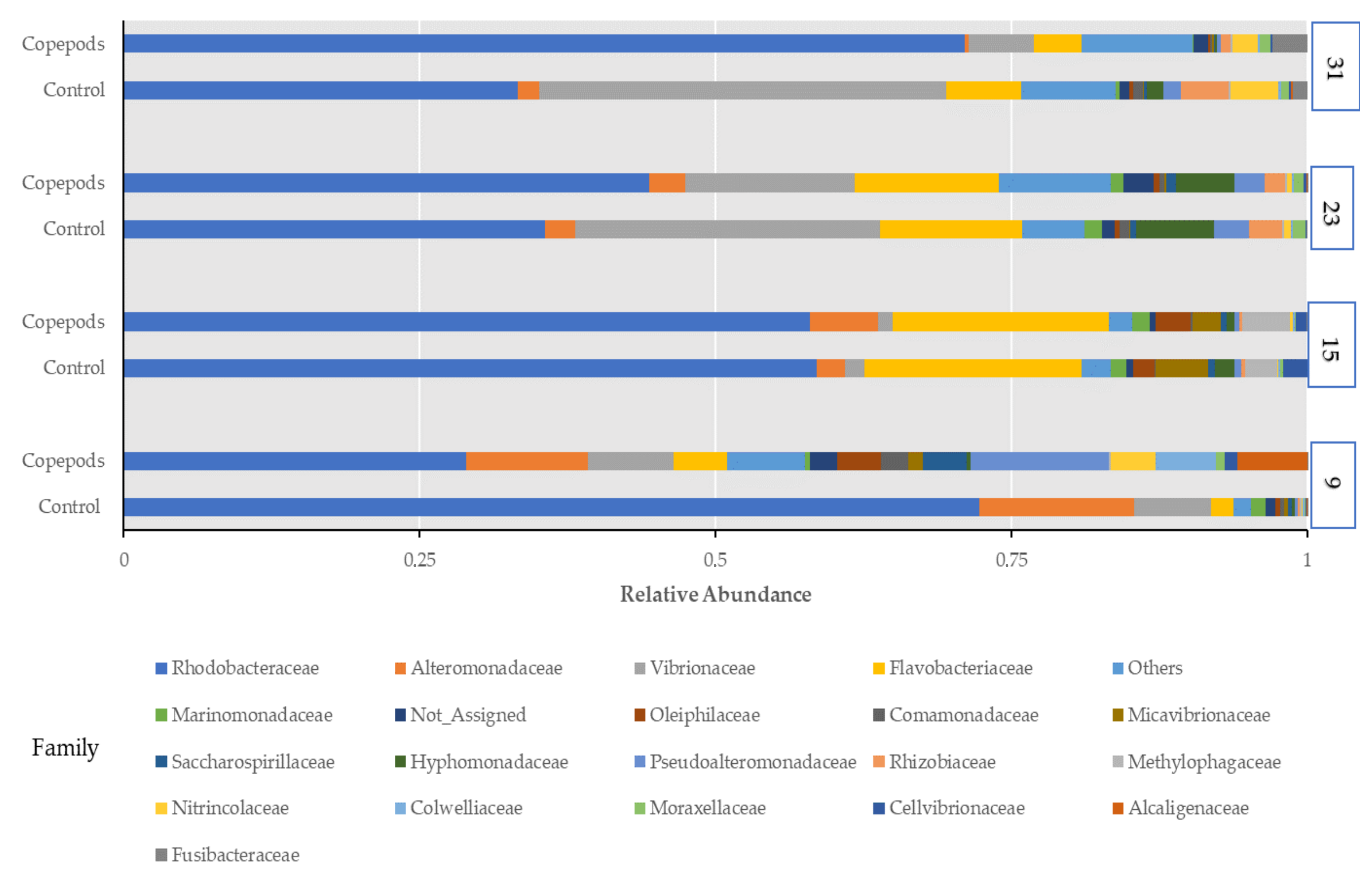
Two bacterial phyla, Proteobacteria and Bacteroidota, accounted for 96.4% of all the retrieved quality filtered sequences. Proteobacteria was the dominant phylum, with an average relative abundance of 85.6% across all the samples followed by Bacteroidota (10.79%), Firmicutes (0.77%), Campilobacterota (0.68%), Actinobacteriota (0.47%), Planctomycetota (0.36%), and Patescibacteria (0.34%) (Figure S1). Amberjack larvae were mostly characterized by bacteria belonging to the Rhodobacteriaceae family with a mean relative abundance for all sampling points at 50.25%, along with the families Vibrionaceae and Flavobacteriaceae, showing respective percentages of 12% and 9.6% (Figure 4), followed by Alteromonadaceae (4.8%), Pseudoalteromonadaceae (2.5%), and Hyphomonadaceae (2%). There was a difference in relative abundances of families between the two groups in 9 dah larvae, as 13 families in larvae from the copepods group, and only 5 families from the control group showed relative abundance greater than 1%. So, a higher diversity was found in the copepods group at the family level. Additionally, the 9 dah larvae from the copepods group appeared to have the highest relative abundances of families from all sampling points, such as Pseudoalteromonas (11.6%), Alcaligenaceae (5.9%), Colwelliaceae (5%), Saccharospirillaceae (3.7%), Oleiphilaceae (3.6%), and Comamonadaceae (2.3%).
A large proportion (average relative abundance at 15%) of the microbial communities was not assigned to a specific genus, whereas the most abundant genus was Thalassobius, exhibiting relative abundance at a mean percentage of 16,5% across all samples, followed by Vibrio (12%), Dokdonia (8.4%), Cognathshimia (7,9%), Ruegeria (7.5%), and Alteromonas (4.5%) (Figure S2). In 9 dah larvae from the copepods group, the genera as Pseudoalteromonas, Alcaligenes, Thalassotalea, Oleiphilus, and Neptuniibacter showed their highest relative abundances at 9.4%, 5.9%, 5%, 3.67%, and 3.5%, respectively, across the experimental period, following the Shannon diversity index, which was higher for larvae from the copepods group compared with the control group (p < 0.05).
The core microbiota is defined as any set of microbial taxa, as well as the associated genomic or functional attributes characteristic of a specific host or environment [48]. To determine the presence of an ASV in a group, its prevalence was set to be higher than 50%, meaning that it was present in at least one sample within each tank and showed 1% of minimum relative abundance. The core microbiota of the two groups was similar (Figure S3). Marinomonas (ASV45) and Thalassococcus (ASV30) were unique to the control and the copepods group, respectively. Nine ASVs were present in the larvae from both feeding protocols at all sampling points. The shared core ASVs belonged to the genera Thalassobius, Ruegeria, Nautella, Dokdonia, Donghicola, Cognatishimia, Vibrio, and Alteromonas; and one ASV was unassigned. In addition, we found significantly enriched taxa in the copepod feeding protocol’s larvae. Specifically, the genus Alcaligenes exhibited a significant (p < 0.05) differential relative abundance of about 6% in the copepods group at 9 dah (Figure S4A). The genus Thalassobius, which belongs to the core genera, was the most abundant genus at all sampling points, and its relative abundance was detected significantly (p < 0.05) higher for the larvae from the copepod feeding protocol larvae (Figure S4B).
Furthermore, to determine the predictive functionality of larvae microbiota for different feeding protocols, functional assessment using PICRUSt2 was performed and assigned to the KEGG (Kyoto Encyclopedia of Genes and Genomes) pathways. One pathway was significantly (p < 0.05) enriched in larvae from the copepods group, related to the perosamine synthetase (KEGG Orthology: K13010), which is related to amino sugar and nucleotide sugar metabolism (Figure 5).

3.2. Bacterial Strains Isolated from Greater Amberjack Larvae and Live Feed

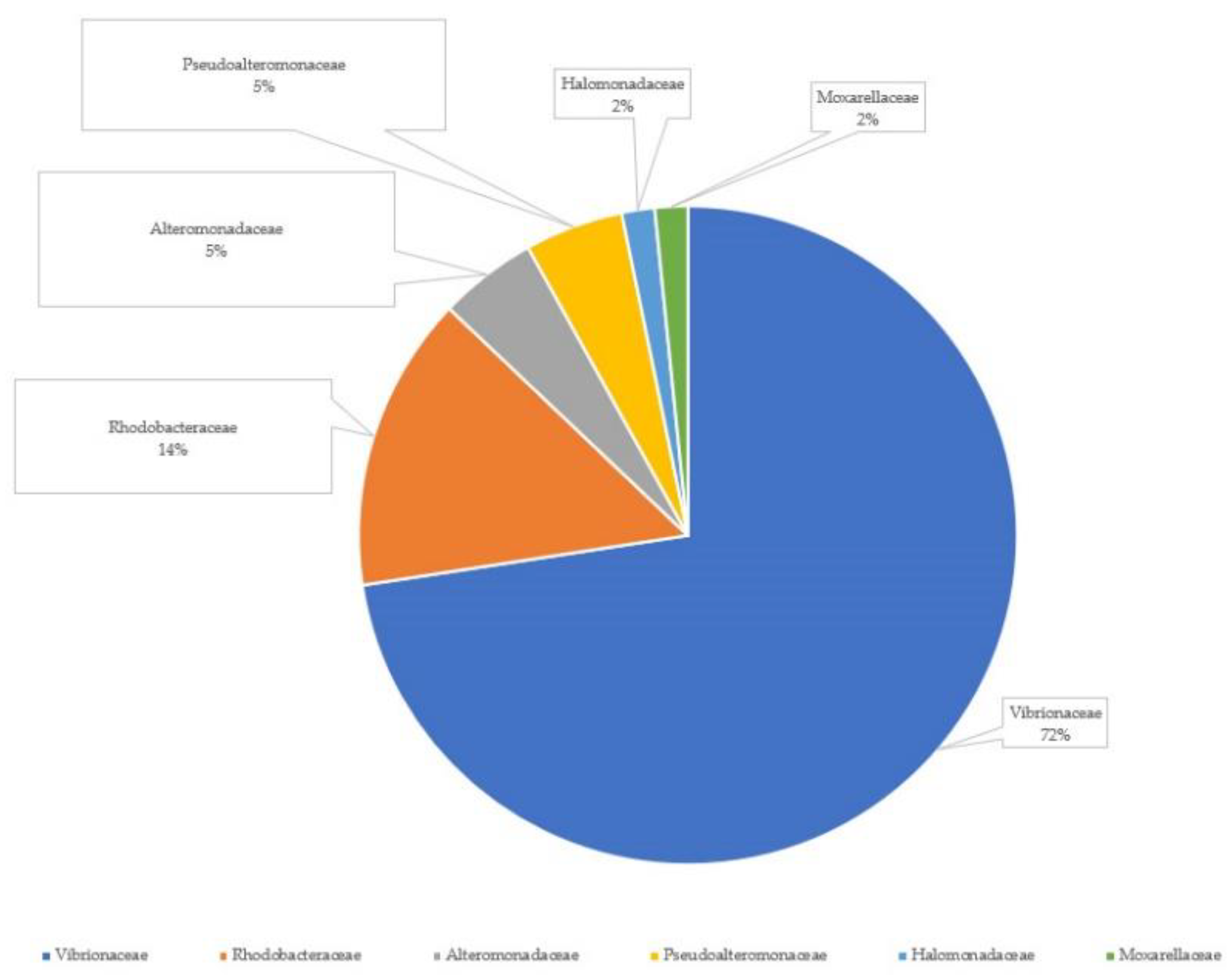
A total of 62 antagonistic culturable bacterial strains were isolated, 59 from larvae homogenates and 3 from homogenated Artemia sp. nauplii. The partial length of the 16S rRNA gene was sequenced to identify the isolated bacterial strains. The bacterial isolates were identified at the family level, and 45 bacterial strains were members of Vibrionaceae, the most abundant antagonistic bacteria. The rest were nine Rhodobacteraceae, three Pseudoalteromonadaceae, three Alteromonadaceae, and one annotation was found for each of the Halomonadaceae and Moxarellaceae families (Figure 6).
Among the isolated strains, there were several putative probiotics belonging to the Rhodobacteraceae family, as shown in Table 3. The isolate from the Halomonadaceae family matched with the Halomonas sp. strain.

3.3. In Vitro Inhibition Tests against Pathogens

The first trial of the double-layer test for the 62 bacterial isolates in vitro inhibition against four fish pathogenic strains, using TSA as the culture medium of the bacterial strains, showed that 12 of them inhibited the growth of the pathogens. Almost all the bacterial isolates from fish belonged to the Vibrionaceae family (Table 4). The twelfth bacterial strain, Halomonas sp., which was isolated from homogenized Artemia sp., exhibited inhibition zones greater than 30 mm in tests with V. alginolyticus and V. harveyi.
The double-layer test that was performed for the isolated bacterial strains, using Marine Agar as a culture medium, showed that four strains exhibited antagonistic activity against the fish pathogens (Table 5). These bacterial strains were Phaeobacter gallaeciensis strain JL2886, Phaeobacter sp. R-52698, and Ruegeria sp. InS-296, which was isolated from amberjack larvae, and one bacterial strain, Rhodobacter sp. 1–5, which was isolated from homogenized Artemia sp. The strain Phaeobacter gallaeciensis strain JL2886 was isolated from homogenized greater amberjack larvae of 9 dah, the strain Phaeobacter sp. R-52698 was isolated from the larvae of 16 dah (both from the copepods group), and the strain Ruegeria sp. InS-296 was isolated from larvae of 16 dah (control group).

4. Discussion

Greater amberjack is a new species for European aquaculture with a high economic value [49]. Despite intensive research on the rearing of amberjack larvae, high mortalities are often observed, which is a bottleneck for the commercialization of this species. This is the first report to characterize the larval microbiome of this species using next-generation sequencing.
The host microbiome may influence the physiology, digestion, and development of the immune system in marine fish larvae [50]. Understanding host-microbiota interactions could be essential for the improvement of rearing protocols or microbial management in larval cultures, for example, using probiotic bacteria.

4.1. Bacterial Dynamics Associated with the Early Developmental Stages of Greater Amberjack Larvae

During the experiment, we observed a gradual change in the alpha diversity of the larval microbiota. Bacterial community richness and diversity (measured as Shannon H’) significantly increased over time (Figure 2A,B). In gilthead seabream, the lowest bacterial richness was found at 1–5 dah [5,6], whereas in cod larvae, the lowest bacterial richness was observed at 17 dah [11]. Regardless of the host species, rearing system, or feed regime, the bacterial diversity in fish larvae tends to increase gradually with age until it reaches a peak when approaching the juvenile stage [2]. Interindividual variation in the gut microbiota composition has been observed in the microbiomes of other fish species, such as zebrafish [51] and Gibel carp [52]. In this study, age was the most significant factor shaping the microbiota in greater amberjack larvae, creating different clusters between 9, 15, and 23 with 31 dah (Figure 3). During the early developmental larvae stages (9 dah and 15 dah), regardless of the diet, there is a stage-specific bacterial community as a result of physiological changes such as gut differentiation, the appearance of acidic conditions in the stomach, or development of the immune system [13,53,54,55]. Generally, the microbiota of fish larvae comprises four dominant phyla: Proteobacteria, Bacteroidota, Firmicutes, and Actinomycetes, with changes in dominance, observed depending on the fish species, developmental stage, rearing system, and feed, as shown in the case of Atlantic cod [11], yellowtail amberjack [8], and gilthead seabream larvae [5]. We observed the same dominant phyla in the greater amberjack larvae. Bacteriodota showed the highest fluctuation in relative abundance among these phyla, with proportions of 4%, 18.6%, 14.4%, and 6% at 9, 15, 23, and 31, respectively. Firmicutes abundance remained low during the whole experiment (0.1–0.6%), reaching their highest average relative abundance (2.23%) in larvae at 31 dah. The presence of Firmicutes has been reported during the weaning process to formulated diet in many marine species [3,56,57] which can explain why the abundance of Firmicutes increased at the last sampling point as commercial feed was provided to the larvae after 23 dah. So, as reported in studies with other fish species [5,8,11], the microbiota of the greater amberjack can be influenced by the type of feed provided to the fish.
At the family level, Rhodobacteraceae dominated and was the most abundant family during the whole experiment (28.9–72.2% mean relative abundance), followed by Vibrionaceae and Flavobacteriaceae. The family Rhodobacteraceae colonizes tank wall biofilms and comprises predominately Κ-strategist species [58,59]. Members of this family showed probiotic activity by producing antimicrobial substances against Vibrios [27,60,61]. Recently, the isolation of the putative probiotic Phaeobacter sp. from marine fish yolk-sac larvae was reported for the first time [24]. In our study, the presence of dominant host-associated microbiota with probiotic characteristics was important, as they may have a greater chance of colonizing the gut and thereby confer a health benefit to the host.
Thalassobius of the Rhodobacteraceae family was the most abundant genus (16.5%), with its relative abundance being at the same level for both groups at the early larval stages but higher in larvae 31 dah from the copepod group. Members of the genus Thalassobius have been isolated from marine environments, particularly from surface coastal seawater and tidal flat samples [62]. Independent of the water treatment system used in the hatcheries, the uptake of bacteria by larvae is dominated by bacteria entering with the live food [55]. However, seawater microbiota also represents a significant contribution since marine fish drink seawater to osmoregulate, so bacteria from the water colonize the digestive tract before active feeding commences [16]. The outer surfaces of the fish larvae and the gills are colonized by bacteria in the rearing water. In addition, all live food organisms are filter-feeders, so to filter the water for food particles, they ingest free-living bacteria from the water. These bacteria may accumulate in the live food organisms and be in turn ingested by the fish larvae. Therefore, the water microbiota has a strong influence on the gut during early life stages [63]. The high abundance of Thalassobius in the microbiota of amberjack larvae in this experiment could be related to either drinking water by the larvae or the consumption of live feed organisms, since it appears to increase after 9 dah, indicating a strong influence of the live feed microbiota.

4.2. Bacterial Composition and Functionality Associated with Different Feeding Protocols in Greater Amberjack Larvae

In our study, it was shown that the age of the larvae had a strong impact on the microbiota of greater amberjack larvae. In contrast, the feeding protocol had only a minor influence on the microbial communities. The addition of copepods led to only a slight increase in the richness and diversity of the larval bacterial community, mainly at 9 dah and 31 dah. More specifically, the larvae microbiota fed either diet was colonized by Proteobacteria and Bacteroidota, sharing approximately 96% of the relative abundance at each sampling point. Both phyla showed the same pattern of fluctuation between the sampling points for the two diets. Interestingly, the relative abundance of Firmicutes differed at 31 dah, with 3.12% for the copepod and 1.34% for the control group.
At the family level, Rhodobacteraceae was the dominant family in both groups but showed a different pattern of relative abundance. Specifically, larvae from the control group showed a gradual decrease from 72.2 to 33.2% over time. On the contrary, the larvae from the copepod group showed a gradual increase from 28.9 to 71% over time. Additionally, while in larvae from both groups, the relative abundance of Rhodobacteraceae was higher compared with Vibrionaceae during the whole experiment, at 31 dah larvae from the control group Vibrionaceae showed similar abundance with Rhodobacteraceae. Members of the Vibrionaceae family are widely found in marine environments, often isolated from the intestinal tract of various marine fish species at the larval and fry stages, such as seabass, turbot, Atlantic cod, Atlantic halibut, and Dover sole [50].
Regarding the presence of Vibrios in the larvae microbiota, due to the feeding protocols, there was a difference in their average relative abundance being 17% for the control (the most abundant genus in this group) and 7% for the copepod group, respectively. Larvae from the control group collapsed at 31 dah, whereas the larvae of the copepods group survived up to 42 dah (Figure 1). At 31 dah, in larvae from the control group, the relative abundances of Vibrionacae and Rhodobacteriaceae families were 34.3% and 33.3%, respectively. The proportions of the same families for larvae of the same age from the copepods group were 5.5% and 71%, respectively. The feeding regime did not influence the richness and diversity of the larval host-associated microbiota, but it seemed that the addition of copepods resulted in higher survival of the larvae, which could be due to the restriction of Vibrionaceae, and enhanced presence of Rhodocteriaceae, revealing antagonism against Vibrio species and possible probiotic activity.
Larvae at 9 dah from the copepods group showed the highest abundances of Pseudoalteromonas, Alcaligenaceae, Colwelliaceae, Saccharospirillaceae, Oleiphilaceae, and Comamonadaceae, families, except Vibrionaceae and Rhobocteriaceae, which resulted in higher diversity than the control group at this age. Fish fed a variety of feed items may support more diverse gut-associated bacterial communities because of a wider range of potential substrates available to the bacteria [17]. Thalassobius was the significantly dominant genus in the greater amberjack larvae microbiota. Copepods have higher protein and free amino acid content compared to Artemia and rotifers [64]. The protein content of copepods is on average 50% higher than that of the protein content of Artemia [31]. Thus, the need of the larvae fed with copepods to adapt to greater amounts of specific dietary elements, such as amino acids, might lead to stable colonization of the larval microbiota of the genus Thalassobius, as this genus is strictly aerobic and uses organic acids and amino acids as carbon sources [65]. The high dietary value of the copepods in the greater amberjack larvae microbiota is supported by the increase in metabolic pathways related to amino acid and glycan biosynthesis in the copepod-fed larvae, indicating a potential nutritional benefit when these live feed organisms are included in the diet of amberjack larvae. Moreover, the genus Alcaligenes showed a significantly higher relative abundance in the copepod group at 9 dah, indicating that this genus may play an essential role in specific larval adaptations as an adaptation to the copepod biochemical composition. Alcaligenes have been recognized as bacterial species isolated from the intestinal tracts of marine fish species at the larval and fry life stages, specifically in Dover sole and turbot [50]. In agreement with our findings, Alcaligenes have been isolated from turbot larvae fed with copepods [66].
In this study, the experiment was run under industrial conditions. The larvae had to be fed according to the routine operations of the hatchery and could not be starved. The larvae were not disinfected because of the need to isolate antagonistic culturable bacteria and were only rinsed with autoclaved seawater. Due to the small body size of the larvae, dissection of the gut was not possible at the sampling points. Thus, the microbiota of greater amberjack larvae described in this study represents bacteria originating from the skin, gills, and gastrointestinal tract.

4.3. Isolation of Cultivable Antagonistic Bacteria with Probiotic Effect

In this study, culturable antagonistic bacteria were isolated from homogenates of greater amberjack larvae and live food, using a novel technique. This method was based on the appearance of inhibition zones in Petri dishes spread with serial dilutions of the homogenates. In this way, it was possible to recognize and isolate bacterial strains with antagonistic activity against other bacteria in the same sample.
In total, 62 antagonistic bacterial strains were isolated; 59 from larvae homogenates and 3 from homogenated Artemia sp. Sequencing and subsequent annotation revealed that the most numerous antagonistic bacterial strains, coming from the larvae, belonged to the Vibrionaceae family (72%). Vibrio is a genus of Gram-negative bacteria that is highly salt-tolerant and unable to survive in freshwater; consequently, it is commonly found in various saline or brackish water environments. Among the Vibrionales order, the Vibrionaceae family comprises aquatic bacteria, mostly living in warm waters that tolerate different levels of salinity, including fresh, brackish, and marine waters. The Vibrionaceae family includes the genera Aliivibrio, Catenococcus, Enterovibrio, Grimontia, Listonella, Photobacterium, Salinivibrio, and Vibrio [67], and it is a genomically, phylogenetically, and functionally diverse group. In agreement with our findings, Vibrio strains have been isolated from the intestinal tracks of marine fish species at larval, juvenile, and adult life stages (seabass, turbot, Atlantic cod, Atlantic halibut, and Dover sole) [50]. Vibrionaceae have been reported to be the most numerous antagonistic genera associated with fish, such as Japanese flounder (Paralichthys olivaceus) [68], and Senegalese sole [44], but also live feeds (rotifer, Artemia, and copepods) [32].
In addition to the members of the Vibrionaceae, representatives from Pseudoalteromonadaceae and Roseobacteraceae families were isolated in proportions of 5% and 14%, respectively, among the antagonistic bacterial strains. Pseudoalteromonas isolates have been used as putative probiotics in different farmed organisms. For instance, Pseudoalteromonas strains isolated from the intestinal tract of Atlantic cod, the gonads, and the intestinal content of S. lalandi, demonstrated antagonistic activity against the pathogen Vibrio anguillarum [60], and improved larval growth and survival in S. lalandi, [69]. These results are in agreement with our findings that Pseudoalteromonadaceae are a part of the greater amberjack larvae microbiota demonstrating antagonistic activity and are putative probiotics.
Probiotics as an alternative to the use of antibiotics and other chemotherapeutic agents can make aquaculture more sustainable and decrease the ecological footprint of the sector, as they have the potential to improve the GI tract of the fish, including development and maturation of the intestine and immune system [70,71], and resistance to infectious pathogenic microbiota [72]. Three Roseobacteraceae bacterial strains were isolated, which were identified as Phaeobacter gallaeciensis, Phaeobacter sp., and Ruegeria sp. Phaeobacter gallaeciensis was isolated from 9 dah and Phaeobacter sp. from 15 dah, larvae from the copepod group, while Ruegeria sp. was isolated from 9 dah larvae from the control group. These strains showed inhibitory activity against Vibrio anguillarum, Vibrio alginolyticus, Vibrio harveyi and Aeromonas veronii strains in vitro. Among others, Phaeobacter species have been shown to reduce the pathogenic load in cultures of microalgae and cod larvae during larviculture [73,74] and increase larval survival and specific growth rate in sea bass larvae [24]. Several Phaeobacter strains have been isolated in marine aquaculture from the seawater and collectors of scallops (Pecten maximus) [75], larval rearing tank walls [67], and different units of Danish turbot farms [76]. As in Makridis et al. [24], Phaeobacter strains were isolated from homogenated greater amberjack larvae. Phaeobacter has been isolated from tank walls or biofilters of the rearing systems, which are aerobic environments, but from the reared larvae. As they were isolated from greater amberjack larvae at the early developmental stages (9 and 15 dah), it appears that they colonized the surface of the larval body, gills, or gut. The sampled amberjack larvae were not surface-sterilized but only washed with sterile water, so Phaeobacter could be isolated from these aerobic surfaces, or they could be a transient part of the gut microbiota.
Three antagonistic bacterial strains were isolated from Artemia sp., Halomonas sp., Psychrobacter sp., and Rhodobacter sp. The isolated Halomonas sp. showed very strong antagonistic activity against Vibrio alginolyticus and Vibrio harveyi, while Rhodobacter sp. showed smaller inhibition zones against Aeromonas veronii and Vibrio harveyi. In previous studies, Psychrobacter sp., isolated from the whole intestine of juvenile grouper E. coioides and as a probiotic when it was administrated with the diet, improved the autochthonous diversity along the gastrointestinal tract of this grouper [77]. It has also been suggested to be capable of producing and secreting effective antimicrobial substances [78]. Following the previous studies, we hypothesize that the isolated strains in our study can be important probiotic candidates for future studies.

5. Conclusions

In our study, we found that greater amberjack larvae reared in a commercial hatchery were colonized by a diverse range of bacteria and that the composition of the host microbiota is mainly influenced by age rather than by diet. Despite this, the slight increase in diversity together with the decrease in potential opportunistic groups such as Vibrios could indicate a positive enrichment of the copepods in the greater amberjack larvae microbiota. Finally, we were able to isolate bacteria from the larvae with antagonistic activity against fish pathogens, mainly from the family Rhodobacteraceae, suggesting that the host-associated microbiota is a good source of probiotics.

Supplementary Materials

The following supporting information can be downloaded at: https://www.mdpi.com/article/10.3390/microorganisms11081889/s1, Figure S1: Relative abundance of bacteria phyla in all sampling points (control, copepods; diet) (9, 15, 23, 31; dah), Figure S2. Relative abundance of prokaryotic genera in all sampling points (control, copepods; diet) (9, 15, 23, 31; dah), Figure S3. Core shared and unique genera of S. dumerili larvae fed with the control and the copepods feeding protocols for the experimental period. Core ASVs, defined as genera present, with a prevalence of 50% and occurring at 1% minimum of relative abundance. Figure S4. Relative abundance of the significantly (p < 0.05) A. differential abundant Alcaligenes genus and B. most abundant Thalassobius genus, between the control and the copepod feeding protocols 9, 15, 23, 31 dah.

Author Contributions

Conceptualization, P.M.; methodology, P.M. and F.K.; formal analysis, V.P.; investigation, V.P. and S.K. riting—original draft preparation, V.P.; writing—review and editing, P.M. and F.K.; funding acquisition, P.M. All authors have read and agreed to the published version of the manuscript.

Funding

This work was funded by the General Secretariat for Research and Innovation (GSRI) through the research project titled “Improvement of broodstock management and fingerling production methods for greater amberjack (Seriola dumerili)”, with grant number MIS 5045873.

Institutional Review Board Statement

The rearing trial of the fish was performed according to the routine operations of the Galaxidi Marine Farms S.A., and sampling procedures conformed to Directive 2010/63/EU.

Data Availability Statement

The data presented in this study are available upon request from the corresponding author. The data are not publicly available due to short time margins.

Acknowledgments

We thank Pantelis Katharios, Institute of Marine Biology, Biotechnology, and Aquaculture (IMBBC), Hellenic Center for Marine Research, Heraklion, Crete, for providing us the four strains of pathogenic strains (Vibrio anguillarum type strain LMG 4437, Vibrio alginolyticus type strain V2, Vibrio harveyi type strain VH2, and Aeromonas veronii biovar sobria) used in the in vitro inhibition tests. We also thank Galaxidi Marine Farm S.A., Greece, and the hatchery manager Tasos Megos for their collaboration in the rearing of greater amberjack larvae.

Conflicts of Interest

The authors declare no conflict of interest.

References

  1. Vadstein, O.; Øie, G.; Olsen, Y.; Salvesen, I.; Skermo, J. A Strategy to Obtain Microbial Control during Larval Development of Marine Fish; Fish Farming Technology, Reinertsen, H., Dahle, L.A., Jorgensen, L., Eds.; Balkema: Rotterdam, The Netherlands, 1993; ISBN 90 5410 3264. [Google Scholar]
  2. Borges, N.; Keller-Costa, T.; Sanches-Fernandes, G.M.M.; Louvado, A.; Gomes, N.C.M.; Costa, R. Bacteriome structure, function, and probiotics in fish larviculture: The good, the bad, and the gaps. Annu. Rev. Anim. Biosci. 2021, 9, 423–452. [Google Scholar] [CrossRef]
  3. Tarnecki, A.M.; Burgos, F.A.; Ray, C.L.; Arias, C.R. Fish intestinal microbiome: Diversity and symbiosis unravelled by metagenomics. J. Appl. Microbiol. 2017, 123, 2–17. [Google Scholar] [CrossRef] [Green Version]
  4. Vadstein, O.; Bergh, Ø.; Gatesoupe, F.-J.; Galindo-Villegas, J.; Mulero, V.; Picchietti, S.; Scapigliati, G.; Makridis, P.; Olsen, Y.; Dierckens, K.; et al. Microbiology and immunology of fish larvae. Rev. Aquacult. 2013, 5, S1–S25. [Google Scholar] [CrossRef] [Green Version]
  5. Nikouli, E.; Meziti, A.; Antonopoulou, E.; Mente, E.; Kormas, K.A. Host-associated bacterial succession during the early embryonic stages and first feeding in farmed gilthead sea bream (Sparus aurata). Genes 2019, 10, 483. [Google Scholar] [CrossRef] [PubMed] [Green Version]
  6. Califano, G.; Castanho, S.; Soares, F.; Ribeiro, L.; Cox, C.J.; Mata, L.; Costa, R. Molecular taxonomic profiling of bacterial communities in a gilthead seabream (Sparus aurata) hatchery. Front. Microbiol. 2017, 8, 204. [Google Scholar] [CrossRef] [PubMed] [Green Version]
  7. Le, D.; Nguyen, P.; Nguyen, D.; Dierckens, K.; Boon, N.; Lacoere, T.; Kerckhof, F.M.; De Vrieze, J.; Vadstein, O.; Bossier, P. Gut microbiota of migrating wild rabbit fish (Siganus guttatus) larvae have low spatial and temporal variability. Microb. Ecol. 2020, 79, 539–551. [Google Scholar] [CrossRef] [Green Version]
  8. Wilkes Walburn, J.; Wemheuer, B.; Thomas, T.; Copeland, E.; O’Connor, W.; Booth, M.; Fielder, S.; Egan, S. Diet and diet associated bacteria shape early microbiome development in yellowtail kingfish (Seriola lalandi). Microb. Biotechnol. 2019, 12, 275–288. [Google Scholar] [CrossRef] [PubMed] [Green Version]
  9. Ingerslev, H.C.; von Gersdorff, J.; Lenz Strube, M.; Larsen, N.; Dalsgaard, I.; Boye, M.; Madsen, L. The development of the gut microbiota in rainbow trout (Oncorhynchus mykiss) is affected by first feeding and diet type. Aquaculture 2014, 424–425, 24–34. [Google Scholar] [CrossRef] [Green Version]
  10. Giatsis, C.; Sipkema, D.; Smidt, H.; Heilig, H.; Benvenuti, G.; Verreth, J.; Verdegem, M. The impact of rearing environment on the development of gut microbiota in tilapia larvae. Sci. Rep. 2015, 5, 18206. [Google Scholar] [CrossRef] [Green Version]
  11. Bakke, I.; Coward, E.; Andersen, T.; Vadstein, O. Selection in the host structures the microbiota associated with developing cod larvae (Gadus morhua). Environ. Microbiol. 2015, 17, 3914–3924. [Google Scholar] [CrossRef]
  12. Bledsoe, J.W.; Peterson, B.C.; Swanson, K.S.; Small, B.C. Ontogenetic characterization of the intestinal microbiota of channel catfish through 16S rRNA gene sequencing reveals insights on temporal shifts and the influence of environmental microbes. PLoS ONE 2016, 11, e0166379. [Google Scholar] [CrossRef] [PubMed] [Green Version]
  13. Lokesh, J.; Kiron, V.; Sipkema, D.; Fernandes, J.M.O.; Moum, T. Succession of embryonic and the intestinal bacterial communities of Atlantic salmon (Salmo salar) reveals stage-specific microbial signatures. Microbiol. Open 2019, 8, e00672. [Google Scholar] [CrossRef] [PubMed]
  14. Wu, S.G.; Tian, J.Y.; Gatesoupe, F.J.; Li, W.X.; Zou, H.; Yang, B.J.; Wang, G.T. Intestinal microbiota of gibel carp (Carassius auratus gibelio) and its origin as revealed by 454 pyrosequencing. World J. Microbiol. Biotechnol. 2013, 29, 1585–1595. [Google Scholar] [CrossRef] [PubMed]
  15. Li, X.; Yu, Y.; Feng, W.; Yan, Q.; and Gong, Y. Host species as a strong determinant of the intestinal microbiota of fish larvae. J. Microbiol. 2012, 50, 29–37. [Google Scholar] [CrossRef]
  16. Hansen, G.H.; Olafsen, J.A. Bacterial interactions in early life stages of marine cold water fish. Microb. Ecol. 1999, 38, 1–26. [Google Scholar] [CrossRef]
  17. Larsen, A.M.; Mohammed, H.H.; Arias, C.R. Characterization of the gut microbiota of three commercially valuable warmwater fish species. J. Appl. Microbiol. 2014, 116, 1396–1404. [Google Scholar] [CrossRef]
  18. Givens, C.E.; Ransom, B.; Bano, N.; Hollibaugh, J.T. A fish tale: Comparison of the gut microbiomes of 12 finfish and 3 shark species. Mar. Ecol. Prog. Ser. 2014, 518, 209–223. [Google Scholar] [CrossRef] [Green Version]
  19. Roeselers, G.; Mittge, E.K.; Stephens, W.Z.; Parichy, D.M.; Cavanaugh, C.M.; Guillemin, K.; Rawls, J.F. Evidence for a core gut microbiota in the zebrafish. ISME J. 2011, 5, 1595–1608. [Google Scholar] [CrossRef] [Green Version]
  20. Blanch, A.R.; Alsina, M.; Simon, M.; Jofre, J. Determination of bacteria associated with reared turbot (Scophthalmus maximus) larvae. J. Appl. Microbiol. 1997, 82, 729–734. [Google Scholar] [CrossRef]
  21. Lauzon, H.L.; Gudmundsdottir, S.; Petursdottir, S.K.; Reynisson, E.; Steinarsson, A.; Oddgeirsson, M.; Bjornsdottir, R.; Gudmundsdottir, B.K. Microbiota of Atlantic cod (Gadus morhua L.) rearing systems at pre- and posthatch stages and the effect of different treatments. J. Appl. Microbiol. 2010, 109, 1775–1789. [Google Scholar] [CrossRef]
  22. McIntosh, D.; Ji, B.; Forward, B.S.; Puvanendran, V.; Boyce, D.; Ritchie, R. Culture-independent characterization of the bacterial populations associated with cod (Gadus morhua L.) and live feed at an experimental hatchery facility using denaturing gradient gel electrophoresis. Aquaculture 2008, 275, 42–50. [Google Scholar] [CrossRef]
  23. Zapata, A.; Diez, B.; Cejalvo, T.; Gutiérrez-de Frías, C.; Cortés, A. Ontogeny of the immune system of fish. Fish Shellfish Immun. 2006, 20, 126–136. [Google Scholar] [CrossRef]
  24. Makridis, P.; Kokou, F.; Bournakas, C.; Papandroulakis, N.; Sarropoulou, E. Isolation of Phaeobacter sp. from larvae of Atlantic bonito (Sarda sarda) in a mesocosmos unit, and its use for the rearing of European seabass larvae (Dicentrarchus labrax L.). Microorganisms 2021, 9, 128. [Google Scholar] [CrossRef]
  25. Verschuere, L.; Rombaut, G.; Sorgeloos, P.; Verstraete, W. Probiotic bacteria as biological control agents in aquaculture. Microbiol. Mol. Biol. R. 2000, 64, 655–671. [Google Scholar] [CrossRef] [Green Version]
  26. Mohapatra, S.; Chakraborty, T.; Kumar, V.; DeBoeck, G.; Mohanta, K.N. Aquaculture and stress management: A review of probiotic intervention. J. Anim. Physiol. Anim. Nutr. 2013, 97, 405–443. [Google Scholar] [CrossRef] [PubMed]
  27. Prol-García, J.M.; Gómez, M.; Lorenzo Sánchez, L.; Pintado, J. Phaeobacter grown in biofilters: A new strategy for the control of Vibrionaceae in aquaculture. Aquac. Res. 2014, 45, 1012–1025. [Google Scholar] [CrossRef]
  28. Verma, G.; Gupta, A. Probiotics Application in Aquaculture: Improving Nutrition and Health. J. Anim. Feed. Sci. Technol. 2015, 3, 53–64. [Google Scholar]
  29. Bjornsdottir, R.; Johannsdottir, J.; Coe, J.; Smaradottir, H.; Agustsson, T.; Sigurgisladottir, S.; Gudmundsdottir, K.B. Survival and quality of halibut larvae (Hippoglossus hippoglossus L.) in intensive farming: Possible impact of the intestinal bacterial community. Aquaculture 2009, 286, 53–63. [Google Scholar] [CrossRef]
  30. Lazado, C.C.; Caipang, C.M.; Estante, E.G. Prospects of host-associated microorganisms in fish and penaeids as probiotics with immunomodulatory functions. Fish Shellfish Immunol. 2015, 45, 2–12. [Google Scholar] [CrossRef]
  31. Conceicao, L.; Yufera, M.; Makridis, P.; Morais, S.; Dinis, M. Live feeds for early stages of fish rearing. Aquac. Res. 2010, 41, 613–640. [Google Scholar] [CrossRef]
  32. Loufi, K.; Sfakianakis, D.G.; Makridis, P. The effect of feeding with the copepod Acartia tonsa during the first days of larval rearing on skeleton ontogeny and skeletal deformities in greater amberjack (Seriola dumerili). In Proceedings of the Aquaculture Europe 2022, Rimini, Italy, 27–30 September 2022; pp. 742–743. Available online: https://eposters.blob.core.windows.net/eas-eposters/AE2022AbstractBook.pdf (accessed on 2 May 2023).
  33. Dittman, K.K.; Rasmussen, B.B.; Melchiorsen, J.; Sonnenschein, E.C.; Lone, G.; Bentzon-Tilia, M. Changes in the microbiome of mariculture feed organisms after treatment with a potentially probiotic strain of Phaeobacter inhibens. Appl. Environm. Microbiol. 2020, 86, e0049920. [Google Scholar] [CrossRef]
  34. Rangel, F.; Enes, P.; Gasko, L.; Gai, F.; Hausmann, B.; Berry, D.; Oliva-Teles, A.; Serra, C.R.; Pereira, F.C. Differential modulation of the European sea bass gut microbiota by distinct insect meals. Front. Microbiol. 2022, 13, 831034. [Google Scholar] [CrossRef]
  35. Callahan, B.J.; McMurdie, P.J.; Rosen, M.J.; Han, A.W.; Johnson, A.J.A.; Holmes, S.P. DADA2: High-resolution sample inference from Illumina amplicon data. Nat. Methods 2016, 13, 581–583. [Google Scholar] [CrossRef] [Green Version]
  36. Kokou, F.; Sasson, G.; Mizrahi, I.; Cnaani, A. Antibiotic effect and microbiome persistence vary along the European seabass gut. Sci. Rep. 2020, 10, 10003. [Google Scholar] [CrossRef] [PubMed]
  37. Wang, Q.; Garrity, M.G.; Tiedje, M.J.; Cole, R.J. Naïve Bayesian Classifier for Rapid Assignment of rRNA Sequences into the New Bacterial Taxonomy. Appl. Environ. Microbiol. 2007, 73, 5261–5267. [Google Scholar] [CrossRef] [PubMed] [Green Version]
  38. McLaren, M.R. Silva SSU Taxonomic Training Data Formatted for DADA2 (Silva Version 138). Available online: https://zenodo.org/record/3986799 (accessed on 15 April 2021).
  39. Pinheiro, J.C.; Bates, D.M. Unconstrained parametrizations for variance-covariance matrices. Stat. Comput. 1996, 6, 289–296. [Google Scholar] [CrossRef]
  40. Anderson, M.J. A new method for non-parametric multivariate analysis of variance. Austral Ecol. 2001, 26, 32–46. [Google Scholar] [CrossRef]
  41. Douglas, G.M.; Maffei, V.J.; Zaneveld, J.R.; Yurgel, S.N.; Brown, J.R.; Taylor, C.M.; Huttenhower, C.; Langille, M.G.I. PICRUSt2 for prediction of metagenome functions. Nat. Biotechnol. 2020, 38, 685–688. [Google Scholar] [CrossRef]
  42. Jensen, S.; Bergh, Ò.; Enger, Ò.; Hjeltnes, B. Use of PCR-RFLP for genotyping 16S rRNA and characterizing bacteria cultured from halibut fry. Can. J. Microbiol. 2002, 48, 379–386. [Google Scholar] [CrossRef]
  43. Lane, D.J. 16S/23S rRNA sequencing. In Nucleic Acid Techniques in Bacterial Systematics, 1st ed.; Stackebrandt, E.M., Goodfellow, M., Eds.; John Wiley and Sons: Chichester, UK, 1991; pp. 177–203. [Google Scholar]
  44. Makridis, P.; Martins, S.; Tsalavouta, M.; Dionisio, L.C.; Kotoulas, G.; Magoulas, A.; Dinis, M.T. Antimicrobial activity in bacteria isolated from Senegalese sole, Solea senegalensis, fed with natural prey. Aquac. Res. 2005, 36, 1619–1627. [Google Scholar] [CrossRef]
  45. Castillo, D.; D’Alvise, P.; Kalatzis, P.G.; Kokkari, C.; Middelboe, M.; Gram, l.; Liu, S.; Katharios, P. Draft genome sequences of Vibrio alginolyticus strains V1 and V2, opportunistic marine pathogens. Genome Announc. 2015, 3, e00729-15. [Google Scholar] [CrossRef] [PubMed] [Green Version]
  46. Castillo Bermúdez, D.E.; D’Alvise, P.; Middelboe, M.; Gram, L.; Liu, S.; Kalatzis, P.; Kokkari, C.; Katharios, P. Draft genome sequences of the fish pathogen Vibrio harveyi strains VH2 and VH5. Genome Announc. 2015, 3, e01062-15. [Google Scholar] [CrossRef] [Green Version]
  47. Smyrli, M.; Prapas, A.; Rigos, G.; Kokkari, C.; Pavlidis, M.; Katharios, P. Aeromonas veronii infection associated with high morbility and mortality in farmed European Seabass Dicentrarchus labrax in the Aegean Sea, Greece. Fish Pathol. 2017, 52, 68–81. [Google Scholar] [CrossRef] [Green Version]
  48. Neu, T.A.; Allen, E.E.; Roy, K. Defining and quantifying the core microbiome: Challenges and prospects. Proc. Natl. Acad. Sci. USA 2021, 118, e2104429118. [Google Scholar] [CrossRef] [PubMed]
  49. Nijssen, J.E.; Reinders, J.M.; Krystallis, A.; Tacken, G. Developing an Internationalization Strategy Using Diffusion Modeling: The Case of Greater Amberjack. Fishes 2019, 4, 12. [Google Scholar] [CrossRef] [Green Version]
  50. Egerton, S.; Culloty, S.; Whooley, J.; Stanton, C.; Ross, R.P. The gut microbiota of marine fish. Front. Microbiol. 2018, 9, 873. [Google Scholar] [CrossRef]
  51. Stephens, W.Z.; Burns, A.R.; Stagaman, K.; Wong, S.; Rawls, J.F.; Guillemin, K.; Bohannan, B.J. 2016. The composition of the Zebrafish intestinal microbial community varies across development. ISME J. 2016, 10, 644–654. [Google Scholar] [CrossRef] [Green Version]
  52. Li, X.; Zhou, L.; Yu, Y.; Ni, J.; Xu, W.; Yan, Q. Composition of Gut Microbiota in the Gibel Carp (Carassius auratus gibelio) Varies with Host Development. Microb. Ecol. 2016, 74, 239–249. [Google Scholar] [CrossRef]
  53. Yan, Q.; Li, J.; Yu, Y.; Wang, J.; He, Z.; Van Nostrand, J.D.; Kempher, M.L.; Wu, L.; Wang, Y.; Liao, L.; et al. Environmental filtering decreases with fish development for the assembly of gut microbiota. Environ. Microbiol. 2016, 18, 4739–4754. [Google Scholar] [CrossRef]
  54. Burns, A.R.; Stephens, W.Z.; Stagaman, K.; Wong, S.; Rawls, J.F.; Guillemin, K.; Bohannan, B.J.M. Contribution of neutral processes to the assembly of gut microbial communities in the zebrafish over host development. ISME J. 2015, 10, 655–664. [Google Scholar] [CrossRef] [Green Version]
  55. Vadstein, O.; Attramadal, J.K.; Bakke, I.; Forberg, T.; Olsen, Y.; Verdegem, M.; Giatsis, C.; Skjermo, J.; Aasen, I.M.; Gatesoupe, F.J.; et al. Managing the microbial community of marine fish larvae: A holistic perspective for larviculture. Front. Microbiol. 2018, 9, 1820. [Google Scholar] [CrossRef]
  56. Kormas, K.A.; Meziti, A.; Mente, E.; Frentzos, A. Dietary differences are reflected on the gut prokaryotic community structure of wild and commercially reared sea bream (Sparus aurata). Microbiol. Open 2014, 3, 718–728. [Google Scholar] [CrossRef] [PubMed]
  57. Ghanbari, M.; Kneifel, W.; Domig, K.J. A new view of the fish gut microbiome: Advances from next-generation sequencing. Aquaculture 2015, 448, 464–475. [Google Scholar] [CrossRef]
  58. Bruhn, J.B.; Gram, R.; Belas, R. Production of antibacterial compounds and biofilm formation by Roseobacter species are influenced by culture conditions. Appl. Environ. Microb. 2007, 73, 442–450. [Google Scholar] [CrossRef] [Green Version]
  59. Cardona, E.; Gueguen, Y.; Magré, K.; Lorgeoux, B.; Piquemal, D.; Pierrat, F.; Noguier, F.; Saulnier, D. Bacterial community characterization of water and intestine of the shrimp Litopenaeus stylirostris in a biofloc system. BMC Microbiol. 2016, 16, 157–166. [Google Scholar] [CrossRef] [Green Version]
  60. Fjellheim, A.J.; Klinkenberg, G.; Skjermo, J.; Aasen, I.M.; Vadstein, O. Selection of candidate probionts by two different screening strategies from Atlantic cod (Gadus morhua L.) larvae. Vet. Microbiol 2010, 144, 153–159. [Google Scholar] [CrossRef]
  61. Copetti, F.; Gregoracci, G.B.; Vadstein, O.; Schveitzer, R. Management of biofloc concentrations as an ecological strategy for microbial control in intensive shrimp culture. Aquaculture 2021, 543, 736969. [Google Scholar] [CrossRef]
  62. Pujalte, M.J.; Lucena, T.; Rodrigo-Torres, L.; Arahal, D.R. Comparative genomics of Thalassobius including the description of Thalassobius activus sp. nov., and Thalassobius autumnalis sp. nov. Front. Microbiol. 2018, 8, 2645. [Google Scholar] [CrossRef] [Green Version]
  63. Makridis, P.; Fjellheim, A.J.; Skjermo, J.; Vadstein, O. Colonization of the gut in first feeding turbot by bacterial strains added to the water or bioencapsulated in rotifers. Aquacult. Int. 2000, 8, 367–380. [Google Scholar] [CrossRef]
  64. van der Meeren, T.; Olsen, R.E.; Hamre, K.; Fyhn, H.J. Biochemical composition of copepods for evaluation of feed quality in production of juvenile marine fish. Aquaculture 2008, 274, 375–397. [Google Scholar] [CrossRef] [Green Version]
  65. Arahal, D.R.; Macian, M.C.; Garay, E.; Pujalte, M.J. Thalassobius mediterraneus gen. nov., sp. nov., and reclassification of Ruegeria gelatinovorans as Thalassobius gelatinovorus comb. nov. Int. J. Syst. Evol. Micr. 2005, 55, 2371–2376. [Google Scholar] [CrossRef] [PubMed] [Green Version]
  66. Hjelm, M.; Bergh, Ø.; Riaza, A.; Nielsen, J.; Melchiorsen, J.; Jensen, S.; Duncan, H.; Ahrens, P.; Birkbeck, H.; Gram, L. Selection and identification of autochthonous potential probiotic bacteria from turbot larvae (Scophthalmus maximus) rearing units. System. Appl. Microbiol. 2004, 27, 360–371. [Google Scholar]
  67. Sampaio, A.; Silva, V.; Poeta, P.; Aonofriesei, F. Vibrio spp.: Life Strategies, Ecology, and Risks in a changing Environment. Diversity 2022, 14, 97. [Google Scholar] [CrossRef]
  68. Sugita, H.; Okano, R.; Suzuki, Y.; Iwai, D.; Mizukami, M.; Akiyama, N.; Matsuura, S. Antibacterial abilities of intestinal bacteria from larval and juvenile Japanese flounder against fish pathogens. Fish Sci. 2002, 68, 1004–1011. [Google Scholar] [CrossRef] [Green Version]
  69. Leyton, Y.; Sayes, C.; Mejias, C.; Abarca, M.; Wilson, R.; Riquelme, C. Increased larval survival of Seriola lalandi using Pseudoalteromonas sp. as probiotics. Rev. Biol. Mar. Oceanog. 2017, 52, 95–101. [Google Scholar] [CrossRef] [Green Version]
  70. Nayak, S. Role of gastrointestinal microbiota in fish. Aquac. Res. 2010, 41, 1553–1573. [Google Scholar] [CrossRef]
  71. Picchietti, S.; Fausto, A.M.; Randelli, E.; Carnevali, O.; Taddei, A.R.; Buonocore, F.; Scapigliati, G.; Abelli, L. Early treatment with Lactobacillus delbrueckii strain induces an increase in intestinal T-cells and granulocytes and modulates immune-related genes of larval Dicentrarchus labrax (L.). Fish Shellfish Immun. 2009, 26, 368–376. [Google Scholar] [CrossRef] [Green Version]
  72. Ringø, E.; Løvmo, L.; Kristiansen, M.; Bakken, Y.; Salinas, I.; Myklebust, R.; Olsen, R.E.; Mayhew, T.M. Lactic acid bacteria vs. pathogens in the gastrointestinal tract of fish: A review. Aquacult. Res. 2009, 41, 451–467. [Google Scholar] [CrossRef]
  73. D’Alvise, P.W.; Lillebø, S.; Prol-Garcia, M.J.; Wergeland, H.I.; Nielsen, K.F.; Bergh, Ø.; Gram, L. Phaeobacter gallaeciensis reduces Vibrio anguillarum in cultures of microalgae and rotifers, and prevents Vibriosis in cod larvae. PLoS ONE 2012, 7, e43996. [Google Scholar] [CrossRef] [Green Version]
  74. D’Alvise, P.W.; Lillebø, S.; Wergeland, H.I.; Nielsen, K.F.; Gram, L.; Bergh, Ø. Protection of cod larvae from vibriosis by Phaeobacter spp.: A comparison of strains and introduction times. Aquaculture 2013, 384–387, 82–86. [Google Scholar] [CrossRef]
  75. Ruiz-Ponte, C.; Cilia, V.; Lambert, C.; Nicolas, J.L. Roseobacter gallaeciencis sp. nov., a new marine bacterium isolated from the rearings and collectors of the scallop Pecten maximus. Int. J. Syst. Bacteriol. 1998, 48, 537–542. [Google Scholar] [CrossRef]
  76. Porsby, C.H.; Nielsen, K.F.; Gram, L. Phaeobacter and Ruegeria species of the Roseobacter clade colonize separate niches in a Danish turbot (Scophthalmus maximus)-rearing farm and antagonize Vibrio anguillarum under different growth conditions. Appl. Environm. Microbiol. 2008, 74, 7356–7364. [Google Scholar] [CrossRef] [PubMed] [Green Version]
  77. Yang, H.; Sun, Y.; Ma, R.; Li, J.; Huang, K. Probiotic Psychrobacter sp. improved the autochthonous microbial diversity along the gastrointestinal tract of grouper Epinephelus coioides. J. Aquac. Res. Dev. 2011, S1, 001. [Google Scholar] [CrossRef] [Green Version]
  78. Wanka, Μ.Κ.; Damerau, T.; Costas, B.; Krueger, A.; Carsten Schulz, C.; Wuertz, S. Isolation and characterization of native probiotics for fish farming. BMC Microbiol. 2018, 18, 119. [Google Scholar] [CrossRef] [PubMed] [Green Version]
Figure 1. Schematic drawing of experimental feeding protocols of greater amberjack larvae during the experimental period (3–42 DAH) (feeding protocols: control and copepods; r: rotifers; A0: newly hatched Artemia sp. nauplii; A1: enriched Artemia sp. metanauplii; F: formulated feed). Arrows indicate the sampling days.
Microorganisms 11 01889 g001
Figure 2. (A) Microbial richness during greater amberjack larvae development as indicated by linear mixed-effect model analysis (group means ±SEM); (B) Shannon’s diversity indices (±SEM) for microbial communities associated with pooled grater amberjack larvae samples between the control and the copepod groups 9, 15, 23, 31 dah.
Microorganisms 11 01889 g002
Figure 3. Principal coordinate analysis using Bray-Curtis as dissimilarity metric, indicating clustering (two-way Permanova, p < 0.05) due to age (numbers) and not feeding protocol (colours) (days after hatching: 9, 15, 23, 31 dah).
Microorganisms 11 01889 g003
Figure 4. Relative abundance of prokaryotic families in all sampling points (control, copepods; diet) (9, 15, 23, and 31 dah).
Microorganisms 11 01889 g004
Figure 5. Gene abundance associated with the perosamine synthetase (K13010) pathway in larvae 9, 15, 23, 31 dah in the two groups (control, copepods).
Microorganisms 11 01889 g005
Figure 6. Family-level summary of BLASTN results in proportions for the identified 62 bacterial strains isolated from homogenated greater amberjack larvae coming and homogenated Artemia sp.
Microorganisms 11 01889 g006
Table 1. Linear mixed-effects model using restricted maximum likelihood (RELM) for age and diet effects on richness and diversity.
Richness
AIC = 165.4049, BIC = 168.3143, logLik = −76.70243
den DFFp value
Age1110.390.008
Diet110.720.41
Age Diet interaction111.510.24
Diversity (Shannon Index H’)
AIC = 50.44739, BIC = 53.35683, logLik = −19.2237
den DFFp-value
Age119.820.009
Diet111.270.28
Age Diet interaction112.320.15
Table 2. Effect of the diet and time on microbial community profiles by two-way Permanova.
Sum of SquareDegree of FreedomMean SquareFp
Diet0.1810.180.90.54
Age1.8030.602.970.0001
Interaction0.5330.180.870.66
Residual1.6280.20
Total4.1415
Table 3. Isolated bacterial representatives from Rhodobacteraceae family according to the BLASTN results (1 isolated strains from homogenated larvae, 2 isolated strains from homogenated Artemia sp.).
Bacterial StrainAccession
Number
Identity
(Pct %)
E ValueBit Score
Phaeobacter gallaeciensis JL2886 1CP015124.19502111
Phaeobacter sp. 1KT185144.19602159
Ruegeria sp.
InS-296 1
MF359524.19602130
Ruegeria sp.
InS-119 1
MF359371.19702200
Ruegeria sp.
InS-264 1
MF070517.19902350
Nautella italica (Phaeobacter italicus) 1HQ908722.19702196
Roseobacter sp. 1KY770280.19702233
Rhodobacteraceae bacterium ZJ3003 1KP301108.19802281
Rhodobacter sp.
1–5 2
AF513400.19101661
Table 4. Antimicrobial activity against pathogenic strains (Aeromonas veronii, Vibrio alginolyticus, Vibrio harveyi, and Vibrio anguillarum) in aquaculture fish using the double layer method in TSA culture medium, and biochemical tests (Gram stain, oxidase test, catalase test) for phenotypic characterization of the bacterial strains isolated from the reared S. dumerili larvae. (+) inhibition zone up to 10 mm, (++) 11–20 mm, (+++) 21–30 mm, (++++) greater than 30 mm, and (-) absence of inhibition zone.
Pathogenic Strain
Bacterial StrainAeromonas veroniiVibrio alginolyticusVibrio harveyiVibrio anguillarumGramOxidaseCatalase
Vibrio parahaemolyticus strain VP1-++++++++++-++
Vibrio alginolyticus strain ZJ-T++++++++++++++++-++
Vibrio alginolyticus strain Xmb025+++++++++++++-++
Vibrio alginolyticus strain Xmb019+++++++++++++-++
Uncultured Vibrio sp. clone HH101352+++++++++++++++-++
Uncultured Vibrio sp. clone HH101334+++++++-+++-++
Uncultured Vibrio sp. clone C0A05-1++++++++++++-++
Uncultured Vibrio sp. clone HH101375+++++++---++
Vibrio sp. PSMJVIT3+++++++++++++-++
Vibrio sp. strain JLT194+++++++++++++++-++
Vibrio sp. CN87+++++++++++++++-++
Table 5. Antimicrobial activity against pathogenic strains in aquaculture fish using the double layer method, in Marine Agar culture medium, of the bacterial strains isolated from reared S. dumerili larvae and Artemia sp. (+) inhibition zone up to 5 mm; (++) 5–10 mm and (-) absence of inhibition zone (1 isolated strains from homogenated larvae, 2 isolated strains from homogenated Artemia sp.).
Pathogenic Strain
Bacterial StrainAeromonas veroniiVibrio harveyiVibrio anguillarumVibrio alginolyticus
Phaeobacter gallaeciensis JL2886 1++--
Phaeobacter sp. 1+++++
Ruegeria sp.InS-296 1++++++++
Rhodobacter sp. 1–5 2++--
Disclaimer/Publisher’s Note: The statements, opinions and data contained in all publications are solely those of the individual author(s) and contributor(s) and not of MDPI and/or the editor(s). MDPI and/or the editor(s) disclaim responsibility for any injury to people or property resulting from any ideas, methods, instructions or products referred to in the content.