Article

Morphometrical Identification and Phylogenetic Analysis of Rhinonyssidae (Acari: Mesostigmata) Parasitizing Avian Hosts: New Molecular Data

by
Susana A. Sánchez-Carrión
1,
Ivan Dimov
2,
Francisco J. Márquez Jiménez
3 and
Manuel de Rojas Álvarez
1,*
1
Departament of Microbiology and Parasitology, Faculty of Pharmacy, University of Sevilla, Profesor García González 2, 41012 Sevilla, Spain
2
Department of Clinical Anatomy and Operative Surgery Named after Professor M.G. Prives, Pavlov First Saint Petersburg State Medical University, L’va Tolstogo Str. 6-8, Saint Petersburg 197022, Russia
3
Department of Animal Biology, Vegetal Biology and Ecology, Faculty of Experimental Sciences, Universidad de Jaén, 23071 Jaén, Spain
*
Author to whom correspondence should be addressed.
Microorganisms 2023, 11(7), 1783; https://doi.org/10.3390/microorganisms11071783
Submission received: 2 June 2023 / Revised: 5 July 2023 / Accepted: 6 July 2023 / Published: 10 July 2023
(This article belongs to the Special Issue Avian Pathogens 2.0)

Abstract

:
Members of the family Rhinonyssidae are tiny hematophagous endoparasitic mites that inhabit the nasal cavities of birds and can cause trauma to their hosts. Traditionally, identifying species in this group has relied on observing their morphometrical characteristics. Nevertheless, determining species within this particular group has become more challenging due to the rising number of newly discovered species. Moreover, the morphometrical traits vary depending on the specific genus or group of species being studied. In this study, the complete internal transcribed spacer ITS1, 5.8S rDNA, and ITS2 regions of the ribosomal DNA from eighteen species of rhinonyssid mites belonging to four genera were sequenced to assess the utility of this genomic region in resolving taxonomic questions in this group and to estimate the phylogenetic relationships among the species. Mites were collected by dissecting the nasal cavities of birds under a stereomicroscope. Specimens used for morphometrical analyses were cleared in 85% lactic acid for 1–48 h and mounted in Hoyer’s medium. Other specimens were preserved at −20 °C for molecular studies. From the data obtained in this study, it can be concluded that a thorough review and an accurate morphometrical identification and determination of the discriminatory traits are needed in this group of mites. Moreover, although the ITS1–5.8S–ITS2 fragment solves different taxonomic and phylogenetic problems at the species level, it would be necessary to test new molecular markers, or even a combination of nuclear and mitochondrial markers or different domains of the nuclear 28S rDNA, to discover a reliable taxonomic situation for rhinonyssids.

1. Introduction

Rhinonyssid mites are permanent hematophagous endoparasites that move slowly and inhabit the nasal cavities of birds, taking advantage of the most vascularized areas to feed [1]. Usually, these mites do not cause significant pathological problems in their hosts, although their way of feeding could cause direct damage to birds (Rhinonyssidosis avium disease) [2] and can cause trauma and rupture of the nasal tissues. The family Rhinonyssidae includes around 600 species described worldwide, which are currently grouped into 11 genera [3]: Larinyssus [4], Locustellonyssus [5], Mesonyssus [6], Ptilonyssoides [7], Ptilonyssus [8], Rallinyssus [9], Rhinoecius [10], Rhinonyssus [11], Sternostoma [8], Tinaminyssus [12], and Vitznyssus [13].
The first studies on these parasites date back to the late 19th century, during which several European researchers mentioned the presence of nasal parasites in birds [14]. Berlesse and Trouessart defined the genus Ptilonyssus and the subfamily Rhinonyssinae in 1889 [8]. It was not until 1935 that new species were included, and, years later, the works of Castro and Pereira pioneered a larger and more complete compendium [13]. Strandtmann, in the middle of the 20th century, increased the morphological information on this family of mites [15]. Fain began his studies on rhinonyssids from Europe and Africa in 1957, followed by a huge scientific work on rhinonyssids [16]. Several authors continued to make important contributions to our knowledge of the family Rhinonyssidae in the second half of the last century.
Furthermore, the classification of the family Rhinonyssidae has always been based on the morphometry of the mites found. However, many of the morphological characteristics that have been used for discriminatory purposes are based on characteristics that gradually change among closely related species, with great variability observed in groups of species that are taxonomically and ecologically close. This eventually leads to ambiguous identifications, as well as groupings of species that may be closely related, such as the “sairae” group of Ptilonyssus [17], the “melloi” group among the Tinaminyssus [18], or the “coniventris” group in the genus Rhinonyssus [19]
The new techniques of molecular biology, through the study of different molecular markers, have allowed us to establish relationships between the genetics of populations and distinct aspects of morphology [20], providing a complementary approach to the accurate identification of species in different groups of parasites and establishing phylogenetic relationships among them [21,22].
Despite these facts, few molecular studies on rhinonyssid mites have been carried out. The small size, the great difficulty of identifying the huge number of morphologically similar species, the lack of adequate descriptions and illustration of the species [23], and the disagreement among acarologists regarding the discriminatory morphological features for identifying genera and species may explain the high number of synonymies published by different authors.
Thus, in the case of the family Rhinonyssidae, there have been few molecular studies and few molecular markers have been tested as nuclear markers, such as the ITS1–5.8S–ITS2 fragment and the 28S region, or as mitochondrial rDNA (e.g., cytochrome oxidase I (COI) and 16S). This means that molecular data in the GenBank database regarding different markers and/or the number of rhinonyssid species are scarce [17,24,25].
The aim of the present study was to determine the level of variation among the ITS1–5.8S–ITS2 sequences of several different species and populations, and to analyze the phylogenetic relationships among rhinonyssid mites. For this purpose, our dataset was aligned with other ITS1–5.8S–ITS2 sequences that were obtained previously [25,26]
The resulting phylogenetic tree was analyzed according to the current taxonomy of the family Rhinonyssidae. Our data addressed some taxonomic questions, such as the status of the taxa included in species complexes that comprise very closely related species, and the usefulness of cheliceral morphology for identifying genera.

2. Materials and Methods

Eighteen species of rhinonyssid mites belonging to four genera were analyzed using new samples collected for this study in Spain and Russia. Three different populations belonging to the same species and two genera were included, alongside some sequences retrieved from GenBank (Table S1).
Mites were collected by dissecting the nasal cavities of birds under a stereomicroscope. The specimens used for morphometrical analyses were cleared in 85% lactic acid for 1–48 h (depending on the original opacity) and mounted in Hoyer’s medium. Other specimens were preserved at −20 °C for the molecular studies (Table S2).
Hosts were obtained in different ways. Hunting birds like Streptopelia turtur and Columba palumbus were provided to us by hunters. The “Centro Zoosanitario de Sevilla”, which is in charge of the bird population control program developed in the city of Seville, captured the individual birds and provided them to us frozen for parasite examination (e.g., Columba livia, Streptopelia decaocto, Passer domesticus). Most of the remaining surveyed bird species were obtained dead under various circumstances: on the roads, because of high-voltage transmission lines, or even due to adverse weather (elevated temperature and lack of water during the summer, etc.) (Table S2).

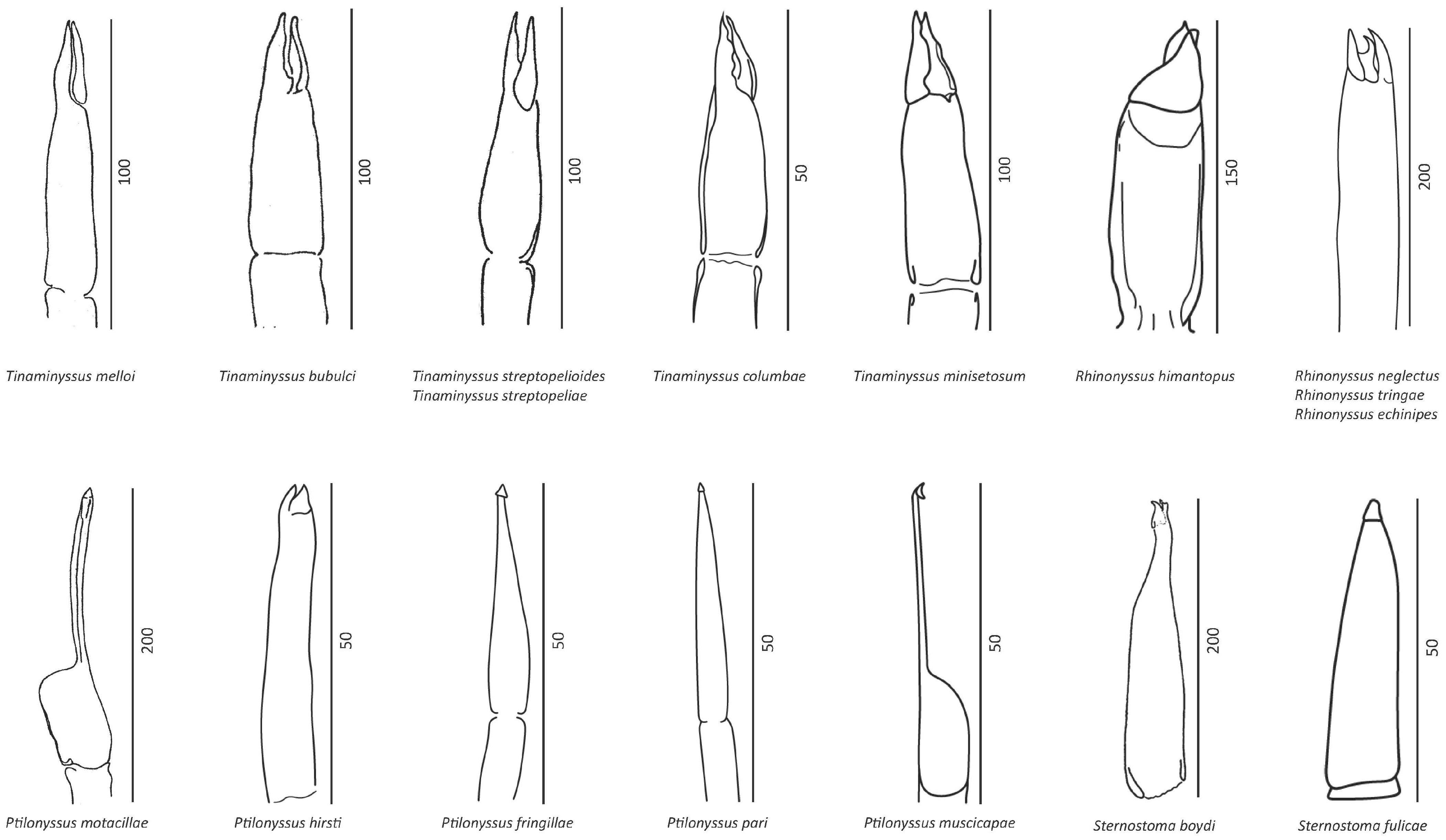
2.1. Morphometrical Study and Identification

For an accurate identification of each of the species and populations which was considered, illustrations from the dorsal and the ventral views were made on paper using a Nikon Eclipse Microscope with a camera lucida.
All specimens from each population were analyzed using the discriminatory morphological features considered by different authors. In addition, a brief description of each genus is reported here.
For Tinaminyssus spp., the most important morphometrical features for discriminating the species are the shape of the chelicerae, a length of 450–630 µm, the presence and shape of the poststigmatal shields, chaetotaxy and the shape of the dorsal podosomal shield, the arrangement of the z6 setae, the length of the Jv1 and Jv2 setae, the gnathosomal formula, the number of ventral setae and convexitas coxae, and the presence of spina coxae [27,28,29].
Ptilonyssus spp. are mites with oblong bodies 380–760 µm long. The dorsal side of the podosoma has only a podosomal shield, the dorsal side of the opisthosoma has only one shield (either opisthosomal or pygidial), and the stigma has oblong peritremes and is located dorsolaterally. There can be presence or absence of the mesosomal shield, an absent poststigmal shield, terminally located gnathosoma, presence or absence of the deutosternal denticles, absence of the tritosternum, presence or absence of the sternal shield, presence of genital and anal shields, an anus with the anal shield located ventrally, the presence of asperos, and absence of the convexitas and spina coxae [3,7,13,16,27,28,30,31].
The Rhinonyssus species consists of mites with generally rounded idiosomes, a length of 430–970 µm, the presence of a podosomal shield, the absence of opisthosomal and pygidial shields, dorsolaterally located stigmata without peritrems, presence or absence of mesosomal shields, the absence of a poststigmal shield, terminally located gnathosoma, the absence of deutosternal denticles, the absence of a tritosternum, presence or absence of the primary scutum, the presence of a genital shield, an anal shield that is present or strongly reduced, an anus located on the ventral or dorsal side of the opisthosoma, the presence or absence of asperos, and an absence of convexities and spines on the coxae [7,15,16,27,28,30,32,33].
The Sternostoma species is characterized by an oval body and a length of 330–630 µm. On the idiosoma, the two dorsal shields are podosomal and opisthosomal. Pygidial shields are absent. The stigmata are located dorsolaterally, and the peritremes are absent. Mesosomal shields are present or absent. Poststigmal shields are absent. The gnathosoma are located ventrally, and only the most distal part can extend beyond the frontal edge of the idiosoma. Deutosternal denticles are present or absent. The tritosternum is absent. Sternal, genital, and anal shields are present. The anus has an anal shield situated ventrally or terminally. Asperos can be present or absent [3,7,16,27,28,31,32,34].
Moreover, twelve morphometrical measures and two ratios between the measures (LOS/WOS and LPS/WPS) were retrieved. Morphometrical measurements of the dorsal and ventral sides of Tinaminyssus spp., Ptilonyssus spp., Rhinonyssus spp., and Sternostoma spp. were made, namely, the length of the body (including palps) (LB), the width of the idiosoma (WI), the length of the podosomal shield (LPS), the width of the podosomal shield (WPS), the length of the opisthosomal shield (LOS), the width of the opisthosomal shield (WOS), the length of the anal shield (LAS), the width of the anal shield (WAS), the length of the ventral view of the gnathosoma (including palps) (LG), and the width of the gnathosoma (WG) [26].
Furthermore, in 2016, Dimov [35] introduced abbreviations and Latin terms for the setae and some other structures, as follows: MS, mesosomal shield (mesosomal scutulum); LMSL, length of the left mesosomal shield; LMSR, length of the right mesosomal shield; WMSL, width of the left mesosomal shield; WMSR, width of the right mesosomal shield; PSS, poststigmal shield; PgS, pygidial shield; Dd, deutosternal denticles; Cc, convexity on the coxa (convexitas coxae); Sc, spines on the coxa (spina coxae); α1–α4, microphytes of the opistosomal shield; dch, chelicera finger; bch, base of the chelicera; An, anus; LPgS, length of the pygidial shield; WPgS, width of the pygidial shield. Some of these features have been considered for morphological identification.

2.2. Molecular Study

DNA was isolated from single mites using the NucleoSpin® Tissue XS DNA extraction kit manufactured by Macherey-Nagel (GmbH & Co. KG, Düren, Germany). Briefly, the mites were coarsely crushed with a plastic pestle in a 1.5 mL microcentrifuge tube containing 20 μL of an extraction buffer according to the manufacturer’s protocol. Each sample was checked on SYBR Safe stained with 2% Tris-borate-ethylenediaminetetraacetic acid and 0.8% agarose gel. The PCR mix for amplifying the ITS region consisted of 10 μL of 10× PCR buffer, 1 μL of 10 mM dNTP, 6 μL and 50 mM MgCl2, 1.5 μL of 10 μM forward primer, 1.5 μL of 10 μM reverse primer, 10 μL of template DNA, and 0.5 μL of Taq DNA polymerase (2.5 units). The samples were autoclaved with double-distilled water for a final volume of 50 μL.
For amplification of the ITS region, the initial denaturation step was carried out at 94 °C for 2 min, followed by 35 cycles of 20 s at 94 °C for denaturation, 30 s at 53 °C for annealing the primers, and 45 s at 72 °C for primer extension. A final extension step was carried out for 7 min at 72 °C. The primers were as follows: forward, 5-AGAGGAAGTAAAAGTCGTAACAAG-3′; reverse, 5-ATATGCTTAAATTCAGGGGG-3 [36].
The PCR products were sequenced in two directions using the Sanger method by the commercial company Allgenetics (A Coruña, Spain). GenBank accession numbers for the sequenced samples are provided in Supplementary Table S1.

Sequence Alignments and Phylogenetic Analyses
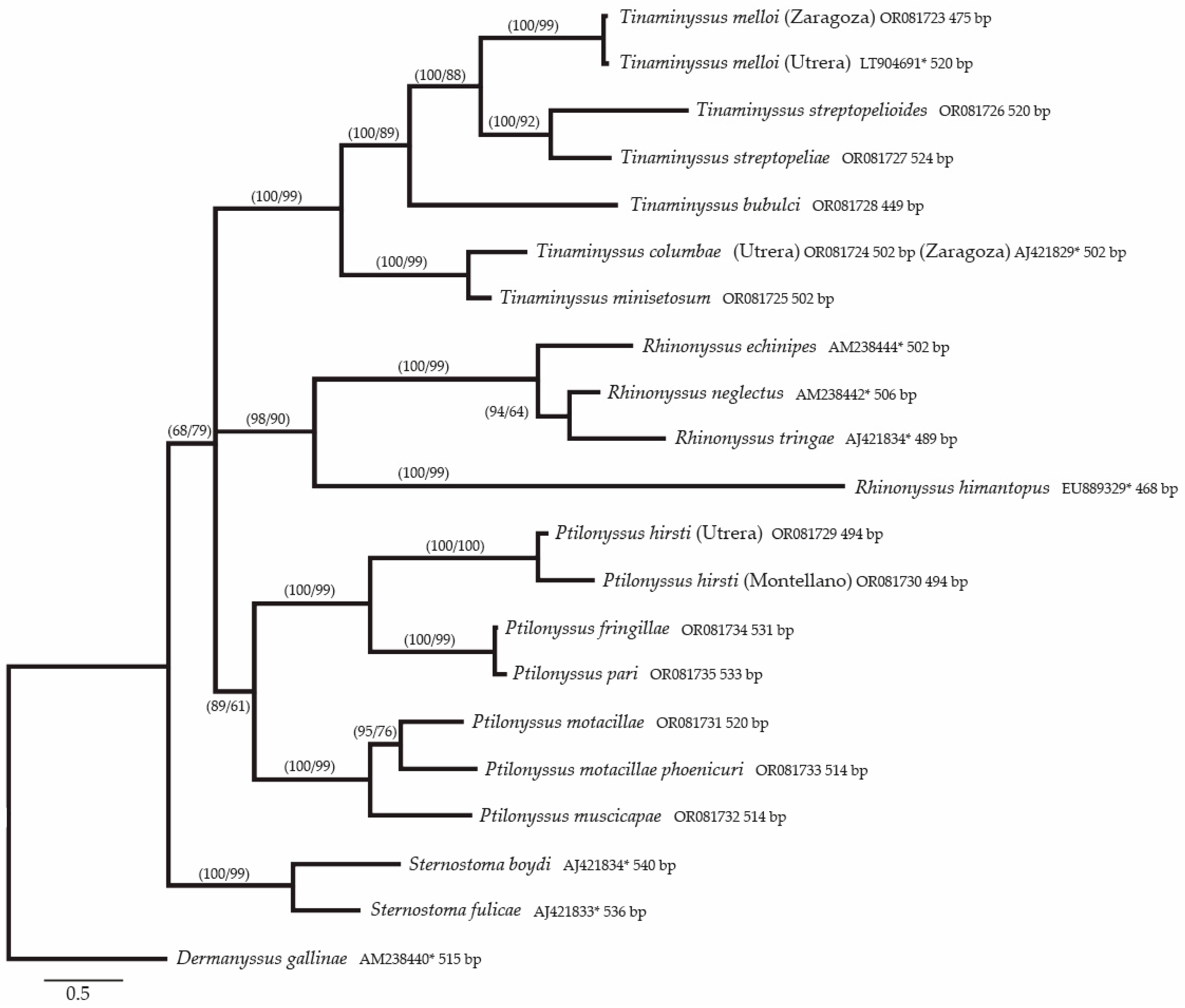
The sequences of the ITS region were aligned using MEGA v.5.2 [37] and adjusted manually, whereas DAMBE v.5.0 [38] was used to optimize the alignments. Pairwise distance matrices were generated using the Kimura two-parameter model (K2P) [39].
Phylogenetic trees were inferred on the basis of the nucleotide data and built using two methods. Maximum likelihood (ML) trees were generated using PHYML v.3.0 [40], and Bayesian inference (BI) trees were built using Mr. Bayes v.3.2.6 [41]. JMODELTEST 2.1.10 [42] was used to select the best-fitting model of nucleotide substitution for the alignment of the ITS region. Models of sequence evolution were chosen for the subsequent analyses according to the Akaike Information Criterion [43,44]. The best-fitting nucleotide substitution models selected by JMODELTEST included a general time-reversible model with a gamma-distributed rate variation of GTR + G for the ITS region. These nucleotide substitution models were used to analyze the phylogenetic relationships using PHYML. Support for the topology was examined by bootstrapping the original dataset 1000 times (the heuristic option) [45]. The best-fitting nucleotide models selected by JMODELTEST and available for BI in MrBayes were GTR + G (nst = 6 with gamma rates) For BI, the standard deviation of the splitting frequency was used to assess convergence; the chains were sampled every 500 generations and each dataset was run for 10 million generations. Burn-in was determined empirically by examining the log-likelihood values of the chains. Phylogenetic trees based on the ITS region were rooted with a species of the superfamily Dermanyssoidea, namely, Dermanyssus gallinae.

3. Results

3.1. Morphometrical Identification

All mites were identified strictly on the basis of the descriptions, measurements, and the best illustrations and redescriptions of females that different authors have carried out, with which the specimens considered in this study were compared. Thus, a summary of the most important discriminatory traits is reported in Supplementary Table S3.
Previous works were used to identify the species [3,6,7,13,15,16,18,19,27,27,31,32,46,47,48,49,50,51,52,53,54,55,56,57,58,59,60,61,62,63]. The specific publications are presented in Supplementary Table S3 within the descriptions of each identified species.

Molecular Results

There was little variability in the lengths of the different sequences, which ranged between 449 and 536 bp, with an average of 506 bp.
The nucleotide composition (Supplementary Table S4) showed a higher content of adenine and thymine (29.6% and 31.7%, respectively), while cytosine had the lowest concentration (14.5%), followed by guanine (24.2%). In the collection of these data, the values of the external elements were not considered.
Table 1 shows the fragment lengths (in base pairs) of the ITS1, 5.8S, and ITS2 fragments. The boundaries were determined by using the conserved areas flanking the 18S, 5.8S, and 28S sequences according to Navajas et al. [36] and de Rojas et al. [64].
Thus, when the fragments of the ITS1–5.8S–TS2 region of the species considered in the present study were aligned, an alignment was obtained with 576 positions, including gaps, with 197 conserved and 353 variables.
The pairwise distance matrix produced by a pairwise comparison of all considered taxa is given in Supplementary Table S5, which also includes the percentages of similarity between the taxa. Table 2 shows a summary of the intergeneric, interspecific, and intrapopulation distances, as well as the percentage of similarity.

3.2. Phylogenetic Study of the Species Included in the Alignment

In the analysis of the sequences aligned with Mega version 11.01 and Clustal W version 2.0, an alignment was obtained for the ITS1–5.8S–ITS2 fragment of 576 base pairs, finding 197 preserved positions and 353 variables.
Phylogenetic trees were constructed as explained in the Materials and Methods section. The topology of the Bayesian tree, including all the considered taxa with Dermanyssus gallinae as an outgroup, is shown in Figure 1. Each node in this tree shows the Bayesian posterior probabilities (PP) followed by the ML bootstrap values (BS).

4. Discussion

The question of delimiting species in molecular genetics remains a problem to be solved because there is no objective limit which indicates that two species are distinct [24]. However, Dobler and Farrell [65] observed that, in the case of parasitic organisms, “gene flow between parasite populations in different host species may be limited, even if the parasites are not 100% specific”. This structure can favor the formation of races or speciation.
The family Rhinonyssidae is believed to have descended from ectoparasitic ancestors related to the Macronyssidae [4]. Rhinonyssid genera vary in their degrees of host specificity, with some genera being restricted to one host family and others found in hosts from different orders [48]. In North America, genera with broad host ranges are Ptilonyssus (passeriform, caprimulgiform, falconiform, and apodiform hosts), Rhinonyssus (anseriform, podicipediform, and charadriiform hosts), Sternostoma (passeriform, piciform, and charadriiform hosts), and Tinaminyssus (ciconiiform and columbiform hosts). Among the more host-specific rhinonyssids, Rhinoecius species parasitize owls (Strigiformes), Rallinyssus species parasitize rails (Rallidae), and Larinyssus species parasitize gulls and terns (Laridae) [3].

4.1. Morphological Analysis

Throughout the course of their study, the principles governing the construction of the natural rhinonyssid system have undergone significant changes. These changes have often required a complete review of the supraspecific classification, with the most challenging aspect being the development of criteria for determining the genera and subfamily ranks, such as the morphology of the chelicerae (Figure 2). Thus, firstly, an accurate identification of the studied species is needed. In the Supplementary Materials, Table S3, a brief description including all discriminatory characteristics of the species identified in this study is shown.
It is noteworthy that, with regard to the genus Tinaminyssus, in 1948, Castro [13] described Neonyssus melloi as a new species from some specimens found in the nasal cavities of Columba livia domestica L. According to this author, the new species was characterized by its very strong legs with highly developed nails, a gnathosome that was small in size in relation to the body, chelicerae of uniform diameter, and peritremal scutes surrounding the stigmata.
In 1960, Fain [6] created the genus Mesonyssus to include the Neonyssus species endowed with long cheliceral fingers. Therefore, Neonyssus melloi became part of the new genus Mesonyssus. However, in 1958, Strandtmann and Wharton [9] had defined the genus Tinaminyssus on the basis of a series of characteristics, including all those assigned by Fain to the genus Mesonyssus. Therefore, the generic name proposed by Strandtmann and Wharton had priority, as indicated by Domrow in 1969 [27], who considered that the correct name for the genus should be Tinaminyssus.

4.2. Molecular Analysis

As shown in the Supplementary Materials, Table S4, the length of ITS1 varied between 232 bp in Sternostoma fulicae and 193 in Rhinonyssus tringae. The 5.8S rDNA gene ranged from 189 bp in Ptilonyssus hirsti to 177 in Rhinonyssus echinipes. The ITS2 fragment was less variable in size, with all species showing values very close to 90. These values are very similar to those found by de Rojas et al. [64] and Úbeda et al. [66]. In addition, according to the GenBank database, members of the Dermanysoidea superfamily (Dermanyssus longipes FM179377 and Androlaelaps casalis AM903317) showed similar values for ITS1 and for the 5.8s rDNA gene, with the ITS2 region being significantly shorter (73 and 62 bp, respectively).
The contents of the different bases are shown in Supplementary Table S4 as percentages. As can be seen in Table S4, our results were practically identical to those obtained by de Rojas et al. [64] for a smaller number of rhinonyssid species.
Likewise, the lengths of the markers considered in the different species studied herein, as shown in Table S4, were also similar to those obtained by Úbeda et al. [66] for species of the genus Rhinonyssus, ranging from 468 bp for R. himantopus to 506 in R. neglectus to 540 bp for Sternostoma boydi [64], as well as to those of other representatives of the superfamily Dermanysoidea, such as Dermanyssus longipes (accession number FM179377).
With regard to the variability of the different fragments (Table 1), as expected, the 5.8S rDNA gene was the most conserved region, while the ITS1 region was the longest and most variable. Finally, ITS2 was the shortest and, unusually, quite preserved fragment, at least among species from the same genus. This fact was observed by Navajas et al. [36] in mites of the family Phytoseiidae, and by de Rojas et al. [64] in representatives of the Rhinonyssidae.

4.3. Analysis of Phylogenetic and Genetic Distances

It was not until 1979 that Pence attempted to address phylogeny in this family of mites. Pence, on the basis of Castro’s observations [13], hypothesized that the chelicerae would have fairly stable morphological characteristics, since they are related to nutrition. According to this hypothesis, the rhinonyssid mites would come from some ancestor of the Macronysidae in which the morphology of the chelicerae had some similarity to those of the genus Tinaminyssus [67]. Figure 2 shows drawings of the chelicerae of all species included in this study.
Thus, according to the phylogenetic tree created as explained in the Materials and Methods sections and shown in the results (Figure 1), several questions can be answered.
The topology of the tree shown in Figure 1 corresponds to that created via the Bayesian inference method, and is similar to that obtained via the maximum likelihood method. However, in the latter case, the bootstrap values were slightly lower than the Bayesian probabilities. In both cases, the four genera considered in this study were shown to be in different clades. This observation coincides with the morphological proposal of Pence (1979) [68] regarding the classification of genera based on the morphology of the chelicerae. However, species of the genus Sternostoma appeared in a separate branch, and the species of the other three genera were gathered together in a polytomy, which means that a more conserved marker must be tested to resolve the phylogeny at the genus level.
The clade that includes the species of the genus Tinaminyssus appears in a common branch with Bayesian posterior probabilities (PP) and ML support (BS) of 100/99. Within this group, two different branches can be distinguished, depending on the morphological characteristics: the “melloi” complex (T. melloi, T. streptopelioides, and T. streptopeliae) and the “columbae” complex (T. minisetosum and T. columbae) [18]. However, Tinaminyssus bubulci, a parasite of pelecaniform birds, appears in the “melloi” group with the species parasitizing columbiform birds (100/99). Furthermore, two of the morphological criteria for including a species in the “columbae” group are a lack of postanal setae and barely developed opisthosomal setae. T. bubulci shows numerous well developed opisthosomal setae (typical of the “melloi” group), but lacks the postanal setae. Thus, this apparent morphological and ecological contradiction must be tested by assaying more isolated or concatenated molecular markers in order to resolve it.
Moreover, T. streptopeliae from and T. streptopelioides show very similar morphologies and measurements that overlap. They are gathered together (100/92), and the genetic distance between them (0.11) is similar to that between T. streptopeliae and T. melloi (0.14), which are different species. This agrees with de Rojas et al. [26], who considered the former species to be a cryptic species.
Finally, T. columbae and T. minisetosum, which were included in the “columbae” group, were gathered together with high support (100/99). However, although these two species were considered to be different species, the genetic distance was the lowest among the species of Tinaminyssus (0.05).
The genus Ptilonyssus is the most numerous in the family Rhinonyssidae. Currently, there is no consensus among acarologists about its taxonomic boundaries nor the number of species which should be included [31]. According to the morphological signs, some species of the genus have been grouped into species complexes on the basis of the chaetotaxy of the podosomal shield on the dorsal aspect of the idiosoma, namely, the “hirsti”, “lanii”, “motacillae”, “orthonychus”, “pari”, and “sairae” groups [68]. This study included three species belonging to the “hirsti” complex, three belonging to the “sairae” complex, and one belonging to the “pari” complex.
The topology of the Bayesian phylogenetic tree included two sister branches with high to medium support (89/61) that contained species of the “hirsti” complex and the “sairae” complex. The former was supported with 100/99, and gathered species with chelicerae without widened bases. Within this group, two different populations of P. hirsti were gathered together with high support (100/100). The other branch gathered P. fringillae and P. pari from Russia (100/100). In this case, the genetic distance between these species was 0.1, even lower than that between populations of P. hirsti. However, P. pari from Parus atricapillus was described by Fain and Hyland [53] on the basis of pairwise comparisons among P. capensis, P. emberizae, P. chloris, and P. hirsti. In 1974, Fain and Sixl [52] described a new species: P. fringillae. When drawings of the specimens were compared, these species showed the same number of shields, but with slight differences in their development. Thus, two of the dorsal opisthosomal seta were included in the opisthosomal shield in P. fringillae, but not in P. pari. Furthermore, P. pari has two poststigmatic shields that are not shown in P. pari. However, the differences in the development of the shields depend on the specimen and the time of collection, as in T. streptopeliae and T. streptopeliodes [34]. Thus, these traits are not reliable discriminatory characteristics. Therefore, further molecular studies are needed in order to clarify the statuses of P. fringillae and P. pari.
The second branch grouping Ptilonyssus species included P. motacillae, P. motacillae phoenicuri, and P. muscicapae, which show chelicerae that become wider at the base. All these species belonged to the “muscicapae” group, and their clustering was well-supported (100/99). It is noteworthy that the genetic distance between P. motacillae phoenicuri and P. motacillae was 0.08; currently, these are considered to be the same species. However, the genetic distance between P. motacillae and P. muscicapae was not much higher (0.10).
All of these data explain the complexity of delimiting species and determining discriminatory traits in groups of closely related species of the family Rhinonyssidae, and further morphological and molecular studies are needed to establish the statuses of species and subspecies within this group.
The last clade in the polytomy, similar to those discussed previously, was well-supported (98/90) and included species of the genus Rhinonyssus, most of which parasitize birds from frequently wet and marshy habitats [69]. In this study, the authors established three species groups within this genus: the “coniventris” group, the “himantopus” group, and the “rhinolethrum” group.
Species of the genus Rhinonyssus were also grouped into a well-supported common trunk (98/90) with two branches. One branch included species belonging to the “coniventris” complex, while the other included R. himantopus, which belonged to the “himantopus” group. Our molecular results were consistent with the morphological data, as R. himantopus, which did not belong to the “coniventris” group, had a very different morphology. Moreover, the distances between R. himantopus and the other species of the genus Rhinonyssus were very high, similar to those obtained between R. himantopus and species from any other genera, ranging from 0.45 to 0.48. For this reason, it would be interesting to study more species from the “himantopus” group, because the cheliceral morphology is slightly different from that of the “coniventris” group.
The most similar closely related species in the family Rhinonyssidae were placed in the “coniventris” group, which Fain established in 1963 to include a series of species that were causing confusion and controversy among Rhinonyssidae specialists. The species that gave its name to the complex is Rhinonyssus coniventris [11].
Individuals of these species have a single poorly sclerotized dorsal shield that, in certain cases, has become so reduced that it practically only includes the muscular insertion areas characteristic of this region of the idiosoma. The sternal shield does not exist as such, and the genital shield is almost always much longer than it is wide. No anal shield can be observed. In the present study, numerous specimens of mites were located and studied, and their morphological characteristics allowed them to be included in the species Rhinonyssus echinipes [57], R. neglectus [57], and R. tringae [54]. These three species show few morphological differences.
In fact, in 1951, Strandtman [15], discussing the Rhinonyssus species described by Hirst, indicated that it was quite probable that a re-examination of the types of R. neglectus [57] and R. echinipes [57], in the light of abundant material from many hosts, would show that they were conspecific with R. coniventris [11]. Later, in 1956, the species R. neglectus [57] and R. echinipes [57] were considered to be synonyms of R. coniventris [34].
In 1963, Fain concluded that: R. coniventris is a polytypic species that includes three subspecies (R. coniventris coniventris, R. c. neglectus and R. c. echinipes), each restricted to one or several closely related genera of Charadriiformes [54].
Finally, in 1984, Butenko [32] concluded that the “coniventris” group included five forms that were assigned species status: R. coniventris [11], R. echinipes [57], R. neglectus [57], R. shcherbininae [32], and R. tringae [54].
In this study, species of the “coniventris” group were gathered together in a well-supported common branch (100/99). However, it is noteworthy that the genetic distances among these three species were 0.07, 0.08, and 0.10, which are similar to those between P. motacillae and P. motacillae phoenicuri (0.08), or between P. motacillae and P. muscicapae, which are very similar (0.10).
Two species of this complex group, R. neglectus and R. tringae, were grouped together and separated from R. echinipes. This branch was well-supported in terms of PP (94) and had medium ML bootstrapping support (64).
Furthermore, the grouping of R. tringae and R. neglectus in the same branch could be explained by the preference of these two species for Charadriiformes hosts of the Scolopacidae family (Tringa totanus and Calidris alpina, respectively). In contrast, R. echinipes is only found in Charadriiforme hosts of the Charadriidae family.
The combination of the molecular analyses and morphological results suggests that the taxonomic statuses of R. echinipes [57], R. neglectus [57], and R. tringae [54] can be accepted as species, although it would be convenient to prove this by means of other molecular markers. Thus, the molecular data confirm our morphological and ecological observations.
Finally, the only branch grouping species from the same genus, which was not present in the polytomy and was well-supported (100/99), included two species of the genus Sternostoma.
In this study, only two species that parasitize aquatic birds could have been included. Sternostoma boydi from Larus argentatus and S. fulicae from Fulica atra. These two species are easily distinguishable on the basis of their morphological traits. However, the genetic distance (0.10) is similar to that of closely related species. This fact could indicate a case of phenotypic plasticity for adaptation to very distantly related birds, namely, the charadriformes and gruiformes.
From all of these data, it can be concluded that, although the ITS1–5.8S–ITS2 fragment solves different taxonomic and phylogenetic problems at the species level, it would be necessary to test new molecular markers that resolve the intergeneric relationships and to concatenate two or more molecular markers to resolve the identification of species in complexes that include very closely related species. Even the combination of nuclear and mitochondrial markers or different domains of the nuclear 28S rDNA would be convenient for solving these situations. Furthermore, a thorough review and an accurate morphometrical identification and determination of the discriminatory traits are needed for this group of mites.

Supplementary Materials

The following supporting information can be downloaded at: https://www.mdpi.com/article/10.3390/microorganisms11071783/s1. Table S1. Species included in this study. Hosts, geographical origin, and number of GenBank Access of the fragment ITS1-5.8S-ITS2 are shown. (*) Sequences retrieved from GenBank. Table S2. Epidemiological information of each parasitic mite species in each host species. The table shows the prevalence with 95% confidence intervals for the number of studied hosts > 2 (in brackets), the mean intensities with their standard errors, and the range of mites collected per host (in brackets). Total number of each mite species collected per host species (n) and total number of infested hosts with each mite species are also shown. Table S3. Morphological description of the species included in this study with the references used for identification. Table S4. Nucleotide composition of the species sequences described in this study. Table S5. Matrix of genetic distances (lower left corner) and percentages of similarity between them (upper right corner) between the different species considered in this study.

Author Contributions

I.D., F.J.M.J. and M.d.R.Á. conceived this study. I.D., S.A.S.-C. and M.d.R.Á. analyzed the data. F.J.M.J. supervised the research. M.d.R.Á. wrote the first draft, which was revised by all other authors. All authors have read and agreed to the published version of the manuscript.

Funding

This research was funded by V Plan Propio de Investigación de la Universidad de Sevilla, Spain grant to Department of Microbiology and Parasitology. University of Seville.

Data Availability Statement

Sequences of each of the species analyzed in this study are available from NCBI (GenBank accession numbers: from OR081723 to OR081735).

Acknowledgments

We thank Centro Municipal Zoosanitario de Sevilla, and especially Francisco Peña Fernández and Rafael Cuadrado Nieto, for providing some of the samples. We also give thanks to Miguel Fernández Almenara for bird identifications.

Conflicts of Interest

The authors declare no conflict of interest.

References

  1. Knee, W.; Galloway, T.D. New Host and Locality Records for Endoparasitic Nasal Mites (Acari: Rhinonyssidae, Turbinoptidae, and Ereynetidae) Infesting Birds in Manitoba, Canada. Can. Entomol. 2017, 149, 89–103. [Google Scholar] [CrossRef] [Green Version]
  2. Dimov, I. Rhinonyssidosis avium. VetPharma 2011, 3–4, 88–90. [Google Scholar]
  3. Knee, W. Five New Species of Rhinonyssidae (Mesostigmata) and One New Species of Dermanyssus (Mesostigmata: Dermanyssidae) from Birds of Alberta and Manitoba, Canada. J. Parasitol. 2008, 94, 348–374. [Google Scholar] [CrossRef] [PubMed]
  4. Strandtmann, R.W. The Mesostigmatic Nasal Mites of Birds. I. Two New Genera from Shore and Marsh Birds. J. Parasitol. 1948, 34, 505. [Google Scholar] [CrossRef]
  5. Bregetova, N.G. Generic grouping of the rhinonyssid mites (Mesostigmata, Rhinonyssidae), parasites of birds. I. The new genus Sternostomoides. Ent. Obozr. 1965, 44, 709–713. [Google Scholar]
  6. Fain, A. Comparative Morphology of Rhinonyssidae. Bull. Ann. Soc. Roy. Ent. Belg. 1960, 96, 303–313. [Google Scholar]
  7. Vitzthum, H.G. Milben aus der Nasenhöhle von Vögeln. J. Fur Ornithol. 1935, 83, 563–587. [Google Scholar] [CrossRef]
  8. Berlese, A.; Trouessart, E. Diagnoses of New or Little-Known Mites. Bull. Bibl. Sci. Quest 1889, 2, 121–143. [Google Scholar]
  9. Strandtmann, R.W.; Wharton, G.W. A Manual of Mesostigmatid Mites Parasitic on Vertebrates; Institute of Allergy: College Park, MD, USA, 1958; Volume 4, 330p. [Google Scholar]
  10. Cooreman, J. Rhinoecius oti n. Gen., n. Sp. Acarien: Rhinonyssidae. Bull. Mus. Hist. Nat. Begl. 1946, 22, 1–4. [Google Scholar]
  11. Trouessart, E.L. Note sur les Acariens Parasites des Fosses Nasales des Oiseaux. C R Soc. Biol. IDe Ser. 1894, 1, 723–724. [Google Scholar]
  12. Strandtmann, R.W. Host Specificity of Bird Nasal Mites (Rhinonyssidae) Is a Function of the Gregariousness of the Host. In Proceedings of the 10th International Congress of Entomology, Montreal, QC, Canada, 17–25 August 1956; Volume 1, pp. 909–911. [Google Scholar]
  13. Castro, M.P. Reestruturacao Generica of the Family Rhinonyssidae Vitzhum, 1935 (Acari: Mesostigmata: Gamasides) and Descricao of Some Species Novas. Arch. Int. Biol. 1948, 18, 253–284. [Google Scholar]
  14. Giebel, C. Ueber einige Milben. Z. Ges. Naturw. 1871, 38, 29–32. [Google Scholar]
  15. Strandtmann, R.W. The Mesostigmatic Nasal Mites of Birds. II. New and Poorly Known Species of Rhinonyssidae. J. Parasitol. 1951, 37, 129–140. [Google Scholar] [CrossRef] [PubMed]
  16. Fain, A. Essai de clasiffication des Rhinonyssidae (Acari: Mesostigmata) avec description de deux genres nouveaux. Ann. Parasitol. Hum. Comp. 1957, 31, 145–157. [Google Scholar]
  17. Pence, D.B.; Casto, S.D. Studies on the Variation and Morphology of the Ptilonyssus “Sairae” Complex (Agarina: Rhinonyssinae) from North American Passeriform Birds. J. Med. Entomol. 1976, 13, 71–95. [Google Scholar] [CrossRef]
  18. Fain, A. Les Rhinonyssides Parasites Des Pigeons. Rev. Zool. Bot. Afr. 1962, 65, 305–324. [Google Scholar]
  19. Fain, A. Le Complexe “Rhinonyssus Coniventris”(Rhinonyssidae: Mesostigmata). Bull. Ann. Soc. R Entomolog. Belg. 1963, 99, 86–100. [Google Scholar]
  20. Huyse, T.; Poulin, R.; Théron, A. Speciation in Parasites: A Population Genetics Approach. Trends Parasitol. 2005, 21, 469–475. [Google Scholar] [CrossRef] [PubMed]
  21. Sunantaraporn, S.; Sanprasert, V.; Pengsakul, T. Molecular Survey of the Head Louse Pediculus Humanus Capitis in Thailand and Its potential Role for Transmitting Acinetobacter spp. Parasites Vectors 2015, 8, 127. [Google Scholar] [CrossRef] [Green Version]
  22. Kelomey, A.E.; Paraiso, A.; Sina, H.; Legout, H.; Garnery, L.; Baba-Moussa, L. Genetic Characterization of the Honeybee Ectoparasitic Mite Varroa destructor from Benin (West Africa) Using Mitochondrial and Microsatellite Markers. Exp. Appl. Acarol. 2017, 72, 61–67. [Google Scholar] [CrossRef]
  23. Baulieu, F.; Knee, W.; Nowell, V.; Schwarzfeld, M.; Lindo, Z.; Behan-Pelletier, V.M.; Lumley, L.; Young, M.R.; Smith, I.; Proctor, H.C.; et al. Acari of Canada. Zookeys 2019, 819, 77–168. [Google Scholar] [CrossRef]
  24. Morelli, M.; Spicer, G.S. Cospeciation between the Nasal Mite Ptilonyssus sairae (Acari: Rhinonyssidae) and Its Bird Hosts. Syst. Appl. Acarol. 2007, 12, 3. [Google Scholar] [CrossRef] [Green Version]
  25. De Rojas, M.; Mora, M.D.; Ubeda, J.M.; Cutillas, C.; Navajas, M.; Guevara, D.C. Phylogenetic Relationships in Rhinonyssid Mites (Acari: Rhinonyssidae) Based on Mitochondrial 16S RDNA Sequences. Exp. Appl. Acarol. 2001, 25, 957–967. [Google Scholar] [CrossRef]
  26. Rojas, D.; Doña, M.; Jovani, J.; Dimov, R.; Zurita, I.; Callejon, A. Evidence of Cryptic Species in the Genus Tinaminyssus (Acari: Rhinonyssidae) Based on Morphometrical and Molecular Data. Exp. Appl. Acarol. 2018, 75, 355–391. [Google Scholar] [CrossRef]
  27. Domrow, R. The Nasal Mites of Queensland Birds (Acari: Dermanyssidae, Ereynetidae, and Epidermoptidae). Proc. Linn. Soc. 1969, 93, 297–426. [Google Scholar]
  28. Pence, D.B.; Casto, S. Two New Species and New Records of Nasal Mites of the Genus Sternostoma (Acarina: Rhinonyssinae) from Birds in Texas. J. Parasitol. 1975, 61, 360–368. [Google Scholar] [CrossRef] [PubMed]
  29. Braza, R.; Úbeda Ontiveros, J.M.; Guevara Benítez, D.C. Ácaros del Género Tinaminyssus Strandtmann y Wharton, 1958, Parásitos Nasícolas de Aves Ciconiformes y Columbiformes Españolas. Rev. Ibérica Parasitol. 1990, 50, 73–80. [Google Scholar]
  30. Dimov, I. A New Nasal Mite of the Genus Ptilonyssus (Rhinonyssidae) from Parus caeruleus (Passeriformes) from Russia. J. Hell. Vet. Med. Soc. 2017, 63, 25. [Google Scholar] [CrossRef] [Green Version]
  31. Dimov, I. Three New Species of Nasal Mites Ptilonyssus (Mesostigmata, Rhinonyssidae) from Russia. Arhimed-J. Sci. Pract. 2020, 9, 41–52. [Google Scholar]
  32. Butenko, O.M. Rhinonyssid Mites of Non-Passerine Birds of the USSR; Academy of Sciences of USSR: Moscow, Russia, 1984; p. 188. [Google Scholar]
  33. Kaneko, K.; Matsudaira, Y.; Masahito, P. Endoparasitic mites of anatid birds collected in Chiba and Saitama prefectures, Japan (Acarina: Rhinonyssidae and Ereynetidae). Jpn. Soc. Med. Entomol. Zool. 1978, 29, 147–154. [Google Scholar] [CrossRef] [Green Version]
  34. Strandtmann, R.V. The Mesostigmatic Nasal Mites of Birds. IV. The Species and Hosts of the Genus Rhinonyssus (Acarina, Rhinonyssidae). Proc. Entom. Soc. Wash. 1956, 58, 129–142. [Google Scholar]
  35. Dimov, I. Taxonomic Diversity and Morphology of Mites of the Family Rhinonyssidae of the Northwest of Russia; ZIN RAS: St. Petersburg, Russia, 2000; 214p, ISBN 978-5-6044687-1-5. [Google Scholar]
  36. Navajas, M.; Lagnel, J.; Fauvel, G.; De Moraes, G. Sequence Variation of Ribosomal Internal Transcribed Spacers (ITS) in Commercially Important Phytoseiidae Mites. Exp. Appl. Acarol. 1999, 23, 851–859. [Google Scholar] [CrossRef] [PubMed]
  37. Tamura, K.; Peterson, D.; Peterson, N.; Stecher, G.; Nei, M.; Kumar, S. MEGA5: Molecular Evolutionary Genetics Analysis Using Maximum Likelihood, Evolutionary Distance, and Maximum Parsimony Methods. Mol. Biol. Evol. 2011, 28, 2731–2739. [Google Scholar] [CrossRef] [PubMed] [Green Version]
  38. Xia, X. DAMBE5: A Comprehensive Software Package for Data Analysis in Molecular Biology and Evolution. Mol. Biol. Evol. 2013, 30, 1720–1728. [Google Scholar] [CrossRef] [Green Version]
  39. Kimura, M. A Simple Method for Estimating Evolutionary Rates of Base Substitutions through Comparative Studies of Nucleotide Sequences. J. Mol. Evol. 1980, 16, 111–120. [Google Scholar] [CrossRef]
  40. Guindon, S.; Dufayard, J.F.; Lefort, V.; Anisimova, M.; Hordijk, W.; Gascuel, O. New algorithms and methods to estimate max-imum-likelihood phylogenies: Assessing the performance of PhyML 3.0. Syst. Biol. 2010, 59, 307–321. [Google Scholar] [CrossRef] [Green Version]
  41. Ronquist, F.; Huelsenbeck, J.P. MrBayes 3: Bayesian phylogenetic inference under mixed models. Bioinformatics 2003, 19, 1572–1574. [Google Scholar] [CrossRef] [Green Version]
  42. Posada, D. jModelTest: Phylogenetic Model Averaging. Mol. Biol. Evol. 2008, 25, 1253–1256. [Google Scholar] [CrossRef]
  43. Huelsenbeck, J.P.; Rannala, B. Phylogenetic Methods Come of Age: Testing Hypotheses in an Evolutionary Context. Science 1997, 276, 227–232. [Google Scholar] [CrossRef] [Green Version]
  44. Posada, D.; Buckley, T.R. Model Selection and Model Averaging in Phylogenetics: Advantages of Akaike Information Criterion and Bayesian Approaches over Likelihood Ratio Tests. Syst. Biol. 2004, 53, 793–808. [Google Scholar] [CrossRef]
  45. Felsenstein, J. Confidence limits on phylogenies: An approach using the bootstrap. Evolution 1985, 39, 783–791. [Google Scholar] [CrossRef]
  46. Domrow, R. Acari Spinturnicidae from Australia and New Guinea. Acarologia 1972, 13, 552–584. [Google Scholar]
  47. Pence, D.B. The Nasal Mites of Birds from Louisiana. I. Dermanyssids (Rhinonyssinae) from Shore and Marsh Birds. J. Parasitol. 1972, 58, 153–168. [Google Scholar] [CrossRef]
  48. Pence, D.B. The Nasal Mites of Birds from Louisiana. IX. Synopsis. J. Parasitol. 1973, 59, 881. [Google Scholar] [CrossRef]
  49. Zumpt, F.; Till, W.M. Nasal Mites of Birds Hitherto Known from the Ethiopian Region, with Keys and Descriptions of Nine New Species (Acarina: Laelaptidae). J. Entomol. Soc. South. Afr. 1955, 18, 50–59. [Google Scholar]
  50. Domrow, R. Fourteen Species of Ptilonyssus from Australian Birds (Acarina, Laelaptidae). Acarologia 1964, 6, 595–623. [Google Scholar]
  51. Fain, A.; Sixl, W. Two New Rhinonyssids from Austrian Birds. Bull. Ann. Soc. Roy. Ent. Belg. 1971, 107, 89–93. [Google Scholar]
  52. Fain, A.; Sixl, W.; Moritsch, C. The Nasal Mites of the Family Rhinonyssidae with Description of a New Species (Acarina). Mitteilungen der Abteilung Zool. Bot. Landesmus. Joanneum Graz 1974, 3, 1–9. [Google Scholar]
  53. Fain, A.; Hyland, K. On Three Species of Rhinonyssids Described by Hirst. Ann. Mag. Nat. Hist. 1962, 5, 341–348. [Google Scholar] [CrossRef]
  54. Fain, A. La spermatheque et ses canaux adducteurs chez les acariens mesostigmatiques parasites des voies respiratoires. Acarologia 1963, 5, 463–479. [Google Scholar]
  55. Gretillat, S. Description de Deux Nouvelles Especes de Rhinonyssidae (Acarina, Mesostigmata) Rallinyssus strandtmanni et Larinyssus petiti. Vie et Milleu 1961, 12, 151–160. [Google Scholar]
  56. Bregetova, N.G. Mites from the nasal cavity of Fly carchers of the genus Muscicapa. Parazitologiya 1970, 4, 59–62. [Google Scholar]
  57. Hirst, S. On Some New Parasitic Mites. Proc. Zool. Soc. 1921, 1922, 769–802. [Google Scholar] [CrossRef]
  58. Guevara Benítez, D.; Úbeda Ontiveros, J.M.; Morillas Márquez, F. Acaros Del Genero Ptilonyssus Berlese y Trouessart, 1889 (Mesostigmata: Rhinonyssidae) Parasito de Fosas Nasales de Aves Paseriformes Españolas, 1: Ptilonyssus hirsti (Castro y Pereira, 1947) Pereira y Castro. Rev. Ibérica Parasitol. 1949, 46, 75–80. [Google Scholar]
  59. Bregetova, N. Ticks Parasitizing in the Nasal Cavity of Birds. Parasit Sb. Zool. Inst. Acad. Sci. USSR 1951, 13, 111–119. [Google Scholar]
  60. TerBush, L.E. Incidence of Nasal Mites in Different Age Classes of Herring Gulls (Larus argentatus). J. Parasitol. 1963, 49, 525. [Google Scholar] [CrossRef]
  61. Spicer, G.S. Two New Nasal Mites of the Genus Ptilonyssus (Mesostigmata: Rhinonyssidae) from Texas. Acarologia 1977, 18, 595–601. [Google Scholar]
  62. Fain, A.; Bafort, J. Deux Nouveaux Acariens Parasites Nasicoles Du Rossignol Du Japon Leioth Rix Lutea Swainson. Bull. La Soc. R. Zool. D’anvers 1963, 31, 7–11. [Google Scholar]
  63. Guevara, D.C.; Ontiveros, J.M. Descripción de La Larva y Redescripción de La Hembra de Sternostoma fulicae Fain y Bafort, 1963 (Acarina: Rhinonyssidae). Rev. Iber. Parasitol. 1975, 35, 81–93. [Google Scholar]
  64. de Rojas, M.; Mora, M.D.; Ubeda, J.M.; Cutillas, C.; Navajas, M.; Guevara, D.C. Phylogenetic Relationships in Rhinonyssid Mites (Acari: Rhinonyssidae) Based on Ribosomal DNA Sequences: Insights for the Discrimination of Closely Related Species. Parasitol. Res. 2002, 88, 675–681. [Google Scholar] [CrossRef]
  65. Dobler, S.; Farrell, B.D. Host Use Evolution in Chrysochus Milkweed Beetles: Evidence from Behaviour, Population Genetics and Phylogeny. Mol. Ecol. 1999, 8, 1297–1307. [Google Scholar] [CrossRef] [PubMed]
  66. Úbeda Ontiveros, J.M.; Guevara Benítez, D.C.; Cutillas Barrios, C.; Ariza Astolfi, C.; de Rojas Alvarez, M. Ácaros de las vías respiratorias de vertebrados. In “In Memoriam” al Profesor Doctor D. Ignacio Navarrete López-Cózar; Tovar Andrada, J.J., Reina Esojo, D., Eds.; Facultad de Veterinaria, Universidad de Extremadura: Cáceres, Spain, 2006; ISBN 84-690-2894-4. [Google Scholar]
  67. Dimov, I. Kleshchi-Rinonissidy Ptic Severo-Zapada Rossii; LLC Zhigulin.: Saint Petersburg, Russia, 2018. [Google Scholar]
  68. Pence, D.B. Congruent Inter-Relationships of the Rhinonyssinae (Dermanyssidae) with Their Avian Hosts. In Recent Advances in Acarology; Elsevier: Amsterdam, The Netherlands, 1979; pp. 371–377. ISBN 9780125922029. [Google Scholar]
  69. Strandtmann, R.W. New Records for Rhinonyssus himantopus and Notes on Other Species of the Genus. J. Kans. Entomol. Soc. 1959, 32, 1. [Google Scholar]
Figure 1. Phylogenetic tree (BI topology) of rhinonyssid mites of different species and geographical origins based on the ITS1-5.8S-ITS2 fragment. Values next to the nodes indicate the Bayesian posterior probabilities (PP), followed by the ML bootstrap values (BS). Support is only indicated on nodes with PP > 0.95 and BS > 75%. Each species shows the GB access number and the length of the amplified fragment. (*) Sequences were retrieved from GenBank.
Microorganisms 11 01783 g001
Figure 2. Morphology of the chelicerae of the species identified in this study. Scales are shown in µm.
Microorganisms 11 01783 g002
Table 1. Lengths of transcribed internal spacers 1 and 2 (ITS1, ITS2) and the rDNA gene 5.8 (in base pairs).
SpeciesITS15.8SITS2
Tinaminyssus melloi (Zaragoza)
Castro, 1948
218184 -
Tinaminyssus melloi (Utrera)
Castro, 1948
218 182 90
Tinaminyssus columbae (Utrera)
Castro, 1948
200 182 90
Tinaminyssus columbae (Zaragoza)
Castro, 1948
200 182 90
Tinaminyssus minisetosum
Butenko, 1984
201 181 90
Tinaminyssus streptopelioides
Butenko, 1984
218 182 90
Tinaminyssus streptopeliae
Fain, 1962
218 182 94
Tinaminyssus bubulci
Zumpt & Till, 1955
198 181 -
Ptilonyssus hirsti (Montellano)
Castro, 1948
214 189 -
Ptilonyssus hirsti (Utrera)
Castro, 1948
214 189 87
Ptilonyssus motacillae
Fain, 1966
214 191 82
Ptilonyssus muscicapae
Bregetova, 1970
213 189 82
Ptilonyssus motacillae phoenicuri
Fain, 1966
213 189 82
Ptilonyssus fringillae (Montellano)
Fain & Sixl, 1971
226 188 87
Ptilonyssus pari (Russia)
Fain & Hyland, 1963
226 188 89
Rhinonyssus echinipes
Hirst, 1921
206 177 89
Rhinonyssus himantopus
Strandtmann, 1951
197 181 90
Rhinonyssus neglectus
Hirst, 1921
210 177 89
Rhinonyssus tringae
Fain, 1963
193 177 89
Sternostoma boydi
Strandtmann, 1951
236 184 90
Sternostoma fulicae
Guevara & Úbeda, 1975
232 184 90
Table 2. Variation in the genetic distances using Kimura’s two-parameter model and the percentage of intergeneric, interspecific, and intrapopulation similarity (*, genetic distance between P. fringillaeP. pari).
LevelGenetic Distance (K2P)%
Genera
Tinaminyssus–Sternostoma 0.26–0.34 77.6–81.1
Tinaminyssus–Rhinonyssus 0.33–0.54 70.5–79.5
Tinaminyssus–Ptilonyssus 0.22–0.39 73.8–82.8
Sternostoma–Rhinonyssus 0.32–0.42 72.7–77.8
Sternostoma–Ptilonyssus 0.23–0.32 76.4–82.5
Ptilonyssus–Rhinonyssus 0.31–0.48 71.7–79.3
Species
Tinaminyssus 0.05–0.27 82.5–96
Sternostoma 0.1 91.5
Ptilonyssus 0.08–0.30 (0.01 *) 72.6–94.1
Rhinonyssus 0.08–0.48 78.3–93.2
Populations
T. melloi (Zaragoza–Utrera) 0 100
T. columbae (Utrera–Zaragoza) 0 100
P. hirsti (Montellano–Utrera) 0.04 96.7
Disclaimer/Publisher’s Note: The statements, opinions and data contained in all publications are solely those of the individual author(s) and contributor(s) and not of MDPI and/or the editor(s). MDPI and/or the editor(s) disclaim responsibility for any injury to people or property resulting from any ideas, methods, instructions or products referred to in the content.

