Article

Expression of β-Glucosidases from the Yak Rumen in Lactic Acid Bacteria: A Genetic Engineering Approach

1
College of Veterinary Medicine, Gansu Agricultural University, Lanzhou 730070, China
2
Center for Anaerobic Microbes, Institute of Biology, Gansu Academy of Sciences, Lanzhou 730000, China
3
Beijing Animal Husbandry Station, Beijing 100107, China
*
Authors to whom correspondence should be addressed.
Microorganisms 2023, 11(6), 1387; https://doi.org/10.3390/microorganisms11061387
Submission received: 16 March 2023 / Revised: 18 May 2023 / Accepted: 22 May 2023 / Published: 25 May 2023
(This article belongs to the Section Microbial Biotechnology)

Abstract

:
β-glucosidase derived from microorganisms has wide industrial applications. In order to generate genetically engineered bacteria with high-efficiency β-glucosidase, in this study two subunits (bglA and bglB) of β-glucosidase obtained from the yak rumen were expressed as independent proteins and fused proteins in lactic acid bacteria (Lactobacillus lactis NZ9000). The engineered strains L. lactis NZ9000/pMG36e-usp45-bglA, L. lactis NZ9000/pMG36e-usp45-bglB, and L. lactis NZ9000/pMG36e-usp45-bglA-usp45-bglB were successfully constructed. These bacteria showed the secretory expression of BglA, BglB, and Bgl, respectively. The molecular weights of BglA, BglB, and Bgl were about 55 kDa, 55 kDa, and 75 kDa, respectively. The enzyme activity of Bgl was significantly higher (p < 0.05) than that of BglA and BglB for substrates such as regenerated amorphous cellulose (RAC), sodium carboxymethyl cellulose (CMC-Na), desiccated cotton, microcrystalline cellulose, filter paper, and 1% salicin. Moreover, 1% salicin appeared to be the most suitable substrate for these three recombinant proteins. The optimum reaction temperatures and pH values for these three recombinant enzymes were 50 °C and 7.0, respectively. In subsequent studies using 1% salicin as the substrate, the enzymatic activities of BglA, BglB, and Bgl were found to be 2.09 U/mL, 2.36 U/mL, and 9.4 U/mL, respectively. The enzyme kinetic parameters (Vmax, Km, Kcat, and Kcat/Km) of the three recombinant strains were analyzed using 1% salicin as the substrate at 50 °C and pH 7.0, respectively. Under conditions of increased K+ and Fe2+ concentrations, the Bgl enzyme activity was significantly higher (p < 0.05) than the BglA and BglB enzyme activity. However, under conditions of increased Zn2+, Hg2+, and Tween20 concentrations, the Bgl enzyme activity was significantly lower (p < 0.05) than the BglA and BglB enzyme activity. Overall, the engineered lactic acid bacteria strains generated in this study could efficiently hydrolyze cellulose, laying the foundation for the industrial application of β-glucosidase.

1. Introduction

β-glucosidase (EC3.2.1.21) is a cellulase that has been discovered in plants, animals, and microorganisms [1]. It is a dual-function enzyme that hydrolyzes glycosidic bonds in alkyl-, amino-, and aryl-β-D-glucosides, as well as in cyanogenic glucosides. In addition, β-glucosidase can also catalyze the formation and degradation of glycosyl bonds between distinct compounds via transglycosylation [2,3]. Unsurprisingly, β-glucosidase had important applications in the food and medicine industries, as well as in energy and feed processing [4,5,6]. β-glucosidase has been isolated from Xylella rickettsii [7], Thermomucor indicae-seudaticae [8], and Acidothermus cellulolyticus [9]. Moreover, the β-glucosidase gene has been isolated from a metagenomic library of cattle rumen [10].
The structure and enzymatic properties of β-glucosidase vary widely depending on its source [11]. Although bacteria and fungi have been used for industrial β-glucosidase production, the β-glucosidase from natural microbial strains has disadvantages such as a low yield, low enzyme activity, and poor thermal stability [12,13].
The overexpression and purification of recombinant proteins via genetic engineering have enabled the widespread application of these proteins [14]. Heterologous expression systems have been adopted to increase β-glucosidase activity. Different β-glucosidase genes have been heterologously expressed in microbial systems such as Lactobacillus lactis [14], Escherichia coli [15], and Pichia pastoris [16]. In particular, L. lactis, which is generally regarded as a safe (GRAS) microorganism and has probiotic properties, is used to express heterologous genes [17,18]. The pMG36e plasmid has been derived from pVW01 and replicated in Lactobacillus spp., Lactococcus spp., Bacillus subtilis, and E. coli [19]. In addition, in order to obtain a cellulase consortium containing different cellulolytic activities, fusion expression has been applied for the synthesis of bifunctional enzymes that enable cellulose hydrolysis [20,21]. It has been proven that individually expressed, fused, and co-expressed endoglucanases and β-glucosidases can promote the hydrolysis of sugarcane bagasse [22].
The yak rumen is inhabited by unique, complex, diverse, and large microbial communities that co-metabolize and efficiently degrade wild grass to provide both energy and nutrients for yaks. Hence, the yak rumen is rich in highly active lignocellulose hydrolases [23,24]. Many studies showed that microbial communities in the yak rumen are one of the main sources of cellulase [25,26,27]. In order to generate genetically engineered bacteria producing high-efficiency β-glucosidase, in this study we cloned two β-glucosidase genes bglA and bglB from the bovine rumen into the constitutive expression vector pMG36e. These genes were cloned to encode independent proteins and fused proteins; accordingly, the expression vectors pMG36e-usp45-bglA, pMG36e-usp45-bglB, and pMG36e-usp45-bglA-usp45-bglB were constructed. These vectors were inserted into the host strain L. lactis NZ9000 to develop three bacterial expression systems. Subsequently, the hydrolysis activity of the β-galactosidases and the optimal growth conditions of the three recombinant strains were analyzed.

2. Materials and Methods

2.1. Bacterial Strains, Plasmids, and Culture Conditions

The bacterial strains and plasmids used in this study are listed in Table 1. Escherichia coli DH5α was cultured in Luria-Bertani (LB) medium (both agar and broth) at 37 °C. Meanwhile, L. lactis NZ9000 was cultured in GM17 medium at 30 °C. Then, 0.1 μg/mL of erythromycin (Takara Biotechnology Co., Ltd., Dalian, China) was used to screen for recombinant L. lactis NZ9000 containing the pMG36e vector.

2.2. Construction of Individual Genes and the Fusion Gene

Primers were designed based on the NCBI bglA and bglB sequences (GenBank Accession No. M60210.1 and M60211.1) (Table 2). Using the CTAB method, the total genomic DNA was extracted from the yak rumen fluid collected from 20 Tianzhu yaks aged 5–6 years (gibbed and male, body weight of about 250 kg, grazed on wild grass at the Wushaoling pasture in the Tibetan Autonomous County of Tianzhu, Gansu Province, China). Both bglA and bglB were amplified via polymerase chain reaction (PCR) with the bglA-F/R and bglB-F/R primers. PCR was performed with a total volume of 50 μL, containing 2 μL of EasyPfu Buffer Polymerase (TransGen Biotech Co., Ltd. Beijing, China), 5 μL 10× EasyPfu Buffer, 5 μL 2.5 mmol/L dNTPs, 2 μL ligation product, 26 μL nuclease-free water, and 2.5 μL of each primer. The PCR procedure was as follows: pre-denaturation at 95 °C for 5 min; 35 cycles of 95 °C for 30 s, 55 °C for 30 s, and 72 °C for 1 min 40 s; and final extension at 72 °C for 10 min. The PCR products of bglA and bglB were purified using the Easy Pure PCR Purification Kit (TransGen Biotech Co., Ltd., Beijing, China).
The primers of bglA-R1 and bglB-F1 were phosphorylated using the T4 polynucleotide kinase enzyme (Beyotime Institute of Biotechnology, Jiangsu, China). Then, the bglA and bglB fragments were amplified by PCR using the bglA-F/R1 and bglB-F1/R primers and the total genomic DNA as a template. The PCR system and procedure were the same as those described above. The PCR products of bglA and bglB were ligated via T4-DNA ligase (TakaRa Biotechnology Co., Ltd. Dalian, China). The fusion bglA-bglB fragment was amplified using bglA-R1 and bglB-F1 specific primers with the ligation product as a template. PCR was performed with a total volume of 50 μL, containing 2 μL of EasyPfu Buffer Polymerase (TransGen Biotech Co., Ltd. Beijing, China), 5 μL of 10× EasyPfu Buffer, 5 μL of 2.5 mmol/L dNTPs, 2 μL of ligation product, 26 μL of nuclease-free water, and 2.5 μL of each primer. The PCR procedure was as follows: pre-denaturation at 95 °C for 5 min; 35 cycles of 95 °C for 30 s, 58 °C for 30 s, and 72 °C for 3 min; and final extension at 72 °C for 10 min. Then, bglA-bglB was recovered using the Easy Pure Quick Gel Extraction Kit (TransGen Biotech Co., Ltd. Beijing, China).

2.3. Construction and Transformation of the Recombinant Plasmid

The PCR products of purified bglA, bglB, and bglA-bglB were digested with Pst I and Hind III (TakaRa Biotechnology Co., Ltd. Dalian, China). The same enzymes were used to digest pMG36e and the products were linked to the vector using T4-DNA ligase (TakaRa Biotechnology Co., Ltd. Dalian, China) to produce the recombinant vectors pMG36e-usp45-bglA, pMG36e-usp45-bglB, and pMG36e-usp45-bglA-usp45-bglB. Then, the recombinant vectors were introduced into the E. coli DH5α cells. Positive E. coli DH5α/pMG36e-usp45-bglA, E. coli DH5α/pMG36e-usp45-bglB, and E. coli DH5α/pMG36e-usp45-bglA-usp45-bglB transformants were verified using PCR and double enzyme digestion (Pst I and Hind III). The verified and correct recombinant plasmids were selected for sequencing and the sequencing results were compared with data from NCBI’s BLAST tool.
Using the Gene Pulser Xcell (Bio-Rad Laboratories, Inc, Hercules, CA, USA), the verified recombinant plasmids pMG36e-usp45-bglA, pMG36e-usp45-bglB, and pMG36e-usp45-bglA-usp45-bglB were inserted into L. lactis NZ9000 via electroporation under the following conditions: a pulse voltage of 2200 V, pulse resistance of 100 Ω, pulse capacitance of 25 μF, and pulse duration of 3 s. Positive transformants were screened using 0.1 μg/mL of erythromycin selection and verified by PCR.
The inoculum of L. lactis NZ9000/pMG36e-usp45-bglA, L. lactis NZ9000/pMG36e-usp45-bglB, and L. lactis NZ9000/pMG36e-usp45-bglA-usp45-bglB was added to GM17 plates containing 1 g/L of carboxymethyl cellulose (CMC) and 0.1 μg/mL of erythromycin (pH 7.0). The plates were cultured at 30 °C for 48 h. The plates were then treated with 1 g/L of Congo red solution. After incubation for 30 min, the plates were washed with 1 mol/L NaCl to reveal clear zones against a red background. The clear zones were indicative of CMC hydrolysis.

2.4. Enzyme Assays and Protein Analysis of Recombinant L. lactis NZ900

The recombinant strains were incubated at 30 °C for 36 h, and, then, the supernatant was collected. The recombinant proteins were purified using the precipitation method [29]. The molecular masses of enzymes from the recombinant strains were estimated via sodium dodecyl sulfate–polyacrylamide gel electrophoresis (SDS-PAGE). With glucose as a standard, the activity of all of these enzymes was determined by measuring the amount of reducing sugars released after enzyme catalysis using the 3,5-dinitrosalicylic acid (DNS) method [30]. Regenerated amorphous cellulose (RAC), sodium carboxymethyl cellulose (CMC-Na), dried cotton, microcrystalline cellulose, filter paper [31], and 1% salicin were used as substrates to test the cellulase activity of individual and recombinant fusion enzymes. Recombinant proteins inactivated by boiling were used as blank controls.

2.5. Enzyme Assays at Different Temperatures and pH Values

The optimum substrate was reacted with the supernatant of the recombinant enzyme at different temperatures (30–90 °C) for 30 min to determine the optimum reaction temperature for the recombinant enzyme. The optimum substrate was reacted with the recombinant enzyme supernatant in several different pH buffers (pH 3.0–9.0) at the optimum temperature for 30 min to determine the optimum pH for recombinase activity.

2.6. Enzyme Kinetic Parameter Assays

The activity of recombinant enzymes was measured using various concentrations (0.01–10 nmol/L) of salicin as the substrate at 50 °C and pH 7.0. The enzyme kinetic parameters (Vmax, Km, Kcat, and Kcat/Km) were estimated using GraphPad Prism 8.4.2 with Michaelis–Menten equation fitting and Lineweaver–Burk plots (double-reciprocal plots).

2.7. Enzyme Assays at Different Metal Ion Concentrations

Furthermore, 1% salicin was used as the substrate at 50 °C and pH 7.0, K+, Mg2+, Mn2+, Zn2+, Hg2+, Fe2+, Co2+, Ca2+, and Cu2+ were added to the recombinant enzyme-containing supernatant and the optimal substrate such that the final concentration of the solutions was 1 mmol/mL or 5 mmol/mL. Tween 20 and Tween 80 solutions were added to the recombinant enzyme-containing supernatant and optimal substrate such that the final concentration of the solutions was 1% or 10%. These solutions were incubated at the optimal temperature and pH for 30 min. The effect of different concentrations of ions and solutions on the activity of the recombinant enzyme was measured. Solutions without metal ions and chemical reagents were used as blank controls.

2.8. Statistical Analysis

To evaluate the influence of various temperatures, pH values, and ions and chemical reagents on the enzymatic activity of the recombinant enzymes, the data were statistically analyzed using GraphPad Prism 8.4.2 one-way ANOVA. Enzyme assays were performed in triplicates. The means were compared using Tukey’s test. The significance was set at p < 0.05.

3. Results

3.1. Identification of Recombinant Plasmids

Maps of the three recombinant plasmids generated in this study using SnapGene software (Version 5.2.4) are shown in Figure 1a–c. The recombinant plasmid contained the promoter P32, the signal peptide usp45, and a ribosomal binding site (RBS). pMG36e-usp45-bglA, pMG36e-usp45-bglB, and pMG36e-usp45-bglA-usp45-bglB were successfully constructed and verified via double digestion (Pst I and Hind III). The sequencing showed that the size of bglA, bglB, and fusion bglA-bglB was 1361 bp, 1361 bp, and 2722 bp, respectively (Figure 1d). Using Blast on NCBI, it was found that the bglA and bglB sequences are 100% identical to Paenibacillus polymyxa (M60210.1 and M60211.1).
As shown in Figure 2, L. lactis NZ9000/pMG36e-usp45-bglA, L. lactis NZ9000/pMG36e-usp45-bglB, and L. lactis NZ9000/pMG36e-usp45-bglA-usp45-bglB induced clear circles around them after Congo red staining. These circles indicated that substrate hydrolysis had occurred. The findings confirmed the successful construction of the three recombinant lactic acid bacteria. Notably, the hydrolysis circle of L. lactis NZ9000/pMG36e-usp45-bglA-usp45-bglB was larger than that of L. lactis NZ9000/pMG36e-usp45-bglA and L. lactis NZ9000/pMG36e-usp45-bglB.

3.2. Enzyme Expression Levels in Recombinants

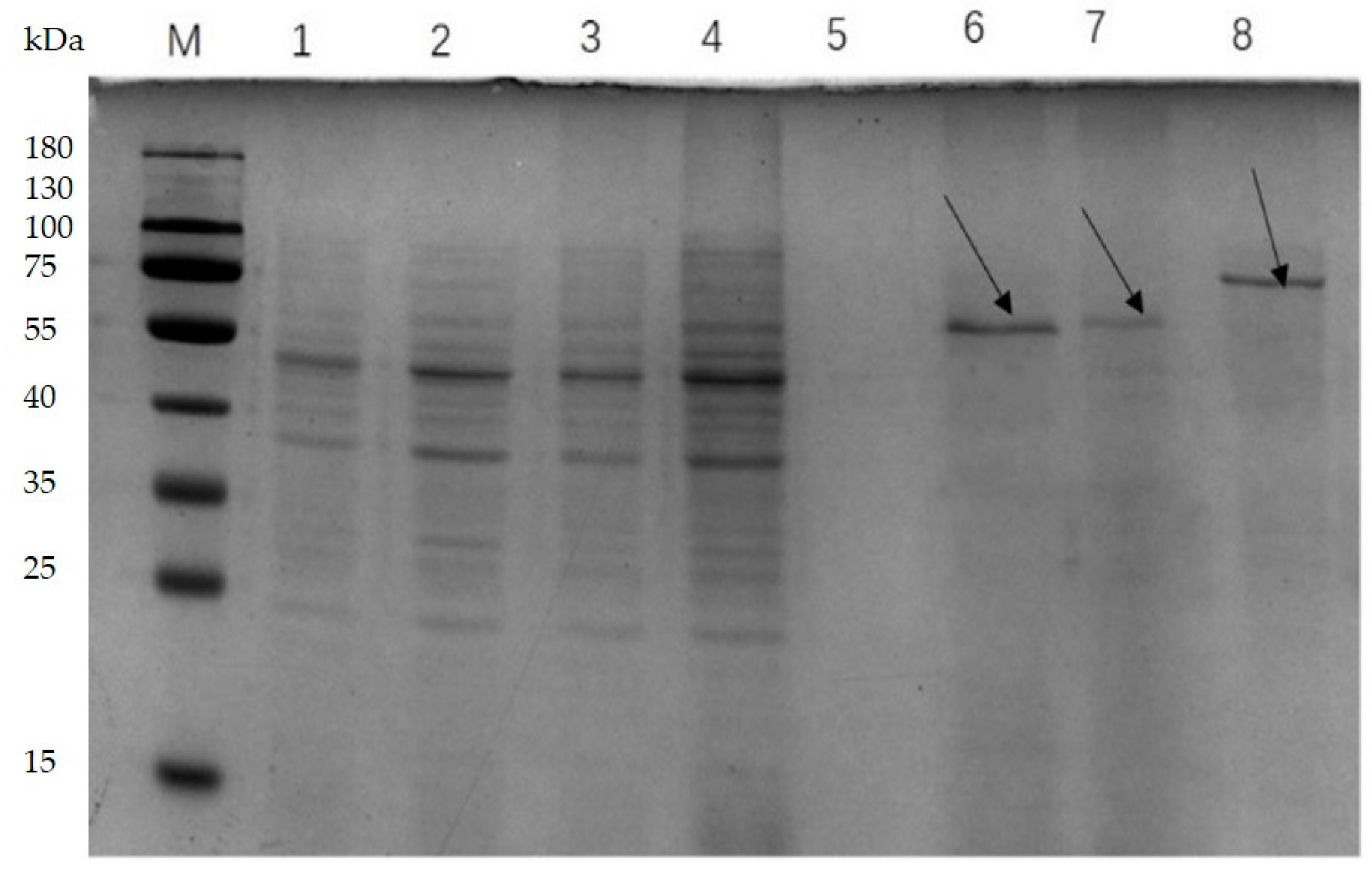
L. lactis NZ9000/pMG36e-usp45-bglA-usp45-bglB, L. lactis NZ9000/pMG36e-usp45-bglA, and L. lactis NZ9000/pMG36e-usp45-bglB showed the secretory expression of the recombinant proteins Bgl, BglA, and BglB, respectively, as determined using SDS-PAGE. The results of the analysis using Quantity One 1-D software (version 4.6.2) showed that the band with a molecular mass of 75 kDa was Bgl. Moreover, BglA and BglB were found to have a molecular mass of 55 kDa (Figure 3).
The enzymatic activity of Bgl was significantly higher than that of BglA and BglB for various substrates. However, the difference in enzymatic activity between BglA and BglB was not significant. Among the substrates tested, the recombinant enzyme had the highest specificity for 1% salicin, followed by CMC-Na. The lowest specificity was observed for absorbable cotton. Using 1% salicin as the substrate, the enzyme activities of BglA, BglB, and Bgl in the supernatant were found to be 2.09 U/mL, 2.36 U/mL, and 9.4 U/mL, respectively. The total enzyme activities of BglA, BglB, and Bgl for cellulose were found to be 0.97 U/mL, 1.12 U/mL, and 2.53 U/mL, respectively, using filter paper as the substrate (Figure 4a).

3.3. Determination of Optimum Reaction Temperature and pH of the Recombinant Enzyme

The enzyme activity of Bgl, BglA, and BglB increased with an increase in temperature within the reaction temperature range of 30–50 °C. The highest enzymatic of Bgl, BglA, and BglB was observed at 50 °C. As the temperature increased beyond 50 °C, the enzyme activity showed a slow decreasing trend. Therefore, the optimum reaction temperature for the fusion enzyme was determined to be 50 °C. The recombinant enzyme activity was around 75% when the temperature was 45–65 °C (Figure 4b). The optimum reaction temperature for these 3 recombinant enzymes was 50 °C.
When the pH was 3.0–7.0, the enzyme activity increased with an increase in pH. The optimal pH for Bgl, BglA, and BglB ranged from 6.0 to 8.0. The activity of these enzymes was above 90% within this pH range and peaked at pH 7.0. When the pH was over 7.0, the enzyme activity decreased, indicating that these enzymes were neutral enzymes (Figure 4c). The optimum reaction pH for these three recombinant enzymes was 7.0.

3.4. Effect of Various Substrates on the Kinetic Parameters of Recombinant Enzymes

We used salicin as the substrate to determine the kinetic parameters of the recombinant enzymes at an optimal temperature and pH (Table 3). The Vmax values were 1343, 1254, and1347 μmol∙min−1mg−1 for the bacterial supernatant of L. lactis NZ9000/pMG36e-usp45-bglA, L. lactis NZ9000/pMG36e-usp45-bglB, and L. lactis NZ9000/pMG36e-usp45-bglA-usp45-bglB, respectively. The Km values were 38.61, 43.99, and 34.8 μmol∙L−1 for the bacterial supernatant of L. lactis NZ9000/pMG36e-usp45-bglA, L. lactis NZ9000/pMG36e-usp45-bglB, and L. lactis NZ9000/pMG36e-usp45-bglA-usp45-bglB, respectively. The Kcat values were 434.8, 348.7, and 1016 s−1 for the bacterial supernatant of L. lactis NZ9000/pMG36e-usp45-bglA, L. lactis NZ9000/pMG36e-usp45-bglB, and L. lactis NZ9000/pMG36e-usp45-bglA-usp45-bglB, respectively. The Kcat/Km values were 11.26, 7.93, and 29.2 L∙s−1∙mol−1 for the bacterial supernatant of L. lactis NZ9000/pMG36e-usp45-bglA, L. lactis NZ9000/pMG36e-usp45-bglB, and L. lactis NZ9000/pMG36e-usp45-bglA-usp45-bglB, respectively.

3.5. Effect of Ions and Chemical Reagents on Recombinant Enzymes

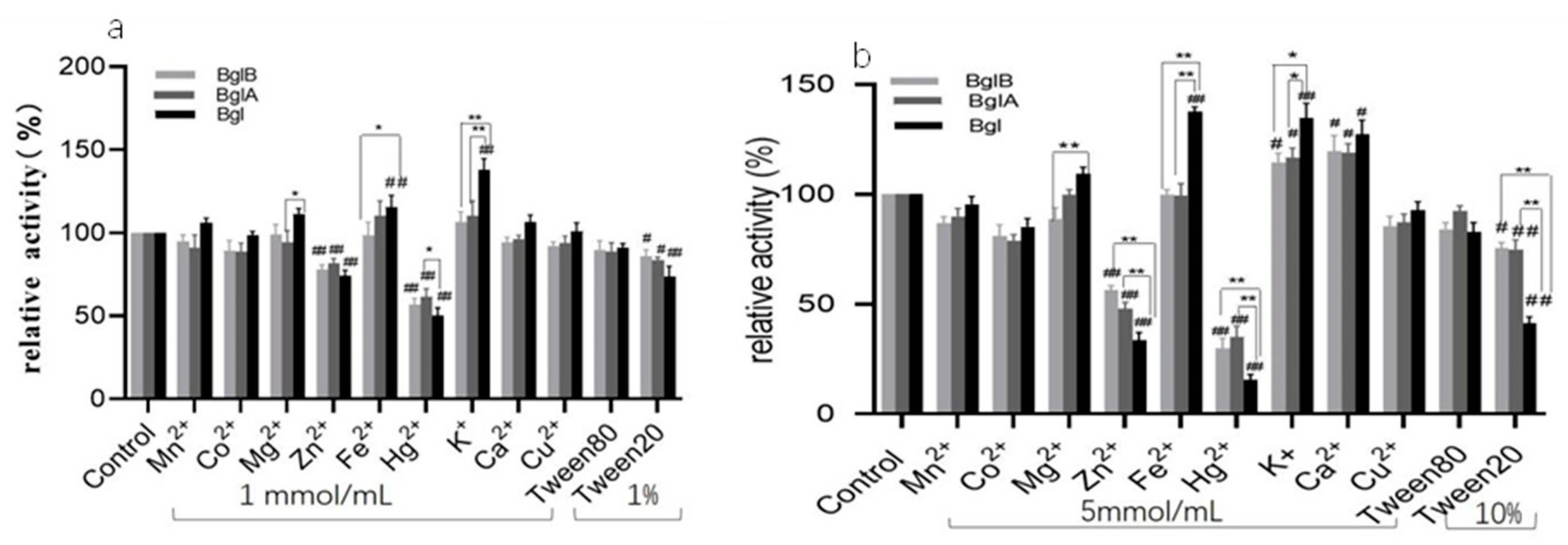
The increase in K+ and Fe2+ ion concentrations significantly increased the activity of the fusion recombinant enzyme but not the single recombinant enzymes. The activity of the Bgl enzyme was much greater than that of the BglA and BglB enzymes. Cu2+, Co2+, Mn2+, and Tween 80 did not affect the activity of the three recombinant enzymes. Ca2+ did not affect the activities of the three recombinases at a final concentration of 1 mmol/L but significantly promoted enzyme activity at a final concentration of 5 mmol/L. The recombinant enzyme was inhibited by Zn2+, Hg2+, and Tween 20, and an increase in their concentrations inhibited Bgl more significantly than BglA and BglB (Figure 5a,b).

4. Discussion

Currently, β-glucosidase is widely used in industrial applications, including in the textile, food processing, biorefinery, brewing, chemical, detergent, animal feed, paper, pharmaceutical, medical, pulp, and waste management industries [32,33]. Microbial communities in the yak rumen are one of the main sources of cellulase. For example, Enterococcus faecalis EF85 and EF83 isolated from the rumen of Tibetan yaks have the potential to degrade cellulose [25]. In addition, high-throughput 16S rRNA sequencing and metagenomic analysis have indicated that the rumen microbiome of plateau yaks has several genes encoding cellulases and hemicelluloses [26,27]. In order to obtain high-efficiency β-glucosidase, in this study two β-glucosidase genes bglA and bglB from the yak rumen were cloned into pMG36e as independent or fused proteins to construct the expression vectors pMG36e-bglA, pMG36e-bglB, and pMG36e-bglA-bglB. The engineered strains L. lactis NZ9000/pMG36e-usp45-bglA, L. lactis NZ9000/pMG36e-usp45-bglB, and L. lactis NZ9000/pMG36e-usp45-bglA-usp45-bglB were successfully constructed thereafter and showed the secretory expression of BglA, BglB, and Bgl, respectively. pMG36e was constructed based on the transcriptional and translational signals of the L. lactis milk lipid subspecies protease gene. It contained a promoter, an RBS, the start of an open reading frame, and a transcriptional terminator, and it was known to be a constitutive expression vector for the inserted gene in L. lactis [19]. pMG36e has previously been used to successfully express β-galactosidase in L. lactis [34]. L. lactis NZ9000 was a manually constructed recombinant host with the nisin-controlled gene expression (NICE) system [28]. In previous studies, L. lactis NZ9000 was used to express various proteins originating from other bacteria [35]. L. lactis, which is a well-recognized probiotic, has advantages over other expression systems in expressing recombinant proteins [28,36]. Unlike E. coli, L. lactis has a single outer membrane and can secrete target proteins directly into the extracellular environment [37]. The total enzyme production in L. lactis is higher than that in E. coli [38]. The ability of L. lactis to efficiently express and secrete proteins in large quantities is dependent on the selection of suitable and effective transcriptional promoters and secretory signal peptides [39]. Usp45, the main signal peptide for the extracellular proteins of L. lactis, plays an important role in guiding proteins to the cytoplasmic membrane [40]. By replacing the natural Nuc signal peptide with Usp45, the efficiency of heterologous protein secretion by L. lactis can be increased greatly [41].
The biodegradation of cellulose requires multiple cellulases that act in concert with each other to make cellulose degradation more efficient [42]. The β-glucosidase of Bacillus subtilis SU40 acts in synergy with the endoglucanase of Bacillus subtilis MG7 to completely degrade rice straw [41]. Fusion proteins containing xylanases and endonucleases with multiple catalytic structural domains can improve the efficiency of cellulose degradation [43]. In one study, three different cellulase genes were fused to construct a BCE with trifunctional cellulase activity. The fused enzyme showed higher specific activities of the β-glucosidase BG, endoglucanase CBH, and exoglucanase EG than did single enzymes under the same conditions [22]. In the present study, the enzymatic activity of the recombinant fusion enzyme in this study was 9.4 U/mL, which was approximately 3.5-fold higher than that of a single cellulase in lactic acid bacteria.
The temperature optima of cellulases in currently available commercial enzyme cocktails is typically around 50 °C [44]. In this study, an analysis of the enzymatic properties of the recombinant cellulase showed that the optimum reaction pH was 7.0 and the optimum reaction temperature was 50 °C. The highest growth and maximum CMCase production by Bacillus licheniformis TLW-3 were recorded at pH 7 and 50 ºC [45]. The Bgl enzyme in this study had a longer activity duration than the bacterial β-glucosidase of Aeromonas alternata, which rapidly lost its activity after incubation at 40 °C [46]. The Bgl in this study also exhibited more pH tolerance than the β-glucosidase unglu135B12, which showed optimal enzymatic activity at pH 5.0 [10]. In the present study, cellulase activity increased when the temperature increased from 30 °C to 50 °C and decreased when the temperature was above 80 °C. This was because as the temperature increased, the effective collision rate between the substrate molecules and the enzyme became greater, and the reaction rate became faster. However, when the temperature was too high, the denaturation of the enzyme was accelerated, leading to a decrease in enzyme activity [47]. The effect of metal ions on cellulase activity was specific to the cellulase being examined.
Different metal ions may have different affinities for different amino acid residues in cellulose, leading to conformational changes that may stimulate or inhibit cellulase activity. In this study, K+ and Fe2+ promoted the activity of the recombinant enzyme, while Cu2+, Co2+, and Mn2+ had little effect on this activity. Meanwhile, Hg2+ and Zn2+ inhibited the activity of the recombinant enzyme. Hg2+ is a heavy metal ion that can disrupt the structure of enzymes and even inactivate them [48]. Fe2+, Co2+, and Mn2+ can stimulate the enzymatic activity of endoglucanase and xylanase isolated from the rumen of goats [49]. The activity of β-glucosidase isolated from Mycobacterium thermophilum was found to be strongly reduced in the presence of Hg2+ and Cu2+ ions [50]. Interestingly, the fused Bgl examined in this study had higher enzymatic activities that the single cellulases. This was probably because the fused cellulase disrupted the crystalline structure of cellulose more deeply, increasing accessibility and making the accessible surface area larger. When the pores of the substrate were large enough, the fused enzyme enhanced its adsorption of cellulose, which improved the hydrolysis efficiency of the cellulase [51]. In a study on cellulose-secreting engineered L. lactis that promote lignocellulose degradation in high-moisture alfalfa, a combination of fusion L. lactis was found to be superior to a combination of cellulase and wild-type lactic acid bacteria subspecies in terms of silage quality and degradation of lignin [52].

5. Conclusions

In this study, the strains L. lactis NZ9000/pMG36e-usp45-bglA, L. lactis NZ9000/pMG36e-usp45-bglB, and L. lactis NZ9000/pMG36e-usp45-bglA-usp45-bglB were successfully engineered. They showed the secretory expression of BglA, BglB, and Bgl, respectively. The molecular weights of BglA, BglB, and Bgl were about 55 kDa, 55 kDa, and 75 kDa, respectively. In subsequent studies, using 1% salicin as the substrate, the optimum reaction temperature and pH for the three recombinant enzymes were found to be 50 °C and 7.0, respectively. The Vmax, Km, Kcat, and Kcat/Km of the three recombinant strains were analyzed using 1% salicin as the substrate at 50 °C and pH 7.0, respectively. The Bgl enzyme activity was significantly higher than the BglB and BglA enzyme activity (p < 0.05). Overall, the results showed that the β-glucosidase gene for lignocellulose degradation from the rumen of Tianzhu yak grazing on the Qinghai-Tibetan Plateau has important application and development prospects in the industrial production of β-glucosidase. Further development and research are warranted.

Author Contributions

Conceptualization: C.W., X.W. and Y.W.; Methodology: C.W. and X.W.; Software: Y.S. and Y.Y.; Validation: C.W. and X.W.; Formal analysis: C.M. and S.T.; Investigation: C.W., Y.W. and Z.Z.; Resources: C.W., Y.Y., S.T., Y.W. and X.W.; Data curation: Y.S., Z.Z. and C.M.; Writing—original draft preparation: C.W. and X.W.; Writing—review and editing: C.W., X.W. and Y.W.; Visualization: C.W. and Y.S.; Supervision: C.W.; Project administration: C.W. and X.W.; Funding acquisition: C.W., Y.W. and X.W. All authors have read and agreed to the published version of the manuscript.

Funding

This work was supported by grants from the Key Research and Development Project Plan of Gansu Provincial Science and Technology Department (18YF1NA077 and 20YF3NA020), the National Natural Science Foundation of China (No. 31500067, 31760614, and 31860659), the Application Research and Development Project of Gansu Academy of Sciences (2020JK-03), and the Science and Technology Industrialization Project of Gansu Academy of Sciences (2020–2022).

Data Availability Statement

No new data were created or analyzed in this study. Data sharing is not applicable to this article.

Acknowledgments

The authors would like to thank all of the reviewers who participated in the review.

Conflicts of Interest

The authors declare no conflict of interest.

References

  1. Gilmore, S.P.; Henske, J.K.; O’Malley, M.A. Driving biomass breakdown through engineered cellulosomes. Bioengineered 2015, 6, 204–208. [Google Scholar] [CrossRef] [PubMed]
  2. Salgado, J.C.S.; Meleiro, L.P.; Carli, S.; Ward, R.J. Glucose tolerant and glucose stimulated β-glucosidases-A review. Bioresour. Technol. 2018, 267, 704–713. [Google Scholar] [CrossRef] [PubMed]
  3. Nigam, P.S. Microbial enzymes with special characteristics for biotechnological applications. Biomolecules 2013, 3, 597–611. [Google Scholar] [CrossRef] [PubMed]
  4. Navarro, R.R.; Otsuka, Y.; Nojiri, M.; Ishizuka, S.; Nakamura, M.; Shikinaka, K.; Matsuo, K.; Sasaki, K.; Sasaki, K.; Kimbara, K.; et al. Simultaneous enzymatic saccharification and comminution for the valorization of lignocellulosic biomass toward natural products. BMC Biotechnol. 2018, 18, 79. [Google Scholar] [CrossRef] [PubMed]
  5. Zhang, M.; Cai, G.; Zheng, E.; Zhang, G.; Li, Y.; Li, Z.; Yang, H.; Wu, Z. Transgenic pigs expressing β-xylanase in the parotid gland improve nutrient utilization. Transgenic. Res. 2019, 28, 189–198. [Google Scholar] [CrossRef]
  6. Angelotti, J.A.F.; Dias, F.F.G.; Sato, H.H.; Fernandes, P.; Nakajima, V.M.; Macedo, J. Improvement of Aglycone Content in Soy Isoflavones Extract by Free and Immobilized Β-Glucosidase and their Effects in Lipid Accumulation. Appl. Biochem. Biotechnol. 2020, 192, 734–750. [Google Scholar] [CrossRef]
  7. Rani, V.; Mohanram, S.; Tiwari, R.; Nain, L.; Arora, A. Beta-Glucosidase: Key Enzyme in Determining Efficiency of Cellulase and Biomass Hydrolysis. J. Bioprocess. Biotech. 2014, 5, 197. [Google Scholar]
  8. Martins, E.D.S.; Gomes, E.; da Silva, R.; Junior, R.B. Production of cellulases by Thermomucor indicae-seudaticae: Characterization of a thermophilic β-glucosidase. Prep. Biochem. Biotechnol. 2019, 49, 830–836. [Google Scholar] [CrossRef]
  9. Li, Y.; Bu, M.; Chen, P.; Li, X.; Chen, C.; Gao, G.; Feng, Y.; Han, W.; Zhang, Z. Characterization of a Thermophilic Monosaccharide Stimulated β-Glucosidase from Acidothermus cellulolyticus. Chem. Res. Chin. Univ. 2018, 34, 212–220. [Google Scholar] [CrossRef]
  10. Li, Y.; Liu, N.; Yang, H.; Zhao, F.; Yu, Y.; Tian, Y.; Lu, X. Cloning and characterization of a new β-glucosidase from a metagenomic library of rumen of cattle feeding with Miscanthus sinensis. BMC Biotechnol. 2014, 14, 85. [Google Scholar] [CrossRef]
  11. Zhang, J.; Zhao, N.; Xu, J.; Qi, Y.; Wei, X.; Fan, M. Exploring the catalytic mechanism of a novel β-glucosidase BGL0224 from Oenococcus oeni SD-2a: Kinetics, spectroscopic and molecular simulation. Enzyme Microb. Technol. 2021, 148, 109814. [Google Scholar] [CrossRef] [PubMed]
  12. Xue, X.; Wu, Y.; Qin, X.; Ma, R.; Luo, H.; Su, X.; Yao, B. Revisiting overexpression of a heterologous β-glucosidase in Trichoderma reesei: Fusion expression of the Neosartorya fischeri Bgl3A to cbh1 enhances the overall as well as individual cellulase activities. Microb. Cell. Fact. 2016, 15, 122. [Google Scholar] [CrossRef] [PubMed]
  13. Acebrón, I.; Plaza-Vinuesa, L.; de Las Rivas, B.; Muñoz, R.; Cumella, J.; Sánchez-Sancho, F.; Mancheño, J.M. Structural basis of the substrate specificity and instability in solution of a glycosidase from Lactobacillus plantarum. Biochim. Biophys. Acta Proteins Proteom. 2017, 1865, 1227–1236. [Google Scholar] [CrossRef] [PubMed]
  14. Tak, J.Y.; Jang, W.J.; Lee, J.M.; Suraiya, S.; Kong, I.S. Expression in Lactococcus lactis of a β-1,3-1,4-glucanase gene from Bacillus sp. SJ-10 isolated from fermented fish. Protein Expr. Purif. 2019, 162, 18–23. [Google Scholar] [CrossRef]
  15. Boonkaew, B.; Udompaisarn, S.; Arthan, D.; Somana, J. Expression and characterization of a recombinant stevioside hydrolyzing β-glycosidase from Enterococcus casseliflavus. Protein Expr. Purif. 2019, 163, 105449. [Google Scholar] [CrossRef]
  16. Xia, W.; Xu, X.; Qian, L.; Shi, P.; Bai, Y.; Luo, H.; Ma, R.; Yao, B. Engineering a highly active thermophilic β-glucosidase to enhance its pH stability and saccharification performance. Biotechnol. Biofuels 2016, 9, 147. [Google Scholar] [CrossRef]
  17. Zegers, N.D.; Kluter, E.; van Der Stap, H.; van Dura, E.; van Dalen, P.; Shaw, M.; Baillie, L. Expression of the protective antigen of Bacillus anthracis by Lactobacillus casei: Towards the development of an oral vaccine against anthrax. J. Appl. Microbiol. 1999, 87, 309–314. [Google Scholar] [CrossRef]
  18. Gaya, P.; Peirotén, Á.; Landete, J.M. Expression of a β-glucosidase in bacteria with biotechnological interest confers them the ability to deglycosylate lignans and flavonoids in vegetal foods. Appl. Microbiol. Biotechnol. 2020, 104, 4903–4913. [Google Scholar] [CrossRef]
  19. van de Guchte, M.; van der Vossen, J.M.; Kok, J.; Venema, G. Construction of a lactococcal expression vector: Expression of hen egg white lysozyme in Lactococcus lactis subsp. lactis. Appl. Environ. Microbiol. 1989, 55, 224–228. [Google Scholar] [CrossRef]
  20. Pang, J.; Liu, Z.Y.; Hao, M.; Zhang, Y.F.; Qi, Q.S. An isolated cellulolytic Escherichia coli from bovine rumen produces ethanol and hydrogen from corn straw. Biotechnol. Biofuels 2017, 10, 165. [Google Scholar] [CrossRef]
  21. Liu, Z.L.; Li, H.N.; Song, H.T.; Xiao, W.J.; Xia, W.C.; Liu, X.P.; Jiang, Z.B. Construction of a trifunctional cellulase and expression in Saccharomyces cerevisiae using a fusion protein. BMC Biotechnol. 2018, 18, 43. [Google Scholar] [CrossRef] [PubMed]
  22. Kurniasih, S.D.; Alfi, A.; Natalia, D.; Radjasa, O.K.; Nurachman, Z. Construction of individual, fused, and co-expressed proteins of endoglucanase and β-glucosidase for hydrolyzing sugarcane bagasse. Microbiol. Res. 2014, 169, 725–732. [Google Scholar] [CrossRef] [PubMed]
  23. An, D.; Dong, X.; Dong, Z. Prokaryote diversity in the rumen of yak (Bos grunniens) and Jinnan cattle (Bos taurus) estimated by 16S rDNA homology analyses. Anaerobe 2005, 11, 207–215. [Google Scholar] [CrossRef]
  24. Wei, Y.Q.; Yang, H.J.; Luan, Y.; Long, R.J.; Wu, Y.J.; Wang, Z.Y. Isolation, identification and fibrolytic characteristics of rumen fungi grown with indigenous methanogen from yaks (Bos grunniens) grazing on the Qinghai-Tibetan Plateau. J. Appl. Microbiol. 2016, 120, 571–587. [Google Scholar] [CrossRef]
  25. Zhao, J.; Shao, T.; Chen, S.; Tao, X.; Li, J. Characterization and identification of cellulase-producing Enterococcus species isolated from Tibetan yak (Bos grunniens) rumen and their application in various forage silages. J. Appl. Microbiol. 2021, 131, 1102–1112. [Google Scholar] [CrossRef]
  26. Jiang, H.; Cao, H.W.; Chai, Z.X.; Chen, X.Y.; Zhang, C.F.; Zhu, Y.; Xin, J.W. Dynamic alterations in yak (Bos grunniens) rumen microbiome in response to seasonal variations in diet. Physiol. Genomics 2022, 54, 514–525. [Google Scholar] [CrossRef]
  27. Zhao, C.; Wang, L.; Ke, S.; Chen, X.; Kenéz, Á.; Xu, W.; Wang, D.; Zhang, F.; Li, Y.; Cui, Z.; et al. Yak rumen microbiome elevates fiber degradation ability and alters rumen fermentation pattern to increase feed efficiency. Anim. Nutr. 2022, 11, 201–214. [Google Scholar] [CrossRef]
  28. Kuipers, O.P.; de Ruyter, P.G.; Kleerebezem, M.; de Vos, W.M. Quorum sensing-controlled gene expression in lactic acid bacteria. J. Biotechnol. 1998, 64, 15–21. [Google Scholar] [CrossRef]
  29. Niu, L.; Zhang, H.; Wu, Z.; Wang, Y.; Liu, H.; Wu, X.; Wang, W. Correction: Modified TCA/acetone precipitation of plant proteins for proteomic analysis. PLoS ONE 2019, 14, e0211612. [Google Scholar] [CrossRef]
  30. Zhang, Y.H.; Hong, J.; Ye, X. Cellulase assays. Methods Mol. Biol. 2009, 581, 213–231. [Google Scholar] [PubMed]
  31. Dashtban, M.; Maki, M.; Leung, K.T.; Mao, C.; Qin, W. Cellulase activities in biomass conversion: Measurement methods and comparison. Crit. Rev. Biotechnol. 2010, 30, 302–309. [Google Scholar] [CrossRef] [PubMed]
  32. Singhvi, M.S.; Zinjarde, S.S. Production of pharmaceutically important genistein and daidzein from soybean flour extract by using β-glucosidase derived from Penicillium janthinellum NCIM 1171. Process. Biochem. 2020, 97, 183–190. [Google Scholar] [CrossRef]
  33. Jang, W.J.; Lee, G.H.; Lee, J.M.; Kim, T.Y.; Jeon, M.H.; Kim, Y.H.; Lee, E.W. Improving enzyme activity, thermostability and storage stability of β-1,3-1,4-glucanase with poly-γ-glutamic acid produced by Bacillus sp. SJ-10. Enzyme Microb. Technol. 2021, 143, 109703. [Google Scholar] [CrossRef] [PubMed]
  34. Zhang, W.; Wang, C.; Huang, C.Y.; Yu, Q.; Liu, H.C.; Zhang, C.W.; Pei, X.F. Construction and secretory expression of beta-galactosidase gene from Lactobacillus bulgaricus in Lactococcus lactis. Biomed. Environ. Sci. 2012, 25, 203–209. [Google Scholar]
  35. Liu, F.; van Heel, A.J.; Chen, J.; Kuipers, O.P. Functional production of clostridial circularin A in Lactococcus lactis NZ9000 and mutational analysis of its aromatic and cationic residues. Front. Microbiol. 2022, 13, 1026290. [Google Scholar] [CrossRef]
  36. King, M.S.; Boes, C.; Kunji, E.R. Membrane protein expression in Lactococcus lactis. Methods Enzymol. 2015, 556, 77–97. [Google Scholar] [PubMed]
  37. Sriraman, K.; Jayaraman, G. Enhancement of recombinant streptokinase production in Lactococcus lactis by suppression of acid tolerance response. Appl. Microbiol. Biotechnol. 2006, 72, 1202–1209. [Google Scholar] [CrossRef] [PubMed]
  38. Liu, Q.H.; Shao, T.; Dong, Z.H.; Bai, Y.F. Solution for promoting egl3 gene of Trichoderma reesei high-efficiency secretory expression in Escherichia coli and Lactococcus lactis. Process. Biochem. 2017, 62, 135–143. [Google Scholar]
  39. Stephenson, D.P.; Moore, R.J.; Allison, G.E. Transformation of, and heterologous protein expression in, Lactobacillus agilis and Lactobacillus vaginalis isolates from the chicken gastrointestinal tract. Appl. Environ. Microbiol. 2011, 77, 220–228. [Google Scholar] [CrossRef]
  40. van Asseldonk, M.; Rutten, G.; Oteman, M.; Siezen, R.J.; de Vos, W.M.; Simons, G. Cloning of usp45, a gene encoding a secreted protein from Lactococcus lactis subsp. lactis MG1363. Gene 1990, 95, 155–160. [Google Scholar] [CrossRef]
  41. Le Loir, Y.; Nouaille, S.; Commissaire, J.; Brétigny, L.; Gruss, A.; Langella, P. Signal peptide and propeptide optimization for heterologous protein secretion in Lactococcus lactis. Appl. Environ. Microbiol. 2001, 67, 4119–4127. [Google Scholar] [CrossRef]
  42. Cao, G.; Guo, W.; Wang, A.; Zhao, L.; Xu, C.; Zhao, Q.; Ren, N. Enhanced cellulosic hydrogen production from lime-treated cornstalk wastes using thermophilic anaerobic microflora. Int. J. Hydrog. Energy 2012, 37, 13161–13166. [Google Scholar] [CrossRef]
  43. Duedu, K.O.; French, C.E. Characterization of a Cellulomonas fimi exoglucanase/xylanase-endoglucanase gene fusion which improves microbial degradation of cellulosic biomass. Enzyme Microb. Technol. 2016, 93–94, 113–121. [Google Scholar] [CrossRef] [PubMed]
  44. Viikari, L.; Alapuranen, M.; Puranen, T.; Vehmaanperä, J.; Siika-Aho, M. Thermostable enzymes in lignocellulose hydrolysis. Adv. Biochem. Eng. Biotechnol. 2007, 108, 121–145. [Google Scholar] [PubMed]
  45. Kiran, T.; Asad, W.; Ajaz, M.; Hanif, M.; Rasool, S.A. Industrially relevant cellulase production by indigenous thermophilic Bacillus licheniformis TLW-3 strain: Isolation-molecular identification and enzyme yield optimization. Pak. J. Pharm. Sci. 2018, 31, 2333–2340. [Google Scholar]
  46. Sun, J.; Wang, W.; Yao, C.; Dai, F.; Zhu, X.; Liu, J.; Hao, J. Overexpression and characterization of a novel cold-adapted and salt-tolerant GH1 β-glucosidase from the marine bacterium Alteromonas sp. L82. J. Microbiol. 2018, 56, 656–664. [Google Scholar] [CrossRef]
  47. Wang, H.; Zhai, L.; Geng, A. Enhanced cellulase and reducing sugar production by a new mutant strain Trichoderma harzianum EUA20. J. Biosci. Bioeng. 2020, 129, 242–249. [Google Scholar] [CrossRef]
  48. Haq, I.U.; Tahir, S.F.; Aftab, M.N.; Akram, F.; Rehman, A.U.; Nawaz, A.; Mukhtar, H. Purification and Characterization of a Thermostable Cellobiohydrolase from Thermotoga petrophila. Protein Pept. Lett. 2018, 25, 1003–1014. [Google Scholar] [CrossRef]
  49. Cheng, J.; Huang, S.; Jiang, H.; Zhang, Y.; Li, L.; Wang, J.; Fan, C. Isolation and characterization of a non-specific endoglucanase from a metagenomic library of goat rumen. World J. Microbiol. Biotechnol. 2016, 32, 12. [Google Scholar] [CrossRef]
  50. Pyeon, H.M.; Lee, Y.S.; Choi, Y.L. Cloning, purification, and characterization of GH3 β-glucosidase, MtBgl85, from Microbulbifer thermotolerans DAU221. PeerJ 2019, 7, e7106. [Google Scholar] [CrossRef]
  51. Sun, S.; Sun, S.; Cao, X.; Sun, R. The role of pretreatment in improving the enzymatic hydrolysis of lignocellulosic materials. Bioresour. Technol. 2016, 199, 49–58. [Google Scholar] [CrossRef] [PubMed]
  52. Liu, Q.; Li, J.; Zhao, J.; Wu, J.; Shao, T. Enhancement of lignocellulosic degradation in high-moisture alfalfa via anaerobic bioprocess of engineered Lactococcus lactis with the function of secreting cellulase. Biotechnol. Biofuels 2019, 12, 88. [Google Scholar] [CrossRef] [PubMed]
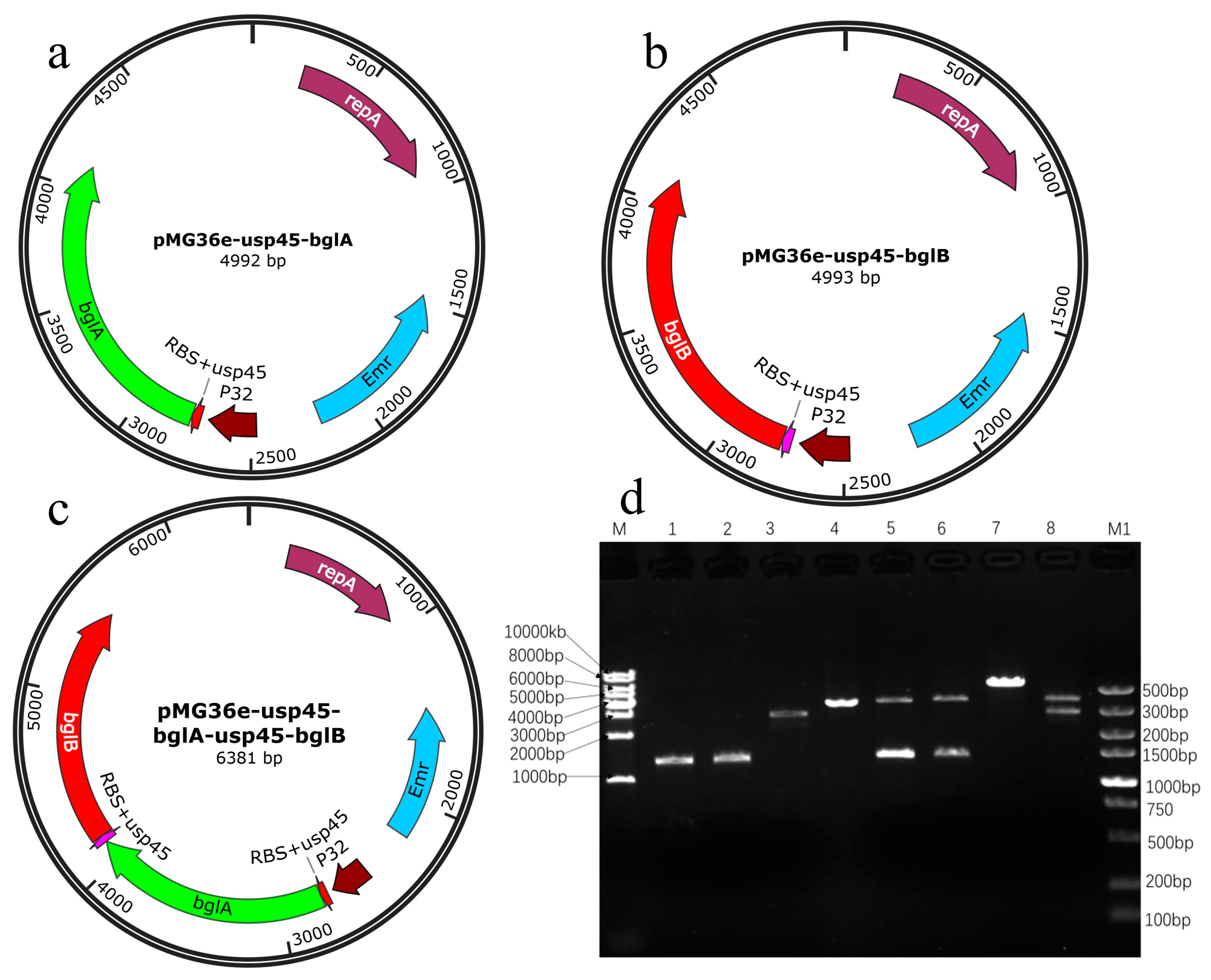
Figure 1. Maps of the three recombinant plasmid constructs and their electropherogram. (a) pMG36e-usp45-bglA recombinant plasmid; (b) pMG36e-usp45-bglB recombinant plasmid; (c) pMG36e-usp45-bglA-usp45-bglB recombinant plasmid; (d) M: DNA molecular weight marker; 1: PCR product of bglA; 2: PCR product of bglB; 3: PCR product of bglA-bglB; 4: pMG36e plasmid after Pst I digestion; 5: pMG36e-bglA plasmid after Pst I and Hind III digestion; 6: pMG36e-bglB plasmid after Pst I and Hind III digestion; 7: pMG36e-bglA-bglB plasmid after Pst I digestion; 8: pMG36e-bglA-bglB plasmid after Pst I and Hind III digestion.
Microorganisms 11 01387 g001
Figure 2. The analysis of recombinant enzymes activity using agar plates with 0.1% sodium carboxymethyl cellulose CMC-Na-stained Congo red. The halos dimeter is proportional to enzyme’s activity. To each well 20 μL of the enzyme with concentration 0.26–0.27 mg/mL of recombinant enzymes was transferred; 1: The inoculum of L. lactis NZ9000/pMG36e-usp45-bglA; 2: The inoculum of L. lactis NZ9000/pMG36e; 3: The inoculum of L. lactis NZ9000/pMG36e-usp45-bglA-usp45-bglB; 4: The inoculum of L. lactis NZ9000/pMG36e-usp45-bglB; 5: Negative control ddH2O; 6: The inoculum of L. lactis NZ9000/pMG36e; 7: β-glucosidase standard.
Microorganisms 11 01387 g002
Figure 3. SDS-PAGE of supernatant from recombinant strains. M: Protein molecular weight marker, 1: Bacterial supernatant precipitate of L. lactis NZ9000/pMG36e; 2: Bacterial supernatant precipitate of L. lactis NZ9000/pMG36e-usp45-bglA; 3: Bacterial supernatant precipitate of L. lactis NZ9000/pMG36e-usp45-bglB; 4: Bacterial supernatant precipitate of L. lactis NZ9000/pMG36e-usp45-bglA-usp45-bglB; 5: TCA/acetone precipitated protein of pMG36e from the L. lactis NZ9000/pMG36e bacterial supernatant; 6: TCA/acetone-precipitated BglA protein from L. lactis NZ9000/pMG36e-usp45-bglA bacterial supernatant; 7: TCA/acetone-precipitated BglB protein from L. lactis NZ9000/pMG36e-usp45-bglB bacterial supernatant; 8: TCA/acetone-precipitated Bgl protein from L. lactis NZ9000/pMG36e-usp45-bglA-usp45-bglB bacterial supernatant.
Microorganisms 11 01387 g003
Figure 4. Effect of temperature, pH, and substrate on recombinant enzyme activity. (a) Substrate specificity of recombinant proteins; (b) Effect of temperature on recombinant enzyme activity; (c) Effect of pH on recombinant enzyme activity; (*: Represents the difference within experimental groups; *: p < 0.05; **: p < 0.01; error bars in graphs represent standard deviations).
Microorganisms 11 01387 g004
Figure 5. Effect of different concentrations of metal ions and chemical reagents on recombinant enzyme activity. (a) Effect of 1 mmol/mL ions and 1% chemical solutions on enzyme activity; (b) Effect of 5 mmol/mL ions and 10% chemical solutions on enzyme activity (#: Represents the difference between experimental groups and the control group; #: p < 0.05; ##: p < 0.01; ###: p < 0.001 *: Represents the difference within experimental groups; *: p < 0.05; **: p < 0.01; Error bars in graphs represent standard deviations).
Microorganisms 11 01387 g005
Table 1. Bacterial strains and plasmids used in this study.
Strain and PlasmidRelevant Trait(s)Source or Reference
StrainsEscherichia coli DH5αsupE44 Δlac U169 (Φ80 lacZ ΔM15) hsdR17 recA1, endA1 gyrA96 thi-l relA1Our laboratory
L. lactis NZ9000Expressing recombinant plasmids[28]
E. coli DH5α/pMG36e-usp45-bglAE. coli DH5α with pMG36e-usp45-bglAThis study
E. coli DH5α/pMG36e-usp45-bglBE. coli DH5α with pMG36e-usp45-bglBThis study
E. coli DH5α/pMG36e-usp45-bglA-usp45-bglBE. coli DH5α with pMG36e-usp45-bglA-usp45-bglBThis study
L. lactis NZ9000/pMG36eL. lactis NZ9000 with pMG36e This study
L. lactis NZ9000/pMG36e-usp45-bglAL. lactis NZ9000 with secretory expression BglAThis study
L. lactis NZ9000/pMG36e-usp45-bglBL. lactis NZ9000 with secretory expression BglBThis study
L. lactis NZ9000/pMG36e-usp45-bglA-usp45-bglBL. lactis NZ9000 with secretory expression BglThis study
PlasmidspMG36eEmr; expression vector with the P32 promoter, multiple cloning sites (MCFs), and prtP translational terminator[19]
pMG36e-usp45-bglAEmr; secretory expression of BglAThis study
pMG36e-usp45-bglBEmr; secretory expression of BglBThis study
pMG36e-usp45-bglA-usp45-bglBEmr; secretory expression of fusion BglThis study
Table 2. Amplification primers used in this study.
PrimerSequence (5′-3′)Restriction Enzyme
bglA-FAACTGCAGAGAGCGCAAAAAAAAGATTATCTCAGCTAATGACTATTTTTCAATTTCCGCPst I
bglA-RTCAAGCTTCTTTGTTTAGCGTCHind III
bglB-FATGCTGCAGAATACCTTTATATTTCPst I
bglB-RCCCAAGCTTGCGCAAAAAAAAGATTATCTCAGCTACTTTTCTATTTAAAACCCGHind III
bglA-R1AACTGCAGAAGAAGGAGATATACATGCAAAAAAAAGATTATCTCAGCTAATGACTATTTTTCAATTTCCGCPst I
bglB-F1CCCAAGCTTCCCTTTTCTATTTAAAACCCGHind III
Single underlines indicate sites of enzyme restriction (cuts); wavy lines indicate the RBS; and double underlines indicate the usp45 signal peptide.
Table 3. Kinetic parameters of recombinant enzymes.
StrainVmax
(μmol∙min−1mg−1)
Km
(μmol∙L−1)
Kcat
(s−1)
Kcat/Km
(L∙s−1∙μmol−1)
L. lactis NZ9000/pMG36e-usp45-bglA134338.61434.811.26
L. lactis NZ9000/pMG36e-usp45-bglB125443.99348.77.93
L. lactis NZ9000/pMG36e-usp45-bglA-usp45-bglB134734.8101629.2
Disclaimer/Publisher’s Note: The statements, opinions and data contained in all publications are solely those of the individual author(s) and contributor(s) and not of MDPI and/or the editor(s). MDPI and/or the editor(s) disclaim responsibility for any injury to people or property resulting from any ideas, methods, instructions or products referred to in the content.