Article

The First Swedish Outbreak with VIM-2-Producing Pseudomonas aeruginosa, Occurring between 2006 and 2007, Was Probably Due to Contaminated Hospital Sinks

1
Department of Infectious Diseases and Hospital Infection Control, Lund University Hospital, SE-22185 Lund, Sweden
2
Section of Clinical Microbiology, Department of Medical Sciences, Uppsala University, SE-75185 Uppsala, Sweden
3
Department of Women’s and Children’s Health, Uppsala University, SE-75185 Uppsala, Sweden
*
Author to whom correspondence should be addressed.
These authors contributed equally to this work.
Microorganisms 2023, 11(4), 974; https://doi.org/10.3390/microorganisms11040974
Submission received: 22 February 2023 / Revised: 5 April 2023 / Accepted: 6 April 2023 / Published: 8 April 2023

Abstract

:
Multidrug-resistant Pseudomonas aeruginosa is an increasing clinical problem worldwide. The aim of this study was to describe the first outbreak of a Verona integron-borne metallo-ß-lactamase (VIM)-2-producing P. aeruginosa strain in Sweden and its expansion in the region. A cluster of multidrug-resistant P. aeruginosa appeared at two neighbouring hospitals in 2006. The isolates were characterized by PCR, pulsed-field gel electrophoresis (PFGE), and whole-genome sequencing. Patient charts, laboratory records, and hygiene routines were reviewed, and patients, staff, and the environment were screened. The investigation revealed a clonal outbreak of a VIM-2-producing P. aeruginosa strain belonging to the high-risk clonal complex 111, susceptible only to gentamicin and colistin. No direct contact between patients could be established, but most of them had stayed in certain rooms/wards weeks to months apart. Cultures from two sinks yielded growth of the same strain. The outbreak ended when control measures against the sinks were taken, but new cases occurred in a tertiary care hospital in the region. In conclusion, when facing prolonged outbreaks with this bacterium, sinks and other water sources in the hospital environment should be considered. By implementing proactive control measures to limit the bacterial load in sinks, the waterborne transmission of P. aeruginosa may be reduced.

1. Introduction

Pseudomonas aeruginosa is a non-fermenting, Gram-negative rod commonly found in soil and aquatic environments. This opportunist has a high ability to form biofilms and develop antibiotic resistance, and it is one of the leading agents of nosocomial infections and outbreaks. The outbreaks are often localized to units with vulnerable patients and a high consumption of broad-spectrum antibiotics, e.g., intensive care units (ICUs) [1,2,3]. Staff, co-patients, visitors, medical equipment, water supplies, sinks, hygiene products, etc., can all take part in the transmission [1,4,5,6,7,8].
β-lactams with or without β-lactamase inhibitors are often the first-line therapy for severe infections caused by P. aeruginosa, but this bacterium may render β-lactams useless through a range of complex resistance mechanisms [9,10,11,12,13]. Several β-lactamases can be carried by P. aeruginosa, but the metallo-β-lactamases (MBLs) have, in recent years, become more frequent. MBLs can hydrolyse all β-lactams used for treating P. aeruginosa-induced infections except aztreonam, and these enzymes have rapidly become a major concern [14,15]. MBL-encoding genes are typically embedded in integrons and they are transferable by plasmids. On the plasmids, the MBL genes are often combined with resistance genes covering other classes of antibiotics. The result is often multidrug-resistant isolates, leaving physicians with few, if any, therapeutic options [14,15,16].
The most prevalent transferable MBL gene in clinical samples is Verona integron-borne metallo-ß-lactamase (VIM)-2 [11]. It was identified in 1996 in a P. aeruginosa isolate from a patient in France [17]. Ever since, the VIM-2 gene has, in the vast majority of cases, been recognized in P. aeruginosa isolates, and, as a consequence, it is involved in most outbreaks of MBL-producing P. aeruginosa [2,3,18,19,20]. The first VIM-2-producing P. aeruginosa in Sweden was isolated in 1999 at Malmö University Hospital in the south of Sweden [21]. The patient died several months later and no new cases were reported until 2004, when two new multidrug-resistant P. aeruginosa isolates appeared a few months apart. Both patients had been discharged from an ICU at Lund University Hospital to two different hospitals within the catchment area of Malmö University Hospital. The investigation showed that both isolates carried VIM-2, and they exhibited the same DNA pattern as the isolate from 1999 with an arbitrarily primed polymerase chain reaction (PCR) [21]. The Department of Infectious Control in Lund was contacted. No control measures were taken.
In 2006, a cluster of cases of multidrug-resistant P. aeruginosa appeared in the neighbouring Blekinge County. The aim of the study was to describe the first outbreak of VIM-2-producing P. aeruginosa in Sweden, as well as the characteristics of the outbreak strain and the patients involved. Furthermore, the most likely source of the outbreak, the infection control measures taken, and the continued clonal expansion in the southern part of Sweden are reported.

2. Materials and Methods

2.1. Settings and Ethics

Blekinge County is in the south-eastern part of Sweden and has a population of approximately 150.000 inhabitants. It has two hospitals, the secondary-level Karlskrona Hospital (KaH) with 330 beds and the primary-level Karlshamn Hospital (KnH) with 120 beds. At the tertiary health care level, the patients are transferred to the university hospitals in Lund or Malmö. These latter two hospitals are only 25 km apart, and patients are often transported between them. During the first three months of 2006, four patients with multidrug-resistant P. aeruginosa were observed at KnH (n = 3) and KaH (n = 1). An epidemiological investigation was initiated after the third patient. As this study was performed according to the Swedish Infection Protection Act and as a part of an outbreak investigation with a direct impact on public health, no ethical approval was needed. Personal identifiers have been removed in order to ensure confidentiality.

2.2. Cases

All patients admitted to KaH and KnH between February 2006 and June 2007 with an infection or carriage of a P. aeruginosa isolate resistant to imipenem, ceftazidime and a positive Etest for MBLs were defined as cases.
The first MBL-producing P. aeruginosa was isolated from a urine sample obtained from patient 1 on 1 February 2006. The patient had contracted a catheter-associated urinary tract infection (CAUTI) during a stay in the ICU at KnH. From 2 February to 6 March 2006, patients 2, 3, and 4 were reported. Patient 2 had ventilator-associated pneumonia and died within a week in the ICU at KnH. Patient 3 was treated for a CAUTI in a medical ward at KnH. Patient 4 was discovered in a surgical ward at KaH after screening. Only carriage was observed, and the patient died within the follow-up period due to underlying conditions. Five months followed without any new patients, and the outbreak seemed to be at an end. However, from 7 August 2006 to 20 May 2007, four additional patients were registered. Patient 5 was admitted to the ICU at KnH with a pneumothorax. After transfer to a medical ward, an MBL-producing P. aeruginosa isolate grew in a sputum sample. The patient was transported to the Department of Infectious Diseases at KaH but died within two weeks. Patient 6 shared the room with patient 5 at the medical ward at KnH, and the MBL-producing P. aeruginosa was isolated from a bronchial aspirate and a leg ulcer. The patient exhibited no signs of infection. Patient 7 had a urinary catheter and was discovered through screening prior to surgery. Patient 8 received a urinary catheter after surgery and MBL-producing P. aeruginosa isolates were found at multiple locations after screening. The patient was transferred to the Department of Infectious Diseases at KaH for isolation, but died within 11 days. A summary of the patient data is shown in Table 1.

2.3. Epidemiological Investigation

The patient charts were reviewed, and infections, underlying conditions, antibiotic treatments, and international travels were registered. In addition, the dates for admissions, transfers, and discharges in the last 3 months prior to culture positivity were extracted, and the room numbers in different wards were noted when possible. The laboratory records were used to find culture data concerning P. aeruginosa with and without MBL, and to follow how long the outbreak patients stayed culture-positive. Policies and procedures were thoroughly controlled, especially concerning hand hygiene, cleaning, and medical equipment.
Patients with wounds and urinary catheters were screened for the presence of MBL-producing P. aeruginosa at medical ward B, KnH, when the bacterium was first isolated from patient 3. Screening was also performed on patients at medical ward A, KnH, and on the staff of the ICU in the same hospital when patients 5 and 6 were diagnosed. Finally, screening of patients alone was performed at the surgery wards at KaH and KnH when patients 7 and 8 were found to be positive. For these patients, rectal samples were added.
From August 2006 to June 2007, 124 environmental samples were collected from contact surfaces and sinks in rooms in the ICU, medical ward A, and the surgery ward at KnH, and in surgery ward A at KaH.

2.4. Cultures and Susceptibility Testing

P. aeruginosa was identified with conventional laboratory methods and/or an API 20 NE® instrument (BioMerieux, Lyon, France). Antimicrobial susceptibility was tested with disk diffusion on Iso-Sensitest Agar® (Oxoid Ltd., London, UK). Breakpoints established by the Swedish Reference Group for Antibiotics was used (nowadays, exchanged for the European Committee on Antimicrobial Susceptibility Testing).
The MBL phenotype was detected with the Etest for MBLs (AB Biodisk, Solna, Sweden). A reduction in imipenem minimum inhibitory concentrations (MICs) by ≥3 twofold dilutions in the presence of ethylenediaminetetraacetic acid (EDTA) was interpreted as being positive for MBL production. An additional double-disk test with imipenem ± EDTA and ceftazidime ± 2-mercaptopropionic acid (MPA) was performed as earlier described [22,23]. The MICs of MBL-positive strains were further determined for imipenem, meropenem, piperacillin/tazobactam, ceftazidime, aztreonam, gentamicin, amikacin, tobramycin, ciprofloxacin, fosfomycin, and colistin.

2.5. Identification of MBL with PCR

Deoxyribonucleic acid (DNA) was prepared by heating a bacterial suspension to 95 °C for 10 min. The template was added to the HotStarTaq master mix (Qiagen AB ®, Solna, Sweden) with earlier described primers [24] (Eurogentech S.A., Seraing, Belgium) in a final volume of 25 μL. The PCR reactions were processed in a GeneAmp PCR System 9700 cycler (PE Applied Biosystems, Foster City, CA, USA), where the program was carried out at 94 °C for 5 min, and was followed by 30 cycles of 1 min at 94 °C, 1 min primer annealing at 55 °C, 1.5 min at 72 °C, and a final extension step at 72 °C for 5 min. The PCR products were separated by electrophoresis on 1.5% agarose gels (GeneChoice Inc., Frederick, MD, USA). The patient isolate from 1999 was used as the positive control.

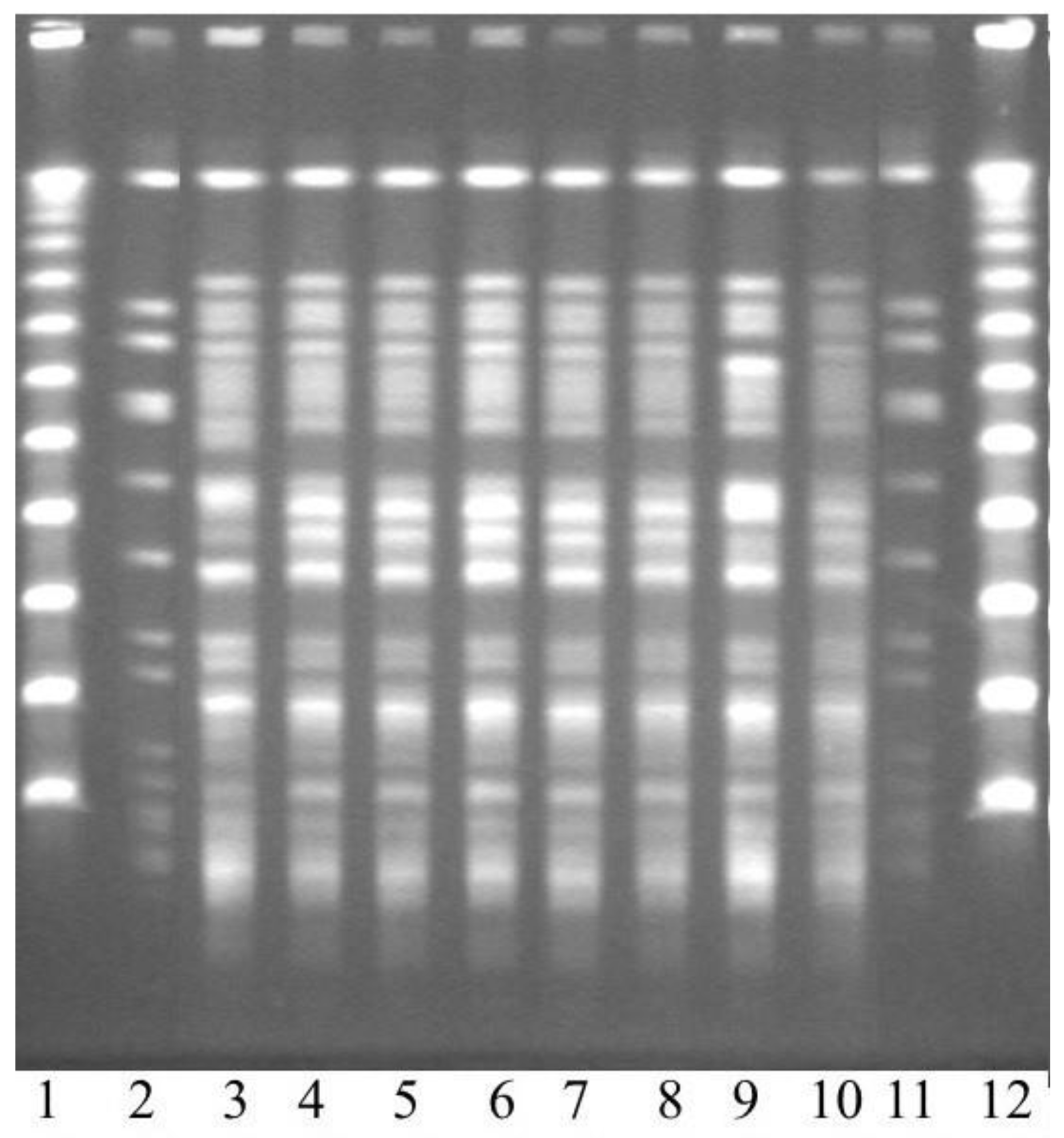
2.6. Pulsed-Field Gel Electrophoresis (PFGE)

PFGE was performed as previously described [25]. Banding patterns were compared visually with BioNumericsSoftware, version 4.0 (Applied Maths Bvba, St.-Martens-Latem, Belgium). Using clustering of a similarity matrix based on band-matching Dice coefficients (tolerance 1% and optimization 1%), dendrograms were created. Isolates showing indistinguishable pulsed-field patterns or closely related band patterns (>90% similarity) were regarded as clonally related.

2.7. Whole-Genome Sequencing (WGS) and Computational Analysis

One of the VIM-2-positive isolates (from patient 3) was randomly chosen for WGS. One colony was incubated overnight in Luria–Bertani broth (Becton Dickinson, Stockholm, Sweden) at 35 °C. DNA was prepared using the BioRobot M48 system (Qiagen GmbH, Hilden, Germany) and the MagAttract® DNA Mini M48 Kit according to the manufacturer’s recommendations. The DNA preparations were transported to SciLifeLab, Uppsala, Sweden. After controlling the DNA quality, sequencing was carried out on an IonTorrent™ PGM instrument (Life Technologies, Carlsbad, CA, USA) with a read length of 400 bp. Read quality was assessed using FastQC software (v0.11.4, http://www. bioinformatics.babraham.ac.uk, accessed on 1 November 2016), according to the developers’ recommendations.
The obtained read was de novo assembled with the methylated-CpG island recovery assay (MIRA) 4.9.5_2 [26]. The assembly statistics were assessed with the Quality Assessment Tool for Genome Assemblies (QUAST) v4.4 [27], and species confirmation was performed with the ribosomal multilocus sequence typing (rMLST) speciation tool at pubmlst.org/rmlst. Seven-gene MLST was carried out [28] using the database hosted on www.pubmlst.org. The IntegronFinder tool [29] was used for the detection of integron loci in the draft genome, and the predicted integrons were illustrated with the statistical software R (v3.3.3, R Foundation for Statistical Computing, Vienna, Austria, www.R-project.org, package genoPlotR). In addition, the isolate was analysed concerning acquired genetic resistance determinants with the Antibiotic Resistance Gene-ANNOTation (ARG-ANNOT) database [30]. For virulence factors, the Virulence Factors of Pathogenic Bacteria database (VFDB, 2016) [31] was applied together with Basic Local Alignment Search Tool (BLAST) searches on the draft genome [32]. Read sequence data are available from the European Nucleotide Archive, Sequence Read Archive, and DNA DataBank of Japan databases under the project reference PRJEB25448.

3. Results

3.1. Bacterial Findings

From February 2006 to June 2007, there were a total of 816 clinical P. aeruginosa isolates from 507 patients. Of these isolates, 69 (8.5%) were resistant to imipenem and 9 (1.1%) were resistant to both imipenem and ceftazidime. The MBL phenotype was confirmed in all but one of these nine isolates. It was excluded since it did not fulfil the case definition. The remaining eight isolates were all PCR-positive for VIM-2 and had band patterns that were identical or exhibited >90% similarity in the PFGE (Figure 1). They were all susceptible to gentamicin (MIC 2 mg/mL) and colistin (MIC 1 mg/mL). The MICs for the remaining tested antibiotic drugs were the following: carbapenems and ciprofloxacin > 32 mg/mL; fosfomycin, 64 mg/mL; piperacillin-tazobactam, 32 mg/mL; and ceftazidime, aztreonam, tobramycin, and amikacin, 16 mg/mL.

3.2. Genetic Data of the Outbreak Strain

A total of 1.2 million sequences with read lengths varying from 8 to 546 bases were obtained. The reads were de novo assembled into a draft genome, resulting in a total assembly length of 7.2 million bp; it was 121 contigs larger than 100,000 bp with an average coverage per contig ranging from 26 to 51, and an N50 of 69,517. The guanine–cytosine (GC) content of the draft genome was 65.73%, and 96.6% of the draft genome could be mapped to the complete genome of P. aeruginosa PA01 (NC_002516.2).
Sequence typing revealed that the isolate belonged to the clonal complex 111. The assembly failed to produce the acs locus, which is why the exact sequence type could not be determined. The remaining alleles were: gua—allele 5; mut—allele 5; nuo—allele 4; pps—allele 4; and trp—allele 3.
IntegronFinder predicted one complete integron class I with the blaVIM-2 and an uncharacterized protein (Figure 2). The integron consisted of the integrase gene (intI), two promotors Pc and Pint, and the attI recombination site, followed by the gene cassette with an open reading frame (ORF) of 218 bp, the attC recombination site, and the beta-lactamase blaVIM-2. Additionally, two clusters of attC sites lacking integron integrases (CALIN) were detected on separated contigs, both with two ORFs with uncharacterized proteins.
The BLAST search in the ARG-ANNOT database on the draft genome yielded the following resistance determinants: two beta-lactamase genes blaVIM-2 and blaOXA-50, two aminoglycoside resistance genes aph3-IIb (O-phosphotransferase) and aacA29b (N-acetyltransferase), the chloramphenicol acetyltransferase catB7 (a chromosome-encoded variant of the cat gene found in P. aeruginosa), and the fluoroquinolone resistance gene oqxB. The oprD gene included frameshifts.
The BLAST search for virulence factors confirmed the existence of all chromosomal loci related to the pathogenicity of P. aeruginosa PA01 in the VFDB, including the HIS-I Hcp1 secretion island I and TTSS type III secretion system.

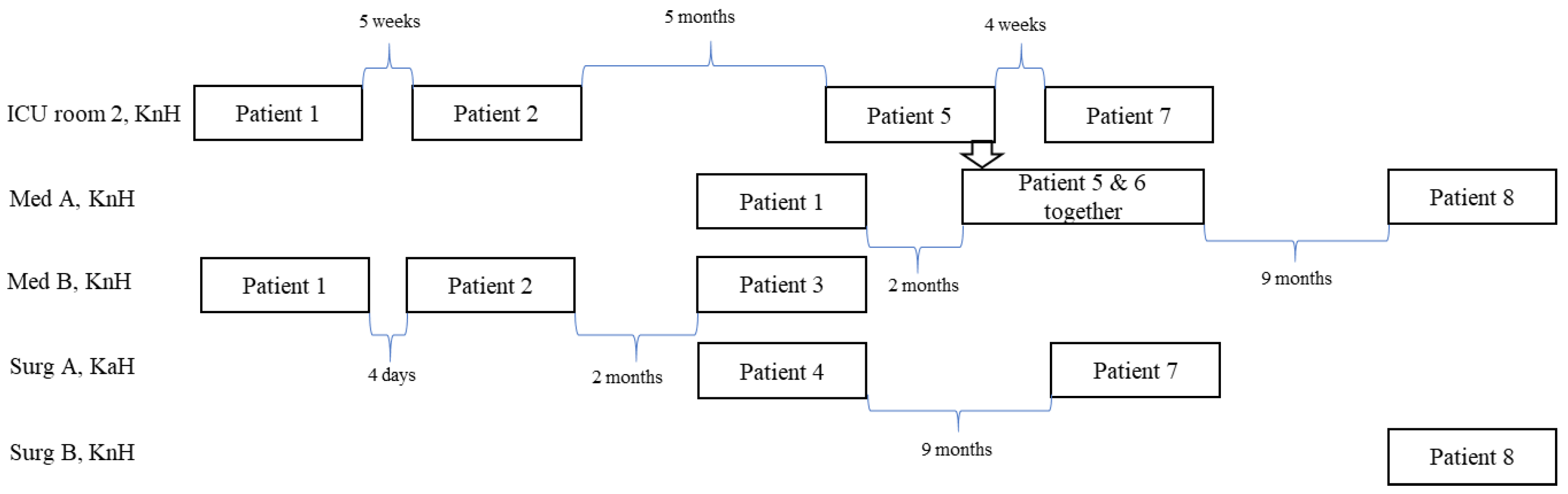
3.3. Epidemiological Investigation

All wards visited by the case patients during the 3-month period prior to culture positivity are listed in Table 1. Wards and rooms shared by patients in relation to time are shown in Figure 3. None of the patients had travelled outside Scandinavia prior to their culture positivity, but two patients (patient 2 and patient 4) had received treatment at Lund University Hospital during 2003.
All patients but one (patient 4) were first admitted to KnH. Patients 1–3 had all stayed in the medical ward B at KnH, but not at the same time (see Figure 3 for the time interval between the patients). Data are missing concerning which room they stayed in after admission to this ward, but it is unlikely that the outbreak strain was transmitted to patient 3 in this ward since the patient was already culture-positive on the day of admittance. Patients 1, 5, 6, and 8 had a period of care in medical ward A at KnH. Patients 5 and 6 had stayed in the same room, whereas patient 1 had been admitted to the ward about 2 months earlier and patient 8 about 9 months later. Patients 1, 2, 5, and 7 had, in addition, stayed in the same room in the ICU at KnH, but not at the same time (Figure 3). At KaH, patients 4 and 7 had been admitted to ward A in the Department of Surgery, but their admission was separated by 9 months.
Of the 124 environmental samples, MBL-producing P. aeruginosa was found in 2. According to PFGE, both isolates belonged to the outbreak strain (not shown). One of the positive environmental isolates was collected on 15 August 2006 from a sink drain in room 2 at the ICU, KnH, where patients 1, 2, and 5 had stayed 34, 26, and 2 weeks earlier, respectively. The second environmental sample was obtained on 24 May 2007 from the sink drain in a room at the surgical ward, KnH, when patient 8 was still there. In no instance was the outbreak strain found among the screened staff.

3.4. Infection Control Measures

Once infection/colonization was established, all case patients were isolated in single rooms with separate toilets and showers. Healthcare workers were instructed to use gowns and gloves when in contact with the patients. Alcohol-based hand rub was used before and after glove use and patient contact. From July 2006, case patients were transferred to and isolated at the Department of Infectious Diseases, KaH. The ICU at KnH was closed for admission for one week in August 2006 when the outbreak strain was discovered in one of the sink drains. This sink was replaced, the other sinks were treated overnight with acetic acid (24%), and the ward was disinfected with Virkon® (Viroderm, Solna, Sweden). No MBL-producing bacteria could thereafter be isolated from any of the sink drains at the ICU. The sink in the surgical ward at KnH was treated with boiling water and 24% acetic acid. Cultures obtained from the sink one week later were negative for MBL-producing P. aeruginosa. During the following 15 years, no more cases were reported.

4. Discussion

In the present study, the first Swedish outbreak involving MBL-producing P. aeruginosa was described. The outbreak was prolonged, and the causal strain was multidrug-resistant, VIM-2-producing, and belonged to the high-risk clonal complex 111. During a period of about 1.5 years, eight patients were colonized and/or infected with the outbreak strain in Blekinge County. Fifty percent of them developed a clinical infection and three died.
Due to the separation in time, patient-to-patient transmission was unlikely in all cases but one. However, several patients had been admitted to the same rooms or wards for shorter or longer time intervals, which is why a source in the environment was most likely. The environmental samples yielded growth of the outbreak strain in two sinks located in rooms where the colonized patients had stayed, providing a likely reservoir. As the sinks were replaced or decontaminated, no more cases occurred. It has remained so during the last 15 years in Blekinge County.
The isolation frequency of VIM-producing P. aeruginosa has been very low in Sweden and other Scandinavian countries. Most isolates have been derived from patients recently hospitalized abroad, suggesting that the import of international clones has been the major route for acquiring this type of bacterium [21]. The only exception is the southern part of Sweden, where the local expansion of ST111 has caused several infections and deaths. Although the intervention was successful in Blekinge County, this clone continued to cause infections. In 2007, four new patients appeared at Lund University Hospital [21]. They were followed by twelve more cases during the years between 2008 and 2013 [33]. Due to the low prevalence, these infections are difficult to recognize and easy to ignore. However, the mortality is high, and in Lund it was 50% [33]. To stop the transmission of VIM-2-producing P. aeruginosa, 24% acetic acid was used and a proactive routine was introduced to reduce the bacterial load; since 2013, the sinks in high-risk wards have been treated with acetic acid. At the beginning it was once weekly, but in later years the interval has been prolonged to once every month or every three months. No more clusters or outbreaks have been recorded.
It is obviously easier to stop an outbreak of P. aeruginosa than to eradicate this bacterium from a plumbing system. The fact that the outbreak strain has resisted ten years of acetic acid treatment emphasizes the importance of acting promptly to avoid the establishment of biofilm further down the drain system. This finding also has future implications, as multidrug-resistant bacteria are expected to become more common. Outbreaks may be on a large scale and cost a lot, but they do not cost as much as constantly present healthcare-associated infections (HAIs). These everyday infections are also more difficult to discover unless the bacteria carry some marker in the form of resistance genes. Recently, a British research group showed that environmental, Gram-negative bacterial populations are largely structured by ward and sink, with a few lineages being widely distributed [34]. They also compared the environmental isolates with contemporaneous patient isolates and reported that sinks may contribute to up to 10% of the infections caused by E. coli [34]. In a small country such as Sweden, HAIs affect approximately 65.000 patients every year at a cost of EUR 150–220 million. About 40% of these infections are urinary tract infections, and a majority of them are caused by E. coli. Several million EUR would probably be saved yearly, not to mention all the unnecessary suffering and deaths that could be avoided, if routines regarding sink practices were improved.
Despite extensive investigation, it was never clarified when or where patient 1 acquired the VIM-2-producing P. aeruginosa or if this patient was the true index patient in Blekinge County. Patients 2 and 4 had been treated at Lund University Hospital three years earlier, but no other connections with the university hospitals in Lund or Malmö were established. It is possible that ST111 could have been introduced repeatedly into this region, but it seems more likely that the clone was present in different sinks for all these years, and that Lund University Hospital played a central role in delivering new patients.
It was also not easy to follow the transmission routes for the patients at KnH. Some of the patients had been admitted to the same rooms or wards, but they were separated in time by weeks or months. Direct contact was unlikely, except for the two patients who had stayed in the same room at the same time. None of the staff were a carrier; thus, it was most likely that patients were colonized from a reservoir in the environment. In support of this hypothesis were the findings in the two sinks, of which one was in room 2 at the ICU where half of the patients had stayed. P. aeruginosa survives for only shorter time periods on dry surfaces, but anything wet or moist in an environment may act as a reservoir [35,36]. The bacterium has been associated with water taps, sinks, plumbing systems, shower drains, and faucets in hospitals. To make the outbreaks cease, replacements of the water fittings, sinks, etc., are often necessary [1,8,37,38,39]. In some hospitals the solution has been more drastic, and waterless patient care has been implemented [40].
The outbreak strain belonged to the high-risk multi-drug resistant CC111, which has been one of the most successful clonal complexes globally [3,39,41,42]. It is known for its virulence and ability to produce biofilms. Apart from blaVIM-2, which was linked to, but not within, the class 1 integron, it had frameshifts in the oprD gene and carried blaOXA-50. This relatively weak β-lactamase is constitutively expressed in P aeruginosa and confers decreased susceptibility to piperacillin, and, interestingly, to meropenem in P. aeruginosa, but not in E. coli [43]. In addition, it was resistant to the most common treatment alternatives, thereby affecting the outcomes for two patients.
In conclusion, the first outbreak of VIM-2-producing P. aeruginosa in Sweden had low prevalence and was caused by the high-risk CC111. It lasted 1.5 years and involved eight patients, of whom half developed an infection. Sinks in the rooms were the most likely source, and sinks continued to play a major role as a source during the following years in this part of Sweden. When facing prolonged outbreaks with this bacterium, sinks and their drain systems should be considered. By implementing proactive control measures to limit the bacterial load in sinks and water fittings, the transmission of P. aeruginosa to vulnerable patients could probably be reduced, if not eliminated.

Author Contributions

Conceptualization, C.-J.F., G.S., S.S. and Å.M.; formal analysis, C.-J.F., G.S., S.S. and Å.M.; funding acquisition, S.S. and Å.M.; methodology, E.T., S.S. and Å.M.; supervision, Å.M.; writing—original draft, C.-J.F., G.S., S.S. and Å.M.; writing—review and editing, C.-J.F., G.S., S.S. and Å.M. All authors have read and agreed to the published version of the manuscript.

Funding

This study was financially supported by ALF-funds, Afa Insurance, Sweden (grant 150411), and the Alf-de-Ruvo Memorial Foundation, Sweden.

Data Availability Statement

More detailed patient data are not openly available to ensure confidentiality, but are available from the corresponding author upon reasonable request.

Acknowledgments

We thank the staff at the National Genomics Infrastructure at the SciLifeLab Uppsala, Sweden, for the generation of sequencing data.

Conflicts of Interest

The authors declare no conflict of interest. The funders had no role in the design of the study; in the collection, analyses, or interpretation of data; in the writing of the manuscript; or in the decision to publish the results.

References

  1. Knoester, M.; de Boer, M.G.; Maarleveld, J.J.; Claas, E.C.J.; Bernards, A.T.; de Jonge, E.; van Dissel, J.T.; Veldkamp, K.E. An integrated approach to control a prolonged outbreak of multidrug-resistant Pseudomonas aeruginosa in an intensive care unit. Clin. Microbiol. Infect. 2014, 20, O207–O215. [Google Scholar] [CrossRef] [PubMed] [Green Version]
  2. Lolans, K.; Queenan, A.M.; Bush, K.; Sahud, A.; Quinn, J.P. First nosocomial outbreak of Pseudomonas aeruginosa producing an integron-borne metallo-beta-lactamase (VIM-2) in the United States. Antimicrob. Agents Chemother. 2005, 49, 3538–3540. [Google Scholar] [CrossRef] [PubMed] [Green Version]
  3. Van der Bij, A.K.; Van Mansfeld, R.; Peirano, G.; Goessens, W.H.F.; Severin, J.A.; Pitout, J.D.D.; Willems, R.; Van Westreenen, M. First outbreak of VIM-2 metallo-beta-lactamase-producing Pseudomonas aeruginosa in The Netherlands: Microbiology, epidemiology and clinical outcomes. Int. J. Antimicrob. Agents 2011, 37, 513–518. [Google Scholar] [CrossRef] [PubMed] [Green Version]
  4. Suarez, C.; Peña, C.; Arch, O.; Dominguez, M.A.; Tubau, F.; Juan, C.; Gavaldá, L.; Sora, M.; Oliver, A.; Pujol, M.; et al. A large sustained endemic outbreak of multiresistant Pseudomonas aeruginosa: A new epidemiological scenario for nosocomial acquisition. BMC Infect. Dis. 2011, 11, 272. [Google Scholar] [CrossRef] [Green Version]
  5. Jongerden, I.P.; Buiting, A.G.; Leverstein-van Hall, M.A.; Speelberg, B.; Zeidler, S.; Kesecioglu, J.; Bonten, M.J. Effect of open and closed endotracheal suctioning on cross-transmission with Gram-negative bacteria: A prospective crossover study. Crit. Care Med. 2011, 39, 1313–1321. [Google Scholar] [CrossRef]
  6. Verfaillie, C.J.; Bruno, M.J.; Holt, A.F.V.I.; Buijs, J.G.; Poley, J.-W.; Loeve, A.J.; Severin, J.A.; Abel, L.F.; Smit, B.J.; de Goeij, I.; et al. Withdrawal of a novel-design duodenoscope ends outbreak of a VIM-2-producing Pseudomonas aeruginosa. Endoscopy 2015, 47, 493–502. [Google Scholar] [CrossRef]
  7. Ferroni, A.; Nguyen, L.; Pron, B.; Quesne, G.; Brusset, M.C.; Berche, P. Outbreak of nosocomial urinary tract infections due to Pseudomonas aeruginosa in a paediatric surgical unit associated with tap-water contamination. J. Hosp. Infect. 1998, 39, 301–307. [Google Scholar] [CrossRef]
  8. Breathnach, A.S.; Cubbon, M.D.; Karunaharan, R.N.; Pope, C.F.; Planche, T.D. Multidrug-resistant Pseudomonas aeruginosa outbreaks in two hospitals: Association with contaminated hospital waste-water systems. J. Hosp. Infect. 2012, 82, 19–24. [Google Scholar] [CrossRef]
  9. Lorusso, A.B.; Carrara, J.A.; Barroso, C.D.N.; Tuon, F.F.; Faoro, H. Role of efflux pumps on antimicrobial resistance in Pseudomonas aeruginosa. Int. J. Mol. Sci. 2022, 23, 15779. [Google Scholar] [CrossRef]
  10. Cameron, D.R.; Pitton, M.; Oberhaensli, S.; Schlegel, K.; Prod’hom, G.; Blanc, D.S.; Jakob, S.M.; Que, Y.-A. Parallel evolution of Pseudomonas aeruginosa during a prolonged ICU-infection outbreak. Microbiol. Spectr. 2022, 10, e0274322. [Google Scholar] [CrossRef]
  11. Yoon, E.-J.; Jeong, S.H. Mobile carbapenemase genes in Pseudomonas aeruginosa. Front. Microbiol. 2021, 12, 614058. [Google Scholar] [CrossRef]
  12. Glen, K.A.; Lamont, I.L. β-lactam resistance in Pseudomonas aeruginosa: Current status, future prospects. Pathogens 2021, 10, 1638. [Google Scholar] [CrossRef] [PubMed]
  13. Castanheira, M.; Doyle, T.B.; Hubler, C.M.; Collingsworth, T.D.; DeVries, S.; Mendes, R.E. The plethora of resistance mechanisms in Pseudomonas aeruginosa: Transcriptome analysis reveals a potential role of lipopolysaccharide pathway proteins to novel β-lactam/β-lactamase inhibitor combinations. J. Glob. Antimicrob. Resist. 2022, 31, 72–79. [Google Scholar] [CrossRef] [PubMed]
  14. Elshamy, A.A.; Aboshanab, K.M. A review on bacterial resistance to carbapenems: Epidemiology, detection and treatment options. Future Sci. OA. 2020, 6, FSO438. [Google Scholar] [CrossRef] [PubMed] [Green Version]
  15. Del Barrio-Tofiño, E.; López-Causapé, C.; Oliver, A. Pseudomonas aeruginosa epidemic high-risk clones and their association with horizontally-acquired β-lactamases: 2020 update. Int. J. Antimicrob. Agents 2020, 56, 106196. [Google Scholar] [CrossRef]
  16. Horcajada, J.P.; Montero, M.; Oliver, A.; Sorlí, L.; Luque, S.; Gómez-Zorrilla, S.; Benito, N.; Grau, S. Epidemiology and treatment of multidrug-resistant and extensively drug-resistant Pseudomonas aeruginosa infections. Clin. Microbiol. Rev. 2019, 32, e00031-19. [Google Scholar] [CrossRef] [PubMed]
  17. Poirel, L.; Collet, L.; Nordmann, P. Carbapenem-hydrolyzing metallo-beta-lactamase from a nosocomial isolate of Pseudomonas aeruginosa in France. Emerg. Infect. Dis. 2000, 6, 84–85. [Google Scholar] [CrossRef]
  18. Pitout, J.D.; Chow, B.L.; Gregson, D.B.; Laupland, K.B.; Elsayed, S.; Church, D.L. Molecular epidemiology of metallo-beta-lactamase-producing Pseudomonas aeruginosa in the Calgary Health Region: Emergence of VIM-2-producing isolates. J. Clin. Microbiol. 2007, 45, 294–298. [Google Scholar] [CrossRef] [Green Version]
  19. Viedma, E.; Juan, C.; Villa, J.; Barrado, L.; Orellana, M.A.; Sanz, F.; Otero, J.R.; Oliver, A.; Chaves, F. VIM-2-producing multidrug-resistant Pseudomonas aeruginosa ST175 clone, Spain. Emerg. Infect. Dis. 2012, 18, 1235–1241. [Google Scholar] [CrossRef]
  20. Pirzadian, J.; Persoon, M.C.; Severin, J.A.; Klaassen, C.H.W.; de Greeff, S.C.; Mennen, M.G.; Schoffelen, A.F.; Wielders, C.C.H.; Witteveen, S.; van Santen-Verheuvel, M.; et al. National surveillance pilot study unveils a multicenter, clonal outbreak of VIM-2-producing Pseudomonas aeruginosa ST111 in the Netherlands between 2015 and 2017. Sci. Rep. 2021, 11, 21015. [Google Scholar] [CrossRef]
  21. Samuelsen, Ö.; Toleman, M.A.; Sundsfjord, A.; Rydberg, J.; Leegaard, T.M.; Walder, M.; Lia, A.; Ranheim, T.E.; Rajendra, Y.; Hermansen, N.O.; et al. Molecular Epidemiology of Metallo-β-Lactamase-Producing Pseudomonas aeruginosa Isolates from Norway and Sweden Shows Import of International Clones and Local Clonal Expansion. Antimicrob. Agents Chemother. 2010, 54, 346–352. [Google Scholar] [CrossRef] [PubMed] [Green Version]
  22. Arakawa, Y.; Shibata, N.; Shibayama, K.; Kurokawa, H.; Yagi, T.; H Fujiwara, H.; Goto, M. Convenient test for screening metallo-beta-lactamase-producing Gram-negative bacteria by using thiol compounds. J. Clin. Microbiol. 2000, 38, 40–43. [Google Scholar] [CrossRef] [PubMed]
  23. Lee, K.; Lim, Y.S.; Yong, D.; Yum, J.H.; Chong, Y. Evaluation of the Hodge test and the imipenem-EDTA double-disk synergy test for differentiating metallo-beta-lactamase-producing isolates of Pseudomonas spp. and Acinetobacter spp. J. Clin. Microbiol. 2003, 41, 4623–4629. [Google Scholar] [CrossRef] [Green Version]
  24. Shibata, N.; Doi, Y.; Yamane, K.; Yagi, T.; Kurokawa, H.; Shibayama, K.; Kato, H.; Kai, K.; Arakawa, Y. PCR typing of genetic determinants for metallo-beta-lactamases and integrases carried by Gram-negative bacteria isolated in Japan, with focus on the class 3 integron. J. Clin. Microbiol. 2003, 41, 5407–5413. [Google Scholar] [CrossRef] [PubMed] [Green Version]
  25. Lytsy, B.; Sandegren, L.; Tano, E.; Torell, E.; Andersson, D.I.; Melhus, Å. The first major extended-spectrum beta-lactamase outbreak in Scandinavia was caused by clonal spread of a multiresistant Klebsiella pneumoniae producing CTX-M-15. APMIS 2008, 116, 302–308. [Google Scholar] [CrossRef] [PubMed]
  26. Chevreux, B.; Wetter, T.; Suhai, S. Genome sequence: Assembly using trace signals and additional sequence information. Comput. Sci. Biol. Proc. Ger. Conf. Bioinform. GCB 1999, 99, 45–56. [Google Scholar]
  27. Gurevich, A.; Saveliev, V.; Vyahhi, N.; Tesler, G. QUAST: Quality assessment tool for genome assemblies. Bioinformatics 2013, 29, 1072–1075. [Google Scholar] [CrossRef] [Green Version]
  28. Curran, B.; Jonas, D.; Grundmann, H.; Pitt, T.; Dowson, C.G. Development of a multilocus sequence typing scheme for the opportunistic pathogen Pseudomonas aeruginosa. J. Clin. Microbiol. 2004, 42, 5644–5649. [Google Scholar] [CrossRef] [Green Version]
  29. Cury, J.; Jove, T.; Touchon, M.; Neron, B.; Rocha, E.P. Identification and analysis of integrons and cassette arrays in bacterial genomes. Nucleic Acids Res. 2016, 44, 4539–4550. [Google Scholar] [CrossRef] [Green Version]
  30. Gupta, S.K.; Padmanabhan, B.R.; Diene, S.M.; Lopez-Rojas, R.; Kempf, M.; Landraud, L.; Rolain, J.-M. ARG-ANNOT, a new bioinformatic tool to discover antibiotic resistance genes in bacterial genomes. Antimicrob. Agents Chemother. 2014, 58, 212–220. [Google Scholar] [CrossRef] [Green Version]
  31. Chen, L.; Zheng, D.; Liu, B.; Yang, J.; Jin, Q. VFDB 2016: Hierarchical and refined dataset for big data analysis—10 years on. Nucleic Acids Res. 2016, 44, D694–D697. [Google Scholar] [CrossRef]
  32. Altschul, S.F.; Gish, W.; Miller, W.; Myers, E.W.; Lipman, D.J. Basic local alignment search tool. J. Mol. Biol. 1990, 215, 403–410. [Google Scholar] [CrossRef]
  33. Stjärne Aspelund, A.; Sjöström, K.; Olsson Liljequist, B.; Mörgelin, M.; Melander, E.; Påhlman, L.I. Acetic acid as a decontamination method for sink drains in a nosocomial outbreak of metallo-beta-lactamase-producing Pseudomonas aeruginosa. J. Hosp. Infect. 2016, 94, 13–20. [Google Scholar] [CrossRef] [PubMed] [Green Version]
  34. Constantinides, B.; Chau, K.K.; Quan, T.P.; Rodger, G.; Andersson, M.I.; Jeffery, K.; Lipworth, S.; Gweon, H.S.; Peniket, A.; Pike, G.; et al. Genomic surveillance of Escherichia coli and Klebsiella spp. in hospital sink drains and patients. Microb. Genom. 2020, 6, e000391. [Google Scholar] [CrossRef] [PubMed]
  35. Katzenberger, R.H.; Rösel, A.; Vonberg, R.P. Bacterial survival on inanimate surfaces: A field study. BMC Res. Notes 2021, 14, 97. [Google Scholar] [CrossRef] [PubMed]
  36. Lewenza, S.; Abboud, J.; Poon, K.; Kobryn, M.; Humplik, I.; Bell, J.R.; Mardan, L.; Reckseidler-Zenteno, S. Pseudomonas aeruginosa displays a dormancy phenotype during long-term survival in water. PLoS ONE 2018, 13, e0198384. [Google Scholar] [CrossRef] [PubMed] [Green Version]
  37. Gillespie, T.A.; Johnson, P.R.; Notman, A.W.; Coia, J.E.; Hanson, M.F. Eradication of a resistant Pseudomonas aeruginosa strain after a cluster of infections in a hematology/oncology unit. Clin. Microbiol. Infect. 2000, 6, 125–130. [Google Scholar] [CrossRef] [Green Version]
  38. Hota, S.; Hirji, Z.; Stockton, K.; Lemieux, C.; Dedier, H.; Wolfaardt, G.; Gardam, M.A. Outbreak of multidrug-resistant Pseudomonas aeruginosa colonization and infection secondary to imperfect intensive care unit room design. Infect. Control Hosp. Epidemiol. 2009, 30, 25–33. [Google Scholar] [CrossRef] [Green Version]
  39. Witney, A.A.; Gould, K.A.; Pope, C.F.; Bolt, F.; Stoker, N.G.; Cubbon, M.D.; Bradley, C.R.; Fraise, A.; Breathnach, A.S.; Butcher, P.D.; et al. Genome sequencing and characterization of an extensively drug-resistant sequence type 111 serotype O12 hospital outbreak strain of Pseudomonas aeruginosa. Clin. Microbiol. Infect. 2014, 20, O609–O618. [Google Scholar] [CrossRef] [Green Version]
  40. Catho, G.; Martischang, R.; Boroli, F.; Chraïti, M.N.; Martin, Y.; Tomsuk, Z.K.; Renzi, G.; Schrenzel, J.; Pugin, J.; Nordmann, P.; et al. Outbreak of Pseudomonas aeruginosa producing VIM carbapenemase in an intensive care unit and its termination by implementation of waterless patient care. Crit. Care 2021, 25, 301. [Google Scholar] [CrossRef]
  41. Wright, L.L.; Turton, J.F.; Livermore, D.M.; Hopkins, K.L.; Woodford, N. Dominance of international ‘high-risk clones’ among metallo-beta-lactamase-producing Pseudomonas aeruginosa in the UK. J. Antimicrob. Chemother. 2015, 70, 103–110. [Google Scholar] [CrossRef] [PubMed]
  42. Oliver, A.; Mulet, X.; Lopez-Causape, C.; Juan, C. The increasing threat of Pseudomonas aeruginosa high-risk clones. Drug Resist. Updat. 2015, 21–22, 41–59. [Google Scholar] [CrossRef] [PubMed]
  43. Girlich, D.; Naas, T.; Nordmann, P. Biochemical characterization of the naturally occurring oxacillinase OXA-50 of Pseudomonas aeruginosa. Antimicrob. Agents Chemother. 2004, 48, 2043–2048. [Google Scholar] [CrossRef] [PubMed] [Green Version]
Figure 1. Pulsed-field gel electrophoresis of the patient isolates. Lanes 1 and 12: DNA size markers; lanes 2 and 11: a control strain; and lanes 3–10: patient isolates from the outbreak in the same order as they were isolated.
Microorganisms 11 00974 g001
Figure 2. Illustration of the class I integron (2518 bp) of the outbreak strain. Abbreviations: intl—class integrase gene; Pc and Pint—gene cassette promotors; attI—integron-associated recombination site; ORF—open reading frame; attC—recombination site of the gene cassette; VIM-2—Verona integron-borne metallo-ß-lactamase-2.
Microorganisms 11 00974 g002
Figure 3. Wards visited by the patients involved in the outbreak. Med A and B—wards in the Department of Medicine, Surg A and B—ward in the Department of Surgery, ICU—intensive care unit, KnH—Karlshamn hospital, KaH—Karlskrona Hospital.
Microorganisms 11 00974 g003
Table 1. Summary of the patient data.
PatientMBL Culture +
Sample
MBL Screening Prior to Admittance
MBL-Induced Infection
Antibiotic Treatment 1Risk FactorsOutcomeFollow-UpPossible Transmission of MBL+ P. aeruginosa 2
12 February 2006
Urine
Not performed
Yes
GEN + CXMUrinary catheterDischarged on day 117MBL−
on days 15, 21, 33, and 221
KnH: Med A, Med B, and ICU
215 February, 2006
BAL 3
Not performed
Yes
GEN + PTZInvasive ventilationDead on day 7-KnH: Med B and ICU
328 February 2006
Urine
Negative
Yes
PTZUrinary catheterDischarged on day 9MBL−
on days 9, 111, and 154
KnH: Med B
46 March 2006
Urine
Not performed
No
-Urinary catheterDischarged on day 4,
dead of other reasons
-KaH: Surg A
57 August 2006
Sputum
Negative
Probable
AZI, ERT, RIM-Dead on day 33-KnH: Med A
612 August 2006
Sputum, wound
Negative
No
CTXLeg ulcerDischarged on day 26MBL−
on days 46, 89, and 300
KnH: Med A and ICU
713 December 2006
Urine
Negative
No
-Urinary catheterDischarged on day 2MBL+
on day 320
KnH: Med A
820 May 2007
Urine
Negative
No
AMXUrinary catheterDead on day 11-KnH: Med A
KnH: Surg B
1 Amoxicillin—AMX, azithromycin—AZI, ciprofloxacin—CIP, clindamycin—CLI, cefoxitin—CTX, cefuroxime—CXM, doxycycline—DOX, ertapenem—ERT, erythromycin—ERY, imipenem—IMI, metronidazole—MTZ, norfloxacin—NOR, piperacillin-tazobactam—PTZ, rifampicin—RIM, sulfamethoxazole-trimetroprim—TSU. 2 Ward and hospital in which a possible transmission of the MBL-producing outbreak strain could have occurred between patients. Med ward—ward in the Department of Medicine, Surg ward—ward in the Department of Surgery, ICU—intensive care unit, Inf—the Department of Infectious Diseases, Ort—the Department of Orthopaedic Surgery, Rehab—the Department of Rehabilitation, Thx—the Department of Thoracic Surgery, KnH—Karlshamn Hospital, KaH—Karlskrona Hospital. 3 Bronchoalveolar lavage.
Disclaimer/Publisher’s Note: The statements, opinions and data contained in all publications are solely those of the individual author(s) and contributor(s) and not of MDPI and/or the editor(s). MDPI and/or the editor(s) disclaim responsibility for any injury to people or property resulting from any ideas, methods, instructions or products referred to in the content.

