Article

Legionella pneumophila and Free-Living Nematodes: Environmental Co-Occurrence and Trophic Link

by
Christin Hemmerling
1,2,
Aurélie Labrosse
2,
Liliane Ruess
1,* and
Michael Steinert
2
1
Institute of Biology, Ecology, Humboldt Universität zu Berlin, Philippstraße 13, 10115 Berlin, Germany
2
Institute of Microbiology, Technische Universität Braunschweig, Spielmannstraße 7, 38106 Braunschweig, Germany
*
Author to whom correspondence should be addressed.
Microorganisms 2023, 11(3), 738; https://doi.org/10.3390/microorganisms11030738
Submission received: 10 February 2023 / Revised: 3 March 2023 / Accepted: 9 March 2023 / Published: 13 March 2023
(This article belongs to the Section Environmental Microbiology)

Abstract

:
Free-living nematodes harbor and disseminate various soil-borne bacterial pathogens. Whether they function as vectors or environmental reservoirs for the aquatic L. pneumophila, the causative agent of Legionnaires’ disease, is unknown. A survey screening of biofilms of natural (swimming lakes) and technical (cooling towers) water habitats in Germany revealed that nematodes can act as potential reservoirs, vectors or grazers of L. pneumophila in cooling towers. Consequently, the nematode species Plectus similis and L. pneumophila were isolated from the same cooling tower biofilm and taken into a monoxenic culture. Using pharyngeal pumping assays, potential feeding relationships between P. similis and different L. pneumophila strains and mutants were examined and compared with Plectus sp., a species isolated from a L. pneumophila-positive thermal source biofilm. The assays showed that bacterial suspensions and supernatants of the L. pneumophila cooling tower isolate KV02 decreased pumping rate and feeding activity in nematodes. However, assays investigating the hypothesized negative impact of Legionella’s major secretory protein ProA on pumping rate revealed opposite effects on nematodes, which points to a species-specific response to ProA. To extend the food chain by a further trophic level, Acanthamoebae castellanii infected with L. pneumphila KV02 were offered to nematodes. The pumping rates of P. similis increased when fed with L. pneumophila-infected A. castellanii, while Plectus sp. pumping rates were similar when fed either infected or non-infected A. castellanii. This study revealed that cooling towers are the main water bodies where L. pneumophila and free-living nematodes coexist and is the first step in elucidating the trophic links between coexisting taxa from that habitat. Investigating the Legionella–nematode–amoebae interactions underlined the importance of amoebae as reservoirs and transmission vehicles of the pathogen for nematode predators.

1. Introduction

The rod-shaped Gram-negative aquatic bacterium Legionella pneumophila is an opportunistic human pathogen and the causative agent of Legionnaire’s disease, a potentially severe pneumonia [1]. Upon inhalation of L. pneumophila-contaminated aerosols, the bacteria infect and replicate primarily within alveolar macrophages [2,3]. L. pneumophila is ubiquitous in nature, where it occurs in both natural freshwater habitats such as lakes and rivers and man-made water systems such as air conditioning units, shower heads, whirlpools and spa baths [4,5,6]. However, the majority of L. pneumophila-induced outbreaks of pneumonia have their origin in technical water systems, as bacterial replication is promoted by elevated temperatures [6,7,8]. In Germany, major outbreaks (2010 and 2013) were traced back to cooling towers [9,10]. Cooling towers pose a particular health risk as they can disperse contaminated aerosols over long distances, with documented infections up to 15 km away from the source [8].
In its aquatic environment, L. pneumophila exists as planktonic bacteria or resides as sessile cells in biofilm communities [11]. Biofilms are defined as complex microbial communities irreversibly attached to a substratum or phase boundary or to each other, embedded in a secreted matrix of extracellular polymeric substances (EPS) [12]. The microbial composition depends on abiotic factors such as water flow, temperature and light exposure, making biofilms a microbially diverse ecosystem harboring a variety of bacteria, fungi, protozoa and algae [13]. In this environment, L. pneumophila infects and multiplies in a wide range of amoebae (e.g., Acanthamoeba, Naegleria and Vermamoeba) and ciliates (e.g., Tetrahymena) [14,15,16]. Beside protists, freshwater and biofilm environments are typically populated with high densities of free-living nematodes with more than one million individuals per m2 [17,18]. However, nematodes are also abundant in cooling towers, where they can constitute over 80% of the eukaryotic population [19]. In their aquatic habitat, nematodes influence key biofilm processes such as oxygen turnover and the release of secondary metabolites [20,21]. Based on specific life strategies, nematode families can be assigned to a colonizer—persister scale (c-p scale) ranging from 1 (extreme colonizers) to 5 (extreme persisters) [22]. Colonizers (r-strategists) have a short life cycle, a high tolerance to disturbances and a high colonization ability [23]. Especially enrichment opportunists (c-p 1) show a strong population growth under food-rich conditions, while general opportunists (c-p 2) have a lower fecundity but higher tolerance to disturbances than the c-p 1 group [24]. On the other hand, persisters (K-strategists) typically have a long life cycle, a low colonization ability and are susceptible to disturbance [23].
A major role for the pathogenesis and ecology of L. pneumophila comes from secreted secondary metabolites. The export of these effector proteins into the extracellular milieu and/or target cells is mediated by the type II secretion (T2SS) and the type IV secretion system (T4SS) [25]. T4SS secretes more than 300 effector proteins, which manipulate pivotal host processes including autophagy, death pathways, protein translation and turnover and innate immunity [3]. T2SS, which also contributes to L. pneumophila pathogenesis, is critical for survival in water, promoting biofilm formation, sliding motility and infection of amoebal hosts such as Acanthamoeba castellanii and Vermamoeba vermiformis [26,27,28]. One of the >25 effectors secreted by T2SS is the zinc metalloprotease ProA, the major secretory protein of L. pneumophila [29,30]. As a virulence factor, ProA regulates the activation of other T2SS effectors, cleaves the structural proteins gelatin and casein and degrades various cytokines; recently, the degradation of collagen IV has also been demonstrated [25,31,32,33]. Furthermore, ProA is required for the optimal infection of the amoebae Naegleria lovaniensis and V. vermiformis, but interestingly not A. castellanii [28,34].
Recently, L. pneumophila was reported to infect the model nematode Caenorhabditis elegans as well as nematodes from environmental samples, and, to establish replicative niches within the intestinal lumen, the gonadal tissue and pseudocoelomic cavity of its nematode host [35,36]. Nematode infection with L. pneumophila includes a shortened lifespan, extrusion of viscera through the vulva and intestinal and anal distensions [37]. Generally, free-living nematodes are known to ingest, harbor and disseminate bacteria, that survive the gut passage, including human pathogens such as Escherichia coli O157:H7, Listeria welshimeri, Pseudomonas aeruginosa, Salmonella enterica and Serratia marcescens [38,39,40]. Accordingly, the role of nematodes as vectors for human pathogens has long been recognized [41,42].
The nematode diet consists of a variety of microorganisms such as algae, fungi, bacteria and protozoa, with bacteria as the main prey for aquatic nematodes [43,44]. A prerequisite for L. pneumophila dissemination by nematodes is a reasonable trophic linkage, i.e., feeding activity, engulfment, or attachment of the bacterium. Moreover, the fitness of the nematodes should not be affected negatively by the consumption of the pathogenic bacteria, e.g., via effector proteins. A suitable approach to address this is the pharyngeal pumping assay, which is frequently used to investigate food uptake by C. elegans in relation to hormones such as eicosanoids, neuromodulators such as serotonin and octopamine, drugs and microplastics [45,46,47,48]. The nematode pharynx is a neuromuscular organ, which connects the buccal cavity with the intestine [49]. This highly muscular tube shows swollen (bulb) and narrower (called propharynx or isthmus) proportions depending on nematode taxa [50]. The pharynx activity shows two distinct muscle movements: Firstly, the pharynx pumps the food into the buccal cavity and accumulates it in its anterior part. Secondly, peristalsis transports the accumulated food further on to the grinder in the terminal bulb, which crushes the food before it is passed into the intestine [51,52].
To obtain a better understanding of the interspecific interactions of L. pneumophila and free-living nematodes in their natural environment, the current study investigates their co-occurrence in biofilms of technical (cooling towers) and natural (swimming lakes) water habitats. This comprehensive field survey revealed that in cooling towers, nematodes could act as potential reservoirs, vectors or grazers of L. pneumophila. Based on this, the nematode species Plectus similis (Zell, 1993) and L. pneumophila, isolated from biofilms of a cooling tower, were cultured. To examine the feeding relationship, pharyngeal pumping assays with P. similis from a cooling tower and Plectus sp., a species isolated from a thermal spa biofilm, where it co-occurred with L. pneumophila [36], were performed. Different L. pneumophila variants (cooling tower isolate KV02, Corby wild-type, Corby ΔproA mutant, complementary mutant strain Corby ΔproA proA) were tested against E. coli OP50 as a control diet. Additionally, the fitness of P. similis and Plectus sp. incubated with the respective L. pneumophila supernatants was analyzed. A further pumping assay investigated the nematode response to A. castellanii infected with L. pneumophila KV02.
This study revealed that a L. pneumophila diet impairs the feeding activity in nematodes compared to E. coli and A. castellanii diets. The impact of the Legionella effector protein ProA on nematode predators varied with diet and nematode species. Finally, Legionella-infected amoebae as nematode prey seemed to act as a “Trojan horse”, facilitating bacterial transmission into nematodes.

2. Materials and Methods

2.1. Field Sampling Campaign

Swimming lakes in the Berlin–Brandenburg region in Germany were investigated for biofilms in August 2018. In 9 out of the 26 lakes surveyed, considerable biofilms were detected. In each of these lakes, the biofilms in three subhabitats were sampled: (1) water surface and algae, (2) reed or macrophytes and (3) submerged stones or litter. Water with biofilms from the different habitats was sampled in 1 l plastic flasks. The samples were transferred to cooling boxes and shipped to Humboldt-Universität Berlin for further processing. Subsamples (volume 173 cm3, including biofilm) were extracted using a modified Bearman method [53]. Nematodes were extracted at room temperature for 24 h, followed by a gradual heating regime for 6 h in 5 °C steps starting at 20 °C and ending at 45 °C. Afterwards, nematodes were fixed in a 5% formaldehyde solution and stored in vials at 8 °C until identification.
A total of seven Legionella-positive cooling towers (CT 1-7) in the Lower Saxony region in Germany were sampled in September 2018. Nematodes associated with L. pneumophila in a thermal source in Aix les Bains, France, were sampled in 2016. Processing details were presented in a previous work [36]. Briefly, water was sampled in 1 l sterile bottles. The biomass was collected by filtration (0.45 µm pores) and DNA was extracted using the DNeasy Power water kit from QIAGEN (Cat. No. 14900-100-NF). To verify species identity of the environmental isolate L. pneumophila strain KV02, PCR amplification and subsequent sequencing of the 16S rRNA gene was performed using the standard primers 27f (5′-AGAGTTTGATCCTGGCTCAG-3′) and 1492r (5′-TACGGCTACCTTGTTACGACT-3′). Resulting sequences were analyzed via NCBI Blast® using the database for 16S ribosomal RNA sequences (Bacteria and Archaea).

2.2. Determination of the Nematode Fauna

Total nematode numbers were counted using a light microscope at 40× magnification (Leitz DIAPLAN, Leitz, Germany). Of each sample, 100 individuals were determined to the genus level at 1000× magnification (Axio Scope.A1, Zeiss, Oberkochen, Germany). In samples containing less than 100 individuals, all specimens were identified. Pictures of nematodes were taken with a five-megapixel digital camera (AxioCam ICc 5, Zeiss, Oberkochen, Germany) coupled with a picture-imaging software (ZEN (blue edition), Zeiss, Oberkochen, Germany) to assess body measurements for morphological identification. Nematodes were identified using the identification keys by Bongers [50], Holovachov and Boström [54], Zell [55] and Andrassy [56], and trophic groups, i.e., plant feeders, fungal feeders, bacterial feeders, omnivores and predators were assigned [57].

2.3. Cultivation of Bacteria, Amoebae and Nematodes

2.3.1. Bacteria

The L. pneumophila isolate KV02 was isolated from CT 3 during the sampling campaign in 2018. The isolate was transformed with pXDC-50 (mCherry) kindly provided by Dr. Xavier Charpentier [58] as described previously [36]. L. pneumophila KV02mCherry, L. pneumophila Corby wild-type, L. pneumophila Corby ΔproA and L. pneumophila Corby ΔproA proA were grown on buffered charcoal–yeast extract (BCYE) agar at 37 °C for 2 days. The BCYE agar was supplemented with 12 µg/mL chloramphenicol (KV02mCherry, Corby ΔproA proA) and/or 20 µg/mL kanamycin (Corby ΔproA, Corby ΔproA proA). The construction and verification of the mutant strains L. pneumophila Corby ΔproA and Corby ΔproA proA were described in Scheithauer et al., 2021 [32].
The Escherichia coli strains OP50 and BL21 were grown on lysogeny broth (LB) agar at 37 °C overnight. Additionally, E. coli BL21 was transformed with a purified vector plasmid to enable the recombinant production of ProA. For this, the proA gene from L. pneumophila Corby was amplified with the primers proA_fw and proA_rv. The PCR fragment was then integrated into the plasmid pET22b(+) after digestion with the restriction enzymes Ndel and Xhol (for details see Scheithauer et al. [32]). The E. coli BL21 pET22b(+)-proA strain was cultured on LB agar (supplemented with 1 nM IPTG) at 37 °C overnight. The functional activity of the proA gene in E. coli BL21 pET22b(+)-proA was demonstrated by Scheithauer et al., 2021 [32].

2.3.2. Amoebae

The axenic strain Acanthamoeba castellanii (ATCC 30234) was grown in 20 mL peptone yeast extract glucose broth (PYG) in cell-culture flasks for 2 days at 37 °C and 5% CO2 before further procession.

2.3.3. Nematodes

Plectus similis from CT 3 and Plectus sp. from the thermal source in Aix les Bains, France, were maintained on Page’s Amoeba saline (PAS) agar at 20 °C with no additional food source. For the following assays, nematodes were age-synchronized by screening the population on the PAS plates using a binocular and hand-transferring only adult individuals to the assay plates.

2.4. Confocal Laser-Scanner Microscopy

P. similis and Plectus sp. were inspected for the ingestion of L. pneumophila via confocal laser-scanning microscopy (CLSM; Leica TCS SP8, Wetzlar, Germany). For this, L. pneumophila KV02mCherry were adjusted to 3 × 10⁴ colony-forming units /mL (cfu/mL) in PAS buffer and transferred to an 8-well ibidi® plate. Then, nematodes were added to the Legionella suspension. After incubation for 72 h in the dark at room temperature, samples were fixed with 20 µL of 16% paraformaldehyde and nematodes were microscopely scanned for the ingestion of bacterial cells.

2.5. Infection of A. castellanii

After amoebae reached confluency after incubation for 2–3 days at 37 °C, adherent cells were centrifuged at 233× g for 20 min and adjusted to 1 × 10⁵ cfu/mL with PAS buffer. 5 mL of the suspension were transferred in cell-culture flasks and incubated for 30–60 min at 37 °C. Then, adherent cells were infected with L. pneumophila KV02mCherry. Therefore, freshly grown colonies of L. pneumophila KV02mCherry were resuspended in phosphate-buffered saline (PBS) and adjusted to 1 × 10⁶ cfu/mL. Then, amoebae were infected with 50 µL of the adjusted bacteria suspension at an MOI (multiplicity of infection) of 0.02 and incubated for 24 h at 37 °C before further procession.

2.6. Pharyngeal Pumping Assay

2.6.1. Experimental Set-Up

To test the impact of bacteria (L. pneumophila and E. coli strains) and amoebae (A. castellanii) as food source on the pharyngeal pumping frequency of the free-living nematodes P. similis and Plectus sp., assay plates were prepared as followed: 300 µL of either bacteria or amoebae suspension was seeded onto nematode growth medium (NGM) agar plates. After the suspension was dried, 140 adult nematodes were hand-picked from a PAS agar plate onto an assay plate (3 replicates with n = 140 individuals). The nematodes were then allowed to adapt before being tested.
The optimal bacteria and amoebae concentrations and nematode adaption time were determined in preliminary tests. Therefore, suspensions with different concentrations (for bacteria 1 × 10⁴, 106 and 108 cfu/mL and amoebae 1 × 10³, 104 and 105 cfu/mL) were seeded onto NGM plates (3 replicates per concentration). Then, 20 nematode individuals (originating from a PAS cultivation plate) were placed onto each assay plate and the nematode activity and feeding behavior was monitored every hour over a period of eight hours. The concentration and time at which the highest proportion of individuals was feeding was chosen for the pumping assay.
As the pumping rate of nematodes is too fast to count correctly in real time, 1 min videos of individuals feeding (500× magnification) were recorded using a VHX-600 digital microscope (Keyence Corporation, Osaka, Japan). For analysis, videos were then played back at half speed to count each individual pump. Generally, videos of 15 animals were recorded per replicate. However, as the tested strains had different effects on the feeding behavior, it was not always possible to record as much as 15 feeding individuals for each treatment and replicate. The L. pneumophila diets resulted in an especially high inactivity of nematodes (presumably caused by toxic secondary metabolites released by the offered bacteria), resulting in less than 15 feeding individuals observed per replicate. Thus, pumping rates were reported as contractions min−1 per individual.

2.6.2. Food Resources

Freshly grown colonies of L. pneumophila strains, i.e., L. pneumophila isolate KV02mCherry, L. pneumophila Corby wild-type and L. pneumophila Corby ΔproA, were resuspended in PBS buffer. A final concentration of 1 × 10⁶ cfu/mL was seeded onto the assay plates and nematode adaption time was 3 h.
Fresh E. coli OP50 colonies were resuspended in PBS buffer and offered to nematodes in a concentration of 1 × 10⁶ cfu/mL. Nematode adaption time was 8 h.
24 h after infection with L. pneumophila KV02mCherry, A. castellanii were seeded onto the assay plates. As a control, nematodes were fed with non-infected A. castellanii. Adaption time was 8 h for both treatments.

2.6.3. Effector Proteins

To test whether the negatively affected nematode-feeding behavior is due to toxic effector proteins released from L. pneumophila, rather than the bacterial cells themselves, E. coli OP50 cells were added to supernatant of the L. pneumophila isolate KV02mCherry. For this, fresh L. pneumophila KV02mCherry colonies were resuspended in PBS buffer (1 × 10⁶ cfu/mL) and incubated for 24 h at 4 °C to allow the release of bacterial compounds into the buffer. Then, the L. pneumophila KV02mCherry suspension was centrifuged at 4000× g for 20 min. The pellet was discarded and the supernatant was sterile-filtered through one layer of 0.2 µm filter paper. The resultant filtrate was tested for the absence of any cells by plating onto BCYE agar. In order to add the filtrate to E. coli OP50, E. coli OP50 in LB medium (1 × 10⁶ cfu/mL) was centrifuged at 3000× g for 20 min. The supernatant was discarded and the E. coli OP50 pellet was resuspended in L. pneumophila filtrate. The final suspension was seeded onto the assay plates and nematode adaption time was 3 h.
In a next step, the collagen IV-degrading protease ProA was tested for its negative effect on feeding activity. Therefore, ProA was isolated from L. pneumophila Corby supernatant and purified after a previously described method [32]. The functional activity of purified ProA in degrading collagen IV was demonstrated by Scheithauer et al., 2021 [32]. Then, nematodes were incubated in 10 µg/mL purified ProA in cryo-DEAE buffer for 2 h. After incubation, nematodes were transferred onto the assay plate prepared with a dried E. coli OP50 lawn (1 × 10⁶ cfu/mL). Adaption time was 8 h for both treatments.
Additionally, freshly grown colonies of E. coli BL21 pET22b(+)-proA were resuspended in PBS buffer and offered to nematodes in a concentration of 1 × 10⁶ cfu/mL. As a control, nematodes were fed with the native E. coli BL21 strain. The PBS buffer as well as the NGM agar of both treatments contained 1 mM IPTG. Adaption time was 8 h for both treatments.

2.7. Nematode Fitness

To test the effect of effector proteins in Legionella supernatant on nematode fitness, nematodes were added to supernatants of the following L. pneumophila strains: L. pneumophila KV02mCherry, L. pneumophila Corby wild-type, L. pneumophila Corby ΔproA and L. pneumophila Corby ΔproA proA, as controls were pure PBS buffer without L. pneumophila contact.
Adult nematodes (35–45 individuals per sample) were hand-picked from PAS agar plates into 15 mL Falcon tubes with 2 mL sterile tab water. After 2 h, the supernatant (1 mL) of the tap water was discarded and 1 mL fresh tap water was added as washing procedure. Then, the nematode samples were incubated overnight at room temperature. The following day, the supernatant (1 mL) of the tapb water was discarded and 2 mL of L. pneumophila filtrate or PBS buffer (control) was added. Each treatment was replicated four times and incubated at room temperature. After 24 h and 48 h, the number of dead nematodes was counted using a light microscope (10× magnification). Nematodes were considered to be dead if they did not move when touched with a fine needle.

2.8. Statistics

The data were tested for normal distribution (Shapiro–Wilk normality test) and homogeneity of variance (Levene’s test). If the test requirements were met by the data, a two-sided analysis of variance (ANOVA) followed by Tukey’s post hoc test (significance level at p < 0.05) was performed. If the data did not meet normal distribution, either the Mann–Whitney U test or the Kruskal–Wallis rank sum test with Dunn’s post hoc test (with Bonferroni correction and a significance level at p < 0.05) was performed according to the number of groups to be compared. All statistical analyses were performed with the software R (version 4.1.2 “Bird Hippie”).

3. Results

3.1. Co-Occurrence of Free-Living Nematodes and Legionella pneumophila in Natural and Technical Water Habitats

In total, only seven nematode genera were found across all investigated cooling towers (CTs, Table 1). The number of genera per individual CT ranged from two (CT 1 & 4), two (CT 3 & 5) and one (CT 6 & 7) to zero (CT 2). Plectus was the most abundant genus with an occurrence in four different CTs. The highest densities were reached by Plectus with 1045 Ind. 100−1 mL in CT 1 and Diploscapter with 162 Ind. 100−1 mL in CT 5. Bacterial feeders were the only trophic group present in five CTs, while in CT 1 and CT 4, the fungal feeder Filenchus also occurred (Table 1). Considering nematode life strategy, i.e., colonizers (c) or persisters (p) [23], the biofilms in CTs were mainly inhabited by opportunistic species with a c-p value of 2, i.e., general opportunists according to the c-p scale introduced by Bongers [23], except CT 5, where only strong r-strategists (c-p 1) were detected. L. pneumophila was detected in all CTs except for CT 6, where biocide shock dosage before sampling prevented its detection.
Compared to CTs, the nematode community in swimming lakes was much more diverse. The most genera were detected on biofilms around algae (14), followed by submerged stones (12) and macrophytes (11) (Table 1). The predominating genus across sub-habitats was Chromadorina, with densities ranging from 63 Ind. 100−1 mL to 136 Ind. 100−1 mL for algae and macrophytes, respectively. All subhabitats were characterized by a complex trophic structure with microbial feeders, omnivores and predators. Moreover, nematodes in the biofilms of swimming lakes showed a wide range of life strategies from r-strategists over opportunists to K-strategists. Three nematode genera, i.e., Filenchus, Heterocephalobus and Plectus, occurred in both natural and technical water habitats. L. pneumophila could not be detected in swimming lakes.

3.2. Ingestion of Legionella pneumophila by Plectus similis and Plectus sp.

Two free-living nematodes, P. similis as the most abundant genus across CTs and Plectus sp. from a thermal spa biofilm, were chosen as model candidates for further experiments. In a first step, P. similis and Plectus sp. were tested for their general ability to take up L. pneumophila. For this, nematodes were fed with the cooling tower isolate L. pneumophila KV02. Species identification of the isolate KV02 was confirmed by 16S rRNA gene sequencing with an alignment of 99.86%. Following the transformation of KV02 with mCherry (red fluorescence), confocal laser-scanner microscopy revealed the presence of KV02mCherry in different regions within the pharynx and intestine of nematodes (Figure 1). In P. similis, a single rod-shaped KV02mCherry cell was located right before the grinder within the terminal bulb (Figure 1A). In Plectus sp., KV02mCherry cells could be detected within the propharynx, grinder and upper intestine (Figure 1B), as well as mid-gut (Figure 1C).

3.3. Legionella pneumophila Affects Pharyngeal Pumping Activity in Plectus similis and Plectus sp.

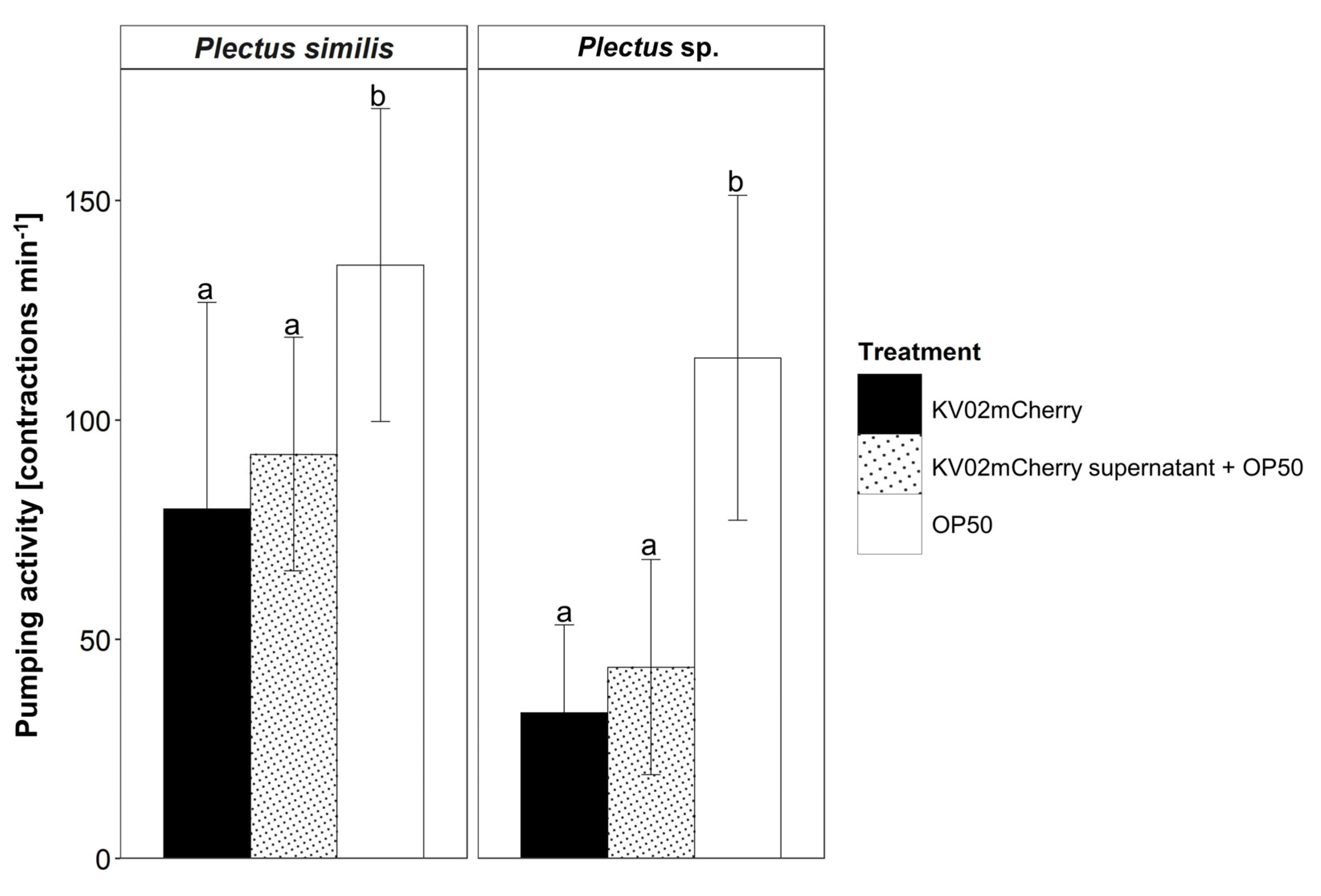
After microscopic confirmation of L. pneumophila ingestion by P. similis and Plectus sp., the effect of L. pneumophila KV02mCherry on the pharyngeal pumping activity of nematodes in comparison to E. coli OP50 was determined (Figure 2). P. similis fed with OP50 showed, with 135 contractions min−1, a 70% higher pumping frequency compared to KV02mCherry, with 80 contractions min−1 (p < 0.001) (Videos S1 and S2). Similarly, Plectus sp. displayed, with 114 contractions min−1, a three-times-higher pumping frequency when fed with OP50 (p < 0.001) compared to Legionella (Videos S3 and S4).
As the pumping rates differed clearly between bacteria species, we hypothesized that the effector proteins secreted by KV02mCherry manipulate the hosts’ feeding activity, inducing an impaired pharyngeal activity. To test this hypothesis, nematodes were fed with OP50 mixed with KV02mCherry supernatant (Figure 2). In this set-up, the pumping frequency of P. similis, with 44 contractions min−1, was 16% higher than when fed with KV02mCherry directly, but still significantly lower (p < 0.001) compared to the sole OP50 diet. A similar picture was displayed by Plectus sp. The pumping frequency, with 44 contractions min−1 when exposed to OP50 plus supernatant, increased by 33% compared to KV02mCherry cells, but this was again significantly slower (p < 0.001) than feeding OP50.

3.4. Effector Protein ProA as a Prospect Candidate for the Regulation of Pumping Activity

The experiments with the addition of L. pneumophila KV02mCherry supernatant to E. coli OP50 point to an impaired pumping frequency of nematodes (Figure 2). One of the most abundant effector proteins in Legionella supernatant is the zinc metalloprotease ProA, with its broad proteolytic and cytotoxic activities [29,33]. ProA degrades collagen IV, which is a major component of the basement membranes covering the pharynx, intestine and gonads of nematodes [32,59]. Consequently, ProA was investigated for its potential to alter pumping activity.
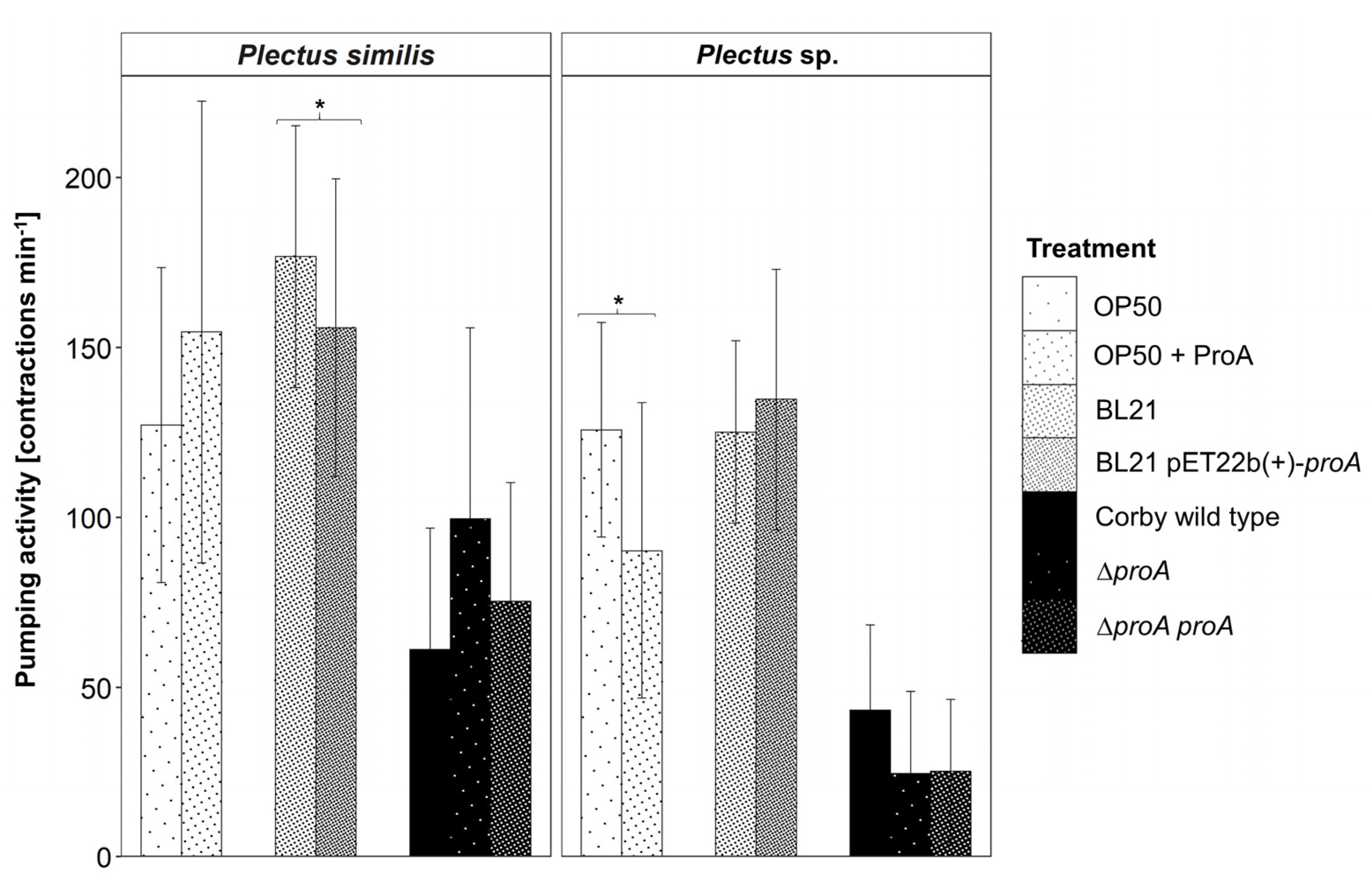
In a first step, pumping frequencies were compared between L. pneumophila Corby wild-type and L. pneumophila Corby ΔproA, a mutant, which does not produce this protease (Figure 3). When fed with the Corby ΔproA mutant, P. similis had, with 100 contractions min−1, a higher pumping frequency compared to feeding Corby wild-type with 61 contractions min−1. Contrary to P. similis, the pumping activity of Plectus sp. was low when offering the Corby ΔproA mutant (25 contractions min−1), but increased to 43 contractions min−1 with Corby wild-type. Additionally, to guarantee that the complementation of the Corby ΔproA deletion rescued the observed phenotype, the complement mutant strain L. pneumophila Corby ΔproA proA was tested (Figure 3). The pumping frequency of P. similis in the presence of the complement mutant strain L. pneumophila Corby ΔproA proA (75 contractions min−1) was in between the frequency in presence of the wild strain (61 contractions min−1) and the frequency with the Corby ΔproA mutant (100 contractions min−1). This tendency was not observed for Plectus sp., as the frequency in the presence of both mutant strains was identically reduced to 25 contractions min−1.
To further analyze the effect of ProA, nematodes were incubated in a cryo-DEAE buffer with 10 µg/mL purified ProA isolated from L. pneumophila Corby wild-type for 2 h, before offering E. coli OP50 as a diet (Figure 3). Only Plectus sp. responded to this treatment with pumps significantly lower (p < 0.05) in the group incubated with ProA, with 90 contractions min−1, than in the group fed with sole E. coli OP50, pumping with 114 contractions min−1. Additionally, a negative control of nematodes incubated in sole cryo-DEAE buffer was performed to exclude the potential negative effects of the cryo-DEAE buffer on the pharyngeal pumping activity.
In a next step, nematodes were fed either with the E. coli strain BL21 pET22b(+)-proA or the E. coli strain BL21 not capable of ProA synthesis. This set-up examines whether pharyngeal pumping of nematodes is affected by the protease, if synthetized by E. coli as bacterium with otherwise good food quality. For P. similis, pumping frequency was significantly reduced (p < 0.05), with 155 and 177 contractions min−1 when feeding BL21 pET22b(+)-proA and the control strain, respectively (Figure 3). For Plectus sp., the pumping activity for both strains was equal, differing by only 10 contractions min−1.

3.5. Acanthamoeba castellanii as Potential Promotor for Legionella pneumophila Ingestion

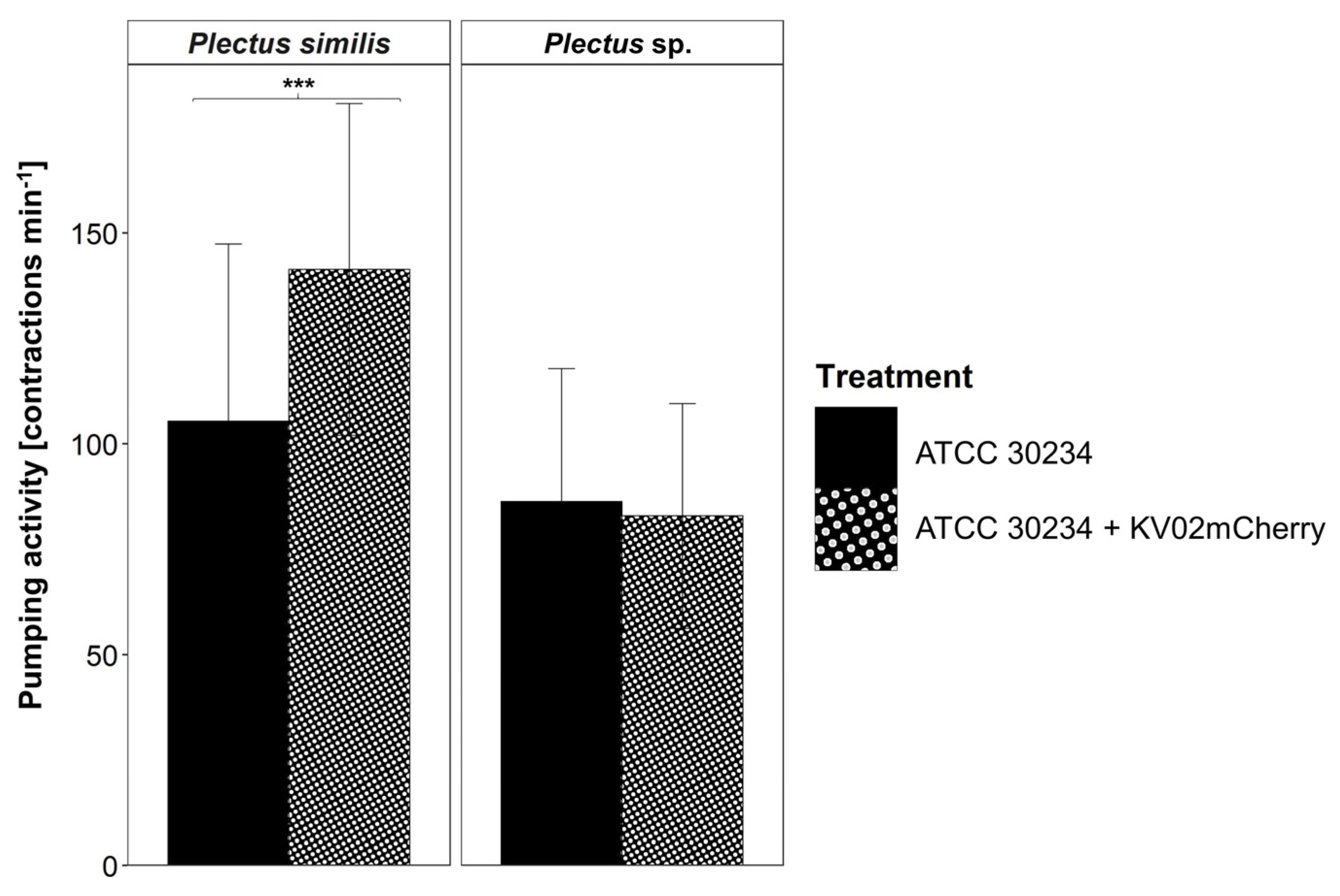
Links between L. pneumophila and nematodes may also include a further trophic level, i.e., free-living amoebae, which are well known as hosts for pathogenic bacteria. To investigate this “Trojan-horse” approach, nematodes were fed with A. castellanii 24 h after its infection with L. pneumophila KV02mCherry (Figure 4). The pumping frequency of P. similis was 34% higher (p < 0.001) when fed with L. pneumophila KV02mCherry infected amoebae (141 contractions min−1) compared to non-infected ones (105 contractions min−1). In contrast, for Plectus sp., no difference in pharyngeal pumping between amoebae with/without L. pneumophila KV02mCherry was detected.

3.6. Influence of Diet on Overall Nematode Feeding Behavior

To determine the impact of different diets on nematode behavior, all individuals on each assay plate were counted after a respective adaption time, i.e., after 3 h for Legionella or 8 h for E. coli and A. castellanii. The following three behaviors were distinguished: (1) “inactive” nematodes with no apparent movement, (2) “active” fast-moving nematodes freely roaming the bacterial lawn and (3) “feeding” nematodes dwelling in a small area on the bacterial lawn, visibly pumping with their pharynx.
For P. similis, when fed with L. pneumophila, nematodes were most active (83%) with Corby ΔproA proA as diet and least active (41%) with Corby wild-type as diet (p < 0.05, Table 2). Otherwise, the proportions of inactive, active and feeding individuals were similar between the different L. pneumophila strains. For Plectus sp., a comparable pattern was observed, but with a higher occurrence of inactive individuals (59–77%).
With E. coli OP50 as diet, the proportions of active and feeding P. similis where highest (87 and 88%, respectively) when fed with OP50 and lowest (24 and 12%, respectively) when individuals were incubated with ProA prior to the assay (p < 0.05, Table 2). On the other hand, significantly more nematodes were inactive (76%) due to preincubation with ProA than without (12%, p < 0.05). For Plectus sp., the activity mode of nematodes was similar between all E. coli OP50 treatments. However, the proportion of feeding individuals was significantly higher (p < 0.05) when fed with OP50 (39%) than when fed with OP50 mixed with KV02mCherry filtrate (11%).
The synthesis of the protease ProA by E. coli did not affect nematode behavior. The proportions of inactive, active and feeding individuals of both P. similis and Plectus sp. were nearly equal between E. coli BL21 pET22b(+)-proA and the control diet E. coli BL21 (Table 2).
For P. similis, when fed with A. castellanii, the proportions of inactive, active and feeding individuals, ranging between 41% to 56%, were similar for infected and non-infected amoebae (Table 2). Comparably, in Plectus sp., no significant response to amoebae carrying L. pneumophila or not was observed. However, the overall behavior to the amoebae as prey differed to that of P. similis. The proportion of active individuals of Plectus sp. was highest (62–69%), and that of inactive nematodes was lowest (31–38%). This difference was significant (p < 0.05) for infected amoebae as diet.
To assign differences in the general feeding behavior of both nematodes related to the four different diet groups, the wild-type strains E. coli OP50, E. coli BL21 and A. castellanii were statistically compared to L. pneumophila KV02mCherry. The pumping activity of P. similis was significantly lower when fed with L. pneumophila KV02mCherry compared to E. coli OP50 (p < 0.05). For Plectus sp., the activity was significantly lower when fed with L. pneumophila KV02mCherry compared to A. castellanii (p < 0.05).

3.7. Effect of Legionella pneumophila Supernatant on Nematode Fitness

As E. coli OP50 mixed with L. pneumophila KV02mCherry filtrate as diet resulted in impaired pharyngeal pumping frequency, the effects of Legionella supernatant on nematode fitness were studied more thoroughly. Nematodes were incubated in four different supernatants or PBS solely as control, and the mortality was determined after 24 h and 48 h. This revealed that in general, P. similis had a higher overall mortality when incubated with Legionella supernatant than Plectus sp. (Figure 5).
The mortality rate of P. similis, ranging from 9% (PBS) to 24% (Corby ΔproA), was more than twice as high for Legionella (except for Corby ΔproA proA) supernatant than for the PBS control after incubation for 24 h (Figure 5). This was significant for the supernatants of Corby ΔproA (p < 0.01) and Corby wild-type (p < 0.05). Additionally, the mortality rate of P. similis was significantly lower for the complementary mutant Corby ΔproA proA when compared to Corby ΔproA (p < 0.01) and Corby wild-type (p < 0.05). After 48 h, the mortality of Corby ΔproA and Corby wild-type supernatant increased by 25% and 45%, respectively, compared to 24 h. Both supernatants increased mortality significantly (p < 0.05) compared to PBS after 48 h. On the other hand, the mortality of Corby ΔproA proA decreased by half compared to 24 h.
Plectus sp. mortality ranged between 8% (Corby ΔproA proA) and 20% (Corby ΔproA) after incubation with the respective supernatant for 24 h (Figure 5). Compared to PBS, the mortality of Plectus sp. increased by about one half to 14% and 15% in the supernatants of KV02mCherry and Corby wild-type, respectively. Further, the mortality of Plectus sp. increased to 20% in the supernatant of the Corby ΔproA mutant. Moreover, the mortality of the Corby ΔproA supernatant was significantly higher than the mortality of Corby ΔproA proA supernatant (p < 0.05). After 48 h, the picture was different: the mortality of nematodes was high in all treatment variants, including the control. As at 24 h, most dead individuals were detected after incubation with Corby ΔproA (27%); however, the lowest numbers occurred with Corby ΔproA proA supernatant (13%, p < 0.01), Corby wild-type (15%, p < 0.05) and KV02mCherry (17%, p < 0.05) supernatant. Additionally, with PBS, more than twice as many individuals died at 48 h compared to 24 h.

4. Discussion

4.1. Free-Living Nematodes and L. pneumophila Co-Occur in Cooling Towers

This survey, investigating natural (swimming lakes) and technical (cooling towers) water bodies, revealed the co-occurrence of L. pneumophila and free-living nematodes within cooling towers. Recently, this was also reported by another cooling-tower study [19]. However, L. pneumophila and nematodes were not associated together in natural swimming lakes, but co-occurred in a natural thermal source.
Cooling towers provide a unique environment for microbial growth, considering temperatures ranging between 25 °C and 35 °C, a neutral pH, and continuous aeration [60]. On the other hand, the high operating temperatures, in combination with the application of biocides, play an important selective role on all biota inhabiting the water body of cooling towers [61]. This was distinctly visible in the extremely low diversity of the nematode community, comprising one to three taxa only, all generalists that are tolerant to pollutants and other disturbances and are, moreover, able to survive food-poor conditions [62].
The widest distribution across cooling towers, with occurrences in four out of seven, showed the genus Plectus and taxa in the family Cephalobidae (i.e., Acrobeloides, Eucephalobus, Heterocephalobus). Their presence matches their opportunistic life strategy, i.e., they are adapted to exploit newly available habitats, and, with their rapid growth rate, quickly establish populations [23]. They can cope with unpredictable or variable environments such as a cooling tower, and here, most of them are additionally favored due to their capacity to tolerate temperatures well above 30 °C [63]. Cephalobidae are the most common group in soils worldwide [64], while Plectidae are also abundant in humid habitats, e.g., mosses, with proportions up to 60% of the nematode community [65,66]. This likely explains the frequent occurrence of Plectus in cooling-tower biofilms. The thermophile Diploscapter sp., with an optimal population growth at 30 °C [67], was only found in a single cooling tower, but findings from wastewater biofilms, trickling filters and activated sludge are documented [67,68]. In sum, all nematode taxa detected in cooling towers (except Filenchus) were bacterial feeders. Apparently, the biofilm community offers few resources, only supporting short food chains lacking large-sized omnivores and predators at higher trophic levels.
Unlike in cooling towers, biofilms in lakes are hotspots for biodiversity, with a wide variety of microorganisms and their grazers in numerous ecological niches [69,70]. However, no L. pneumophila could be detected in the investigated swimming lakes, although the water temperature (mean: 25.1 °C, Table S1) during the sampling period was suitable for its growth [71,72]. In multispecies biofilms, bacteria compete for a better accessibility and utilization of nutrients, thus faster-growing species, e.g., P. aeruginosa, likely outcompete L. pneumophila [73]. Moreover, some bacteria exhibit inhibitory effects on L. pneumophila, e.g., Aeromonas hydrophila, Acidovorax sp. and Sphingomonas sp. [74]. Evidently, L. pneumophila can better use the framework of its ecological potency in low-diversity habitats such as cooling towers.
As for other biofilm biota, the nematode populations were distinctly more diverse in the biofilms of swimming lakes than those of cooling towers, reflecting the greater microbial diversity and the related number of trophic niches [75]. The most abundant across lake subhabitats was Chromadorina sp., a genus which predominates in freshwater biofilms [17,76,77]. Interestingly, taxa which occurred in both natural and technical water habitats, i.e., Filenchus, Heterocephalobus and Plectus, where much more abundant in the cooling towers than in swimming lakes. The cooling tower biofilms likely offered an environment with less predation and/or resource competition, fostering population growth in adapted taxa. The fact that only single or very few species were encountered an entire cooling tower habitat suggests the random, windborne dispersal of nematodes.
As in cooling towers, bacterial feeders were the dominant trophic group in all three subhabitats of the swimming lakes. However, in the lake biofilms, high numbers of omnivores and predators were also detected. These K-strategists, with long lifespans and high sensitivity to disturbance, reflect the balanced environmental conditions as well as stability due to a better nutrient supply in lakes [18].

4.2. L. pneumophila Diet Reduces Feeding Activity in Free-Living Nematodes

Free-living nematodes take up food by rhythmic contractions of the pharynx muscles, which makes these pharyngeal pumps a reliable indicator of food ingestion [78]. In turn, the availability, quality and familiarity of the food affects the rate of pharyngeal pumping [79]. Analyzing the pumping rates of P. similis and Plectus sp. revealed that both species pumped significantly more when fed with E. coli OP50 compared to L. pneumophila KV02mCherry. As pharyngeal pumping shows a graded response to food availability [80], this indicates E. coli OP50 as a quality resource. In contrast, slow pumping punctured with long pauses was observed for Legionella, a behavior typical for worms without food [80]. Comparing synthetic beads and E. coli OP50, Fueser et al. [47] showed that C. elegans restricts pumping for particles with low nutritional value to a basic rate. This behavior prevents the nematode from wasting energy by high-frequency pumping, but still allows screening for food. One factor contributing to food quality is the bacterial cell size, yet with a length of 1 to 3 µm, Legionella is in the same range as E. coli OP50 [81,82]. In conclusion, the downregulated pumping rate in P. similis and Plectus sp. suggests that L. pneumophila is a poor or unsuitable (e.g., well-defended—see Section 4.3) food source for Plectus.
The food quality can be further assessed by the foraging behavior of nematodes, which exhibits two discrete foraging states called roaming and dwelling [83]. Roaming is a rapid, straight movement to explore the environment, whereas dwelling is characterized by slow movements with frequent reversals and turns [84]. On poor food, roaming strongly increases, while dwelling predominates on high quality food [84,85]. When fed with the E. coli strains OP50 and BL21 or the amoeba A. castellanii, the nematodes moved slowly, equaling dwelling, and proportions of moving and feeding individuals were more-or-less similar (personal observation). In contrast, in the presence of L. pneumophila strains, foraging activity switched to an exploratory behavior, i.e., the proportion of moving individuals strongly increased compared to slow-moving, feeding specimens. This change from dwelling to roaming again points to a lower food quality of L. penumophila for Plectus compared to the common diet E. coli, but also to A. castellanii.

4.3. L. pneumophila Reduces Fitness of Free-Living Nematodes

Secondary metabolites secreted by bacteria can act not only as repellents but also as toxins for nematodes [86,87]. For example, the production of cyanide by P. aeruginosa ceases pharyngeal pumping followed by progressive paralysis and death in C. elegans [88]. To test whether L. pneumophila effectors regulate the pharyngeal pumping rate, we offered E. coli OP50 suspended in Legionella KV02mCherry supernatant as diet. Interestingly, the pumping rate of P. similis and Plectus sp. decreased significantly compared to pure E. coli OP50 and was almost as low as for the KV02mCherry diet. This strongly suggests that secondary metabolites in the supernatant of L. pneumophila, e.g., the Legionella major secretory protein ProA, can critically impair the pharyngeal pumping activity of Plectus.
Supporting this, in P. similis, the pumping rate was higher (albeit not significantly) when fed with a L. pneumophila Corby ΔproA mutant strain compared to the Corby wild-type and Corby ΔproA proA strains, respectively (Figure 3). Moreover, feeding on E. coli BL21 pET22b(+)-proA decreased the pumping rate. Due to the high standard deviations for E. coli BL21 pET22b(+)-proA and its control BL21, the statistically assigned significant reduction in the pumping rates should be regarded more as a trend. In contrast, Plectus sp. decreased the pumping rate when fed with the L. pneumophila strains ΔproA and ΔproA proA, but not with E. coli BL21 pET22b(+)-proA. However, the incubation with purified ProA also impaired the pumping rate. As mentioned above, taking into account the biological variability in pumping activity for E. coli OP50 + ProA and its control without ProA, this points to a trend rather than a distinct decrease in the pumping rate in the presence of ProA.
In sum, the impact of ProA on nematode feeding activity varied with diet, and further showed species-specific differences. Moreover, no direct toxicity of ProA was observed, as the incubation of nematodes with the supernatant of the L. pneumophila ΔproA strain for 24 and 48 h, respectively, did not result in a reduced mortality compared to the Corby wild-type, Corby ΔproA proA and KV02mCherry strains (Figure 5).
Besides impaired feeding, bacterial defense can expose negative impact on nematode fitness [89,90]. Secondary metabolites with nematicidal potential are reported for common soil and freshwater bacteria such as Pseudomonas e.g., [91,92]. In line with this, the mortality of Plectus increased after incubation in L. pneumophila supernatant (except for Corby ΔproA proA) for 24 h when compared to PBS buffer. However, the high mortality in PBS (control) after 48 h for Plectus sp. does not allow for a clear statement about the mortality of Legionella supernatant after 48 h. Besides ProA, other potentially destructive enzymes in L. pneumophila supernatant that degrade host cell components are T2SS-dependent proteases, peptidases, acid phosphatases, lipases, phospholipases A and C, lysophospholipase A, a cholesterol acyltransferase and an RNase [34]. Such secretions of L. pneumophila have the potential to reduce nematode fitness, e.g., protease and lipase activity can lead to structural damage in the cuticle, followed by a decrease in motility. Similarly, an extracellular serine protease from Bacillus sp. was reported to degrade the cuticle of the bacterial feeder Panagrellus redivivus causing the nematodes’ death [93]. Further, Siddiqui et al. [94] showed that the exposure of the root-knot nematode Meloidogyne incognita to culture filtrate of Pseudomonas fluorescens CHA0 killed nematodes, which was induced by the extracellular protease AprA. Further studies are needed to clarify which of the potential destructive secondary metabolites are the causative agent for this negative Legionella–nematode interaction.

4.4. A. castellanii as Trojan Horse for L. pneumophila Transmission

L. pneumophila resists degradation by amoebae and other protozoa and multiplies intracellularly [16]. Such hosts of bacterial pathogens, such as the free-living amoeba A. castellanii, are considered the “Trojan horse” of the microbial world [95]. Virtually nothing is known regarding whether these Legionella—amoeba interactions involve higher trophic levels, i.e., predators of A. castellanii such as nematodes.
Pharyngeal pumping assays investigating this trophic link revealed that when fed with L. pneumophila-infected A. castellanii (i.e., 24 h post infection with L. pneumophila KV02mCherry), the pumping rate of nematodes was similar (Plectus sp.) or even significantly higher (P. similis) compared to non-infected amoebae. The video recordings showed that the nematodes do not swallow the amoeba cells whole, but rather rupture the cell membrane and suck in the cell contents (Videos S5 and S6). It is likely that the disruption of the physical and structural integrity of the amoebal plasma membrane, facilitated by the pore-forming activity of intracellular L. pneumophila, allowed for easy access to amoebal cytoplasma, and in the case of P. similis, also stimulated pumping activity.
By their sucking feeding mode, nematodes incorporated the liberated pathogen from the amoeba diet. Similarly, Rasch et al. [36] observed Legionella cells, but no intact Legionella-infected A. castellanii within the digestive tract of C. elegans, and thus argued against a “typical” Trojan horse transmission of L. pneumophila via A. castellanii into nematodes. On the other hand, the tested Plectus species did not discriminate against infected amoebae, as indicated by the observed pumping rates. It is possible that L. pneumophila “hides” from nematode detection inside the amoeba and, in this way, could access nematodes as a new host without being detected before ingestion. Moreover, nematodes preferred L. pneumophila-infected amoebae over a sole L. pneumophila diet, pointing towards a “Trojan horse-like transmission” strategy of Legionella in Plectus.

5. Conclusions

This study identified cooling towers as technical water habitats, where free-living nematodes and L. pneumophila co-occur in biofilm communities. The species P. similis (coolingtower isolate) and Plectus sp. (thermalspa isolate) were chosen in order to examine the feeding relationship between L. pneumophila and nematodes. Pharyngeal pumping assays revealed that L. pneumophila impairs the pumping rate of Plectus compared to E. coli and A. castellanii diets. However, the impact of the Legionella major secretory protein ProA on nematode feeding activity varied with diet and nematode species.
Overall, reduced feeding activity induced by L. pneumophila suggests that Legionella is no common resource of the genus Plectus, as shown in an artificial set-up on agar plates. However, the ingestion of L. pneumophila, resulting in an intracellular infection of the nematodes’ digestive tract, can occur and has to be considered in pathogen dissemination. Moreover, trophic interactions between Plectus- and Legionella-infected amoebae point to a “Trojan horse-like transmission” of L. pneumophila into nematodes. In order to further decipher the Legionella–nematode interaction, other coolingtower taxa, e.g., the thermophilic Diploscapter, have to be tested for their potential as reservoirs or vectors for L. pneumophila in future studies. Additionally, feeding assays in (artificial) biofilms are necessary to obtain a more realistic picture on the predator–prey relationship between nematodes and L. pneumophila.

Supplementary Materials

The following supporting information can be downloaded at: https://www.mdpi.com/article/10.3390/microorganisms11030738/s1, Table S1: List of surveyed swimming lakes; Video S1: P. similis + KV02mCherry; Video S2: P. similis + E. coli OP50; Video S3: Plectus sp. + KV02mCherry; Video S4: Plectus sp. + E. coli OP50; Video S5: P. similis + A. castellanii infected with KV02mCherry; Video S6: Plectus sp. + A. castellanii infected with KV02mCherry [96,97,98,99,100,101,102,103].

Author Contributions

Conceptualization, supervision, project administration and funding acquisition, L.R. and M.S.; methodology, validation and writing—review and editing, C.H., L.R. and M.S.; formal analysis, C.H.; investigation and resources, C.H. and A.L.; visualization and writing—original draft preparation, C.H. All authors have read and agreed to the published version of the manuscript.

Funding

This work was funded by the Deutsche Forschungsgemeinschaft (DFG) with the grant numbers RU 780/19-1 and STE 838/12-1.

Data Availability Statement

Data are contained within the article.

Acknowledgments

We acknowledge the help of Andreas Richter during the field survey of swimming lakes. The L. pneumophila mutant strains L. pneumophila Corby ΔproA and L. pneumophila Corby ΔproA proA, as well as native purified ProA and E. coli strain BL21 pET22b(+)-proA were kindly provided by Lina Scheithauer at the Technische Universität Braunschweig. We further thank the reviewers whose comments helped improve and clarify this manuscript.

Conflicts of Interest

The authors declare no conflict of interest.

References

  1. Mondino, S.; Schmidt, S.; Rolando, M.; Escoll, P.; Gomez-Valero, L.; Buchrieser, C. Legionnaires’ disease: State of the art knowledge of pathogenesis mechanisms of Legionella. Annu. Rev. Pathol. Mech. Dis. 2020, 15, 439–466. [Google Scholar]
  2. Horwitz, M.A.; Silverstein, S.C. Legionnaires’ disease bacterium (Legionella pneumophila) multiplies intracellularly in human monocytes. J. Clin. Investig. 1980, 66, 441–450. [Google Scholar]
  3. Finsel, I.; Hilbi, H. Formation of a pathogen vacuole according to Legionella pneumophila: How to kill one bird with many stones. Cell. Microbiol. 2015, 17, 935–950. [Google Scholar]
  4. Atlas, R.M. Legionella: From environmental habitats to disease pathology, detection and control. Environ. Microbiol. 1999, 1, 283–293. [Google Scholar]
  5. Borella, P.; Guerrieri, E.; Marchesi, I.; Bondi, M.; Messi, P. Water ecology of Legionella and protozoan: Environmental and public health perspectives. Biotechnol. Annu. Rev. 2005, 11, 355–380. [Google Scholar] [PubMed]
  6. Newton, H.J.; Ang, D.K.; Van Driel, I.R.; Hartland, E.L. Molecular pathogenesis of infections caused by Legionella pneumophila. Clin. Microbiol. Rev. 2010, 23, 274–298. [Google Scholar] [PubMed]
  7. Rogers, J.; Dowsett, A.B.; Dennis, P.J.; Lee, J.V.; Keevil, C. Influence of temperature and plumbing material selection on biofilm formation and growth of Legionella pneumophila in a model potable water system containing complex microbial flora. Appl. Environ. Microbiol. 1994, 60, 1585–1592. [Google Scholar] [PubMed]
  8. Phin, N.; Parry-Ford, F.; Harrison, T.; Stagg, H.R.; Zhang, N.; Kumar, K.; Lortholary, O.; Zumla, A.; Abubakar, I. Epidemiology and clinical management of Legionnaires’ disease. Lancet Infect. Dis. 2014, 14, 1011–1021. [Google Scholar] [PubMed]
  9. Lueck, C.; Brzuszkiewicz, E.; Rydzewski, K.; Koshkolda, T.; Sarnow, K.; Essig, A.; Heuner, K. Subtyping of the Legionella pneumophila “Ulm” outbreak strain using the CRISPR–Cas system. Int. J. Med. Microbiol. 2015, 305, 828–837. [Google Scholar]
  10. Maisa, A.; Brockmann, A.; Renken, F.; Lück, C.; Pleischl, S.; Exner, M.; Jurke, A. Epidemiological investigation and case–control study: A Legionnaires’ disease outbreak associated with cooling towers in Warstein, Germany, August–September 2013. Eurosurveillance 2015, 20, 30064. [Google Scholar]
  11. Molofsky, A.B.; Swanson, M.S. Differentiate to thrive: Lessons from the Legionella pneumophila life cycle. Mol. Microbiol. 2004, 53, 29–40. [Google Scholar] [CrossRef] [PubMed]
  12. Donlan, R.M.; Costerton, J.W. Biofilms: Survival mechanisms of clinically relevant microorganisms. Clin. Microbiol. Rev. 2002, 15, 167–193. [Google Scholar] [CrossRef] [PubMed]
  13. Taylor, M.; Ross, K.; Bentham, R. Legionella, protozoa, and biofilms: Interactions within complex microbial systems. Microb. Ecol. 2009, 58, 538–547. [Google Scholar] [CrossRef] [PubMed]
  14. Rowbotham, T.J. Preliminary report on the pathogenicity of Legionella pneumophila for freshwater and soil amoebae. J. Clin. Pathol. 1980, 33, 1179–1183. [Google Scholar] [CrossRef]
  15. Fields, B.S.; Benson, R.F.; Besser, R.E. Legionella and Legionnaires’ disease: 25 years of investigation. Clin. Microbiol. Rev. 2002, 15, 506–526. [Google Scholar] [CrossRef]
  16. Swart, A.L.; Harrison, C.F.; Eichinger, L.; Steinert, M.; Hilbi, H. Acanthamoeba and Dictyostelium as cellular models for Legionella infection. Front. Cell. Infect. Microbiol. 2018, 8, 61. [Google Scholar] [CrossRef]
  17. Schroeder, F.; Traunspurger, W.; Pettersson, K.; Peters, L. Temporal changes in periphytic meiofauna in lakes of different trophic states. J. Limnol. 2012, 71, e23. [Google Scholar] [CrossRef]
  18. Traunspurger, W.; Wilden, B.; Majdi, N. An overview of meiofaunal and nematode distribution patterns in lake ecosystems differing in their trophic state. Hydrobiologia 2020, 847, 2665–2679. [Google Scholar] [CrossRef]
  19. Paranjape, K.; Bédard, É.; Shetty, D.; Hu, M.; Choon, F.C.P.; Prévost, M.; Faucher, S.P. Unravelling the importance of the eukaryotic and bacterial communities and their relationship with Legionella spp. ecology in cooling towers: A complex network. Microbiome 2020, 8, 157. [Google Scholar] [CrossRef]
  20. Sabater, S.; Vilalta, E.; Gaudes, A.; Guasch, H.; Munoz, I.; Romani, A. Ecological implications of mass growth of benthic cyanobacteria in rivers. Aquat. Microb. Ecol. 2003, 32, 175–184. [Google Scholar] [CrossRef]
  21. Mathieu, M.; Leflaive, J.; Ten-Hage, L.; De Wit, R.; Buffan-Dubau, E. Free-living nematodes affect oxygen turnover of artificial diatom biofilms. Aquat. Microb. Ecol. 2007, 49, 281–291. [Google Scholar] [CrossRef]
  22. Bongers, T.; Ferris, H. Nematode community structure as a bioindicator in environmental monitoring. Trends Ecol. Evol. 1999, 14, 224–228. [Google Scholar] [CrossRef] [PubMed]
  23. Bongers, T. The maturity index: An ecological measure of environmental disturbance based on nematode species composition. Oecologia 1990, 83, 14–19. [Google Scholar] [CrossRef] [PubMed]
  24. Ferris, H.; Bongers, T.; de Goede, R.G.M. A framework for soil food web diagnostics: Extension of the nematode faunal analysis concept. Appl. Soil Ecol. 2001, 18, 13–29. [Google Scholar] [CrossRef]
  25. McCoy-Simandle, K.; Stewart, C.R.; Dao, J.; DebRoy, S.; Rossier, O.; Bryce, P.J.; Cianciotto, N.P. Legionella pneumophila type II secretion dampens the cytokine response of infected macrophages and epithelia. Infect. Immun. 2011, 79, 1984–1997. [Google Scholar] [CrossRef]
  26. White, R.C.; Cianciotto, N.P. Assessing the impact, genomics and evolution of type II secretion across a large, medically important genus: The Legionella type II secretion paradigm. Microb. Genom. 2019, 5, e000273. [Google Scholar] [CrossRef] [PubMed]
  27. Tyson, J.Y.; Vargas, P.; Cianciotto, N.P. The novel Legionella pneumophila type II secretion substrate NttC contributes to infection of amoebae Hartmannella vermiformis and Willaertia magna. Microbiology 2014, 160, 2732–2744. [Google Scholar] [CrossRef] [PubMed]
  28. White, R.C.; Truchan, H.K.; Zheng, H.; Tyson, J.Y.; Cianciotto, N.P. Type II secretion promotes bacterial growth within the Legionella-containing vacuole in infected amoebae. Infect. Immun. 2019, 87, e00374-19. [Google Scholar] [CrossRef] [PubMed]
  29. Hell, W.; Essig, A.; Bohnet, S.; Gatermann, S.; Marre, R. Cleavage of tumor necrosis factor-α by Legionella exoprotease. Apmis 1993, 101, 120–126. [Google Scholar] [CrossRef] [PubMed]
  30. Tyson, J.Y.; Pearce, M.M.; Vargas, P.; Bagchi, S.; Mulhern, B.J.; Cianciotto, N.P. Multiple Legionella pneumophila Type II secretion substrates, including a novel protein, contribute to differential infection of the amoebae Acanthamoeba castellanii, Hartmannella vermiformis, and Naegleria lovaniensis. Infect. Immun. 2013, 81, 1399–1410. [Google Scholar] [CrossRef]
  31. Conlan, J.W.; Baskerville, A.; Ashworth, L.A.E. Separation of Legionella pneumophila Proteases and Purification of a Protease Which Produces Lesions Like Those of Legionnaires Disease in Guinea Pig Lung. Microbiol. 1986, 132, 1565–1574. [Google Scholar] [CrossRef]
  32. Scheithauer, L.; Thiem, S.; Schmelz, S.; Dellmann, A.; Büssow, K.; Brouwer, R.M.; Ünal, C.M.; Blankenfeldt, W.; Steinert, M. Zinc metalloprotease ProA of Legionella pneumophila increases alveolar septal thickness in human lung tissue explants by collagen IV degradation. Cell. Microbiol. 2021, 23, e13313. [Google Scholar] [CrossRef]
  33. Scheithauer, L.; Thiem, S.; Ünal, C.M.; Dellmann, A.; Steinert, M. Zinc metalloprotease ProA from Legionella pneumophila inhibits the pro-inflammatory host response by degradation of bacterial flagellin. Biomolecules 2022, 12, 624. [Google Scholar] [CrossRef]
  34. Rossier, O.; Dao, J.; Cianciotto, N.P. The type II secretion system of Legionella pneumophila elaborates two aminopeptidases, as well as a metalloprotease that contributes to differential infection among protozoan hosts. Appl. Environ. Microbiol. 2008, 74, 753–761. [Google Scholar] [CrossRef] [PubMed]
  35. Hellinga, J.R.; Garduño, R.A.; Kormish, J.D.; Tanner, J.R.; Khan, D.; Buchko, K.; Jimenez, C.; Pinette, M.M.; Brassinga, A.K.C. Identification of vacuoles containing extraintestinal differentiated forms of Legionella pneumophila in colonized Caenorhabditis elegans soil nematodes. Microbiol. Open 2015, 4, 660–681. [Google Scholar] [CrossRef] [PubMed]
  36. Rasch, J.; Krüger, S.; Fontvieille, D.; Ünal, C.M.; Michel, R.; Labrosse, A.; Steinert, M. Legionella-protozoa-nematode interactions in aquatic biofilms and influence of Mip on Caenorhabditis elegans colonization. Int. J. Med. Microbiol. 2016, 306, 443–451. [Google Scholar] [CrossRef] [PubMed]
  37. Brassinga, A.K.C.; Kinchen, J.M.; Cupp, M.E.; Day, S.R.; Hoffman, P.S.; Sifri, C.D. Caenorhabditis is a metazoan host for Legionella. Cell. Microbiol. 2010, 12, 343–361. [Google Scholar] [CrossRef]
  38. Kurz, C.L.; Ewbank, J.J. Caenorhabditis elegans for the study of host–pathogen interactions. Trends Microbiol. 2000, 8, 142–144. [Google Scholar] [CrossRef]
  39. Anderson, G.L.; Caldwell, K.N.; Beuchat, L.R.; Williams, P.L. Interaction of a free-living soil nematode, Caenorhabditis elegans, with surrogates of foodborne pathogenic bacteria. J. Food Prot. 2003, 66, 1543–1549. [Google Scholar] [CrossRef]
  40. Kroupitski, Y.; Pinto, R.; Bucki, P.; Belausov, E.; Ruess, L.; Spiegel, Y.; Sela, S. Acrobeloides buetschlii as a potential vector for enteric pathogens. Nematology 2015, 17, 447–457. [Google Scholar] [CrossRef]
  41. Walters, J.V.; Holcomb, R.R. Isolation of enteric pathogen from sewage-borne nematode. Nematologica 1967, 13, 155. [Google Scholar]
  42. Wasilewska, L.; Webster, J.M. Free-living nematodes as disease factors of man and his crops. Int. J. Environ. Stud. 1975, 7, 201–204. [Google Scholar] [CrossRef]
  43. Moens, T.; Van Gansbeke, D.; Vincx, M. Linking estuarine nematodes to their suspected food. A case study from the Westerschelde Estuary (south-west Netherlands). J. Mar. Biol. Assoc. UK 1999, 79, 1017–1027. [Google Scholar] [CrossRef]
  44. Moens, T.; Traunspurger, W.; Bergtold, M. Feeding ecology of free-living benthic nematodes. In Freshwater Nematodes. Ecology and Taxonomy; Abebe, E., Traunspurger, W., Andrássy, I., Eds.; CAB International Publishing: Wallingford, UK, 2006; pp. 105–131. [Google Scholar]
  45. Zhou, Y.; Falck, J.R.; Rothe, M.; Schunck, W.H.; Menzel, R. Role of CYP-eicosanoids in the regulation of pharyngeal pumping and food uptake in C. elegans. J. Lipid Res. 2015, 56, 2110–2123. [Google Scholar] [CrossRef] [PubMed]
  46. Liu, H.; Qin, L.W.; Li, R.; Zhang, C.; Al-Sheikh, U.; Wu, Z.X. Reciprocal modulation of 5-HT and octopamine regulates pumping via feedforward and feedback circuits in C. elegans. Proc. Natl. Acad. Sci. USA 2019, 116, 7107–7112. [Google Scholar] [CrossRef]
  47. Fueser, H.; Rauchschwalbe, M.T.; Höss, S.; Traunspurger, W. Food bacteria and synthetic microparticles of similar size influence pharyngeal pumping of Caenorhabditis elegans. Aquat. Toxicol. 2021, 235, 105827. [Google Scholar] [CrossRef]
  48. Gaur, A.V.; Agarwal, R. Risperidone induced alterations in feeding and locomotion behavior of Caenorhabditis elegans. Curr. Res. Toxicol. 2021, 2, 367–374. [Google Scholar] [CrossRef]
  49. Chiang, J.T.A.; Steciuk, M.; Shtonda, B.; Avery, L. Evolution of pharyngeal behaviors and neuronal functions in free-living soil nematodes. J. Exp. Biol. 2006, 209, 1859–1873. [Google Scholar] [CrossRef]
  50. Bongers, T. De Nematoden van Nederland; Stichting Uitgeverij van de Koninklijke Natuurhistorische Vereniging: Utrecht, The Netherlands, 1994; pp. 1–408. [Google Scholar]
  51. Fang-Yen, C.; Avery, L.; Samuel, A.D. Two size-selective mechanisms specifically trap bacteria-sized food particles in Caenorhabditis elegans. Proc. Natl. Acad. Sci. USA 2009, 106, 20093–20096. [Google Scholar] [CrossRef]
  52. Song, B.M.; Avery, L. The pharynx of the nematode C. elegans: A model system for the study of motor control. Worm 2013, 2, e21833. [Google Scholar] [CrossRef]
  53. Ruess, L. Studies on the nematode fauna of an acid forest soil: Spatial distribution and extraction. Nematologica 1995, 41, 229–239. [Google Scholar] [CrossRef]
  54. Holovachov, O.; Boström, S. Identification of Plectida (Nematoda); EUMAINE, Gent and Nematology; UC Riverside: Riverside, CA, USA, 2010; Available online: http://www.nrm.se/download/18.9ff3752132fdaeccb6800015609/PLECTIDA%5B1%5D.pdf (accessed on 12 November 2020).
  55. Zell, H. Die Gattung Plectus Bastian, 1865 Sensu lato (Nematoda, Plectidae)–ein Beitrag zur Ökologie, Biogeographie, Phylogenie und Taxonomie der Plectidae; Staatliches Museum für Naturkunde Karlsruhe: Karlsruhe, Germany, 1993; pp. 1–172. [Google Scholar]
  56. Andrássy, I. Free-Living Nematodes of Hungary (Nematoda errantia), I; Hungarian Natural History Museum: Budapest, Hungary, 2005; pp. 1–518. [Google Scholar]
  57. Yeates, G.W.; Bongers, T.; De Goede, R.G.W.; Freckman, D.W.; Georgieva, S.S. Feeding habits in soil nematode families and genera—An outline for soil ecologists. J. Nematol. 1993, 25, 315–331. [Google Scholar] [PubMed]
  58. Charpentier, X.; Kay, E.; Schneider, D.; Shuman, H.A. Antibiotics and UV radiation induce competence for natural transformation in Legionella pneumophila. J. Bacteriol. 2011, 193, 1114–1121. [Google Scholar] [CrossRef] [PubMed]
  59. Graham, P.L.; Johnson, J.J.; Wang, S.; Sibley, M.H.; Gupta, M.C.; Kramer, J.M. Type IV collagen is detectable in most, but not all, basement membranes of Caenorhabditis elegans and assembles on tissues that do not express it. J. Cell Biol. 1997, 137, 1171–1183. [Google Scholar] [CrossRef] [PubMed]
  60. Liu, Y.; Zhang, W.; Sileika, T.; Warta, R.; Cianciotto, N.P.; Packman, A. Role of bacterial adhesion in the microbial ecology of biofilms in cooling tower systems. Biofouling 2009, 25, 241–253. [Google Scholar] [CrossRef] [PubMed]
  61. Di Gregorio, L.; Tandoi, V.; Congestri, R.; Rossetti, S.; Di Pippo, F. Unravelling the core microbiome of biofilms in cooling tower systems. Biofouling 2017, 33, 793–806. [Google Scholar] [CrossRef]
  62. Bongers, T.; Bongers, M. Functional diversity of nematodes. Appl. Soil Ecol. 1998, 10, 239–251. [Google Scholar] [CrossRef]
  63. Majdi, N.; Traunspurger, W.; Fueser, H.; Gansfort, B.; Laffaille, P.; Maire, A. Effects of a broad range of experimental temperatures on the population growth and body-size of five species of free-living nematodes. J. Therm. Biol. 2019, 80, 21–36. [Google Scholar] [CrossRef]
  64. Yeates, G.W. Nematodes as soil indicators: Functional and biodiversity aspects. Biol. Fertil. Soils 2003, 37, 199–210. [Google Scholar] [CrossRef]
  65. Schenk, J.; Traunspurger, W.; Ristau, K. Genetic diversity of widespread moss-dwelling nematode species in German beech forests. Eur. J. Soil Biol. 2016, 74, 23–31. [Google Scholar] [CrossRef]
  66. Shevchenko, V.L.; Zhylina, T.M. Taxonomic structure of nematode communities of epiphytic mosses in green plantations of Chernihiv, Ukraine. Вестнuк 3ooлoгuu 2016, 50, 477–482. [Google Scholar] [CrossRef]
  67. Gibbs, D.S.; Anderson, G.L.; Beuchat, L.R.; Carta, L.K.; Williams, P.L. Potential role of Diploscapter sp. strain LKC25, a bacterivorous nematode from soil, as a vector of food-borne pathogenic bacteria to preharvest fruits and vegetables. Appl. Environ. Microbiol. 2005, 71, 2433–2437. [Google Scholar] [CrossRef]
  68. Bergtold, M.; Mayr, G.; Traunspurger, W. Nematodes in wastewater biofilms—Appearance and density of species in three biofilter reactors. Water Res. 2007, 4, 145–151. [Google Scholar] [CrossRef] [PubMed]
  69. Vadeboncoeur, Y.; Steinman, A.D. Periphyton function in lake ecosystems. Sci. World J. 2002, 2, 1449–1468. [Google Scholar] [CrossRef]
  70. Gubelit, Y.I.; Grossart, H.P. New Methods, New Concepts: What Can Be Applied to Freshwater Periphyton? Front. Microbiol. 2020, 11, 1275. [Google Scholar] [CrossRef] [PubMed]
  71. Żbikowska, E.; Kletkiewicz, H.; Walczak, M.; Burkowska, A. Coexistence of Legionella pneumophila bacteria and free-living amoebae in lakes serving as a cooling system of a power plant. Water Air Soil Pollut. 2014, 225, 2066. [Google Scholar] [CrossRef]
  72. Schwake, D.O.; Alum, A.; Abbaszadegan, M. Legionella Occurrence beyond Cooling Towers and Premise Plumbing. Microorganisms 2021, 9, 2543. [Google Scholar] [CrossRef]
  73. Declerck, P. Biofilms: The environmental playground of Legionella pneumophila. Environ. Microbiol. 2010, 12, 557–566. [Google Scholar] [CrossRef]
  74. Abu Khweek, A.; Amer, A.O. Factors mediating environmental biofilm formation by Legionella pneumophila. Front. Cell. Infect. Microbiol. 2018, 8, 38. [Google Scholar] [CrossRef] [PubMed]
  75. Gaudes, A.; Sabater, S.; Vilalta, E.; Muñoz, I. The nematode community in cyanobacterial biofilms in the river Llobregat, Spain. Nematology 2006, 8, 909–919. [Google Scholar]
  76. Croll, N.A.; Zullini, A. 1972. Observations on the bionomics of the freshwater nematode Chromadorina bioculata. J. Nematol. 1972, 4, 256–260. [Google Scholar]
  77. Majdi, N.; Traunspurger, W.; Boyer, S.; Mialet, B.; Tackx, M.; Fernandez, R.; Gehner, S.; Ten-Hage, L.; Buffan-Dubau, E. Response of biofilm-dwelling nematodes to habitat changes in the Garonne River, France: Influence of hydrodynamics and microalgal availability. Hydrobiologia 2011, 673, 229–244. [Google Scholar] [CrossRef]
  78. Abada, E.A.E.; Sung, H.; Dwivedi, M.; Park, B.J.; Lee, S.K.; Ahnn, J. C. elegans behavior of preference choice on bacterial food. Mol. Cells 2009, 28, 209–213. [Google Scholar] [CrossRef] [PubMed]
  79. Scholz, M.; Lynch, D.J.; Lee, K.S.; Levine, E.; Biron, D. A scalable method for automatically measuring pharyngeal pumping in C. elegans. J. Neurosci. Methods 2016, 274, 172–178. [Google Scholar] [CrossRef] [PubMed]
  80. Lee, K.S.; Iwanir, S.; Kopito, R.B.; Scholz, M.; Calarco, J.A.; Biron, D.; Levine, E. Serotonin-dependent kinetics of feeding bursts underlie a graded response to food availability in C. elegans. Nat. Commun. 2017, 8, 14221. [Google Scholar] [CrossRef]
  81. Buse, H.Y.; Ashbolt, N.J. Counting Legionella cells within single amoeba host cells. Appl. Environ. Microbiol. 2012, 78, 2070–2072. [Google Scholar] [CrossRef] [PubMed]
  82. Gomez, F.; Monsalve, G.C.; Tse, V.; Saiki, R.; Weng, E.; Lee, L.; Srinivasan, C.; Frand, A.R.; Clarke, C.F. Delayed accumulation of intestinal coliform bacteria enhances life span and stress resistance in Caenorhabditis elegans fed respiratory deficient E. coli. BMC Microbiol. 2012, 12, 300. [Google Scholar] [CrossRef]
  83. Flavell, S.W.; Pokala, N.; Macosko, E.Z.; Albrecht, D.R.; Larsch, J.; Bargmann, C.I. Serotonin and the neuropeptide PDF initiate and extend opposing behavioral states in C. elegans. Cell 2013, 154, 1023–1035. [Google Scholar] [CrossRef] [PubMed]
  84. Shtonda, B.B.; Avery, L. Dietary choice behavior in Caenorhabditis elegans. J. Exp. Biol. 2006, 209, 89–102. [Google Scholar] [CrossRef]
  85. Ben Arous, J.; Laffont, S.; Chatenay, D. Molecular and sensory basis of a food related two-state behavior in C. elegans. PLoS ONE 2009, 4, e7584. [Google Scholar] [CrossRef]
  86. Bargmann, C.I.; Hartwieg, E.; Horvitz, H.R. Odorant-selective genes and neurons mediate olfaction in C. elegans. Cell 1993, 74, 515–527. [Google Scholar] [CrossRef] [PubMed]
  87. Khan, F.; Jain, S.; Oloketuyi, S.F. Bacteria and bacterial products: Foe and friends to Caenorhabditis elegans. Microbiol. Res. 2018, 215, 102–113. [Google Scholar] [CrossRef] [PubMed]
  88. Darby, C.; Cosma, C.L.; Thomas, J.H.; Manoil, C. Lethal paralysis of Caenorhabditis elegans by Pseudomonas aeruginosa. Proc. Natl. Acad. Sci. USA 1999, 96, 15202–15207. [Google Scholar] [CrossRef] [PubMed]
  89. Jousset, A.; Rochat, L.; Péchy-Tarr, M.; Keel, C.; Scheu, S.; Bonkowski, M. Predators promote defence of rhizosphere bacterial populations by selective feeding on non-toxic cheaters. ISME J. 2009, 3, 666–674. [Google Scholar] [CrossRef] [PubMed]
  90. Richter, A.; Kern, T.; Wolf, S.; Struck, U.; Ruess, L. Trophic and non-trophic interactions in binary links affect carbon flow in the soil micro-food web. Soil Biol. Biochem. 2019, 135, 239–247. [Google Scholar] [CrossRef]
  91. Jousset, A. Ecological and evolutive implications of bacterial defences against predators. Environ. Microbiol. 2012, 14, 1830–1843. [Google Scholar] [CrossRef]
  92. Guo, J.; Jing, X.; Peng, W.L.; Nie, Q.; Zhai, Y.; Shao, Z.; Zheng, L.; Cai, M.; Li, G.; Zuo, H.; et al. Comparative genomic and functional analyses: Unearthing the diversity and specificity of nematicidal factors in Pseudomonas putida strain 1A00316. Sci. Rep. 2016, 6, 29211. [Google Scholar] [CrossRef]
  93. Lian, L.H.; Tian, B.Y.; Xiong, R.; Zhu, M.Z.; Xu, J.; Zhang, K.Q. Proteases from Bacillus: A new insight into the mechanism of action for rhizobacterial suppression of nematode populations. Lett. Appl. Microbiol. 2007, 45, 262–269. [Google Scholar] [CrossRef]
  94. Siddiqui, I.A.; Haas, D.; Heeb, S. Extracellular protease of Pseudomonas fluorescens CHA0, a biocontrol factor with activity against the root-knot nematode Meloidogyne incognita. Appl. Environ. Microbiol. 2005, 71, 5646–5649. [Google Scholar] [CrossRef]
  95. Brouse, L.; Brouse, R.; Brouse, D. Natural pathogen control chemistry to replace toxic treatment of microbes and biofilm in cooling towers. Pathogens 2017, 6, 14. [Google Scholar] [CrossRef]
  96. Badegewässer Landkreis Oder-Spree. Available online: https://www.landkreis-oder-spree.de/media/custom/2689_1595_1.PDF?1530869752 (accessed on 20 October 2022).
  97. Flughafensee: Wassertemperatur. Available online: https://wasserportal.berlin.de/station.php?anzeige=g&sgrafik=mw&thema=owt&station=5800303 (accessed on 20 October 2022).
  98. Badegewässer Landkreis Barnim. Available online: https://presse.barnim.de/documents/tabelle-badewasserqualitaet-80833 (accessed on 20 October 2022).
  99. Badewarnungen für drei Seen. Available online: https://hohen-neuendorf.de/de/stadt-leben/aktuelles/badewarnungen-fuer-drei-seen (accessed on 20 October 2022).
  100. Am Wolfsschluchtkanal: Wassertemperatur. Available online: https://wasserportal.berlin.de/station.php?anzeige=g&sgrafik=tw&thema=owt&station=5800107 (accessed on 20 October 2022).
  101. Plötzensee: Wassertemperatur. Available online: https://wasserportal.berlin.de/station.php?anzeige=g&sgrafik=mw&thema=owt&station=5800312 (accessed on 20 October 2022).
  102. Biesdorfer Baggersee: Wassertemperatur. Available online: https://wasserportal.berlin.de/station.php?anzeige=g&sgrafik=mw&thema=owt&station=5800317 (accessed on 20 October 2022).
  103. Nayhauss, A. Ab ins Wasser! Das sind die besen Badeseen in Berlin. Available online: https://www.morgenpost.de/berlin/article214942137/Sportlich-schick-romantisch-Badeseen-fuer-jeden-Typ.html (accessed on 20 October 2022).
Figure 1. Confocal laser-scanner microscopy of L. pneumophila KV02mCherry (cooling tower isolate) ingested by the nematodes Plectus similis and Plectus sp. Single rod-shaped cells of L. pneumophila KV02mCherry are visible in red. (A) P. similis with L. pneumophila KV02mCherry right before the grinder (gr) located within the terminal bulb (tb). Plectus sp. with several bacterial cells in the propharynx (pph), terminal bulb and upper intestine (int) (B), as well as a single cell in the mid-gut (C).
Microorganisms 11 00738 g001
Figure 2. Pharyngeal pumping activity per individual [contractions min−1 ± SD] of the nematode species Plectus similis and Plectus sp. fed with L. pneumophila KV02mCherry (cooling tower isolate), E. coli OP50 or E. coli OP50 mixed with L. pneumophila KV02mCherry supernatant. Bars with the same letter are not statistically different according to Dunn’s post hoc test (p < 0.05).
Microorganisms 11 00738 g002
Figure 3. Pharyngeal pumping activity per individual [contractions min−1 ± SD] of the nematode species Plectus similis and Plectus sp. fed with different diets, i.e., L. pneumophila (Corby wild-type, ProA-lacking Corby ΔproA mutant and its respective complementary mutant Corby ΔproA proA), E. coli BL21 (BL21, ProA-producing strain) and E. coli OP50. Additionally, nematodes were incubated for 2 h in ProA prior to feeding on E. coli OP50 (OP50 + ProA). Statistical differences according to the Dunn test (three L. pneumophila strains) and Mann–Whitney U test (two E. coli OP50 and two E. coli BL21 strains), respectively. * p < 0.05.
Microorganisms 11 00738 g003
Figure 4. Pharyngeal pumping activity per individual [contractions min−1 ± SD] of the nematode species Plectus similis and Plectus sp. fed with the A. castellanii strain ATCC 30234. A. castellanii were infected with L. pneumophila KV02mCherry (cooling tower isolate) and fed to nematodes 24 h after infection. Statistical differences according to the Mann–Whitney U test. *** p < 0.001.
Microorganisms 11 00738 g004
Figure 5. Mortality [% ± SD] of the nematode species Plectus similis and Plectus sp. after incubation with different supernatants of Legionella for 24 and 48 h. The strains tested were L. pneumophila KV02mCherry (cooling tower isolate), L. pneumophila Corby wild-type and a L. pneumophila Corby ΔproA mutant and its respective complementary mutant strain L. pneumophila Corby ΔproA proA. Control = PBS buffer. Statistical differences according to Tukey’s post hoc test (p < 0.05). Bars with the same letter are not statistically different according to Dunn’s post hoc test (p < 0.05).
Microorganisms 11 00738 g005
Table 1. Co-occurrence of nematode genera (Ind. 100−1 mL ± SD) and Legionella pneumophila (+, detection; -, no detection) in natural and technical water habitats. Nematode genera with assigned c-p values [23] arranged by trophic group.
Trophic GroupGenusc-pSwimming Lakes (n = 9)Cooling Towers (n = 7)
Water Surface, AlgaeRead,
Macrophytes
Submerged Stones, Litter CT 1CT 2CT 3CT 4CT 5CT 6CT 7
Plant feedersAphelenchoides21.5 ± 1.32.8 ± 4.01.7 ± 3.0-------
Filenchus2--3.5 ± 6.021.5--25.6---
Bacterial
feeders
Acrobeloides2-----28.676.9---
Alaimus41.5 ± 1.32.1 ± 3.6--------
Bastiania30.7 ± 1.2---------
Chromadorina363.3 ± 36.7136.2 ± 64.268.2 ± 45.0-------
Diplogasteritus1-------9.5 ± 16.5--
Diploscapter1-------161.9 ± 16.5--
Eucephalobus2---------28.6
Eumonhystera116.1 ± 17.36.6 ± 5.71.1 ± 0.9-------
Heterocephalobus20.4 ± 0.7--10.8------
Monhystrella10.7 ± 1.2---------
Panagrolaimus12.2 ± 2.1---------
Paraphanolaimus3--2.4 ± 4.1-------
Plectus23.4 ± 4.21.5 ± 1.31.1 ± 0.91044.6-28.6282.1-28.6-
Rhabdolaimus3-18.8 ± 32.57.0 ± 9.2-------
OmnivoresAchromadora30.8 ± 1.3-6.1 ± 6.1-------
Epidorylaimus4--1.2 ± 2.1-------
Eudorylaimus40.7 ± 1.2---------
Laimydorus47.7 ± 13.338.3 ± 64.21.0 ± 1.7-------
Mesodorylaimus46.0 ± 9.488.3 ± 150.84.2 ± 3.8-------
PredatorsIronus4-2.2 ± 3.6--------
Mononchus4-0.8 ± 1.4--------
Tobrilus360.7 ± 49.212.4 ± 10.717.9 ± 25.0-------
Total number of nematode genera1411123023211
Detection Legionella pneumophila---+++++- 1+
1 Biocide shock dosage before sampling.
Table 2. Nematode activity [% ± SD] of Plectus similis and Plectus sp. fed with different diets, i.e., L. pneumophila (cooling-tower isolate KV02mCherry, Corby wild-type, ProA-lacking Corby ΔproA mutant and its respective complementary mutant Corby ΔproA proA), E. coli OP50 (OP50, OP50 mixed with KV02mCherry filtrate), E. coli BL21 (BL21, ProA-producing strain) and non-infected and Legionella (KV02mCherry)-infected A. castellanii. Additionally, nematodes were incubated for 2 h in ProA prior to feeding on OP50 (OP50 + ProA). Statistical differences according to Dunn’s post hoc test (p < 0.05) were examined between activity modes (i.e., columns) for each diet separately. Data with the same or no letters are not statistically different.
StrainPlectus similisPlectus sp.
InactiveActiveFeedingInactiveActiveFeeding
L. pneumophila
KV02mCherry46 ± 17 ab54 ± 17 ab9 ± 577 ± 923 ± 914 ± 2
Corby wild-type59 ± 12 a41 ± 12 a7 ± 371 ± 2429 ± 2413 ± 1
ΔproA33 ± 8 ab67 ± 8 ab7 ± 168 ± 1632 ± 1614 ± 2
ΔproA proA17 ± 4 b83 ± 4 b16 ± 559 ± 341 ± 312 ± 0
E. coli OP50
OP5012 ± 1 a88 ± 1 a87 ± 1 a52 ± 948 ± 939 ± 2 a
OP50 + KV02mCherry filtrate21 ± 9 ab79 ± 9 ab21 ± 2 ab53 ± 847 ± 811 ± 1 b
OP50 + ProA76 ± 3 b24 ± 3 a12 ± 3 b66 ± 834 ± 821 ± 2 ab
E. coli BL21
BL21 pET22b(+)-proA46 ± 654 ± 651 ± 641 ± 459 ± 454 ± 6
BL2154 ± 246 ± 244 ± 360 ± 1140 ± 1138 ± 12
A. castellanii
ATCC 3023456 ± 744 ± 741 ± 638 ±1262 ± 1252 ± 18
ATCC 30234 + KV02mCherry46 ± 1054 ± 1049 ± 1131 ±469 ± 458 ± 8
Disclaimer/Publisher’s Note: The statements, opinions and data contained in all publications are solely those of the individual author(s) and contributor(s) and not of MDPI and/or the editor(s). MDPI and/or the editor(s) disclaim responsibility for any injury to people or property resulting from any ideas, methods, instructions or products referred to in the content.