Article

Plant-Associated Representatives of the Bacillus cereus Group Are a Rich Source of Antimicrobial Compounds

1
Proteomics and Spectroscopy Unit (ZBS6), Center for Biological Threats and Special Pathogens, Robert Koch Institute, 13353 Berlin, Germany
2
Division of Pathology and Phyto-Immunology, Plant Protection Research Institute (PPRI), Duc Thang, Bac Tu Liem, Hanoi, Vietnam
3
Bioinformatics and Systems Biology, Justus-Liebig Universität Giessen, 35392 Giessen, Germany
4
Highly Pathogenic Microorganisms Unit (ZBS2), Center for Biological Threats and Special Pathogens, Robert Koch Institute, 13353 Berlin, Germany
5
Institute of Marine Biotechnology e.V. (IMaB), 17489 Greifswald, Germany
6
Pharmaceutical Biotechnology, University of Greifswald, 17489 Greifswald, Germany
7
Institute of Biology, Humboldt University Berlin, 10115 Berlin, Germany
*
Author to whom correspondence should be addressed.
Microorganisms 2023, 11(11), 2677; https://doi.org/10.3390/microorganisms11112677
Submission received: 8 October 2023 / Revised: 26 October 2023 / Accepted: 27 October 2023 / Published: 31 October 2023
(This article belongs to the Special Issue Biological Control of the Plant Pathogens)

Abstract

:
Seventeen bacterial strains able to suppress plant pathogens have been isolated from healthy Vietnamese crop plants and taxonomically assigned as members of the Bacillus cereus group. In order to prove their potential as biocontrol agents, we perform a comprehensive analysis that included the whole-genome sequencing of selected strains and the mining for genes and gene clusters involved in the synthesis of endo- and exotoxins and secondary metabolites, such as antimicrobial peptides (AMPs). Kurstakin, thumolycin, and other AMPs were detected and characterized by different mass spectrometric methods, such as MALDI-TOF-MS and LIFT-MALDI-TOF/TOF fragment analysis. Based on their whole-genome sequences, the plant-associated isolates were assigned to the following species and subspecies: B. cereus subsp. cereus (6), B. cereus subsp. bombysepticus (5), Bacillus tropicus (2), and Bacillus pacificus. These three isolates represent novel genomospecies. Genes encoding entomopathogenic crystal and vegetative proteins were detected in B. cereus subsp. bombysepticus TK1. The in vitro assays revealed that many plant-associated isolates enhanced plant growth and suppressed plant pathogens. Our findings indicate that the plant-associated representatives of the B. cereus group are a rich source of putative antimicrobial compounds with potential in sustainable agriculture. However, the presence of virulence genes might restrict their application as biologicals in agriculture.

1. Introduction

At present, the replacement of harmful chemical pesticides by environmentally friendly biological means is a pressing need in agriculture worldwide. Microbes, such as bacteria and fungi, have been proven to be promising candidates for the development of efficient agents useful in sustainable agriculture. At present, endospore-forming Bacillus spp. and Gram-negative Pseudomonas spp. are the most used constituents of bioformulations applied in biological plant protection. The main advantage of bioformulations based on Bacillus endospores is their longevity, which makes their stability comparable with that of chemical fungicides [1].
During a survey of plant-beneficial bacteria as part of the microbiome of different plant-associated sites, such as the rhizosphere, the tissues of the inner root, and the attached insect larvae of Vietnamese crop plants (black pepper, coffee and orange trees, brown mustard, and tomato), a number of Gram-positive, endospore-forming bacteria, able to suppress common plant pathogens, were isolated. Based on their draft genome sequences, the isolates were taxonomically assigned as being members of the Bacillaceae family, representing four main taxonomic groups: Lysinibacillus spp., Brevibacillus spp., the Bacillus subtilis species complex, and the Bacillus cereus group [2]. Our further studies revealed that, in contrast to Lysinibacillus sp., the plant-associated Brevibacilli harbored a multitude of interesting antimicrobial peptides with a strong potential to suppress phytopathogenic bacteria, fungi, and nematodes [3]. The Bacillus velezensis isolates TL7 and S1, members of the B. subtilis species complex, were identified in large-scale trials as the most promising candidates for developing efficient biocontrol agents [4]. In this study, we focus on the plant-associated isolates belonging to the B. cereus group in order to investigate their potential for biocontrol and plant growth promotion.
The B. cereus group, also known as B. cereus sensu lato (s.l.), comprises a steadily increasing number of species, but is still plagued by taxonomic inconsistencies. Several phenotypic traits important for taxonomic assignment, such as the synthesis of the anthrax toxin and capsule, entomopathogenic crystal proteins, and the synthesis of emetic toxins (cereulide), are plasmid-encoded and can be lost during strain evolution [5]. Well-known members of the B. cereus group are the human-pathogenic B. anthracis, the entomopathogenic B. thuringiensis, and the opportunistic pathogen B. cereus sensu stricto (s.s.). The three species are able to cause human diseases with different severity [6]. They are closely related and harbor very similar genome sequences, which do not necessarily justify their delineation in different species. Traditionally, they have been discriminated due to properties mainly encoded by extrachromosomal elements.
B. anthracis (risk group 3) was identified in 1876 by the German physician Robert Koch as the causative agent of anthrax [7], the first disease that was linked to a microbe. Its virulence is based on the ability to form exotoxins and a capsule, which are encoded by the plasmids pXO1 and pXO2 [8].
The plasmid-encoded production of the highly toxic cereulide is restricted to rare emetic B. cereus strains occurring in some foods, whilst the production of the diarrheal-inducing enterotoxins (hemolysin BL, HBL; non-hemolytic enterotoxin, NHE; and cytotoxin K, CytK) is common in B. cereus s.l. [9]. Due to the extreme stability of the cyclic dodecadepsipeptide celeuride, which withstands current food processing techniques, their emetic B. cereus producer strains are of particular concern for human health [9].
B. thuringiensis (Bt), isolated 1901 by Ishikawa as “B. sotto” from the silkworm, and some years later as B. thuringiensis by Berliner from the meal moth [10], is an insect pathogen that is successfully used in agriculture as a biopesticide based on the production of diverse crystal toxins, also known as δ-endotoxins [11].
However, the B. cereus taxonomy solely based on the presence of virulence plasmids with a specific function becomes increasingly questionable in light of the recent phylogenomic data. The occurrence of B. cereus strains containing pXO1-like plasmids [12] and of crystal protein-harboring B. thuringiensis strains, which are phylogenetically related to B. anthracis [13], make the identification of these species a difficult task. Moreover, the occurrence of virulence genes in the B. cereus s.l. species cannot be excluded. Therefore, B. cereus s.l. strains with potential for use in sustainable agriculture can be a risk for public health and need to be carefully checked for their genomic content, also in the case of their taxonomic delineation suggests them as a “safe” species.
In this study, we aim to elucidate the potential of plant-associated members of the B. cereus species complex as biological plant protection. In the utilization of these strains as biocontrol agents, their potential due to their rich biosynthetic potential, but also the risks connected with the presence of virulence genes, need to be considered. Genome-based phylogenetic analyses revealed that most of the isolates were clustered within two subspecies of the B. cereus s.s. species. Interestingly, the isolate B. cereus TK1 harbored genes encoding two different crystal proteins and one vegetative insectopathogenic toxin (Vip3). Genome mining for biosynthetic gene clusters probably involved in the synthesis of antimicrobial peptides (AMPs) and direct mass-spectrometric investigation of the synthesized AMPs revealed that the isolates are promising candidates for use in sustainable agriculture. In this context, the lipopeptides kurstakin and thumolycin seem to be of special importance. A special highlight of our research is resolving the primary structure of the plasmid-encoded thumolycin pentapeptide by LIFT-MALDI-TOF/TOF fragment analysis. The inhibiting action of the B. cereus s.l. isolates against plant pathogens was corroborated in direct assays performed with pathogenic oomycetes, fungi, and nematodes. Finally, the plant-growth-promoting activity of some of the isolates is demonstrated. Regardless of these promising results, we have to consider the risk for public health when the B. cereus s.l. isolates are applied as biological means in agriculture.

2. Materials and Methods

2.1. Strain Isolation and Cultivation

Isolation from Vietnamese healthy crop plants, and insects attached on plant surfaces (Table 1), and the purification of the strains were performed as described previously [2,4]. In order to exclude vegetative cells, the samples were heat-treated at 80 °C for 20 min. Only isolates able to suppress fungal plant pathogens were selected for further characterization [2]. The cultivation of the bacterial strains and DNA isolation have been previously described [14]. The B. cereus group strains were cultivated on Cereus Ident agar and Cereus-selective agar, as described in Section 2.4.

2.2. Reconstruction of the Complete Genomes

The genome sequences of Bacillus cereus A22, B. cereus A24, B. cereus HD1.4B, and B. cereus HD2.4 were reconstructed using a combined approach of two sequencing technologies that generated short paired-end reads and long reads. The resulting sequences were then used for hybrid assembly. Short-read sequencing has been previously described [14]. Long-read sequencing was conducted in house with the Oxford Nanopore MinION with the flowcell (R9.4.1), as described previously [3]. The quality of assemblies was assessed by determining the ratio of falsely trimmed proteins by using Ideel (https://github.com/phiweger/ideel, accessed on 1 November 2021). The genome coverage of the obtained contigs was 50× in average. Genome annotation and visualization was performed as described previously [3].

2.3. Screening of the Virulence Genes

The screening of virulence genes in whole-genome shotgun sequences (WGS) and complete genomes was performed by using a combined analysis of the PATRIC annotation system [15] and tblastN in the 17 genomes. The most characteristic genes from B. anthracis Vollum, including four genes of the pXO1 plasmid (cya, lef, pagA, and repX) and six genes of the pXO2 plasmid (capA, capB, capC, capD, capE, and repS), were used as the reference sequences. The tblastN threshold for both similarity and coverage was >30%, and all BLAST results were cross-checked against the PATRIC annotation, available at the Bacterial and Viral Bioinformatics Resource Center, BV-BRC, https://www.bv-brc.org/, accessed on 1 November 2021.
Table 1. The plant-associated B. cereus group isolates and their collection sites. Two B. cereus genomosubspecies, A, and B, were distinguished (Figure 1). The crop plants used for isolating the strains were black pepper (Piper nigrum) trees, tomato (Lycopersicon esculentum) plants, orange (Citrus sinensis) trees, maize (Zea mays), and brown mustard (Brassica juncea). Samples obtained from the inner root tissues were obtained after the sterilization of the root surface [2].
AccessionSizeG + CGenes SampleGTDBCollection
bp%CodingRNANameGenomospeciesPlantOrganSiteDate
CP085501.15,268,01835.75533149A24B. cereus ssp. Ablack pepperrootVietnam24 May 2018
CP085506.15,183,31234.95450154HD2.4BB. cereus ssp. Atomato plantsrhizosphereVietnam, Hoai Duc, Ha Noi23 April 2019
CP085510.15,183,87034.95450157HD1.4BB. cereus ssp. Atomato plantrhizosphereVietnam, Hoai Duc, Ha Noi23 April 2019
JABSVB000000000.15,796,35834.8566480HB3.1B. cereus ssp. Aorange treerhizosphereVietnam, Cao Phong, Hoa Binh17 April 2019
VDDR00000000.15,071,71635.4561963A8B. cereus ssp. Acoffee treerootVietnam9 May 2018
VEPT00000000.15,319,67835.3533263A31B. cereus ssp. Ablack pepper rootVietnam22 May 2018
VEPS00000000.26,195,29934.7625478TK1B. cereus ssp. Bblack pepper rhizosphereVietnam9 May 2018
CP085498.15,310,79135.25640143A22B. cereus ssp. Bcoffee treerootVietnam9 May 2018
JABSVF000000000.15,869,33634.8564080M2.1BB. cereus ssp. BmaizerhizosphereVietnam, Phu An, Thanh Da 6 December 2019
VEPQ00000000.15,604,01135.6546663A42B. cereus ssp. Bblack pepper rootVietnam12 May 2018
VEPR00000000.15,594,61735.6548067SN4-3B. cereus ssp. Bmaizedead insectVietnam28 May 2018
VEPV00000000.15,335,51335.2533958SN1B. tropicus ssp. BOstrinia nubilalis Vietnam28 May 2018
VEPW00000000.15,958,60635.8584393CD3-2B. tropicus ssp. brown mustardrhizosphereVietnam28 May 2018
VEPU00000000.15,443,80135.2538972SN4.1B. pacificus ssp. BOstrinia nubilalis Vietnam28 May 2018
JABSVD000000000.15,695,94035.1553487HD1.3Bacillus sp.tomato plantrhizosphereVietnam, Hoai Duc, Ha Noi23 April 2019
VEPX00000000.15,695,94035.3513665CD3-5Bacillus sp.brown mustard rhizosphereVietnam28 May 2018
VEPY00000000.15,150,56035.2517561CD3-1aBacillus sp.brown mustardrhizosphereVietnam28 May 2018
The criteria for the presence of virulence plasmids were established as described by Liu et al. [16]. Sequences representing the different types of δ-endotoxins (Supplementary Table S1) were extracted from the NCBI data bank. Searches for the presence of genes encoding crystal proteins and toxins in the 17 plant-associated B. cereus genome sequences were performed with tblastN using the respective protein sequences as query.

2.4. Genotypic and Phenotypic Characterization of the Isolate B. cereus CD3-1a

In addition to the B. anthracis virulence genes mentioned above, the genome of strain B. cereus CD3-1a was also screened for presence of the four B. anthracis-specific prophage regions (dhp) described by Radnedge et al. [17]. These in silico analyses were complemented by real-time PCR assays targeting pagA, capB, rpoB, and dhp61.183. Colony morphology was examined on Columbia blood agar, blood trimethoprim agar, Cereus Ident agar, and Cereus-selective agar [18].

2.5. Taxonomical Phylogeny Assessment

Species and subspecies delineation were performed using the Type (Strain) Genome Server (TYGS) platform [4]. Information on nomenclature was provided by the List of Prokaryotic names with Standing in Nomenclature (LPSN), available at https://lpsn.dsmz.de (accessed on 1 November 2021) [19]. The EDGAR3.0 pipeline [20] was used for elucidating taxonomic relationships as described previously [4].

2.6. Genome Mining

The in silico prediction of gene clusters involved in secondary metabolite synthesis was performed using the antiSMASH pipeline version 6 [21], the bioinformatic tool described by Bachmann and Ravel [22], and BAGEL4 [23].

2.7. Sample Preparation and Mass-Spectrometric Detection of the Bioactive Peptides

The bioactive compounds of the investigated B. cereus s.l. strains were detected and identified by MALDI-TOF MS, as outlined previously [24,25]. A Bruker Autoflex Speed TOF/TOF mass spectrometer (Bruker Daltonics; Bremen, Germany) was used with Smartbeam laser technology applying a 1 kHz frequency-triple Nd-YAG laser (λex = 355 nm). Samples (2 µL) of the colony surface extracts and culture supernatants were mixed with a 2 µL matrix solution (a saturated solution of α-hydroxy-cinnamic acid in 50% aqueous ACN containing 0.1% TFA) spotted on the target, air-dried, and measured. Mass spectra were obtained by positive-ion detection in the reflector mode. The monoisotopic masses were observed. Parent ions were detected with a resolution of 10.000. The sequence analysis of peptide products was performed by MALDI-LIFT-TOF/TOF mass spectrometry in the laser induction decay (LID) mode [26]. The product ions in the LIFT-TOF/TOF fragment spectra were obtained with a resolution of 1000.

2.8. Antifungal, Nematocidal, and Plant-Growth-Promoting Activity Assays

Assays for activity against plant pathogens (oomycetes, fungi, and nematodes) were performed as previously described [4]. In brief, antifungal activities were assayed by placing agar plugs containing the respective fungi onto potato dextrose agar (PDA). The test bacteria were then streaked between the plugs, and the diameter of the fungal colonies as indicative for direct growth inhibition was recorded daily.
The bioassays for nematocidal activity were performed with Caenorhabditis elegans N2 and Meloidogyne sp., as described previously [4]. In the slow killing test, the nematodes were added to an agar-solidified growth medium containing the test bacteria and incubated for a period from 3 to 5 days at 25 °C. In the liquid fast killing test, overnight cultures of the test bacteria were transferred into 12-well plates containing the nematodes in a liquid M9 medium. The mortality of nematodes was defined as the ratio of dead (non-motile) nematodes to the total number of nematodes [4].
The root-knot nematode Meloidogyne sp. was isolated from the roots of infested pepper plants, according to Hooper et al. [27]. Tomato plantlets were grown in pots with sterilized alluvial Red River soil under subtropical climate conditions in the local greenhouse [4]. Test bacteria and second-stage juvenile (J2) nematodes were added to the pots two weeks after transplanting. Ten weeks after infesting with the nematodes, the number of knots in tomato plants was estimated [28].
Plant growth promotion assays were performed with Arabidopsis thaliana seedlings, as described previously [29]. Seven-day-old seedlings were dipped into a spore suspension of the test bacteria and transferred into a square Petri dish with a half-strength Murashige–Skoog medium solidified with 1% agar. After three weeks of incubation at 22 °C and a daily photoperiod of 14 h, the fresh weight of the plants was measured.

2.9. Data Analysis

The data obtained from the biocontrol and plant growth promotion experiments were analyzed using a one-factorial analysis of variance (ANOVA). The mean values were calculated from the results of the replicates (n ≥ 3). The Fisher´s least significant difference (LSD) test was conducted as a post hoc test for estimating significant differences (p≤ 0.05) between the mean values as described previously [4].

2.10. Gene Bank Accession Numbers of the Complete Genome Sequences

Bacillus cereus A22 chromosome: CP085498.1, Bacillus cereus A22 plasmid P1: CP085499.1, Bacillus cereus A22 plasmid P2: CP085500.1, Bacillus cereus A24 chromosome: CP085501.1, Bacillus cereus A24 plasmid P1: CP085502.1, Bacillus cereus A24 plasmid P2: CP085503.1, Bacillus cereus HD1.4B chromosome: CP0855510.1, Bacillus cereus HD1.4B plasmid P1: CP085511.1, Bacillus cereus HD1.4B plasmid P2: CP085512.1, Bacillus cereus HD1.4B plasmid P3: CP085513.1, Bacillus cereus HD2.4 chromosome: CP0855506.1, Bacillus cereus HD2.4 plasmid P1: CP085507.1, Bacillus cereus HD2.4 plasmid P2: CP085508.1, Bacillus cereus HD2.4 plasmid P3: CP085509.1.

3. Results and Discussion

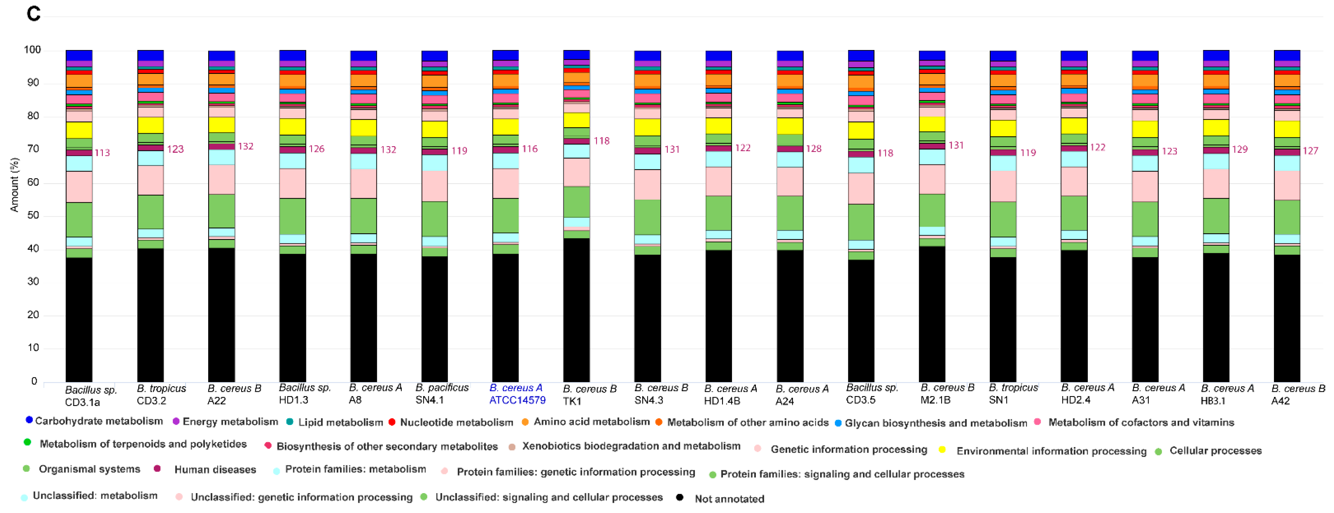
3.1. Comparative Genome Analysis of the Isolates from Vietnamese Crop Plants Representing the Bacillus cereus s.l. Complex

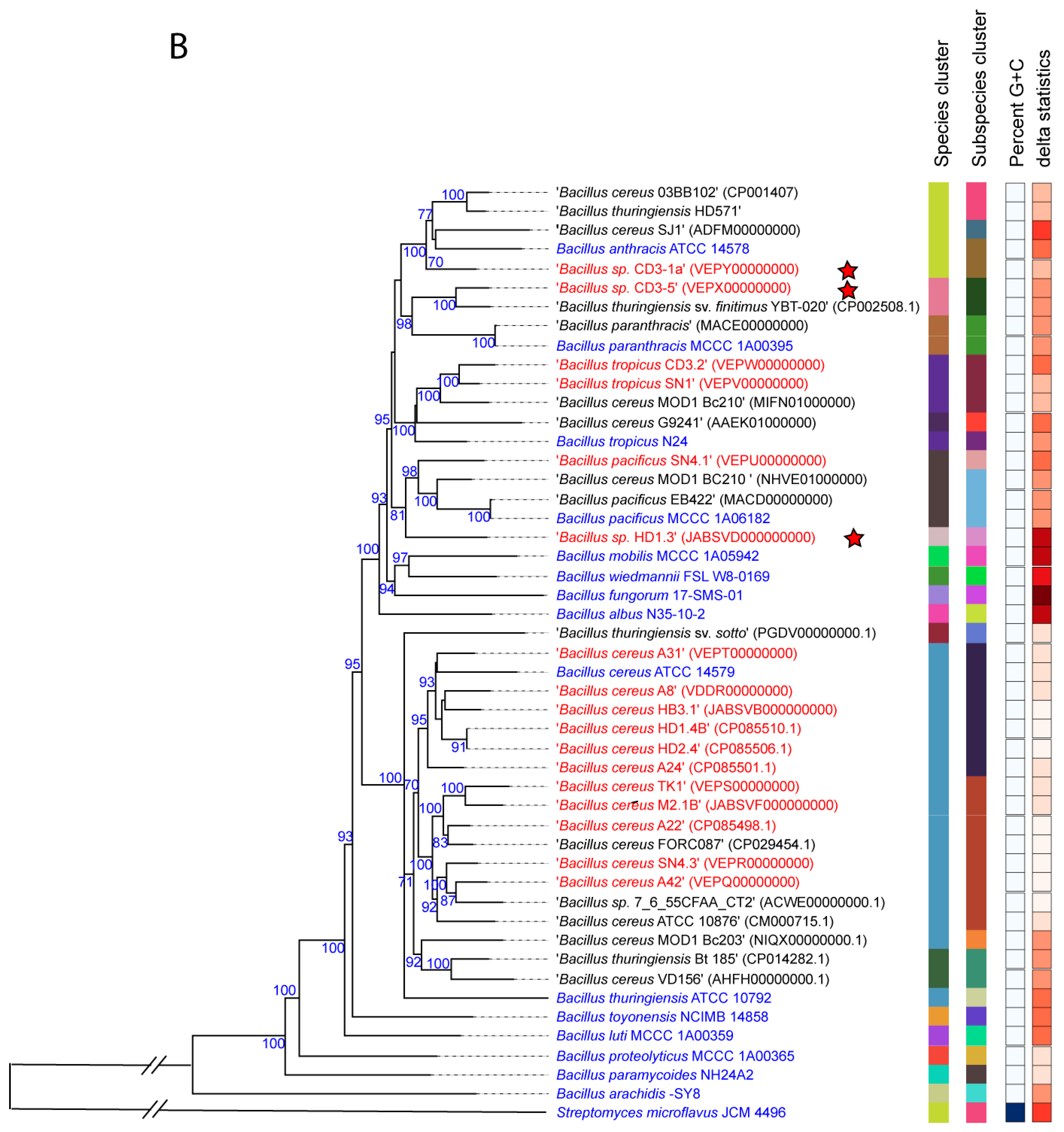
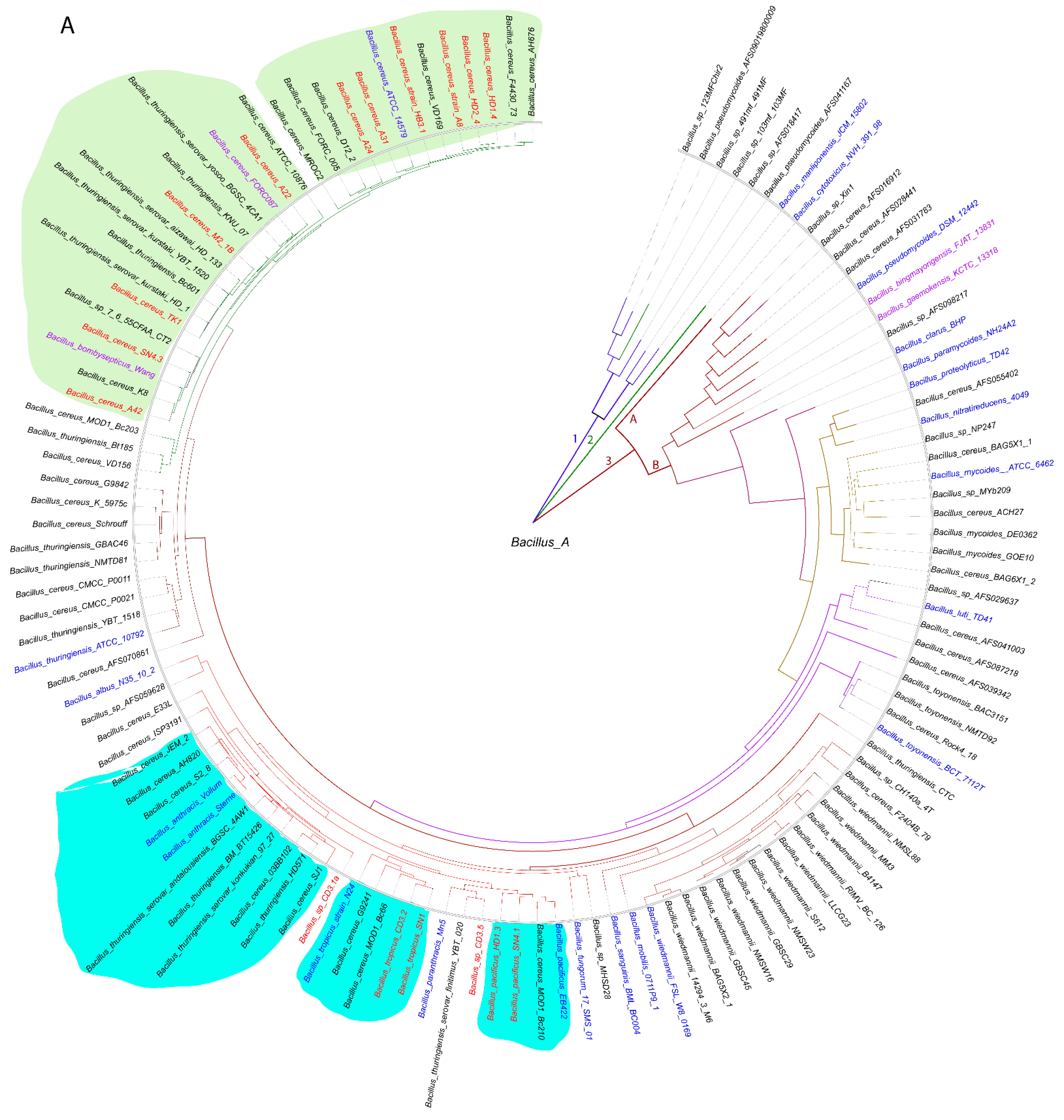
3.1.1. Genome-Based Species and Subspecies Delineation of the Plant-Associated Isolates Belonging to the B. cereus Group

Seventeen of the endospore-forming bacterial strains, isolated from Vietnamese crop plants and insects attached at their surface (Table 1), were previously assigned to the Bacillus cereus s.l. group [2]. All isolates displayed the typical features of B. cereus: they developed phospholipase C and hemolytic activity when cultivated on Cereus Ident and sheep blood agar plates. The phylogenetic tree obtained from the 16S rRNA sequences supported their previous taxonomic assignment as members of the B. cereus s.l. group, but possessed an average branch support of only 28.5% (Supplementary Figure S1), which is not sufficient for robust species delineation.
We used a whole-genome-based approach for the robust delineation of the taxonomic position of the plant-associated B. cereus s.l. isolates. The phylogenomic tree containing a total of 128 B. cereus s.l. genomes, mainly extracted from the NCBI data bank, yielded three main branches (1–3). Branch 3 was subdivided into clusters 3A and 3B. All of our isolates were distributed within the cluster 3B and were to be found related to the clusters formed by the type strains of B. cereus, B. anthracis, B. tropicus, and B. pacificus (Figure 1A).
Figure 1. (A) Approximately maximum likelihood phylogenetic tree for 128 Bacillus cereus group genomes, calculated by EDGAR3.0 using the FastTree software (http://www.microbesonline.org/fasttree/ (accessed on 1 November 2021)). The unrooted tree was built out of a core of 1054 genes per genome. The core has 280,349 AA residues/bp per genome and 35,884,672 in total. Bacillus_A is a term used from GTDB (https://gtdb.ecogenomic.org (accessed on 1 November 2021)) for the genomospecies belonging to the B. cereus group. The tree is divided into three main branches (1–3). Cluster 3 is further divided into the subclusters 3A and 3B. The 21 type strains are labelled in blue letters. The isolates investigated in this study are indicated by red letters and all belong to the subcluster 3B. Related subclusters containing the plant-associated Vietnamese isolates are marked by irregular-colored fields. The ANI values are documented in Supplementary Figure S3. (B) Bacillus cereus tree inferred with FastMe 2.01 [30] from GBDP distances calculated from whole-proteome data using the Type (Strain) Genome Server TYGS (https://tygs.dsmz.de (accessed on 1 November 2021)). Analysis was performed using both maximum likelihood and maximum parsimony, with 16 type strains (blue letters) and 17 genome sequences obtained from the Bacillus cereus strains isolated from Vietnamese crop plants (red letters). In addition, 16 strains with similar proteomes obtained from the NCBI data bank were included, yielding a total of 49 proteomes. The branch lengths are scaled in terms of the GBDP distance formula d5. Putative novel genomospecies were indicated by red stars. The numbers above the branches are GBDP pseudo-bootstrap support values > 60% from the replications, with an average branch support of 87.5%. The first two colored columns to the right of each name refer to the genome-based species and subspecies clusters, specified by dDDH cutoff values of 70% and 79%, respectively. (C) Functional KEGG category analysis of plant-associated B. cereus group isolates. The type strain B. cereus ATCC 14,579 was included in the analysis. The number of genes associated with human diseases is indicated. A total of 125,216 KEGG functional categories (including non-annotated sequences) were found in the selected 18 contigs.
Microorganisms 11 02677 g001aMicroorganisms 11 02677 g001bMicroorganisms 11 02677 g001c
Similar, but more detailed, results were obtained when using the Type (Strain) Genome server TYGS [31]. Our survey resulted in assigning six species and seven subspecies clusters (Figure 1B, Supplementary Figure S2). A total of 15 of the isolates were assigned to four valid species, B. cereus (11), Bacillus pacificus (1), Bacillus tropicus (2), and Bacillus anthracis (1). In the case of B. cereus, the dDDH values obtained after the comparison of 11 isolates with the type strain ATCC14579 exceeded the species cut off (>70%, Supplementary Table S2). At the genomic level, two subclusters were distinguished: six isolates, yielding dDDH values above the subspecies cutoff (>79%), represented the subspecies ‘A’ (B. cereus subsp. cereus), whilst five isolates showed dDDH values ranging from 72% to 74%, when compared with ATCC14579. The latter cluster formed a second subcluster ‘B’ together with B. cereus FORC087.1, which was clearly distinguished from subcluster ‘A’ (Supplementary Table S2). When the genomes of the members of the B. cereus subcluster ‘B’ were compared with the genome of Bacillus bombysepticus Wang [32], their dDDH values exceeded the subspecies cutoff (>79%, Supplementary Table S2). The direct comparison of FORC087 with B. bombysepticus Wang yielded a dDDH value of 87.8%, suggesting their close relationship. In the Genome Taxonomy Database, GTDB [33], the genomospecies Bacillus_A bombysepticus harbored 667 members, whilst the B. cereus genomospecies represented by B. cereus ATCC 14,579 harbored only 310 genomes (GTDB release 08-RS214, 28th April 2023).
Although B. bombysepticus is still not listed as a valid species in the List of Prokaryotic names with Standing in Nomenclature, LPSN [34], we propose to designate the B. cereus subcluster ‘B’ as genomosubspecies B. cereus subsp. bombysepticus, taking into account that the members of the ‘bombysepticus group’ shared dDDH values above the species cutoff with B. cereus ATCC 14579.
The genomes of two other isolates, SN1 and CD3-2, were assigned, according to their dDDH and Fast ANI values, to Bacillus tropicus. However, when compared with the B. tropicus type strain N24, their dDDH and ANI values were below the subspecies cutoff, indicating that these isolates form the subcluster ‘B’ together with B. cereus MOD1 Bc210, distinct from the B. tropicus type strain (Figure 1B, Supplementary Table S2).
The isolate Bacillus sp. CD3-1a clustered together with the B. anthracis type strains. However, this species delineation appeared to be questionable, since we did not detect the B. anthracis virulence plasmids pXO1 and pXO2 in the draft genome of CD3-1a (see next section).
Two isolates, Bacillus sp. CD3-5 and Bacillus sp. HD1.3, although distantly related to B. tropicus and B. pacificus, could not be assigned to any species present in the TYGS database (17 April 2023) and might represent novel genomospecies (Figure 1B).

3.1.2. Occurrence of Virulence Genes Might Restrict the Application of B. cereus s.l. Isolates

The functional KEGG analysis revealed the presence of genes possibly involved in human disease in the genomes of all plant-associated B. cereus s.l. isolates (Figure 1C).
Within the B. cereus group, the occurrence of virulence factors, which are closely linked to disease symptoms [35,36], and of entomopathogenic Cry toxins [37] have been reported. In the past, these elements have been widely applied to the assignation of B. anthracis, B. cereus, and B. thuringiensis.
Since the genome sequence of the isolate CD3-1a formed a cluster together with the B. anthracis type strain ATCC 14,578 (Figure 1), we checked the CD3-1a draft genome for the presence of sequences of the characteristic anthrax toxin plasmids pXO1 and pXO2. No sequences similar to the genes encoding the Rep proteins RepX (pXO1) and RepS (pXO2) were detected in CD3-1a. Moreover, no sequences exhibiting a significant similarity with the anthrax genes of pXO1 (cya, pagA, and lef) and pXO2 (capABCDE) were found in CD3-1a and in the other plant-associated B. cereus s.l. isolates, excluding their taxonomic delineation as representative of the human-pathogenic B. anthracis species (Supplementary Table S3).
To distinguish “true” B. anthracis isolates from non-anthrax-causing representatives of the B. cereus group, a tblastN search within the genome sequence of the dhp chromosomal marker sequences, which indicate the presence of B. anthracis-specific prophages, was proposed [17]. None of the B. anthracis-specific dhp fragments could be detected in the CD3-1a genome. In addition, the real-time PCR amplification of the protective antigen pagA gene, the capsule capB gene, and the dhp61.183 gene (one of the prophage regions) using CD3-1a DNA was not achieved. A delayed amplification signal was observed for the B. anthracis-specific rpoB gene, which is known for non-anthrax strains of the B. cereus group [17]. In contrast to B. anthracis, but similar to the other isolates, CD3-1a was hemolytic when cultivated in Columbia blood agar or blood trimethoprim agar, and the genes encoding the hemolysin BL (HBL) toxin were present on the chromosome (Supplementary Table S3). The isolate also displayed phospholipase C and lecithinase activity, like the typical strains of the B. cereus group.
Interestingly, the isolate B. pacificus SN4.1 harbored a pXO1-like repX gene, and the isolate B. tropicus CD3.2 harbored a sequence resembling the pXO2-like repS gene in their draft genomes (Figure 2, Supplementary Table S3), suggesting that the rep genes characteristic for the pOX plasmids can occur in other members of the B. cereus s.l. species complex. This is in line with the previous findings of Liu et al. [16].
Next, we probed the B. cereus s.l. isolates for the presence of other virulence genes involved in the production of toxins responsible for foodborne diseases in human beings. Cereulide, the causative agent of the emetic syndrome [9], is known to be non-ribosomally synthesized by giant peptide synthetases encoded by the ces gene cluster. None of our isolates harbored this gene cluster (Figure 2), suggesting that the plant-associated isolates did not represent emetic B. cereus strains.
By contrast, the HBL/NHE enterotoxin operons encoding the non-hemolytic enterotoxin A (NHE) and the hemolysin component BL (HBL) [38] occurred in nearly all isolates, with one exception. B. pacificus SN4.1 harbored the genes responsible for synthesis of NHE enterotoxin, but not the genes for hemolysin synthesis (Supplementary Table S3). HBL and NHE are the causative agents of the diarrheal syndrome in human beings, which is caused by ingestion of vegetative cells and spores that produce enterotoxins in the small intestine [39].
Due to these findings, we cannot exclude that the plant-associated B. cereus s.l. isolates can cause the diarrheal syndrome in human beings. The application of plant-associated B. cereus s.l. strains in crop protection agents represents a possible risk for public health and should be considered with care.

3.1.3. Genes Encoding Insecticidal Proteins in B. cereus subsp. Bombysepticus TK1

Furthermore, we proved the occurrence of cry genes encoding entomocidal proteins (δ-endotoxins). Sequences that completely matched the crystal proteins Cry1A1 and Cry2Ba1 were detected in B. cereus subsp. bombysepticus TK1. The gene encoding the cytolytic CytK protein was detected in all B. cereus group isolates (Figure 2). The synthesis of δ-endotoxins is considered as a typical feature of B. thuringiensis [40]. However, in line with our results, Liu et al. [16] found that the ability to synthesize δ-endotoxins is widespread in different members of the B. cereus species complex. Thus, the presence or absence of cry genes cannot be considered to discriminate between the B. cereus and B. thuringiensis species.
In addition to Cry proteins, TK1 harbored a gene for the synthesis of the vegetative insecticidal protein, Vip3. Vip proteins are referred as second-generation insecticidal proteins. Vip3 proteins have insecticidal activity against Lepidopteran pests [41] and can be used for the management of various detrimental pests.

3.1.4. Plasmid-Encoded Virulence Genes and Biosynthetic Gene Clusters in B. cereus Isolates A22, A24, HD1.4B, and HD2.4

A first survey of the draft genome sequences for the presence of gene clusters encoding lipopeptides revealed that B. cereus ssp. bombysepticus A22 and the B. cereus ssp. cereus strains A24, HD1.4B, and HD2.4 harbored gene clusters similar to the thumolycin gene cluster, previously detected in B. thuringiensi BMB171 [42]. This finding prompted us to sequence completely the four strains using the nanopore sequencing technology (see Materials and Methods). The complete genomes consisted of one single chromosomal DNA molecule and extrachromosomal DNA elements, bearing the features of plasmid DNA (Figure 3). The chromosomes of all four isolates contained more than 5000 kb. The large P1 plasmids of A22 and A24 contained 480,744 bps and 471,669 bps, respectively. The smaller P2 plasmid of A22 contained 93,778 bps. Small plasmids not exceeding 12 kb were detected in A24 (P2), HD1.4B (P3), and HD2.4 (P3). The DNA elements found in HD1.4B and HD2.4 were nearly identical, suggesting that both isolates represented clones of the same strain. Both harbored one chromosome and three plasmids of nearly identical size and gene content (Supplementary Table S4). The presence of plasmid-specific Rep proteins in all extrachromosomal elements was corroborated by using the SEED and the RAST annotation system [43] (Supplementary Figure S4).
Comparing B. cereus ssp. cereus A22 with the representatives of the B. cereus bombysepticus clade (B. bombysepticus Wang, FORC087, ATCC10876, A42, M2.1B, SN4.3, and TK1) yielded 113 singletons, including two catalases, 5-methylcytosine-specific restriction enzyme A, and other restriction enzymes. Comparing B. cereus ssp. bombysepticus A24 with the representatives of the B. cereus cereus clade (ATCC14579, A22, HD1.4B, HD2.4, A8, HB3.1, and A31) revealed 367 singletons, including proteins involved in conjugative transfer, the AlwI family type II restriction endonuclease, and urease subunits and associated proteins.
The potential virulence factor phosphatidylinositol-specific phospholipase C (PI-PLC), a characteristic marker of the B. cereus group [38], was encoded by the large P1 plasmids of A22, A24, HD1.4B, and HD2.4. PI-PLCs catalyze the cleavage of the membrane lipid phosphatidylinositol (PI), or its phosphorylated derivatives, to produce diacylglycerol (DAG) and the water-soluble head group, phosphorylated myo-inositol [44].
The annotation of the chromosomal elements detected in A22, A24, HD1.4B, and HD2.4 is summarized in Supplementary Table S4. Surprisingly, the plasmid P1 sequences from HD1.4B and HD2.4 harbored three genes with similarity to the NHE/HBL enterotoxin operons. These genes are known to be located on the chromosome. In fact, the chromosomes of the four isolates, including HB1.4 and HD2.4B, harbored the complete NHE/HBL gene set (Supplementary Figure S5).
Many metabolic features were found to be encoded by the large P1 plasmids harboring more than 500 coding genes. In addition to the thumolycin gene cluster, present in the large plasmids of all four isolates, two other BGCs encoding pulcheriminic acid and the bacteriocin cerein 7B precursor were found to be located in the large plasmids of HD1.4B and HD2.4.
Interestingly, in addition to the chromosomal-encoded type 1 restriction modification systems (RM) [45], type 1 RM gene clusters encoding the subunits M, S, and R were present in the 481 kb plasmid P1 of A24 and in the 94 kb plasmid P2 of A22. A fragmentary type III RM system consisting of RMIII helicase and the methylation subunit flanked by UvrD helicase and a transposase was detected in the P1 plasmid of HD2.4
A gene cluster detected in plasmid P1 of the B. cereus strains was similar to the anthrose BGC, previously described in B. anthracis Sterne [46]. The anthrose-containing oligosaccharide attached at the surface of the exosporium might contribute to enhanced survival rates under multiple stress conditions. Our results are in line with previous results of Dong et al. [47] demonstrating that the presence of anthrose-containing exosporia is not restricted to B. anthracis.
The complete operon for myo-inositol catabolism was detected in the large plasmids of all the four B. cereus isolates. The gene cluster was found to harbor the genes encoding the same enzymes as the myo-inositol operon previously detected in the chromosome of B. subtilis [48]. The presence of repeats and mobile elements in the flanking regions suggested that the operon might be acquired by horizontal gene transfer (Supplementary Figure S6).

3.2. Genome Mining for Biosynthetic Gene Clusters (BGCs) Encoding Secondary Metabolites

Antimicrobial compounds belong to structurally diverse groups of molecules, such as non-ribosomal peptides (NRPs) and polyketides (PKs), ribosomally synthesized and post-translationally modified (RiPPs) and unmodified (class 2 bacteriocins) peptides [49,50]. Genome mining using the software pipelines of antiSMASH6.0 [20], PKS/NRPS Analysis [21], and BAGEL4 [22] was performed with the genomes of all the Vietnamese isolates of the B. cereus sensu lato complex. The results were subsequently compared with the MIBiG database [51] in order to distinguish between characterized and uncharacterized BGCs. Our survey yielded a total of 209 BGCs representing 36 different gene clusters involved in the biosynthesis of secondary metabolites. Only a few, such as the siderophores petrobactin (BGC0000942) and bacillibactin (BGC0000309), zwittermicin (BGC0001059), locillomycin (BGC0001005), and pulcherrimic acid (BGC0002103), were listed in the MIBiG data bank. Two BGCs, kurstakin and thumolycin, although not listed in the MIBiG repository, were identified due to their similarity to genes already deposited in the NCBI data bank. Most of the BGCs exhibited no or only low similarity to the known BGCs present in the MIBiG data bank. Five BGCs encoding bacillibactin, RiPPs (2), betalactone (1), and terpene (1) were found conserved in all B. cereus s.l. isolates (Figure 2). An overview about the BGC species detected in the plant-associated B. cereus isolates is presented in Supplementary Table S5.

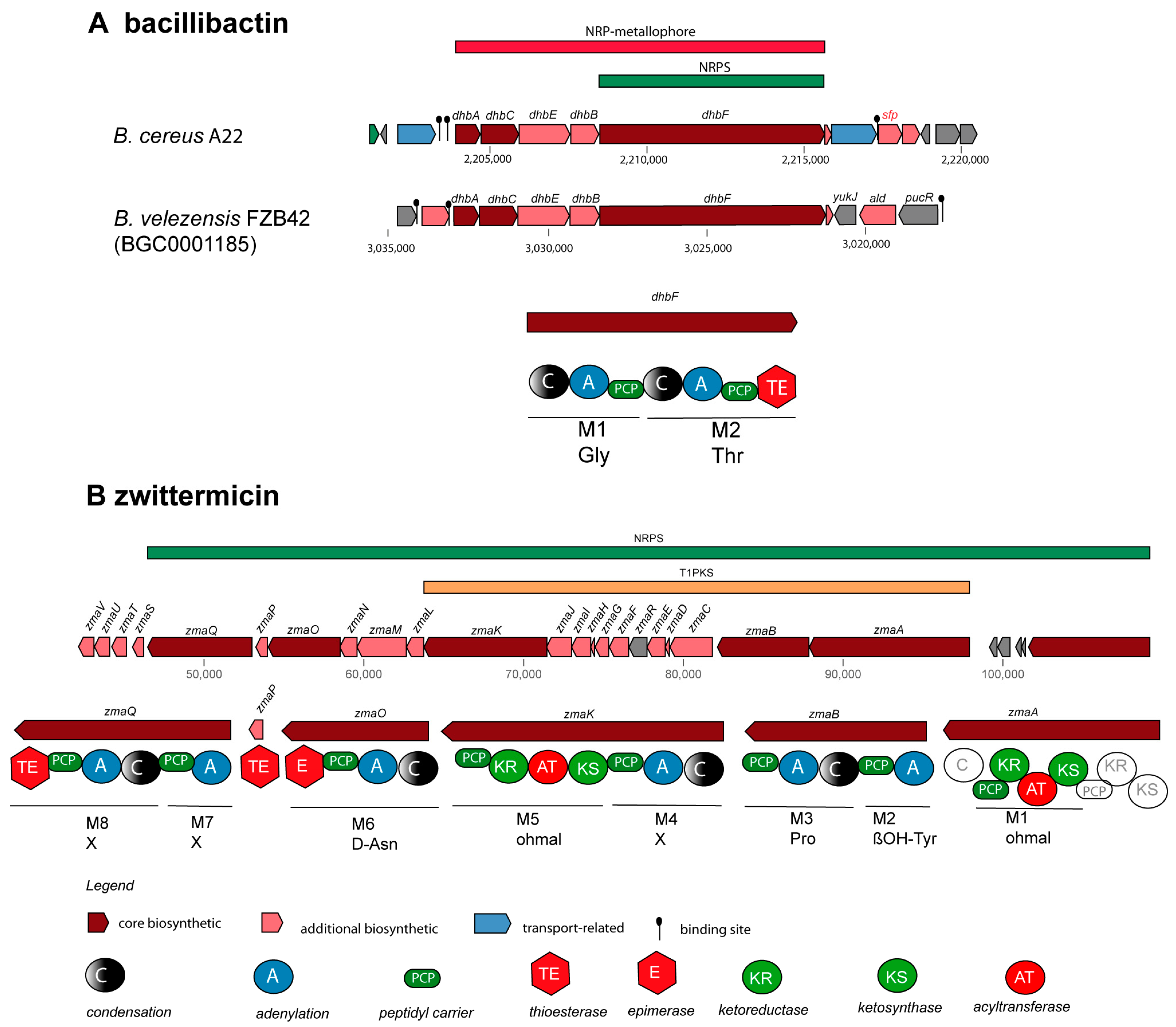
3.2.1. Non-Ribosomally Synthesized Antimicrobial Peptides (NRPs) and Polyketides (PKs)

NRPs are secondary metabolites that are synthesized through giant multi-modular peptide synthetases [52]. The complete krs gene cluster (BGC5) encoding the cyclic lipoheptapeptide kurstakin [53] was found to be widely distributed and occurs in the genomes of most B. cereus sensu lato isolates, except B. pacificus SN4.1, B. tropicus SN1, and Bacillus sp. CD3-1a and CD3.5. Kurstakin is responsible for biofilm formation [54]. Although kurstakin was present in the majority of the investigated isolates (13/17 strains), the kurstakin gene cluster, containing the genes krsE, krsA, krsB, krsC, sfp, and krsD (Figure 4A), is not listed in the MIBiG data bank.
The lipopeptide thumolycin, recently detected in B. thuringiensis BMB171, enabled the bacterium to develop a broad spectrum of antimicrobial and nematocidal activities [42]. Unfortunately, the structure of the lipopeptide is still not resolved. We detected the thumolycin (tho) gene cluster (BGC14) in plasmids of the B. cereus strains A22, A24, HD1.4B, and HD2.4. The genes of the tho cluster spanned around 30 kb. Two multimodular non-ribosomal peptide synthetases (ThoH and ThoI) synthesized a putative pentapeptide Orn-D-X-Leu/Ile-XS-Leu (Figure 4B). The thoC-, thoD-, and thoE-encoded proteins are probably involved in the synthesis of the fatty acid chain [42].
Fragments of the locillomycin gene cluster [55] (BGC34) were detected in B. cereus A8 (Supplementary Table S5). To the best of our knowledge, to date, the locillomycin gene cluster has been detected only in members of the B.subtilis species complex. The gene cluster for the synthesis of the catecholic iron siderophore bacillibactin, 2,3-dihydroxybenzoyl-Gly-Thr trimeric ester, has been previously reported in the genomes of B. subtilis [56] (BGC0000309) and B. velezensis FZB42 [57] (BGC0001185). Its non-ribosomal synthesis was found to be dependent on Sfp (phosphopantetheinyl transferase) [58]. BGCs with a similar structure as that of BGC0000309 and BGC0001185 were detected in all 17 B. cereus sensu lato isolates investigated in this study. Whilst the core structure of the bacillibactin transcription unit was well conserved, a sfp gene in the flanking region was identified as a unique feature for the B. cereus bacillibactin operon (Figure 5A). This is in contrast to the operon structure in the B. subtilis species complex, where the sfp gene is located in a more remote location, downstream flanking the surfactin operon [58].
The gene cluster for the synthesis of the aminopolyol antibiotic zwittermicin was detected in four B. cereus genomes. The highly polar zwittermicin A (ZmA) possesses antiprotist and antibacterial activities, and consists of numerous ethanolamine and glycolyl moieties flanked by N-terminal D-serine and an unusual amide generated from ß-ureidoalanine. The aminopolyol structure of the final product results from different processing events of the NRPS/PK hybrid precursor molecule (Figure 5B), in which a multitude of gene products of the zma gene cluster are involved [59].
In addition, numerous uncharacterized NRPs, PKS, and NRP/PKS hybrids were found (Supplementary Tables S5 and S6). A unique gene in B. tropicus CD3.2, located downstream of an uncharacterized NRP + PK cluster (BGC37, Supplementary Figure S7), encoded a putative necrose-inducing protein (NPP1 family) [60].

3.2.2. Gene Clusters Representing RiPPs and Bacteriocins

In contrast to polyketides and peptides, which are synthesized independently from ribosomes, numerous peptides with antimicrobial activity (bacteriocins) are synthesized by a ribosome-dependent mechanism. According to Zhao and Kuipers [49], several groups of ribosomally synthesized peptides (RiPPs) can be identified:
  • Class I: post-translationally modified peptides smaller than 10 kDa.
  • Class II: small (<10 Da), unmodified peptides with or without a leader sequence.
  • Class III: peptides larger than 10 kDa.
RiPPs, such as lanthipeptides (class1 and class2), linear azol(in)e-containing peptides (LAPs), lassopeptides, sactipeptides, thiopeptides, and representatives of the class II unmodified bacteriocins, such as UviB peptides (holin-like proteins), were detected in the B. cereus group isolates applying the antiSMASH and BAGEL4 toolkits (Supplementary Figures S8 and S9). Many RiPP biosynthetic proteins recognize and bind their cognate precursor peptide through a domain known as the RiPP recognition element (RRE) [61]. The detection of RRE domains using antiSMASH-supported genome mining was helpful in identifying the BGCs involved in the synthesis of RiPPs, which did not contain known core peptide-encoding sequences [62].
A total of 19 different BGCs encoding RiPPs and unmodified class 2 bacteriocins were detected. Only six encoded precursor peptides with an apparent similarity to known RiPPs: BGC20 (thuricin), BGC17 and BGC18 (sublancin/CER074), BGC29 (paeninodin), and BGC26 (thurincin H) (Supplementary Tables S5 and S7).
Antimicrobial lanthipeptides (lantibiotics) are post-translationally highly modified and contain the thioether amino acid lanthionine as well as several other modified amino acids [63]. LanA precursor peptides consist of an N-terminal leader peptide and a C-terminal core region. The first step in post-translational modification is the activation and elimination of water from the Ser and Thr residues forming dehydroalanine (DhA) and dehydrobutyrine (DhB), respectively. Then, ß-thioether cross-links are generated between the DhA, DhB, and the Cys residues. The modifying enzymes involved in the formation of the thioether link in class AI lanthipeptides are the dehydratase LanB and the cyclase LanC. The modification of A2 lanthipeptides is accomplished by LanM, containing the dehydratase and the cyclase domain in one protein. Classes A3 and A4 lanthipeptides are modified by LanKC and LanL, respectively [64]. We detected a BGC encoding a representative of the A1 lanthipeptides in B. cereus SN4.3 (BGC25). Four genes encoding precursor peptides similar to paenibacillin and subtilomycin were identified within BGC25. BGCs encoding A2 lantibiotics similar to plantaricin (BGC19), thuricin (BGC20), lichenicidin (BGC21), salivaricin (BGC23), and paenibacillin (BGC25) occurred in several B. cereus isolates (Supplementary Tables S5 and S7).
Gene clusters encoding LAPS were found to be widely distributed in B. cereus and related species. LAPS are characterized by the post-translational modification of the precursor peptide, yielding thiazol(in)e and (methyl)oxazol(in)e heterocycles. Modifying enzymes are the FMN-dependent dehydrogenase (SagB) and cyclodehdratase (SagC and YcaO) [65]. BGC2 (Supplementary Table S7) was identified as being member of the TOMM class (thiazole/oxazole-modified microcins), characterized by a gene cluster consisting of a cyclodehydratase gene and associated genes encoding dehydrogenase and a maturation protein. The core region of the TOMM precursor leader peptide contained a region enriched with Cys residues (BGC7/8), which is typically for the hetero-cycloanthracin/sonorensin family [66].
A glyocin-encoding gene cluster (BGC17) was detected in the plasmid P2 of B. cereus A24. Glyocins are defined as post-translationally glycosylated RiPPs with antimicrobial activity [65,67]. BGC17 resembled sublancin, which is an S-linked glycopeptide coding a SunS family peptide S-glcosyltransferase and a bacillicin CER074 peptide (BGC0001863) containing a glucose attached to a cysteine residue [68]. A second gene cluster (BGC33) harboring genes encoding a glycosyltransferase and a putative 75 aa precursor peptide was detected in B. cereus HB3.1 (Supplementary Table S5).
Lassopeptides are characterized by an N-terminal macrolactam ring threaded by the C-terminal tail. A cysteine protease B and a lactam synthetase C are necessary for the post-translational modification of the precursor peptide [69]. Two gene clusters, probably encoding lassopeptides, were identified. BGC29 harbored, in addition to a structural gene (paeA) for the synthesis of paeninodin lasso peptide [70], the genes for the synthesis of the essential components of post-translational modification, paeB1 (PQQD family protein), paeB2 (cysteine protease), and paeC (lactam ring closing cyclase). Four copies of the lasso precursor gene and split B1 and B2 genes were detected in BGC30 (Supplementary Table S7).
Two gene clusters (BGC26 and BGC28), encoding the radical S-adenosylmethionine (rSAM) enzyme, necessary for the post-translational modification of sactipeptides, occurred in the representatives of the B. cereus group. A well-known representative of sactipeptides is subtilosin A (SboA), synthesized by Bacillus subtilis. The rSAM enzyme (AlbA) catalyzes the linkage of a thiol with an α-carbon of a functional amino acid residue [71]. BGC26 harbored genes involved in the synthesis and rSAM-dependent modification of a thurincin H-like precursor peptide. A gene encoding a protein containing an N-terminal radical SAM domain (pfam04055) and a C-terminal pfam08756 domain with a CxCxxxxC motif (BmbF) was detected in BGC28. In contrast to B. subtilis, the YfkA and YfkB regions, originally reported as separate ORFs in B. subtilis, were found fused in the B. cereus gene cluster (Supplementary Table S7).
Like the structurally related sactipeptides, the thioether linkage in ranthipeptides is generated via a radical-initiated mechanism. However, ranthipeptides do not contain α-carbon links and were recently designated as non-α thioether peptides [72]. The ranthipeptide gene cluster (BGC15) detected in four B. cereus isolates harbored a gene encoding the rSAM protein belonging to the MoaA/NifB/PqqE/SkfB superfamily (Supplementary Table S7).
A gene cluster (BGC7/8), involved in the synthesis and modification of an 82 aa precursor thiopeptide belonging to the heterocycloanthracin/sonorensin family [73], occurred in all B. cereus group isolates. Its C-terminal region contained an extended repeat region with Cys at every third residue (Supplementary Table S7).
Two different subclasses of bacteriocin class II peptides were detected: the holin-like BhlA encoding genes (BGC12 and BGC16) and a cluster (BGC13) harboring a gene encoding a cerein-like prepeptide belonging to the Blp family. Similar as in lanthipeptides, the Blp family prepeptides are characterized by a conserved GlyGly processing site between the N-terminal leader and the C-terminal core peptide region [74]. The BhlA holin of Bacillus pumilus causes bacterial death by cell membrane disruption [75]. In addition to the genes encoding leaderless BhlA peptides, the holin gene clusters harbored genes encoding muramidases (GH25 glycosyl hydrolases) that hydrolyze the peptidoglycan cell wall (Supplementary Table S7, Supplementary Figures S8 and S9).

3.2.3. Other Antimicrobial Secondary Metabolites

Seven BGCs encoding other antimicrobial secondary metabolites were identified in the plant-associated B. cereus isolates (Supplementary Figure S7, Supplementary Table S8). Three of them, BGC3 (=BGC0000942, petrobactin), BGC11 (=BGCBGC0002103, pulcherriminic acid), and BGC32 (=BGC0000914, furan), were similar to the known BGCs deposited in the MiBIG data bank.
The asbABCDEF gene cluster is responsible for the biosynthesis of petrobactin, a catecholate siderophore that functions in both iron acquisition and virulence [76]. We detected the petrobactin gene cluster in the genomes of 14 isolates. Only B. pacificus SN4-1 and HD1-3 and Bacillus sp. CD3.5 did not harbor the BGC0000942 cluster, which is common in most representatives of the B. cereus group [77].
Pulcherriminic acid is a cyclic dipeptide able to chelate Fe3+ [78]. Due to its high affinity to Fe ions, Bacillus strains producing pulcherriminic acid compete successfully with other microorganisms in low iron environments. A gene cluster similar to the pulcherriminic acid synthesis cluster in B. subtilis (BGC0000914) was detected in the B. cereus HD1.4B and HD2.4 plasmid sequences (Supplementary Table S4) and in the draft genomes of B. cereus A8 and B. tropicus CD3-2.
Several genes of BGC32, possibly involved in the synthesis of a furan-like metabolite, showed a striking similarity to the methylenomycin A gene cluster in Streptomyces coelicolor [79].
Four BGCs did not show similarity to any characterized biosynthetic gene clusters. BGC6 possibly encoded ß-lactone-harbored genes with similarity to the genes flanking the plipastatin BGC in B. subtilis. BGC24 contained several genes of the carbohydrate metabolism, probably involved in the synthesis of ladderane. BGC10 and BGC31 encoded enzymes for the synthesis of terpene and nucleoside metabolites, respectively.

3.3. Detection of Bioactive Peptides by MALDI-TOF Mass Spectrometry

The genome mining data summarized in Figure 2 indicate the presence of BGCs at the genomic level. However, the real biosynthetic capacity of the investigated isolates can only be verified by the isolation and structural analysis of the compounds actually produced. In Figure 6, we demonstrate the production of the non-ribosomally formed secondary metabolites of strain A22 as a representative for B. cereus detected by MALDI-TOF MS. As an overview, Figure 6A–C show mass spectra for the compounds found in a surface extract of A22 taken from cell materials grown on agar plates in the Landy medium for 48 h. Two prominent products were observed. Figure 6B shows the mass peaks for two kurstakins with chain lengths of their fatty acid component of 12 and 13 carbon atoms, respectively. The following mass data were found: C12-kurstakins: [M + H,Na,K]+ = 892,5/914,5/930.5 Da; C13-kurstakins: [M + H,Na,K]+ = 906.5/928.5/944.5 Da. In addition, as yet unknown compounds with the mass numbers of 1051.8 and 1065.9 were found, which dominate the MALDI-TOF mass spectrum of the surface extract in Figure 6A. Presumably, there are two isomers that differ by a methylene group (Figure 6C). This compound cannot be correlated to any of the BGCs of the antiSMASH profile of strain A22.
Figure 6D–F show the mass spectra of the products of A22 released into the culture broth for growth in liquid cultures for 48 in the Landy medium. Here, the arylpolyene lipopeptide thumolycin and both siderophores bacillibactin and petrobactin were detected. Figure 6E exhibits the mass peaks of thumolycin (m/z = 697.8) and petrobactin (m/z = 720.2). A relatively small part of the kurstakins was released into the culture filtrate, while the main part remained attached at the outer surface of A22. In Figure 6F, kurstakin mass peaks (m/z = 906.8/930.8 and 944.8) overlap with those of the siderophore bacillibactin [M + H,Na,K]+ = 883.6/905.6/921 Da. These results demonstrate that kurstakins were predominantly found attached to the outer surface of B. cereus cells, while thumolycin and both siderophores bacillibacin and petrobactin were released into the culture medium. Similar profiles were obtained for strains A24, HD1.4B, and HD2.4. All other investigated B, cereus isolates did not produce thumolycin.
The structure of both lipopeptide products of strain A22, kurstakin and thumolycin, were investigated in detail by LIFT-MALDI-TOF/TOF fragment analysis [26]. Table 2 shows the sequence determination of C13 kurstakin with a parent ion [M + H}+ = 906.504 Da derived from product ions obtained by LIFT-MALDI-TOF/TOF fragment ion spectra. In Table 3, the structure of this compound is modelled from the nearest neighbor relationships using di-, tri-, and tetrapeptide fragments.
Table 2. Mass spectrometric sequence determination of C13-kurstakin produced by B. cereus A22 with a parent ion [M + H]+ = 906.504 derived from product ion patterns obtained by LIFT-MALDI-TOF/TOF fragment ion spectra. FA: fatty acid component.
bn₋H2O (found)-280.123337.149408.107495.159632.284760.445-
bn (found)197.030298.133355.112---778.446906.504
bn (calc.)197.190298.238355.259426.296513.328650.387778.446906.504
C13-FAThr
(1)
Gly
(2)
Ala
(3)
Ser
(4)
His
(5)
Gln
(6)
Gln
(7)
yn (calc.)906.502710.322609.275552.253481.216394.184257.125129.066
yn (found)906.504710.266609.163552.133481.106394.084257.050129.066
yn₋H2O (found)-692.185--463.103---
Table 3. Modeling of the structure of C13-kurstakin (m/z = 906.5) by nearest-neighbor relationships obtained by MALDI-TOF MS.
(A) Dipeptide fragments m/zm/z
Calc.found
C13-FA-Thr298.238298.141/280.129
     Thr-Gly159.077159.007/141.022
         Gly-Ala129.066129.016
            Ala-Ser159.077159.007/141.022
               Ser-His225.099225.034/207.019
                   His-Gln266.125266.060
                       Gln-Gln257.125257.050
(B) Tripeptide fragments
C13-FA-Thr-Gly341.244341.170/323.167
     Thr-Gly-Ala230.114230.026/212.025
         Gly-Ala-Ser216.098216.024/198.022
            Ala-Ser-His296.136-/278.050
               Ser-His-Gln353.157353.084/335.070
                   His-Gln-Gln394.184394.101/335.070
(C) Tetrapeptide fragments
C13-FA-Thr-Gly-Ala426.296-/408.121
     Thr-Gly-Ala-Ser317.146317.057/299.070
         Gly-Ala-Ser-His353.157353.084/335.070
            Ala-Ser-His-Gln424.194-
               Ser-His-Gln-Gln481.216481.120/463.120
Using the same technique, we investigated thumolycin, which is a combination of a pentapeptide attached to a yet unknown arylpolyene lipidic residue. By LIFT-MALDI-TOF/TOF fragment analysis, we obtained the complete sequence of the pentapeptide part for the first time, which is compatible with the initial results from Zheng et al. [42] and the module organization of the corresponding BGC derived from antiSMASH 6.0 genome mining. The structure of this pentapeptide is shown in Table 4.
In summary, by MALDI-TOF mass spectrometry, we detected all compounds produced by B. cereus strains non-ribosomally. The investigation of the RiPPs, such as lanthipeptides, sactipeptides, and bacteriocins, is in still progress.

3.4. B. cereus s.l. Strains Suppressed Plant Pathogens and Promoted Plant Growth

3.4.1. Antifungal and Nematocidal Activity

Our in vitro bioassays revealed that only few of the B. cereus group isolates efficiently inhibited phytopathogenic fungi and nematodes. Antifungal activity was examined in vitro using Fusarium oxysporum, known for causing fusarium wilt disease [80], and Phytophthora palmivora, one of the most detrimental plant pathogens in Vietnam [81]. Whilst most of the isolates did not significantly suppress the growth of the pathogenic fungi and oomycetes, a strong antimicrobial activity was exerted by B. cereus HB3.1 and Bacillus sp. HD.3 (Figure 7).
Root-knot nematodes, such as Meloidogyne spp., are one of the most important plant pathogens in tropical and temperate agriculture, and are responsible for the significant harvest losses of main Vietnamese crops, such as coffee trees (Coffea arabica and Coffea canephora) and black pepper plants [82]. In order to analyze the antagonistic activity of the B. cereus isolates, we tested at first their suppressing effect against the model nematode Caenorhabditis elegans. Fast and slow death rates were estimated in a bioassay under laboratory conditions. Most of the investigated B. cereus isolates were able to kill considerable amounts of the nematodes, as revealed in both test systems (Figure 8).
In order to examine the inhibiting effects against phytopathogenic nematodes more directly, we isolated a representative strain of Meloidogyne sp. from the galls of infested black pepper plant roots according to the hypochlorite procedure [83]. The inhibiting effect exerted by the test bacteria on disease development was examined in a greenhouse experiment. Ten weeks after the transplanting of the tomato plantlets into the soil, the formation of root knots was visually registered and used as a measure for calculating the disease index according to Bridge and Page [28]. All B. cereus isolates were found to be efficient against nematodes. In the presence of HB3.1, HD1.4B, HD2.4, MS2.1B, and SN4.3, the killing rates (estimated as slow and fast killing rates) of Caenorhabditis elegans exceeded 60% (Figure 8A). The Melodoine sp.-caused disease index of tomato plants was reduced by more than 50% after the application of B. cereus HB3.1, HD2.4, MS2.1B, and SN4.3 (Figure 8B). Similar rates were previously detected in representatives of B. velezensis [4] and Brevibacillus spp. [3].

3.4.2. Plant Growth Promotion

We examined the effect of the Vietnamese B. cereus isolates on the Arabidopsis thaliana biotest system [29]. B. cereus HD1.4B and B. cereus HD2.4 enhanced the growth of the Arabidopsis seedlings by more than 20% (Figure 9). However, using the same biotest system, the increase rates observed for some plant-associated Brevibacillus and B. velezensis strains isolated during the same survey [2] were higher and estimated to range from 30 to 40% [3,4].

4. Conclusions

In this study, we showed that plant-associated representatives of the B. cereus group were able to suppress important plant pathogens, such as fungi (Fusarium oxysporum), oomycetes (Phytophthora palmivora), and root-knot forming nematodes (Meloidogyne sp.) The plant-growth-promoting activity of some of the isolates could also be demonstrated.
Genome mining revealed that the members of the B. cereus group are rich in gene clusters probably involved in the synthesis of antimicrobial peptides that are efficient in inhibiting plant pathogens and triggering plant-induced systemic resistance [84]. A total of 36 different biosynthetic gene clusters (BGCs), many of them not listed in the MiBIG data bank, were detected in the 17 isolates obtained from Vietnames crop plants. Mass-spectrometric analysis revealed that, in addition to some hitherto unknown compounds, several species of the antimicrobial lipopeptides kurstakin and thumolycin and the siderophores bacillibactin and petrobactin were expressed in many of the isolates. The arylpolyene lipopeptide thumolycin was reported to possess interesting antimicrobial and nematocidal activities, but its primary structure was not resolved [42]. Here, the primary structure of the pentapeptide part was resolved, and the Orn residue was identified as being linked with the yet unknown arylpolyene lipid part.
In addition to antimicrobial peptides, the biocontrol of plant pathogens can be exerted by δ-endotoxins (parasporal inclusion proteins), traditionally known to be produced by B. thuringiensis, a close relative of B. cereus. Cry genes encoding the entomocidal crystal proteins Cry1Aa1 and Cry2Ba1 were detected in the genome of B. cereus ssp. Bombysepticus TK1. In the same strain, another gene encoding the vegetative insecticidal protein Vip3 was also detected, suggesting that the presence of insecticidal proteins is not restricted to B. thuringiensis. The encouraging results described above might lead to the development of the selected representatives of the B. cereus group as biocontrol agents. However, as known food poisoning organisms and members of the risk group 2, the potential of the B. cereus group isolates to produce toxins needs to be carefully examined before they can be applied in sustainable agriculture.
In this context, we showed that no gene clusters encoding B. anthracis pXO plasmid-related toxins were present in all the B. cereus s.l. genomes investigated here. Furthermore, we could rule out the synthesis of the heat-stable cereulide toxin, the causative agent of the emetic syndrome. However, the regular appearance of chromosomally localized virulence genes, encoding the heat-labile enterotoxins HBL and NHE, might restrict the direct application of B. cereus s.l. strains in biological plant protection.
In order to avoid such conflicts, the utilization of interesting B. cereus BGCs and genes encoding entomocidal proteins can be achieved by their heterologous expression in safe plant-beneficial host strains, which has been already demonstrated with B. velezensis FZB42 [85,86].

Supplementary Materials

The following supporting information can be downloaded at: https://www.mdpi.com/article/10.3390/microorganisms11112677/s1. Figure S1: Tree inferred with FastMe 2.1.6.1 from GBDP distances calculated from 16S rDNA gene sequences. Figure S2: GBDP tree (whole-genome-sequence-based) inferred with FastMe 2.1.6.1 from GBDP distances calculated from genome sequences. Figure S3: Heat map of the FastANI values of 128 B. cereus group genomes calculated and drawn by the EDGAR3.0 software package. Figure S4: Environment of the Rep protein genes in the plasmid sequences of A22, A24, HD1.4B, and HD2.4; Figure S5: Localization of the virulence genes and gene clusters on the chromosomes and plasmids of B. cereus isolates. The cytK gene and the NHE/HBL gene clusters were chromosomally localized. The complete set of NHE and HBL genes was chromosomally localized in all four completely sequenced strains (A22, A24, HD1.4B, and HD2.4). The P1 plasmid sequences of HD1.4B and HD2.4 harbored genes with similarity to the NHE/HBL enterotoxin family. Figure S6: Plasmid-encoded catabolic operons, biosynthetic gene clusters (BGCs), and restriction/modification systems. Figure S7: BGCs in the B. cereus group isolates encoding NRPS/PKS and other secondary metabolites. Figure S8: RiPP gene clusters detected by applying the BAGEL4 software (http://bagel4.molgenrug.nl/ (accessed on 1 November 2021)) in the genomes of the Vietnamese Bacillus cereus group genomes. Figure S9: RiPP gene clusters detected by applying the antiSMASH version 6 software in the Vietnamese Bacillus cereus group genomes. Table S1: Types of entomocidal cry toxins. Table S2: Species and subspecies delineation of the 17 Vietnamese B. cereus sensu lato isolates according to their dDDH and Fast ANI values. Table S3: List of the 17 B. cereus group strains used in this study with detailed annotation. Table S4: Genome annotation of the four completely sequenced B. cereus strains by RASTk. Table S5: Gene clusters (BGCs) of the Vietnamese Bacillus cereus group isolates. Table S6: Gene clusters (BGCs) of the Vietnamese Bacillus cereus group isolates involved in the non-ribosomal synthesis of antimicrobial peptides (RiPPs and bacteriocins). Table S7: Gene clusters (BGCs) of the Vietnamese Bacillus cereus group isolates involved in the ribosomal synthesis of antimicrobial peptides (RiPPs and bacteriocins). Table S8: Gene clusters (BGCs) of the Vietnamese Bacillus cereus group isolates involved in the synthesis of other secondary metabolites.

Author Contributions

Conceptualization, R.B., L.T.T.T., T.S. and P.L.; software, J.B.; investigation, J.V., J.J., S.H., L.T.T.T., A.S., P.T.L., L.T.P.T., S.R.K. and R.B.; resources, T.S. and P.L.; data curation, J.J., S.H., J.V. and C.B.; writing—original draft preparation, R.B., J.V. and L.T.T.T.; writing—review and editing, J.V., S.R.K., T.S., P.L., S.R.K. and R.B.; project administration, R.B., T.S. and P.L.; funding acquisition, R.B., T.S., P.L. and L.T.T.T. All authors have read and agreed to the published version of the manuscript.

Funding

This research was supported through the project ENDOBICA by the Bundesministerium für Bildung und Forschung (BMBF) (grant no. 031B0582A/031B0582B), the National Foundation for Science and Technology Development (NAFOSTED; code no. 106.03-2017.28), and the Ministry of Science and Technology (MOST) in Vietnam (code no. NDT.40.GER/18).

Data Availability Statement

Gene bank accession numbers of the complete genome sequences available in the NCBI data bank are listed in Section 2, Materials and Methods.

Acknowledgments

Silke Becker and Petra Lochau are thanked for their excellent technical support.

Conflicts of Interest

The authors declare no conflict of interest.

References

  1. Borriss, R. Use of plant-associated Bacillus strains as biofertilizers and biocontrol agents. In Bacteria in Agrobiology: Plant Growth Responses; Maheshwari, D.K., Ed.; Springer: Berlin/Heidelberg, Germany, 2011; pp. 41–76. [Google Scholar] [CrossRef]
  2. Tam, L.T.T.; Jähne, J.; Luong, P.T.; Thao, L.T.P.; Chung, L.T.K.; Schneider, A.; Blumenscheit, C.; Lasch, P.; Schweder, T.; Borriss, R. Draft genome sequences of 59 endospore-forming Gram-positive bacteria associated with crop plants grown in Vietnam. Microbiol. Resour. Announc. 2020, 9, e01154-20. [Google Scholar] [CrossRef]
  3. Jähne, J.; Le Thi, T.T.; Blumenscheit, C.; Schneider, A.; Pham, T.L.; Le Thi, P.T.; Blom, J.; Vater, J.; Schweder, T.; Lasch, P.; et al. Novel Plant-Associated Brevibacillus and Lysinibacillus Genomospecies Harbor a Rich Biosynthetic Potential of Antimicrobial Compounds. Microorganisms 2023, 11, 168. [Google Scholar] [CrossRef]
  4. Thanh Tam, L.T.; Jähne, J.; Luong, P.T.; Phuong Thao, L.T.; Nhat, L.M.; Blumenscheit, C.; Schneider, A.; Blom, J.; Kim Chung, L.T.; Anh Minh, P.L.; et al. Two plant-associated Bacillus velezensis strains selected after genome analysis, metabolite profiling, and with proved biocontrol potential, were enhancing harvest yield of coffee and black pepper in large field trials. Front. Plant. Sci. 2023, 14, 1194887. [Google Scholar] [CrossRef] [PubMed]
  5. Carroll, L.M.; Cheng, R.A.; Wiedmann, M.; Kovac, J. Keeping up with the Bacillus cereus group: Taxonomy through the genomics era and beyond. Crit. Rev. Food Sci. Nutr. 2022, 62, 7677–7702. [Google Scholar] [CrossRef]
  6. Ehling-Schulz, M.; Lereclus, D.; Koehler, T.M. The Bacillus cereus Group: Bacillus Species with Pathogenic Potential. Microbiol. Spectr. 2019, 7. [Google Scholar] [CrossRef]
  7. Koch, R. Die Ätiologie der Milzbrand-Krankheit, begründet auf die Entwicklungsgeschichte des Bacillus Anthracis. Cohns beiträge ur Biol. Der Pflanz. 1876, 2, 277. [Google Scholar]
  8. Moayeri, M.; Leppla, S.H.; Vrentas, C.; Pomerantsev, A.P.; Liu, S. Anthrax Pathogenesis. Annu. Rev. Microbiol. 2015, 69, 185–208. [Google Scholar] [CrossRef] [PubMed]
  9. Yang, S.; Wang, Y.; Liu, Y.; Jia, K.; Zhang, Z.; Dong, Q. Cereulide and Emetic Bacillus cereus: Characterizations, Impacts and Public Precautions. Foods 2023, 12, 833. [Google Scholar] [CrossRef]
  10. Berliner, E. Über die Schlafsucht der Mehlmottenraupe (Ephestia kühniella Zell) und ihren Erreger Bacillus thuringiensis n. sp. Z. fAngew. Entomol. 1915, 2, 29–56. [Google Scholar] [CrossRef]
  11. Palma, L.; Muñoz, D.; Berry, C.; Murillo, J.; Caballero, P. Bacillus thuringiensis toxins: An overview of their biocidal activity. Toxins 2014, 6, 3296–3325. [Google Scholar] [CrossRef]
  12. Baldwin, V.M. You Can’t B. cereus—A Review of Bacillus cereus Strains That Cause Anthrax-Like Disease. Front. Microbiol. 2020, 11, 1731. [Google Scholar] [CrossRef]
  13. Kolstø, A.B.; Tourasse, N.J.; Økstad, O.A. What sets Bacillus anthracis apart from other Bacillus species? Annu. Rev. Microbiol. 2009, 63, 451–476. [Google Scholar] [CrossRef] [PubMed]
  14. Blumenscheit, C.; Jähne, J.; Schneider, A.; Blom, J.; Schweder, T.; Lasch, P.; Borriss, R. Genome sequence data of Bacillus velezensis BP1.2A and BT2.4. Data Brief. 2022, 41, 107978. [Google Scholar] [CrossRef] [PubMed]
  15. Olson, R.D.; Assaf, R.; Brettin, T.; Conrad, N.; Cucinell, C.; Davis, J.J.; Dempsey, D.M.; Dickerman, A.; Dietrich, E.M.; Kenyon, R.W.; et al. Introducing the Bacterial and Viral Bioinformatics Resource Center (BV-BRC): A resource combining PATRIC, IRD and ViPR. Nucleic Acids Res. 2023, 51, D678–D689. [Google Scholar] [CrossRef] [PubMed]
  16. Liu, Y.; Lai, Q.; Göker, M.; Meier-Kolthoff, J.P.; Wang, M.; Sun, Y.; Wang, L.; Shao, Z. Genomic insights into the taxonomic status of the Bacillus cereus group. Sci. Rep. 2015, 5, 14082. [Google Scholar] [CrossRef]
  17. Radnedge, L.; Agron, P.G.; Hill, K.K.; Jackson, P.J.; Ticknor, L.O.; Keim, P.; Andersen, G.L. Genome differences that distinguish Bacillus anthracis from Bacillus cereus and Bacillus thuringiensis. Appl. Environ. Microbiol. 2003, 69, 2755–2764. [Google Scholar] [CrossRef]
  18. Klee, S.R.; Ozel, M.; Appel, B.; Boesch, C.; Ellerbrok, H.; Jacob, D.; Holland, G.; Leendertz, F.H.; Pauli, G.; Grunow, R.; et al. Characterization of Bacillus anthracis-like bacteria isolated from wild great apes from Cote d’Ivoire and Cameroon. J. Bacteriol. 2006, 188, 5333–5344. [Google Scholar] [CrossRef]
  19. Meier-Kolthoff, J.P.; Sardà Carbasse, J.; Peinado-Olarte, R.L.; Göker, M. TYGS and LPSN: A database tandem for fast and reliable genome-based classification and nomenclature of prokaryotes. Nucleic Acid Res. 2022, 50, D801–D807. [Google Scholar] [CrossRef]
  20. Dieckmann, M.A.; Beyvers, S.; Nkouamedjo-Fankep, R.C.; Hanel, P.H.G.; Jelonek, L.; Blom, J.; Goesmann, A. EDGAR3.0, Comparative genomics and phylogenomics on a scalable infrastructure. Nucleic Acids Res. 2021, 49, W185–W192. [Google Scholar] [CrossRef]
  21. Blin, K.; Shaw, S.; Kloosterman, A.M.; Charlop-Powers, Z.; van Wezel, G.P.; Medema, M.H.; Weber, T. antiSMASH 6.0, Improving cluster detection and comparison capabilities. Nucleic Acids Res. 2021, 49, W29–W35. [Google Scholar] [CrossRef]
  22. Bachmann, B.O.; Ravel, J. Chapter 8. Methods for in silico prediction of microbial polyketide and nonribosomal peptide biosynthetic pathways from DNA sequence data. Methods Enzymol. 2009, 458, 181–217. [Google Scholar] [CrossRef] [PubMed]
  23. van Heel, A.J.; de Jong, A.; Song, C.; Viel, J.H.; Kok, J.; Kuipers, O.P. BAGEL4, A user-friendly web server to thoroughly mine RiPPs and bacteriocins. Nucleic Acids Res. 2018, 46, W278–W281. [Google Scholar] [CrossRef] [PubMed]
  24. Vater, J.; Herfort, S.; Doellinger, J.; Weydmann, M.; Borriss, R.; Lasch, P. Genome Mining of the Lipopeptide Biosynthesis of Paenibacillus polymyxa E681 in Combination with Mass Spectrometry: Discovery of the Lipoheptapeptide Paenilipoheptin. Chembiochem. 2018, 19, 744–753. [Google Scholar] [CrossRef]
  25. Mülner, P.; Schwarz, E.; Dietel, K.; Junge, H.; Herfort, S.; Weydmann, M.; Lasch, P.; Cernava, T.; Berg, G.; Vater, J. Profiling for Bioactive Peptides and Volatiles of Plant Growth Promoting Strains of the Bacillus subtilis Complex of Industrial Relevance. Front. Microbiol. 2020, 11, 1432. [Google Scholar] [CrossRef] [PubMed]
  26. Suckau, D.; Resemann, A.; Schuerenberg, M.; Hufnagel, P.; Franzen, J.; Holle, A. A novel MALDI LIFT-TOF/TOF mass spectrometer for proteomics. Anal. Bioanal. Chem. 2003, 376, 952–956. [Google Scholar] [CrossRef] [PubMed]
  27. Hooper, D.J.; Hallmann, J.; Subbotin, S.A. Methods for extraction, processing and detection of plant and soil nematodes. In Plant Parasitic Nematodes in Subtropical and Tropical Agriculture; Luc, M., Sikora, R., Bridge, J., Eds.; CAB International: Wallingford, UK, 2005; pp. 53–86. [Google Scholar]
  28. Bridge, J.; Page, S.L.J. Estimation of Root Knot Nematode Infestation Levels in Roots Using a Rating Chart. Trop. Pest Manag. 1980, 26, 296–298. [Google Scholar] [CrossRef]
  29. Budiharjo, A.; Chowdhury, S.P.; Dietel, K.; Beator, B.; Dolgova, O.; Fan, B.; Bleiss, W.; Ziegler, J.; Schmid, M.; Hartmann, A.; et al. Transposon mutagenesis of the plant-associated Bacillus amyloliquefaciens ssp. plantarum FZB42 revealed that the nfrA and RBAM17410 genes are involved in plant-microbe-interactions. PLoS ONE 2014, 9, e98267. [Google Scholar] [CrossRef]
  30. Lefort, V.; Desper, R.; Gascuel, O. FastME 2.0, A comprehensive, accurate, and fast distance-based phylogeny inference program. Mol. Biol. Evol. 2015, 32, 2798–2800. [Google Scholar] [CrossRef]
  31. Meier-Kolthoff, J.P.; Göker, M. TYGS is an automated high-throughput platform for state-of-the-art genome-based taxonomy. Nat. Commun. 2019, 10, 2182. [Google Scholar] [CrossRef]
  32. Cheng, T.; Lin, P.; Jin, S.; Wu, Y.; Fu, B.; Long, R.; Liu, D.; Guo, Y.; Peng, L.; Xia, Q. Complete Genome Sequence of Bacillus bombysepticus, a Pathogen Leading to Bombyx mori Black Chest Septicemia. Genome Announc. 2014, 2, e00312-14. [Google Scholar] [CrossRef]
  33. Parks, D.H.; Chuvochina, M.; Rinke, C.; Mussig, A.J.; Chaumeil, P.A.; Hugenholtz, P. GTDB: An ongoing census of bacterial and archaeal diversity through a phylogenetically consistent, rank normalized and complete genome-based taxonomy. Nucleic Acids Res. 2022, 50, D785–D794. [Google Scholar] [CrossRef] [PubMed]
  34. Parte, A.C.; Sardà Carbasse, J.; Meier-Kolthoff, J.P.; Reimer, L.C.; Göker, M. List of Prokaryotic names with Standing in Nomenclature (LPSN) moves to the DSMZ. Int. J. Syst. Evol. Microbiol. 2020, 70, 5607–5612. [Google Scholar] [CrossRef]
  35. Agata, N.; Ohta, M.; Mori, M. Production of an emetic toxin, cereulide, is associated with a specific class of Bacillus cereus. Curr. Microbiol. 1996, 33, 67–69. [Google Scholar] [CrossRef] [PubMed]
  36. Baillie, L.; Read, T.D. Bacillus anthracis, a bug with attitude! Curr. Opin. Microbiol. 2001, 4, 78–81. [Google Scholar] [CrossRef] [PubMed]
  37. Bravo, A.; Gómez, I.; Porta, H.; García-Gómez, B.I.; Rodriguez-Almazan, C.; Pardo, L.; Soberón, M. Evolution of Bacillus thuringiensis Cry toxins insecticidal activity. Microb. Biotechnol. 2013, 6, 17–26. [Google Scholar] [CrossRef]
  38. Beecher, D.J.; Wong, A.C.L. Cooperative, synergistic and antagonistic haemolytic interactions between haemolysin BL, phosphatidylcholine phospholipase C and sphingomyelinase from Bacillus cereus. Microbiology 2000, 146 Pt 12, 3033–3039. [Google Scholar] [CrossRef]
  39. Dietrich, R.; Jessberger, N.; Ehling-Schulz, M.; Märtlbauer, E.; Granum, P.E. The Food Poisoning Toxins of Bacillus cereus. Toxins 2021, 13, 98. [Google Scholar] [CrossRef]
  40. Sanahuja, G.; Banakar, R.; Twyman, R.M.; Capell, T.; Christou, P. Bacillus thuringiensis: A century of research, development and commercial applications. Plant Biotechnol. J. 2011, 9, 283–300. [Google Scholar] [CrossRef]
  41. Gupta, M.; Kumar, H.; Kaur, S. Vegetative Insecticidal Protein (Vip): A Potential Contender From Bacillus thuringiensis for Efficient Management of Various Detrimental Agricultural Pests. Front. Microbiol. 2021, 12, 659736. [Google Scholar] [CrossRef]
  42. Zheng, D.; Zeng, Z.; Xue, B.; Deng, Y.; Sun, M.; Tang, Y.J.; Ruan, L. Bacillus thuringiensis produces the lipopeptide thumolycin to antagonize microbes and nematodes. Microbiol. Res. 2018, 215, 22–28. [Google Scholar] [CrossRef]
  43. Overbeek, R.; Olson, R.; Pusch, G.D.; Olsen, G.J.; Davis, J.J.; Disz, T.; Edwards, R.A.; Gerdes, S.; Parrello, B.; Shukla, M.; et al. The SEED and the Rapid Annotation of microbial genomes using Subsystems Technology (RAST). Nucleic Acids Res. 2014, 42, D206-14. [Google Scholar] [CrossRef] [PubMed]
  44. Griffith, O.H.; Ryan, M. Bacterial phosphatidylinositol-specific phospholipase C: Structure, function, and interaction with lipids. Biochim. Biophys. Acta 1999, 1441, 237–254. [Google Scholar] [CrossRef] [PubMed]
  45. Gao, Y.; Cao, D.; Zhu, J.; Feng, H.; Luo, X.; Liu, S.; Yan, X.X.; Zhang, X.; Gao, P. Structural insights into assembly, operation and inhibition of a type I restriction-modification system. Nat. Microbiol. 2020, 5, 1107–1118. [Google Scholar] [CrossRef] [PubMed]
  46. Dong, S.; McPherson, S.A.; Wang, Y.; Li, M.; Wang, P.; Turnbough, C.L., Jr.; Pritchard, D.G. Characterization of the enzymes encoded by the anthrose biosynthetic operon of Bacillus anthracis. J. Bacteriol. 2010, 192, 5053–5062. [Google Scholar] [CrossRef]
  47. Dong, S.; McPherson, S.A.; Tan, L.; Chesnokova, O.N.; Turnbough, C.L., Jr.; Pritchard, D.G. Anthrose biosynthetic operon of Bacillus anthracis. J. Bacteriol. 2008, 190, 2350–2359. [Google Scholar] [CrossRef]
  48. Yoshida, K.; Yamaguchi, M.; Morinaga, T.; Kinehara, M.; Ikeuchi, M.; Ashida, H.; Fujita, Y. myo-Inositol catabolism in Bacillus subtilis. J. Biol. Chem. 2008, 283, 10415–10424. [Google Scholar] [CrossRef]
  49. Zhao, X.; Kuipers, O.P. Identification and classification of known and putative antimicrobial compounds produced by a wide variety of Bacillales species. BMC Genom. 2016, 17, 882. [Google Scholar] [CrossRef] [PubMed]
  50. Tracanna, V.; de Jong, A.; Medema, M.H.; Kuipers, O.P. Mining prokaryotes for antimicrobial compounds: From diversity to function. FEMS Microbiol. Rev. 2017, 41, 417–429. [Google Scholar] [CrossRef] [PubMed]
  51. Terlouw, B.R.; Blin, K.; Navarro-Muñoz, J.C.; Avalon, N.E.; Chevrette, M.G.; Egbert, S.; Lee, S.; Meijer, D.; Recchia, M.J.J.; Reitz, Z.L.; et al. MIBiG 3.0, a community-driven effort to annotate experimentally validated biosynthetic gene clusters. Nucleic Acids Res. 2023, 51, D603–D610. [Google Scholar] [CrossRef]
  52. Finking, R.; Marahiel, M.A. Biosynthesis of nonribosomal peptides. Annu. Rev. Microbiol. 2004, 58, 453–488. [Google Scholar] [CrossRef]
  53. Béchet, M.; Caradec, T.; Hussein, W.; Abderrahmani, A.; Chollet, M.; Leclère, V.; Dubois, T.; Lereclus, D.; Pupin, M.; Jacques, P. Structure, biosynthesis, and properties of kurstakins, nonribosomal lipopeptides from Bacillus spp. Appl. Microbiol. Biotechnol. 2012, 95, 593–600. [Google Scholar] [CrossRef] [PubMed]
  54. Yu, Y.Y.; Zhang, Y.Y.; Wang, T.; Huang, T.X.; Tang, S.Y.; Jin, Y.; Mi, D.D.; Zheng, Y.; Niu, D.D.; Guo, J.H.; et al. Kurstakin Triggers Multicellular Behaviors in Bacillus cereus AR156 and Enhances Disease Control Efficacy Against Rice Sheath Blight. Plant Dis. 2023, 107, 1463–1470. [Google Scholar] [CrossRef] [PubMed]
  55. Luo, C.; Liu, X.; Zhou, X.; Guo, J.; Truong, J.; Wang, X.; Zhou, H.; Li, X.; Chen, Z. Unusual Biosynthesis and Structure of Locillomycins from Bacillus subtilis 916. Appl. Environ. Microbiol. 2015, 81, 6601–6609. [Google Scholar] [CrossRef]
  56. May, J.J.; Wendrich, T.M.; Marahiel, M.A. The dhb operon of Bacillus subtilis encodes the biosynthetic template for the catecholic siderophore 2,3-dihydroxybenzoate-glycine-threonine trimeric ester bacillibactin. J. Biol. Chem. 2001, 276, 7209–7217. [Google Scholar] [CrossRef] [PubMed]
  57. Chen, X.H.; Koumoutsi, A.; Scholz, R.; Borriss, R. More than anticipated—Production of antibiotics and other secondary metabolites by Bacillus amyloliquefaciens FZB42. J. Mol. Microbiol. Biotechnol. 2009, 16, 14–24. [Google Scholar] [CrossRef] [PubMed]
  58. Chen, X.H.; Koumoutsi, A.; Scholz, R.; Eisenreich, A.; Schneider, K.; Heinemeyer, I.; Morgenstern, B.; Voss, B.; Hess, W.R.; Reva, O.; et al. Comparative analysis of the complete genome sequence of the plant growth-promoting bacterium Bacillus amyloliquefaciens FZB42. Nat. Biotechnol. 2007, 25, 1007–1014. [Google Scholar] [CrossRef] [PubMed]
  59. Kevany, B.M.; Rasko, D.A.; Thomas, M.G. Characterization of the complete zwittermicin A biosynthesis gene cluster from Bacillus cereus. Appl. Environ. Microbiol. 2009, 75, 1144–1155. [Google Scholar] [CrossRef]
  60. Fellbrich, G.; Romanski, A.; Varet, A.; Blume, B.; Brunner, F.; Engelhardt, S.; Felix, G.; Kemmerling, B.; Krzymowska, M.; Nürnberger, T. NPP1, a Phytophthora-associated trigger of plant defense in parsley and Arabidopsis. Plant J. 2002, 32, 375–390. [Google Scholar] [CrossRef]
  61. Burkhart, B.J.; Hudson, G.A.; Dunbar, K.L.; Mitchell, D.A. A prevalent peptide-binding domain guides ribosomal natural product biosynthesis. Nat. Chem. Biol. 2015, 11, 564–570. [Google Scholar] [CrossRef]
  62. Kloosterman, A.M.; Shelton, K.E.; van Wezel, G.P.; Medema, M.H.; Mitchell, D.A. RRE-Finder: A Genome-Mining Tool for Class-Independent RiPP Discovery. mSystems 2020, 5, e00267. [Google Scholar] [CrossRef]
  63. Sahl, H.G.; Jack, R.W.; Bierbaum, G. Biosynthesis and biological activities of lantibiotics with unique post-translational modifications. Eur. J. Biochem. 1995, 230, 827–853. [Google Scholar] [CrossRef]
  64. Wang, J.; Ma, H.; Ge, X.; Zhang, J.; Teng, K.; Sun, Z.; Zhong, J. Bovicin HJ50-like lantibiotics, a novel subgroup of lantibiotics featured by an indispensable disulfide bridge. PLoS ONE 2014, 9, e97121. [Google Scholar] [CrossRef]
  65. Walker, M.C.; Eslami, S.M.; Hetrick, K.J.; Ackenhusen, S.E.; Mitchell, D.A.; van der Donk, W.A. Precursor peptide-targeted mining of more than one hundred thousand genomes expands the lanthipeptide natural product family. BMC Genom. 2020, 21, 387. [Google Scholar] [CrossRef]
  66. Arnison, P.G.; Bibb, M.J.; Bierbaum, G.; Bowers, A.A.; Bugni, T.S.; Bulaj, G.; Camarero, J.A.; Campopiano, D.J.; Challis, G.L.; Clardy, J.; et al. Ribosomally synthesized and post-translationally modified peptide natural products: Overview and recommendations for a universal nomenclature. Nat. Prod. Rep. 2013, 30, 108–160. [Google Scholar] [CrossRef]
  67. Ren, H.; Biswas, S.; Ho, S.; van der Donk, W.A.; Zhao, H. Rapid Discovery of Glycocins through Pathway Refactoring in Escherichia coli. ACS Chem. Biol. 2018, 13, 2966–2972. [Google Scholar] [CrossRef] [PubMed]
  68. Oman, T.J.; Boettcher, J.M.; Wang, H.; Okalibe, X.N.; van der Donk, W.A. Sublancin is not a lantibiotic but an S-linked glycopeptide. Nat. Chem. Biol. 2011, 7, 78–80. [Google Scholar] [CrossRef] [PubMed]
  69. Hegemann, J.D.; Zimmermann, M.; Xie, X.; Marahiel, M.A. Lasso peptides: An intriguing class of bacterial natural products. Acc. Chem. Res. 2015, 48, 1909–1919. [Google Scholar] [CrossRef] [PubMed]
  70. Zhu, S.; Hegemann, J.D.; Fage, C.D.; Zimmermann, M.; Xie, X.; Linne, U.; Marahiel, M.A. Insights into the Unique Phosphorylation of the Lasso Peptide Paeninodin. J. Biol. Chem. 2016, 291, 13662–13678. [Google Scholar] [CrossRef] [PubMed]
  71. Flühe, L.; Knappe, T.A.; Gattner, M.J.; Schäfer, A.; Burghaus, O.; Linne, U.; Marahiel, M.A. The radical SAM enzyme AlbA catalyzes thioether bond formation in subtilosin A. Nat. Chem. Biol. 2012, 8, 350–357. [Google Scholar] [CrossRef]
  72. Hudson, G.A.; Burkhart, B.J.; DiCaprio, A.J.; Schwalen, C.J.; Kille, B.; Pogorelov, T.V.; Mitchell, D.A. Bioinformatic Mapping of Radical S-Adenosylmethionine-Dependent Ribosomally Synthesized and Post-Translationally Modified Peptides Identifies New Cα, Cβ, and Cγ-Linked Thioether-Containing Peptides. J. Am. Chem. Soc. 2019, 141, 8228–8238. [Google Scholar] [CrossRef]
  73. Chopra, L.; Singh, G.; Choudhary, V.; Sahoo, D.K. Sonorensin: An antimicrobial peptide, belonging to the heterocycloanthracin subfamily of bacteriocins, from a new marine isolate, Bacillus sonorensis MT93. Appl. Environ. Microbiol. 2014, 80, 2981–2990. [Google Scholar] [CrossRef] [PubMed]
  74. Franz, C.M.; van Belkum, M.J.; Holzapfel, W.H.; Abriouel, H.; Gálvez, A. Diversity of enterococcal bacteriocins and their grouping in a new classification scheme. FEMS Microbiol. Rev. 2007, 31, 293–310. [Google Scholar] [CrossRef]
  75. Aunpad, R.; Panbangred, W. Evidence for two putative holin-like peptides encoding genes of Bacillus pumilus strain WAPB4. Curr. Microbiol. 2012, 64, 343–348. [Google Scholar] [CrossRef] [PubMed]
  76. Lee, J.Y.; Janes, B.K.; Passalacqua, K.D.; Pfleger, B.F.; Bergman, N.H.; Liu, H.; Håkansson, K.; Somu, R.V.; Aldrich, C.C.; Cendrowski, S.; et al. Biosynthetic analysis of the petrobactin siderophore pathway from Bacillus anthracis. J. Bacteriol. 2007, 189, 1698–1710. [Google Scholar] [CrossRef] [PubMed]
  77. Koppisch, A.T.; Dhungana, S.; Hill, K.K.; Boukhalfa, H.; Heine, H.S.; Colip, L.A.; Romero, R.B.; Shou, Y.; Ticknor, L.O.; Marrone, B.L.; et al. Petrobactin is produced by both pathogenic and non-pathogenic isolates of the Bacillus cereus group of bacteria. Biometals. 2008, 21, 581–589. [Google Scholar] [CrossRef]
  78. Yuan, S.; Yong, X.; Zhao, T.; Li, Y.; Liu, J. Research Progress of the Biosynthesis of Natural Bio-Antibacterial Agent Pulcherriminic Acid in Bacillus. Molecules 2020, 25, 5611. [Google Scholar] [CrossRef]
  79. Corre, C.; Song, L.; O’Rourke, S.; Chater, K.F.; Challis, G.L. 2-Alkyl-4-hydroxymethylfuran-3-carboxylic acids, antibiotic production inducers discovered by Streptomyces coelicolor genome mining. Proc. Natl. Acad. Sci. USA 2008, 105, 17510–17515. [Google Scholar] [CrossRef]
  80. Arie, T. Fusarium diseases of cultivated plants, control, diagnosis, and molecular and genetic studies. J. Pestic. Sci. 2019, 44, 275–281. [Google Scholar] [CrossRef]
  81. Chi, N.M.; Thu, P.Q.; Nam, H.B.; Quang, D.Q.; Phong, L.V.; Van, N.D.; Trang, T.T.; Kien, T.T.; Tam, T.T.T.; Dell, B. Management of Phytophthora palmivora disease in Citrus reticulata with chemical fungicides. J. Gen. Plant Pathol. 2020, 86, 494–502. [Google Scholar] [CrossRef]
  82. Eisenback, J.D.; and Triantaphyllou, H.H. Root-knot nematodes: Meloidogyne species and races. In Manual of Agricultural Nematology; Nickle, W.R., Ed.; Marcell Dekker: New York, NY, USA, 1991; pp. 191–274. [Google Scholar]
  83. Hussey, P.S.; Barker, K.R. A comparison of methods of collecting inocula of Meloidogyne spp., including a new technique. Plant Dis. Report. 1973, 57, 1025–1028. [Google Scholar]
  84. Chowdhury, S.P.; Hartmann, A.; Gao, X.; Borriss, R. Biocontrol mechanism by root-associated Bacillus amyloliquefaciens FZB42—A review. Front. Microbiol. 2015, 6, 780. [Google Scholar] [CrossRef] [PubMed]
  85. Qiao, J.-Q.; Wu, H.-J.; Huo, R.; Gao, X.-W.; Borriss, R. Stimulation and biocontrol by Bacillus amyloliquefaciens subsp. plantarum FZB42 engineered for improved action. Chem. Biol. Technol. Agric. 2014, 1, 12. [Google Scholar] [CrossRef]
  86. Wu, L.; Wu, H.J.; Qiao, J.; Gao, X.; Borriss, R. Novel Routes for Improving Biocontrol Activity of Bacillus Based Bioinoculants. Front. Microbiol. 2015, 6, 1395. [Google Scholar] [CrossRef] [PubMed]
Figure 2. Occurrence of genes encoding entomopathogenic crystal (light blue) and vegetative (blue) proteins, the replication proteins RepS (grey) and RepX (dark grey) of B. anthracis pXO plasmids, and virulence genes (HBL/NHE, cytK). The gene cluster for synthesizing celeuride (ces) was not detected in any of the isolates. The 36 biosynthetic gene clusters (BGCs) encoding secondary metabolites in the 17 B. cereus s.l. isolates were identified by AntiSMASH6.0 and BAGEL4. The location of the BGC on either the chromosome (C) or the plasmids (P1, P3) is indicated when available. Further information is presented in Supplementary Tables S3–S8.
Microorganisms 11 02677 g002
Figure 3. Circular plots of the Bacillus cereus A22 chromosome (A) and plasmid P1 (B) generated with Biocircos. The Venn diagrams below show the comparison of A22 with the chromosomes and plasmid P1 of B. cereus A24 and HD1.4B. From outer to inner circle: Genes (CDS) on +(1)/− strand (2); core genome, brown (3); GC content (1000 bp window) above the mean: black, below mean: grey (4); GC skew ((G-C)/(G+C)) (1000 bp window), above mean: purple, below mean: light green (5). The grey line within the inner circle shows deviations of the average GC content. The 30 kb thumolycin gene cluster is part of the core genome in all four plasmid P1 species.
Microorganisms 11 02677 g003aMicroorganisms 11 02677 g003b
Figure 4. Gene cluster and domain structure involved in the non-ribosomal synthesis of cyclic lipopeptides in B. cereus A22. (A) The kurstakin (krs) gene cluster is located on the chromosome of A22 in the range of 2325–2355 kb. The amino acid sequence deduced from the adenylation domains was experimentally corrected and completed by LIFT-MALDI-TOF/TOF MS (see Table 2). (B) The thumolycin (tho) gene cluster resides in the B. cereus A22 plasmid 1 between 435 kb and 465 kb. The domain structure of ThoH and ThoI, including the amino acids deduced from their adenylation domains, is shown. The complete amino acid sequence determined by LIFT-MALDI -TOF/TOF MS is shown in Table 3. Further domains were detected in ThoC (A), ThoE (KS), ThoK (TE), and ThoL (ACPS).
Microorganisms 11 02677 g004
Figure 5. Gene clusters involved in the non-ribosomal synthesis of bacillibactin and zwittermicin. The siderophore gene cluster in the B. cereus A22 chromosome. (A) Comparison of the corresponding B. velezensis FZB42 gene cluster revealed the presence of the sfp gene downstream of the ballibactin transcription unit as a unique feature. (B) The gene cluster for the synthesis of the PK/NRP hybrid zwittermicin in B. cereus SN4-3.
Microorganisms 11 02677 g005
Figure 6. Bioactive compounds produced by B. cereus A22. (AC) Compounds detected on the surface extracts of this strain. (A) MALDI-TOF mass spectrum of a surface extract of strain A22 grown on agar plates in the Landy medium in the mass range of m/z = 650–1150. (B) Kurstakins observed in the range of m/z = 880–980. (C) Detection of a yet unknown prominent product with mass numbers of 1051.83 and 1065.86 Da. (DF) Compounds found in the culture filtrate of strain A22 grown in the Landy medium for 48 h. (D) MALDI-TOF mass spectrum of a culture filtrate of strain A22 grown in the Landy medium for 48 h in the mass range of m/z = 690–1120. (E) Detection of the arylpolyene-lipopeptide thumolycin and the siderophore petrobactin with mass numbers of 697.77 and 720.20. Da. (F) Kurstakins and the siderophore bacillibactin ([M + H;K] + = 883.58 and 921.55 Da) found in the mass range of m/z = 880–950.
Microorganisms 11 02677 g006
Figure 7. In vitro assay of the antifungal activity of B. cereus group strains isolated from Vietnamese crop plants. (A) Suppression of Phytophthora palmivora. (B) Suppression of Fusarium oxysporum. Strains with enhanced antimicrobial action are indicated in red. All diagrams show the means of at least three replicates (n ≥ 3). Negative controls were performed without treatment with the bacteria. Columns with superscripts with the same letter are not significantly different according to the Fisher’s least significance difference (LSD) test (p ≤ 0.05).
Microorganisms 11 02677 g007
Figure 8. Nematocidal activity of B. cereus isolates. (A) Bioassay with Caenorhabditis elegans. Slow killing activity was determined in NGM plates and fast killing activity in a liquid medium as described previously [3]. (B) Determination of the biocontrol action of the B. cereus isolates on the root-knot nematode Meloidogyne sp. in greenhouse experiments. Tomato plants infested with Meloidogyne sp. were used for the test (counting of “knots” in the tomato roots). The increase compared to the control without adding with the Bacillus isolates is shown. All experiments were conducted with three independent repetitions and a randomized design. The bars above the columns indicate the standard error (SE). Different letters at each treatment indicate significance between inoculated and uninoculated conditions, at a p ≤ 0.05 level after the t-test.
Microorganisms 11 02677 g008
Figure 9. Growth-promoting effects of B. cereus isolates on Arabidopsis thaliana seedlings. The blue columns in the diagram represent the fresh weight obtained after 21 days under controlled conditions in the growth chamber. The % increase compared to the untreated control (red columns) is indicated on top of the columns. Each treatment value is presented as the means of three replications (n = 3) with the standard error. Different letters at each treatment indicate significance between inoculated and uninoculated conditions, at a p ≤ 0.05 level after the t-test.
Microorganisms 11 02677 g009
Table 4. Sequence of the pentapeptide part of the lipopeptide thumolycin derived from the product ion pattern obtained by LIFT-MALDI-TOF/TOF fragment ion spectra. The yet unknown arylpolyene lipid part of thumolycin of unknown length is linked to the Orn residue. The amino acids Orn, Ile, and Leu were also predicted by their adenylation domain sequences from genome mining using antiSMASH 6.0 (Figure 3B).
b ions →
bn (found)-216.17328.25457.38(p)??
bn (calc.)115.09216.14329.22457.28(P)570.36
Orn
(1)
Thr
(2)
Ile
(3)
Gln
(4)
Leu
(5)
yn (calc.)(p)570.36456.28355.24242.15114.09
yn (found)(p)??456.39355.25242.17-
<-- y ions
Disclaimer/Publisher’s Note: The statements, opinions and data contained in all publications are solely those of the individual author(s) and contributor(s) and not of MDPI and/or the editor(s). MDPI and/or the editor(s) disclaim responsibility for any injury to people or property resulting from any ideas, methods, instructions or products referred to in the content.

