Article

Haloalkalitolerant Fungi from Sediments of the Big Tambukan Saline Lake (Northern Caucasus): Diversity and Antimicrobial Potential

by
Marina L. Georgieva
1,2,*,
Elena N. Bilanenko
2,
Valeria B. Ponizovskaya
2,
Lyudmila Y. Kokaeva
2,3,
Anton A. Georgiev
2,
Tatiana A. Efimenko
1,
Natalia N. Markelova
1,
Anastasia E. Kuvarina
1 and
Vera S. Sadykova
1,*
1
Gause Institute of New Antibiotics, St. Bolshaya Pirogovskaya 11, 119021 Moscow, Russia
2
Faculty of Biology, Lomonosov Moscow State University, 1-12 Leninskie Gory, 119234 Moscow, Russia
3
Faculty of Soil Sciences, Lomonosov Moscow State University, 1-12 Leninskie Gory, 119991 Moscow, Russia
*
Authors to whom correspondence should be addressed.
Microorganisms 2023, 11(10), 2587; https://doi.org/10.3390/microorganisms11102587
Submission received: 12 September 2023 / Revised: 13 October 2023 / Accepted: 16 October 2023 / Published: 19 October 2023

Abstract

:
We have performed a characterization of cultivated haloalkalitolerant fungi from the sediments of Big Tambukan Lake in order to assess their biodiversity and antimicrobial activity. This saline, slightly alkaline lake is known as a source of therapeutic sulfide mud used in sanatoria of the Caucasian Mineral Waters, Russia. Though data on bacteria and algae observed in this lake are available in the literature, data on fungi adapted to the conditions of the lake are lacking. The diversity of haloalkalitolerant fungi was low and represented by ascomycetes of the genera Acremonium, Alternaria, Aspergillus, Chordomyces, Emericellopsis, Fusarium, Gibellulopsis, Myriodontium, Penicillium, and Pseudeurotium. Most of the fungi were characterized by moderate alkaline resistance, and they tolerated NaCl concentrations up to 10% w/v. The analysis of the antimicrobial activity of fungi showed that 87.5% of all strains were active against Bacillus subtilis, and 39.6% were also determined to be effective against Escherichia coli. The majority of the strains were also active against Aspergillus niger and Candida albicans, about 66.7% and 62.5%, respectively. These studies indicate, for the first time, the presence of polyextremotolerant fungi in the sediments of Big Tambukan Lake, which probably reflects their involvement in the formation of therapeutic muds.

1. Introduction

High salt concentrations and alkaline pH values in natural habitats yield the emergence of specific communities of prokaryotic and eukaryotic organisms, with a pronounced dominance of a small number of adapted species [1,2,3,4,5,6]. Every part of the community plays its own role and is strongly connected to the other parts [7,8,9]. Studying cultivated filamentous fungi and their involvement in salt- and alkalitolerant organisms’ communities is of great importance for the examination of fungal taxonomic diversity, physiological characteristics, and the evaluation of antimicrobial potential [10,11,12,13,14]. Investigations of fungi from the coasts of lakes with different degrees of salinity and alkalinity demonstrated that obligate alkaliphilic and halophilic fungi are quite rare and more often confined to specific extreme environments [15,16,17,18], while haloalkalitolerant fungi, i.e., growing at high pH values and salt concentrations but not requiring them for their development, are widespread [14,17,19,20]. It is surprising that some obligate alkaliphilic fungi can be found at the bottom of lakes with neutral chloride salinization, like Lake Baskunchak [21]. The explanation of this phenomenon might be based on the fact that cyanobacterial films and mats, often found in salt lakes, create localized alkaline conditions as a result of photosynthesis and autotrophic sulfate reduction [17]. These films and mats then decompose to form bottom sediments with therapeutic effects (therapeutic muds).
Stress-tolerant fungi are a crucial functional component of the degrader community in an environment with increased salinity and alkalinity [8,9]. Adaptation to one stress factor is usually coupled with adaptation to other factors, i.e., polystress. For example, high salt concentrations cause a lack of free water and are usually associated with periodically high temperatures. Fungi living under such conditions appear to be polyextremophiles [2,3,22,23]. These unique conditions lead to the production of new metabolites (enzymes, antibiotics, etc.) with great biotechnological potential. New antimicrobial peptides named emericellipsins A–E and hydrophobins with antibacterial and antifungal activities were recently discovered to be produced by the alkaliphilic fungi Emericellopsis alkalina and Sodiomyces alkalinus [24,25]. These findings demonstrate the significance of conducting antimicrobial activity studies on the fungi present in these extreme environments.
Big Tambukan Lake, a saline and slightly alkaline drainless lake located in southern Russia, is the main source of therapeutic mud used for treatments in the sanatoria of the Caucasian Mineral Waters [26,27,28,29,30]. The use of muds (peloids) in medical therapy dates back to ancient times, mainly in the treatment of locomotor system pathologies and dermatology [30,31,32,33,34,35,36]. The mud consists of various organic and inorganic matter and their mixture [37,38,39]. Microorganisms play a distinct role in the maturation process of mud mixtures: together with prokaryotes, there are also reports of algae (diatoms, Dunaliella, Vaucheria) [11,40,41,42,43,44,45]. The Russian microbiologist B.L. Isachenko was the first to study the genesis of therapeutic mud from Big Tambukan Lake and established the role of sulfur-cycle bacteria in this process [44]. The salinity of the brine lake over the last 100 years has decreased from 481.1 (in 1930) to 21.4 (in 1997) g/L, but the chloride–sulfate magnesium–sodium composition of the brine has been preserved. The water pH values vary from 8 to 9 [26,28]. Because of this, significant changes occurred in the biota of the reservoir and influenced the formation of therapeutic mud [29,46]. No information could be found in the literature concerning mycological studies of the sediments or muds of this lake. The high salinity and alkaline pH values of the water of this lake caused the focus to be placed on haloalkalitolerant fungi, among which it is possible to find producers of valuable antimicrobial compounds.
The objective of the present study is to provide a description of haloalkalitolerant fungi (based on morphological characters and molecular–genetic analysis) isolated from sediments of Big Tambukan Lake and their ecophysiological parameters (growth at different pH values, NaCl concentrations, and antimicrobial activity). To our knowledge, this is the first report of the presence of haloalkalitolerant fungi in Big Tambukan Lake and their antimicrobial activities.

2. Materials and Methods
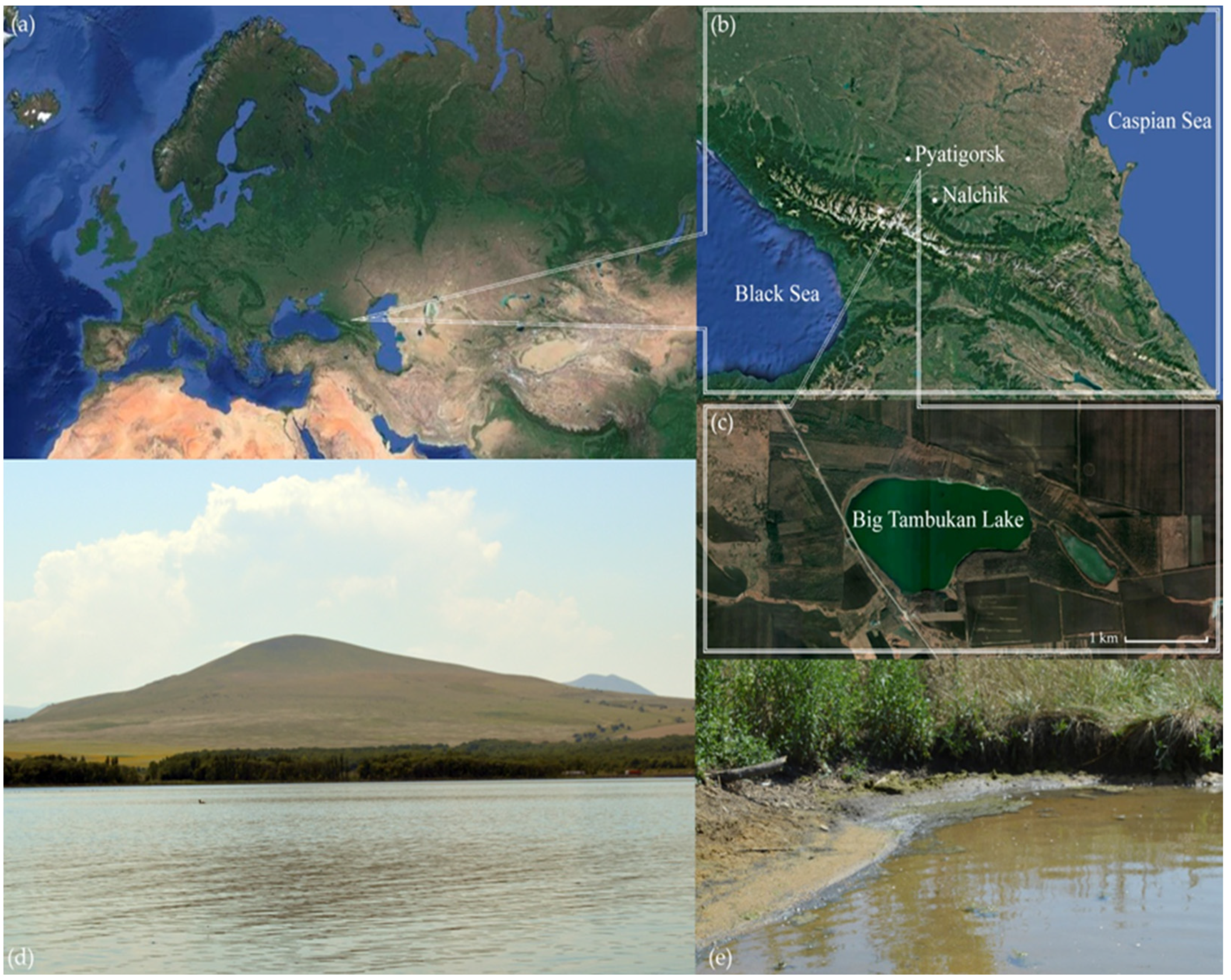
2.1. Samples

Ten samples were collected in August 2018 on the north coast of Big Tambukan Lake (hereinafter Tambukan), Northern Caucasus, Russia (43°57′52.6″ N 43°09′56.8″ E) (Figure 1). The lake’s surface area is 1.87 square kilometers, and its depth is about 3 m. During sampling, the salinity of the lake water was 28 g/L, and its pH level was 8.2. The samples were the top 0–5 cm layer of underwater sediments collected along the coastline. They included clay, sand, silt, mudflat soils, and cyanobacterial mats’ remains. The pH of the samples was 7.5–8.0. The shores of the lake are overgrown with dense vegetation, and among trees, oak and beech dominate. From the bottom of the central part of the lake, mud is derived.

2.2. Culture Media

The selective isolation of haloalkalitolerant fungi was carried out using an alkaline agar medium (AA, pH 10) prepared from malt–yeast extract and carbonate–bicarbonate buffer and containing the following salts: NaCl—6 g/L; KNO3—1 g/L; K2HPO4—1 g/L. Medium preparation is described in detail by Grum-Grzhimaylo et al. [47]. Bacterial growth was inhibited by rifampicin (2 g/L), which was the most efficient antibiotic among 21 preparations tested on AA [16]. Oatmeal agar (OA), Czapek-Dox agar (CZA), and malt–yeast agar (MYA) were used as standard media to culture the obtained micromycetes.
Buffer choices for generating pH values were made according to Grum-Grzhimaylo et al. [16]: Na2HPO4/NaH2PO4 system for pH 8 and Na2CO3/NaHCO3 system for pH 10. Thus, media buffered at pH 8.0 and 10.1 consisted of two components: the buffer component and MYA as the nutrient component. These components were autoclaved separately and then mixed afterward, making up the complete medium. Buffered medium preparation is described in detail by Grum-Grzhimaylo et al. [47].
The reaction of the isolates to the various NaCl concentrations on MYA medium containing either 0%, 2.5%, 5%, 7.5%, or 10% NaCl (w/v) was studied.

2.3. Isolation Techniques

Inoculation was performed using a soil-lump technique, i.e., via the distribution of soil lumps on the surfaces of Petri plates filled with AA. Inoculated plates were wrapped in Parafilm and incubated at 22 °C. After 10–21 days of incubation, the colonies were enumerated, and the retrieved fungal isolates were purified and pre-identified by morphological characters. Forty-eight isolates with different colony morphologies or, in the case of similar phenotypic variants, belonging to different sediment samples were selected for further studies. The 48 newly acquired strains were deposited at the Collection of Extremophilic Fungi of the Department of Mycology and Algology of Moscow State University (MSU) (Russia).
The fungal communities were analyzed according to the frequency of occurrence and the abundance of isolated species. The frequency of the occurrence of a particular species was defined as the percent composition of the number of samples in which a particular species occurred relative to the total number of all samples obtained. The abundance of a particular species was defined as the percent composition of the colony-forming units (CFU) of a particular species relative to the total CFU of all species obtained.

2.4. Morphological Identification of Fungal Isolates

The macromorphology of colonies and their micromorphological characters were observed for 14-day-old colonies grown on AA, MYA, OA, and CZA. The study of fungal morphology was investigated using a light microscope (LM) and scanning electron microscope (SEM). LM observations were performed on a Leica DM 2500 microscope equipped with a DFC 495 camera. SEM specimens were prepared by fixing them in approximately 4% formaldehyde or 2.5% glutaraldehyde, followed by washing and dehydration steps (20 min each) with a series of ethanol (30%, 50%, 70%, and 96% concentrations) and acetone before drying at the critical point in CO2, followed by metal sputter coating. SEM observations were performed using JSM-6380LA (JEOL, Tokyo, Japan) and Quattro S (Thermo Scientific, Brno, Czechia) microscopes. Images were acquired and elaborated using MicroCapture (version 1.0) software.
Manuals, identification guides, and original articles on fungal taxonomy were used for identification. Names and systematic positions of fungi are given in accordance with MycoID [48], Westerdijk Fungal Biodiversity Institute [49], and The National Center for Biotechnology Information (GenBank) [50].

2.5. Molecular Identification of Fungal Isolates

Molecular identification of fungal strains was performed by PCR amplification of the rDNA internal transcribed spacer region (ITS) using the primers ITS1F (TCCGTAGGTGAACTTGCG) and ITS4R (TCCTCCGCTTATTGATATGC) [51]. Genomic DNA was extracted using a genomic DNA isolation kit (LLC Biolabmix, Novosibirsk, Russia) as per the manufacturer’s protocol. The 50 µL PCR reaction mixture consisted of 25 µL of BioMaster HS-Taq PCR-Spec (2×) reaction mix (LLC Biolabmix, Novosibirsk, Russia), 50 mmol MgCl2, 0.1 nmol of each primer, 1–100 ng of isolated DNA, and nuclease-free water. PCR was carried out on a Thermal Cycler 2720 (Applied Biosystems, Foster City, CA, USA) with the following protocol: (1) 95 °C for 5 min, (2) 33 cycles of denaturation at 95 °C for 1 min, annealing at 51 °C for 1 min, extension at 72 °C for 1 min, and (3) final extension at 72 °C for 7 min. DNA fragments were subjected to Sanger sequencing on an Applied Biosystems 3500 Series Genetic Analyzer (Applied Biosystems, Beverly, MA, USA) using the primers ITS1F, ITS2R (GCTGCGTTCTTCATCGATGC), ITS3F (GCATCGATGAAGAACGCAGC), and ITS4R. Additionally, beta-tubulin (b-tub) gene amplification was performed for Emericellopsis species using the βt2a (GGTAACCAAATCGGTGCTGCTTTC) and βt2b (ACCCTCAGTGTAGTGACCCTTGGC) primers.

2.6. Phylogenetic Analysis

We generated an ITS alignment to fit all available sequences that could be reliably aligned with our own. To this end, we retrieved sequences from GenBank (Supplementary Table S1). Alignments were calculated through the MAFFT 7.429 online server [52] using the L-INS-I strategy. Emericellopsis isolates were identified via the sequencing of two gene loci, ITS rDNA and b-tub. The nucleotide sequences were submitted to GenBank [50] with their assigned accession numbers (Table 1).
For the taxonomic identification of species, phylogenetic analysis was carried out using the maximum likelihood (ML). Several phylogenetic trees were constructed at different taxonomic levels to achieve a more precise determination of the taxonomic positions of individual isolates. Nucleotide sequences for these constructions were retrieved from relevant fungal group articles and the NCBI database [50]. ML was run on RAxML-NG1.1.0 [53] with one thousand bootstrap replicates. For ML analyses, IQ-TREE 1.2.2 [54] with the best-fitted model option was used. Bootstrapping was performed using the standard nonparametric bootstrap algorithm with the number of replicates set to 1000. Support for nodes is indicated with bootstrap values ≥ 70.

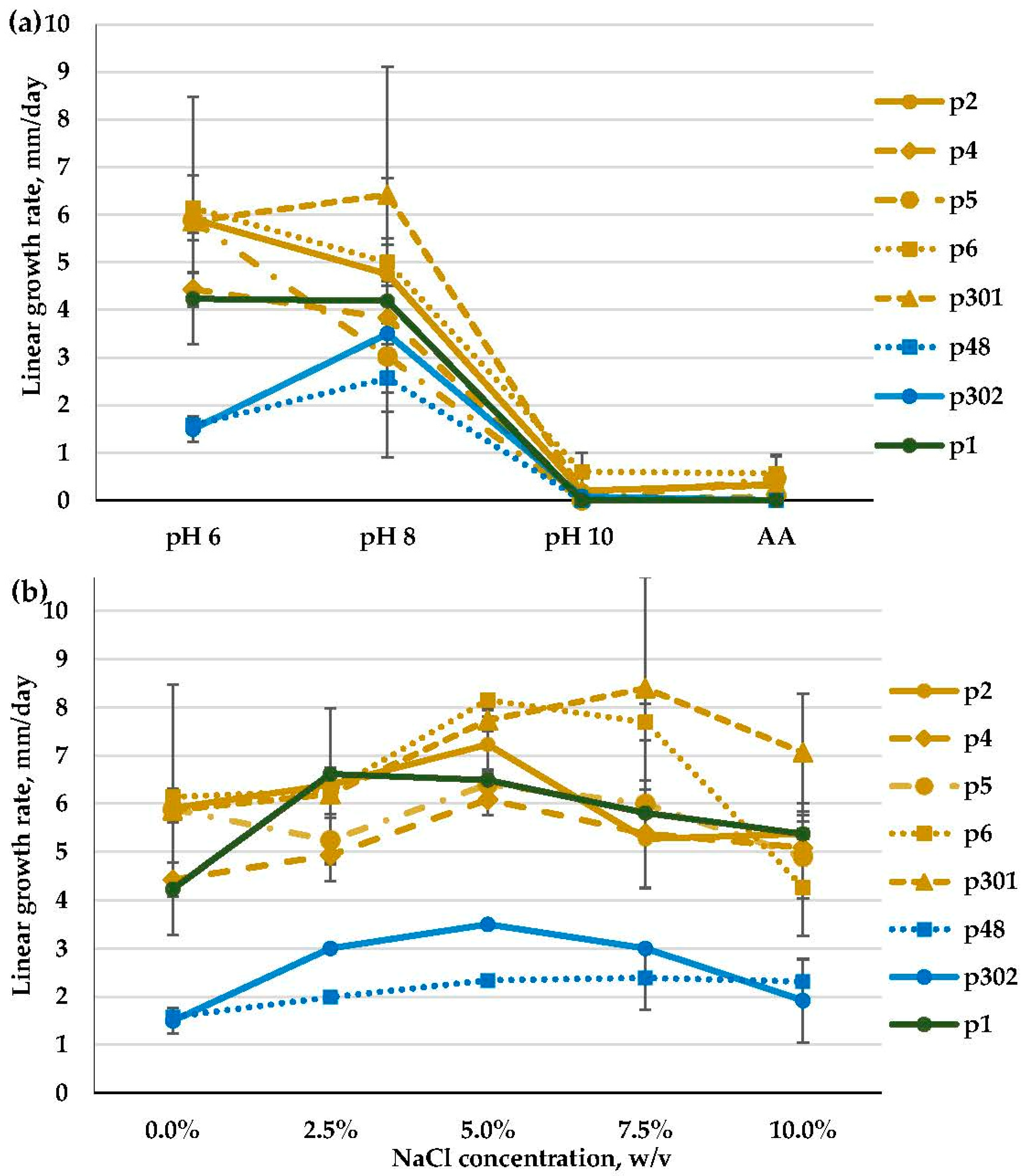
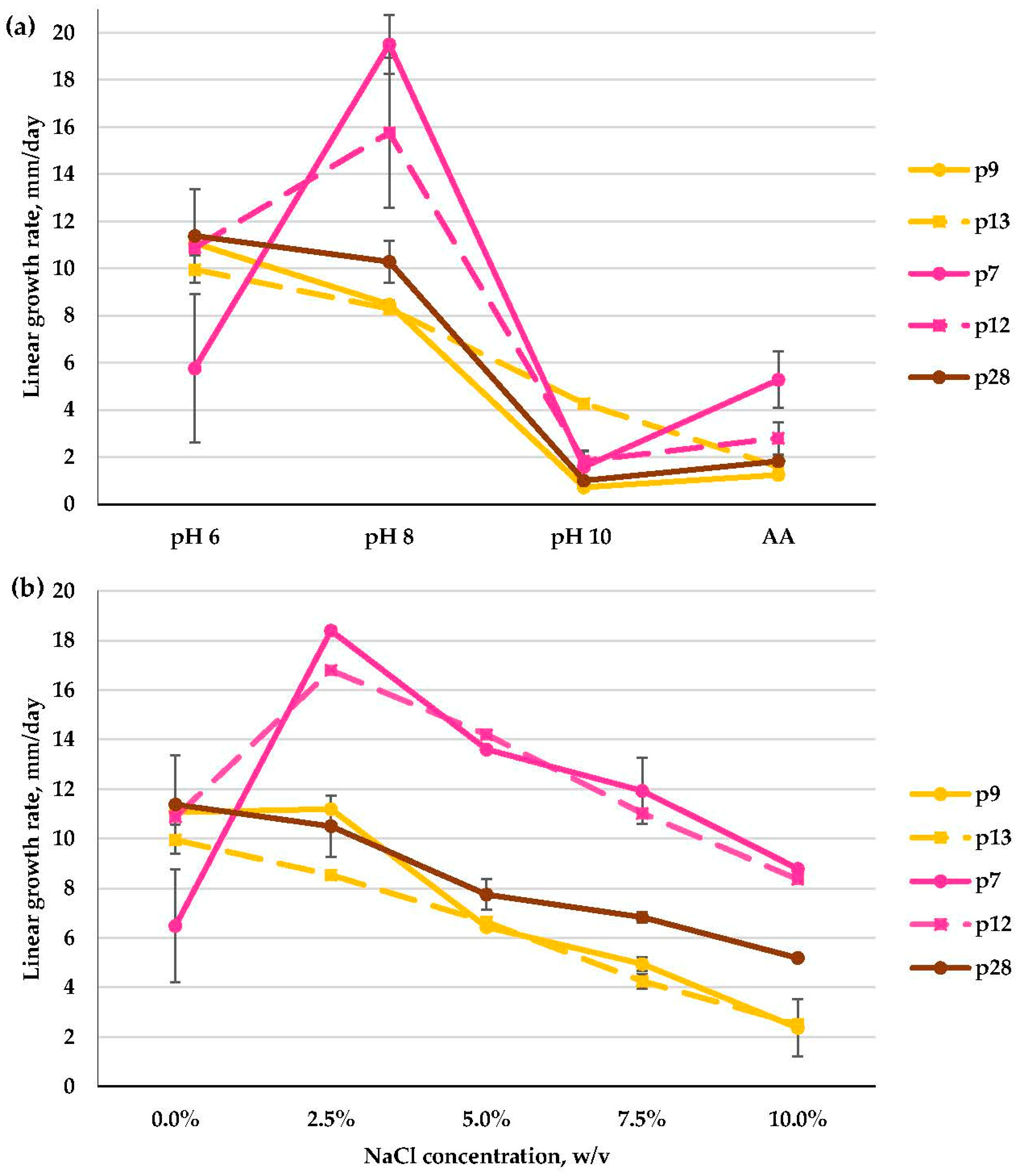
2.7. Growth at Different pH Values and Salt Concentrations

To elucidate the pH preferences of the strains, three media were used. They were MYA (pH 6.1) and two MYA-based media buffered at pH levels 8.0 and 10.1. Grum-Grzhimaylo with co-authors [16] pointed out that sometimes, the presence of extra Na+ in small amounts in the medium could significantly enhance fungal growth in conditions of high pH stress. In this way, to reveal the fungal growth type more completely, in addition to the listed media for growth experiments, we used AA medium (pH 10) containing 0.6% NaCl (w/v).
To reveal halotolerant properties, we studied the reaction of the isolates to the various NaCl concentrations on the MYA medium.
Petri dishes were inoculated at the center, then sealed thoroughly with Parafilm to avoid desiccation, and put at 25 °C in the dark. The growth expansion was followed for 2–4 weeks, depending on the strain. Every two or three days, we measured the diameters of fungal colonies in two perpendicular directions. To determine the linear growth of the strains, a linear regression model was used. The regression coefficient numerically corresponded to the linear growth rate of the colony. The confidence intervals (CIs) for the means were calculated according to Student’s t-distribution (n = 4, α = 0.05) and plotted on the graphs.

2.8. Antimicrobial Assays

The strains of fungi were grown on CZA, MYA, and AA. Our prior research demonstrated that an alkaline medium stimulates the synthesis of peptaibols with antimicrobial activity [55,56,57]. The detection of the peptaibol emericellipsin A in the crude extracts from Emericellopsis strains was made by HPLC and MALDI-TOF MS spectra in accordance with previously obtained data [24,58].
The antimicrobial activity of the strains was measured by the disk diffusion method on test cultures of mycelial and yeast microscopic fungi and on bacteria from the collection of cultures of the Gause Institute of New Antibiotics (Moscow, Russia). Opportunistic mold and yeast test cultures of the fungal species Aspergillus niger INA 00760, Candida albicans ATCC 2091, and test cultures of Gram-positive strains such as Bacillus subtilis ATCC 6633 and Gram-negative bacteria such as Escherichia coli ATCC 25922 were used.
Disks with a 6 mm diameter containing 40 µL of the culture broth were deposited on agar plates. The diameters of the inhibition zones were measured after 24 h at 28 °C. Amphotericin B (for fungi) and ampicillin (for bacteria) solutions (Sigma-Aldrich, St. Louis, MO, USA) were used as positive controls. Cultures were considered to be highly active if the zone of growth retardation of the test organism was 20 mm or more; moderately active cultures had a growth retardation zone of 10–20 mm, and weakly active cultures had a zone of less than 10 mm (disk diameter 6 mm). The test culture of B. subtilis ATCC 6633 was grown on Gause medium no. 2 with the following composition (g/L): 2.5 tryptone (or 30 mL Hottinger broth), 5 peptone, 5 sodium chloride, 10 glucose. E. coli ATCC 25922 was grown on lysogen-broth LB medium (tryptone soy agar). Cultures of the fungi A. niger INA 00760 and C. albicans ATCC 2091 were grown on CZA. The cultures were preliminarily grown in test tubes with nutrient agar slants, after which their cells were suspended in saline to a turbidity of 0.5 according to the McFarland standard HiMedia R092R Tube, Maharashtra, India (1.5 × 10⁸ CFU/mL) and were used within 15 min. One-day cultures of bacteria and 5-day cultures of molds and yeasts were used.

3. Results

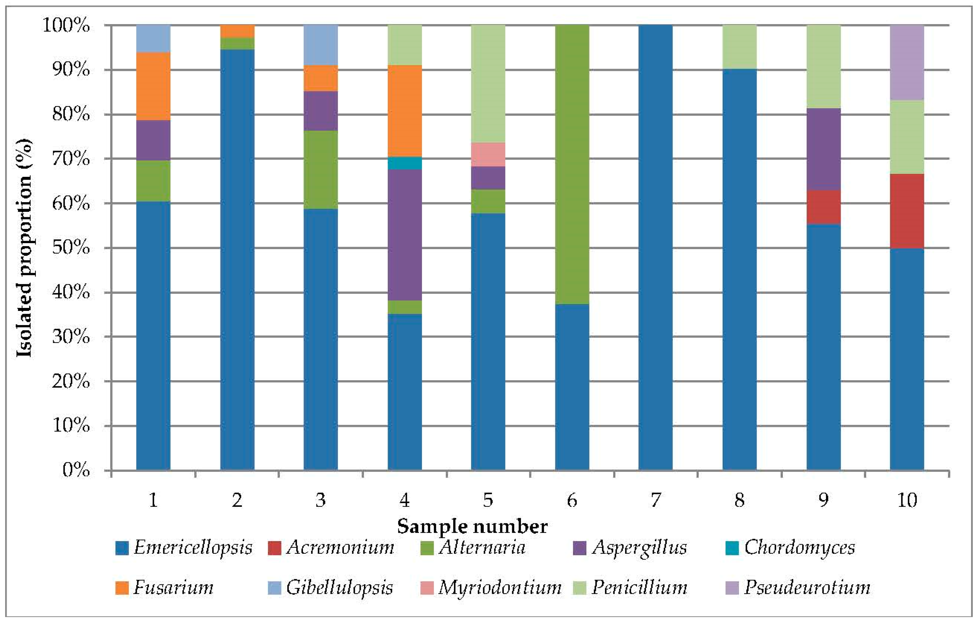
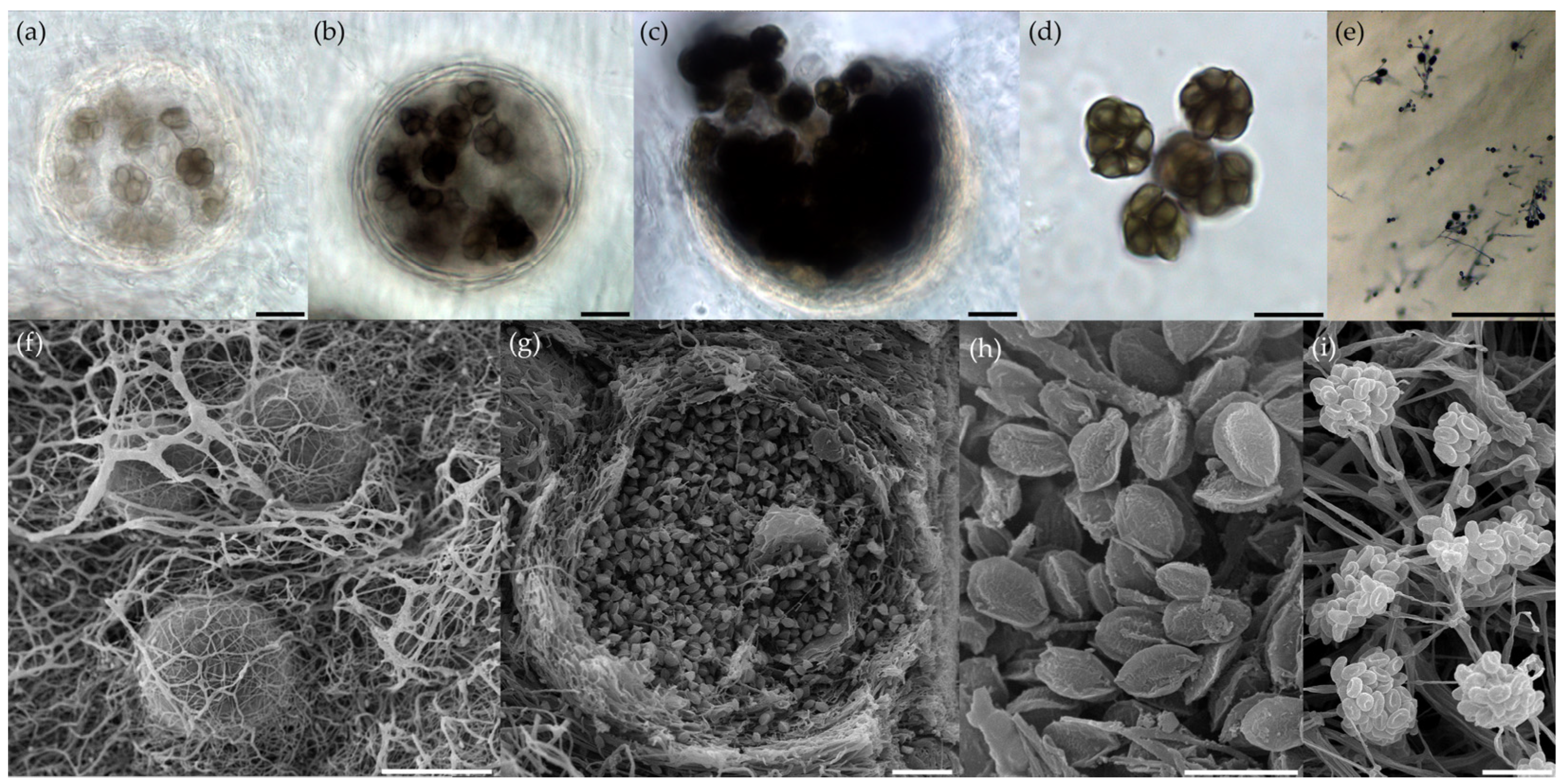
3.1. Analysis of Cultivated Haloalkalitolerant Fungi

From the sediments of Tambukan Lake, 272 fungal CFU were obtained. The retrieved isolates were represented by 10 genera belonging to four classes, i.e., Sordariomycetes, Dothideomycetes, Eurotiomycetes, and Leotiomycetes, all within the phylum Ascomycota. The largest number among all detected species belonged to the Emericellopsis genus. Other genera recorded with more than one species were Aspergillus, Fusarium, Penicillium, and Gibellulopsis (Table 2).
We detected the presence of fungi in all samples of sediments obtained (Supplementary Figure S1). The number of isolated strains ranged from 6 to 37 per sample, reaching, on average, 27 strains per sample (Figure 2).
Emericellopsis was the most frequently occurring genus (100%) and was isolated from all of the sediment samples (Table 2). Other genera detected with a high frequency of occurrence were Alternaria (60%), Aspergillus (50%), Penicillium (50%), and Fusarium (40%). The rest of the detected genera occurred in one or two samples, and their occurrence was lower than 20%.
The abundance of the Emericellopsis genus was 66.2%, varying from 35% (sample no. 4) to 100% (sample no. 7) in a particular sample. Then, Alternaria alternata possessed a high abundance (10%). The maximum abundance was noted in sample no. 6 and amounted to 62.5%. High abundance was also noted for Aspergillus (8.1%), Penicillium (6.3%), and Fusarium (5.5%). The abundance of other isolates did not exceed 2%.

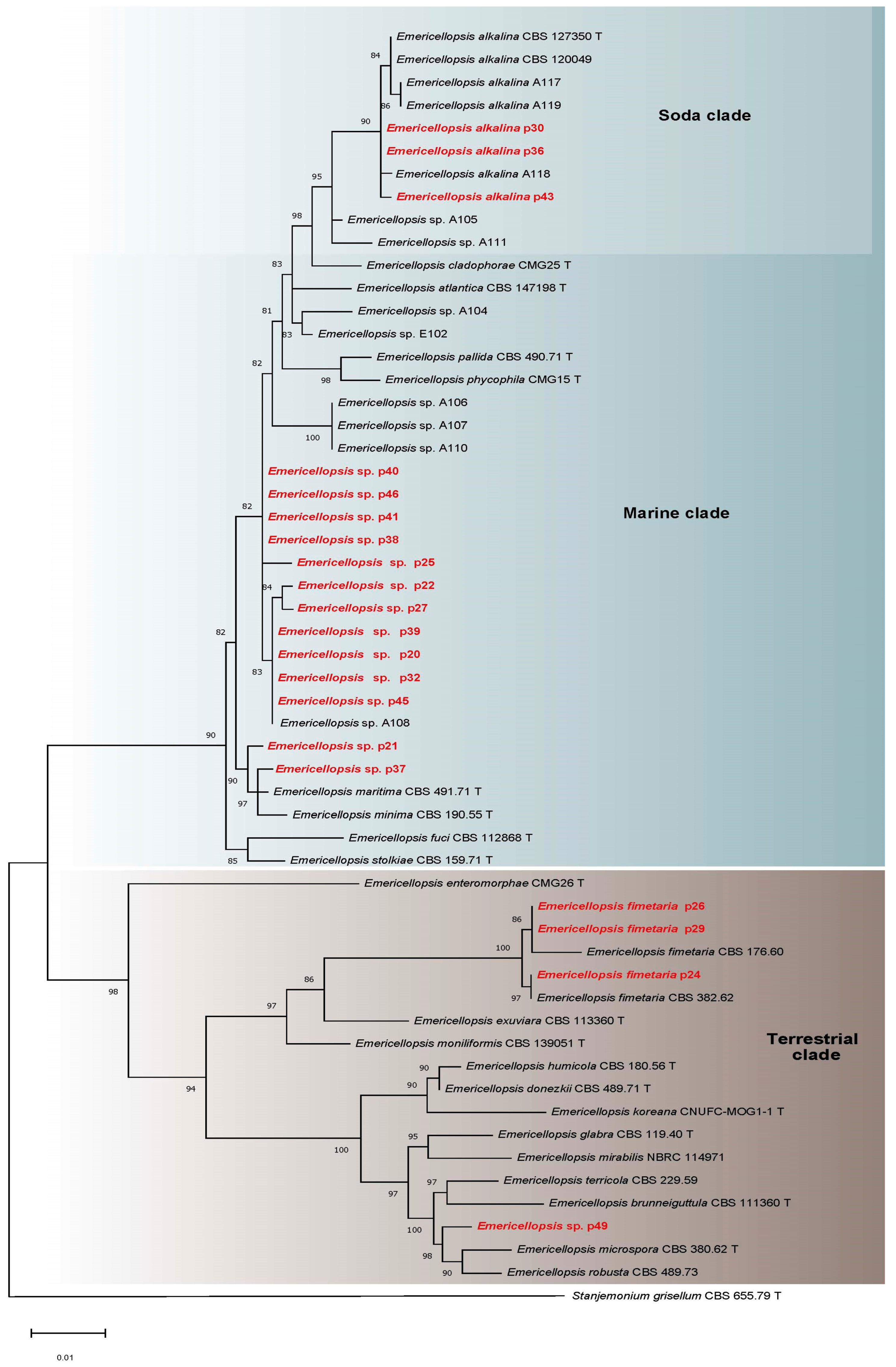
3.2. Molecular Identification, Phylogenetic Analyses, and Salt and Alkaline Preferences of the Obtained Isolates

Forty-eight isolates were analyzed. The vast majority of them were able to develop in an alkaline medium (i.e., pH 8), though they significantly reduced their growth rate in a strongly alkaline medium (i.e., pH 10), and thus, we treat them as alkalitolerants. Pseudeurotium bakeri (p47) was the single isolate obtained that could not tolerate pH 8. We further divide alkalitolerants according to their growth characteristics into strong, moderate, and weak [16]. Strong alkalitolerants grew at pH 8 considerably better than at pH 6 and showed little reduction in growth at pH 10. Moderate alkalitolerants showed better growth at pH 8 than at pH 6, or the same at both pH levels, and strongly reduced growth at pH 10. Weak alkalitolerants preferred a slightly acidic medium (pH 6), and their growth was hindered as pH increased.
Among obtained isolates, no halophiles were detected [18]. The tested isolates can be categorized into three groups—strong halotolerants that can grow optimally at 7.5% (w/v) NaCl and above, moderate halotolerants that can grow optimally at 2.5% and/or 5% (w/v) NaCl, and weak halotolerants that grow optimally in a medium without NaCl and whose growth rates decrease with increasing NaCl concentration.

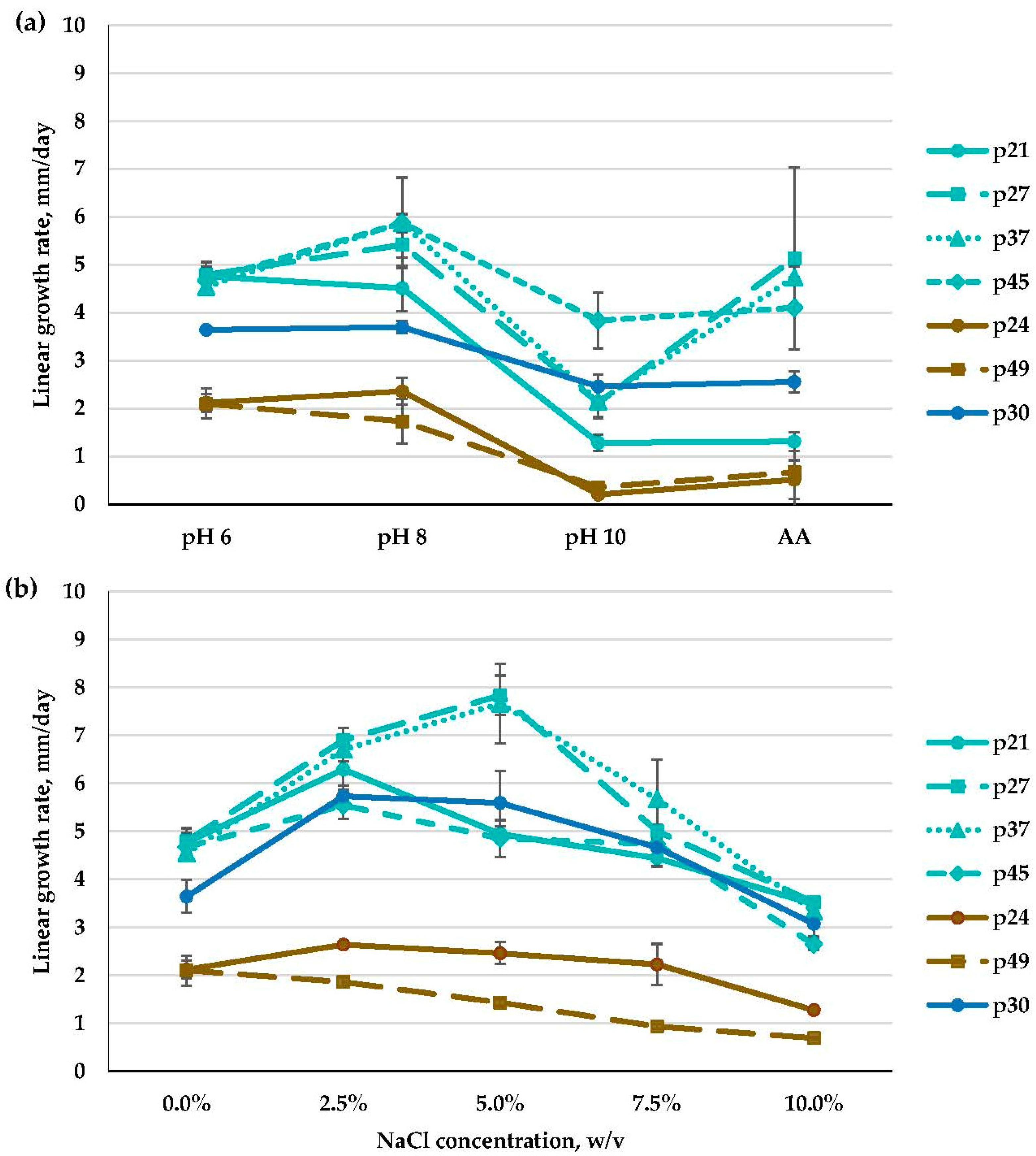
3.2.1. Emericellopsis spp. (Bionectriaceae, Hypocreales)

Fungi of the Emericellopsis genus dominated in the studied samples, possessing the highest frequency of occurrence and abundance (Table 2; Figure 2). ITS and b-tub sequences were generated for 20 isolates from sediments of Tambukan Lake, and these were aligned with 45 ITS and 36 b-tub sequences retrieved from GenBank, including 1 outgroup taxon (Stanjemonium grisellum CBS 655.79 T) (Table 1; Supplementary Table S1). Emericellopsis strains from the sediments of Tambukan Lake were clustered into three ecological clades, outlined previously by Zuccaro et al. [59], Grum-Grzhimaylo et al. [60], and Hagestad et al. [61]. Three strains were in the Soda clade; thirteen strains were in the Marine clade; and four strains were in the Terrestrial clade (Figure 3; Supplementary Figure S2).
Three strains (p30, p36, p43) within the Soda clade were identified as Emericellopsis alkalina, as they clustered together with the ex-type strain of E. alkalina (CBS 127350 T) and other strains of E. alkalina (A117, A118, A119, and CBS 120049).
The majority of Emericellopsis isolates from the sediments of Tambukan Lake were classified within the Marine clade. Phylogenetic analysis based on the ITS locus showed that seven isolates (p37, p45, p32, p20, p39, p27, p22) grouped with the known ex-type strain of E. maritima (CBS 491.71 T), while the remaining six isolates (p38, p41, p40, p46, p21, p25) differed from the ex-type strain E. maritima and formed a distinct clade (Supplementary Figure S2). Phylogenetic reconstructions based on two genes revealed considerable heterogeneity among isolates of this genus obtained from sediments of Tambukan Lake (Figure 3). Only strains p37 and p21 were found to be closely related to the ex-type strain of E. maritima, but they did not group together in a distinct clade. Instead, they formed sister groups with it. The other 11 isolates formed several distinct clades, strongly supported within their respective groups, and did not group with any known species of the genus. Morphologically, these isolates displayed similarities, forming Acremonium-like asexual sporulation-producing conidial heads on solitary, sometimes branched phialides, while the sexual stage was not observed.
Four strains, namely, p24, p26, p29, and p49, were placed in the Terrestrial clade. Strain p49 was identified as Emericellopsis sp., as it formed a separate sister clade in the phylogenetic analysis based on two loci, despite being grouped within the known ex-type strains of E. terricola (CBS 120.40 T) and E. terricola (CBS 229.59) in the ITS-based analysis (Figure 3; Supplementary Figure S2). This isolate also lacked the teleomorph stage and only exhibited an Acremonium-like anamorph. Three strains (p24, p26, p29) within the Terrestrial clade were grouped together with Emericellopsis fimetaria in both the ITS-based and two-loci (ITS and b-tub) phylogenetic analyses (Figure 3; Supplementary Figure S2). The isolates from the sediments of Tambukan Lake showed high support (100%) within this group. All three isolates formed abundant conidiation. Only the p24 isolate in culture (on MA and OA media) formed fruiting bodies typical of the Emericellopsis genus (Figure 4).
Ascomata developing in aerial and submerged mycelia were globose, non-ostiolate, and hyaline at the beginning and became black as ascospores matured, reaching 108–190 μm in diameter. Asci were eight-spored and (sub-) globose, with thin, evanescent, hyaline walls. Ascospores were unicellular and ellipsoid (6.5–8.0 × 3.8–5.4 μm) with 3–5 longitudinal wings (0.4–1.6 μm wide), most of which extended from pole to pole of the spore and which had an undulating or sometimes ragged margin. Colonies on OA at 25 °C after 14 days reached 40–42 mm in diameter and were felty, with reverse yellowish-white and, later, black zones appearing as a result of the formation of abundant ascomata. Colonies on MYA at 25 °C, after 14 days, were 34–36 mm in diameter and white, slightly pinkish, sometimes later darkening in the center due to the formation of ascomata (Supplementary Figure S3). Conidiophores had repeated mesotonous verticillate branching, with branches diverging in acute angles. Stilbella-like synnemata did not form. Conidia were 3.1–3.7 × 1.7–1.9 mm, shorter than the ascospores.
Within the Emericellopsis genus, physiological characters, i.e., pH and NaCl concentration preferences, differed depending on the isolate (Figure 5; Supplementary Figures S4 and S5).
All of the isolates from the Soda and the Marine clades preferred the weakly alkaline medium (pH 8) and were strong or moderate alkalitolerants that slightly or significantly reduced their growth rate in the strongly alkaline medium at pH 10. All of the obtained isolates from the Soda clade (p30, p36, and p43) were moderate alkalitolerants. In the Marine clade, ten isolates (p20, p22, p25, p27, p32, p37, p38, p39, p40, p45) revealed a strong alkalitolerant phenotype, and the remaining three isolates (p21, p41, p46) had a moderate one. In the Terrestrial clade, isolates were slow-growing, and all of them (p26, p29, p49, and p24) were moderate alkalitolerants with similarly high growth rates at both pH 6 and pH 8 (Figure 5a; Supplementary Figure S4).
It is worth mentioning that at high alkaline ambient pH (10), the majority of isolates from the Marine clade (p20, p25, p27, p37, p41, p46) increased their growth rates on the AA medium compared to the medium without salt. Some of them (p20, p25, p27, p37), though, had restricted growth at pH 10 without NaCl, but in the presence of NaCl (0.6% w/v) in the AA medium, they demonstrated a growth rate comparable to that at pH 8, and thus, we assigned these isolates to the group of strong alkalitolerants. Other Emericellopsis isolates did not increase their growth rates on the AA medium (Figure 5a; Supplementary Figure S4). Interestingly, at pH 10, all the Emericellopsis isolates demonstrated a prolonged adaptation period of about ten days, during which they grew extremely slowly. After that period, their growth rate was noticeably accelerated.
As for NaCl preferences, all of the isolates from the Soda clade (p30, p43, p36) were moderate halotolerants, as they preferred a medium containing NaCl, growing optimally at 2.5–5% NaCl. Then, eleven isolates from the Marine clade were moderate halotolerants (p20, p21, p22, p27, p32, p37, p39, p40, p41, p45, p46), with optimal growth at 2.5% and/or 5% NaCl and a decreased growth rate when the NaCl concentration exceeded 5%. The remaining two isolates (p25, p38) were strong halotolerants that grew optimally in a very wide range of NaCl concentrations, including 7.5% NaCl. In the Terrestrial clade, E. fimetaria isolates were moderate (p26 and p29) and strong (p24) halotolerants. They preferred media with both 0% and 2.5% NaCl (p26, p29) or grew on media containing 2.5–7.5% NaCl (p24). Finally, p49 was a moderate halotolerant that preferred media with both 0% and 2.5% NaCl (Figure 5b; Supplementary Figure S5).

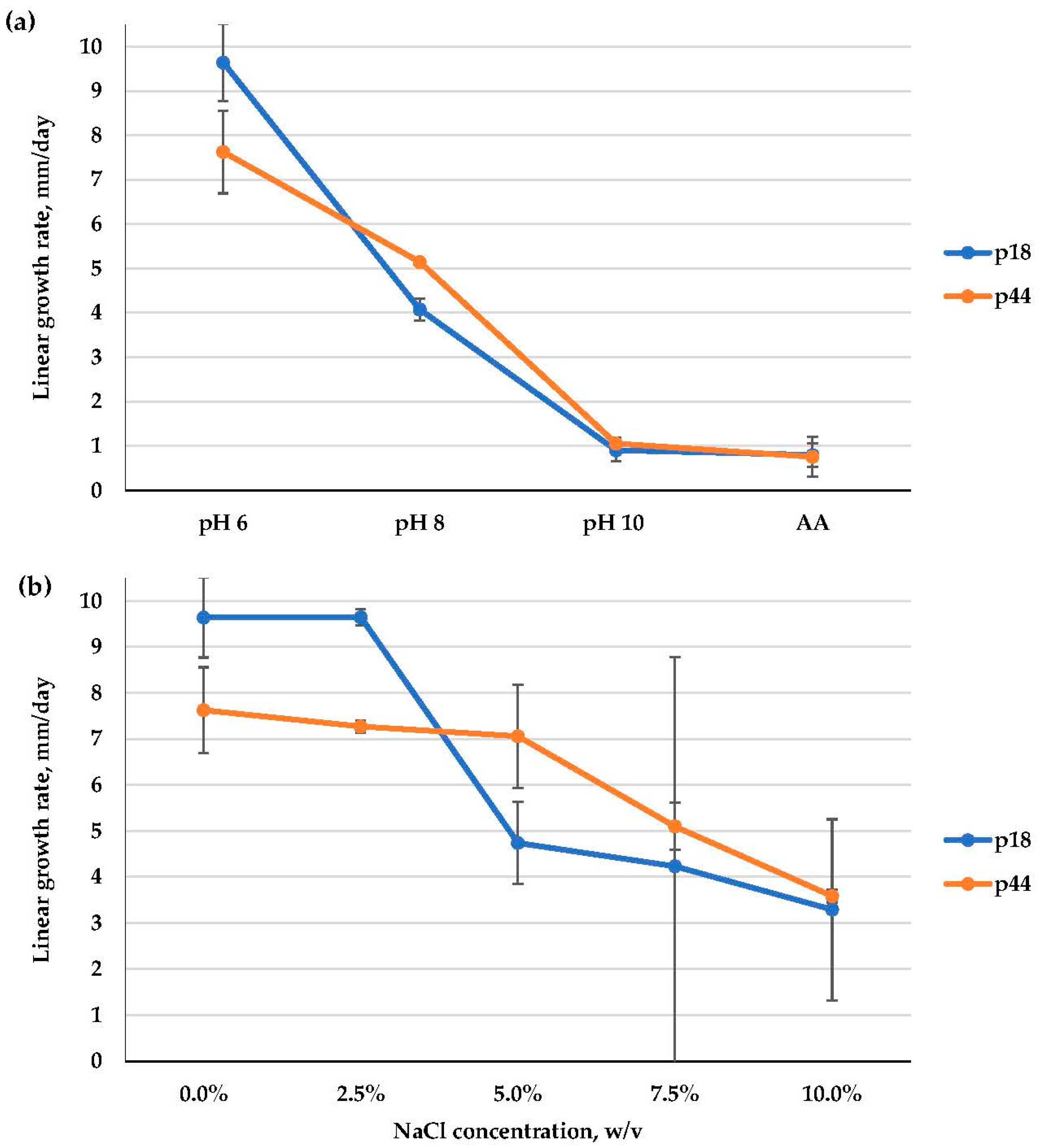
3.2.2. Alternaria alternata (Pleosporaceae, Pleosporales)

ITS sequences were obtained for 2 isolates (p18, p44) and subsequently aligned with 13 sequences retrieved from GenBank, including 1 outgroup taxon (Curvularia homomorpha CBS 156.60 T) (Table 1; Supplementary Table S1). The phylogenetic analysis revealed that our A. alternata (p18 and p44) isolates clustered within a highly supported clade (99% ML) together with the ex-type strain of A. alternata (CBS 916.96 T) (Supplementary Figure S6).
As for ecophysiological characters, A. alternata isolates were weak alkalitolerants, as they grew optimally at pH 6 and were able to grow at alkaline pH levels, though rather slowly. Then, these isolates were moderate halotolerants that preferred 0–2.5% NaCl (p18) or 0–5% NaCl (p44) (Figure 6; Supplementary Figure S7).

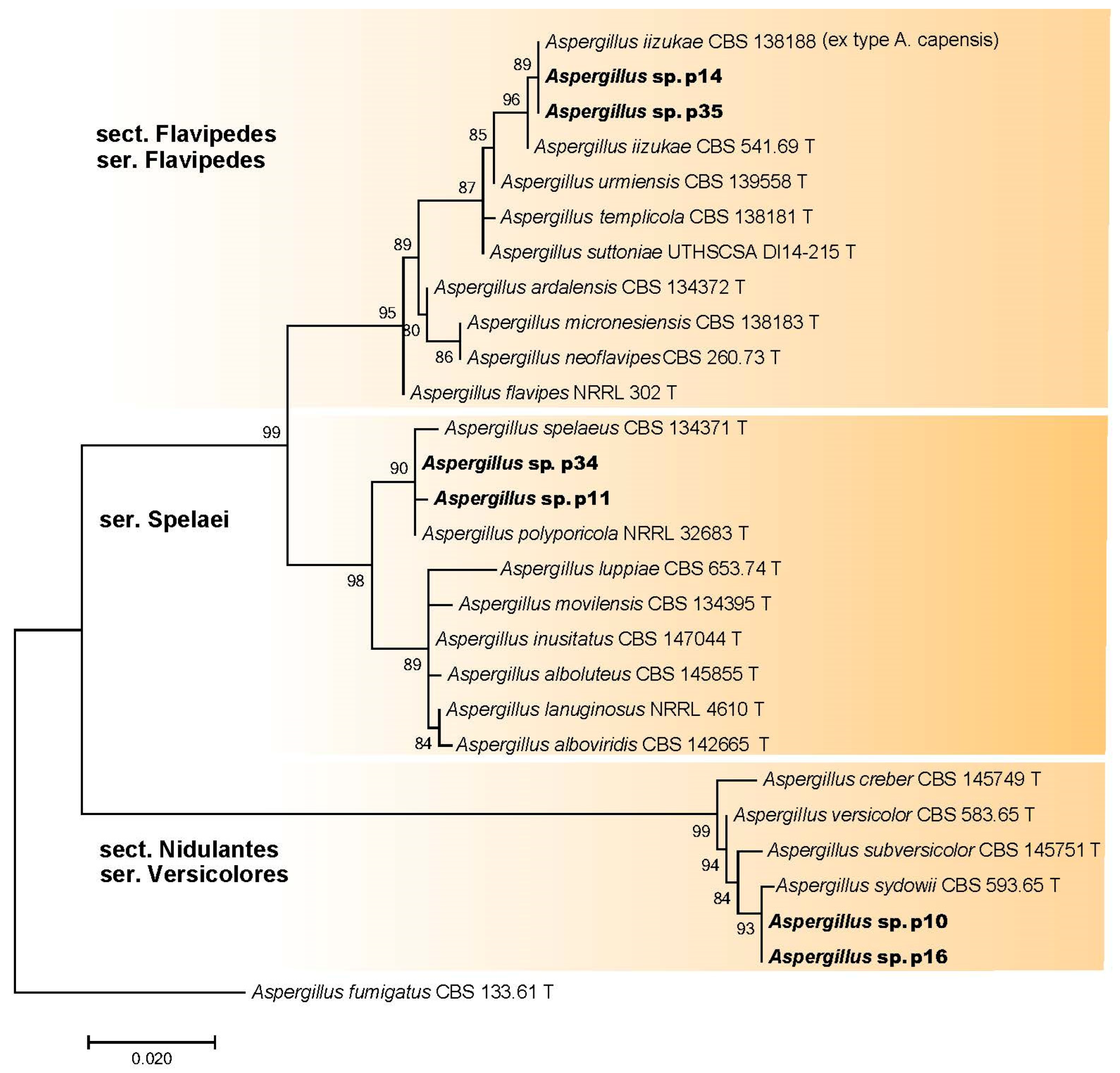
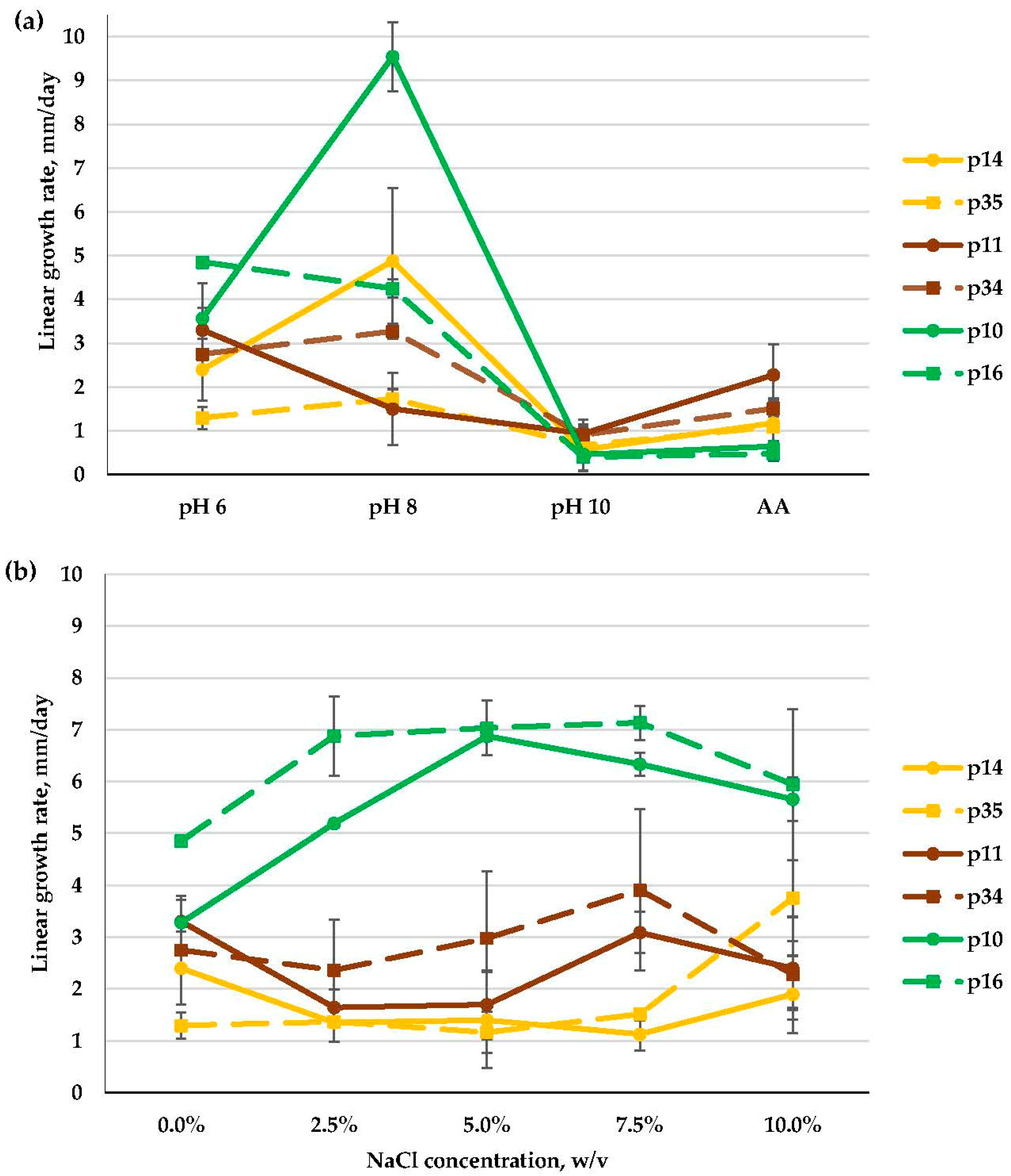
3.2.3. Aspergillus spp. (Aspergillaceae, Eurotiales)

The morphological analysis identified isolates p10, p11, p14, p16, p34, and p35 as Aspergillus species. Subsequently, ITS sequences were obtained for these six isolates and aligned with 22 sequences retrieved from GenBank, which included one outgroup taxon (Aspergillus fumigatus CBS 133.61 T) (Table 1; Supplementary Table S1). The phylogenetic analysis resulted in the clustering of the sequenced isolates into three distinct clades (Figure 7). The majority of isolates belonged to the Flavipedes section from the Flavipedes series (p35, p14) and Spelaei series (p34, p11). Additionally, two strains (p10, p16) were classified as members of the Nidulantes section from the Versicolores series (Figure 7).
Four Aspergillus isolates (i.e., p10, p14, p34, and p35) were moderate alkalitolerants, as their growth rate at pH 8 was higher than (p10, p14, p34) or the same as (p35) that at pH 6 and was strongly restricted at pH 10. The remaining two isolates (p11 and p16) were weak alkalitolerants, as they preferred pH 6 (Figure 8a).
In general, the Aspergillus isolates studied here preferred media containing NaCl at concentrations 7.5% and above, and thus, they were strong halotolerants. Aspergillus isolates from the Versicolores series showed high growth rates at 5–7.5% NaCl (p10) or at 2.5–10% NaCl (p16) and a severe growth decrease on a medium without added salt. Isolates among the Spelaei series (p11 and p34) and Flavipedes series (p14 and p35) grew well at all salt concentrations tested from 0 to 10% NaCl (Figure 8b).

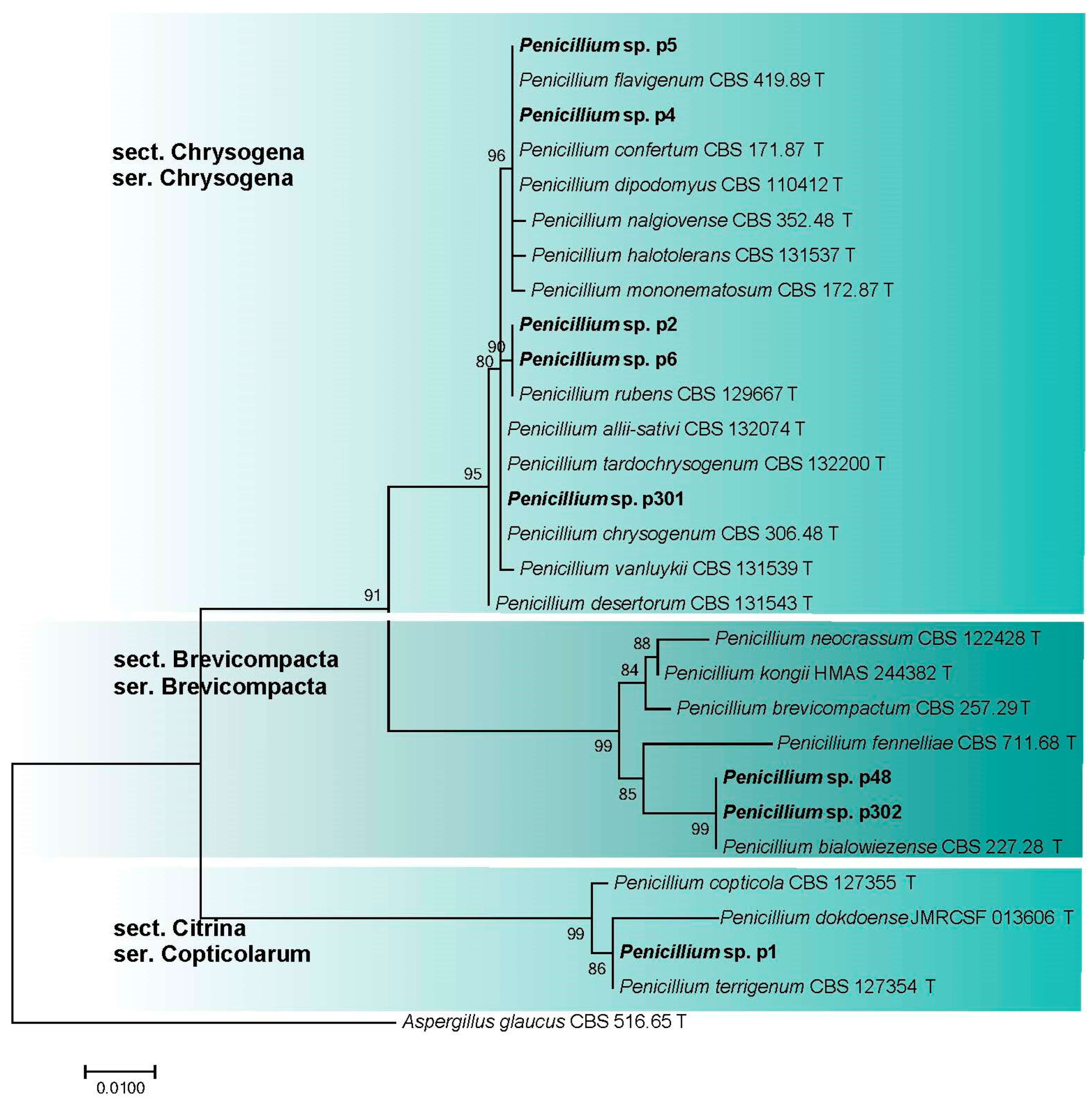
3.2.4. Penicillium spp. (Aspergillaceae, Eurotiales)

The morphological analysis identified isolates p1, p2, p4, p5, p6, p48, p301, and p302 as Penicillium species. Subsequently, ITS sequences were obtained for these 8 isolates and aligned with 21 sequences retrieved from GenBank, which included 1 outgroup taxon (Aspergillus glaucus CBS 516.65 T) (Table 1; Supplementary Table S1). The phylogenetic analysis resulted in the clustering of the sequenced isolates into three distinct clades (Figure 9). Most of the isolates (p2, p4, p5, p6, p301) formed a clade corresponding to Penicillium section Chrysogena series Chrysogena. Two isolates (p48, p302) clustered with the ex-type isolate of P. bialowiezense (CBS 227.28 T) and other species of the section Brevicompacta series Brevicompacta. The remaining isolate (p1) clustered with the ex-type isolate of P. terrigenum (CBS 127354 T) and other species of section Citrina series Copticolarum.
The vast majority of the studied Penicillium isolates, except for a single strain (p5), successfully developed on the slightly alkaline medium (pH 8). Six isolates were moderate alkalitolerants: their growth rate at pH 8 was a little higher than (p302) or the same as (p1, p4, p6, p48, p301) that at pH 6. Another two isolates (p2, p5) were weak alkalitolerants showing optimal growth at pH 6. At pH 10, the growth of all of the isolates was strongly reduced. The presence of extra salt in the AA medium did not have a visible effect on the growth pattern (Figure 10a).
In general, Penicillium isolates preferred NaCl-containing media and were moderate or strong halotolerants. Isolates among the Chrysogena series were moderate halotolerants, with optimal growth on media that included 5% NaCl (p2, p5), or strong halotolerants that grew well at all salt concentrations tested from 0 to 10% NaCl (p301) or preferred 5–10% NaCl (p4) or 5–7.5% NaCl (p6). A Brevicompacta series isolate (p48) preferred 5–10% NaCl and thus was strongly halotolerant, while the isolate p302 grew best at 5% NaCl and was moderate. Finally, Penicillium sp. series Copticolarum (p1) showed the highest growth rate at 2.5–5% NaCl and was a moderate halotolerant (Figure 10b).

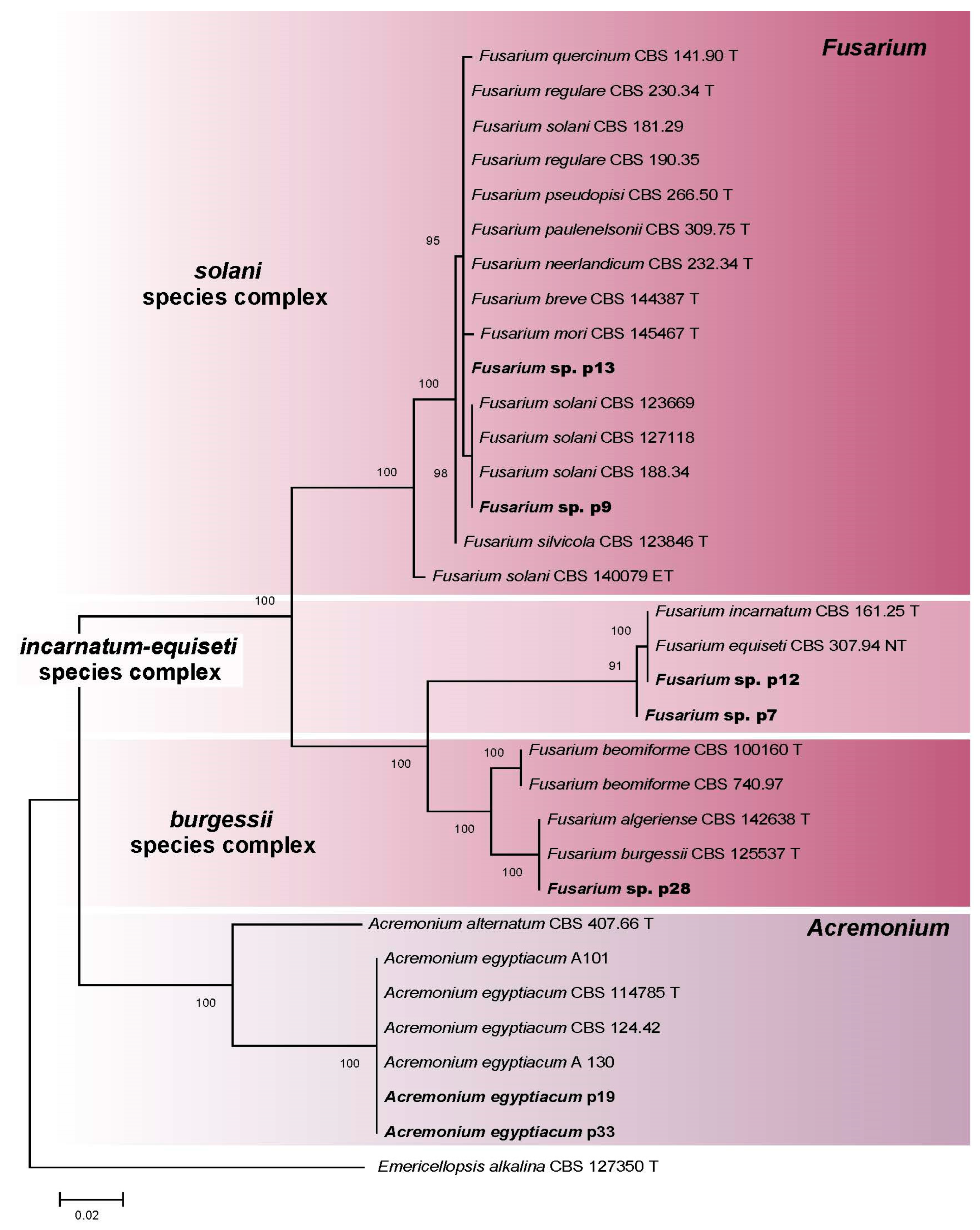
3.2.5. Fusarium spp. and Acremonium spp. (Hypocreales)

Based on the morphological analysis, isolates p7, p9, p12, p13, and p28 were identified as Fusarium species, whereas isolates p19 and p33 were identified as Acremonium species. Subsequently, ITS sequences were generated for the 7 isolates and aligned with 26 sequences retrieved from GenBank, including 1 outgroup taxon (E. alkalina CBS 127350 T) (Table 1; Supplementary Table S1). The phylogenetic analysis revealed that the isolates sequenced in this study clustered into four distinct clades (Figure 11). The Fusarium isolates formed three species complexes: Solani (p9, p13), Incarnatum-Equiseti (p7, p12), and Burgessii (p28). The Acremonium isolates (p19, p33) clustered together with the ex-type isolate of A. egyptiacum (CBS 114785 T) and other isolates of this species (CBS 124.42, A101, A130).
All Fusarium isolates successfully grew at a slightly alkaline pH of 8. Incarnatum-Equiseti complex isolates (p7, p12) and the Burgessii complex isolate (p28) turned out to reveal a moderate alkalitolerant phenotype, as they grew best at pH 8 (p7) or in the range 6–8 pH (p12, p28) and had drastically reduced growth at pH 10. Solani complex isolates (p9, p13) were weak alkalitolerants, as their growth slowed down at pH 8 and preferred a slightly acidic medium (pH 6). At pH 10, extra salt in the medium improved the growth of three isolates (p7, p9, p28) (Figure 12a).
As for NaCl preferences, Incarnatum-Equiseti complex isolates (p7, p12) possessed a moderate halotolerant phenotype, as they preferred 2.5% NaCl and grew much slower without salt. The Burgessii complex isolate (p28) was also a moderate halotolerant that preferred 0–2.5% NaCl. One isolate (p9) of the Solani complex was a moderate halotolerant, growing best at 0–2.5% NaCl, while another (p13) was a weak halotolerant, as it preferred a medium without salt, and its growth rate gradually decreased with increasing salt concentrations (Figure 12b).
A. egyptiacum isolates were moderate (p19) and weak (p33) alkalitolerants, as they preferred pH 6 or pH 8 (p19) and pH 6 (p33) and had drastically reduced growth at pH 10. Extra salt in the AA medium improved the growth of these strains (Figure S8). Both isolates were moderate halotolerants that preferred 2.5% NaCl (p19) or 0–2.5% NaCl (p33), and their growth rate gradually decreased as the salt concentration increased, though even at 10% NaCl, they grew successfully (Supplementary Figure S8).

3.2.6. Low-Frequency Isolates from Sediments of Big Tambukan Lake

The isolates that possessed a low frequency of occurrence (i.e., 10%) and occurred in samples of sediments once were Gibellulopsis nigrescens (p8), G. serrae (p17), and Chordomyces sp. (p42) (Plectosphaerellaceae, Glomerellales); Myriodontium keratinophilum (Incertae sedis, Onygenales) (p15); and Pseudeurotium bakeri (Pseudeurotiaceae, Thelebolales) (p47).
A phylogenetic analysis of Plectosphaerellaceae was conducted using ITS sequences from 3 isolates that were aligned to 12 sequences from GenBank, including 1 outgroup taxon (Plectosphaerella cucumerina CBS 137.37 T) (Table 1; Supplementary Table S1; Supplementary Figure S9). Two strains (p8, p17) were identified as members of the Gibellulopsis genus. Isolate p8 clustered with the ex-type isolate of G. serrae (CBS 290.30 T), while isolate p17 clustered with the neotype isolate of G. nigrescens (CBS 120949 NT). Isolate p42, with a high support value (97%), was grouped with representatives of the Chordomyces genus.
The sequence similarity searches for isolate p15 performed in GenBank using the BLAST sequence analysis tool showed the closest match (100% similarity) in the ITS locus with M. keratinophilum CBS 256.81 (GenBank: MH861337.1). The observed micromorphological characteristics also confirmed the identification.
Isolate p47, obtained as a single isolate from sample no. 10 and grown on nutrient media, exhibited the ability to produce numerous fruiting bodies typical of P. bakeri (Supplementary Figure S10). Our results showed that isolate p47 is most closely related to the ex-type strains of P. bakeri CBS 878.71 T and P. bakeri CBS 128112, with a bootstrap value of 99% (Table 1; Supplementary Table S1; Supplementary Figure S11).
The listed low-frequency isolates showed different pH preferences. Indeed, Plectosphaerellaceae isolates (p8, p17, p42) were strong alkalitolerants that grew optimally at pH 8 and pH 10 (p8 and p42) or successfully developed at pH 6, pH 8, and pH 10 (p17). At the same time, M. keratinophilum (p15) was a moderate alkalitolerant with equally high growth rates at pH 6 and pH 8 and noticeably decreased growth at pH 10, while P. bakeri (p47) could not grow at alkaline pH values at all and was the single non-alkalitolerant isolate. The addition of extra salt to the medium at pH 10 slightly increased the growth of Plectosphaerellaceae isolates and did not have a visible effect on M. keratinophilum (Figure 13a).
NaCl preferences were different among Plectosphaerellaceae. G. serrae (p8) was a weak halotolerant and preferred a medium without salt, while G. nigrescens (p17) and Chordomyces sp. (p42) were moderate halotolerants with optimal growth on media including 2.5% and 5% NaCl. All Plectosphaerellaceae isolates had drastically reduced growth at 10% NaCl (Figure 13b). Then, M. keratinophilum (p15) was strongly halotolerant and grew optimally in an extremely wide range of 0–7.5% NaCl. Finally, P. bakeri (p47) was a weak halotolerant. It had significantly decreased growth when the NaCl concentration increased and did not grow at 7.5% NaCl (Figure 13b).

3.3. Antimicrobial Activity

The antimicrobial characterization of the fungi from sediments of Tambukan Lake against test cultures is given in Table 3 and Supplementary Tables S2 and S3. The analysis of the antimicrobial activity of different genera against test cultures of fungi and bacteria revealed that the majority of the strains are defined by their antibacterial activity. Thus, the proportion of strains with antifungal activity against A. niger INA 00760 was 66.7%, while those with antibacterial activity against B. subtilis ATCC 6633 made up 87.5% of the total number of studied strains (Table 3).
As expected, the targets of antimicrobial activity varied between the representatives of the different fungal genera. Representatives of micromycetes from the Emericellopsis genus showed both antibacterial activity (i.e., predominantly against B. subtilis ATCC 6633) and antifungal activity (i.e., against A. niger INA 00760 and C. albicans ATCC 2091). It is important to highlight that, in the present study, Emericellopsis strains were more frequent than the other isolated fungal cultures. Weak antifungal activity but a strong antibacterial effect was shown for Penicillium and Aspergillus genera. It was shown that the Fusarium genus produced compounds that were effective against fungi but not against E. coli ATCC 25922.
Most of the fungal cultures studied exhibited antibacterial activity against B. subtilis, with a zone of inhibition from 9 to 25 mm, rather than against Escherichia coli (Table 4).
Thirteen strains from the Emericellopsis genus were determined to be highly active against A. niger INA 00760 (Table 5). Moreover, two highly active strains against A. niger INA 00760 were detected among Acremonum and Chordomyces genera.
Strains of Emericellopsis showed the maximum activity against bacteria when cultivated on CZA; the maximum activity against test cultures of fungi was found when cultivated on AA. At the same time, isolates representing other genera showed approximately the same activity against B. subtilis ATCC 6633 and A. niger INA 00760, both when cultivated on CZA and when cultivated on MYA. The maximum activity against C. albicans ATCC 2091 was observed when cultivated on MYA, and that against E. coli ATCC 25922 occurred when cultivated on CZA. The results are presented in Supplementary Tables S2 and S3. The peptaibol emericellipsin’s complex was found in two strains from the Terrestrial clade (p26 and p29) and one from the Soda clade (p46).

4. Discussion

Nature possesses a wide variety of environmental niches with diverse abiotic conditions. In some locations, abiotic factors (such as ion content and pH) may deviate from those of most habitats. This leads to the formation of specific zones termed “extreme habitats” that restrict the growth of most organisms. It is now widely known that a wide range of extreme-tolerant and a smaller range of extremophile fungi not only survive but also function in these conditions. Such fungi degrade organic matter, form associations with plants, participate in biogeochemical cycles, etc. However, their success depends on the peculiarities of their adaptation to various environmental factors and their combinations. Most data on polyextremophilia concern prokaryotes, while the data on eukaryotes, including cultivated fungi, are scarce. The use of modern methodological techniques for the isolation and analysis of mycobiota shows that, in saline lakes and their surrounding soils, there are mycelial fungi characterized by unique adaptation features, including new taxa [10,14,16,62,63,64].
The first study of cultivated fungi resistant to saline and alkaline conditions from the sediments of the saline and slightly alkaline Tambukan Lake showed their low taxonomic diversity and abundance. All identified fungi were ascomycetes, most of them belonging to Sordariomycetes, Eurotiomycetes, and Dothideomycetes. A high occurrence and abundance were noted for fungi that belong to Emericellopsis, Alternaria, Fusarium, Aspergillus, and Penicillium. Sporadic Acremonium, Gibellulopsis, Chordomyces, Pseudeurotium, and Myriodontium were detected. Most isolates were alkalitolerant (except for p47) and halotolerant. No alkaliphiles or halophiles were detected.
The highest occurrence and abundance in the samples of sediments of Tambukan Lake were observed for Emericellopsis spp. Although, in general, species of the genus Emericellopsis can be isolated from a variety of substrates worldwide, including agricultural and forest soils, peat deposits, freshwater, and estuarine and marine mudflats [65,66], many researchers have noted that aquatic systems may be rich sources of species of this genus [59,61,67,68]. Currently, the genus comprises 27 species [61,66,68,69,70]. Phylogenetic analysis shows the separation of species of this genus into two large, well-supported clades: the Marine clade and the Terrestrial clade [59]. Later, a separate clade, the Soda clade [60], was proposed for strains of Emericellopsis isolated from saline soils surrounding inland lakes. This division only partially reflects the characteristic habitat of specific species of the genus Emericellopsis. Thus, some species isolated from marine habitats are included in the Terrestrial clade, for example, E. brunneiguttula, E. enteromorphae, and E. tubakii [66,68]. But for most species, a relationship between the habitat and a certain clade could be traced, which makes it possible to predict the resistance of isolated strains to the action of natural stress factors such as salt and pH. Strains of Emericellopsis from Tambukan Lake sediments belonging to the Soda (p30, p36, p43), Marine (13 strains), and Terrestrial (p24, p26, p29, p49) clades showed different degrees of adaptation to pH values and salt concentrations.
Species from the Terrestrial clade (including 18 Emericellopsis species) are generally found in soils. Some strains from this clade were also detected in estuarine sediments and unusual biotopes: E. exuviara from the skin of Corucia zebrata (Scincidae) [71]; E. moniliformis from forest soil, from volcanic ash soil, and from human nails [66,72]; and E. fimetaria from the soil and from the dung of rabbits [66]. This group is characterized by weak pH tolerance [56,60]. Most isolates of Emericellopsis from the Tambukan Lake sediments from the Terrestrial clade (E. fimetaria, Emericellopsis sp.) were moderate haloalkalitolerants. Species of the Marine clade (comprising eight species) are quite often found in saline marine biotopes on a variety of substrates—in deep-sea muds, on the surface of macrophyte algae, and on invertebrate animals [59,60,61,68]. For E. atlantica and E. cladophorae, the presence in the genome of enzymes capable of degrading algae-containing substrates has been shown [61,73]. However, the species diversity of this clade is probably significantly higher [56,60]. Phylogenetic analysis of Emericellopsis from the Tambukan Lake sediments also indicates isolates forming distinct clades within this highly supported group (Figure 3; Supplementary Figure S2), which, in further studies, can be described as new species. The Soda clade is close to the Marine clade and includes only one species of E. alkalina, as well as several anamorphic representatives of this genus, forming separate clades with high support [60]. All Emericellopsis isolates from the Soda and Marine clades from the Tambukan Lake sediments were strong or moderate haloalkalitolerants. Some Emericellopsis spp. isolates showed greater resistance to high pH when NaCl was present in the medium. The detection of Emericellopsis spp. in various biotopes probably reflects not only the adaptive features of species of this genus but also the diversity of enzymes in them that allow the use of a wide variety of substrates on the shores of saline lakes, from cyanobacterial mats to the remains of Artemia salina crustaceans and halophytes [60,61,64,73,74]. The investigated sediments from Tambukan Lake included similar substrates. The detection of numerous isolates of Emericellopsis spp., which are resistant to the pH and NaCl concentration, indicates their participation in degradation processes.
Widespread in different habitats, Alternaria includes species with a variety of tolerances to pH and salt concentrations. For example, the Soda section includes species isolated from soda soils and demonstrable facultative alkaliphiles [16]. A. alternata, the only species known to be widely distributed and usually associated with plants, was isolated from the sediments of Tambukan Lake with a frequency of occurrence of 60%. Isolates of the species showed poor resistance to pH above 8 and salt concentrations above 2.5%. In comparison with other studied saline biotopes, the absence of other Pleosporales species here, which are often important subdominants or dominants under similar conditions, was unexpected [16,63,64]. This is probably explained by the absence of plant remains in the samples we studied.
The high occurrence and high abundance of Eurotiomycetes (Penicillium spp. and Aspergillus spp.) in the sediments of Tambukan Lake was surprising. They showed weak or moderate alkalitolerance and preferred media containing NaCl, and they were strong halotolerants. The greatest salt resistance among all isolated Aspergillus is characteristic of the Flavipedes series strains, which showed an increase in growth rate at 10% NaCl. Previously, Aspergillus spp. from the same series were isolated from the hypersaline Baskunchak Lake [21]. Isolates of these widely distributed genera are found under various saline conditions [75,76,77,78]. Some species serve as models for studying salt and pH adaptations [14]. Our study confirms that the haloalkalitolerance of Penicillium and Aspergillus is common among different sections and series.
Fusarium spp. and Acremonium spp. often show persistence over a wide range of pH values [16]. In the sediments of Tambukan Lake, the occurrence of Fusarium spp. was 50%. Acremonium is represented by a single species, A. egyptiacum. This species was also isolated from the hypersaline Baskunchak Lake [21]. Strains from Tambukan Lake had haloalkalitolerance, showing similar growth rates at pH 6 and 8 and reduced growth at pH 10. Some species of Plectospherellaceae are dominant in conditions of high salinity and high pH, for example, in soda lakes [16,63,64]. At the same time, they can be found in salt lakes [21] and in non-saline soils [19,79]. In the sediments of Tambukan Lake, we found only Gibellulopsis spp. and Chordomyces sp. They are characterized by strong alkalitolerance and weak or moderate halotolerance.
The detection of the keratinophilic fungus M. keratinophilum was interesting, with an occurrence of 10%. Although the fungus does not belong to the dominant species, its detection in the sediments should be considered, as there are cases of its indication as an agent of human keratomycoses [80]. In the hypersaline Baskunchak Lake, we also detected, with a small frequency of occurrence, the keratinophilic fungus Chrysosporium lobatum [21]. Unexpectedly, P. bakeri was detected, which was previously considered confined to cold biotopes [81]; however, it has been detected in the mudflats and salterns of South Korea [12].
The water–salt balance of Tambukan Lake has changed greatly over the past 100 years, which is reflected in the biota of the lake. For example, the salt-loving crustacean Artemia salina, which is characteristic of hypersaline water bodies, disappeared [46]. Monitoring of degraders in these changing conditions is a necessary link in the sequence of measures to preserve the natural properties of the lake. However, detection in salty conditions does not yet mean active participation in the community under these conditions [82]. Difficulties arise in separating permanent inhabitants and active participants in destruction processes from “visitors”, whose spores are always present in the environment without a significant influence on the community. Actual participation in the degrader community may be indicated by the fitness of isolates of certain species in the extreme conditions arising in saline lakes. The use of metabarcoding to study the mycobiota of salt lakes would undoubtedly reveal a greater diversity of OTUs that can be found in samples [83,84]. Nevertheless, this does not exclude them but makes it necessary to use methods that allow for isolating these organisms in culture, assessing their adaptive capabilities, and searching for valuable secondary metabolites.
Thus, in the complex of haloalkalitolerant fungi isolated from the bottom sediments of Lake Tambukan, there are groups of species adapted to different degrees of high salt concentrations and pH values of the medium. This may reflect significant fluctuations in the mineralization of the lake, leading to the dominance of different types of fungi in different periods. The diversity of substrates at the bottom of the lake allows the development of both saprotrophes and highly specialized species (for example, keratinophils). It can be assumed that with further desalination of the lake, changes in the fungal community will occur in the direction of the increasing occurrence of Penicillium, Aspergillus, Alternaria, Fusarium, and others that are, to a lesser extent, associated with cyanobacterial complexes at the bottom of the lake.
As is known, micromycetes from unusual ecotopes may be of interest as producers of novel biologically active antimicrobial compounds [12,13]. Since there is a minimum of information on the biodiversity and potential activity of micromycetes of these rarely studied biotopes, one of the objectives was to assay each isolate’s antimicrobial activity against a number of test bacteria and fungi in vitro. Interest in alkaline fungi is related to the search for new antibiotic compounds that organisms can make under extreme conditions. In general, about 88% of all isolates used in operation are active against B. subtilis, and a practically equal number of isolates show activity against A. niger and C. albicans—about 67% and 63%, respectively. A smaller proportion of the isolates are active against E. coli—40%. The results we obtained reflect the general trend in the search for new antimicrobial compounds in recent years: an acute shortage of antibiotics active against Gram-negative bacteria and mycosis pathogens.
Of particular interest are species of the genus Emericellopsis as possible producers of new antibiotics. Previously, there was a boom (50–60 years of the 20th century) in the study of their ability to produce B-lactam antibiotics, particularly cephalosporins [85]. Later, several more new antibiotics were discovered. Since the early 2000s, the ability of fungi in this group to produce peptaibols has aroused interest. The identification of different peptaibol groups from filamentous fungi stimulated research on their isolation and characterization [86,87,88]. Further, the threat posed by antibiotic resistance accelerated research on these peptaibiotics. This resulted in a rapid increase in the number of identified compounds, which in turn demanded efficient data registration, organization, and retrieval methods [89]. Responding to this need, a few databases were developed. Among the antimicrobial peptides, peptaibols nowadays represent the largest group, with more than 1400 compounds summarized in the offline version of the “Comprehensive Peptaibiotics Database” [90] and online Norine databases [91]. At the moment, fungi of the genus Emericellopsis are known to have potential as producers of antimicrobial peptaibols. Thus, zervamicins produced by E. salmosynnemata [92,93] have intrinsic neuroleptic activity. Bergofungins A and B, produced by E.donezkii, and bergofungins C and D, produced by E. salmosynnemata, have antifungal activity [94,95,96], and heptaibine and emerimicins that synthesize E. minima have antibacterial potency [97,98,99]. Previously, Emericellopsis alkalina was reported to produce the unique peptaibols emericellipsins A-E, as well as a novel class of antifungal compounds [24,58]. Our research recently showed that emericellipsins A-E are synthesized not only by isolates of E. alkalina but also by other species of this genus, which are also active against B. subtilis, E.coli, and A. niger [56]. From the information presented in this study, we can offer evidence that the Emericellopsis strains obtained from the sediments of Tambukan Lake can also be beneficial from an antimicrobial standpoint. In the current study, in two strains from the Terrestrial clade (p26 and p29) and one from the Soda clade (p43), emericellipsins A-E were detected. At the same time, interesting activity was observed with respect to fungal test strains (the mold micromycete Aspergillius niger INA 00760 and the yeast Candida albicans ATCC 2091). When cultivated on MYA and CZA, almost all isolates were either inactive or weakly active against fungal test strains, and when cultivated on an alkaline medium, these same isolates exhibited moderate or high antifungal activity. The collection of haloalkalitolerant strains of Emericellopsis spp. obtained from Tambukan Lake is of particular interest for the search for producers of new antibiotics from the peptaibol group.

5. Conclusions

Mycobiota from the bottom sediments of Tambukan Lake include cultivated salt- and alkali-resistant mycelial fungi, which can be considered an important link between degraders and participants in the initial stages of the formation of therapeutic mud. The abundance and species diversity of haloalkalitolerant micromycetes are low, and all isolated fungi are ascomycetes. The specifics of the structure of the fungal complex reflect the desalination that has been taking place in the lake recently.
The complex differs from that in bottom sediments of soda and hypersaline chloride lakes with a low occurrence of Plectosphaerellaceae representatives, unconditional dominance of Emericellopsis spp., and a significant proportion of halotolerant Penicillium spp. and Aspergillus spp. The isolated fungi are adapted to different degrees to high salt concentrations and medium pH values. Among haloalkalitolerant fungi from Tambukan Lake, there are promising strains for further investigation of antimicrobial activity.

Supplementary Materials

The following supporting information can be downloaded at https://www.mdpi.com/article/10.3390/microorganisms11102587/s1: Table S1: Sequences for fungal species used in this study with their GenBank accession numbers. Figure S1: Samples of sediments of Big Tambukan Lake on AA medium after 14 days of incubation: a—sample no. 6; b—sample no. 7; c—sample no. 8; d—sample no. 9. Figure S2: Maximum likelihood tree for the Emericellopsis genus based on partial sequences of the ITS rDNA (including 5.8S rDNA) region. Branch lengths are proportional to the estimated number of nucleotide substitutions. The BP values are displayed on the nodes (BP; 1000 replicates). Emericellopsis spp. and related species were clustered into the “Marine”, “Soda”, or “Terrestrial” clade. Taxa names of the isolates obtained in this study are in bold. “T” beside each strain name indicates the strain as the ex-type strain. Figure S3: Emericellopsis fimetaria p24, 14-day-old (25 °C, dark regime) colonies in 9 cm Petri plates: a—on OA; b—on MYA; c—on MYA-based medium buffered at pH 10; d—45-day-old colony on AA; e–h—on MYA containing 2.5%, 5%, 7.5%, and 10%, respectively, (w/v) NaCl. Figure S4: Linear growth rate patterns of Emericellopsis strains at different pH values. a—Marine clade: Emericellopsis sp. (p20, p22, p25, p32, p38, p39, p40, p41, p46); b—Terrestrial clade: E. fimetaria (p26, p29); Soda clade: E. alkalina (p36, p43) (n = 4, α = 0.05, mean ± CI). Figure S5: Linear growth rate patterns of Emericellopsis strains at different NaCl concentrations. a—Marine clade: Emericellopsis sp. (p20, p22, p25, p32, p38, p39, p40, p41, p46); b—Terrestrial clade: E. fimetaria (p26, p29); Soda clade: E. alkalina (p36, p43) (n = 4, α = 0.05, mean ± CI). Figure S6: Phylogeny of Alternaria alternata isolates’ sequences (marked in bold) and closely related species based on ITS rDNA. ML support values are displayed over each node. T—ex-type strains. Figure S7: Alternaria alternata. a–g Fourteen-day-old (25 °C, dark regime) colonies in 9 cm Petri plates (isolate p44): a—on MYA medium (pH 6; 0% NaCl); b—on MYA-based media buffered at pH 10; c—on AA; d–g—on MYA containing 2.5%, 5%, 7.5%, and 10%, respectively, (w/v) NaCl. Figure S8: Linear growth rate patterns of Acremonium egyptiacum (p19 and p33): a—at different pH values; b—at different NaCl concentrations (n = 4, α = 0.05, mean ± CI). Figure S9: Phylogenetic analysis of Plectosphaerellaceae (Gibellulopsis and Chordomyces) isolates (marked in bold) based on ITS rDNA sequences. ML support values are displayed over each node. T—ex-type strains. NT—neotype strain. Figure S10: Pseudeurotium bakeri (p47). a–c Twenty-one-day-old (25 °C, dark regime) colonies in 9 cm Petri plates: a–b—on MYA containing 5% and 2.5%, respectively, (w/v) NaCl; c—on MYA (0% NaCl, pH 6); d—colony margin on MYA with enclosed fruiting bodies (LM); e—ascomata fruiting bodies (SEM); f—broken asomata cleistothecium with ascospores (note multilayered wall) (SEM); g—ascospores (SEM). Scale bars: d = 1 mm; e = 100 μm; f = 20 μm; g = 5 μm. Figure S11: ITS rDNA-based phylogeny of the obtained Pseudeurotium bakeri strain p47 (marked in bold) and closely related species. ML support values are displayed over each node. T—ex-type strains. Table S2: Antimicrobial activity of Emericellopsis genus. Table S3: Antimicrobial activity of other genera obtained.

Author Contributions

Conceptualization, E.N.B., V.S.S. and M.L.G.; methodology, M.L.G. and V.S.S.; software, A.E.K., A.A.G., V.B.P. and L.Y.K.; validation, M.L.G. and V.S.S.; formal analysis, A.E.K., T.A.E., N.N.M., V.B.P., L.Y.K. and M.L.G.; investigation, A.E.K., T.A.E., N.N.M., V.B.P., L.Y.K., V.S.S., E.N.B. and M.L.G.; data curation, M.L.G., V.S.S. and E.N.B.; writing—original draft preparation, A.E.K., V.B.P., L.Y.K., V.S.S., E.N.B. and M.L.G.; writing—review and editing, E.N.B., V.B.P., A.E.K., M.L.G. and V.S.S.; visualization, V.B.P., L.Y.K., A.A.G. and M.L.G.; supervision, V.S.S. and M.L.G.; project administration, V.S.S. and M.L.G.; funding acquisition, V.S.S. All authors have read and agreed to the published version of the manuscript.

Funding

This study was supported by the Ministry of Science and Higher Education of the Russian Federation within the framework of the Federal Scientific and Technical Program for the Development of Genetic Technologies for 2019–2027 (agreement №. 075-15-2021-1345, unique identifier RF—193021X0012).

Data Availability Statement

All sequence data are available in NCBI GenBank under the accession numbers in the manuscript.

Acknowledgments

SEM studies were carried out at the Shared Research Facility “Electron microscopy in life sciences” at Moscow State University (Unique Equipment “Three-dimensional electron microscopy and spectroscopy”). The authors are grateful to O. S. Samylina (Vinogradsky Institute of Microbiology, Scientific Research Center of Biotechnology, Russian Academy of Sciences) for participation in the discussion of the study. We are very grateful to Ekaterina Yu. Blagoveshchenskaya (Lomonosov Moscow State University) for providing help with statistical analysis. The authors express their deepest gratitude to Timur Sadykov (Plekhanov Russian University of Economics), who has swum across Big Tambukan Lake in its most inaccessible part, thereby enabling the collection of samples that the investigation reported in the present paper is based upon.

Conflicts of Interest

The authors declare no conflict of interest.

References

  1. Gunde-Cimerman, N.; Oren, A.; Plemenitaš, A. Adaptation to Life at High Salt Concentrations in Archaea, Bacteria, and Eukarya; Springer: Dordrecht, Germany, 2005; 576p. [Google Scholar] [CrossRef]
  2. Mesbah, N.M.; Wiegel, J. Life under Multiple Extreme Conditions: Diversity and Physiology of the Halophilic Alkalithermophiles. Appl. Environ. Microbiol. 2012, 78, 4074–4082. [Google Scholar] [CrossRef] [PubMed]
  3. Gunde-Cimerman, N.; Plemenitaš, A.; Oren, A. Strategies of Adaptation of Microorganisms of the Three Domains of Life to High Salt Concentrations. FEMS Microbiol. Rev. 2018, 42, 353–375. [Google Scholar] [CrossRef]
  4. Samylina, O.S.; Namsaraev, Z.B.; Grouzdev, D.S.; Slobodova, N.V.; Zelenev, V.V.; Borisenko, G.V.; Sorokin, D.Y. The Patterns of Nitrogen Fixation in Haloalkaliphilic Phototrophic Communities of Kulunda Steppe Soda Lakes (Altai, Russia). FEMS Microbiol. Ecol. 2019, 95, fiz174. [Google Scholar] [CrossRef] [PubMed]
  5. Zgonik, V.; Mulec, J.; Eleršek, T.; Ogrinc, N.; Jamnik, P.; Ulrih, N.P. Extremophilic Microorganisms in Central Europe. Microorganisms 2021, 9, 2326. [Google Scholar] [CrossRef] [PubMed]
  6. Das, T.; Al-Tawaha, A.R.; Pandey, D.K.; Nongdam, P.; Shekhawat, M.S.; Dey, A.; Choudhary, K.; Sahay, S. Halophilic, Acidophilic, Alkaliphilic, Metallophilic, and Radioresistant Fungi: Habitats and Their Living Strategies. In Extremophilic Fungi: Ecology, Physiology and Applications; Sahay, S., Ed.; Springer Nature: Singapore, 2022; pp. 171–193. [Google Scholar]
  7. Cantrell, S.A.; Tkavc, R.; Gunde-Cimerman, N.; Zalar, P.; Acevedo, M.; Báez-Félix, C. Fungal Communities of Young and Mature Hypersaline Microbial Mats. Mycologia 2013, 105, 827–836. [Google Scholar] [CrossRef]
  8. Carreira, C.; Lønborg, C.; Kühl, M.; Lillebø, A.I.; Sandaa, R.-A.; Villanueva, L.; Cruz, S. Fungi and Viruses as Important Players in Microbial Mats. FEMS Microbiol. Ecol. 2020, 96, fiaa187. [Google Scholar] [CrossRef]
  9. Coleine, C.; Stajich, J.E.; Selbmann, L. Fungi Are Key Players in Extreme Ecosystems. Trends Ecol. Evol. 2022, 37, 517–528. [Google Scholar] [CrossRef]
  10. Orwa, P.; Mugambi, G.; Wekesa, V.; Mwirichia, R. Isolation of Haloalkaliphilic Fungi from Lake Magadi in Kenya. Heliyon 2020, 6, e02823. [Google Scholar] [CrossRef]
  11. Andrei, A.-Ş.; Baricz, A.; Robeson, M.S.; Păuşan, M.R.; Tămaş, T.; Chiriac, C.; Szekeres, E.; Barbu-Tudoran, L.; Levei, E.A.; Coman, C.; et al. Hypersaline Sapropels Act as Hotspots for Microbial Dark Matter. Sci. Rep. 2017, 7, 6150. [Google Scholar] [CrossRef]
  12. Heo, Y.M.; Lee, H.; Kim, K.; Kwon, S.L.; Park, M.Y.; Kang, J.E.; Kim, G.-H.; Kim, B.S.; Kim, J.-J. Fungal Diversity in Intertidal Mudflats and Abandoned Solar Salterns as a Source for Biological Resources. Mar. Drugs 2019, 17, 601. [Google Scholar] [CrossRef]
  13. Martínez, G.M.; Pire, C.; Martínez-Espinosa, R.M. Hypersaline Environments as Natural Sources of Microbes with Potential Applications in Biotechnology: The Case of Solar Evaporation Systems to Produce Salt in Alicante County (Spain). Curr. Res. Microb. Sci. 2022, 3, 100136. [Google Scholar] [CrossRef]
  14. Fernández-López, M.G.; Batista-García, R.A.; Aréchiga-Carvajal, E.T. Alkaliphilic/Alkali-Tolerant Fungi: Molecular, Biochemical, and Biotechnological Aspects. J. Fungi 2023, 9, 652. [Google Scholar] [CrossRef] [PubMed]
  15. Oren, A. Diversity of Halophilic Microorganisms: Environments, Phylogeny, Physiology, and Applications. J. Ind. Microbiol. Biotechnol. 2002, 28, 56–63. [Google Scholar] [CrossRef] [PubMed]
  16. Grum-Grzhimaylo, A.A.; Georgieva, M.L.; Bondarenko, S.A.; Debets, A.J.M.; Bilanenko, E.N. On the Diversity of Fungi from Soda Soils. Fungal Divers. 2016, 76, 27–74. [Google Scholar] [CrossRef]
  17. Kevbrin, V.V. Isolation and Cultivation of Alkaliphiles. In Alkaliphiles in Biotechnology. Advances in Biochemical Engineering/Biotechnology; Mamo, G., Mattiasson, B., Eds.; Springer International Publishing: Cham, Switzerland, 2019; Volume 172, pp. 53–84. [Google Scholar] [CrossRef]
  18. Gostinčar, C.; Stajich, J.E.; Kejžar, A.; Sinha, S.; Nislow, C.; Lenassi, M.; Gunde-Cimerman, N. Seven Years at High Salinity—Experimental Evolution of the Extremely Halotolerant Black Yeast Hortaea werneckii. J. Fungi 2021, 7, 723. [Google Scholar] [CrossRef] [PubMed]
  19. Bondarenko, S.A.; Georgieva, M.L.; Bilanenko, E.N. Alkalitolerant Micromycetes in Acidic and Neutral Soils of the Temperate Zone. Microbiology 2016, 85, 737–744. [Google Scholar] [CrossRef]
  20. Ayva, F.; Demirel, R.; İlhan, S.; Kyzy, L.U.; Çiğdem, U.; Zorluer, N.C.; İrdem, E.; Özbiçen, E.; Ocak, E.; Tunca, G. Haloalkalitolerant and Haloalkaliphilic Fungal Diversity of Acıgöl/Turkey. Mantar Derg. 2021, 12, 33–41. [Google Scholar]
  21. Bondarenko, S.A.; Georgieva, M.L.; Kokaeva, L.Y.; Bilanenko, E.N. The First Discovery of Alkali-Resistant Fungi on the Coast of Chloride Lake Baskunchak. Mosc. Univ. Biol. Sci. Bull. 2019, 74, 57–62. [Google Scholar] [CrossRef]
  22. Mamo, G. Challenges and Adaptations of Life in Alkaline Habitats. In Alkaliphiles in Biotechnology. Advances in Biochemical Engineering/Biotechnology; Mamo, G., Mattiasson, B., Eds.; Springer International Publishing: Cham, Switzerland, 2020; Volume 172, pp. 85–133. [Google Scholar] [CrossRef]
  23. Gostinčar, C.; Lenassi, M.; Gunde-Cimerman, N.; Plemenitaš, A. Fungal Adaptation to Extremely High Salt Concentrations. In Advances in Applied Microbiology; Laskin, A.I., Sariaslani, S., Gadd, G.M., Eds.; Academic Press: Burlington, VT, USA, 2011; Volume 77, pp. 71–96. [Google Scholar] [CrossRef]
  24. Kuvarina, A.E.; Gavryushina, I.A.; Kulko, A.B.; Ivanov, I.A.; Rogozhin, E.A.; Georgieva, M.L.; Sadykova, V.S. The Emericellipsins A–E from an Alkalophilic Fungus Emericellopsis alkalina Show Potent Activity against Multidrug-Resistant Pathogenic Fungi. J. Fungi 2021, 7, 153. [Google Scholar] [CrossRef] [PubMed]
  25. Kuvarina, A.E.; Rogozhin, E.A.; Sykonnikov, M.A.; Timofeeva, A.V.; Serebryakova, M.V.; Fedorova, N.V.; Kokaeva, L.Y.; Efimenko, T.A.; Georgieva, M.L.; Sadykova, V.S. Isolation and Characterization of a Novel Hydrophobin, Sa-HFB1, with Antifungal Activity from an Alkaliphilic Fungus, Sodiomyces alkalinus. J. Fungi 2022, 8, 659. [Google Scholar] [CrossRef]
  26. Kazankin, A.P.; Florinskiy, O.S. Hydrological Regime of Lake Bol’shoi Tambukan. Water Resour. 2007, 34, 132–139. [Google Scholar] [CrossRef]
  27. Efimenko, N.V.; Romanova, E.V.; Kochieva, D.P.; Lobzhanidze, P.B.; Karagulov, H.G. Balneohomeopathic Drugs Based on the Silt Sulphide Mud of Big Tambukan Lake. Meditsinskiy Vestn. Yuga Ross. 2012, 70–73. [Google Scholar]
  28. Fedorov, Y.A. Hydrological and hydrochemical study of the sulphide lake Bolshoy Tambukan. Izv. Vuzov. North Cauc. Region. Nat. Sci. 2013, 81–88. [Google Scholar]
  29. Bondareva, G.L.; Derkacheva, M.G. Formation conditions, the current state and action for preservation of the field of therapeutic mud of the Lake Big Tambukan. Razved. I Okhrana Nedr. 2017, 51–56. [Google Scholar]
  30. Fedorov, Y.A.; Ruban, D.A. Peloids as Important Resource for Regional Sustainable Development: Conceptual Considerations. Rev. Espac. 2018, 39, 21. [Google Scholar]
  31. Gomes, C.; Carretero, M.I.; Pozo, M.; Maraver, F.; Cantista, P.; Armijo, F.; Legido, J.L.; Teixeira, F.; Rautureau, M.; Delgado, R. Peloids and Pelotherapy: Historical Evolution, Classification and Glossary. Appl. Clay Sci. 2013, 75–76, 28–38. [Google Scholar] [CrossRef]
  32. Carbajo, J.M.; Maraver, F. Sulphurous Mineral Waters: New Applications for Health. Evid. Based Complement. Alternat. Med. 2017, 2017, e8034084. [Google Scholar] [CrossRef]
  33. Gálvez, I.; Torres-Piles, S.; Ortega-Rincón, E. Balneotherapy, Immune System, and Stress Response: A Hormetic Strategy? Int. J. Mol. Sci. 2018, 19, 1687. [Google Scholar] [CrossRef]
  34. Maraver, F.; Armijo, F.; Fernandez-Toran, M.A.; Armijo, O.; Ejeda, J.M.; Vazquez, I.; Corvillo, I.; Torres-Piles, S. Peloids as Thermotherapeutic Agents. Int. J. Environ. Res. Public Health 2021, 18, 1965. [Google Scholar] [CrossRef]
  35. Mourelle, M.L.; Gómez, C.P.; Legido, J.L. Microalgal Peloids for Cosmetic and Wellness Uses. Mar. Drugs 2021, 19, 666. [Google Scholar] [CrossRef]
  36. Malkhazova, S.; Orlov, D.; Shartova, N.; Starikov, S.; Puzanova, T. Mud Springs (Peloids). In Healing Springs of Russia; Malkhazova, S., Orlov, D., Shartova, N., Starikov, S., Puzanova, T., Eds.; Springer International Publishing: Cham, Switzerland, 2022; pp. 71–80. [Google Scholar] [CrossRef]
  37. Carretero, M.I. Clays in Pelotherapy. A Review. Part I: Mineralogy, Chemistry, Physical and Physicochemical Properties. Appl. Clay Sci. 2020, 189, 105526. [Google Scholar] [CrossRef]
  38. Morer, C.; Roques, C.-F.; Françon, A.; Forestier, R.; Maraver, F. The Role of Mineral Elements and Other Chemical Compounds Used in Balneology: Data from Double-Blind Randomized Clinical Trials. Int. J. Biometeorol. 2017, 61, 2159–2173. [Google Scholar] [CrossRef]
  39. Fedorov, Y.A.; Ruban, D.A. Geoheritage Resource of Small Mud Lakes in the Semi-Arid Environments of the Russian South. Resources 2019, 8, 75. [Google Scholar] [CrossRef]
  40. Vadlja, D.; Bujak, M.; Čož-Rakovac, R.; Roje, M.; Čižmek, L.; Horvatić, A.; Svetličić, E.; Diminić, J.; Rakovac, S.; Oros, D.; et al. Bioprospecting for Microorganisms in Peloids—Extreme Environment Known for Its Healing Properties. Front. Mar. Sci. 2022, 9, 822139. [Google Scholar] [CrossRef]
  41. Carretero, M.I. Clays in Pelotherapy. A Review. Part II: Organic Compounds, Microbiology and Medical Applications. Appl. Clay Sci. 2020, 189, 105531. [Google Scholar] [CrossRef]
  42. Shadrin, N.V.; Anufriieva, E.V.; Shadrina, S.N. Brief Review of Phototrophs in the Crimean Hypersaline Lakesand Lagoons: Diversity, Ecological Role, the Possibility of Using. Mar. Biol. J. 2017, 2, 80–85. [Google Scholar] [CrossRef]
  43. Pesciaroli, C.; Viseras, C.; Aguzzi, C.; Rodelas, B.; González-López, J. Study of Bacterial Community Structure and Diversity during the Maturation Process of a Therapeutic Peloid. Appl. Clay Sci. 2016, 132–133, 59–67. [Google Scholar] [CrossRef]
  44. Isachenko, B.L. Mikrobiologicheskie Issledovaniya nad Gryazevymi Ozerami (Microbiological Analysis over Mud Lakes); Selected Works; Akad. Nauk SSSR: Moscow, Russia, 1951; Volume 2, pp. 26–142. [Google Scholar]
  45. Kolotilova, N.N. Vertical movements of a phototrophic bacteria population in a laboratory microcosm. Life Earth 2022, 44, 377–382. [Google Scholar] [CrossRef]
  46. Potapov, E.G.; Sibukaev, E.S. The current state and problems of Tambukan field of therapeutic mud. Kurortnaya Meditsina 2021, 31–44. [Google Scholar] [CrossRef]
  47. Grum-Grzhimaylo, A.A.; Debets, A.J.M.; van Diepeningen, A.D.; Georgieva, M.L.; Bilanenko, E.N. Sodiomyces alkalinus, a New Holomorphic Alkaliphilic Ascomycete within the Plectosphaerellaceae. Persoonia-Mol. Phylogeny Evol. Fungi 2013, 31, 147–158. [Google Scholar] [CrossRef]
  48. Mycobank. Available online: https://www.mycobank.org/ (accessed on 8 September 2023).
  49. Westerdijk Institute. Available online: https://wi.knaw.nl/ (accessed on 8 September 2023).
  50. National Center for Biotechnology Information. Available online: https://www.ncbi.nlm.nih.gov/ (accessed on 8 September 2023).
  51. White, T.J.; Bruns, T.; Lee, S.; Taylor, J.; Innis, M.A.; Gelfand, D.H.; Sninsky, J.J. Amplification and Direct Sequencing of Fungal Ribosomal RNA Genes for Phylogenetics. In PCR Protocols; Innis, M.A., Gelfand, D.H., Sninsky, J.J., White, T.J., Eds.; Academic Press: San Diego, CA, USA, 1990; pp. 315–322. [Google Scholar]
  52. Katoh, K.; Rozewicki, J.; Yamada, K.D. MAFFT Online Service: Multiple Sequence Alignment, Interactive Sequence Choice and Visualization. Brief. Bioinform. 2019, 20, 1160–1166. [Google Scholar] [CrossRef]
  53. Kozlov, A.M.; Darriba, D.; Flouri, T.; Morel, B.; Stamatakis, A. RAxML-NG: A Fast, Scalable and User-Friendly Tool for Maximum Likelihood Phylogenetic Inference. Bioinformatics 2019, 35, 4453–4455. [Google Scholar] [CrossRef] [PubMed]
  54. Nguyen, L.-T.; Schmidt, H.A.; von Haeseler, A.; Minh, B.Q. IQ-TREE: A Fast and Effective Stochastic Algorithm for Estimating Maximum-Likelihood Phylogenies. Mol. Biol. Evol. 2015, 32, 268–274. [Google Scholar] [CrossRef] [PubMed]
  55. Baranova, A.A.; Georgieva, M.L.; Bilanenko, E.N.; Andreev, Y.A.; Rogozhin, E.A.; Sadykova, V.S. Antimicrobial Potential of Alkalophilic Micromycetes Emericellopsis alkalina. Appl. Biochem. Microbiol. 2017, 53, 703–710. [Google Scholar] [CrossRef]
  56. Kuvarina, A.E.; Gavryushina, I.A.; Sykonnikov, M.A.; Efimenko, T.A.; Markelova, N.N.; Bilanenko, E.N.; Bondarenko, S.A.; Kokaeva, L.Y.; Timofeeva, A.V.; Serebryakova, M.V.; et al. Exploring Peptaibol’s Profile, Antifungal, and Antitumor Activity of Emericellipsin A of Emericellopsis Species from Soda and Saline Soils. Molecules 2022, 27, 1736. [Google Scholar] [CrossRef] [PubMed]
  57. Kuvarina, A.E.; Sukonnikov, M.A.; Timofeeva, A.V.; Serebryakova, M.V.; Baratova, L.A.; Buzurnyuk, M.N.; Golyshkin, A.V.; Georgieva, M.L.; Sadykova, V.S. Uncovering the Effects of the Cultivation Condition on Different Forms of Peptaibol’s Emericellipsins Production from an Alkaliphilic Fungus, Emericellopsis alkalina. Fermentation 2023, 9, 422. [Google Scholar] [CrossRef]
  58. Rogozhin, E.A.; Sadykova, V.S.; Baranova, A.A.; Vasilchenko, A.S.; Lushpa, V.A.; Mineev, K.S.; Georgieva, M.L.; Kul’ko, A.B.; Krasheninnikov, M.E.; Lyundup, A.V.; et al. A Novel Lipopeptaibol Emericellipsin A with Antimicrobial and Antitumor Activity Produced by the Extremophilic Fungus Emericellopsis alkalina. Molecules 2018, 23, 2785. [Google Scholar] [CrossRef]
  59. Zuccaro, A.; Summerbell, R.C.; Gams, W.; Schroers, H.-J.; Mitchell, J.I. A New Acremonium Species Associated with Fucus spp., and Its Affinity with a Phylogenetically Distinct Marine Emericellopsis Clade. Stud. Mycol. 2004, 50, 283–297. [Google Scholar]
  60. Grum-Grzhimaylo, A.A.; Georgieva, M.L.; Debets, A.J.M.; Bilanenko, E.N. Are Alkalitolerant Fungi of the Emericellopsis Lineage (Bionectriaceae) of Marine Origin? IMA Fungus 2013, 4, 213–228. [Google Scholar] [CrossRef]
  61. Hagestad, O.C.; Hou, L.; Andersen, J.H.; Hansen, E.H.; Altermark, B.; Li, C.; Kuhnert, E.; Cox, R.J.; Crous, P.W.; Spatafora, J.W.; et al. Genomic Characterization of Three Marine Fungi, Including Emericellopsis atlantica sp. nov. with Signatures of a Generalist Lifestyle and Marine Biomass Degradation. IMA Fungus 2021, 12, 21. [Google Scholar] [CrossRef]
  62. Gostinčar, C.; Zalar, P.; Gunde-Cimerman, N. No Need for Speed: Slow Development of Fungi in Extreme Environments. Fungal Biol. Rev. 2022, 39, 1–14. [Google Scholar] [CrossRef]
  63. Bondarenko, S.A.; Georgieva, M.L.; Bilanenko, E.N. Fungi Inhabiting the Coastal Zone of Lake Magadi. Contemp. Probl. Ecol. 2018, 11, 439–448. [Google Scholar] [CrossRef]
  64. Georgieva, M.L.; Bondarenko, S.A.; Markelova, N.N.; Bilanenko, E.N. Alkali-Resistant Filamentous Fungi of the Coastal Zone of the Dauria Saline Lakes. Contemp. Probl. Ecol. 2023, 16, 391–402. [Google Scholar] [CrossRef]
  65. Domsch, K.H.; Gams, W.; Anderson, T.-H. Compendium of Soil Fungi, 2nd ed.; IHW-Verlag: Eching, Germany, 2007; 672p. [Google Scholar]
  66. Hou, L.W.; Giraldo, A.; Groenewald, J.Z.; Rämä, T.; Summerbell, R.C.; Huang, G.Z.; Cai, L.; Crous, P.W. Redisposition of Acremonium-like Fungi in Hypocreales. Stud. Mycol. 2023, 105, 23–203. [Google Scholar] [CrossRef]
  67. Tubaki, K. Aquatic Sediment as a Habitat of Emericellopsis, with a Description of an Undescribed Species of Cephalosporium. Mycologia 1973, 65, 938–941. [Google Scholar] [CrossRef]
  68. Gonçalves, M.F.M.; Vicente, T.F.L.; Esteves, A.C.; Alves, A. Novel Halotolerant Species of Emericellopsis and Parasarocladium Associated with Macroalgae in an Estuarine Environment. Mycologia 2020, 112, 154–171. [Google Scholar] [CrossRef]
  69. Phookamsak, R.; Hyde, K.D.; Jeewon, R.; Bhat, D.J.; Jones, E.B.G.; Maharachchikumbura, S.S.N.; Raspé, O.; Karunarathna, S.C.; Wanasinghe, D.N.; Hongsanan, S.; et al. Fungal Diversity Notes 929–1035: Taxonomic and Phylogenetic Contributions on Genera and Species of Fungi. Fungal Divers. 2019, 95, 1–273. [Google Scholar] [CrossRef]
  70. Hyde, K.D.; Hongsanan, S.; Jeewon, R.; Bhat, D.J.; McKenzie, E.H.C.; Jones, E.B.G.; Phookamsak, R.; Ariyawansa, H.A.; Boonmee, S.; Zhao, Q.; et al. Fungal Diversity Notes 367–490: Taxonomic and Phylogenetic Contributions to Fungal Taxa. Fungal Divers. 2016, 80, 1–270. [Google Scholar] [CrossRef]
  71. Sigler, L.; Zuccaro, A.; Summerbell, R.; Mitchell, J.I.; Paré, J. Acremonium exuviarum sp. nov., a lizard-associated fungus with affinity to Emericellopsis. Stud. Mycol. 2004, 50, 409–413. [Google Scholar]
  72. Giraldo, A.; Gené, J.; Sutton, D.A.; Wiederhold, N.; Guarro, J. New Acremonium-like Species in the Bionectriaceae and Plectosphaerellaceae. Mycol. Prog. 2017, 16, 349–368. [Google Scholar] [CrossRef]
  73. Gonçalves, M.F.M.; Hilário, S.; Van de Peer, Y.; Esteves, A.C.; Alves, A. Genomic and Metabolomic Analyses of the Marine Fungus Emericellopsis cladophorae: Insights into Saltwater Adaptability Mechanisms and Its Biosynthetic Potential. J. Fungi 2022, 8, 31. [Google Scholar] [CrossRef] [PubMed]
  74. Carreira, C.; Staal, M.; Falkoski, D.; de Vries, R.P.; Middelboe, M.; Brussaard, C.P.D. Disruption of Photoautotrophic Intertidal Mats by Filamentous Fungi. Environ. Microbiol. 2015, 17, 2910–2921. [Google Scholar] [CrossRef]
  75. Houbraken, J.; Kocsubé, S.; Visagie, C.M.; Yilmaz, N.; Wang, X.-C.; Meijer, M.; Kraak, B.; Hubka, V.; Bensch, K.; Samson, R.A.; et al. Classification of Aspergillus, Penicillium, Talaromyces and Related Genera (Eurotiales): An Overview of Families, Genera, Subgenera, Sections, Series and Species. Stud. Mycol. 2020, 95, 5–169. [Google Scholar] [CrossRef] [PubMed]
  76. Rodríguez-Andrade, E.; Stchigel, A.M.; Cano-Lira, J.F. New Xerophilic Species of Penicillium from Soil. J. Fungi 2021, 7, 126. [Google Scholar] [CrossRef] [PubMed]
  77. Jurjević, Ž.; Houbraken, J.; Sklenář, F.; Kolařík, M.; Arendrup, M.C.; Jørgensen, K.M.; Siqueira, J.P.Z.; Gené, J.; Yaguchi, T.; Ezekiel, C.N.; et al. Re-Examination of Species Limits in Aspergillus Section Flavipedes Using Advanced Species Delimitation Methods and Description of Four New Species. Stud. Mycol. 2021, 99, 100120. [Google Scholar] [CrossRef]
  78. Sklenář, F.; Glässnerová, K.; Jurjević, Ž.; Houbraken, J.; Samson, R.A.; Visagie, C.M.; Yilmaz, N.; Gené, J.; Cano, J.; Chen, A.J.; et al. Taxonomy of Aspergillus Series Versicolores: Species Reduction and Lessons Learned about Intraspecific Variability. Stud. Mycol. 2022, 102, 53–93. [Google Scholar] [CrossRef]
  79. Giraldo, A.; Hernández-Restrepo, M.; Crous, P.W. New Plectosphaerellaceous Species from Dutch Garden Soil. Mycol. Prog. 2019, 18, 1135–1154. [Google Scholar] [CrossRef]
  80. Kochhar, S.; Gupta, V.S.; Sethi, H.S.; Capoor, M.R. A Rare Case of Keratomycosis Due to Myriodontium keratinophilum. Delhi. J. Ophthalmol. 2018, 29, 54–56. [Google Scholar] [CrossRef]
  81. Kochkina, G.A.; Ozerskaya, S.M.; Ivanushkina, N.E.; Chigineva, N.I.; Vasilenko, O.V.; Spirina, E.V.; Gilichinskii, D.A. Fungal Diversity in the Antarctic Active Layer. Microbiology 2014, 83, 94–101. [Google Scholar] [CrossRef]
  82. Kohlmeyer, J.; Volkmann-Kohlmeyer, B. Fungi from Coral Reefs: A Commentary. Mycol. Res. 2003, 107, 386–387. [Google Scholar] [CrossRef]
  83. Maza-Márquez, P.; Lee, M.D.; Bebout, B.M. The Abundance and Diversity of Fungi in a Hypersaline Microbial Mat from Guerrero Negro, Baja California, México. J. Fungi 2021, 7, 210. [Google Scholar] [CrossRef]
  84. Jeilu, O.; Gessesse, A.; Simachew, A.; Johansson, E.; Alexandersson, E. Prokaryotic and Eukaryotic Microbial Diversity from Three Soda Lakes in the East African Rift Valley Determined by Amplicon Sequencing. Front. Microbiol. 2022, 13, 999876. [Google Scholar] [CrossRef]
  85. Abraham, E.P.; Newton, G.G.F. Penicillins and Cephalosporins. In Biosynthesis; Gottlieb, D., Shaw, P.D., Eds.; Springer: Berlin/Heidelberg, Germany, 1967; pp. 1–16. [Google Scholar]
  86. Hou, X.; Sun, R.; Feng, Y.; Zhang, R.; Zhu, T.; Che, Q.; Zhang, G.; Li, D. Peptaibols: Diversity, Bioactivity, and Biosynthesis. Eng. Microbiol. 2022, 2, 100026. [Google Scholar] [CrossRef]
  87. Rima, M.; Rima, M.; Fajloun, Z.; Sabatier, J.-M.; Bechinger, B.; Naas, T. Antimicrobial Peptides: A Potent Alternative to Antibiotics. Antibiotics 2021, 10, 1095. [Google Scholar] [CrossRef] [PubMed]
  88. Zhang, Q.-Y.; Yan, Z.-B.; Meng, Y.-M.; Hong, X.-Y.; Shao, G.; Ma, J.-J.; Cheng, X.-R.; Liu, J.; Kang, J.; Fu, C.-Y. Antimicrobial Peptides: Mechanism of Action, Activity and Clinical Potential. Mil. Med. Res. 2021, 8, 48. [Google Scholar] [CrossRef] [PubMed]
  89. Gavryushina, I.A.; Georgieva, M.L.; Kuvarina, A.E.; Sadykova, V.S. Peptaibols as Potential Antifungal and Anticancer Antibiotics: Current and Foreseeable Development (Review). Appl. Biochem. Microbiol. 2021, 57, 556–563. [Google Scholar] [CrossRef]
  90. Stoppacher, N.; Neumann, N.K.N.; Burgstaller, L.; Zeilinger, S.; Degenkolb, T.; Brückner, H.; Schuhmacher, R. The Comprehensive Peptaibiotics Database. Chem. Biodivers. 2013, 10, 734–743. [Google Scholar] [CrossRef]
  91. Flissi, A.; Ricart, E.; Campart, C.; Chevalier, M.; Dufresne, Y.; Michalik, J.; Jacques, P.; Flahaut, C.; Lisacek, F.; Leclère, V.; et al. Norine: Update of the Nonribosomal Peptide Resource. Nucleic Acids Res. 2020, 48, D465–D469. [Google Scholar] [CrossRef] [PubMed]
  92. Ovchinnikova, T.V.; Levitskayab, N.G.; Voskresenskayab, O.G.; Kamenskiib, A. Neuroleptic Properties of the Ion-Channel-Forming Peptaibol Zervamicin: Locomotor Activity and Behavioral Effects. Chem. Biodivers. 2007, 4, 1374. [Google Scholar] [CrossRef]
  93. Balashova, T.A.; Shenkarev, Z.O.; Tagaev, A.A.; Ovchinnikova, T.V.; Raap, J.; Arseniev, A.S. NMR Structure of the Channel-Former Zervamicin IIB in Isotropic Solvents. FEBS Lett. 2000, 466, 333–336. [Google Scholar] [CrossRef]
  94. Gessmann, R.; Axford, D.; Brückner, H.; Berg, A.; Petratos, K. A Natural, Single-Residue Substitution Yields a Less Active Peptaibiotic: The Structure of Bergofungin A at Atomic Resolution. Acta Crystallogr. Sect. F Struct. Biol. Commun. 2017, 73, 95–100. [Google Scholar] [CrossRef] [PubMed]
  95. Berg, A.; Ritzau, M.; Ihn, W.; Schlegel, B.; Fleck, W.F.; Heinze, S.; Gräfe, U. Isolation and Structure of Bergofungin, a New Antifungal Peptaibol from Emericellopsis donezkii HKI 0059. J. Antibiot. 1996, 49, 817–820. [Google Scholar] [CrossRef]
  96. Berg, A.; Schlegel, B.; Ihn, W.; Demuth, U.; Gräfe, U. Isolation and Structural Elucidation of New Peptaibols, Bergofungins B, C and D, from Emericellopsis donezkii HKI 0059. J. Antibiot. 1999, 52, 666–669. [Google Scholar] [CrossRef] [PubMed]
  97. Ishiyama, D.; Satou, T.; Senda, H.; Fujimaki, T.; Honda, R.; Kanazawa, S. Heptaibin, a Novel Antifungal Peptaibol Antibiotic from Emericellopsis Sp. BAUA8289. J. Antibiot. 2000, 53, 728–732. [Google Scholar] [CrossRef]
  98. De Zotti, M.; Biondi, B.; Peggion, C.; Park, Y.; Hahm, K.-S.; Formaggio, F.; Toniolo, C. Synthesis, Preferred Conformation, Protease Stability, and Membrane Activity of Heptaibin, a Medium-Length Peptaibiotic. J. Pept. Sci. 2011, 17, 585–594. [Google Scholar] [CrossRef] [PubMed]
  99. Inostroza, A.; Lara, L.; Paz, C.; Perez, A.; Galleguillos, F.; Hernandez, V.; Becerra, J.; González-Rocha, G.; Silva, M. Antibiotic Activity of Emerimicin IV Isolated from Emericellopsis Minima from Talcahuano Bay, Chile. Nat. Prod. Res. 2018, 32, 1361–1364. [Google Scholar] [CrossRef]
Figure 1. Sampling location. (a,b)—Location of Big Tambukan Lake; (c,d)—general view of the lake; (e)—view of the sampling place.
Microorganisms 11 02587 g001
Figure 2. Proportions of filamentous fungal genera in the samples of sediments of Tambukan Lake.
Microorganisms 11 02587 g002
Figure 3. ITS rDNA and b-tub phylogeny of Emericellopsis isolates (marked in red bold) and closely related species, with ML support values displayed over each node. Emericellopsis spp. and related species were clustered into Marine, Soda, or Terrestrial clades. T—ex-type strains.
Microorganisms 11 02587 g003
Figure 4. Emericellopsis fimetaria p24: (ac) early and late stages of ascomata formation (LM); (d)—asci with ascospores (LM); (e)—conidial sporulation (LM); (f)—ascomata covered by hyphal strands with conidiophores (SEM); (g)—broken ascomata with ascospores (note multilayered wall) (SEM); (h)—ascospores with wings (SEM); (i)—conidiophores with conidia (SEM). Scale bars: (ad,fi) = 10 μm; (e) = 100 μm.
Microorganisms 11 02587 g004
Figure 5. Linear growth rate patterns of Emericellopsis strains: (a)—at different pH values; (b)—at different NaCl concentrations (n = 4, α = 0.05, mean ± CI). Soda clade: E. alkalina (p30); Marine clade: Emericellopsis sp. (p21, p27, p37, p45); Terrestrial clade: E. fimetaria (p24) and Emericellopsis sp. (p49).
Microorganisms 11 02587 g005
Figure 6. Linear growth rate patterns of Alternaria alternata: (a)—at different pH values; (b)—at different NaCl concentrations (n = 4, α = 0.05, mean ± CI).
Microorganisms 11 02587 g006
Figure 7. Phylogenetic analysis of Aspergillus isolates (marked in bold) and closely related species based on ITS rDNA sequences. ML support values are displayed over each node. T—ex-type strains.
Microorganisms 11 02587 g007
Figure 8. Linear growth rate patterns of Aspergillus sp. ser. Flavipedes (p14, p35); Spelaei (p11, p34); Versicolores (p10, p16): (a)—at different pH values; (b)—at different NaCl concentrations (n = 4, α = 0.05, mean ± CI).
Microorganisms 11 02587 g008
Figure 9. Phylogenetic analysis of Penicillium isolates (marked in bold) and closely related species based on ITS rDNA sequences. ML support values are displayed over each node. T—ex-type strains.
Microorganisms 11 02587 g009
Figure 10. Linear growth rate patterns of Penicillium sp. series Chrysogena (p2, p4, p5, p6, p301); Brevicompacta (p48, p302); Copticolarum (p1): (a)—at different pH values; (b)—at different NaCl concentrations (n = 4, α = 0.05, mean ± CI).
Microorganisms 11 02587 g010
Figure 11. Phylogenetic analysis of Fusarium and Acremonium isolates (marked in bold) and closely related species based on ITS rDNA sequences. ML support values are displayed over each node. T—ex-type strains; NT—neotype strain.
Microorganisms 11 02587 g011
Figure 12. Linear growth rate patterns of Fusarium sp. species complexes: Solani (p9, p13); Incarnatum-Equiseti (p7, p12); Burgessii (p28): (a)—at different pH values; (b)—at different NaCl concentrations (n = 4, α = 0.05, mean ± CI).
Microorganisms 11 02587 g012
Figure 13. Linear growth rate patterns of Gibellulopsis serrae (p8), G. nigrescens (p17), Chordomyces sp. (p42), Myriodontium keratinophilum (p15), and Pseudeurotium bakeri (p47): (a)—at different pH values; (b)—at different NaCl concentrations (n = 4, α = 0.05, mean ± CI).
Microorganisms 11 02587 g013
Table 1. Accession numbers of sequences generated in this study.
SpeciesIsolate No.GenBank Accession No.
ITSBeta-Tubulin
Acremonium egyptiacump19 */SLF 0218.0913 **OR335864-
p33/SLF 0218.1001OR335865-
Alternaria alternatap18/SLF 0218.0604OR335862-
p44/SLF 0218.0306OR335861-
Aspergillus sp.
sect. Flavipedes ser. Flavipedes
p14/SLF 0218.0503OR335848-
Aspergillus sp.
sect. Flavipedes ser. Flavipedes
p35/SLF 0218.0110OR335851-
Aspergillus sp.
sect. Flavipedes ser. Spelaei
p11/SLF 0218.0402OR335847-
Aspergillus sp.
sect. Flavipedes ser. Spelaei
p34/SLF 0218.0911OR335850-
Aspergillus sp.
sect. Nidulantes ser. Versicolores
p10/SLF 0218.0315OR335846-
Aspergillus sp.
sect. Nidulantes ser. Versicolores
p16/SLF 0218.0914OR335849-
Chordomyces sp.p42/SLF 0218.0408OR335860-
Emericellopsis alkalinap30/SLF 0218.0608OR335874OR287057
p36/SLF 0218.0313OR335876OR287059
p43/SLF 0218.1002OR335882OR287065
Emericellopsis fimetariap24/SLF 0218.0620OR335869OR287052
p26/SLF 0218.0308OR335871OR287054
p29/SLF 0218.0609OR335873OR287056
Emericellopsis sp.p20/SLF 0218.0117OR335866OR287049
Emericellopsis sp.p21/SLF 0218.0708OR335867OR287050
Emericellopsis sp.p22/SLF 0218.0101OR335868OR287051
Emericellopsis sp.p25/SLF 0218.0601OR335870OR287053
Emericellopsis sp.p27/SLF 0218.0504OR335872OR287055
Emericellopsis sp.p32/SLF 0218.0908OR335875OR287058
Emericellopsis sp.p37/SLF 0218.0401OR335877OR287060
Emericellopsis sp.p38/SLF 0218.0203OR335878OR287061
Emericellopsis sp.p39/SLF 0218.0701OR335879OR287062
Emericellopsis sp.p40/SLF 0218.0702OR335880OR287063
Emericellopsis sp.p41/SLF 0218.0312OR335881OR287064
Emericellopsis sp.p45/SLF 0218.0201OR335883OR287066
Emericellopsis sp.p46/SLF 0218.0801OR335884OR287067
Emericellopsis sp.p49/SLF 0218.1006OR335885OR287068
Fusarium sp.
complex Burgessii
p28/SLF 0218.0204OR335857-
Fusarium sp.
complex Incarnatum-Equiseti
p7/SLF 0218.0106OR335853-
Fusarium sp.
complex Incarnatum-Equiseti
p12/SLF 0218.0404OR335855-
Fusarium sp.
complex Solani
p9/SLF 0218.0309OR335854-
Fusarium sp.
complex Solani
p13/SLF 0218.0405OR335856-
Gibellulopsis nigrescensp17/SLF 0218.0103OR335859-
Gibellulopsis serraep8/SLF 0218.0302OR335858-
Myriodontium keratinophilump15/SLF 0218.0510OR335852-
Penicillium sp. sect. Brevicompacta ser. Brevicompactap302/SLF 0218.0562OR335841-
Penicillium sp. sect. Brevicompacta ser. Brevicompactap48/SLF 0218.1005OR335845-
Penicillium sp. sect. Chrysogena ser. Chrysogenap2/SLF 0218.0902OR335839-
Penicillium sp. sect. Chrysogena ser. Chrysogenap301/SLF 0218.0561OR335840-
Penicillium sp. sect. Chrysogena ser. Chrysogenap4/SLF 0218.0915OR335842-
Penicillium sp. sect. Chrysogena ser. Chrysogenap5/SLF 0218.0903OR335843-
Penicillium sp. sect. Chrysogena ser. Chrysogenap6/SLF 0218.0802OR335844-
Penicillium sp. sect. Citrina
ser. Copticolarum
p1/SLF 0218.0407OR335838-
Pseudeurotium bakerip47/SLF 0218.1004OR335863-
*—working isolate number; **—isolate number in the Collection of Extremophilic Fungi of the Department of Mycology and Algology of Moscow State University.
Table 2. Haloalkalitolerant ascomycetes isolated from the sediments of Tambukan Lake.
TaxonFrequency of Occurrence, %Abundance, %
SORDARIOMYCETES, Hypocreales, Bionectriaceae
Acremonium egyptiacum201.1
Emericellopsis alkalina503.3
Emericellopsis fimetaria201.1
Emericellopsis spp. Marine clade10061.4
Emericellopsis sp. Terrestrial clade100.4
SORDARIOMYCETES, Hypocreales, Nectriaceae
Fusarium sp. complex Burgessii100.4
Fusarium sp. complex Incarnatum-Equiseti202.9
Fusarium sp. complex Solani202.2
SORDARIOMYCETES, Glomerellales, Plectosphaerellaceae
Chordomyces sp.100.4
Gibellulopsis nigrescens100.7
Gibellulopsis serrae101.1
DOTHIDEOMYCETES, Pleosporales, Pleosporaceae
Alternaria alternata609.9
EUROTIOMYCETES, Eurotiales, Aspergillaceae
Aspergillus sp. sect. Flavipedes ser. Flavipedes301.8
Aspergillus sp. sect. Flavipedes ser. Spelaei204.0
Aspergillus sp. sect. Nidulantes ser. Versicolores202.2
Penicillium sp. sect. Brevicompacta ser. Brevicompacta201.1
Penicillium sp. sect. Chrysogena ser. Chrysogena304.0
Penicillium sp. sect. Citrina ser. Copticolarum101.1
EUROTIOMYCETES, Onygenales, Incertae sedis
Myriodontium keratinophilum100.4
LEOTIOMYCETES, Thelebolales, Pseudeurotiaceae
Pseudeurotium bakeri100.4
Table 3. Total number of strains (%) showing antimicrobial activity.
GeneraNumber of StrainsTest Organisms
Escherichia coli
ATCC 25922
Bacillus subtilis ATCC 6633Aspergillius niger
INA 00760
Candida albicans ATCC 2091
Emericellopsis2013 (65% *)19 (95%)18 (90%)16 (80%)
Penicillium82 (25%)6 (75%)3 (37.5%)2 (25%)
Aspergillus62 (33.3%)5 (83.3%)3 (50%)4 (66.7%)
Fusarium50 (0%)4 (80%)3 (60%)2 (40%)
Gibellulopsis21 (50%)1 (50%)1 (50%)0 (0%)
Alternaria20 (0%)2 (100%)2 (100%)2 (100%)
Acremonium21 (50%)2 (100%)1 (50%)1 (50%)
Myriodontinum10 (0%)1 (100%)0 (0%)1 (100%)
Pseudeurotium10 (0%)1 (100%)0 (0%)1 (100%)
Chordomyces10 (0%)1 (100%)1 (100%)1 (100%)
Total number of strains showing activity4819 (39.6% **)42 (87.5%)32 (66.7%)30 (62.5%)
*—Of the total number of isolated strains within the genus. **—Of the total number of strains.
Table 4. Ratio of the studied strains from different genera with antibacterial activity.
GeneraTotal StrainsInactiveWeakly ActiveModerately ActiveHighly Active
Escherichia coliBacillus subtilisEscherichia coliBacillus subtilisEscherichia coliBacillus subtilisEscherichia coliBacillus subtilis
Emericellopsis20713091118
Penicillium862022400
Aspergillus641111400
Fusarium551010300
Table 5. Ratio of the studied strains from different genera with antifungal activity.
GeneraTotal StrainsInactiveWeakly ActiveModerately ActiveHighly Active
Aspergillius nigerCandida albicansAspergillius nigerCandida albicansAspergillius nigerCandida albicansAspergillius nigerCandida albicans
Emericellopsis202410451311
Penicillium856221000
Aspergillus632013300
Fusarium523003200
Disclaimer/Publisher’s Note: The statements, opinions and data contained in all publications are solely those of the individual author(s) and contributor(s) and not of MDPI and/or the editor(s). MDPI and/or the editor(s) disclaim responsibility for any injury to people or property resulting from any ideas, methods, instructions or products referred to in the content.

