Article

First Report of Autochthonous Canine Leishmaniasis in Hong Kong

1
Department of Veterinary Clinical Sciences, City University of Hong Kong, Kowloon, Hong Kong
2
Acorn Veterinary Hospital, G/F 68-80 Second St., Sai Yung Pun, Hong Kong
3
Koret School of Veterinary Medicine, The Hebrew University of Jerusalem, Rehovot 91905, Israel
*
Author to whom correspondence should be addressed.
Microorganisms 2022, 10(9), 1873; https://doi.org/10.3390/microorganisms10091873
Submission received: 25 August 2022 / Revised: 13 September 2022 / Accepted: 14 September 2022 / Published: 19 September 2022
(This article belongs to the Special Issue Vector-Borne Diseases in Temperate and Tropical Regions)

Abstract

:
Canine leishmaniasis is a zoonotic disease caused by Leishmania infantum; transmitted by the bite of phlebotomine sand flies. Leishmania infantum amastigotes were identified by cytology from a locally born Hong Kong dog exhibiting nasal, cutaneous, and systemic disease who was part of a kennel of eight dogs. All eight kennel dogs were subsequently tested serologically by enzyme-linked immunosorbent assay (ELISA) and by polymerase chain reaction (PCR) followed by DNA sequencing for L. infantum infection. The local dog was seropositive and blood and splenic tissue were PCR positive for L. infantum whilst the other kennel dogs were negative on serology and PCR. Autochthonous transmission was suspected for the local dog as Hong Kong lacks known vectors of L. infantum. Either vertical transmission from the deceased dam who had previously died with disease suspicious for leishmaniasis or horizontal transmission from a second non-locally born kennel dog who had been diagnosed previously with leishmaniasis was possible. This is the first recorded autochthonous case of canine leishmaniasis in Hong Kong. Leishmaniasis should be considered as a differential for cutaneous or systemic illness in local untraveled dogs in Hong Kong. In addition, as dogs serve as L. infantum reservoirs for human infection attention should be paid to the possibility of leishmaniasis emerging in Hong Kong.

1. Introduction

Leishmaniasis is an important disease of animals and humans which affects four eco-epidemiological regions of the world, including South-East Asia [1]. Canine and human leishmaniasis caused by Leishmania infantum are transmitted by the bite of phlebotomine sand flies. Infected dogs are the main domestic reservoir of the parasite for transmission to both humans and animals. Leishmaniasis is endemic in various parts of China [2] and transmitted in China by four different sand fly species: Phlebotomus chinensis, P. longiductus, P. wui, and P. alexandri [1]. According to the Hong Kong Food and Environmental Hygiene Department, the government body responsible for monitoring vector-borne diseases of humans, no human leishmaniasis cases have been reported in Hong Kong. There are currently no specific sand fly monitoring procedures by the department in Hong Kong but insect monitoring has identified various flies belonging to the Psychodidae with no Phlebotomus spp. found (personal communication; M. W. Lee, Pest Control Advisory Section, Food and Environmental Hygiene Department, Hong Kong). Canine leishmaniasis was diagnosed unexpectedly in a locally born dog, who had never left Hong Kong, residing in a kennel of eight dogs which included another imported dog previously found to have leishmaniasis but was clinically cured following treatment. Autochthonous transmission was suspected to have occurred either by horizontal or vertical mechanisms.

2. Materials and Methods
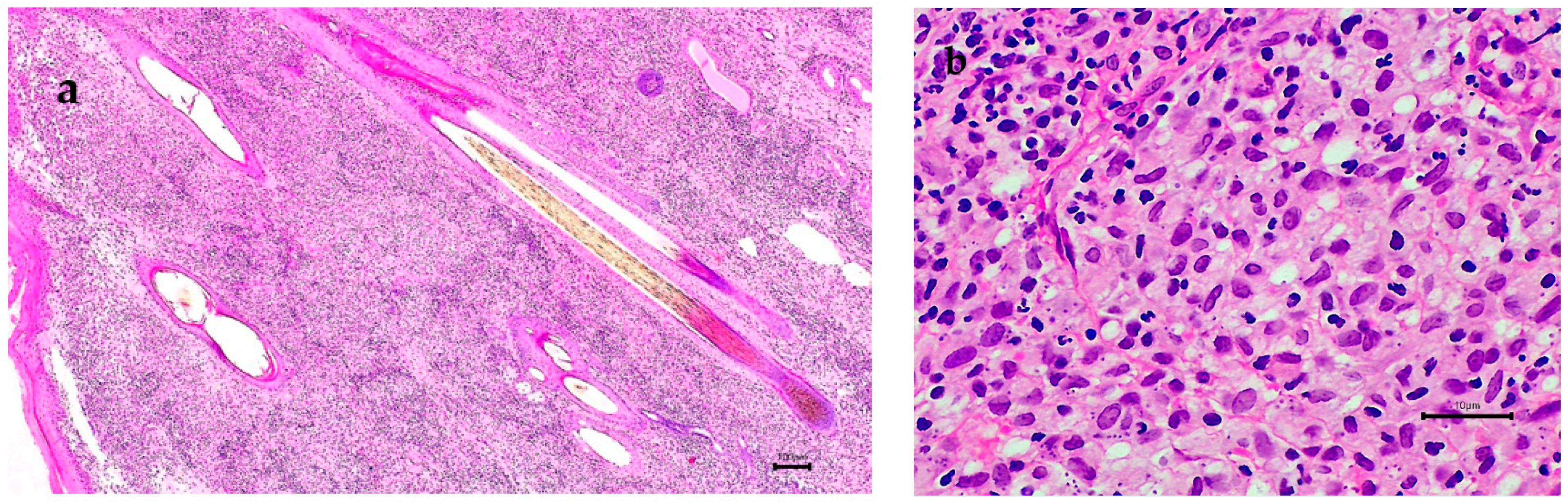
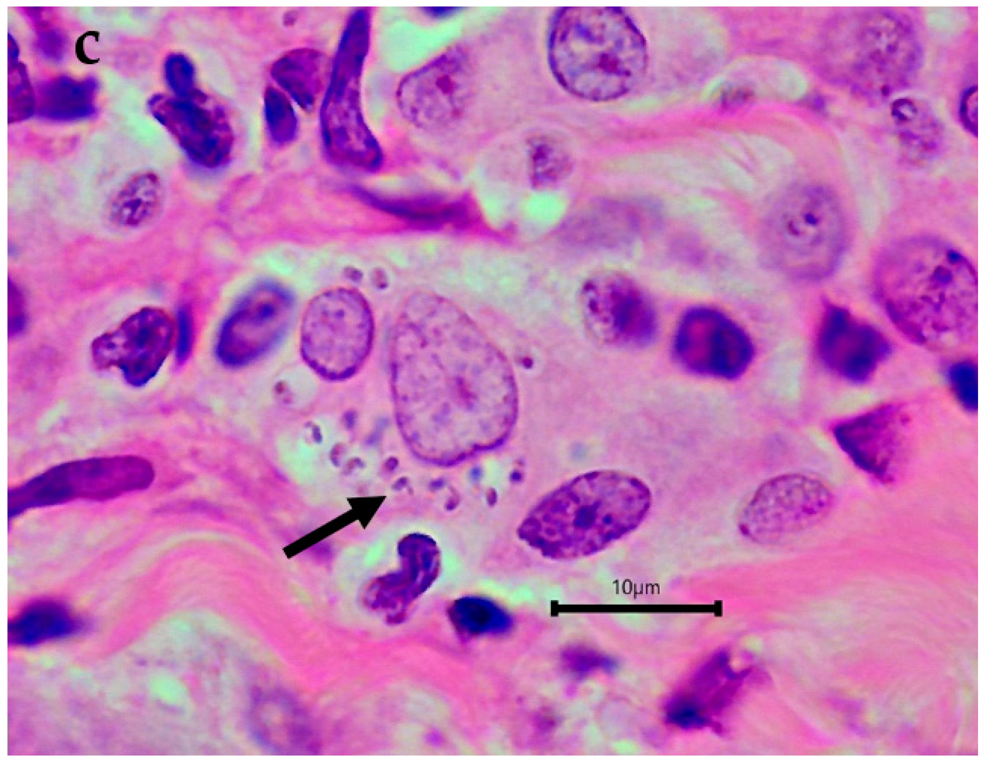
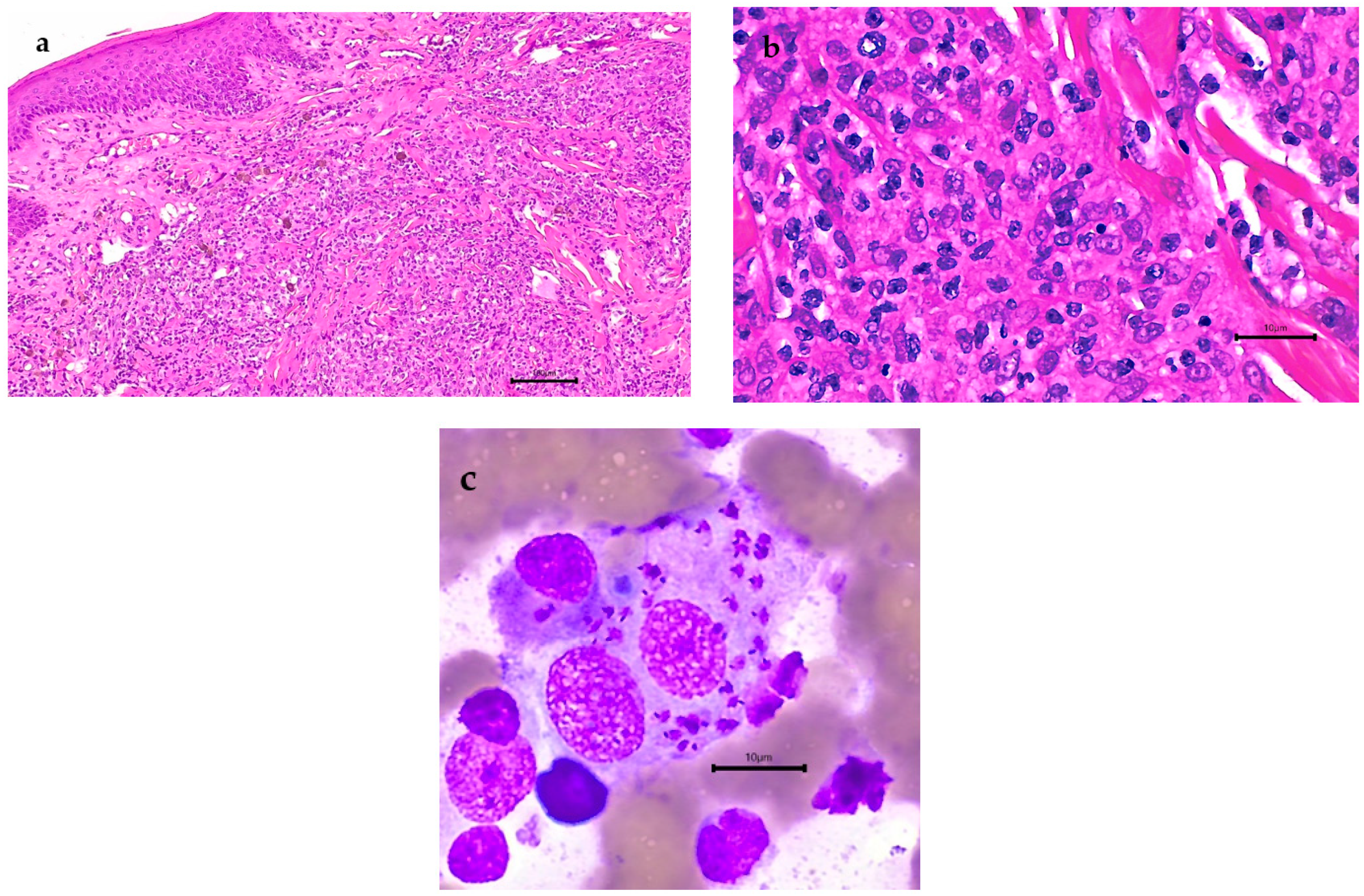
Eight privately owned, adult Belgian Malinois, 4 males and 4 females, aged between 6 and 10 years, resided on a single, Hong Kong Island property, working as guard dogs (Table 1). Four dogs were born in Hong Kong (dogs 2, 3, 5, 6), two in the Netherlands (dogs 4, 8), one in the United States of America (USA) (dog 1), and one in Sweden (dog 7). Two dogs (dog 2 and 3) were littermates, both born in Hong Kong to a dam imported from the USA and dog 5 was the progeny of dog 1 (Figure 1). All kennel dogs were under the veterinary care of author 2 (AM) and all testing and medical record retrievals were performed as part of disease investigation with permission from the owner.
Over a 5-year period, four kennel dogs (dogs 1, 2, 3, 4) had presented to the attending veterinarian with dermatitis (dogs 1, 3, 4) or dermatitis with systemic illness (dog 2). Skin biopsies from dog 1 were collected for histopathology in 2015 along with an EDTA blood sample which was submitted for quantitative PCR to IDEXX Reference Laboratories, UK. In 2019, aspirates of spleen and liver were collected from the systemically ill dog (dog 2), which were stained with Wright’s Giemsa (WG). Following cytological identification of amastigotes in dog 2, additional aspirates of the spleen were placed into a sterile tube for PCR. Skin biopsies were collected in 2019, from 2 other kennel dogs with dermatitis (dogs 3, 4), with each sample split, with half of the sample placed into sterile saline for PCR, and the other half processed for histopathology. All biopsy samples for histopathology were fixed in formalin and processed into paraffin blocks and stained with haematoxylin and eosin (H and E), Grocott’s methenamine silver (GMS), periodic acid Schiff (PAS), Ziehl Neelsen (ZN), Gram and Giemsa for histology. Whole blood was collected from the cephalic vein of all eight kennel dogs between 2019 and 2020 for PCR and serology. PCR was also conducted on the paraffin block containing skin collected in 2015, with all tests performed by the Koret School of Veterinary Medicine, Hebrew University, Israel.
In summary, PCR for Leishmania detection was performed on EDTA blood from all eight dogs, punch biopsies of fresh skin placed into sterile saline from the two dogs with active skin disease (dogs 3, 4), paraffin-embedded skin from dog number 1 collected in 2015 when ulcerative skin disease first appeared, and aspirated splenic tissue collected in a plain tube from the systemically ill dog (dog 2). DNA was extracted from all blood and tissue samples using the Qiagen EZ1 Advanced XL automated system (QIAGEN GmbH, QIAGEN Strasse 1, 40724 Hilden, Germany). DNA was stored at −7 °C before being sent to the Hebrew University in Israel. DNA from the paraffin-embedded tissue was extracted from 3 different pieces of the same block using the Quick-DNATM FFPE kit (Zymo research, Irvine, CA, USA) according to the manufacturer’s instructions.
The presence of Leishmania DNA in samples was tested using two PCR protocols for amplification of different targets. A 265 bp fragment of the Leishmania internal transcribed spacer 1 (ITS1) region of the L. infantum rRNA operon was amplified by real-time PCR using primers ITS-219 F (AGCTGGATCATTTTCCGATG) and ITS-219R (ATCGCGACACGTTATGTGAG) and then evaluated by high-resolution melt (HRM) analysis as previously described [3]. In addition, 120 bp of the Leishmania kinetoplast DNA (kDNA) minicircle were amplified by real-time PCR using primers JW11 (CCTATTTTACACCAACCCCCAGT) and JW12 (GGGTAGGGGCGTTCTGCGAAA) and then evaluated by melt curve analysis as previously described [3,4]. All positive PCR products were sequenced using the BigDye Terminator v3.1 Cycle Sequencing Kit and an ABI PRISM 3100 Genetic Analyzer (Applied Biosystems, Foster City, CA, USA), at the Center for Genomic Technologies, Hebrew University of Jerusalem, Israel. DNA sequences were evaluated with the ChromasPro software version 2. 1.1 (Technelysium Pty Ltd., Brisbane, QLD, Australia) and compared for similarity with sequences available in GenBank®, using the BLAST program (http://www.ncbi.nlm.nih.gov/BLAST/, accessed on 10 September 2022).
Leishmania serology was performed on blood samples collected from the cephalic vein of all 8 dogs collected between 2019 and 2020. Serum was stored at −70 °C before being sent to the Koret School of Veterinary Medicine, Hebrew University, Israel. Serology for anti-leishmanial antibodies was performed by enzyme-linked immunosorbent assay (ELISA) using L. infantum antigen, as described previously [5]. Dog 2 had a previous round of serology with the first blood sample collected at the time of systemic disease emergence, which was sent to the Veterinary Medical Diagnostic Laboratory at Texas A&M University, College Station, TX, USA.

3. Results

Dog no. 1, born in the USA, presented in 2015 with ulcerative, granulomatous dermatitis affecting the nail bed of a single digit on the right forelimb which was biopsied to reveal abundant Leishmania amastigotes on histopathology (Figure 2) but quantitative PCR on EDTA blood at this time was negative for Leishmania (IDEXX Reference Laboratories, Wetherby, UK), (Table 1). Treatment over a 2-year period (2015–2017) with allopurinol (generic, China) resolved the skin disease but, in 2018, 12 months after the drug was discontinued, multiple digits on the hindlimbs developed ulcerative dermatitis. Biopsy was declined by the owner at this time, allopurinol treatment was recommenced, and is currently ongoing, and no skin disease is currently present. PCR and DNA sequencing performed at the Hebrew University in Israel on DNA extracted from paraffin-embedded tissue from the nail bed collected in 2015 and stored for five years confirmed leishmaniasis was caused by L. infantum. ELISA serology and PCR performed on blood collected in 2020, five years after the initial diagnosis when the dog was clinically well with no cutaneous lesions, were negative for leishmaniasis (Table 1).
Dog 2 was born in, and had never travelled outside of, Hong Kong (Table 1). Information about the sire and dam was unavailable, but anecdotally, the dam, born in the USA, had died at an unknown time point prior to the dog’s cutaneous disease presentation in 2018, from disease with similar manifestations to leishmaniasis. In 2018, ulcerative skin disease affecting multiple limbs as well as the nasal vestibule, causing nasal stertor, progressed to culminate in overt systemic infection in 2019 with splenomegaly, hepatomegaly, and lymphadenomegaly of the popliteal, prescapular, and sub-mandibular lymph nodes. Disease progression occurred despite various treatments involving the antibiotics cephalexin (Stada Pharmaceuticals (Asia) Limited, Kwun Tong, Hong Kong), and enrofloxacin (Dechra Veterinary Products (Australia) Pty Ltd., Somersby, NSW, Australia), the steroid prednisolone (Mavlab Pty Ltd., Slacks Creek, QLD, Australia), and the anti-fungal agent itraconazole (Europharm Lab Co., Ltd., Tai Po, Hong Kong). Incisional biopsies of the skin and nasal mucosa, collected in 2018, stained with H and E revealed pyogranulomatous inflammation within the dermis and the nasal submucosa, but no amastigotes were seen (Figure 3). GMS, PAS, ZN, Gram and Giemsa staining did not provide additional information, so clinically, leishmaniasis was not suspected. Systemic infection in 2019 manifested as lethargy and dullness with mild regenerative, normocytic, hypochromic anaemia (PCV: 31% reference range 37–54%; reticulocytes 128.1 × 10⁹/L reference range 11–92 × 109/L; MCHC 321 g/L reference range 330–360 g/L), with 18 nucleated RBCs/100 white blood cells and a left shift of neutrophils (9 neutrophilic bands/100 WBCs) with a mild increase in plasma proteins (81 g/L reference range 59–78 g/L). Serum biochemistry changes included mild hyperglobulinaemia (43 g/L reference range 19–36 g/L) and hypoalbuminaemia (30 g/L reference range 32–44 g/L). There was mildly increased ALP (104 U/L reference range 17–100 U/L), AST (95.1 U/L reference range 15–57), and CK (769 U/L reference range 48–261 U/L). Spleen and liver cytology by fine needle aspirate showed granulomatous to pyogranulomatous splenitis and hepatitis with abundant Leishmania amastigotes within macrophages in both organs (Figure 3). Urinalysis was unremarkable.
The presence of amastigotes seen on cytological assessment of the spleen and liver prompted confirmation of leishmaniasis infection. An indirect fluorescent antibody test (IFAT) performed at the Texas A&M veterinary medical diagnostic laboratory on blood detected an antibody titre of 2048 against L. infantum. ELISA serology and PCR on blood taken two months later and performed at the Hebrew University, as well as aspirated splenic tissue, were positive for L. infantum. The ELISA result had an optical density (OD) of 1.436 (cut off 0.4 OD) and PCR, which detected the ITS-1 spacer using the ITS219 F/R primers, produced sequences from the blood and spleen which were 100% similar to one another and had 100% identity to L. infantum (MN503527.1) (Table 1). Dog 2 was successfully treated with allopurinol which has led to clinical resolution of all clinical signs.
The remaining six ‘in contact’ dogs (dogs 3, 4, 5, 6, 7, 8) included two dogs with chronic skin disease at the time of sampling (dogs 3 and 4) whilst the remaining four dogs (dogs 5, 6, 7, 8) were, and remain, clinically healthy. Dog 3 was born in Hong Kong and was a full sister of the systemically ill, Leishmania-positive dog (dog 2) (Table 1). This dog had a chronic history of alopecia with hyperpigmentation on the dorsum of the thorax and rump regions, which clinically resembled flea allergy dermatitis. Skin punch biopsies collected from alopecic regions were PCR negative for Leishmania and lacked amastigotes on histology on H and E, GMS, PAS, ZN, Giemsa, and Gram staining, and the histological diagnosis reported changes consistent with chronic hyperplastic dermatitis. Dog 4 was born in the Netherlands, and presented with chronic, pyogranulomatous, ulcerative dermatitis on the left stifle, but histology failed to demonstrate amastigotes or any infectious agents on H and E, GMS, PAS, ZN, Giemsa, and Gram staining, and PCR from this skin biopsy was negative for Leishmania. All six dogs were seronegative for Leishmania by ELISA and their PCR from blood and skin samples (dogs 3 and 4) was negative.

4. Discussion

This report is the first to describe leishmaniasis due to L. infantum, in either animals or humans, in Hong Kong. The presence of leishmaniasis in the canine population is of potential public health concern as dogs are the main reservoir for human infection and canine leishmaniasis has been shown to precede human disease [6]. Canine L. infantum infection is often sub-clinical [7,8,9], however, clinically and sub-clinically infected dogs are infectious to sand fly vectors that can transmit infection to humans and other dogs [10,11]. Although both dogs with leishmaniasis in this report were clinically stable due to ongoing allopurinol treatment at the time of this report, it is likely that these two dogs remained infected as allopurinol, as well as all other drugs used for the treatment of canine leishmaniasis, are not known to cure dogs parasitologically and completely eliminate infection [9].
Leishmania infantum is transmitted by female sand fly bites [12] and only specific sand fly species serve as vectors. Although no suitable sand fly vector has been identified in Hong Kong, the natural distribution range of Phlebotomus chinensis Newstead, 1843, a proven vector of visceral leishmaniasis [2,13], is close to Hong Kong in the Guangdong and Hainan districts of mainland China (pers comm from senior agricultural officer from Agriculture, Fisheries and Conservation Department of Hong Kong). As geographic spread of this vector into Hong Kong is possible, the medical community needs to be vigilant about the possible emergence of leishmaniasis.
The diagnosis of leishmaniasis in dogs 1 and 2 was unexpected. Dog 1 had been imported from the USA where leishmaniasis is not endemic but is mainly acquired during travel or military service to areas where the disease is endemic, or is transmitted predominantly via vertical autochthonous pathways [7,14,15,16,17,18]. Chronic dermatitis of the nail bed is not pathognomonic for leishmaniasis, so unless histopathology was performed, leishmaniasis was unlikely to be considered high on the differential list for this dog.
Diagnosis of leishmaniasis in dog 2 was considered unlikely due to a number of reasons. The first is that this dog had never travelled outside of Hong Kong and leishmaniasis is not an endemic disease in Hong Kong. The second is the lack of amastigotes seen on histology of the skin and nasal mucosal lesions, which is consistent with previous reports [19,20], with one study reporting that amastigotes were only seen by histology in 20% of dogs with cutaneous disease [21]. The third reason, and the focus of this paper, is that Hong Kong lacks known vectors of this disease, so despite the fact that there was a chronically infected dog in this kennel (dog 1), autochthonous transmission was not a clinical consideration. It was not until several rounds of various treatment regimens had failed and the disease became systemic did cytology reveal abundant amastigotes in the liver and spleen. The high numbers of visceral amastigotes produced positive PCR results conducted on blood and splenic tissue and serology was also positive, confirming the diagnosis, allowing appropriate treatment to begin.
Non-vector borne transmission of leishmaniasis in dogs has been associated with in utero infection, exposure to parasites within blood products, venereal transmission, and potentially by direct contact including dog fights [17,22,23,24,25,26,27,28,29,30,31,32] with similar incubation periods of 3 months to 7 years [26,27]. The two most likely routes of infection for dogs 1 and 2 were in utero infection and direct contact. No information was available on the sire or dam of dog 1. The dam of dog 2 and 3 died with clinical disease suspicious for leishmaniasis, but unfortunately no investigation into the cause of death was made and medical records were not available, so the date of death in relation to the diagnosis of leishmaniasis in dog 2 was unable to be determined. In utero infection from asymptomatic or symptomatic parents at the time of mating or whelping results in some or all puppies in a litter becoming infected [24,26,28]. It has been shown that maternal Leishmania disease status significantly predicts the likelihood of offspring infection in dogs [22] so it would have been interesting to know whether the dam was infected and clinically ill with L. infantum at the time that dog 2 and 3 were born. Dog 3, a littermate of dog 2, had no evidence of vertical transmission from the dam, being clinically healthy apart from chronic dermatitis attributable to flea allergy, and was serologically negative and PCR negative on blood and skin biopsy for leishmaniasis.
Horizontal transmission may have occurred between dogs 1 and 2, as multiple episodes of dog fights occurred after leishmaniasis was diagnosed in dog 1. Resultant wounds bled freely and many lacerations required veterinary treatment. However, this mode of transmission has not been proven in dogs [29,30].
Leishmania infantum infection in dogs has two main patterns: sub-clinical or clinical disease, with about 5–10% of dogs naturally infected progressing to overt clinical disease [7]. The cutaneous lesion of ulcerative dermatitis affecting the nail beds of both dogs has been previously described in canine leishmaniasis [33,34]. Interestingly, although the gross lesions were similar between dogs 1 and 2, the numbers of amastigotes detected by microscopy varied, with large numbers seen on histology in dog 1, confirmed by PCR, with no obvious organisms seen in cutaneous lesions in dog 2, consistent with previous reports where the numbers of amastigotes can vary [21,33]. Large numbers of amastigotes are typically associated with exfoliative dermatitis in dogs, which was not present in either kennel dog, despite this pattern being commonly reported in canine leishmaniasis [20,33]. No amastigotes were seen in the nasal lesions from dog 2, despite amastigotes being previously reported in the nasal epithelium of infected dogs [31,35]. Dog 2 developed systemic disease with granulomatous splenitis and hepatitis as well as hyperglobulinaemia, similar to previous reports in dogs, with abundant amastigotes seen on microscopy [9,17,25,32,34,36]. Hyperglobulinaemia is associated with anti-Leishmania IgG antibodies, detectable via serology which are non-immunoprotective [37], and are responsible for the formation of immune-complex glomerulonephritis, which is a common cause of mortality in infected dogs [9,38].
This study highlights difficulties in clinically diagnosing canine leishmaniasis, especially in non-endemic geographical regions. The two kennel dogs had conflicting results on various tests during their clinical workup. Leishmaniasis in dog 1 was diagnosed on skin biopsy, but PCR remained negative on whole blood samples, collected 5 years apart, despite the abundant amastigotes seen when the first blood sample was collected. Negative PCR results from infected dogs is not surprising as, in a previous study, 25% of symptomatic dogs were negative using quantitative real-time PCR on peripheral blood [39]. Serology performed on blood 5 years after the initial leishmaniasis diagnosis was also negative, despite the dog probably remaining infected, as this dog relapsed 12 months after completing a 2-year treatment regimen with allopurinol [40,41]. Low parasite burdens due to treatment contribute to negative serological screening tests and, overall, serology fails to detect a large portion of sub-clinically infected dogs [8,42]. Skin and nasal mucosal biopsies from dog 2 failed to reveal amastigotes, but over time, amastigotes were identified on liver and splenic aspirates, confirmed by PCR on splenic aspirates and blood. Veterinarians should consider canine leishmaniasis as a potential diagnosis for skin disease in non-endemic regions and not just as a travel-acquired or imported condition. Veterinarians should also understand the limitations in the detection techniques of leishmaniasis in tissues when amastigotes are sparse in number, or on blood and serum of affected dogs when levels of circulating organisms are low.

5. Conclusions

This report is the first to document autochthonous canine leishmaniasis in Hong Kong. Veterinary practitioners and public health officials need to consider leishmaniasis in local dogs from geographical areas that currently lack suitable vectors. Canine Leishmania infection is mostly sub-clinical, however, sub-clinically infected dogs could be infectious to sand fly vectors whose geographical range may expand, and canine infection may precede human disease [7,8,9,10,11]. Therefore, monitoring to detect phlebotomine vectors in Hong Kong is warranted and medical practitioners in non-endemic regions need to be vigilant for potential disease in humans.

Author Contributions

Conceptualisation, J.S.; methodology, J.S. and G.B.; investigation, J.S. and A.M.; resources, J.S. and A.M.; data curation, J.S. and A.M.; writing—original draft preparation, J.S.; writing—review and editing, J.S., G.B., A.M. and Y.N.-B. All authors have read and agreed to the published version of the manuscript.

Funding

This research received no external funding. The study represented data collected from veterinary disease investigation in a kennel of dogs. Serology and PCR were funded by the corresponding author’s internal resources (G.B.).

Institutional Review Board Statement

Ethics approval and consent to participate not applicable. Dogs in this study were naturally infected, tested for leishmaniasis and evaluated clinically as part of their clinical veterinary management.

Informed Consent Statement

The kennel owner gave permission for access to medical records for the purpose of publication of this paper.

Data Availability Statement

All relevant data have been included in this submission. Additional data exist, incorporated as part of confidential medical records of the attending veterinary hospital.

Acknowledgments

We thank Abbey Veterinary Services, Devon, TQ12 2BG, UK for providing a copy of the H and E-stained histology slide for dog 1 as well as the matching paraffin block to enable PCR analysis.

Conflicts of Interest

The authors declare no conflict of interest.

References

  1. José, A.R.-P.; Saurabh, J.; Alexei, M.; Ana, N.M.-E.; Samantha, V.; Supriya, W.; Mona, O.; Zaw, L.; Abate, B.; Aya, Y.; et al. Global Leishmaniasis Surveillance: 2019–2020, a Baseline for the 2030 Roadmap. Available online: https://www.who.int/publications/i/item/who-wer9635-401-419 (accessed on 2 September 2022).
  2. Lun, Z.R.; Wu, M.S.; Chen, Y.F.; Wang, J.Y.; Zhou, X.N.; Liao, L.F.; Chen, J.P.; Chow, L.M.; Chang, K.P. Visceral Leishmaniasis in China: An Endemic Disease under Control. Clin. Microbiol. Rev. 2015, 28, 987–1004. [Google Scholar] [CrossRef]
  3. Nicolas, L.; Milon, G.; Prina, E. Rapid differentiation of Old World Leishmania species by LightCycler polymerase chain reaction and melting curve analysis. J. Microbiol. Methods 2002, 51, 295–299. [Google Scholar] [CrossRef]
  4. Talmi-Frank, D.; Nasereddin, A.; Schnur, L.F.; Schönian, G.; Töz, S.O.; Jaffe, C.L.; Baneth, G. Detection and identification of old world Leishmania by high resolution melt analysis. PLoS Negl. Trop. Dis. 2010, 4, e581. [Google Scholar] [CrossRef] [PubMed]
  5. Baneth, G.; Nachum-Biala, Y.; Zuberi, A.; Zipori-Barki, N.; Orshan, L.; Kleinerman, G.; Shmueli-Goldin, A.; Bellaiche, M.; Leszkowicz-Mazuz, M.; Salant, H.; et al. Leishmania infection in cats and dogs housed together in an animal shelter reveals a higher parasite load in infected dogs despite a greater seroprevalence among cats. Parasit. Vectors 2020, 13, 115. [Google Scholar] [CrossRef]
  6. Michalsky, E.M.; Rocha, M.F.; da Rocha Lima, A.C.; França-Silva, J.C.; Pires, M.Q.; Oliveira, F.S.; Pacheco, R.S.; dos Santos, S.L.; Barata, R.A.; Romanha, A.J.; et al. Infectivity of seropositive dogs, showing different clinical forms of leishmaniasis, to Lutzomyia longipalpis phlebotomine sand flies. Vet. Parasitol. 2007, 147, 67–76. [Google Scholar] [CrossRef] [PubMed]
  7. Toepp, A.J.; Petersen, C.A. The balancing act: Immunology of leishmaniosis. Res. Vet. Sci. 2020, 130, 19–25. [Google Scholar] [CrossRef]
  8. Marcondes, M.; Day, M.J. Current status and management of canine leishmaniasis in Latin America. Res. Vet. Sci. 2019, 123, 261–272. [Google Scholar] [CrossRef]
  9. Baneth, G.; Koutinas, A.F.; Solano-Gallego, L.; Bourdeau, P.; Ferrer, L. Canine leishmaniosis—new concepts and insights on an expanding zoonosis: Part one. Trends Parasitol. 2008, 24, 324–330. [Google Scholar] [CrossRef]
  10. Laurenti, M.D.; Rossi, C.N.; da Matta, V.L.; Tomokane, T.Y.; Corbett, C.E.; Secundino, N.F.; Pimenta, P.F.; Marcondes, M. Asymptomatic dogs are highly competent to transmit Leishmania (Leishmania) infantum chagasi to the natural vector. Vet. Parasitol. 2013, 196, 296–300. [Google Scholar] [CrossRef]
  11. Magalhães-Junior, J.T.; Mota, T.F.; Porfirio-Passos, G.; Larangeira, D.F.; Franke, C.R.; Barrouin-Melo, S.M. Xenodiagnosis on dogs with visceral leishmaniasis: Canine and sand fly aspects related to the parasite transmission. Vet. Parasitol. 2016, 223, 120–126. [Google Scholar] [CrossRef]
  12. Serafim, T.D.; Iniguez, E.; Oliveira, F. Leishmania infantum. Trends Parasitol. 2020, 36, 80–81. [Google Scholar] [CrossRef] [PubMed]
  13. Young, C.W.; Hertig, M. The development of flagellates in Chinese sandflies (phlebotomus) fed on hamsters infected with Leishmania donovani. Proc. Soc. Exp. Biol. Med. 1926, 23, 611–615. [Google Scholar] [CrossRef]
  14. Duprey, Z.H.; Steurer, F.J.; Rooney, J.A.; Kirchhoff, L.V.; Jackson, J.E.; Rowton, E.D.; Schantz, P.M. Canine visceral leishmaniasis, United States and Canada, 2000–2003. Emerg. Infect. Dis. 2006, 12, 440–446. [Google Scholar] [CrossRef] [PubMed]
  15. Petersen, C.A. Leishmaniasis, an emerging disease found in companion animals in the United States. Top. Companion Anim. Med. 2009, 24, 182–188. [Google Scholar] [CrossRef]
  16. Boggiatto, P.M.; Gibson-Corley, K.N.; Metz, K.; Gallup, J.M.; Hostetter, J.M.; Mullin, K.; Petersen, C.A. Transplacental transmission of Leishmania infantum as a means for continued disease incidence in North America. PLoS Negl. Trop. Dis. 2011, 5, e1019. [Google Scholar] [CrossRef]
  17. de Almeida, M.E.; Spann, D.R.; Bradbury, R.S. Leishmania infantum in US-Born Dog. Emerg. Infect. Dis. 2020, 26, 1882–1884. [Google Scholar] [CrossRef]
  18. Alonso, F.H.; Vasilatis, D.M.; Veluvolu, S.M.; Willcox, J.L.; Scorza, B.M.; Petersen, C.A.; Kol, A. Canine leishmaniasis in Northern California-A case report. Vet. Clin. Pathol. 2021, 50, 71–75. [Google Scholar] [CrossRef]
  19. Fondevila, D.; Vilafranca, M.; Ferrer, L. Epidermal immunocompetence in canine leishmaniasis. Vet. Immunol. Immunopathol. 1997, 56, 319–327. [Google Scholar] [CrossRef]
  20. Ferrer, L.; Rabanal, R.; Fondevila, D.; Ramos, J.; Domingo, M. Skin lesions in canine leishmaniasis. J. Small Anim. Pract. 1988, 29, 381–388. [Google Scholar] [CrossRef]
  21. Ordeix, L.; Dalmau, A.; Osso, M.; Llull, J.; Montserrat-Sangrà, S.; Solano-Gallego, L. Histological and parasitological distinctive findings in clinically-lesioned and normal-looking skin of dogs with different clinical stages of leishmaniasis. Parasit. Vectors 2017, 10, 121. [Google Scholar] [CrossRef] [Green Version]
  22. Toepp, A.J.; Bennett, C.; Scott, B.; Senesac, R.; Oleson, J.J.; Petersen, C.A. Maternal Leishmania infantum infection status has significant impact on leishmaniasis in offspring. PLoS Negl. Trop. Dis. 2019, 13, e0007058. [Google Scholar] [CrossRef] [PubMed]
  23. Mancianti, F.; Sozzi, S. Isolation of Leishmania from a newborn puppy. Trans. R. Soc. Trop. Med. Hyg. 1995, 89, 402. [Google Scholar] [CrossRef]
  24. Díaz-Espiñeira, M.M.; Slappendel, R.J. A case of autochthonous canine leishmaniasis in The Netherlands. Vet. Q. 1997, 19, 69–71. [Google Scholar] [CrossRef] [PubMed]
  25. Gaskin, A.A.; Schantz, P.; Jackson, J.; Birkenheuer, A.; Tomlinson, L.; Gramiccia, M.; Levy, M.; Steurer, F.; Kollmar, E.; Hegarty, B.C.; et al. Visceral leishmaniasis in a New York foxhound kennel. J. Vet. Intern. Med. 2002, 16, 34–44. [Google Scholar] [CrossRef] [PubMed]
  26. Salant, H.; Nachum-Biala, Y.; Feinmesser, B.; Perelmutter, M.; Baneth, G. Early onset of clinical leishmaniosis in a litter of pups with evidence of in utero transmission. Parasit. Vectors. 2021, 14, 326. [Google Scholar] [CrossRef]
  27. Owens, S.D.; Oakley, D.A.; Marryott, K.; Hatchett, W.; Walton, R.; Nolan, T.J.; Newton, A.; Steurer, F.; Schantz, P.; Giger, U. Transmission of visceral leishmaniasis through blood transfusions from infected English foxhounds to anemic dogs. J. Am. Vet. Med. Assoc. 2001, 219, 1076–1083. [Google Scholar] [CrossRef]
  28. Svobodova, V.; Svoboda, M.; Friedlaenderova, L.; Drahotsky, P.; Bohacova, E.; Baneth, G. Canine leishmaniosis in three consecutive generations of dogs in Czech Republic. Vet. Parasitol. 2017, 237, 122–124. [Google Scholar] [CrossRef]
  29. Karkamo, V.; Kaistinen, A.; Näreaho, A.; Dillard, K.; Vainio-Siukola, K.; Vidgrén, G.; Tuoresmäki, N.; Anttila, M. The first report of autochthonous non-vector-borne transmission of canine leishmaniosis in the Nordic countries. Acta Vet. Scand. 2014, 56, 84. [Google Scholar] [CrossRef]
  30. Naucke, T.J.; Amelung, S.; Lorentz, S. First report of transmission of canine leishmaniosis through bite wounds from a naturally infected dog in Germany. Parasit. Vectors 2016, 9, 256. [Google Scholar] [CrossRef]
  31. Petanides, T.A.; Koutinas, A.F.; Mylonakis, M.E.; Day, M.J.; Saridomichelakis, M.N.; Leontides, L.S.; Mischke, R.; Diniz, P.; Breitschwerdt, E.B.; Kritsepi, M.; et al. Factors associated with the occurrence of epistaxis in natural canine leishmaniasis (Leishmania infantum). J. Vet. Intern. Med. 2008, 22, 866–872. [Google Scholar] [CrossRef]
  32. Foglia Manzillo, V.; Di Muccio, T.; Cappiello, S.; Scalone, A.; Paparcone, R.; Fiorentino, E.; Gizzarelli, M.; Gramiccia, M.; Gradoni, L.; Oliva, G. Prospective study on the incidence and progression of clinical signs in naïve dogs naturally infected by Leishmania infantum. PLoS Negl. Trop. Dis. 2013, 7, e2225. [Google Scholar] [CrossRef] [PubMed]
  33. Saridomichelakis, M.N.; Koutinas, A.F. Cutaneous involvement in canine leishmaniasis due to Leishmania infantum (syn. L. chagasi). Vet. Dermatol. 2014, 25, 61-e22–71-e22. [Google Scholar] [CrossRef] [PubMed]
  34. Squarre, D.; Chambaro, H.M.; Hayashida, K.; Moonga, L.C.; Qiu, Y.; Goto, Y.; Oparaocha, E.; Mumba, C.; Muleya, W.; Bwalya, P.; et al. Autochthonous Leishmania infantum in Dogs, Zambia, 2021. Emerg. Infect. Dis. 2022, 28, 888–890. [Google Scholar] [CrossRef]
  35. Levy, E.; Mylonakis, M.E.; Saridomichelakis, M.N.; Polizopoulou, Z.S.; Psychogios, V.; Koutinas, A.F. Nasal and oral masses in a dog. Vet. Clin. Pathol. 2006, 35, 115–118. [Google Scholar] [CrossRef] [PubMed]
  36. Paltrinieri, S.; Gradoni, L.; Roura, X.; Zatelli, A.; Zini, E. Laboratory tests for diagnosing and monitoring canine leishmaniasis. Vet. Clin. Pathol. 2016, 45, 552–578. [Google Scholar] [CrossRef] [PubMed]
  37. Courtenay, O.; Quinnell, R.J.; Garcez, L.M.; Shaw, J.J.; Dye, C. Infectiousness in a cohort of brazilian dogs: Why culling fails to control visceral leishmaniasis in areas of high transmission. J. Infect. Dis. 2002, 186, 1314–1320. [Google Scholar] [CrossRef]
  38. Costa, F.A.; Goto, H.; Saldanha, L.C.; Silva, S.M.; Sinhorini, I.L.; Silva, T.C.; Guerra, J.L. Histopathologic patterns of nephropathy in naturally acquired canine visceral leishmaniasis. Vet. Pathol. 2003, 40, 677–684. [Google Scholar] [CrossRef] [PubMed]
  39. Solcà Mda, S.; Bastos, L.A.; Guedes, C.E.; Bordoni, M.; Borja, L.S.; Larangeira, D.F.; da Silva Estrela Tuy, P.G.; Amorim, L.D.; Nascimento, E.G.; de Sá Oliveira, G.G.; et al. Evaluating the accuracy of molecular diagnostic testing for canine visceral leishmaniasis using latent class analysis. PLoS ONE 2014, 9, e103635. [Google Scholar] [CrossRef]
  40. Gonçalves, A.A.M.; Leite, J.C.; Resende, L.A.; Mariano, R.; Silveira, P.; Melo-Júnior, O.A.O.; Ribeiro, H.S.; de Oliveira, D.S.; Soares, D.F.; Santos, T.A.P.; et al. An Overview of Immunotherapeutic Approaches Against Canine Visceral Leishmaniasis: What Has Been Tested on Dogs and a New Perspective on Improving Treatment Efficacy. Front. Cell. Infect. Microbiol. 2019, 9, 427. [Google Scholar] [CrossRef]
  41. Ribeiro, R.R.; Michalick, M.S.M.; da Silva, M.E.; Dos Santos, C.C.P.; Frézard, F.J.G.; da Silva, S.M. Canine Leishmaniasis: An Overview of the Current Status and Strategies for Control. Biomed. Res. Int. 2018, 2018, 3296893. [Google Scholar] [CrossRef] [Green Version]
  42. Miró, G.; Cardoso, L.; Pennisi, M.G.; Oliva, G.; Baneth, G. Canine leishmaniosis--new concepts and insights on an expanding zoonosis: Part two. Trends Parasitol. 2008, 24, 371–377. [Google Scholar] [CrossRef] [PubMed]
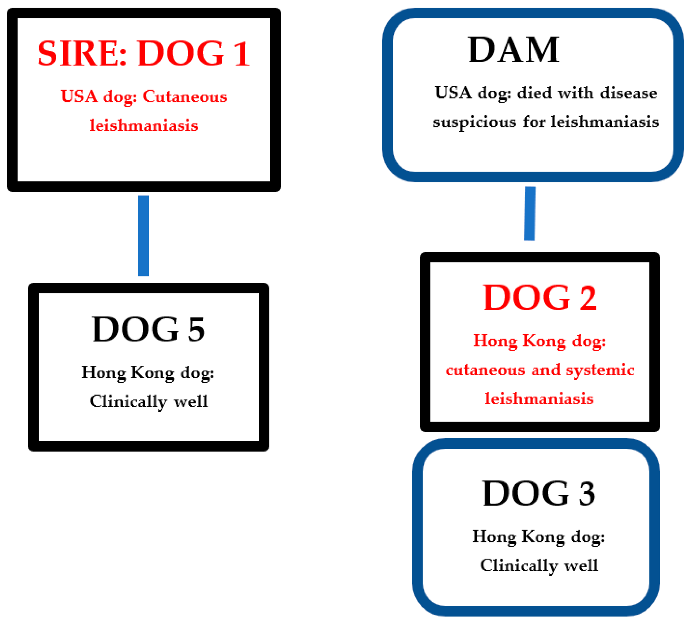
Figure 1. Leishmaniasis disease status and relationships of dogs within the Hong Kong kennel. USA: United States of America. Black rectangles: Male dogs. Blue rounded rectangles: Female dogs.
Microorganisms 10 01873 g001
Figure 2. Nail bed skin histopathology from dog 1 collected in 2015: (a) Granulomatous dermatitis. Haematoxylin and eosin staining; bar = 100 um. (b) Granulomatous dermatitis with macrophages admixed with neutrophils and fewer plasma cells and lymphocytes. Haematoxylin and eosin staining; bar = 10 um. (c) Multiple Leishmania amastigotes within the cytoplasm of a macrophage. Arrow points to a kinetoplast at right angles to the amastigote nucleus. Haematoxylin and eosin staining; bar = 10 um.
Microorganisms 10 01873 g002aMicroorganisms 10 01873 g002b
Figure 3. Nasal mucosa histology and splenic aspirates from dog 2: (a) Histopathology of nasal mucosa with granulomatous inflammation. Haematoxylin and eosin staining; bar = 100 um. (b) Nasal sub-mucosa with macrophages admixed with fewer neutrophils, plasma cells, and small lymphocytes. Haematoxylin and eosin staining; bar = 10 um. (c) Splenic aspirate. Wright’s Giemsa stain. Leishmania amastigotes within the cytoplasm of macrophages. Bar = 10 um.
Microorganisms 10 01873 g003
Table 1. Signalment, origin, main test results, and Leishmania treatment outcome of eight dogs living in the same kennel investigated for leishmaniasis.
Dog Dog Country of OriginRelationship with Other Dogs in KennelYear of BirthYear of TestDetection and Anatomic Location of Leishmania Amastigotes by MicroscopyELISA SerologyPCR Result and TissueLeishmania Treatment/Outcome
1USASire of dog 520112015Yes: Skin ND− (blood) *Skin disease resolved but relapsed
2020ND-− (blood)
+ (skin) **
Skin disease resolved
2Hong KongFull brother of dog 320122018Suspected: Skin and nasal mucosaNDNDSkin disease resolved but relapsed and became systemic
2019Yes: Spleen and liver+ ***+ (blood, spleen)Disease resolved
3Hong KongFull sister of dog 220122019No-− (blood and skin)NA
4NetherlandsNone20092019No-− (blood and skin)NA
5Hong KongSon of dog 120132020ND-− (blood)NA
6Hong KongNone20112020ND-− (blood)NA
7SwedenNone20112020ND-− (blood)NA
8NetherlandsNone20122020ND-− (blood)NA
− Negative result; + positive result; ND: Not done; NA: Not applicable. * Dog 1 PCR testing on EDTA blood. Quantitative Leishmania PCR was performed by the IDEXX Reference Laboratory, United Kingdom. All other PCRs performed by Koret School of Veterinary Medicine, the Hebrew University, Israel. ** Dog 1 PCR conducted on stored skin sample collected in 2015. *** Dog 2 had two rounds of ELISA serology; one performed by Texas A&M veterinary medical diagnostic laboratory. All other ELISA serology performed by Koret School of Veterinary Medicine, the Hebrew University, Israel.
Publisher’s Note: MDPI stays neutral with regard to jurisdictional claims in published maps and institutional affiliations.

