Article

The Effects of Secondary Growth of Spartina alterniflora after Treatment on Sediment Microorganisms in the Yellow River Delta

1
School of Biological & Environmental Engineering, Binzhou University, Binzhou 256600, China
2
College of Marine Life Sciences, Ocean University of China, Qingdao 266005, China
3
Department of Natural Resources, First Institute of Oceanography, Qingdao 266100, China
4
Shandong Provincial Key Laboratory of Eco-Environmental Science for Yellow River Delta, Binzhou University, Binzhou 256600, China
*
Authors to whom correspondence should be addressed.
Microorganisms 2022, 10(9), 1722; https://doi.org/10.3390/microorganisms10091722
Submission received: 5 August 2022 / Revised: 22 August 2022 / Accepted: 23 August 2022 / Published: 26 August 2022
(This article belongs to the Topic Fungal Diversity)

Abstract

:
As a typical invasive species, Spartina alterniflora is widely recognized as one of the primary threats to biodiversity in various habitats, including wetlands. Although the invasion by S. alterniflora has been managed in multiple ways, it may reappear after treatment. How S. alterniflora affects the soil microbial community in coastal wetlands during its regeneration process has not yet been clarified. Here, rhizosphere soil samples (RSPs) and bulk soil samples (SSPs) were collected in the S. alterniflora community and a high-throughput sequencing method was conducted to analyze the composition and diversity of soil microorganisms. Meanwhile, we also obtain the soil physicochemical properties. In the present study, there was no significant difference in the alpha diversity of both bacterial and fungal communities in the SSP and RSP groups. The PCoA (principal coordinate analysis) also showed that the microbial community structure did not differ significantly between the SSP and RSP groups. The results showed that except for pH, the total sulfur (TS) content, total nitrogen (TN) content, and electrical conductivity (EC) did not differ significantly (p > 0.05) between the SSP and RSP groups. The composition of the bacterial and fungal community in the rhizosphere of S. alterniflora was similar to that found in the surrounding soils. The top two dominant bacterial phyla were Proteobacteria and Desulfobacterota in the present study. Venn diagram results also support this view; most OTUs belong to the common OTUs of the two groups, and the proportion of unique OTUs is relatively small. The LEfSe (LDA effect size) analysis showed that Campylobacterota (at the phylum level) and Sulfurimonas (at the genus level) significantly increased in the RSP group, implying that the increased Sulfurimonas might play an essential role in the invasion by S. alterniflora during the under-water period. Overall, these results suggest that the bacterial and fungal communities were not significantly affected by the S. alterniflora invasion due to the short invasion time.

1. Introduction

Biological invasion is an essential factor driving global environmental change and has profoundly influenced the biological structure of ecosystems and attracted great attention. The intertidal wetlands are the transition zone between marine and terrestrial environments and are one of the most environmentally stressed regions on Earth. These wetlands are vulnerable to biological invasion due to the alternation of land and water environments [1]. Biological invasion can affect the biodiversity, the ecosystem balances, and the natural wetland landscape succession [2]. As a typical invasive plant species, Spartina alterniflora is widely recognized as one of the primary threats to biodiversity in various habitats, including wetlands [3,4]. The invasion by S. alterniflora has caused the loss of habitats of native communities and decreased the native species richness, influencing the biodiversity of the intertidal wetland ecosystems [5].
S. alterniflora, native to North America, is a perennial and deep-rooted salt marsh grass and is among the most successful invasive species [6]. It was first introduced to China in 1979 for coastal erosion control and sediment stabilization [7]. Due to its greater biomass, higher growth rate, and more vital reproductive capacity, S. alterniflora has spread rapidly, out-competing native plants and becoming one of the dominant species in the invaded ecosystems [8,9]. The invasion by S. alterniflora can change the circulation process of soil elements in the invasion area. Previous studies found that the sediment organic carbon and total nitrogen (TN) were significantly increased by the invasion of S. alterniflora due to the plant deposits [10], leading to a change in the composition of microorganisms in the sediment. It has been reported that S. alterniflora provided more nutrition and energy sources, promoted the rhizosphere soil’s microbial community structure, and improved the microbial community’s function, thus creating a better soil condition and facilitating the invasion [11,12]. With the deepening of research, people pay more and more attention to the role of soil microorganisms in the ecosystem. The role of soil microorganisms in the process of biological invasion has also become a research hotspot. It has been reported that S. alterniflora provided more nutrition and energy sources, promoted the microbial community structure of the rhizosphere soil, and improved the function of the microbial community, thus creating a better soil condition, which in turn facilitates the invasion [13,14]. Therefore, the invasion by S. alterniflora needs to be controlled and studied at the macrolevel and analyzed and summarized at the microlevel, especially with regard to the role and function of microorganisms in the invasion process. By regulating the composition of soil microorganisms, the competitiveness under the ground can change and then affect the survival of the native plants [15]. At the same time, the invasion by S. alterniflora also mediates population competition and changes the composition of the benthos [16,17]. The invasion by S. alterniflora has been managed in various ways. Many methods have been adopted to control S. alterniflora in the YRD, including the cutting plus tilling treatment, the mechanical rolling treatment, and the drowned treatment [18]. Our previous study found that different methods for controlling S. alterniflora significantly changed the soil microbial community structure [18].
The Yellow River Delta (YRD) is the youngest wetland ecosystem in China’s warm-temperate zone and it provides multiple ecological services, including soil conservation, water purification, and biodiversity maintenance [19]. Over the past few decades, this delta has experienced significant environmental change due to the invasion by S. alterniflora. Many methods have been adopted to control S. alterniflora in the YRD. Through monitoring, we found that S. alterniflora had a certain proportion of regeneration after treatment. With the progress of microbiome research technology, the study of microorganisms has changed from quantity to quality and from community structure to function. Second-generation sequencing technology and mass spectrum technology derived from acer genome, transcriptome, and the macrometabolism and the macroprotein group can provide a more comprehensive perspective and systematic analysis of the structure and function of microbial groups; thus, the progress of microbial proteomics research methods for the exploration of plant invasion mechanism opens a new route. To explore the influence of S. alterniflora regeneration on the YRD, we used a high-throughput sequencing method to analyze the composition and diversity of soil microorganisms. The main objective of this study was to explore how S. alterniflora affected soil microorganisms in coastal wetlands during this regeneration process and find the differences in soil properties and microbial communities between the rhizosphere and the bulk soil instead of characterizing them.

2. Materials and Methods

2.1. Study Area and Soil Sampling

The study was conducted in the Yellow River Delta (37°16′ N–38°16′ N, 118°20′ E–119°20′ E) in Shandong Province, China. This area has a typical continental monsoon climate, characterized by four distinct seasons. The mean annual temperature and precipitation are 12.3 °C and 542.3 mm, respectively. Salty soil is the main soil type in this area. The vegetation that is widely distributed includes Suaeda salsa and Tamarix chinensis [20].
In this area, S. alterniflora is discontinuously distributed in the invasive range. The density of S. alterniflora is 80–110 per square meter in the study area. The S. alterniflora was controlled by the cutting plus tilling treatment in August 2021 and the newly invasive S. alterniflora first appeared in April 2022. In May 2022, four sampling sites were randomly selected and there were two plots set at each site. Each plot was 1 m × 1 m and the distance between the adjacent plots in the same site was 100 m. Two rhizosphere and bulk soil samples were collected from each plot, respectively, and a total of 16 soil samples were obtained. After removing the soil that was indirectly attached to the root by gently shaking the plant, the rhizosphere soil was collected by brushing the soil from the root surface with a sterile soft-bristled paint brush, and the bulk soil at the 0–10 cm layer was collected using the soil auger. The rhizosphere soil samples were marked as the RSP group and the bulk soil samples were marked as the SSP group. The SSP samples were collected from the bulk soil and the plot was carefully checked to see whether there were roots/rhizomes in the bare soil area. The sampling site was abandoned immediately if any roots/rhizomes were found. The RSP samples were taken according to the rhizosphere microbial sampling method [15]. After collection, all samples were placed in a dry ice box and brought back to the laboratory. All soil samples were divided into two parts. One part was air dried and stored at 4 °C for determining soil physicochemical properties, and the other was stored at −80 °C for DNA extraction.

2.2. Soil Physicochemical Properties Analysis

Soil pH was measured in the supernatant with 1:5 soil–water mixtures by a pH meter (Sartorius PB-10, Gottingen, Germany). The total nitrogen (TN) was measured on an Elemental Analyzer (CHOS, Elemental Analyzer, Vario EL, Germany). Electrical conductivity (EC) was measured using a conductivity meter with dry soil–water ratio of 1:5 (Hanna HI98192, Italy). The sulfur was determined by an inductively coupled plasma atomic emission spectrometry (ICP/AES).

2.3. Soil DNA Extraction, Amplification, and Sequencing

Total genomic DNA was extracted from soil samples using TGuide S96 Magnetic Soil DNA Kit (DP812, Tiangen Biotech Co., Ltd., Beijing, China) according to the manufacturers’ instructions. Then, the extracted DNA quality and purity were measured by agarose gel electrophoresis and an ultraviolet spectrophotometer, respectively. The V3-V4 region of the 16S rRNA gene was amplified using forward primers 338 (5′-ACTCCTACGGGAGGCAGCA-3′) and reverse primers 806 (5′-GGACTACHVGGGTWTCTAAT-3′). The ITS1 region of the fungal rRNA gene was amplified using forward primers ITS1 (5′-CTTGGTCATTTAGAGGAAGTAA-3′) and reverse primers ITS2 (5′-GCTGCGTTCTTCATCGATGC-3′). Polymerase chain reaction (PCR) was performed under the following conditions: the initial denaturation was at 95 °C for 5 min, followed by 25 cycles at 95 °C for 30 s, 50 °C for the 30 s, 72 °C for 40 s, and the final extension was at 72 °C for 7 min. The PCR reaction volume was 20 µL: 13.25 µL H2O, 2.0 µL 10 × PCR ExTaq Buffer, 0.5 µL DNA template, 1.0 µL prime1, 1.0 µL prime2, 2.0 µL dNTP, and 0.25 µL ExTaq. PCR-amplified products were detected insusing 1.8% agarose gel electrophoresis, purified using VAHTSTM DNA Clean Beads (Vazyme, Nanjing, China), and then quantified using a Quant-iT™ dsDNA HS Assay Kit (Thermo Fisher Scientific, Waltham, MA, USA). Finally, paired-end sequencing was conducted on an Illumina Novaseq 6000 platform at Biomarker Technologies Corporations (Beijing, China). The raw sequence data in the present study were deposited at the Sequence Read Archive (SRA) database of NCBI under accession numbers SAMN29837827 to SAMN29837842 for bacteria and SAMN29837843 to SAMN29837858 for fungi.

2.4. Sequence Processing and Statistical Analysis

To obtain high-quality clean reads, the raw reads were filtered using Trimmomatic v0.33, and primer sequences were identified and cut using cutadapt 1.9.1 [21,22]. Then, the clean reads were merged depending on overlap using Usearch v10 [23]. Finally, the denoising and removal of chimeric sequences were conducted to obtain non-chimeric reads using DADA2 in QIIME2 2020.6 [24,25]. The non-chimeric reads were clustered into the operational taxonomic unit (OTU) at the 97% similarity level, whereafter the final OTUs that were used were purified at the 0.005% threshold. Bacterial and fungal taxonomic identities were determined using the SILVA v138 database [26]. Alpha diversity indices (AEC, Chao1, Shannon and Simpson indices) and beta diversity were calculated using QIIME2 software (version 2020.6) [25]. Venn diagrams were constructed to show the shared and unique OTUs between the SSP and RSP groups. The principal coordinate analysis (PCoA) based on the Bray–Curtis distance was used to visualize the difference in the microbial community structure between the two groups. LDA effect size (LEfSe) was used to identify the significant difference in the bacterial and fungal community composition at the different taxonomic levels. The criterion for LEfSe was set as p < 0.05, LDA > 4.0. Redundancy analyses (RDA) were conducted to investigate the relationship between the soil physicochemical properties and the microbial community composition. All figures were constructed using R v3.4.4 [27]. The differences in the soil physicochemical properties and the soil alpha diversity indices were tested by Student’s t-test using SPSS 23.0 (IBM SPSS Inc., Chicago, IL, USA). The difference in the microbial community structure was tested using permutational multivariate analysis of variance (PERMANOVA). Significant differences were defined as p < 0.05.

3. Results

3.1. Soil Physicochemical Properties

The soil physicochemical properties of samples were analyzed in the present study. Our results showed that the average values of all soil physicochemical properties were higher in the SSP group than in the RSP group. Soil pH changed significantly (p < 0.05), whereas the sulfur content, the total nitrogen (TN) content, and the electrical conductivity (EC) did not differ significantly (p > 0.05) between the SSP and RSP groups (Table 1).

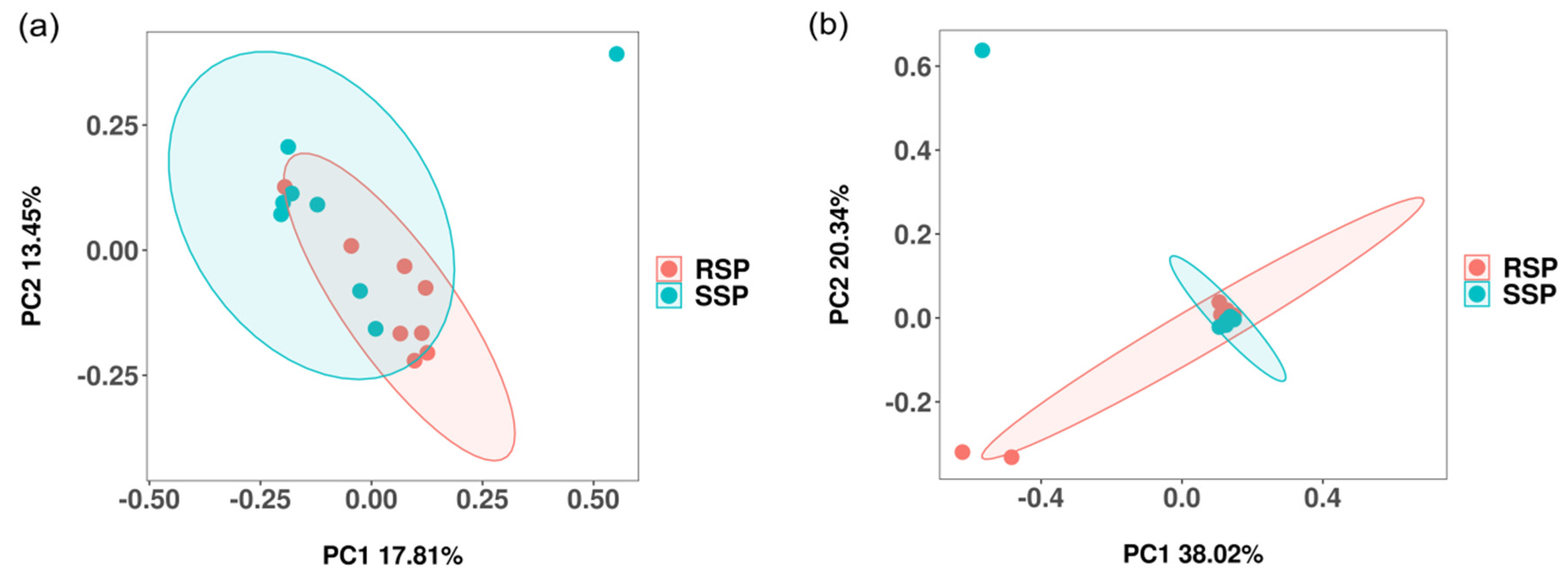
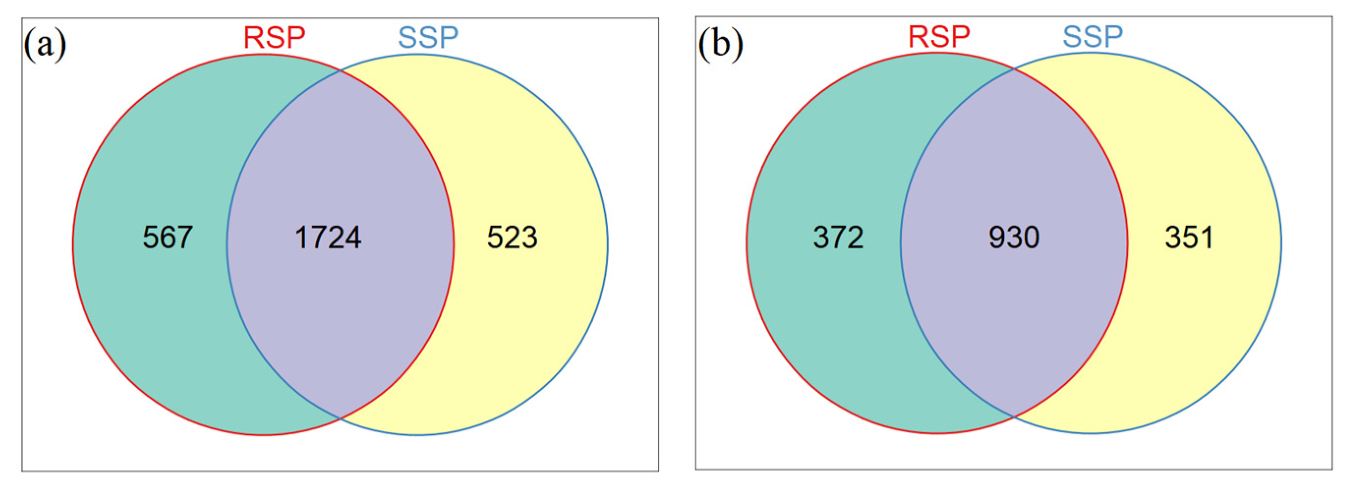
3.2. Bacterial and Fungal Community Diversity

For the bacterial community, 1,273,100 raw reads were obtained, and after filtering, 1,264,325 clean reads were obtained. For the fungal diversity, 1,280,048 raw reads were obtained and after filtering, 1,266,239 clean reads were obtained. Diversity indices (Shannon and Simpson) and richness indices (Chao1 and ACE indices) were calculated to analyze microbial alpha diversity. In the present study, the bacterial and fungal community diversity did not differ significantly (p > 0.05) between the SSP and RSP groups (Table 2 and Table 3). The Chao1, Shannon, and Simpson indices for the bacterial community were higher in the SSP group, whereas the ACE index was higher in the RSP group. All diversity and richness indices for the fungal community were higher in the SSP group
The principal coordinate analysis (PCoA) based on the Bray–Curtis distance was used to exhibit the differences in bacterial and fungal community structure between the SSP and RSP groups. For the bacterial community, the first principal component (PC1) explained 17.81% of the variation and the second principal component (PC2) explained 13.45% of the variation, respectively (Figure 1a). PC1 had the greatest impact. In total, 31.26% of the variation was explained by the two principal components. For the fungal community, PC1 explained 38.02% of the variation and PC2 explained 20.34% of the variation (Figure 1b). PC1 had the greatest impact. In total, the two principal components explained 58.36% of the variation. To statistically support the clustering of bacterial and fungal communities between the two groups, the p-value was calculated via PERMANOVA. The community structure of bacteria and fungi did not exhibit a significant difference between the SSP and RSP groups (R2 = 0.080, p > 0.05; R2 = 0.081, p > 0.05).
The Venn diagram showed the number of shared and unique OTUs between the SSP and RSP groups. For the bacterial community, there were 1724 shared OTUs, and 567 unique OTUs in the RSP group and 523 unique OTUs in the SSP group, respectively (Figure 2a). For the fungal community, there were 930 shared OTUs, 372 unique OTUs in the RSP group and 351 unique OTUs in the SSP group, respectively (Figure 2b).

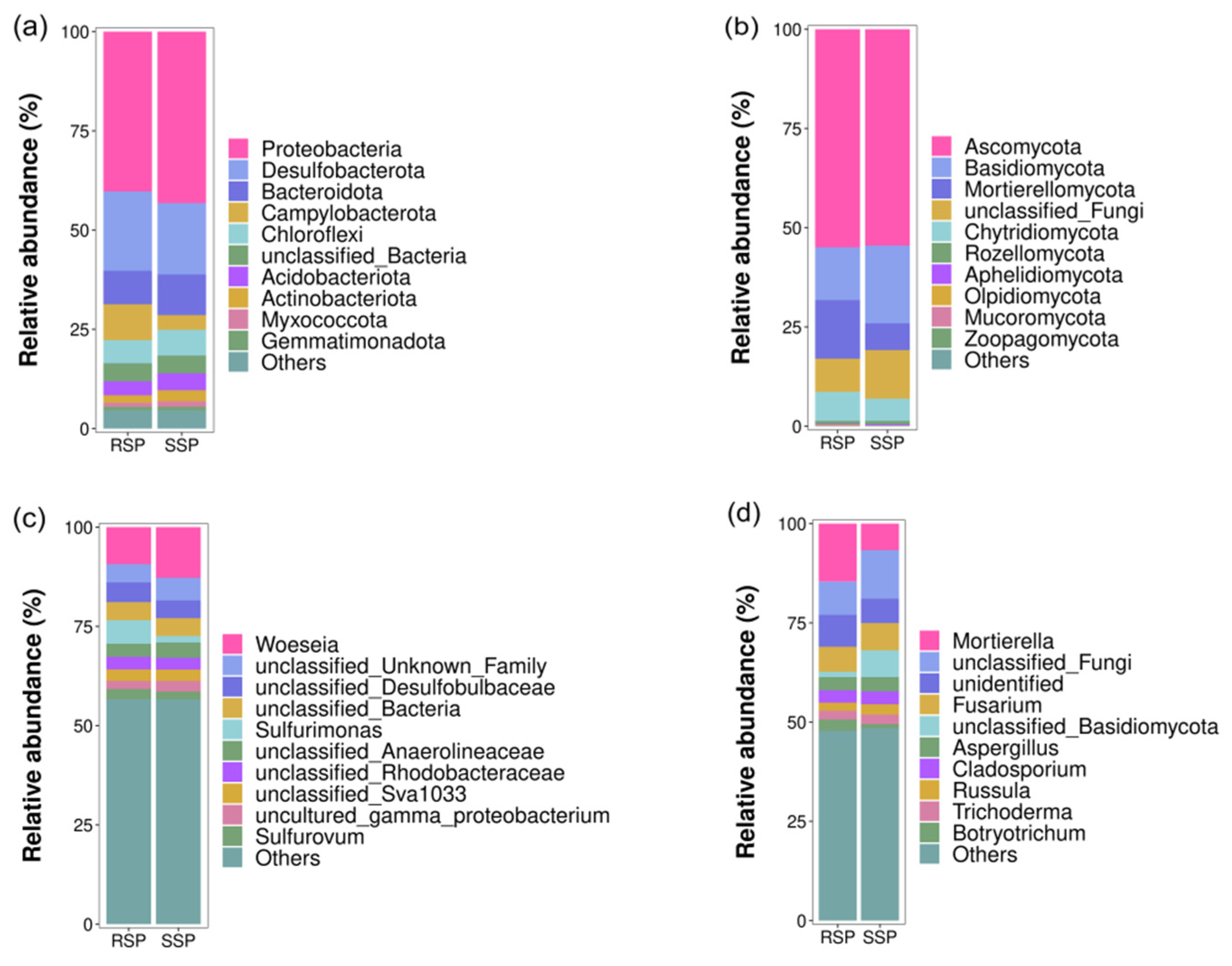
3.3. Bacterial and Fungal Community Composition

The relative abundance of different bacterial and fungal taxa at the phylum and genus level was shown in Figure 3. The dominant bacterial phyla (relative abundance of more than 1%) were Proteobacteria (40.29–43.20%), Desulfobacterota (17.97–19.95%), Campylobacterota (3.69–9.04%), Bacteroidota (8.44–10.25%), Chloroflexi (5.83–6.50%), Acidobacteriota (3.50–4.23%), Actinobacteriota (1.88–2.81%), and Myxococcota (1.03–1.36%) (Figure 3a). The relative abundance of Proteobacteria was the highest, followed by the relative abundance of Desulfobacterota, in both the SSP and RSP groups. At the genus level, except for the unclassified genera, the dominant bacterial taxa were Woeseia (9.34–12.75%), Sulfurovum (1.94–2.53%), and Sulfurimonas (1.65–5.92%) (Figure 3c). Woeseia was the most abundant genus in both the RSP and SSP groups. For the fungal community, the dominant phyla were Ascomycota (54.52–54.94%), Basidiomycota (13.30–19.59%), Mortierellomycota (6.73–14.73%), and Chytridiomycota (5.60–7.29%) (Figure 3b). Ascomycota was the most abundant phylum in both the RSP and SSP groups. Except for the unclassified genera, the dominant fungal genera were Fusarium (6.32–6.87%), Mortierella (6.69–14.61%), Aspergillus (3.32–3.62%), Cladosporium (3.09–3.22%), Russula (2.05–2.63%), Trichoderma (2.22–2.38%), and Botryotrichum (1.08–2.84%) (Figure 3d). Fusarium was the most abundant genus in the SSP group, while Mortierella had the highest relative abundance in the RSP group.

3.4. LefSe Analysis

The LefSe analysis was used to compare the significant difference in the relative abundance of some taxa between the two groups (LDA > 4.0, p < 0.05). For the bacterial community, the relative abundance of the phylum Campylobacterota, the class Campylobacteria, the order Enterobacterales and Campylobacterota, the family Sulfurimonadaceae, and the genus Sulfurimonas were significantly higher in the RSP group than in the SSP group. The relative abundance of the order Steroidoloacterales, the family Woeseiaceae, and the genus Woeseia significantly increased in the SSP group (Figure 4a). Except for the unclassified taxa for the fungal community, only the phylum Basidiomycota rose markedly in the SSP group and the class Chytridiomycetes significantly increased in the RSP group (Figure 4b).

3.5. Redundancy Analysis (RDA) Analysis

RDA was performed to explore the relationship between soil physicochemical factors and microbial community structure. The top five bacterial and fungal phyla in relative abundance and the soil physicochemical factors of sulfur, pH, TN, and EC were used as parameters, respectively. Proteobacteria was strongly correlated with the TN and EC for the bacterial community and Chloroflexi was strong in sulfur content. The two axes of the RDA explained 25.62% and 7.366% of the bacterial community variation, respectively (Figure 5a). For the fungal community, Basidiomycota was strongly correlated with pH. The two axes of the RDA explained 40.55% and 6.194% of the fungal community variation, respectively (Figure 5b)

4. Discussion

Invasive plants can affect the structure and function of the microbial community of invaded areas in various ways. Invasive plants can release chemical residues and change the soil matrix and physical and chemical properties [28]. Invasive plants break the original material circulation pattern and affect soil microorganisms’ structure composition and function. They also change the processing, decomposition, and transformation of rhizosphere microorganisms of native plants, thus affecting the absorption of nutrients, energy flow, and genetic information transfer of native plants [29]. Researchers found that the plant microbiome mainly drives short-term changes in the plant environment. In contrast, long-term changes are driven by ecological and evolutionary interactions between the plant microbiome and its host [30]. Thus, exploring the dynamic changes in soil microbial community structure of intertidal wetlands under the S. alterniflora invasion is meaningful. S. alterniflora has a high tolerance and adaptability to environmental stressors.
In this study, the alpha diversity indices of the bacterial and fungal communities did not differ significantly between the SSP and RSP groups. Although diversity did not differ significantly between the SSP and RSP groups, higher values in the RSP group suggested that the microbial communities were influenced by plant root exudates and soil physicochemical properties. The rhizosphere microorganisms mainly rely on plant root exudates as a food source, and this is closely related to the diversity of microbial communities. Conversely, microorganisms in the bulk soil are lightly affected by plant root exudates, with a consequent lower level of microbial diversity [31]. Our result was not consistent with the above finding. It was reported that the decreased pH could inhibit the bacterial enzymatic and metabolic activities, which is not conducive to the growth of bacteria [32]. The pH value in the RSP group decreased significantly compared to the SSP group, which may explain the lower diversity of the rhizosphere bacterial community. A previous study found that a higher microbial alpha diversity might improve the resilience of S. alterniflora and be an effective strategy for successful invasion [33]. We speculated that the bacterial and fungal communities were not significantly affected by the S. alterniflora invasion due to the short invasion time after the control treatment in August 2021. Venn diagram results also support this view, showing that most OTUs belong to the common OTUs of the two groups and the proportion of unique OTUs is relatively small.
The relationship between the soil’s physical and chemical properties and the plants and microorganisms was complex. Our results showed that the sediment pH was significantly different between the groups. The sediment pH was changed by the S. alterniflora invasion with values decreasing, similar to the previous study [34,35]. We speculated that S. alterniflora was accompanied by the release of H+ in sediments, and that caused a decrease in pH. The sulfur content, TN content, and EC did not differ significantly (p > 0.05) between the SSP and RSP groups. A previous study found that S. alterniflora invasion could substantially affect the sulfur [15] and TN content [35]. At the same time, the soil’s physical and chemical properties varied considerably within the group. We speculated that the soil’s physical and chemical properties were not significantly affected due to the short invasion time of the revived S. alterniflora.
For the bacteria, the dominant phyla in wetland soil were Proteobacteria, Bacteroidota, Chloroflexi, Acidobacteriota, and Actinobacteriota, and the frequently observed dominant genera were Woeseia, Sulfurovum, and Sulfurimonas. For fungi, the dominant phyla were Ascomycota, Basidiomycota, and Chytridiomycota [18]. The above microbial taxa were also found in both the SSP and RSP groups. The top two dominant bacterial phyla were Proteobacteria and Desulfobacterota in the present study, whereas Proteobacteria and Bacteroidota were detected as the most abundant bacterial phyla in our previous study [15]. Though the composition of the bacterial community in the rhizosphere of S. alterniflora was similar to that in the surrounding sediments, some differences were still identified in composition. The LEfSe analysis showed that the Campylobacterota (at the phylum level) and the Sulfurimonas (at the genus level) significantly increased in the RSP group. Previous studies found that Sulfurimonas reduced sulfur compounds as electron donors and nitrate, nitrite, and oxygen as electron acceptors, and Sulfurimonas used CO2 as the sole carbon source [36]. Zhang et al. found that Sulfurimonas has a better adaptation to lower oxygen concentrations and lower available carbon conditions [37]. S. alterniflora is distributed in an intertidal zone that was submerged in seawater for quite a long time. Thus, we speculated that the increased Sulfurimonas might play an essential role in the invasion by S. alterniflora during the under-water time. Physical and chemical properties significantly affect the structure of the microbial community. In the present study, the RDA results showed that the fungal phylum Basidiomycota was strongly correlated with pH, which was consistent with previous studies, demonstrating that the richness of Basidiomycota was explained by a positive response to soil pH [38,39].

5. Conclusions

In the present study, we explored the effects of the secondary growth of S. alterniflora on sediment microorganisms in the Yellow River Delta to deeply understand the scientific basis for the rational management of S. alterniflora. In conclusion, other than the pH, the sulfur content, TN content, and EC did not differ significantly between the SSP and RSP groups. The top two dominant bacterial phyla were Proteobacteria and Desulfobacterota in the present study. The Venn diagram results also support this view, showing that most OTUs belong to the common OTUs of the two groups, and the proportion of unique OTUs is relatively small. Additionally, the bacterial and fungal communities were not significantly affected by the S. alterniflora invasion due to the short invasion time.

Author Contributions

Conceptualization, S.S. and L.L.; formal analysis, Z.Z.; investigation, Y.Z. and J.C.; data curation, J.W. and T.W.; writing—original draft preparation, S.S. and L.L.; writing—review and editing, J.X. and X.T. All authors have read and agreed to the published version of the manuscript.

Funding

This work was supported by the Natural Science Foundation of Shandong Province (ZR2021QD082), the NSFC-Shandong Joint Found (No. U1806213), National Natural Science Foundation of China (No. 42176154), and the Taishan Scholars Program of Shandong Province, China (No. TSQN201909152).

Institutional Review Board Statement

Not applicable.

Informed Consent Statement

Not applicable.

Data Availability Statement

The data that support the findings of this study are available in NCBI (https://www.ncbi.nlm.nih.gov/ (accession number (SAMN29837827-SAMN29837858) (accessed on 20 July 2022).

Conflicts of Interest

There are no declaration of interest by the authors.

References

  1. Alp, M.; Pichon, C.L. Getting from Sea to Nurseries: Considering Tidal Dynamics of Juvenile Habitat Distribution and Connectivity in a Highly Modified Estuarine Riverscape. Ecosystems 2020, 24, 583–601. [Google Scholar] [CrossRef]
  2. Wigginton, R.D.; Pearson, J.; Whitcraft, C.R. Invasive plant ecosystem engineer facilitates community and trophic level alteration for brackish marsh invertebrates. Ecosphere 2014, 5, 40. [Google Scholar] [CrossRef]
  3. Blanco, A.; Larrinaga, A.R.; Neto, J.M.; Troncoso, J.; Mendez, G.; Dominguez-Lapido, P.; Ovejero, A.; Pereira, L.; Mouga, T.M.; Gaspar, R.; et al. Spotting intruders: Species distribution models for managing invasive intertidal macroalgae. J. Environ. Manag. 2021, 281, 111861. [Google Scholar] [CrossRef]
  4. Flood, P.J.; Duran, A.; Barton, M.; Mercado-Molina, A.E.; Trexler, J.C. Invasion impacts on functions and services of aquatic ecosystems. Hydrobiologia 2020, 847, 1571–1586. [Google Scholar] [CrossRef]
  5. Chen, J.; Wang, L.; Li, Y.; Zhang, W.; Fu, X.; Le, Y. Effect of Spartina alterniflora invasion and its controlling technologies on soil microbial respiration of a tidal wetland in Chongming Dongtan, China. Ecol. Eng. 2012, 41, 52–59. [Google Scholar] [CrossRef]
  6. Li, H.; Zhang, L. An experimental study on physical controls of an exotic plant Spartina alterniflora in Shanghai, China. Ecol. Eng. 2008, 32, 11–21. [Google Scholar] [CrossRef]
  7. Yuan, J.; Ding, W.; Liu, D.; Kang, H.; Freeman, C.; Xiang, J.; Lin, Y. Exotic Spartina alterniflora invasion alters ecosystem-atmosphere exchange of CH4 and N2O and carbon sequestration in a coastal salt marsh in China. Glob. Chang. Biol. 2015, 21, 1567–1580. [Google Scholar] [CrossRef]
  8. Yuan, L.; Zhang, L.; Xiao, D.; Huang, H. The application of cutting plus waterlogging to control Spartina alterniflora on saltmarshes in the Yangtze Estuary, China. Estuar. Coast. Shelf Sci. 2011, 92, 103–110. [Google Scholar] [CrossRef]
  9. Zhang, G.; Bai, J.; Jia, J.; Wang, W.; Wang, X.; Zhao, Q.; Lu, Q. Shifts of soil microbial community composition along a short-term invasion chronosequence of Spartina alterniflora in a Chinese estuary. Sci. Total Environ. 2019, 657, 222–233. [Google Scholar] [CrossRef]
  10. Lin, H.-J.; Hsu, C.-B.; Liao, S.-H.; Chen, C.-P.; Hsieh, H.-L. Effects of Spartina alterniflora Invasion on the Abundance and Community of Meiofauna in a Subtropical Wetland. Wetlands 2015, 35, 547–556. [Google Scholar] [CrossRef]
  11. Delgado-Baquerizo, M.; Grinyer, J.; Reich, P.B.; Singh, B.K.; Allen, E. Relative importance of soil properties and microbial community for soil functionality: Insights from a microbial swap experiment. Funct. Ecol. 2016, 30, 1862–1873. [Google Scholar] [CrossRef]
  12. Kowalchuk, G.A.; Buma, D.S.; de Boer, W.; Klinkhamer, P.G.; van Veen, J.A. Effects of above-ground plant species composition and diversity on the diversity of soil-borne microorganisms. Antonie Van Leeuwenhoek 2002, 81, 509–520. [Google Scholar] [CrossRef]
  13. Cao, M.; Cui, L.; Sun, H.; Zhang, X.; Zheng, X.; Jiang, J. Effects of Spartina alterniflora Invasion on Soil Microbial Community Structure and Ecological Functions. Microorganisms 2021, 9, 138. [Google Scholar] [CrossRef] [PubMed]
  14. Liu, Q.; Wang, S.; Li, K.; Qiao, J.; Guo, Y.; Liu, Z.; Guo, X. Responses of soil bacterial and fungal communities to the long-term monoculture of grapevine. Appl. Microbiol. Biotechnol. 2021, 105, 7035–7050. [Google Scholar] [CrossRef] [PubMed]
  15. Shang, S.; Hu, S.; Liu, X.; Zang, Y.; Chen, J.; Gao, N.; Li, L.; Wang, J.; Liu, L.; Xu, J.; et al. Effects of Spartina alterniflora invasion on the community structure and diversity of wetland soil bacteria in the Yellow River Delta. Ecol. Evol. 2022, 12, e8905. [Google Scholar] [CrossRef]
  16. Su, Z.; Qiu, G.; Fan, H.; Li, M.; Fang, C. Changes in carbon storage and macrobenthic communities in a mangrove-seagrass ecosystem after the invasion of smooth cordgrass in southern China. Mar. Pollut Bull. 2020, 152, 110887. [Google Scholar] [CrossRef]
  17. Jiang, S.; Zhang, C.; Chen, L.; Liu, C.; Yan, L.; Li, B. Effects of Smooth Cordgrass Spartina alterniflora Invasion on Macrobenthic Fauna in the Yellow River Delta. Wetlands 2022, 42, 1–12. [Google Scholar] [CrossRef]
  18. Li, L.; Jiang, X.; Zhou, Q.; Chen, J.; Zang, Y.; Zhang, Z.; Gao, C.; Tang, X.; Shang, S. Responses of Soil Microbiota to Different Control Methods of the Spartina alterniflora in the Yellow River Delta. Microorganisms 2022, 10, 1122. [Google Scholar] [CrossRef]
  19. Yu, Y.; Wang, H.; Liu, J.; Wang, Q.; Shen, T.; Guo, W.; Wang, R. Shifts in microbial community function and structure along the successional gradient of coastal wetlands in Yellow River Estuary. Eur. J. Soil Biol. 2012, 49, 12–21. [Google Scholar] [CrossRef]
  20. Liu, F.; Mo, X.; Kong, W.; Song, Y. Soil bacterial diversity, structure, and function of Suaeda salsa in rhizosphere and non-rhizosphere soils in various habitats in the Yellow River Delta, China. Sci. Total Environ. 2020, 740, 140144. [Google Scholar] [CrossRef]
  21. Martin, M. Cut adapt removes adapter sequences from high-throughput sequencing reads, EMBnet. EMBnet 17:10-12. Embnet J. 2011, 17, 10–12. [Google Scholar] [CrossRef]
  22. Bolger, A.M.; Lohse, M.; Usadel, B. Trimmomatic: A flexible trimmer for Illumina sequence data. Bioinformatics 2014, 30, 2114–2120. [Google Scholar] [CrossRef] [PubMed]
  23. Edgar, R.C. UPARSE: Highly accurate OTU sequences from microbial amplicon reads. Nat. Methods 2013, 10, 996–998. [Google Scholar] [CrossRef] [PubMed]
  24. Callahan, B.J.; McMurdie, P.J.; Rosen, M.J.; Han, A.W.; Johnson, A.J.; Holmes, S.P. DADA2: High-resolution sample inference from Illumina amplicon data. Nat. Methods 2016, 13, 581–583. [Google Scholar] [CrossRef]
  25. Bolyen, E.; Rideout, J.R. Reproducible, interactive, scalable and extensible microbiome data science using QIIME 2. Nat. Biotechnol. 2019, 37, 852–857. [Google Scholar] [CrossRef]
  26. Quast, C.; Pruesse, E.; Yilmaz, P.; Gerken, J.; Schweer, T.; Yarza, P.; Peplies, J.; Glockner, F.O. The SILVA ribosomal RNA gene database project: Improved data processing and web-based tools. Nucleic Acids Res. 2013, 41, D590–D596. [Google Scholar] [CrossRef]
  27. McMurdie, P.J.; Holmes, S. phyloseq: An R package for reproducible interactive analysis and graphics of microbiome census data. PLoS ONE 2013, 8, e61217. [Google Scholar] [CrossRef]
  28. Callaway, R.M.; Thelen, G.C.; Rodriguez, A.; Holben, W.E. Soil biota and exotic plant invasion. Nature 2004, 427, 731–733. [Google Scholar] [CrossRef]
  29. Zhang, P.; Nie, M.; Li, B.; Wu, J. The transfer and allocation of newly fixed C by invasive Spartina alterniflora and native Phragmites australis to soil microbiota. Soil Biol. Biochem. 2017, 113, 231–239. [Google Scholar] [CrossRef]
  30. Trivedi, P.; Batista, B.D.; Bazany, K.E.; Singh, B.K. Plant–microbiome interactions under a changing world: Responses, consequences and perspectives. New Phytol. 2022, 6, 1951–1959. [Google Scholar] [CrossRef]
  31. Li, Z.; Zu, C.; Wang, C.; Yang, J.; Yu, H.; Wu, H. Different responses of rhizosphere and non-rhizosphere soil microbial communities to consecutive Piper nigrum L. monoculture. Sci. Rep. 2016, 6, 35825. [Google Scholar] [CrossRef] [PubMed]
  32. Qu, Z.; Liu, B.; Ma, Y.; Xu, J.; Sun, H.; Gallery, R. The response of the soil bacterial community and function to forest succession caused by forest disease. Funct. Ecol. 2020, 34, 2548–2559. [Google Scholar] [CrossRef]
  33. Zheng, J.; Li, J.; Lan, Y.; Liu, S.; Zhou, L.; Luo, Y.; Liu, J.; Wu, Z. Effects of Spartina alterniflora invasion on Kandelia candel rhizospheric bacterial community as determined by high-throughput sequencing analysis. J. Soils Sediments 2018, 19, 332–344. [Google Scholar] [CrossRef]
  34. Yang, W.; An, S.; Zhao, H.; Xu, L.; Qiao, Y.; Cheng, X. Impacts of Spartina alterniflora invasion on soil organic carbon and nitrogen pools sizes, stability, and turnover in a coastal salt marsh of eastern China. Ecol. Eng. 2016, 86, 174–182. [Google Scholar] [CrossRef]
  35. Huang, X.; Feng, J.; Dong, J.; Zhang, J.; Yang, Q.; Yu, C.; Wu, M.; Zhang, W.; Ling, J. Spartina alterniflora invasion and mangrove restoration alter diversity and composition of sediment diazotrophic community. Appl. Soil Ecol. 2022, 177, 104519. [Google Scholar] [CrossRef]
  36. Takai, K. Sulfurimonas paralvinellae sp. nov., a novel mesophilic, hydrogen- and sulfur-oxidizing chemolithoautotroph within the Epsilonproteobacteria isolated from a deep-sea hydrothermal vent polychaete nest, reclassification of Thiomicrospira denitrificans as Sul. Int. J. Syst. Evol. Microbiol. 2006, 56, 1725–1733. [Google Scholar] [CrossRef]
  37. Zhang, X.; Liu, S.; Jiang, Z.; Wu, Y.; Huang, X. Gradient of microbial communities around seagrass roots was mediated by sediment grain size. Ecosphere 2022, 13, e3942. [Google Scholar] [CrossRef]
  38. Liu, D.; Liu, G.; Chen, L.; Wang, J.; Zhang, L. Soil pH determines fungal diversity along an elevation gradient in Southwestern China. Sci. China Life Sci. 2018, 61, 718–726. [Google Scholar] [CrossRef]
  39. Li, J.; Wang, Y.; Qiu, Y.; Xie, Z.; Zhang, Y.; Hua, C. Decomposition effects of Lanzhou lily (Lilium davidii var. unicolor) flowers on soil physical and chemical properties and microbial community diversity. Sci. Cold Arid. Reg. 2022, 14, 212–222. [Google Scholar] [CrossRef]
Figure 1. The PcoA based on the Bray–Curtis distance showing the variation in the bacterial (a) and the fungal (b) community structure. Different colors represent the samples from different groups.
Microorganisms 10 01722 g001
Figure 2. Venn diagrams showing the number of shared and unique OTUs between groups. (a) Bacteria and (b) fungi. Each circle represents sampled compartments. Values within intersections represent shared OTUs, values outside intersections represent unique OTUs.
Microorganisms 10 01722 g002
Figure 3. The relative abundance of the top 10 bacterial (a,c) and fungal (b,d) taxa at the phylum (a,b) and genus (c,d) level.
Microorganisms 10 01722 g003
Figure 4. LEfSe analysis showing the significant difference at different bacterial (a) and fungal (b) taxonomic levels between the SSP and RSP groups. Different colors represent different groups.
Microorganisms 10 01722 g004
Figure 5. RDA of the relationship between the soil physicochemical properties and bacterial (a) and fungal (b) genera. RSP, rhizosphere soil samples; SSP, bulk soil samples; TS, total sulfur; TN, total nitrogen; EC, soil electrical conductivity.
Microorganisms 10 01722 g005
Table 1. Changes in soil physicochemical properties between the SSP and RSP groups.
GroupsSulfur (mg/kg)TN (mg/kg)pHEC (ms/cm)
SSP1.39 ± 0.11 a232.88 ± 40.20 a8.26 ± 0.49 a2.49 ± 0.21 a
RSP1.33 ± 0.15 a169.13 ± 29.38 a7.89 ± 0.49 b1.98 ± 0.15 a
TN means total nitrogen and EC means electrical conductivity. Values are the means ± standard error (n = 8). Lowercase letters in the same column indicate significant differences between the two groups (p < 0.05).
Table 2. Bacterial diversity indices in different groups.
GroupsACEChao1SimpsonShannon
SSP707.73 ± 35.83712.83 ± 36.530.9965 ± 0.00048.79 ± 0.11
RSP708.90 ± 20.64709.83 ± 20.790.9955 ± 0.00058.68 ± 0.08
Values are the means ± standard error (n = 8).
Table 3. Fungal diversity indices in different groups.
GroupsACEChao1SimpsonShannon
SSP421.61 ± 45.59421.69 ± 45.610.9809 ± 0.00577.24 ± 0.28
RSP409.13 ± 28.15409.84 ± 28.190.9586 ± 0.01216.81 ± 0.27
Values are the means ± standard error (n = 8).
Publisher’s Note: MDPI stays neutral with regard to jurisdictional claims in published maps and institutional affiliations.

