Article

Tooth-Specific Streptococcus mutans Distribution and Associated Microbiome

1
Section of Pediatric Dentistry, School of Dentistry, University of California, Los Angeles, CA 90095, USA
2
Section of Oral Biology, School of Dentistry, University of California, Los Angeles, CA 90095, USA
3
Division of General Internal Medicine and Health Services Research, Department of Medicine Statistics Core, University of California, Los Angeles, CA 90095, USA
4
The Forsyth Institute, Microbiology, Cambridge, MA 02142, USA
5
Section of Periodontics, School of Dentistry, University of California, Los Angeles, CA 90095, USA
*
Author to whom correspondence should be addressed.
Microorganisms 2022, 10(6), 1129; https://doi.org/10.3390/microorganisms10061129
Submission received: 4 May 2022 / Revised: 22 May 2022 / Accepted: 26 May 2022 / Published: 31 May 2022
(This article belongs to the Special Issue Human Oral Microbiome in Health and Disease)

Abstract

:
Dental caries is multifactorial and polymicrobial in nature and remains one of the most common oral diseases. While caries research has focused on Streptococcus mutans as the main etiological pathogen, its impact at the tooth level is not fully understood. In this cross-sectional study, the levels and distribution of S. mutans in the posterior teeth at different dentition stages were investigated along with the corresponding tooth-specific microbiome. Occlusal plaque samples of 87 individual posterior teeth were collected from thirty children in three dentition stages (primary, mixed, and permanent). The S. mutans levels in the occlusal plaque of individual posterior teeth were quantified with qPCR, and those with preferential colonization were selected for tooth-specific microbiome analysis using 16S rRNA sequencing. Results: Quantification of S. mutans levels in the occlusal plaque confirmed the preferential colonization on the first primary and permanent molars. These teeth were selected for further tooth-specific microbiome sequencing, as they also displayed high caries experience. There were significant differences in the relative abundance of the four most abundant genera: Neisseria, Streptococcus, Rothia, and Veillonella. Furthermore, the tooth-level caries experience was correlated with a reduction in the microbiome diversity. Analyzing the different tooth-associated microbial communities, distinct tooth-specific core microbiomes were identified. Conclusions: Our findings suggest that caries susceptibility at the tooth level, depending on tooth type and dentition stage, is influenced by individual species as well as plaque community.

1. Introduction

Dental caries is the most prevalent chronic oral infectious disease, which affects over 40% of children and adolescents in the US and is a global oral health burden [1,2]. Caries-associated pain and infection in childhood often have life-long consequences for overall health and well-being [3]. The etiology of this multifactorial disease involves complex interactions between the various genetic, microbial, behavioral, and environmental factors [4]. In children, the oral cavity is a dynamic environment with several transitions between the various dentition stages (primary, mixed, and permanent) [5]. Oral health or disease between these stages are strongly correlated, as caries experience in primary teeth often influences the caries outcome in permanent dentition [6]. The caries experience in primary posterior teeth has been established as a reliable predictor for the subsequent decay in permanent dentition [7,8]. These posterior teeth have an increased caries susceptibility due to their distinctive pit and fissure occlusal topography, arch position as well as eruption status that provides a favorable niche for bacterial colonization [9,10,11].
Streptococcus mutans, a major etiological agent for caries, is prevalent on these occlusal surfaces and has been explored as a risk predictor of the disease [12,13,14]. A number of studies reported a direct association between the levels of S. mutans and the higher probability of caries experience [15,16,17,18,19,20], while others noted a lack of correlation [21,22]. In recent years, the paradigm of S. mutans as the principal cariogenic pathogen has shifted with increasing evidence supporting the polymicrobial nature of dental caries. Especially in children, the oral cavity is a highly heterogeneous ecological system. The transition between dentition stages impacts the microbial habitats, leading to a significant shift in the oral microbiota diversity and composition [23,24,25,26,27].
To date, the correlation between the oral microbiome and caries in various dentition stages has been examined using saliva [28,29,30] or pooled dental plaque samples from multiple tooth surfaces [22,29,31,32,33,34,35]. However, only a few studies have focused on the microbial profiles of the tooth, using pooled multiple occlusal surfaces [31,36,37] or site-specific supragingival plaque [35,38], as well as interdental biofilm [39], and found substantial differences in the supragingival dental plaque microbiome among tooth surfaces and caries experience of children. In addition to S. mutans, other species such as Catonella [40,41,42] and Veillonella were reported in active caries, which supports the ecological nature of caries etiology.
While additional species may play a role in dental caries development, a considerable amount of research has established S. mutans as a primary cariogenic pathogen. Detailed analysis of the preferential localization of S. mutans at the tooth level in different dentition stages and its impact on the tooth-specific microbiome are not fully explored. In this cross-sectional study, we evaluated the levels and distribution of S. mutans in the occlusal plaque of posterior teeth throughout the dentition stages and the associated tooth-specific microbiome in caries-active children.

2. Materials and Methods

2.1. Ethics

The study was reviewed and approved by the University of California Los Angeles Institutional Review Board (#16-000289). Parents or legal guardians of all participants signed written informed consent prior to the initiation of the study. The study was conducted in accordance with the declaration of Helsinki for Biomedical Research Involving Human Subjects.

2.2. Recruitment and Sampling

Study participants were recruited from the pediatric patient population of the University of California, Los Angeles (UCLA) Children’s Dental Center during their periodic oral examination. Thirty healthy children were enrolled and stratified into three groups of ten subjects per group according to their age and dentition stage: primary (2–5 years), mixed (6–10 years), and permanent dentition (11–15 years). The participants’ wide age range was necessary to capture posterior tooth types from different dentition stages. Each participant completed a brief questionnaire, which provided demographic information, oral hygiene habits, and treatment history. Participants were eligible based on the following inclusion criteria: healthy children with caries experience (dmft/DMFT score > 0) who were not taking any medication and had no antibiotic use within the last six months. Participants were excluded from the study if they had generalized rampant caries, periodontitis, halitosis, open sores or ulcerations, chronic systemic diseases, reduced saliva production, or other medical conditions that are known to influence the oral cavity’s microbiome.
Oral clinical evaluation was performed by a trained clinical investigator. The caries experience of each posterior tooth within the different dentition stages: primary first (D’s) and second (E’s) molars, permanent first (4’s) and second (5’s) pre-molars, permanent first (6’s), and second (7’s) molars, was evaluated using decayed, missing, and filled tooth (DMFT) index, according to the criteria proposed by the World Health Organization [43].
The dental plaque collection was standardized, and all participants were asked to refrain from any oral hygiene activity, eating, and drinking in the morning before oral sampling. A total of 87 samples were collected, details per tooth type are described in Table 1 and Table A1, from the individual occlusal surfaces of specific posterior teeth (D’s, E’s, 4’s, 5’s, 6’s, and 7’s) using a sterile Pikster® #3 brush per tooth type (Piksters, Macksville, Australia). Samples derived from the same tooth type in all four quadrants were pooled (four-quadrant sample) into sterile 1.5 mL micro-centrifuge tubes (Eppendorf, Hamburg, Germany) containing PBS and glycerol. After collection, all samples were immediately frozen and stored at −80 °C.

2.3. DNA Extraction

Total genomic DNA was extracted from the 87 occlusal plaque samples using the Epicenter MasterPureTM DNA purification kit (Lucigen, Middleton, WI, USA), following the manufacturer’s instructions with modifications. Briefly, plaque samples were subjected to mechanical grinding with glass beads followed by lysozyme treatment for two hours at 37 °C [44]. The DNA quantity and quality were measured with NanoDrop 2000 (ThermoFisher Scientific, Waltham, MA, USA). The extracted DNA was stored at −20 °C until further use.

2.4. Quantitative Polymerase Chain Reaction (q-PCR)

The total bacteria and S. mutans levels in the 87 occlusal plaque samples were assessed using Real-Time Quantitative Polymerase Chain Reaction (qPCR). Primers targeting the conserved region of the 16S rRNA gene and S. mutans-specific primers targeting glucosyltransferase B encoding gene were used for the total bacteria [45] and S. mutans [46] detection, respectively.
The qPCR reactions were performed in 96-well plates (Biorad, Hercules, CA, USA) using Bio-Rad iCycler Thermal Cycler with iQ5 Multicolor Real-Time PCR Detection System (BioRad iQ 5). Briefly, the reaction mix comprised of 0.5 µM of each primer with 1X SYBR Green Master Mix (BioRad) and 15 ng of DNA in a reaction volume of 20 µL. The qPCR program consisted of an initial denaturation for 5 min at 95 °C, followed by 40 cycles of denaturation at 95 °C for 30 s, annealing at 64 °C for 30 s, and extension s at 72 °C for 30 s. The qPCRs were performed with three technical replicates for each sample. The iQ5 Optical System Software generated quantification cycle values (Cq) were analyzed along with melting point data. A standard curve was generated from a serial dilution of S. mutans reference strain (UA140) DNA.

2.5. Microbial Community Analysis

A total of 36 occlusal plaque samples: primary Dprim’s (n = 10), Dmixed’s (n = 8), and permanent 6mixed’s (n = 8), 6perm’s (n = 10) first molars were selected for sequencing (Table 1). Briefly, genomic DNA was used to amplify the V4 region of the 16S gene and sequenced on the Illumina Miseq sequencer system at the UCLA Microbiome Core, using a 150 bp paired-end protocol. The sequencing data were analyzed using QIIME (Quantitative Insights into Microbial Ecology) version 1.9.1 [47]. Sequences were clustered into operational taxonomic units (OTUs) using UCLUST [48], and the taxonomy was assigned with the Human Oral Microbiome Database (HOMD) [49] as a reference. A total of 3,761,710 high-quality sequences from thirty-six samples, with a mean of 104,491 ± 2732, were generated after processing the data obtained from 16S rRNA gene sequencing. For alpha diversity, OTU tables were rarefied to 23,000 reads, Shannon index was calculated [50]. For beta diversity, UniFrac distances were calculated, followed by principal coordinates analysis (PCoA). The OTUs lists generated for the several core analyses were compared using Venn diagram software packages [51].

2.6. Power Analysis

Comparing between D’s and 6’s, with the sample size of 10 per group, we had 80% power to detect significant differences at a 5% significance level using analysis for the repeated measures assuming a correlation of 0.7 between the teeth from the same subject. This calculation assumed that the mean difference of 2.8 would be detected (the means in caries experience: 3.3 for D’s vs. 0.5 for 6’s) with the standard deviation of 1.7.

2.7. Statistical Analysis

The statistical tests were performed using GraphPad Prism (version 8.0.0, GraphPad Software, San Diego, CA, USA), and the statistical significance was defined as follows: * p < 0.05, ** p < 0.005, and *** p < 0.0005. The demographic and S. mutans distribution data analysis was assessed by one-way ANOVA or Kruskal–Wallis test for comparing more than 2 independent groups. Wilcoxon signed-rank test was performed in comparing dmft/DMFT scores and S. mutans levels that were collected within the same subjects. The Spearman correlation analysis was performed between the demographic and oral hygiene parameters. The association between dmft/DMFT scores and S. mutans levels was evaluated by linear regression. The tooth microbiome statistical analysis was determined with the Kruskal–Wallis test, applying the false discovery rate to correct for multiple comparisons. Differences in weighted Unifrac distances between the groups were analyzed with analysis of similarity (ANOSIM).

3. Results

3.1. Study Demographics

A total of thirty pediatric participants were enrolled in this cross-sectional study and were divided into three groups according to their age and type of dentition. In brief, the gender distribution was 17 females (57%) and 13 males (43%). The study population comprised 87% Hispanic and 13% non-Hispanic with an average age of 3.9 (±1.0), 8.6 (±2.1), and 12.4 (±1.8) for primary, mixed, and permanent dentition groups, respectively. The frequency of their oral hygiene habits, such as professional cleaning, their tooth brushing and flossing routine, and mouth rinse usage, were similar between the groups. All subjects reported not using fluoride supplements.

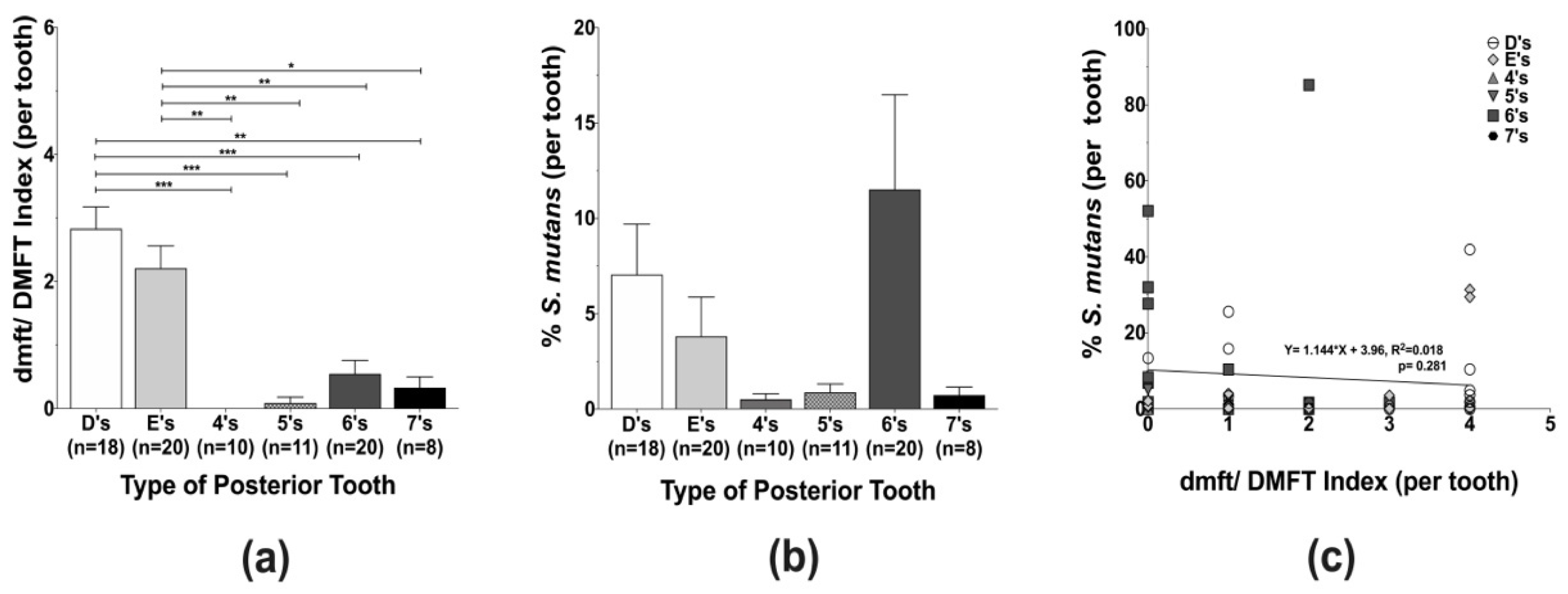
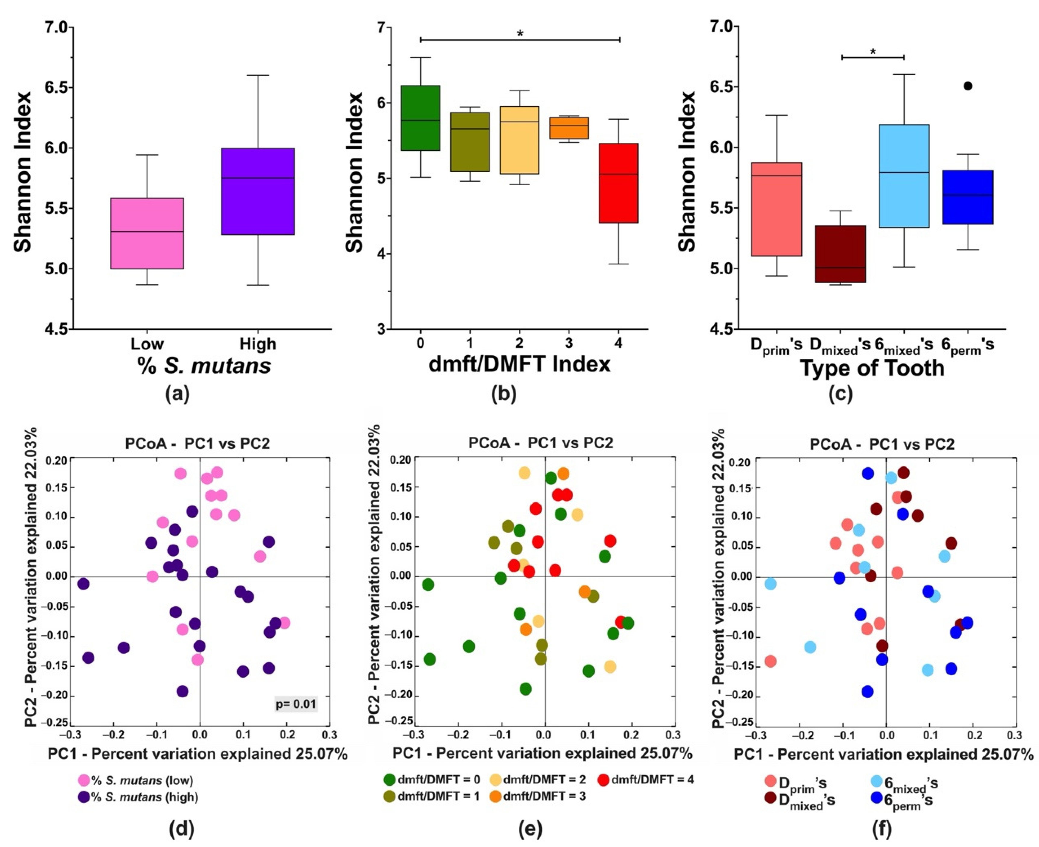
3.2. Caries Experience and Streptococcus mutans Localization in Different Types of Posterior Teeth

The caries status of individual teeth per type of posterior tooth (D’s, E’s, 4’s, 5’s, 6’s, and 7’s), a total of 87 samples, was assessed by the dmft/DMFT index and revealed that the caries experience of the first primary molars (D’s) was significantly higher in comparison to all other teeth, except for the primary second molars (E’s). Among the permanent teeth, the first permanent molars (6’s) exhibited the highest caries experience (Figure 1a). Although not statistically significant, a preferential localization of S. mutans was observed in the primary and permanent first molars, the posterior teeth that erupt first within each dentition (Figure 1b). Even though linear regression analysis demonstrated that S. mutans levels and tooth-specific caries experience were not directly correlated (Figure 1c), it is important to highlight that D’s and 6’s appeared to have higher caries experience and harbor more S. mutans.

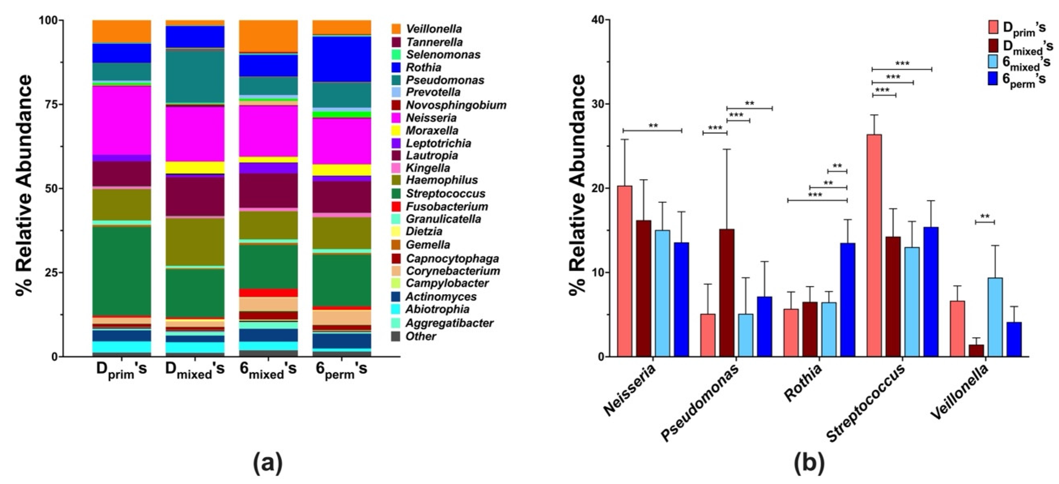
3.3. Community Profiling of D’s and 6’s

Next, we investigated the influence of S. mutans colonization on the local tooth microbiome of D’s and 6’s in the different dentition stages (Dprim, Dmixed, 6mixed, 6perm), a total of 36 posterior teeth. The alpha diversity Shannon index revealed that the S. mutans levels (Figure 2a) did not affect the tooth microbial diversity. In contrast, a significant decrease in diversity was observed on teeth with high caries compared to caries-free teeth (Figure 2b) and on Dmixed in comparison to 6mixed, when posterior teeth per dentition were compared (Figure 2c).
Further, microbial community changes were examined using weighted Unifrac distances according to S. mutans levels, tooth-specific caries experience, and tooth type per dentition (D’s, 6’s) (Figure 2d–f). The S. mutans levels were categorized into two groups: low (<1%) and high (≥1%) (unpublished data) based on qPCR-quantification, significantly clustered according to S. mutans percentage (Figure 2d). For the tooth-specific caries experience, an apparent trend was observed for the teeth with high DMFT but was not statistically significant (Figure 2e). Additional beta diversity analyses considering the tooth type in the different dentition stages did not reveal a specific pattern (Figure 2f).
The local microbial composition of D’s and 6’s in different dentition stages at the genus level revealed a total of forty-five genera, but for further analysis, only those with a mean abundance value of ≥ 0.1% were included (Figure 3a). Of the 25 of these genera, significant differences between the tooth types were only observed for Streptococcus, Neisseria, Rothia, and Veillonella (Figure 3b). While the abundance of Neisseria was significantly higher in the Dprim’s (20.3% ± 7.3) in comparison to the 6perm’s (13.6% ± 11.5), the relative abundance of Rothia and Streptococcus were highest in 6perm’s (13.5% ± 8.7) and Dprim’s (26.5% ± 7.2), respectively, and differed significantly from the other tooth types. Additionally, significantly higher levels of Veillonella were observed in 6mixed’s (9.4% ± 1.4) in comparison to Dmixed’s (2.4% ± 1.3) (Figure 3b).

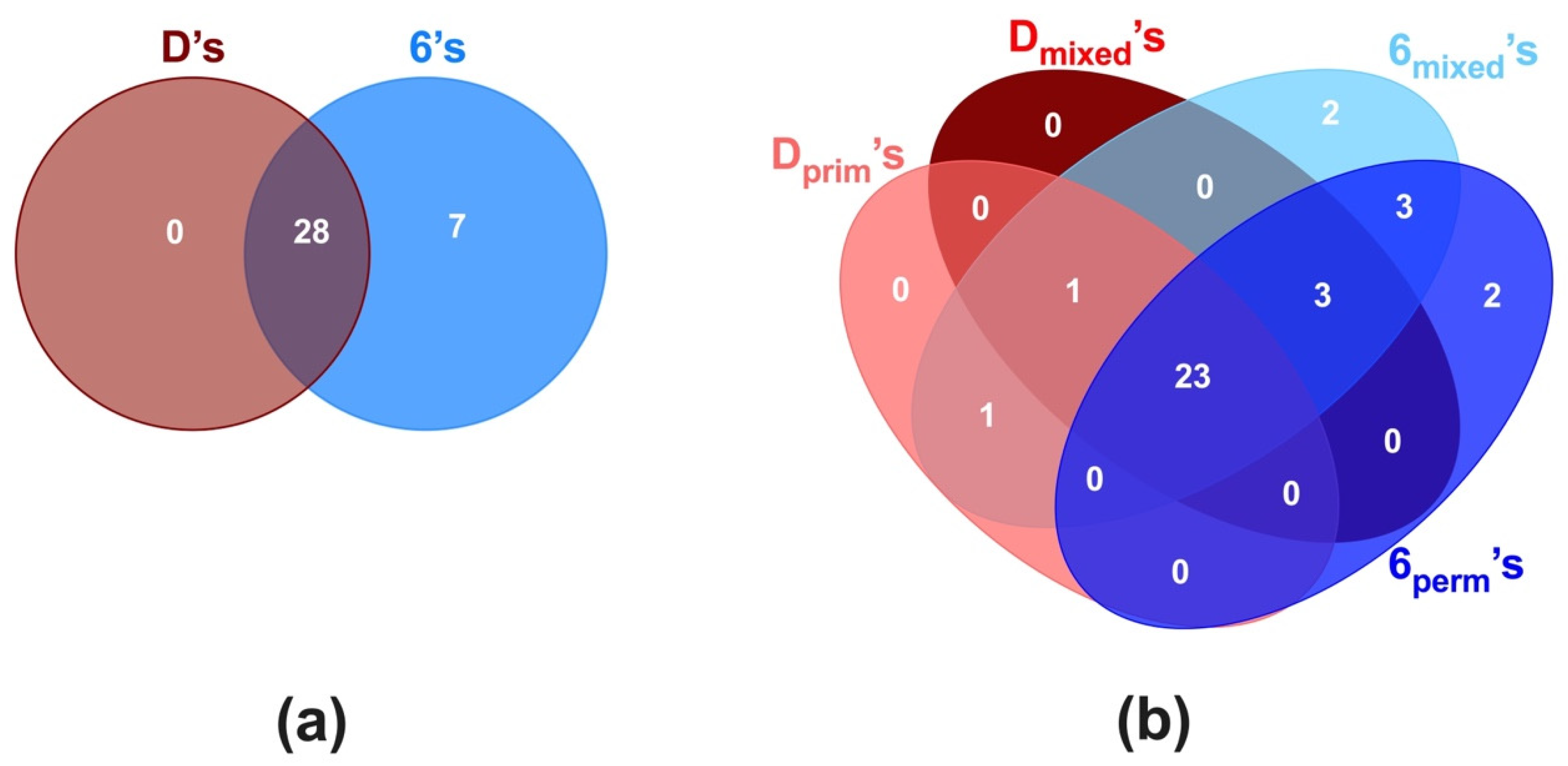
3.4. Core Microbiome of D’s and 6’s

The different tooth-associated microbial communities were analyzed for their respective shared and unique genera. In all posterior teeth (D’s and 6’s), the core microbiome consisted of twenty-eight genera (80.0%), including the highly prevalent genera, Streptococcus, Neisseria, Rothia, and Veillonella, among others (Figure 4a).
When comparing between D’s and 6’s besides the core genera, seven genera (Catonella, Eikenella, Klebsiella, Lachnoanaerobaculum, Peptococcus, Peptostreptococcus, and Tannerella) were specific to the 6’s tooth type. Further analysis of the D’s and 6’s based on their dentition stages revealed that besides a similar core microbiome of 23 genera (66%), specific genera were associated to particular tooth depending on dentition (Figure 4b). The Dprim’s and Dmixed’s had only one genus (Cardiobacterium) in common, while 6mixed’s and 6perm’s shared three genera, Klebsiella, Lachnoanaerobaculum, and Tannerella. Interestingly, four genera were found to be distinct to 6’s according to the dentition stage; Catonella and Eikenella were specific to 6mixed’s, while Peptococcus and Peptostreptococcus were unique to 6perm’s.

4. Discussion

Streptococcus mutans, the principal etiological agent of dental caries, is frequently employed for caries risk assessment [12,52], even though the levels are not always correlated with caries experience [21,22,53,54]. Despite evidence supporting a higher prevalence of S. mutans in posterior teeth [55], the pattern of S. mutans levels in individual posterior teeth across dentition stages is not fully characterized. Therefore, in this cross-sectional study, we investigated the levels of S. mutans and caries experience in individual types of posterior teeth (D’s, E’s, 4’s, 5’s, 6’s, and 7’s), the localization pattern of this bacterium, and explored how S. mutans influences the local tooth microbiome.
This study indicates a differential caries susceptibility and S. mutans localization at the tooth level. The highest caries scores were observed in the first primary molars (D’s) for the primary dentition and the first permanent molars (6’s) for the permanent dentition (Figure 1a). These results are in agreement with other studies, albeit using a different caries index system [56] and in older populations [57]. In particular, the posterior teeth that erupted first and resided longer, Dprim’s and Dmixed’s, displayed the higher caries scores. Interestingly, although not statistically significant, S. mutans appears to favor colonization of D’s and 6’s, the first molars within each dentition stage (Figure 1b), with 6’s having the highest S. mutans levels among the evaluated teeth. The apparent preferential localization of S. mutans could be explained by the fact that these teeth are the first to erupt and are located more anteriorly with a distinct pit and fissure morphology that may promote efficient S. mutans adhesion and plaque accumulation [36,52,55].
To explore the influence of S. mutans and caries experience on the local tooth microbiome, we selected representative teeth (Dprim’s, Dmixed’s, 6mixed’s, and 6perm’s) for sequencing analysis. Overall, species diversity was similar between all the teeth, independent of S. mutans levels. On the contrary, a previous study suggested that the dominance of S. mutans may decrease community diversity [38]. Regarding caries experience, significantly reduced microbial diversity was observed on teeth with high caries compared to caries-free teeth. This observation was similar to other studies that described decreasing microbial diversity with the severity of caries [31,37,38]. Interestingly, in this study, significantly higher species abundance was observed in the 6mixed’s than in the corresponding Dmixed’s, highlighting a potential tooth-level impact on species diversity.
At the genus level, the four most abundant genera were identified, including Streptococcus, Neisseria, Rothia, and Veillonella, which were also reported in previous investigations [24,32]. The members of the Streptococcus and Neisseria genus, predominant in this study, are early colonizers and often dominate the dental plaque in the primary dentition. In concurrence with our findings, previous studies reported that Streptococcus and Neisseria levels tend to decrease as dentition matures, and late colonizers further contribute to diversity [58,59]. In contrast, the genus Rothia increased from primary to permanent dentition, with the highest levels in 6perm’s, indicating that this genus is present in all dentitions with higher abundance in the permanent dentition [24,25].
Moreover, this genus has been associated with either health [28,30,42] or caries [22,29,33], depending on the species. Although not significantly different, the 6’s had the highest caries experience in comparison to the other permanent teeth (4’s, 5’s, and 7’s). Previously, a higher abundance of Veillonella has been associated with permanent dentition [59]. However, in this study, low levels of Veillonella were observed in 6 mixed’s, suggesting an ongoing transition in the oral microbiome.
While transitioning through dentition stages is a dynamic process, part of the microbiome remains constant [23,25]. To better understand the microbe–habitat relationship, the ‘core microbiome’ of D’s and 6’s in caries-active children was evaluated. Within the core microbiome of 28 genera observed between D’s and 6’s (Figure 4b), 19 genera were consistent with those reported previously for the core microbial community in healthy children with a similar age range [23,24]. Additional genera were detected in our study, which focused on caries-active children transitioning through various dentition stages.
Further investigation into the dentition-specific cores of D’s and 6’s revealed that Dprim’s and Dmixed’s shared the genus Cardiobacterium, and 6mixed’s and 6perm’s shared three genera, Klebsiella, Lachnoanaerobaculum, and Tannerella. While Cardiobacterium has been associated with healthy primary dentition [40], Klebsiella is reported in permanent dentition of healthy individuals [35]. On the other hand, Tannerella was only present in the saliva of caries active children with mixed dentition, suggesting the effect of both caries and transition of dentition stages [35]. Furthermore, the presence of Peptococcus and Peptostreptococcus only in 6perm’s suggests the association of these genera with the mature oral microbiome. Similar to the observations of this study, previous studies with healthy individuals identified Peptococcus and Peptostreptococcus in the later stage of primary and permanent dentitions [26]. In this study, the sequencing of the V4 region of the 16S gene did not allow the microbiome analysis at the species level. Although 16S rRNA gene sequencing can provide adequate phylogenetic information to identify the bacteria at the species level, for some taxa, such as different streptococcal species, the identification is limited. In the future, it would be interesting to explore within the identified genera which species are determinant to the tooth-specific microbiome in the caries context.
This cross-sectional study highlights the clinical importance of individual tooth susceptibility to dental caries. The strength of our study was the tooth-type specific plaque investigation compared to other studies, which utilized pooled occlusal [31,36,37] and supragingival [35,38] dental plaque or saliva [28,29,30]. This study revealed that S. mutans preferentially colonized D’s and 6’s and detected differences at the phylogenetic level in the tooth-specific microbiome associated with dental caries throughout dentition stages. Our study design attempted to unravel the microbiome shifts through dentition stages which would be challenging to accomplish in a longitudinal study. Building on our pilot study, which indicated microbial shifts occurring between the different dentition stages, future studies with an increased number of samples per tooth type and the inclusion of all the posterior teeth for microbiome analysis would contribute valuable data to understanding the tooth-level and dentition-stage-dependent caries susceptibility.

Author Contributions

M.D. contributed to conception, data acquisition, analysis, and interpretation, drafted, and critically revised the manuscript; W.T. contributed to data acquisition, analysis, and interpretation, drafted, and critically revised the manuscript; M.-S.S. Sim computed statistical analysis and critically revised the manuscript; M.A., X.H., W.S. and R.L. contributed to conception, data interpretation, and critically revised the manuscript; N.C.T. contributed to conception, design, data acquisition, analysis, interpretation, drafted, and critically revised the manuscript. All authors have read and agreed to the published version of the manuscript.

Funding

This study was funded by the UCLA Council on Research, Faculty Grant Program and partially supported by C3 Jian, Inc.

Institutional Review Board Statement

The study was conducted according to the guidelines of the Declaration of Helsinki and approved by the University of California Los Angeles Institutional Review Board (#16-000289).

Informed Consent Statement

Parents or legal guardians of all participants signed written informed consent prior to the initiation of the study.

Data Availability Statement

The data that support the findings of the study are available from the corresponding authors upon request.

Acknowledgments

The authors would like to thank Joshua Lin, Alejandra Rivera, Loliya Stewart, and dental student volunteers for their support during subject recruitment. We also like to thank Bhumika Shokeen and Farnoosh Haghighi for critical review of this manuscript.

Conflicts of Interest

The authors declare that Dr. W. Shi was a part-time Chief Science Officer of C3J Therapeutics, Inc., which has licensed technologies from UC Regents that could be indirectly related to this research project. The authors declare that the research was conducted in the absence of any commercial or financial relationships that could be construed as a potential conflict of interest.

Appendix A

Table A1. Summary of the samples collected in the study. The occlusal plaque of individual posterior teeth (87 samples) was collected from the thirty enrolled participants, and the samples were stratified into three groups according to the dentition stage. The caries experience of each tooth was evaluated using dmft/DMFT index, and detailed caries experience per tooth type are indicated in the table.
Dentition Stage
Primary (2–5 y)
(n = 10 Participants)
Mixed (6–10 y)
(n = 10 Participants)
Permanent (11–15 y)
(n = 10 Participants)
dmft/DMFT Index
Type of Posterior ToothCodedmfn° Toothd/Dm/Mf/Fn° ToothDMFn° Tooth
Primary 1st molarD’s 0.3 ± 0.70.0 ± 0.01.8 ± 1.7n = 100.4 ± 1.00.3 ± 0.72.6 ± 1.4n = 8
Primary 2nd molarE’s 0.4 ± 0.70.0 ± 0.01.4 ± 1.4n = 100.4 ± 0.90.0 ± 0.01.7 ± 1.2n = 10
Permanent 1st pre-molar4’s 0.0 ± 0.00.0 ± 0.00.0 ± 0.0n = 10.0 ± 0.00.0 ± 0.00.0 ± 0.0n = 9
Permanent 2nd
pre-molar
5’s 0.0 ± 0.00.0 ± 0.00.0 ± 0.0n = 10.0 ± 0.00.0 ± 0.00.1 ± 0.3n = 10
Permanent 1st molar6’s 0.2 ± 0.60.0 ± 0.00.1 ± 0.3n = 100.0 ± 0.00.0 ± 0.00.8 ± 1.1n = 10
Permanent 2nd molar7’s 0.0 ± 0.00.0 ± 0.00.0 ± 0.0n = 10.1 ± 0.30.0 ± 0.00.2 ± 0.4n = 7
Code—tooth nomenclature using Palmer notion; dmft/DMFT—decay missing filled tooth index, per individual tooth type. For the sequencing experiments, we selected (highlighted in grey): primary first molars from primary (Dprim’s, n = 10) and mixed (Dmixed’s, n = 8) dentitions; permanent first molars from mixed (6mixed’s, n = 8) and permanent (6perm’s, n = 10) dentitions.

References

  1. Dye, B.A.; Thornton-Evans, G.; Li, X.; Iafolla, T.J. Dental Caries and Sealant Prevalence in Children and Adolescents in the United States, 2011–2012; NCHS Data Brief; CDC: Atlanta, GA, USA, 2015. [Google Scholar]
  2. Kassebaum, N.J.; Smith, A.G.C.; Bernabé, E.; Fleming, T.D.; Reynolds, A.E.; Vos, T.; Murray, C.J.L.; Marcenes, W. Global, regional, and national prevalence, incidence, and disability-adjusted life years for oral conditions for 195 countries, 1990–2015: A systematic analysis for the global burden of diseases, injuries, and risk factors. J. Dent. Res. 2017, 96, 380–387. [Google Scholar] [CrossRef] [PubMed]
  3. Guarnizo-Herreño, C.C.; Wehby, G.L. Children’s dental health, school performance, and psychosocial well-being. J. Pediatr. 2012, 161, 1153–1159. [Google Scholar] [CrossRef] [PubMed] [Green Version]
  4. De Soet, J.J.; Van Gemert-Schriks, M.C.M.; Laine, M.L.; Van Amerongen, W.E.; Morré, S.A.; Van Winkelhoff, A.J. Host and microbiological factors related to dental caries development. Caries Res. 2008, 42, 340–347. [Google Scholar] [CrossRef] [PubMed]
  5. Lynch, R.J. The primary and mixed dentition, post-eruptive enamel maturation and dental caries: A review. Int. Dent. J. 2013, 63, 3–13. [Google Scholar] [CrossRef]
  6. Saethre-Sundli, H.B.; Wang, N.J.; Wigen, T.I. Do enamel and dentine caries at 5 years of age predict caries development in newly erupted teeth? A prospective longitudinal study. Acta Odontol. Scand. 2020, 78, 509–514. [Google Scholar] [CrossRef] [Green Version]
  7. Zhang, Q.; Van Palenstein Helderman, W.H. Caries experience variables as indicators in caries risk assessment in 6–7-year-old Chinese children. J. Dent. 2006, 34, 676–681. [Google Scholar] [CrossRef]
  8. Topaloglu-Ak, A.; Eden, E. Caries in primary molars of 6–7-year-old Turkish children as risk indicators for future caries development in permanent molars. J. Dent. Sci. 2010, 5, 150–155. [Google Scholar] [CrossRef] [Green Version]
  9. Carvalho, J.C. Caries process on occlusal surfaces: Evolving evidence and understanding. Caries Res. 2014, 48, 339–346. [Google Scholar] [CrossRef]
  10. Carvalho, J.C.; Ekstrand, K.R.; Thylstrup, A. Dental plaque and caries on occlusal surfaces of first permanent molars in relation to stage of eruption. J. Dent. Res. 1989, 68, 773–779. [Google Scholar] [CrossRef]
  11. Ekstrand, K.R.; Nielsen, L.A.; Carvalho, J.C.; Thylstrup, A. Dental plaque and caries on permanent first molar occlusal surfaces in relation to sagittal occlusion. Scand. J. Dent. Res. 1993, 101, 9–15. [Google Scholar] [CrossRef]
  12. Tanzer, J.; Livingston, J.; Thompson, A. The microbiology of primary dental caries in humans. J. Dent. Edu. 2001, 65, 1028–1037. [Google Scholar] [CrossRef]
  13. Loesche, W.J.; Rowan, J.; Straffon, L.H.; Loos, P.J. Association of Streptococcus mutants with human dental decay. Infect. Immun. 1975, 11, 1252–1260. [Google Scholar] [CrossRef] [PubMed] [Green Version]
  14. Loesche, W.J.; Straffon, L.H. Longitudinal investigation of the role of Streptococcus mutans in human fissure decay. Infect. Immun. 1979, 26, 498. [Google Scholar] [CrossRef] [PubMed] [Green Version]
  15. Köhler, B.; Bratthall, P.B.D. Streptococcus mutans in plaque and saliva and the development of caries. Scand. J. Dent. Res. 1981, 89, 19–25. [Google Scholar] [CrossRef] [PubMed]
  16. Damle, S.G.; Loomba, A.; Dhindsa, A.; Loomba, A.; Beniwal, V. Correlation between dental caries experience and mutans streptococci counts by microbial and molecular (polymerase chain reaction) assay using saliva as microbial risk indicator. Dent. Res. J. 2016, 13, 552–559. [Google Scholar] [CrossRef]
  17. Alaluusua, S.; Renkonen, O.V. Streptococcus mutans establishment and dental caries experience in children from 2 to 4 years old. Scand. J. Dent. Res. 1983, 91, 453–457. [Google Scholar] [CrossRef]
  18. Köhler, B.; Andréen, I.; Jonsson, B. The earlier the colonization by mutans streptococci, the higher the caries prevalence at 4 years of age. Oral Microbiol. Immunol. 1988, 3, 14–17. [Google Scholar] [CrossRef]
  19. Granath, L.; Cleaton-Jones, P.; Fatti, L.P.; Grossman, E.S. Prevalence of dental caries in 4- to 5-year-old children partly explained by presence of salivary mutans streptococci. J. Clin. Microbiol. 1993, 31, 66–70. [Google Scholar] [CrossRef] [Green Version]
  20. Loesche, W.J.; Eklund, S.; Earnest, R.; Burt, B. Longitudinal investigation of bacteriology of human fissure decay: Epidemiological studies in molars shortly after eruption. Infect. Immun. 1984, 46, 765–772. [Google Scholar] [CrossRef] [Green Version]
  21. Carlsson, P.; Olsson, B.; Bratthall, D. The relationship between the bacterium Streptococcus mutans in the saliva and dental caries in children in Mozambique. Arch. Oral Biol. 1985, 30, 265–268. [Google Scholar] [CrossRef]
  22. Aas, J.A.; Griffen, A.L.; Dardis, S.R.; Lee, A.M.; Olsen, I.; Dewhirst, F.E.; Leys, E.J.; Paster, B.J. Bacteria of dental caries in primary and permanent teeth in children and young adults. J. Clin. Microbiol. 2008, 46, 1407. [Google Scholar] [CrossRef] [PubMed] [Green Version]
  23. Xu, X.; He, J.; Xue, J.; Wang, Y.; Li, K.; Zhang, K.; Guo, Q.; Liu, X.; Zhou, Y.; Cheng, L.; et al. Oral cavity contains distinct niches with dynamic microbial communities. Environ. Microbiol. 2015, 17, 699–710. [Google Scholar] [CrossRef] [PubMed]
  24. Shi, W.; Qin, M.; Chen, F.; Xia, B. Supragingival microbial profiles of permanent and deciduous teeth in children with mixed dentition. PLoS ONE 2016, 11, e0146938. [Google Scholar] [CrossRef] [PubMed] [Green Version]
  25. Mason, M.R.; Chambers, S.; Dabdoub, S.M.; Thikkurissy, S.; Kumar, P.S. Characterizing oral microbial communities across dentition states and colonization niches. Microbiome 2018, 6, 67. [Google Scholar] [CrossRef]
  26. Shi, W.; Tian, J.; Xu, H.; Zhou, Q.; Qin, M. Distinctions and associations between the microbiota of saliva and supragingival plaque of permanent and deciduous teeth. PLoS ONE 2018, 13, e0200337. [Google Scholar] [CrossRef]
  27. Liu, S.; Wang, Y.; Zhao, L.; Sun, X.; Feng, Q. Microbiome succession with increasing age in three oral sites. Aging 2020, 12, 7874–7907. [Google Scholar] [CrossRef]
  28. Xu, L.; Chen, X.; Wang, Y.; Jiang, W.; Wang, S.; Ling, Z.; Chen, H. Dynamic alterations in salivary microbiota related to dental caries and age in preschool children with deciduous dentition: A 2-year follow-up study. Front. Physiol. 2018, 9, 342. [Google Scholar] [CrossRef]
  29. Jiang, S.; Gao, X.; Jin, L.; Lo, E.C.M. Salivary microbiome diversity in caries-free and caries-affected children. Int. J. Mol. Sci. 2016, 17, 1978. [Google Scholar] [CrossRef] [Green Version]
  30. Luo, A.H.; Yang, D.Q.; Xin, B.C.; Paster, B.J.; Qin, J. Microbial profiles in saliva from children with and without caries in mixed dentition. Oral Dis. 2012, 18, 595–601. [Google Scholar] [CrossRef]
  31. Gross, E.L.; Leys, E.J.; Gasparovich, S.R.; Firestone, N.D.; Schwartzbaum, J.A.; Janies, D.A.; Asnani, K.; Griffen, A.L. Bacterial 16S sequence analysis of severe caries in young permanent teeth. J. Clin. Microbiol. 2010, 48, 4121. [Google Scholar] [CrossRef] [Green Version]
  32. Crielaard, W.; Zaura, E.; Schuller, A.A.; Huse, S.M.; Montijn, R.C.; Keijser, B.J.F. Exploring the oral microbiota of children at various developmental stages of their dentition in the relation to their oral health. BMC Med. Genom. 2011, 4, 22. [Google Scholar] [CrossRef] [PubMed] [Green Version]
  33. Mannaa, A.; Carlén, A.; Campus, G.; Lingström, P. Supragingival plaque microbial analysis in reflection to caries experience. BMC Oral Health 2013, 13, 5. [Google Scholar] [CrossRef] [PubMed] [Green Version]
  34. Johansson, I.; Witkowska, E.; Kaveh, B.; Lif Holgerson, P.; Tanner, A.C.R. The microbiome in populations with a low and high prevalence of caries. J. Dent. Res. 2015, 95, 80–86. [Google Scholar] [CrossRef]
  35. Ribeiro, A.A.; Azcarate-Peril, M.A.; Cadenas, M.B.; Butz, N.; Paster, B.J.; Chen, T.; Bair, E.; Arnold, R.R. The oral bacterial microbiome of occlusal surfaces in children and its association with diet and caries. PLoS ONE 2017, 12, e0180621. [Google Scholar] [CrossRef] [PubMed]
  36. Brailsford, S.R.; Sheehy, E.C.; Gilbert, S.C.; Clark, D.T.; Kidd, E.A.M.; Zoitopoulos, L.; Adams, S.E.; Visser, J.M.; Beighton, D. The microflora of the erupting first permanent molar. Caries Res. 2005, 39, 78–84. [Google Scholar] [CrossRef] [PubMed]
  37. Gross, E.L.; Beall, C.J.; Kutsch, S.R.; Firestone, N.D.; Leys, E.J.; Griffen, A.L. Beyond Streptococcus mutans: Dental caries onset linked to multiple species by 16S rRNA community analysis. PLoS ONE 2012, 7, e47722. [Google Scholar] [CrossRef] [PubMed]
  38. Richards, V.P.; Alvarez, A.J.; Luce, A.R.; Bedenbaugh, M.; Mitchell, M.L.; Burne, R.A.; Nascimento, M.M. Microbiomes of site-specific dental plaques from children with different caries status. Infect. Immun. 2017, 85, e00106–e00117. [Google Scholar] [CrossRef] [Green Version]
  39. Inquimbert, C.; Bourgeois, D.; Bravo, M.; Viennot, S.; Tramini, P.; Llodra, J.C.; Molinari, N.; Dussart, C.; Giraudeau, N.; Carrouel, F. The oral bacterial microbiome of interdental surfaces in adolescents according to carious risk. Microorganisms 2019, 7, 319. [Google Scholar] [CrossRef] [Green Version]
  40. Peterson, S.N.; Snesrud, E.; Liu, J.; Ong, A.C.; Kilian, M.; Schork, N.J.; Bretz, W. The dental plaque microbiome in health and disease. PLoS ONE 2013, 8, e58487. [Google Scholar] [CrossRef] [Green Version]
  41. Xiao, C.; Ran, S.; Huang, Z.; Liang, J. Bacterial diversity and community structure of supragingival plaques in adults with dental health or caries revealed by 16S Pyrosequencing. Front. Microbiol. 2016, 7, 1145. [Google Scholar] [CrossRef]
  42. Hernández, M.; Planells, P.; Martínez, E.; Mira, A.; Carda-Diéguez, M. Microbiology of molar–incisor hypomineralization lesions. A pilot study. J. Oral Microbiol. 2020, 12, 1766166. [Google Scholar] [CrossRef] [PubMed]
  43. Petersen, P.E.; Baez, R.J.; World Health Organization. Oral Health Surveys: Basic Methods, 5th ed.; World Health Organization: Geneva, Switzerland, 2013.
  44. Dinis, M.; Agnello, M.; Cen, L.; Shokeen, B.; He, X.; Shi, W.; Wong, D.T.W.; Lux, R.; Tran, N.C. Oral Microbiome: Streptococcus mutans/Caries Concordant-Discordant Children. Front. Microbiol. 2022, 13, 782825. [Google Scholar] [CrossRef] [PubMed]
  45. Muyzer, G.; De Waal, E.C.; Uitterlinden, A.G. Profiling of complex microbial populations by denaturing gradient gel electrophoresis analysis of polymerase chain reaction-amplified genes coding for 16S rRNA. Appl. Environ. Microbiol. 1993, 59, 695–700. [Google Scholar] [CrossRef] [PubMed] [Green Version]
  46. Jung, W.-S.; Yang, I.L.H.; Lim, W.H.; Baek, S.-H.; Kim, T.-W.; Ahn, S.-J. Adhesion of mutans streptococci to self-ligating ceramic brackets: In vivo quantitative analysis with real-time polymerase chain reaction. Eur. J. Orthod. 2015, 37, 565–569. [Google Scholar] [CrossRef] [Green Version]
  47. Caporaso, J.G.; Kuczynski, J.; Stombaugh, J.; Bittinger, K.; Bushman, F.D.; Costello, E.K.; Fierer, N.; Peña, A.G.; Goodrich, J.K.; Gordon, J.I.; et al. QIIME allows analysis of high-throughput community sequencing data. Nat. Methods 2010, 7, 335–336. [Google Scholar] [CrossRef] [Green Version]
  48. Edgar, R.C. Search and clustering orders of magnitude faster than BLAST. Bioinformatics 2010, 26, 2460–2461. [Google Scholar] [CrossRef] [Green Version]
  49. Chen, T.; Yu, W.-H.; Izard, J.; Baranova, O.V.; Lakshmanan, A.; Dewhirst, F.E. The Human Oral Microbiome Database: A web accessible resource for investigating oral microbe taxonomic and genomic information. J. Biol. Databases Curation 2010, 2010, baq013. [Google Scholar] [CrossRef]
  50. Kuczynski, J.; Stombaugh, J.; Walters, W.A.; González, A.; Caporaso, J.G.; Knight, R. Using QIIME to analyze 16S rRNA gene sequences from microbial communities. Curr. Protoc. Microbiol. 2012, 1, 1E.5. [Google Scholar] [CrossRef] [Green Version]
  51. Bardou, P.; Mariette, J.; Escudié, F.; Djemiel, C.; Klopp, C. Jvenn: An interactive Venn diagram viewer. BMC Bioinform. 2014, 15, 293. [Google Scholar] [CrossRef] [Green Version]
  52. Lemos, J.A.; Palmer, S.R.; Zeng, L.; Wen, Z.T.; Kajfasz, J.K.; Freires, I.A.; Abranches, J.; Brady, L.J. The Biology of Streptococcus mutans. Microbiol. Spectrum 2019, 7. [Google Scholar] [CrossRef]
  53. Matee, M.I.N.; Mikx, F.H.M.; De Soet, J.S.; Maselle, S.Y.; De Graaff, J.; Van Palenstein Helderman, W.H. Mutans streptococci in caries-active and caries-free infants in Tanzania. Oral Microbiol. Immunol. 1993, 8, 322–324. [Google Scholar] [CrossRef]
  54. Matee, M.I.N.; Mikx, F.H.M.; Maselle, S.Y.M.; Van Palenstein Helderman, W.H. Mutans Streptococci and Lactobacilli in breast-fed children with rampant caries. Caries Res. 1992, 26, 183–187. [Google Scholar] [CrossRef]
  55. Lindquist, B.; Emilson, C.G. Distribution and prevalence of mutans streptococci in the human dentition. J. Dent. Res. 1990, 69, 1160–1166. [Google Scholar] [CrossRef] [PubMed]
  56. Baginska, J.; Rodakowska, E.; Milewski, R.; Kierklo, A. Dental caries in primary and permanent molars in 7–8-year-old schoolchildren evaluated with Caries Assessment Spectrum and Treatment (CAST) index. BMC Oral Health 2014, 14, 74. [Google Scholar] [CrossRef] [PubMed] [Green Version]
  57. Bhardwaj, V. Dental caries prevalence in individual tooth in primary and permanent dentition among 6–12-year-old school children in Shimla, Himachal Pradesh. Int. J. Health Allied Sci. 2014, 3, 125–128. [Google Scholar] [CrossRef] [Green Version]
  58. Sampaio-Maia, B.; Monteiro-Silva, F. Acquisition and maturation of oral microbiome throughout childhood: An update. Dent. Res. J. 2014, 11, 291–301. [Google Scholar]
  59. Gomez, A.; Nelson, K.E. The oral microbiome of children: Development, disease, and implications beyond oral health. Microbial. Ecol. 2017, 73, 492–503. [Google Scholar] [CrossRef] [Green Version]
Figure 1. Caries experience and Streptococcus mutans levels quantification among posterior tooth in different dentition stages. (a) The individual posterior tooth (D’s, E’s, 4’s, 5’s, 6’s, and 7’s) caries status was assessed using decayed, missing, and filled tooth (dmft/DMFT) criteria, and the figure represents the average caries experience per tooth type. (b) S. mutans levels were evaluated by qPCR using S. mutans-specific primers (Table 1). (c) Linear regression analysis of S. mutans levels and dmft/DMFT index. Differences in significance between groups were analyzed using one-way ANOVA followed by Kruskal–Wallis multiple comparisons tests; * p < 0.05, ** p < 0.005, and *** p < 0.0005.
Microorganisms 10 01129 g001
Figure 2. Alpha and beta diversity analyses of the microbial community associated with first primary and permanent molars within the different dentition stages (Dprim’s, Dmixed’s, 6mixed’s, and 6perm’s). Alpha diversity was analyzed using Shannon index (upper panel, (ac)), and beta diversity was calculated by principal coordinates analysis (PCoA) of weighted Unifrac distances (lower panel, (df)). The analyses were performed based on the following parameters: (a,d) S. mutans levels, low (% < 1%) and high (% ≥ 1%); (b,e) posterior tooth dmft/DMFT index; and (c,f) tooth type per dentition. The differences in the significance of the groups were performed by one-way ANOVA followed by Kruskal–Wallis multiple comparisons tests; differences in unweighted Unifrac distances between the groups were analyzed with analysis of similarity (ANOSIM); * p < 0.05. The black dot in Figure 2c represents an outlier.
Microorganisms 10 01129 g002
Figure 3. Genus level analysis of plaque communities associated with first primary and permanent molars within the different dentition stages (Dprim’s, Dmixed’s, 6mixed’s, and 6perm’s). Relative abundances at the genus level are shown for microbial profiles (a) and selected genus (b). The statistical analysis of the tooth microbiome at the genus level was determined with the Kruskal–Wallis test, controlling the false discovery rate to correct for multiple comparisons. Statistical significance between groups was evaluated by two-way ANOVA followed by Tukey’s multiple comparisons tests; ** p< 0.005, and *** p < 0.0005.
Microorganisms 10 01129 g003
Figure 4. Core microbiome at the genus level of first primary and permanent molars (Dprim’s, Dmixed’s, 6mixed’s, and 6perm’s). The analyses of the first primary and permanent molars teeth core microbiome are displayed as Venn diagrams, based on (a) type of tooth and (b) tooth type per dentition.
Microorganisms 10 01129 g004
Table 1. Summary of the samples collected in the study. The occlusal plaque of individual posterior teeth (87 samples) was collected from the thirty enrolled participants, and the samples were stratified into three groups according to the dentition stage. The caries experience of each tooth was evaluated using dmft/DMFT index; the respective average per tooth type is indicated in the table.
Dentition Stage
Primary
(2–5 y)
(n = 10
Participants)
Mixed
(6–10 y)
(n = 10
Participants)
Permanent
(11–15 y)
(n = 10
Participants)
dmft/DMFT Index
Type of Posterior ToothCodedmftdmft/DMFTDMFT
Primary 1st molarD’s 2.1 ± 1.7 (n = 10)3.3 ± 1.1 (n = 8)
Primary 2nd molarE’s 2.0 ± 1.8 (n = 10)2.1 ± 1.5 (n = 10)
Permanent 1st pre-molar4’s 0.0 ± 0.0 (n = 1)0.0 ± 0.0 (n = 9)
Permanent 2nd pre-molar5’s 0.0 ± 0.0 (n = 1)0.1 ± 0.3 (n = 10)
Permanent 1st molar6’s 0.3 ± 0.7 (n = 10)0.8 ± 1.1 (n = 10)
Permanent 2nd molar7’s 0.0 ± 0.0 (n = 1)0.3 ± 0.5 (n = 7)
Code—tooth nomenclature using Palmer notion; dmft/DMFT—decay missing filled tooth index, per individual tooth type. For the sequencing experiments, we selected (highlighted in grey): primary first molars from primary (Dprim’s, n = 10) and mixed (Dmixed’s, n = 8) dentitions; permanent first molars from mixed (6mixed’s, n = 8) and permanent (6perm’s, n = 10) dentitions.
Publisher’s Note: MDPI stays neutral with regard to jurisdictional claims in published maps and institutional affiliations.

