Review

Gram-Negative Bacterial Envelope Homeostasis under Oxidative and Nitrosative Stress

by
Thibault Chautrand
,
Djouhar Souak
,
Sylvie Chevalier
and
Cécile Duclairoir-Poc
*
Research Unit Bacterial Communication and Anti-infectious Strategies (UR CBSA), Rouen Normandy University, Normandy University, 55 rue Saint-Germain, 27000 Evreux, France
*
Author to whom correspondence should be addressed.
Microorganisms 2022, 10(5), 924; https://doi.org/10.3390/microorganisms10050924
Submission received: 19 March 2022 / Revised: 15 April 2022 / Accepted: 21 April 2022 / Published: 28 April 2022
(This article belongs to the Special Issue Microbial Adaptation and Tolerance to Environmental Stresses)

Abstract

:
Bacteria are frequently exposed to endogenous and exogenous reactive oxygen and nitrogen species which can damage various biomolecules such as DNA, lipids, and proteins. High concentrations of these molecules can induce oxidative and nitrosative stresses in the cell. Reactive oxygen and nitrogen species are notably used as a tool by prokaryotes and eukaryotes to eradicate concurrent species or to protect themselves against pathogens. The main example is mammalian macrophages that liberate high quantities of reactive species to kill internalized bacterial pathogens. As a result, resistance to these stresses is determinant for the survival of bacteria, both in the environment and in a host. The first bacterial component in contact with exogenous molecules is the envelope. In Gram-negative bacteria, this envelope is composed of two membranes and a layer of peptidoglycan lodged between them. Several mechanisms protecting against oxidative and nitrosative stresses are present in the envelope, highlighting the importance for the cell to deal with reactive species in this compartment. This review aims to provide a comprehensive view of the challenges posed by oxidative and nitrosative stresses to the Gram-negative bacterial envelope and the mechanisms put in place in this compartment to prevent and repair the damages they can cause.

1. Oxidative and Nitrosative Stresses: An Omnipresent Challenge

Oxidative stress is the result of an imbalance between the presence of reactive oxygen species (ROS) and the ability of a biological system to detoxify them and their side products. Similarly, nitrosative stress is an imbalance of reactive nitrogen species (RNS). Some of these molecules can play an important role in signalization in both eukaryotes and prokaryotes. For example, nitric oxide (NO) is involved in the dispersion of biofilm in multiple bacterial species [1] and in the regulation of vasoconstriction in mammals [2,3]. However, the strong redox potential of ROS and RNS allows them to damage a wide range of biomolecules, and any excess can jeopardize cell homeostasis. As a result, most biological organisms possess defense mechanisms allowing them to manage these stresses. Some reactive oxygen and nitrogen species are endogenously produced by the bacterial metabolism, in particular in aerobic bacteria. In addition, bacteria can often encounter exogenous oxidative and nitrosative stresses in their environment, one glaring example being cellular immune response.
The envelope is a complex structure whose integrity is critical for cell survival, serving as a selective barrier at the interface between the inner and the outer bacterial environments. It is also the first bacterial structure to encounter ROS and RNS. Correct membrane function is essential for the survival of bacteria, as well as peptidoglycan integrity and synthesis. Since oxidative and nitrosative stresses can affect a large variety, if not all of the envelope components, it is critical for the cell to maintain envelope homeostasis to survive these stresses. This review aims at gathering the current understanding of the impacts of oxidative and nitrosative stresses on the bacterial envelope and the bacterial mechanisms implemented to cope with such stresses. This review first will address the main actors of oxidative and nitrosative stresses and their exogenous sources, then it will focus on the damages induced by RNS and ROS on the envelope of Gram-negative bacteria, including numerous challenging bacteria in terms of health, and finally, it will address the bacterial response mechanisms to maintain envelope homeostasis, whether by preventing or repairing these damages.

2. The Gram-Negative Bacterial Envelope

The envelope of Gram-negative bacteria is composed of the outer (OM) and inner (IM) membranes and a thin peptidoglycan (PG) layer located in the periplasmic space, which is a highly oxidizing compartment.

2.1. The Outer Membrane (OM)

The OM is asymmetric, with an inner leaflet made of phospholipids facing the periplasm, and an outer layer of anionic glycolipids, including lipopolysaccharides (LPS). Typically, LPS are composed of a lipid A, also known as endotoxin, a core oligosaccharide, and a polysaccharide forming a distal chain known as O-antigen [4]. The OM functions as a selective physical permeability barrier, by controlling the entry into the cell. Most OM transmembrane proteins are β-barrel proteins, which are composed of β-sheets forming a cylinder [5]. Escherichia coli possesses approximately 100 of these proteins, whose functions are mostly unknown. The known β-barrel proteins notably include three major porins: OmpF and OmpC, which both allow the selective passage of small cationic molecules, and PhoE, which is dedicated to the entry of anionic molecules [6]. The other main group of proteins present on the OM are lipoproteins, which possess lipid moieties attached to a terminal cysteine residue [7]. Lipoproteins were considered to be overwhelmingly bound to the IM, and it is only recently that the importance of OM lipoproteins has been highlighted. Lipoproteins are anchored to the OM inner leaflet by their lipid tail facing the periplasm for most of them, while others, called surface lipoproteins, decorate the outer surface of Gram-negative bacteria [8]. They are involved in various processes such as nutrient acquisition, cell signaling, and virulence [8,9]. These functions are not exclusive to protein, however, as phospholipids also participate in the homeostasis of both the outer and inner membranes [10].

2.2. The Inner Membrane (IM)

The IM is composed of a symmetrical bilayer of phospholipids and possesses a much greater protein diversity than the OM. As bacteria lack the intracellular organelles present in eukaryotic cells, the membrane-associated functions of bacterial metabolism take place at the IM [11]. These functions notably include lipid biosynthesis, energy production, and protein secretion. All the components of the Gram-negative bacteria envelopes are synthetized directly in the cytoplasm or at the inner leaflet of the IM. Therefore, all the envelope components must be translocated, secreted, or flipped through the IM. Furthermore, the IM acts as a second layer of protection and selection against the entry of exogenous molecules [12].

2.3. The Periplasm and the Peptidoglycan (PG)

Located between both membranes, the periplasm is an oxidative environment containing a high density of proteins. In this environment, the structure of proteins containing cysteines is usually stabilized by disulfide bonds. The periplasmic compartment fills various roles for the cell. Notably, it sequesters enzymes that would be toxic to the cell in the cytoplasmic space, such as RNAses and alkaline phosphatases, and allows specific protein folding and oxidation and regulation of cell division [13]. In addition, the periplasm harbors the PG, which allows the cell to retain its shape. PG is composed of polymerized glycans that form linear strands cross-linked by short peptides. These glycans strands are made of β-1,4-connected N-acetylglucosamine residues alternating with N-acetylmuramic acid residues [12]. PG thus forms a net-like polymer around the cell that protects it against turgor and serves as a scaffold to attach proteins, lipoproteins, and other molecules [12]. Its basic composition is well conserved between bacteria, but specific variations can alter its properties.
The envelope serves as an interface between the cell and the environment by selecting which substances are able to cross it. Furthermore, the envelope is the first cell component to be affected by the cell environment and must be able to withstand the various stresses it encounters via complex regulatory pathways known as the cell envelope stress response (ESR) [14]. As a result, maintaining membrane homeostasis is critical for the cell. Aerobic bacteria are commonly exposed to oxidative and nitrosative stresses from their environment, resulting in the excessive presence of reactive oxygen or nitrogen species, respectively. These processes can alter multiple envelope components, such as phospholipids and proteins, causing damage to this cell compartment [15,16]. It is necessary to consider the complex interactions between these species and biological molecules to understand the challenges bacteria face.

3. Reactive Oxygen and Nitrogen Species: A Complex Reactions Network

3.1. Oxidative Stress Chemistry

Molecular oxygen (O2) is a small non-polar molecule that diffuses freely across usual biological membranes [17]. Therefore, the bacterial intracellular O2 concentration is similar to their environment. As a result, bacteria either have to avoid oxidative stress by living in anaerobic or microaerobic environments or survive elevated internal oxygen levels. Oxygen is not toxic by itself, as it is practically unreactive with the molecules structuring biological organisms that are lipids, proteins, carbohydrates, and nucleic acids. However, the reduction of O2 can generate various ROS, such as the superoxide anion (O2), hydrogen peroxide (H2O2), and hydroxyl radicals (OH) [18]. In aerobic environments, endogenous ROS may be produced in bacteria via the reaction between O2 and univalent electron donors. These donors can be metal centers, dihydroflavin cofactors, or quinones [18]. The main endogenous source of O2 and H2O2 is the autoxidation of non-respiratory flavoproteins [19] by electron transfer between O2 and the dihydroflavin of the reduced flavoproteins. This reaction leads to the formation of O2, which generally goes through another electron transfer before escaping the active site of the enzyme, generating H2O2. Aside from its conversion into H2O2, O2 is in equilibrium with the hydroperoxyl radical (HO2) through the reversible reaction O2•− + H+ ⇋ HO2 [20]. Since the pKa of this reaction is 4.88, it is estimated that the HO2 form represents less than 1% of the total superoxide in the cellular cytoplasm [21]. However, cytoplasmic pH may not be uniform and may be significantly lowered in the proximity of membranes containing negatively charged phospholipids such as cardiolipin, phosphatidylserine, and phosphatidylinositol [22]. Therefore, local pH decrease could allow the formation of significant quantities of HO2 near the IM. Unlike O2•−, HO2 is hydrophobic, allowing it to cross to the IM lipidic core. As a result, HO2 is critical in the process of lipid peroxidation. In the presence of light, photosynthetic organisms also generate the highly reactive singlet oxygen (1O2) through the pigments of their photosystems [23]. This stress can also occur in non-photosynthetic microorganisms through other cellular cofactors such as rhodopsin, quinones, flavins, and porphyrins [23].

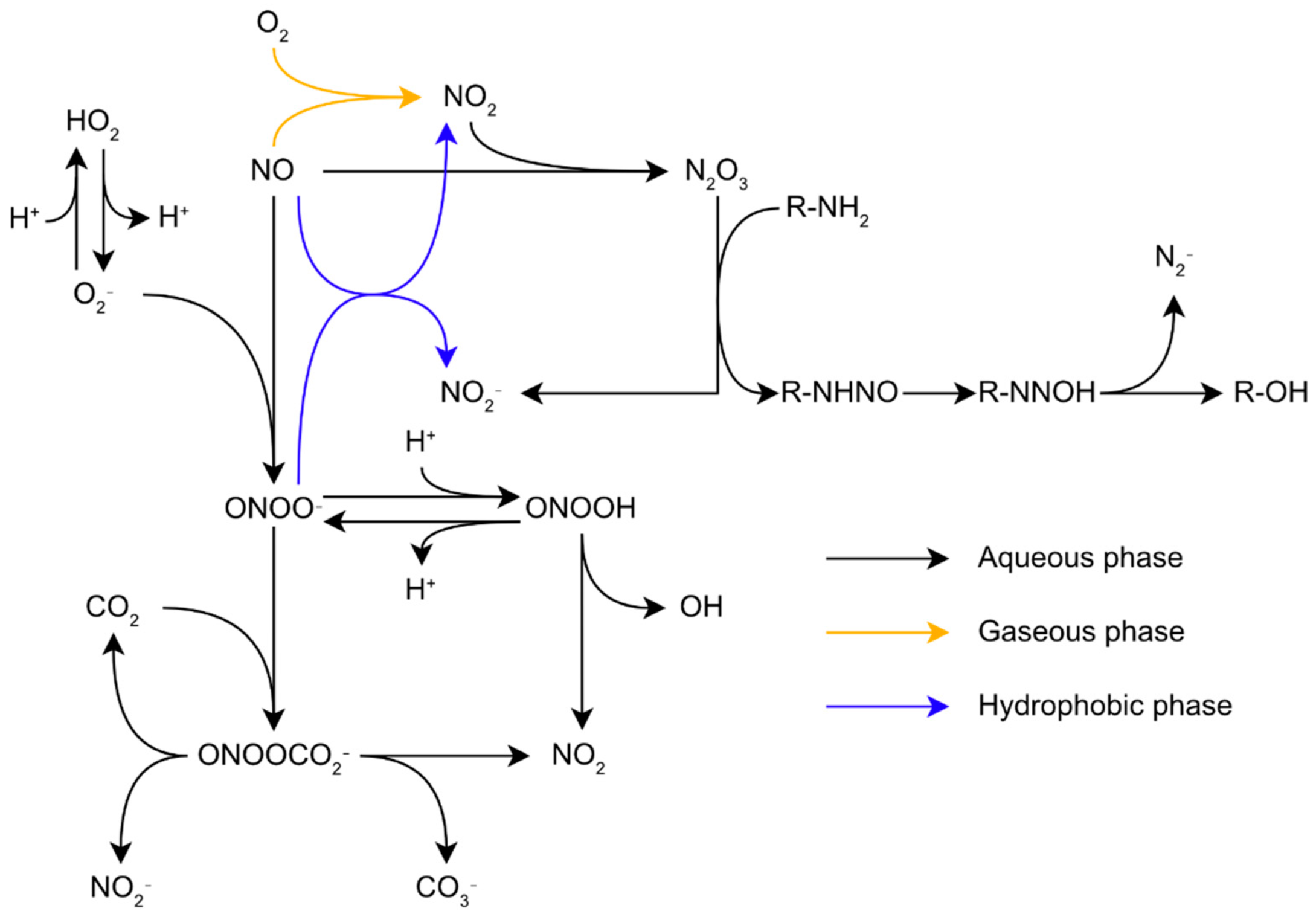
3.2. Nitrosative Stress Chemistry

The range of reactive molecules created by oxidative stress is not limited to ROS. Indeed, RNS production is tightly linked to the presence of ROS in the cells. Nitric oxide (NO) is a small lipophilic radical which diffuses across biological membranes. It is an important molecule for signalization in biological organisms. In mammals, NO notably controls blood pressure and acts as a messenger in the central nervous system [24]. Despite its role as a signaling molecule, NO is toxic for biological organisms at high concentrations, and this molecule is synthetized by macrophages to combat pathogens during the immune response through their inducible nitric oxide synthases (iNOS), making resistance to RNS critical for pathogens [25,26]. NO is more reactive than oxygen regarding the structural components of biological organisms, particularly proteins. Its toxicity comes from its ability to inhibit haem enzymes binding dioxygen, react with Fe-S centers, and indirectly induce the nitrosation of proteins [27]. Similar to oxygen, NO is relatively unreactive with most biological molecules. Direct biological targets of NO are limited to radicals and metal complexes, especially Fe-containing complexes. However, these reactions with intracellular molecules can generate other reactive species much more harmful to the cell. For example, NO with O2•− reacts at a diffusion-controlled rate to form the peroxynitrite anion (ONOO) and its conjugated acid, peroxynitrous acid (ONOOH) (Figure 1) [28]. Since these two species coexist and are in an acid-base equilibrium in common biological conditions, the term peroxynitrite is generally used to describe both species [16]. Peroxynitrite is an impactful biological oxidant. ONOOH isomerizes to nitrate at rates of 0.095 s−1, 1.3 s−1, and 4.5 s−1 at 5 °C, 25 °C, and 37 °C, respectively [29] through the formation of both OH and NO2, hereafter simply noted OH and NO2 [16]. However, in biological conditions, peroxynitrite reacts with its biological targets, such as proteins, lipids, and nucleic acids, at a much faster rate, making this reaction irrelevant. Due to its high reactivity, peroxynitrite-induced damages will mostly depend on the kinetics of the reactions between peroxynitrite and its surrounding targets in priority protein metal centers, thiols, and selenols [16]. Another important RNS is NO2. This may appear alongside OH, in an aqueous environment, through the isomerization of ONOOH (Figure 1). In an air polluted by NO, such as smog, for instance, NO autoxidation in the gaseous phase leads to the formation of NO2 through the equation 2NO+O2 → 2NO2. Further reactions between NO and NO2 leads to the formation of N2O3 through the equation NO2+NO ⇋ N2O3 [30]. In aqueous media, such reactions may occur too fast to produce a significant amount of NO2 intermediate. However, these reactions still have biological relevancy as they may take place in non-aqueous media such as the lipid bilayers of membranes [31].

3.3. Exogenous Sources of Oxidative/Nitrosative Stress

3.3.1. Biotic Stress Sources

In addition to the endogenous ROS and RNS synthesis through metabolic processes described above, bacteria have to deal with exogenous stresses from various sources.
The most studied one is the oxidative burst released by phagocytic cells during the immune response. During this process, macrophages and neutrophils cause an oxidative burst on phagocyted bacteria while they synthetize O2•− through the activity of the membrane nicotinamide adenine dinucleotide (NADPH)-oxidase. Half of the O2•− reacts with H+ to form H2O2. In addition to ROS, macrophages also release large quantities of NO, synthetized by their iNOS. The simultaneous release of O2•− and NO in biological conditions lead to the formation of peroxynitrite [16]. The mammalian immune system also induces oxidative stress inside bacteria using PG Recognition Proteins (PGRPs). These proteins kill bacteria by induction of oxidative, thiol, and metal stresses in bacteria. PGRPs induce oxidative stress by blocking the bacterial respiratory chain, which promotes the production of H2O2 inside the cell [32]. In the context of host infection, bacteria are also exposed to other envelope-damaging factors such as the membrane attack complex and lysozyme, which synergize to degrade bacterial envelopes [33]. In addition to its role in bacterial killing through peptidoglycan hydrolysis, lysozyme possesses immune-dampening properties. It is notably able to decrease the neutrophil oxidative burst [34] and neutralize the prooxidant advanced glycation end products [35], which block its bactericidal activity [36].
The use of ROS as a weapon against microorganisms is not exclusive to the animal immune system. Some plants and microorganisms excrete ROS or redox-cycling compounds to suppress the growth of their competitors. For example, Gram-positive lactic acid bacteria are able to release important concentrations of H2O2 in their environment. In fact, they lack full respiratory chains, but many of them still use oxygen as a direct electron acceptor, thanks to lactate and pyruvate oxidases [37,38]. The obtained product is converted by an acetate kinase in another ATP molecule(s), as well as H2O2. In favorable experimental conditions, lactic acid bacteria can produce millimolar concentrations of H2O2, inhibiting the growth of other bacteria [37,39,40]. However, in natural conditions, H2O2 might be carried away or otherwise degraded; therefore, such concentrations might not be reached. Another example is the production of pyocyanin by P. aeruginosa. This secondary metabolite is able to inhibit the respiration chain, leading to the impairment of energy-dependent transport systems and the production of oxidative species, such as H2O2 [41,42].
Another source of oxidative stress could be antibiotics. Oxidative stress is not directly the primary mode of action of antibiotics to suppress bacterial growth, although new antimicrobial relying on the production of ROS and RNS are gaining interest [43]. Indeed, antibiotics mainly target peptidoglycan biosynthesis, protein synthesis, or DNA replication and repair. However, oxidative stress could be a secondary effect of some antibiotics. For instance, some bactericidal antibiotics [44,45] could lead to the intracellular production of ROS such as O2•− and H2O2 [43,46,47], although these results have been challenged [44,45]. Furthermore, the response to some antibiotics depends on their species [48]. Overall, the impact of oxidative stress in the modes of action of traditional antibiotics on Gram-negative bacteria is a complex question requiring further clarification. In addition to their oxidative potential, antibiotics often induce protective responses leading to the reshaping of the envelope, especially by modulating the efflux pumps and porins at the outer membrane through the triggering of envelope regulators, such as the Cpx complex [49].

3.3.2. Abiotic Stress Sources

Environmental bacteria are also subjected to oxidative and nitrosative stresses through multiple abiotic sources. An important source is ultraviolet (UV) radiations, which can induce oxidative stress inside the cells by direct exposure or through the generation of H2O2 in surface water through UV photochemistry [50,51,52]. Anthropogenic activities also create the conditions for oxidative and nitrosative stresses in the environment. The most glaring example is the release of vast amounts of ROS and RNS into the atmosphere by fuel combustion processes used in transport and industry [53,54], which can affect bacterial physiology [55,56].

3.4. Targets of ROS and RNS

Overall, the exposure of bacteria to excessive oxidative and nitrosative stresses destabilize the envelope, impairing its proper functions, inducing membrane permeabilization [56] and hyperpolarization [57] and eventually leading to cell death. The harm caused by ROS and RNS to the bacterial envelope comes from their ability to react with biomolecules and alter their biochemical properties, disturbing the biochemical processes necessary for cell survival. The vast range of ROS and RNS leads to a wide panel of biological targets. In the envelope, the main targets for oxidative and nitrosative stresses are proteins, and in some cases, phospholipids. These molecules can undergo various alterations discussed below, constituting a real challenge for cells to maintain their envelope integrity.

3.4.1. Phospholipids

ROS and RNS are relatively unreactive with the phospholipids forming biological membranes, except for phospholipids containing polyunsaturated fatty acids (PUFAs), on which ROS and RNS induce their lipid peroxidation. Most bacteria do not synthetize PUFAs, and as a result, most bacterial membranes are composed of saturated or monounsaturated phospholipids [58], thence theoretically insensitive to ROS and RNS. However, studies on P. fluorescens showed modifications of the membrane phospholipid composition after exposure to NO2, despite the absence of PUFAs in the strain [55,56]. By contrast, in various water sources and fish microbiota, bacteria, mostly from the Shewanella genus, synthetize PUFAs, such as eicosapentaenoic acid and docosahexaenoic acid, which are prone to peroxidation [59]. Moreover, even bacteria that are unable to synthetize them may incorporate PUFAs from their environment into their membranes. For example, various Vibrio species, such as V. cholerae, V. vulnificus, and V. parahaemolyticus, possess the mechanisms required to accumulate and incorporate PUFAs into their membranes [60].
Noticeably, the vertebrate immune system takes advantage of such a PUFA incorporation mechanism to initiate lipid peroxidation in bacteria. In vertebrates, arachidonic acid is released concomitantly with RNS and ROS during the oxidative burst. In the context of the immune response, PUFAs are toxic for a wide range of bacteria such as Acinetobacter baumannii [61], Listeria monocytogenes [62], Pseudomonas aeruginosa [62], including gram-positive species, such as Staphylococcus aureus [63], Cutibacterium acnes [63], and Streptococcus pneumoniae [64]. The PUFAs toxicity depends on their capacity to esterify the fatty acids into the bacterial membranes [65]. In the case of the Gram-positive Staphylococcus aureus, the toxicity of PUFAs was shown to be specifically mediated by lipid peroxidation [66]. As a result, even though bacteria are relatively less sensitive to lipid peroxidation than other genera, this process still plays a crucial role in bacterial pathogenicity.

3.4.2. Peptidoglycan

Although the effects of oxidative and nitrosative stress on peptidoglycan are poorly understood, a few studies reported links between oxidative or nitrosative stress and proteins associated with peptidoglycan or its synthesis [67,68,69,70]. The clearest demonstrated effect of oxidative stress on peptidoglycan comes from a recent study by Giacomucci et al. This study showed that the absence of the protein ElyC of unknown function induces the overproduction of OH in the periplasm, which leads to a direct or indirect interruption of peptidoglycan synthesis [71].

3.4.3. Envelope Proteins

ROS and RNS can react with a wide variety of protein features and virtually with any amino acid. However, the susceptibility of amino acids to ROS and RNS oxidation varies, and the reactions taking place are determined by their reaction rate. ROS react especially well with the amino acids containing sulfur, methionine, and cysteine [72]. The oxidation of methionine residues by ROS forms one of two diastereoisomers of methionine sulfoxide (Met-O): methionine-S-sulfoxide or methionine-R-sulfoxide. None of the couple of Met-O stereoisomers is preferably formed over the other, but different reductases are required to reduce each one [73]. ROS oxidation of cysteine residues first generates a highly reactive sulfenic acid derivative (RSOH). Depending on its microenvironment, RSOH may also further react with a nearby cysteine to form a disulfide bond or be oxidized to sulfinic (RSO2H) and sulfonic (RSO3H) acid. Sulfenic acid and methionine sulfoxide formation may induce misfolding, inactivation, or degradation of proteins [74,75].

3.4.4. Protein Carbonylation

ROS can also induce irreversible protein modifications through protein carbonylation. So ROS react with the side chains of amino acids to form carbonyl groups [76]. Protein carbonylation is directly induced by ROS such as H2O2 on the side chains of amino acids: arginine, lysine, proline, and threonine [77]. Furthermore, aldehydes formed by lipid peroxidation can also indirectly target the side chains of lysine, histidine, and cysteine through the Michael reaction [78,79]. Similar to methionine sulfoxide and sulfenic acid, carbonylation can impair protein functions. Protein carbonyls are more difficult to induce than methionine sulfoxide and sulfenic acid and then are considered markers of a more intense oxidative stress [80]. In E. coli, for example, the entry into the stationary phase caused by nitrogen or carbon starvation leads to an increase in protein carbonylation [81].

3.4.5. Protein S-Nitrosylation

Cysteines are also prone to S-nitrosylation, a process by which NO moieties covalently bind to thiols to form an S-nitrosothiol (SNO). Its formation is related to reaction with different RNS, such as NO and N2O3, or NO carriers such as other nitrosothiols. This process may alter protein function. Protein S-nitrosylation is reversible and is often enzymatically mediated by organisms to protect their proteins against untargeted S-nitrosylation from exogenous nitrosative stress [82].

3.4.6. Tyrosine Nitration

The most characteristic protein modification induced by peroxynitrite is tyrosine nitration. Its first step consists of the one-electron oxidation of the tyrosine phenolic ring to form a tyrosine radical, Tyr. This step is performed by various ROS and RNS, such as OH, NO2, or secondary produces of peroxynitrite reactions, CO3, and oxo-metal complexes. In hydrophobic environments, tyrosine nitration may also be initiated by the intermediates of lipid peroxidation, such as lipid peroxyl LOO and alkoxyl LO [83,84]. These radicals contribute to tyrosine nitration within lipid bilayers. During the second step of the tyrosine nitration, Tyr reacts with NO2 to form NO2Tyr. The complete process impairs protein function and leads to loss of function but is compensated by a gain in proteins implicated in regulatory cascades [16].

4. Defenses against Oxidative and Nitrosative Stresses

Considering the wide array of alterations that the envelope biomolecules can undergo under oxidative and nitrosative stresses, bacteria have developed numerous mechanisms to protect themselves and maintain their envelope integrity. These mechanisms rely on two main strategies. The first strategy consists of the scavenging and reduction of ROS and RNS to prevent the biomolecules damages induced by these species with peroxidases and superoxide dismutases. The second strategy consists of the repair of damaged biomolecules. These mechanisms are under the regulation of global oxidative stress sensors and regulators such as OxyR, SoxRS, or RpoS, while others are regulated by the ESR pathways [15,85].

4.1. Reducing Systems

4.1.1. Superoxide Dismutases

The first defense of bacteria against ROS and RNS is scavenging enzymes: superoxide dismutases (SODs). They are synthetized by most organisms to catalyse the dismutation of O2•− into O2 and H2O2 (Figure 2). Since O2•− does not diffuse through biological membranes easily, different SODs are present in different cell compartments [86]. In E. coli, SodA and SodB are cytoplasmic [87,88], while SodC is targeted to the periplasm [89]. In physiological conditions, SodC is assumed to reduce O2•− released in the cytoplasm by the respiratory chain. Periplasmic SODs are involved in the resistance of Salmonella typhimurium to macrophages. SodC mutants are especially susceptible to the combination of O2•− and NO, suggesting that SodC protects the cell against peroxynitrite formation by diverting O2•− [90].

4.1.2. Catalases/Peroxidases

Most organisms possess peroxidases and catalases able to reduce H2O2. E. coli possesses several cytoplasmic peroxidases such as (i) alkyl hydroperoxide reductase AhpCF, which reduces H2O2 into water, (ii) thiol peroxidase Tpx, which is involved in the reduction of bulky hydroperoxides, and (iii) peroxiredoxin Bcp which reduces a broad range of molecules with lesser efficiency [15]. While cytoplasmic peroxidases are common, few have been found in the periplasm [15]. This could be explained by the ability of H2O2 to easily cross biological membranes, making the compartmentalization of peroxidases less relevant to the promotion of damage. However, the discovery of periplasmic peroxidases indicates that some bacteria require H2O2 to be scavenged in this compartment. The first protein identified as a periplasmic peroxidase was PprX, which reduces H2O2 and cumene hydroperoxide [91].

4.1.3. NO-Reductases

Most bacteria are able to use nitrogen anions as final electron acceptors in the respiratory chain under limited O2 conditions [92]. This process, named denitrification, involves successive reductions from NO3 to NO2, NO, N2O, and finally N2. Denitrifying bacteria possess nitric oxide reductases (Nor) divided into two families depending on the use of electron donors. In fact, qNor uses quinol, whereas cNor uses soluble proteins such as cytochrome c [93]. While Nor predominantly acts in the respiratory pathway, it plays a role in resistance to endogenous and exogenous nitrosative stress. This resistance proves useful in bacterial pathogenesis, as Nor is linked to the virulence of several bacteria, such as E.coli and P. aeruginosa [94].

4.1.4. Periplasmic Cytochrome c Nitrite Reductase

The periplasmic cytochrome c nitrite reductase NrfA can convert nitrite (NO2) enzymatically into ammonium (NH4+). Unlike the nitrite reductase, Nir presents in denitrifiers and converts nitrites to NO; the reaction mechanism of NrfA is thought to occur in multiple steps of electron transfer from its five hemes to its substrate. This reaction creates NO and hydroxylamine (NH2OH) intermediates. It has also been shown that NrfA is able to convert NO into NH4+. The ability of NrfA to detoxify NO at the periplasmic level is important for the growth of E. coli in high NO concentrations [95]. This protein also plays an important role in the resistance of Campylobacter jejuni, Salmonella Enterica, and Wolinella succinogenes to nitrosative stress [96,97,98]. Despite the ability of NO to cross bacterial membranes, localized detoxification remains crucial for cell survival. An explanation could be that some NO-derived reactive species, such as ONOO, are not membrane permeant.

4.1.5. Cytochrome bd

Cytochrome bd is a tri-haem membrane protein and is a terminal quinol oxidase of the respiratory chain in bacteria under poor growth conditions. This enzyme reduces molecular oxygen to water by using quinols as electron donors [99]. Cytochrome bd possesses a peroxidase activity, decomposes both H2O2 and ONOO, and converts NO into NO2 (Figure 2) [100]. Therefore, cytochrome bd is involved in the virulence and resistance to the immune response of various bacterial species, such as Shigella flexneri [101], Brucella abortus [102], or Salmonella enterica [103].

4.2. Repair Systems

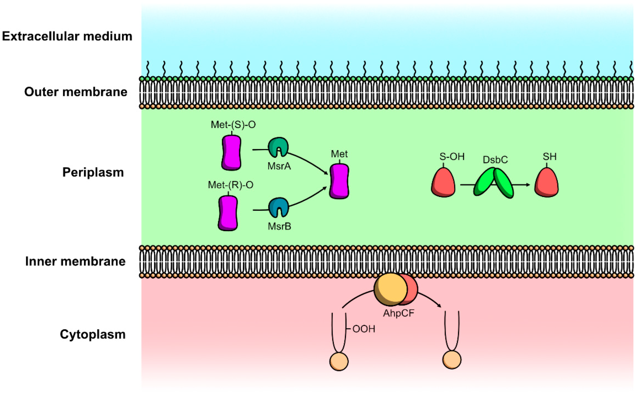
4.2.1. Protein Repair Mechanisms

Unrepaired sulfonic acid, Met-O, S-nitrosylation, and tyrosine nitration may lead to protein misfolding, inactivation, or degradation [74,75]. The periplasm is a more oxidizing environment than the cytoplasm [104], and most cysteine residues of the periplasmic proteins are engaged in disulfide bonds, preventing their oxidation by ROS. However, a small proportion of periplasmic proteins possess cysteine residues in their reduced thiol form [105], leaving them vulnerable to oxidation by ROS. Periplasmic proteins depending on a cysteine residue for their activity, are protected by a system regulating their redox state. Disulfide bond formation protein A (DsbA) possesses a labile disulfide bond transferred to proteins that translocate to the periplasm. The reduced DsbA is then recycled by the IM DsbB. DsbA preferentially introduces disulfide bonds between consecutive cysteine residues, resulting in potential mismatches [15]. Such mismatches are then corrected by the chaperone disulfide isomerase DsbC, which also repairs oxidized cysteine residues. One of the few periplasmic proteins possessing reduced thiols is YbiS, an L,D-transpeptidase catalyzing the covalent attachment between the lipoprotein Lpp and peptidoglycan discovered in E. coli. The catalytic activity of YbiS requires only the cysteine residue of this enzyme [106].
Met-O residues formed by ROS and RNS are reduced to methionine by the methionine sulfoxide reductase (Msr) system. This system revolves around the activity of MsrA and MsrB polypeptides, two methionine sulfoxide reductases with no identity at the sequence or structural level [107]. MsrA specifically reduces methionine-S-sulfoxide while MsrB specifically reduces methionine-R-sulfoxide (Figure 3). Both polypeptides are highly conserved between organisms; however, the genetic organization of msr varies widely among bacteria [108]. Some bacteria, such as E. coli, transcribe msrA and msrB independently, whereas others, such as Helicobacter pylori and Treponema pallidum, msrA and msrB compose a single transcription unit, resulting in the synthesis of a polypeptide with two active sites. The localization of active Msr polypeptides is also variable depending on the species. In H. pylori, Msr possesses SecA-dependent signal sequences involved in the secretion of Msr. The polypeptide is present in the cytoplasmic and membrane fractions but is only active in the membrane fraction [109,110]. In contrast, no activity was found in the membrane fraction of Actinobacillus actinomycetemcomitans, while the cytoplasmic and periplasmic fractions showed Msr activity [111]. Furthermore, Msr was also detected on the surface of bacteria for the first time on Ochrobactrum anthroporium [112].

4.2.2. PG Repair Mechanisms

Aconitases (Acn) are iron-sulfur proteins present in eukaryotes and prokaryotes. They are responsible for the conversion of citrate into isocitrate and, inversely, during the Krebs cycle [113]. When their [4FeS] cluster is lost, some Acns are also able to bind to specific mRNA sequences [114]. The best-studied Acn is the eukaryotic iron-responsive protein 1 (IRP1) [113]. The formation of apo-Acns occurs during exposure to ROS or RNS, and after long-term iron starvation [115]. In H. pylori, AcnB regulates the expression of the PG deacetylase PgdA under oxidative conditions [116]. PgdA is involved in the preservation of peptidoglycan integrity through the deacetylation of peptidoglycan N-acetylglucosamine residues [117]. This deacetylation prevents the formation of β-1,4 bonds between N-acetylglucosamine and N-acetylmuramic acid residues, which are the target of hydrolysis by lysozyme [117]. The deacetylation catalyzed by PdgA also mitigates host immune detection and increases the survival of H. pylori in murine stomachs [118].

4.2.3. Membrane Repair Mechanisms

In E. coli, the NADH peroxidase Ahp catalyzes the reduction of H2O2 to water using NADH, which is converted into unstable NAD+ [119]. In addition to its role in limiting the amount of ROS in the cell, Ahp also plays an important role in the protection of membranes against oxidative stress by reducing fatty acid peroxides (Figure 3) [120].

4.3. Envelope Stress Response (ESR)

Cellular homeostasis is dependent on the integrity of the cell envelope. Bacteria possess a wide array of mechanisms to cope with various stresses that can compromise their envelope integrity [121]. The various pathways involved in the maintenance of the envelope under stress form the ESR [14,122]. Outside of stressful environments, ESRs also play a housekeeping role by regulating envelope precursors and ensuring adequate envelope biogenesis. These pathways possess specificities but often overlap and are not necessarily specific to a single stress. Several of these ESRs regulate proteins involved in protection against oxidative-stress damage [85].

4.3.1. Cpx Complex in E. coli

The Cpx ESR is controlled by a two-component system involving CpxA and CpxR. CpxA is a histidine kinase sensor bound to the IM, and CpxR is its cytoplasmic DNA-binding response regulator [123]. Briefly, when activated, CpxA autophosphorylates conserved histidine residues, and the resulting phosphate is transferred to a conserved aspartate residue of CpxR. The phosphorylated CpxR then regulates the transcription of over 100 genes to relieve envelope stress. The Cpx response also impacts respiratory complexes in E. coli [124]. Notably, CpxRA causes the mitigation of PGRP-induced oxidative stress, which blocks respiratory chains [32].

4.3.2. σ Factors

Bacterial sigma factors are transcription factors that regulate a set of genes in response to specific stimuli, including cell-envelope stress responses. They are subunits of the RNA polymerase, allowing them to target specific promoters. Sigma factors are divided into the σ70 and σ54 families based on their similarities with those of E. coli in terms of structure, amino acids sequence, and mechanism of action. The σ70 family is split into four phylogenetic groups. Group 4, also called the extracytoplasmic function (ECF) sigma factors subfamily, is the largest of these groups and includes the more divergent sigma factors that are involved in various environmental responses. ECFs are extremely diverse, and a lot of them are still unknown from a mechanistic and functional point of view. ECFs take part in a wide range of processes, including starvation response, metal homeostasis, and virulence [14]. ECF activity is regulated by sequestration with a specific inhibitor called the anti-sigma factor, which is often membrane-bound and co-transcribed with its relative ECFs.
The most studied ECF RpoE, also called σE, is involved in the protection of the cell against various stresses. RpoE was originally discovered in E. coli for its ability to induce the transcription of the gene of the sigma factor RpoH, responsible for the transcription of heat-shock genes, at lethal temperatures (50 °C) [125]. Today, the best-characterized model of activation for RpoE is through the proteolysis of its anti-sigma factor RseA by both DegS and RseB. These two proteins are activated by misfolding of OM proteins and by the accumulation of LPS in the periplasmic compartment, respectively [126]. Overall, RpoE detects signals caused by OM malfunction and activates damage repair pathways. RpoE notably elicits an envelope stress response in response to the misfolding of OM proteins induced by oxidative stress. In the photosynthetic bacterium Rhodobacter sphaeroides, RpoH activates oxidative stress defenses under the control of RpoE in response to 1O2 [127]. RpoE also regulates the expression of the highly conserved chaperone protease, DepG. This periplasmic protein is found in most Gram-negative bacteria and ensures survival in animal hosts. The function of DepG is to degrade misfolded proteins in the periplasm to prevent their accumulation.
Other sigma factors respond to oxidative stress. Notably, oxidative stress activates the ECF σT, which promotes the transcription of various genes, including those involved in membrane homeostasis [128]. Furthermore, the ECF RpoS regulates the C-tail anchored IM protein ElaB in coordination with OxyR. While its precise function is unclear, ElaB is upregulated during oxidative stress and seems to play a role in the preservation of membrane integrity under oxidative stress, and is strongly upregulated in ampicillin-tolerant E. coli [129,130].

5. Conclusions

Exogenous ROS and RNS are a frequent occurrence in bacterial life, whether they come from (i) the immune system of their host, (ii) other microorganisms, or (iii) abiotic sources. The oxidative and nitrosative stresses induced by such molecules have a wide array of deleterious effects for bacteria, caused by a complex network of reactions both between these reactive species and bacterial biomolecules. ROS and RNS can notably alter the proteins and lipids composing the Gram-negative bacterial envelope. Maintaining the integrity of the envelope is crucial for these bacteria to survive under oxidative and nitrosative stresses, and a wide array of protection mechanisms have been selected by evolution to lessen and repair the damages caused by these stresses. Overall, the actors of both oxidative and nitrosative stresses resistance in the cytoplasm are better known than those acting at the level of the periplasm. However, more and more proteins involved in oxidative and nitrosative stresses resistance in the periplasm are discovered, often with functions mimicking their cytoplasmic counterparts. This suggests that the localization of the oxidative and nitrosative stresses response is a crucial factor for cell survival, even when dealing with reactive species able to pass through biological membranes.

Author Contributions

Conceptualization, T.C. and C.D.-P.; data research, T.C. and D.S.; data curation T.C. and C.D.-P.; writing—original draft preparation, T.C. and C.D.-P.; writing—review and editing, D.S. and S.C.; supervision, C.D.-P., funding acquisition, S.C. and C.D.-P. All authors have read and agreed to the published version of the manuscript.

Funding

This research was funded by a PhD grant from the GRR SESA (Région Normandie) and was supported by the European Union (European Regional Development Fund, ERDF: P106 N° 0002082 and 18E017461/18P02897), “Région Normandie” and “Évreux Portes de Normandie” (France).

Institutional Review Board Statement

Not applicable.

Informed Consent Statement

Not applicable.

Data Availability Statement

Not applicable.

Conflicts of Interest

The authors declare no conflict of interest. The funders had no role in the design of the study; in the collection, analyses, or interpretation of data; in the writing of the manuscript, or in the decision to publish the results.

References

  1. Barraud, N.; Hassett, D.J.; Hwang, S.-H.; Rice, S.A.; Kjelleberg, S.; Webb, J.S. Involvement of Nitric Oxide in Biofilm Dispersal of Pseudomonas aeruginosa. J. Bacteriol. 2006, 188, 7344–7353. [Google Scholar] [CrossRef] [PubMed] [Green Version]
  2. Ignarro, L.J.; Buga, G.M.; Wood, K.S.; Byrns, R.E.; Chaudhuri, G. Endothelium-derived relaxing factor produced and released from artery and vein is nitric oxide. Proc. Natl. Acad. Sci. USA 1987, 84, 9265–9269. [Google Scholar] [CrossRef] [PubMed] [Green Version]
  3. Palmer, R.M.J.; Ferrige, A.G.; Moncada, S. Nitric oxide release accounts for the biological activity of endothelium-derived relaxing factor. Nature 1987, 327, 524–526. [Google Scholar] [CrossRef] [PubMed]
  4. Raetz, C.R.H.; Whitfield, C. Lipopolysaccharide Endotoxins. Annu. Rev. Biochem. 2002, 71, 635–700. [Google Scholar] [CrossRef] [PubMed] [Green Version]
  5. Chevalier, S.; Bouffartigues, E.; Bodilis, J.; Maillot, O.; Lesouhaitier, O.; Feuilloley, M.G.J.; Orange, N.; Dufour, A.; Cornelis, P. Structure, function and regulation of Pseudomonas aeruginosa porins. FEMS Microbiol. Rev. 2017, 41, 698–722. [Google Scholar] [CrossRef]
  6. Vergalli, J.; Bodrenko, I.V.; Masi, M.; Moynié, L.; Acosta-Gutiérrez, S.; Naismith, J.H.; Davin-Regli, A.; Ceccarelli, M.; van den Berg, B.; Winterhalter, M.; et al. Porins and small-molecule translocation across the outer membrane of Gram-negative bacteria. Nat. Rev. Microbiol. 2020, 18, 164–176. [Google Scholar] [CrossRef]
  7. Sankaran, K.; Wu, H.C. Lipid modification of bacterial prolipoprotein. Transfer of diacylglyceryl moiety from phosphatidylglycerol. J. Biol. Chem. 1994, 269, 19701–19706. [Google Scholar] [CrossRef]
  8. Cole, G.B.; Bateman, T.J.; Moraes, T.F. The surface lipoproteins of gram-negative bacteria: Protectors and foragers in harsh environments. J. Biol. Chem. 2020, 296, 100147. [Google Scholar] [CrossRef]
  9. Wilson, M.M.; Bernstein, H.D. Surface-Exposed Lipoproteins: An Emerging Secretion Phenomenon in Gram-Negative Bacteria. Trends Microbiol. 2016, 24, 198–208. [Google Scholar] [CrossRef]
  10. Kondakova, T.; D’Heygère, F.; Feuilloley, M.J.; Orange, N.; Heipieper, H.J.; Duclairoir Poc, C. Glycerophospholipid synthesis and functions in Pseudomonas. Chem. Phys. Lipids 2015, 190, 27–42. [Google Scholar] [CrossRef]
  11. Silhavy, T.J.; Kahne, D.; Walker, S. The Bacterial Cell Envelope. Cold Spring Harb. Perspect. Biol. 2010, 2, a000414. [Google Scholar] [CrossRef] [PubMed]
  12. Pazos, M.; Peters, K. Peptidoglycan. In Bacterial Cell Walls and Membranes; Kuhn, A., Ed.; Subcellular Biochemistry; Springer International Publishing: Cham, Switzerland, 2019; pp. 127–168. ISBN 978-3-030-18768-2. [Google Scholar]
  13. Miller, S.I.; Salama, N.R. The gram-negative bacterial periplasm: Size matters. PLoS Biol. 2018, 16, e2004935. [Google Scholar] [CrossRef] [PubMed]
  14. Chevalier, S.; Bouffartigues, E.; Bazire, A.; Tahrioui, A.; Duchesne, R.; Tortuel, D.; Maillot, O.; Clamens, T.; Orange, N.; Feuilloley, M.G.J.; et al. Extracytoplasmic function sigma factors in Pseudomonas aeruginosa. Biochim. Biophys. Acta BBA Gene Regul. Mech. 2019, 1862, 706–721. [Google Scholar] [CrossRef] [PubMed]
  15. Arts, I.S.; Gennaris, A.; Collet, J.-F. Reducing systems protecting the bacterial cell envelope from oxidative damage. FEBS Lett. 2015, 589, 1559–1568. [Google Scholar] [CrossRef] [PubMed] [Green Version]
  16. Ferrer-Sueta, G.; Campolo, N.; Trujillo, M.; Bartesaghi, S.; Carballal, S.; Romero, N.; Alvarez, B.; Radi, R. Biochemistry of Peroxynitrite and Protein Tyrosine Nitration. Chem. Rev. 2018, 118, 1338–1408. [Google Scholar] [CrossRef]
  17. Ligeza, A.; Tikhonov, A.N.; Hyde, J.S.; Subczynski, W.K. Oxygen permeability of thylakoid membranes: Electron paramagnetic resonance spin labeling study. Biochim. Biophys. Acta 1998, 1365, 453–463. [Google Scholar] [CrossRef] [Green Version]
  18. Imlay, J.A. The molecular mechanisms and physiological consequences of oxidative stress: Lessons from a model bacterium. Nat. Rev. Microbiol. 2013, 11, 443–454. [Google Scholar] [CrossRef] [Green Version]
  19. Korshunov, S.; Imlay, J.A. Two sources of endogenous H2O2 in Escherichia coli. Mol. Microbiol. 2010, 75, 1389–1401. [Google Scholar] [CrossRef] [Green Version]
  20. Bielski, B.H.J. Reevaluation of the Spectral and Kinetic Properties of Ho2 and O2- Free Radicals. Photochem. Photobiol. 1978, 28, 645–649. [Google Scholar] [CrossRef]
  21. McCord, J.M.; Fridovich, I. Superoxide Dismutase: An enzymic function for erythrocuprein (hemocuprein). J. Biol. Chem. 1969, 244, 6049–6055. [Google Scholar] [CrossRef]
  22. Panov, A.V.; Dikalov, S.I. Cardiolipin, Perhydroxyl Radicals, and Lipid Peroxidation in Mitochondrial Dysfunctions and Aging. Oxid. Med. Cell. Longev. 2020, 2020, 1323028. [Google Scholar] [CrossRef] [PubMed]
  23. Glaeser, J.; Nuss, A.M.; Berghoff, B.A.; Klug, G. Chapter 4—Singlet Oxygen Stress in Microorganisms. In Advances in Microbial Physiology; Poole, R.K., Ed.; Academic Press: Cambridge, MA, USA, 2011; Volume 58, pp. 141–173. [Google Scholar]
  24. Garcia, X.; Stein, F. Nitric Oxide. Semin. Pediatr. Infect. Dis. 2006, 17, 55–57. [Google Scholar] [CrossRef] [PubMed]
  25. Fang, F.C.; Frawley, E.R.; Tapscott, T.; Vazquez-Torres, A. Bacterial Stress Responses during Host Infection. Cell Host Microbe 2016, 20, 133–143. [Google Scholar] [CrossRef] [PubMed] [Green Version]
  26. Flint, A.; Stintzi, A.; Saraiva, L.M. Oxidative and nitrosative stress defences of Helicobacter and Campylobacter species that counteract mammalian immunity. FEMS Microbiol. Rev. 2016, 40, 938–960. [Google Scholar] [CrossRef] [Green Version]
  27. Poole, R.K. Nitric oxide and nitrosative stress tolerance in bacteria. Biochem. Soc. Trans. 2005, 33, 176–180. [Google Scholar] [CrossRef]
  28. Hughes, M.N. Relationships between nitric oxide, nitroxyl ion, nitrosonium cation and peroxynitrite. Biochim. Biophys. Acta BBA Bioenerg. 1999, 1411, 263–272. [Google Scholar] [CrossRef] [Green Version]
  29. Koppenol, W.H.; Moreno, J.J.; Pryor, W.A.; Ischiropoulos, H.; Beckman, J.S. Peroxynitrite, a cloaked oxidant formed by nitric oxide and superoxide. Chem. Res. Toxicol. 1992, 5, 834–842. [Google Scholar] [CrossRef]
  30. Wink, D.A.; Ford, P.C. Nitric Oxide Reactions Important to Biological Systems: A Survey of Some Kinetics Investigations. Methods 1995, 7, 14–20. [Google Scholar] [CrossRef]
  31. Hughes, M.N. Chapter One—Chemistry of Nitric Oxide and Related Species. In Methods in Enzymology; Poole, R.K., Ed.; Globins and Other Nitric Oxide-Reactive Proteins, Part A; Academic Press: Cambridge, MA, USA, 2008; Volume 436, pp. 3–19. [Google Scholar]
  32. Kashyap, D.R.; Kuzma, M.; Kowalczyk, D.A.; Gupta, D.; Dziarski, R. Bactericidal peptidoglycan recognition protein induces oxidative stress in Escherichia coli through a block in respiratory chain and increase in central carbon catabolism. Mol. Microbiol. 2017, 105, 755–776. [Google Scholar] [CrossRef] [Green Version]
  33. Heesterbeek, D.A.C.; Muts, R.M.; van Hensbergen, V.P.; de Saint Aulaire, P.; Wennekes, T.; Bardoel, B.W.; van Sorge, N.M.; Rooijakkers, S.H.M. Outer membrane permeabilization by the membrane attack complex sensitizes Gram-negative bacteria to antimicrobial proteins in serum and phagocytes. PLOS Pathog. 2021, 17, e1009227. [Google Scholar] [CrossRef]
  34. Liu, H.; Zheng, F.; Cao, Q.; Ren, B.; Zhu, L.; Striker, G.; Vlassara, H. Amelioration of oxidant stress by the defensin lysozyme. Am. J. Physiol.-Endocrinol. Metab. 2006, 290, E824–E832. [Google Scholar] [CrossRef] [PubMed]
  35. Li, Y.M.; Tan, A.X.; Vlassara, H. Antibacterial activity of lysozyme and lactoferrin is inhibited by binding of advanced glycation–modified proteins to a conserved motif. Nat. Med. 1995, 1, 1057–1061. [Google Scholar] [CrossRef] [PubMed]
  36. Ragland, S.A.; Criss, A.K. From bacterial killing to immune modulation: Recent insights into the functions of lysozyme. PLoS Pathog. 2017, 13, e1006512. [Google Scholar] [CrossRef] [PubMed]
  37. Seki, M.; Iida, K.; Saito, M.; Nakayama, H.; Yoshida, S. Hydrogen peroxide production in Streptococcus pyogenes: Involvement of lactate oxidase and coupling with aerobic utilization of lactate. J. Bacteriol. 2004, 186, 2046–2051. [Google Scholar] [CrossRef] [Green Version]
  38. Spellerberg, B.; Cundell, D.R.; Sandros, J.; Pearce, B.J.; Idanpaan-Heikkila, I.; Rosenow, C.; Masure, H.R. Pyruvate oxidase, as a determinant of virulence in Streptococcus pneumoniae. Mol. Microbiol. 1996, 19, 803–813. [Google Scholar] [CrossRef]
  39. Liu, X.; Ramsey, M.M.; Chen, X.; Koley, D.; Whiteley, M.; Bard, A.J. Real-time mapping of a hydrogen peroxide concentration profile across a polymicrobial bacterial biofilm using scanning electrochemical microscopy. Proc. Natl. Acad. Sci. USA 2011, 108, 2668–2673. [Google Scholar] [CrossRef] [Green Version]
  40. Tong, H.; Chen, W.; Merritt, J.; Qi, F.; Shi, W.; Dong, X. Streptococcus oligofermentans inhibits Streptococcus mutans through conversion of lactic acid into inhibitory H2O2: A possible counteroffensive strategy for interspecies competition. Mol. Microbiol. 2007, 63, 872–880. [Google Scholar] [CrossRef]
  41. Baron, S.S.; Terranova, G.; Rowe, J.J. Molecular mechanism of the antimicrobial action of pyocyanin. Curr. Microbiol. 1989, 18, 223–230. [Google Scholar] [CrossRef]
  42. Hassan, H.M.; Fridovich, I. Mechanism of the antibiotic action pyocyanine. J. Bacteriol. 1980, 141, 156–163. [Google Scholar] [CrossRef] [Green Version]
  43. Mourenza, Á.; Gil, J.A.; Mateos, L.M.; Letek, M. Oxidative Stress-Generating Antimicrobials, a Novel Strategy to Overcome Antibacterial Resistance. Antioxidants 2020, 9, 361. [Google Scholar] [CrossRef]
  44. Keren, I.; Wu, Y.; Inocencio, J.; Mulcahy, L.R.; Lewis, K. Killing by bactericidal antibiotics does not depend on reactive oxygen species. Science 2013, 339, 1213–1216. [Google Scholar] [CrossRef] [PubMed] [Green Version]
  45. Mahoney, T.F.; Silhavy, T.J. The Cpx Stress Response Confers Resistance to Some, but Not All, Bactericidal Antibiotics. J. Bacteriol. 2013, 195, 1869–1874. [Google Scholar] [CrossRef] [PubMed] [Green Version]
  46. Albesa, I.; Becerra, M.C.; Battán, P.C.; Páez, P.L. Oxidative stress involved in the antibacterial action of different antibiotics. Biochem. Biophys. Res. Commun. 2004, 317, 605–609. [Google Scholar] [CrossRef] [PubMed]
  47. Goswami, M.; Mangoli, S.H.; Jawali, N. Involvement of Reactive Oxygen Species in the Action of Ciprofloxacin against Escherichia coli. Antimicrob. Agents Chemother. 2006, 50, 949–954. [Google Scholar] [CrossRef] [PubMed] [Green Version]
  48. Ladjouzi, R.; Bizzini, A.; Lebreton, F.; Sauvageot, N.; Rincé, A.; Benachour, A.; Hartke, A. Analysis of the tolerance of pathogenic Enterococci and Staphylococcus aureus to cell wall active antibiotics. J. Antimicrob. Chemother. 2013, 68, 2083–2091. [Google Scholar] [CrossRef]
  49. Dam, S.; Pagès, J.-M.; Masi, M. Stress responses, outer membrane permeability control and antimicrobial resistance in Enterobacteriaceae. Microbiol. Read. Engl. 2018, 164, 260–267. [Google Scholar] [CrossRef]
  50. Lesser, M.P. Oxidative stress in marine environments: Biochemistry and Physiological Ecology. Annu. Rev. Physiol. 2006, 68, 253–278. [Google Scholar] [CrossRef] [Green Version]
  51. Meslé, M.M.; Beam, J.P.; Jay, Z.J.; Bodle, B.; Bogenschutz, E.; Inskeep, W.P. Hydrogen Peroxide Cycling in High-Temperature Acidic Geothermal Springs and Potential Implications for Oxidative Stress Response. Front. Mar. Sci. 2017, 4, 130. [Google Scholar] [CrossRef] [Green Version]
  52. Santos, A.L.; Gomes, N.C.M.; Henriques, I.; Almeida, A.; Correia, A.; Cunha, Â. Growth conditions influence UVB sensitivity and oxidative damage in an estuarine bacterial isolate. Photochem. Photobiol. Sci. 2013, 12, 974–986. [Google Scholar] [CrossRef]
  53. Depayras, S.; Kondakova, T.; Heipieper, H.J.; Feuilloley, M.G.; Orange, N.; Duclairoir-Poc, C. The Hidden Face of Nitrogen Oxides Species: From Toxic Effects to Potential Cure? IntechOpen: London, UK, 2018; ISBN 978-1-78923-385-8. [Google Scholar]
  54. Skalska, K.; Miller, J.S.; Ledakowicz, S. Trends in NOx abatement: A review. Sci. Total Environ. 2010, 408, 3976–3989. [Google Scholar] [CrossRef]
  55. Chautrand, T.; Souak, D.; Kondakova, T.; Depayras, S.; Machour, N.; Heipieper, H.; Feuilloley, M.; Orange, N.; Poc, C. Air pollution and other environmental stresses: Gaseous NO2 exposure leads to specific alterations of Pseudomonas fluorescens. WIT Trans. Ecol. Environ. 2020, 244, 53–63. [Google Scholar]
  56. Depayras, S.; Kondakova, T.; Merlet-Machour, N.; Heipieper, H.J.; Barreau, M.; Catovic, C.; Feuilloley, M.; Orange, N.; Poc, C.D. Impact of gaseous NO2 on P. fluorescens strain in the membrane adaptation and virulence. Int. J. Environ. Impacts 2018, 1, 183–192. [Google Scholar] [CrossRef]
  57. Blee, J.A.; Roberts, I.S.; Waigh, T.A. Membrane potentials, oxidative stress and the dispersal response of bacterial biofilms to 405 nm light. Phys. Biol. 2020, 17, 036001. [Google Scholar] [CrossRef] [PubMed]
  58. Strahl, H.; Errington, J. Bacterial Membranes: Structure, Domains, and Function. Annu. Rev. Microbiol. 2017, 71, 519–538. [Google Scholar] [CrossRef]
  59. Dailey, F.E.; McGraw, J.E.; Jensen, B.J.; Bishop, S.S.; Lokken, J.P.; Dorff, K.J.; Ripley, M.P.; Munro, J.B. The Microbiota of Freshwater Fish and Freshwater Niches Contain Omega-3 Fatty Acid-Producing Shewanella Species. Appl. Environ. Microbiol. 2015, 82, 218–231. [Google Scholar] [CrossRef] [PubMed] [Green Version]
  60. Moravec, A.R.; Siv, A.W.; Hobby, C.R.; Lindsay, E.N.; Norbash, L.V.; Shults, D.J.; Symes, S.J.K.; Giles, D.K. Exogenous Polyunsaturated Fatty Acids Impact Membrane Remodeling and Affect Virulence Phenotypes among Pathogenic Vibrio Species. Appl. Environ. Microbiol. 2017, 83, e01415-17. [Google Scholar] [CrossRef] [PubMed] [Green Version]
  61. Jiang, J.-H.; Hassan, K.A.; Begg, S.L.; Rupasinghe, T.W.T.; Naidu, V.; Pederick, V.G.; Khorvash, M.; Whittall, J.J.; Paton, J.C.; Paulsen, I.T.; et al. Identification of Novel Acinetobacter baumannii Host Fatty Acid Stress Adaptation Strategies. mBio 2019, 10, e02056-18. [Google Scholar] [CrossRef] [Green Version]
  62. Shin, S.Y.; Bajpai, V.K.; Kim, H.R.; Kang, S.C. Antibacterial activity of bioconverted eicosapentaenoic (EPA) and docosahexaenoic acid (DHA) against foodborne pathogenic bacteria. Int. J. Food Microbiol. 2007, 113, 233–236. [Google Scholar] [CrossRef]
  63. Desbois, A.P.; Lawlor, K.C. Antibacterial Activity of Long-Chain Polyunsaturated Fatty Acids against Propionibacterium acnes and Staphylococcus aureus. Mar. Drugs 2013, 11, 4544–4557. [Google Scholar] [CrossRef] [Green Version]
  64. Eijkelkamp, B.A.; Begg, S.L.; Pederick, V.G.; Trapetti, C.; Gregory, M.K.; Whittall, J.J.; Paton, J.C.; McDevitt, C.A. Arachidonic Acid Stress Impacts Pneumococcal Fatty Acid Homeostasis. Front. Microbiol. 2018, 9, 813. [Google Scholar] [CrossRef]
  65. Krute, C.N.; Ridder, M.J.; Seawell, N.A.; Bose, J.L.Y. Inactivation of the exogenous fatty acid utilization pathway leads to increased resistance to unsaturated fatty acids in Staphylococcus aureus. Microbiology 2021, 165, 197–207. [Google Scholar] [CrossRef] [PubMed]
  66. Beavers, W.N.; Monteith, A.J.; Amarnath, V.; Mernaugh, R.L.; Roberts, L.J.; Chazin, W.J.; Davies, S.S.; Skaar, E.P. Arachidonic Acid Kills Staphylococcus aureus through a Lipid Peroxidation Mechanism. mBio 2019, 10, e01333-19. [Google Scholar] [CrossRef] [PubMed] [Green Version]
  67. Thibessard, A.; Fernandez, A.; Gintz, B.; Leblond-Bourget, N.; Decaris, B. Effects of rodA and pbp2b disruption on cell morphology and oxidative stress response of Streptococcus thermophilus CNRZ368. J. Bacteriol. 2002, 184, 2821–2826. [Google Scholar] [CrossRef] [PubMed] [Green Version]
  68. Wang, G.; Olczak, A.; Forsberg, L.S.; Maier, R.J. Oxidative Stress-induced Peptidoglycan Deacetylase in Helicobacter pylori. J. Biol. Chem. 2009, 284, 6790–6800. [Google Scholar] [CrossRef] [Green Version]
  69. Davis, M.M.; Brock, A.M.; DeHart, T.G.; Boribong, B.P.; Lee, K.; McClune, M.E.; Chang, Y.; Cramer, N.; Liu, J.; Jones, C.N.; et al. The peptidoglycan-associated protein NapA plays an important role in the envelope integrity and in the pathogenesis of the lyme disease spirochete. PLoS Pathog. 2021, 17, e1009546. [Google Scholar] [CrossRef]
  70. Chautrand, T.; Depayras, S.; Souak, D.; Kondakova, T.; Barreau, M.; Kentache, T.; Hardouin, J.; Tahrioui, A.; Thoumire, O.; Konto-Ghiorghi, Y.; et al. Gaseous NO2 induces various envelope alterations in Pseudomonas fluorescens MFAF76a. Sci. Rep. 2022; in press. [Google Scholar]
  71. Giacomucci, S.; Alvarez, L.; Rodrigues, C.D.A.; Cava, F.; Paradis-Bleau, C. Hydroxyl Radical Overproduction in the Envelope: An Achilles’ Heel in Peptidoglycan Synthesis. Microbiol. Spectr. 2022, 10, e01203-21. [Google Scholar] [CrossRef]
  72. Ezraty, B.; Gennaris, A.; Barras, F.; Collet, J.-F. Oxidative stress, protein damage and repair in bacteria. Nat. Rev. Microbiol. 2017, 15, 385–396. [Google Scholar] [CrossRef]
  73. Lee, B.C.; Gladyshev, V.N. The biological significance of methionine sulfoxide stereochemistry. Free Radic. Biol. Med. 2011, 50, 221–227. [Google Scholar] [CrossRef] [Green Version]
  74. Davies, M.J. The oxidative environment and protein damage. Biochim. Biophys. Acta 2005, 1703, 93–109. [Google Scholar] [CrossRef]
  75. Roos, G.; Messens, J. Protein sulfenic acid formation: From cellular damage to redox regulation. Free Radic. Biol. Med. 2011, 51, 314–326. [Google Scholar] [CrossRef] [PubMed]
  76. Suzuki, Y.J.; Carini, M.; Butterfield, D.A. Protein Carbonylation. Antioxid. Redox Signal. 2010, 12, 323–325. [Google Scholar] [CrossRef] [PubMed]
  77. Stadtman, E.R.; Levine, R.L. Protein oxidation. Ann. N. Y. Acad. Sci. 2000, 899, 191–208. [Google Scholar] [CrossRef] [PubMed]
  78. Grimsrud, P.A.; Xie, H.; Griffin, T.J.; Bernlohr, D.A. Oxidative Stress and Covalent Modification of Protein with Bioactive Aldehydes. J. Biol. Chem. 2008, 283, 21837–21841. [Google Scholar] [CrossRef] [Green Version]
  79. Sayre, L.M.; Lin, D.; Yuan, Q.; Zhu, X.; Tang, X. Protein adducts generated from products of lipid oxidation: Focus on HNE and one. Drug Metab. Rev. 2006, 38, 651–675. [Google Scholar] [CrossRef]
  80. Dalle-Donne, I.; Giustarini, D.; Colombo, R.; Rossi, R.; Milzani, A. Protein carbonylation in human diseases. Trends Mol. Med. 2003, 9, 169–176. [Google Scholar] [CrossRef]
  81. Ballesteros, M.; Fredriksson, Å.; Henriksson, J.; Nyström, T. Bacterial senescence: Protein oxidation in non-proliferating cells is dictated by the accuracy of the ribosomes. EMBO J. 2001, 20, 5280–5289. [Google Scholar] [CrossRef] [Green Version]
  82. Seth, D.; Hausladen, A.; Stamler, J.S. Anaerobic Transcription by OxyR: A Novel Paradigm for Nitrosative Stress. Antioxid. Redox Signal. 2020, 32, 803–816. [Google Scholar] [CrossRef]
  83. Bartesaghi, S.; Wenzel, J.; Trujillo, M.; López, M.; Joseph, J.; Kalyanaraman, B.; Radi, R. Lipid peroxyl radicals mediate tyrosine dimerization and nitration in membranes. Chem. Res. Toxicol. 2010, 23, 821–835. [Google Scholar] [CrossRef] [Green Version]
  84. Folkes, L.K.; Bartesaghi, S.; Trujillo, M.; Radi, R.; Wardman, P. Kinetics of oxidation of tyrosine by a model alkoxyl radical. Free Radic. Res. 2012, 46, 1150–1156. [Google Scholar] [CrossRef]
  85. Raivio, T.L. MicroReview: Envelope stress responses and Gram-negative bacterial pathogenesis. Mol. Microbiol. 2005, 56, 1119–1128. [Google Scholar] [CrossRef] [PubMed]
  86. Korshunov, S.S.; Imlay, J.A. A potential role for periplasmic superoxide dismutase in blocking the penetration of external superoxide into the cytosol of Gram-negative bacteria. Mol. Microbiol. 2002, 43, 95–106. [Google Scholar] [CrossRef] [PubMed]
  87. Yost, F.J.; Fridovich, I. An Iron-containing Superoxide Dismutase from Escherichia coli. J. Biol. Chem. 1973, 248, 4905–4908. [Google Scholar] [CrossRef]
  88. Keele, B.B.; McCord, J.M.; Fridovich, I. Superoxide Dismutase from Escherichia coli B: A new manganese-containing enzyme. J. Biol. Chem. 1970, 245, 6176–6181. [Google Scholar] [CrossRef]
  89. Benov, L.T.; Fridovich, I. Escherichia coli expresses a copper- and zinc-containing superoxide dismutase. J. Biol. Chem. 1994, 269, 25310–25314. [Google Scholar] [CrossRef]
  90. De Groote, M.A.; Ochsner, U.A.; Shiloh, M.U.; Nathan, C.; McCord, J.M.; Dinauer, M.C.; Libby, S.J.; Vazquez-Torres, A.; Xu, Y.; Fang, F.C. Periplasmic superoxide dismutase protects Salmonella from products of phagocyte NADPH-oxidase and nitric oxide synthase. Proc. Natl. Acad. Sci. USA 1997, 94, 13997–14001. [Google Scholar] [CrossRef] [Green Version]
  91. Cho, S.-H.; Parsonage, D.; Thurston, C.; Dutton, R.J.; Poole, L.B.; Collet, J.-F.; Beckwith, J. A new family of membrane electron transporters and its substrates, including a new cell envelope peroxiredoxin, reveal a broadened reductive capacity of the oxidative bacterial cell envelope. mBio 2012, 3, e00291-11. [Google Scholar] [CrossRef] [Green Version]
  92. Shapleigh, J.P. The Denitrifying Prokaryotes. In The Prokaryotes: Volume 2: Ecophysiology and Biochemistry; Dworkin, M., Falkow, S., Rosenberg, E., Schleifer, K.-H., Stackebrandt, E., Eds.; Springer: New York, NY, USA, 2006; pp. 769–792. ISBN 978-0-387-30742-8. [Google Scholar]
  93. Shiro, Y. Structure and function of bacterial nitric oxide reductases: Nitric oxide reductase, anaerobic enzymes. Biochim. Biophys. Acta BBA Bioenerg. 2012, 1817, 1907–1913. [Google Scholar] [CrossRef] [Green Version]
  94. Vázquez-Torres, A.; Bäumler, A. Nitrate, nitrite and nitric oxide reductases: From the last universal common ancestor to modern bacterial pathogens. Curr. Opin. Microbiol. 2016, 29, 1–8. [Google Scholar] [CrossRef] [Green Version]
  95. Poock, S.R.; Leach, E.R.; Moir, J.W.B.; Cole, J.A.; Richardson, D.J. Respiratory Detoxification of Nitric Oxide by the Cytochromec Nitrite Reductase of Escherichia coli. J. Biol. Chem. 2002, 277, 23664–23669. [Google Scholar] [CrossRef] [Green Version]
  96. Kern, M.; Volz, J.; Simon, J. The oxidative and nitrosative stress defence network of Wolinella succinogenes: Cytochrome c nitrite reductase mediates the stress response to nitrite, nitric oxide, hydroxylamine and hydrogen peroxide. Environ. Microbiol. 2011, 13, 2478–2494. [Google Scholar] [CrossRef] [PubMed]
  97. Pittman, M.S.; Elvers, K.T.; Lee, L.; Jones, M.A.; Poole, R.K.; Park, S.F.; Kelly, D.J. Growth of Campylobacter jejuni on nitrate and nitrite: Electron transport to NapA and NrfA via NrfH and distinct roles for NrfA and the globin Cgb in protection against nitrosative stress. Mol. Microbiol. 2007, 63, 575–590. [Google Scholar] [CrossRef] [PubMed]
  98. Runkel, S.; Wells, H.C.; Rowley, G. Chapter Three—Living with Stress: A Lesson from the Enteric Pathogen Salmonella enterica. In Advances in Applied Microbiology; Sariaslani, S., Gadd, G.M., Eds.; Academic Press: Cambridge, MA, USA, 2013; Volume 83, pp. 87–144. [Google Scholar]
  99. Borisov, V.B.; Verkhovsky, M.I. Oxygen as Acceptor. EcoSal Plus 2009, 3, 1–31. [Google Scholar] [CrossRef] [PubMed]
  100. Borisov, V.B.; Forte, E.; Siletsky, S.A.; Arese, M.; Davletshin, A.I.; Sarti, P.; Giuffrè, A. Cytochrome bd protects bacteria against oxidative and nitrosative stress: A potential target for next-generation antimicrobial agents. Biochemistry 2015, 80, 565–575. [Google Scholar] [CrossRef] [PubMed]
  101. Way, S.S.; Sallustio, S.; Magliozzo, R.S.; Goldberg, M.B. Impact of either Elevated or Decreased Levels of Cytochrome bd Expression on Shigella flexneri Virulence. J. Bacteriol. 1999, 181, 1229–1237. [Google Scholar] [CrossRef] [PubMed] [Green Version]
  102. Endley, S.; McMurray, D.; Ficht, T.A. Interruption of the cydB Locus inBrucella abortus Attenuates Intracellular Survival and Virulence in the Mouse Model of Infection. J. Bacteriol. 2001, 183, 2454–2462. [Google Scholar] [CrossRef] [PubMed] [Green Version]
  103. Jones-Carson, J.; Husain, M.; Liu, L.; Orlicky, D.J.; Vázquez-Torres, A. Cytochrome bd-Dependent Bioenergetics and Antinitrosative Defenses in Salmonella Pathogenesis. mBio 2016, 7, e02052-16. [Google Scholar] [CrossRef] [Green Version]
  104. Depuydt, M.; Messens, J.; Collet, J.-F. How Proteins Form Disulfide Bonds. Antioxid. Redox Signal. 2011, 15, 49–66. [Google Scholar] [CrossRef]
  105. Dutton, R.J.; Boyd, D.; Berkmen, M.; Beckwith, J. Bacterial species exhibit diversity in their mechanisms and capacity for protein disulfide bond formation. Proc. Natl. Acad. Sci. USA 2008, 105, 11933–11938. [Google Scholar] [CrossRef] [Green Version]
  106. Mainardi, J.-L.; Hugonnet, J.-E.; Rusconi, F.; Fourgeaud, M.; Dubost, L.; Moumi, A.N.; Delfosse, V.; Mayer, C.; Gutmann, L.; Rice, L.B.; et al. Unexpected Inhibition of Peptidoglycan LD-Transpeptidase from Enterococcus faecium by the β-Lactam Imipenem. J. Biol. Chem. 2007, 282, 30414–30422. [Google Scholar] [CrossRef] [Green Version]
  107. Sasindran, S.J.; Saikolappan, S.; Dhandayuthapani, S. Methionine sulfoxide reductases and virulence of bacterial pathogens. Future Microbiol. 2007, 2, 619–630. [Google Scholar] [CrossRef] [PubMed]
  108. Ezraty, B.; Aussel, L.; Barras, F. Methionine sulfoxide reductases in prokaryotes. Biochim. Biophys. Acta BBA Proteins Proteom. 2005, 1703, 221–229. [Google Scholar] [CrossRef] [PubMed]
  109. Alamuri, P.; Maier, R.J. Methionine sulphoxide reductase is an important antioxidant enzyme in the gastric pathogen Helicobacter pylori. Mol. Microbiol. 2004, 53, 1397–1406. [Google Scholar] [CrossRef] [PubMed]
  110. Cao, P.; McClain, M.S.; Forsyth, M.H.; Cover, T.L. Extracellular Release of Antigenic Proteins by Helicobacter pylori. Infect. Immun. 1998, 66, 2984–2986. [Google Scholar] [CrossRef] [PubMed] [Green Version]
  111. Mintz, K.P.; Moskovitz, J.; Wu, H.; Fives-Taylor, P.M.Y. Peptide methionine sulfoxide reductase (MsrA) is not a major virulence determinant for the oral pathogen Actinobacillus actinomycetemcomitansaa. Microbiology 2002, 148, 3695–3703. [Google Scholar] [CrossRef] [PubMed] [Green Version]
  112. Tamburro, A.; Robuffo, I.; Heipieper, H.J.; Allocati, N.; Rotilio, D.; Di Ilio, C.; Favaloro, B. Expression of glutathione S-transferase and peptide methionine sulphoxide reductase in Ochrobactrum anthropi is correlated to the production of reactive oxygen species caused by aromatic substrates. FEMS Microbiol. Lett. 2004, 241, 151–156. [Google Scholar] [CrossRef] [Green Version]
  113. Artymiuk, P.J.; Green, J. The Double Life of Aconitase. Structure 2006, 14, 2–4. [Google Scholar] [CrossRef]
  114. Goss, D.J.; Theil, E.C. Iron Responsive mRNAs: A Family of Fe2+ Sensitive Riboregulators. Acc. Chem. Res. 2011, 44, 1320–1328. [Google Scholar] [CrossRef] [Green Version]
  115. Soum, E.; Drapier, J.-C. Nitric oxide and peroxynitrite promote complete disruption of the [4Fe-4S] cluster of recombinant human iron regulatory protein 1. JBIC J. Biol. Inorg. Chem. 2003, 8, 226–232. [Google Scholar] [CrossRef]
  116. Austin, C.M.; Maier, R.J. Aconitase-Mediated Posttranscriptional Regulation of Helicobacter pylori Peptidoglycan Deacetylase. J. Bacteriol. 2013, 195, 5316–5322. [Google Scholar] [CrossRef] [Green Version]
  117. Dziarski, R. Recognition of bacterial peptidoglycan by the innate immune system. Cell. Mol. Life Sci. CMLS 2003, 60, 1793–1804. [Google Scholar] [CrossRef] [PubMed]
  118. Wang, G.; Maier, S.E.; Lo, L.F.; Maier, G.; Dosi, S.; Maier, R.J. Peptidoglycan Deacetylation in Helicobacter pylori Contributes to Bacterial Survival by Mitigating Host Immune Responses. Infect. Immun. 2010, 78, 4660–4666. [Google Scholar] [CrossRef] [PubMed] [Green Version]
  119. Dolin, M.I. DPNH peroxidase: Effector activities of DPN+. Biochem. Biophys. Res. Commun. 1977, 78, 393–400. [Google Scholar] [CrossRef] [Green Version]
  120. Farr, S.B.; Kogoma, T. Oxidative stress responses in Escherichia coli and Salmonella typhimurium. Microbiol. Rev. 1991, 55, 561–585. [Google Scholar] [CrossRef] [PubMed]
  121. Hews, C.L.; Cho, T.; Rowley, G.; Raivio, T.L. Maintaining Integrity Under Stress: Envelope Stress Response Regulation of Pathogenesis in Gram-Negative Bacteria. Front. Cell. Infect. Microbiol. 2019, 9, 313. [Google Scholar] [CrossRef] [PubMed] [Green Version]
  122. Mitchell, A.M.; Silhavy, T.J. Envelope stress responses: Balancing damage repair and toxicity. Nat. Rev. Microbiol. 2019, 17, 417–428. [Google Scholar] [CrossRef]
  123. Raivio, T.L. Everything old is new again: An update on current research on the Cpx envelope stress response. Biochim. Biophys. Acta BBA Mol. Cell Res. 2014, 1843, 1529–1541. [Google Scholar] [CrossRef] [Green Version]
  124. Guest, R.L.; Wang, J.; Wong, J.L.; Raivio, T.L. A Bacterial Stress Response Regulates Respiratory Protein Complexes To Control Envelope Stress Adaptation. J. Bacteriol. 2017, 199, e00153-17. [Google Scholar] [CrossRef] [Green Version]
  125. Erickson, J.W.; Gross, C.A. Identification of the sigma E subunit of Escherichia coli RNA polymerase: A second alternate sigma factor involved in high-temperature gene expression. Genes Dev. 1989, 3, 1462–1471. [Google Scholar] [CrossRef] [Green Version]
  126. Kim, D.Y. Two stress sensor proteins for the expression of sigmaE regulon: DegS and RseB. J. Microbiol. 2015, 53, 306–310. [Google Scholar] [CrossRef]
  127. Nuss, A.M.; Glaeser, J.; Klug, G. RpoHII Activates Oxidative-Stress Defense Systems and Is Controlled by RpoE in the Singlet Oxygen-Dependent Response in Rhodobacter sphaeroides. J. Bacteriol. 2009, 191, 220–230. [Google Scholar] [CrossRef] [PubMed] [Green Version]
  128. Alvarez-Martinez, C.E.; Lourenço, R.F.; Baldini, R.L.; Laub, M.T.; Gomes, S.L. The ECF sigma factor σT is involved in osmotic and oxidative stress responses in Caulobacter crescentus. Mol. Microbiol. 2007, 66, 1240–1255. [Google Scholar] [CrossRef] [PubMed]
  129. Guo, Y.; Li, Y.; Zhan, W.; Wood, T.K.; Wang, X. Resistance to oxidative stress by inner membrane protein ElaB is regulated by OxyR and RpoS. Microb. Biotechnol. 2019, 12, 392–404. [Google Scholar] [CrossRef] [PubMed] [Green Version]
  130. Sulaiman, J.E.; Lam, H. Proteomic Study of the Survival and Resuscitation Mechanisms of Filamentous Persisters in an Evolved Escherichia coli Population from Cyclic Ampicillin Treatment. mSystems 2020, 5, e00462-20. [Google Scholar] [CrossRef]
Figure 1. Reactions network of reactive oxygen and nitrogen species in biological conditions.
Microorganisms 10 00924 g001
Figure 2. Various mechanisms used by Gram-negative bacteria to detoxify ROS and RNS in the periplasmic space.
Microorganisms 10 00924 g002
Figure 3. Envelope repair systems of Gram-negative bacteria against oxidative and nitrosative stresses induced damage.
Microorganisms 10 00924 g003
Publisher’s Note: MDPI stays neutral with regard to jurisdictional claims in published maps and institutional affiliations.

