Article

Characterization of Bifidobacterium asteroides Isolates

1
Department of Agricultural, Food and Environment, University of Catania, 95123 Catania, Italy
2
ProBioEtna S.r.l., Spin-Off of University of Catania, 95123 Catania, Italy
3
Department of Biology, Faculty of Natural Sciences and Life, University of Oran1, Oran 31000, Algeria
4
Department of Biomedical and Biotechnological Sciences, University of Catania, 95123 Catania, Italy
5
Department of R&D, Local Noto Unit, Fidia Farmaceutici S.p.A., 96017 Noto, Italy
*
Authors to whom correspondence should be addressed.
Microorganisms 2022, 10(3), 655; https://doi.org/10.3390/microorganisms10030655
Submission received: 8 February 2022 / Revised: 4 March 2022 / Accepted: 11 March 2022 / Published: 18 March 2022
(This article belongs to the Section Veterinary Microbiology)

Abstract

:
Bifidobacteria have long been recognized as bacteria with probiotic and therapeutic features. The aim of this work is to characterize the Bifidobacterium asteroides BA15 and BA17 strains, isolated from honeybee gut, to evaluate its safety for human use. An in-depth assessment was carried out on safety properties (antibiotic resistance profiling, β-hemolytic, DNase and gelatinase activities and virulence factor presence) and other properties (antimicrobial activity, auto-aggregation, co-aggregation and hydrophobicity). Based on phenotypic and genotypic characterization, both strains satisfied all the safety requirements. More specifically, genome analysis showed the absence of genes encoding for glycopeptide (vanA, vanB, vanC-1, vanC-2, vanD, vanE, vanG), resistance to tetracycline (tetM, tetL and tetO) and virulence genes (asa1, gelE, cylA, esp, hyl).

1. Introduction

Bifidobacteria are Gram-positive, non-motile and non-spore-forming bacteria with a curved and clubbed shape, often branched with Y and V forms. Their genome harbors a high G + C content. Increased interest in LAB and bifidobacteria has been registered in the last two decades [1]. Bifidobacteria are commonly isolated from the gastrointestinal tract of various animals such as mammals, birds and insects. However, strains ascribed to Bifidobacterium denticolens, Bifidobacterium dentium, Bifidobacterium inopinatum and Bifidobacterium animalis subsp. lactis species have been isolated from different ecological niches, such as the human oral cavity [2,3,4] and fermented milk products [5,6]. Bifidobacterium asteroides, Bifidobacterium coryneforme and Bifidobacterium indicum are species that were isolated and characterized in the 1960s in pollinating insects, including honeybees Apis mellifera, Apis cerana and Apis dorsata [2,7]. In the last decades, the new species of Bifidobacterium bombi, Bifidobacterium actinocoloniiforme, Bifidobacterium bohemicum and Bifidobacterium commune have been identified from Bombus spp. gut [8]. While gut microbiota in some insects is acquired from food and the environment, bifidobacterial populations in honey, wasps and bumble bees are stable, and different molecules have been characterized as mediators of their cross-talk with the host [8]. In fact, similarly to humans, honeybees have a core in their gut microbiota that matures in the early stages of their life and remains stable throughout adult life. The gut of mammals, birds and insects is known as an anaerobic environment, and bifidobacterial growth has been described as being inhibited by oxygen, which exerts toxic effects. However, the Bifidobacterium asteroides species has been found to tolerate oxygen levels higher than 20%, which represents the upper limit of tolerance for Bifidobacterium animalis subsp. lactis, Bifidobacterium boum, Bifidobacterium psychraerophilum and Bifidobacterium thermophilum [7]. While the long history of safe use of some species, such as Bifidobacterium adolescentis, Bifidobacterium animalis, Bifidobacterium longum, Bifidobacterium breve and Bifidobacterium bifidum, has led to them being proposed for European Safety Authority (EFSA) Qualified Presumption of Safety (QPS) status, other species, such as Bifidobacterium dentium, cannot be used because they are linked to cariogenic processes. Moreover, the current guidelines do not authorize the use in humans of strains of non-human origin [9].
Recently, researchers have turned their attention to different species of honeybees including Apis mellifera, on account of their elevated mortality rates. Honeybees and other pollinating species play a key role in production and global food security, which amounts to an estimated value of 150 billion euro. Many funding programs for pollinator insects have set up grants for the protection of specific species and their habitats. Decades-long investigations on insect physiology have made it possible to understand the single and combined effects of environmental and host stress factors such as pests, pathogens, toxins and nutrient-limited food sources. Moreover, even if the vast majority of probiotic strains have been isolated from human or fermented foods, mainly dairy, the scientific research has been recently focused on the selection of new probiotics from unconventional sources such as meat, fruit, vegetables, cereals, honeybees and beehive products. In addition, the Internal Scientific Association for Probiotics and Prebiotics (ISAPP) recognized probiotic properties of strains belonging to Akkermansia muciniphila, Faecalibacterium prausnitzii, Roseburia spp. and Eubacterium hallii, not previously included in the QPS list [10,11,12,13,14].
Nowadays, studies on the gut microbiota of pollinators have clarified the role of the genera Bifidobacterium and Lactobacillus in immunity preservation, disease tolerance and resistance to environmental stressors [15]. The core of honey bees’ gut microbiota, whatever their geographical origin, is composed of Gilliamella apicola, Lactobacillus Firm-5 (L. melliventris, L. kimbladii, L. kullabergensis, L. helsinborgensis), Lactobacillus Firm-4 (L. mellis, L. mellifer), Snodgrassella alvi and Bifidobacterium asteroides [16]. The gut microbiota and its symbiotic bacteria exert a key role in the innate immunity system of bees, despite the production of antimicrobial peptides with a highly selective activity against pathogenic species [15]. The disruption of the microbial balance in the bee’s gut is due to the perturbation of insect immunity through treatments with microbicidal or microbiostatic compounds; the exposure to pesticides or herbicides at a sublethal concentration (called hidden treatment) leads to a decrease in Firmicutes and Actinobacteria and to an increase in Gammaproteobacteria, such as Gilliamella apicola and Escherichia coli. Gut microbiota dysbiosis and the dysregulation of the innate immunity system expose bees to attacks by parasites such as fungi (Nosema spp.) and trypanosomes (Crithidia and Lotmaria spp.). Moreover, honeybee gut plays a key role in their metabolic activity and nutrition status, especially impacting on vitamin biosynthesis.
Based on knowledge of human gut dysbiosis, two strategies have recently been used to control honeybee dysbiosis and dysregulation: a prebiotic strategy through diet supplementation with sucrose-based solutions or pollen-based feed, and gut microbiota manipulation through supplementation with safe strains from the Bifidobacterium and Lactobacillus genera [16,17].
There is a lack of studies in the literature reporting the selection of Bifidobacterium strains as feed supplementation for honeybees. However, several regulatory authorities have suggested that these strains could possess some important properties that make them suitable for human use. Studying the resistance genes is important for the confirmation of the isolates as non-resistant bacteria. Antibiotic resistance can be due to phenotypic or genotypic features, and the antibiotic resistance profiles of the Bifidobacterium asteroides species could be the result of long-term exposure of honeybees to antibiotics [18]. Recent studies have indicated horizontal antibiotic resistant gene transfer between bacteria residing in the guts of humans and animals. These genes are delivered by mobile genetic elements; the collection of the mobile elements of a microorganism has recently been defined as a mobilome and includes transposases, insertion elements, plasmids and prophages (bifidoprophages). The entire mobilome and resistome have recently been reconstructed. The most abundant antibiotic resistant gene in the Bifidobacterium resistome is the one conferring glycopeptide resistance (43%, 5999 putative enzymes), followed by the methyltransferase class (19%, 2618 putative enzymes), β-lactamase class (17%, 2437 putative enzymes), tetracycline class (16%, 2178 putative enzymes), sulfonamide class (4%) and metronidazole and aminoglycosides classes (0.5%). Analysis of the putative mobile resistome of the Bifidobacterium genus reveals the presence of: hypothetic conjugative transposon that harbors the tetW gene, responsible for protection from tetracycline activity; putative prophage-like elements, which harbor the BacA gene responsible for protection from bacitracin; transposase that harbors a 23S rRNA methylase, which confers resistance towards erythromycin and clindamycin. Moreover, among the predicted transposase encoding genes, the presence of the vanZ gene has been observed, which may confer low-level resistance to glycopeptide antibiotics [19].
One of the main selection criteria for defining the probiotic action of bifidobacterial and lactic acid bacteria is adhesion to human intestinal cells. Adherent strains could exert metabolic and immunomodulatory functions, stabilizing the intestinal mucus barrier and providing the competitive exclusion of pathogenic bacteria [20,21,22,23,24]. Exopolysaccharides can be involved in the adhesion to mucus [25]. The presence of genes and gene clusters encoding for pilus-like structures has also recently been demonstrated in bifidobacterial genomes [26]. The degree of in vitro adhesion depends on many factors, such as the substrate used for the assay (abiotic or biotic surface) and growth medium composition.
Bifidobacterium asteroides has peculiar properties, such as the ability to tolerate oxygen and other metabolic features for carbohydrate metabolism, which have not been already determined in the genus of Bifidobacterium. Genome analysis of the strain Bifidobacterium asteroides PLR2011species demonstrated the presence of a “malolactate fermentation pathway”, responsible for the conversion of malic acid to lactate, with the addition of the characteristic “fructose-6-phosphate pathway”. The latter is uniquely responsible for fructose and glucose fermentation and contains fructose-6-phosphate phosphoketolase (F6PPK), but glucose-6-phosphate is absent [7] Moreover, the Bifidobacterium asteroides species is reported to possess catalase [27].
In the present study, two Bifidobacterium strains isolated from honeybee gut were evaluated as to their safety for human use and other properties.

2. Materials and Methods

2.1. Reference Strains and Culture Conditions

The hemolytic Gram-positive strains Streptococcus pyogenes ATCC 19615 and Streptococcus pneumoniae ATCC 6303 were cultured on Brain Heart Infusion (BHI, Becton Dickinson GmbH, Heidelberg, Germany) at 37 °C under 5% CO2 conditions. Escherichia coli ATCC 25922, E. coli ATCC 9637, Staphylococcus aureus ATCC 6538 and ATCC 29213 were routinely cultured on Trypticase Soy Broth medium (Oxoid, Milan, Italy) at 37 °C under aerobic conditions. Listeria monocytogenes DSM 12464, Salmonella enterica serovar typhimurium ATCC 14028 and Salmonella enterica subsp. enterica serovar enteritidis ATCC 13076 were revitalized in BHI broth at 30 °C under aerobic conditions. The probiotic strain Bifidobacterium animalis BB12 (Christian Hansen AS, Hoersholm, Denmark), was grown in Bifidus Selective Medium Broth (BSM, Sigma Aldrich, Milan, Italy) or in Man Rogosa and Sharpe (MRS, Oxoid, Milan, Italy) supplemented with 0.25% L-cysteine (Sigma Aldrich, Milan, Italy) (MRSc) at 37 °C, under anaerobic conditions, and included in every experiment for comparison.

2.2. Identification of Isolates

Two bacterial isolates named BA15 and BA17, previously obtained from honeybee gut and subjected to morphological characterization (unpublished data), were analyzed for physiological and biochemical properties (catalase, oxidase, spore formation, gelatinase activities, production of indole, NH3 from arginine and CO2 from glucose) and with the use of the API rapid ID 32 A kit (BioMérieux, Grassina, Italy). Based on the enzymatic profile, typical of the Bifidobacterium genus, both isolates were identified at species level. The total genomic DNA was extracted following the method previously described [28] and 16S rDNA was amplified using the primer pairs Bif164 and Bif662 [29,30]. PCR products were purified using a Qiaquick PCR purification kit (Qiagen Hilden, Germany) and subjected to 16S rDNA sequencing. Comparison with sequences held in the BLAST database allowed both stains to be ascribed to the Bifidobacterium asteroides species. The accession numbers of the sequenced strains were as follows (code and identity percentage of isolates in parentheses): Bifidobacterium asteroides MG650026.1 (BA15, 99.60%) and Bifidobacterium asteroides CP017696.1 (BA17, 99.41%).

2.3. Safety Assessment

2.3.1. Antimicrobial Susceptibility and MIC Determination

Antimicrobial susceptibility was determined according to the ISO 10932:2010 [31] broth microdilution procedure using eight antimicrobial agents (ampicillin sodium salt, chloramphenicol, clindamycin hydrochloride, erythromycin, gentamicin sulphate, streptomycin sulphate salt, tetracycline, vancomycin), all purchased from Sigma Aldrich (St. Louis, MO, USA). The Minimum Inhibitory Concentration (MIC), defined as the lowest concentration of antibiotic giving a complete inhibition of visible growth in comparison to an antibiotic-free control well, was determined by the microdilution method according to Russo et al. (2018) [32]. The experiments were conducted in triplicate.
The genomes of the BA15 and BA17 strains were analyzed for the presence of antibacterial resistant genes and other gene associations that can influence the safety profile of the strains. The analysis was performed using the Pathosystems Resource Integration Center (PATRIC) database [33].

2.3.2. PCR Assay on Virulence Factors

The presence of virulence genes (asa1, cylA, gelE, hyl, esp) was evaluated by multiplex PCR following the method described by Vankerckhoven et al. (2004) [34] and using the primer pairs reported in Table 1. PCR reactions were performed in a final volume of 25 µL containing 1.0 µL of genomic DNA, 1.0 µL of each primer (100 mM) and 12.5 µL of 5-PRIME MasterMix including HotStarTaq DNA polymerase (Eppendorf, Milan, Italy). Amplification was carried out as follows: an initial activation step at 94 °C for 15 min, where DNA polymerase was activated; 30 amplification cycles of denaturation (94 °C for 1 min), annealing (56 °C for 1 min) and extension (72 °C for 1 min); followed by one final extension step consisting of 10 min at 72 °C. PCR products were analyzed by electrophoresis in a 1.5% w/v of agarose gel for 1 h at 90 V in 1.0 × TAE buffer solution. After treatment with ethidium bromide solution, the amplicons were detected by UV light. The Lamba DNA/HindIII marker (Thermo Fisher Scientific, Rodano, Italy) and the Φ174 DNA Marker Hae III Digest (Sigma Aldrich, Milan, Italy) were used as DNA ladders [35].

2.3.3. Other Assessments: Hemolytic Activity and DNase and Gelatinase Activities

B. asteroides BA15 and BA17 strains, grown in BSM broth for 18–24 h at 37 °C under anaerobic conditions, were streaked onto blood agar plates containing sheep blood (Biolife, Milan, Italy), and anaerobically incubated at 37 °C for 24–48 h. After incubation, the plates were visually analyzed for the presence or absence of microbial hemolytic properties and distinguished as β-hemolysis, α-hemolysis or γ-hemolysis, based on the appearance of a clear zone, green halo or no zones around the colonies, respectively [28]. S. pyogenes ATCC 19615 and S. pneumoniae ATCC 6303 were used as positive controls. DNase and gelatinase activities were tested in triplicate, as suggested by Pino et al. (2019) [36]. In detail, to evaluate the DNase activity, 5 μL of a liquid culture was spotted onto DNase agar plates (Oxoid, Milan, Italy). After incubation at 37 °C for 48 h, plates were overlain with HCl 1 N for 5 min. Gelatinase activity was assessed by spotting 5 μL of an overnight culture of the strain on BHA supplemented with 0.04% gelatine.

2.4. Hydrophobicity, Auto-Aggregation and Co-Aggregation Abilities

Hydrophobicity (H%), auto-aggregation (Auto-A%) and co-aggregation (Co%) abilities were tested as described by Pino et al. (2021) [37]. Hydrophobicity was determined as bacterial adhesion to hydrocarbons (BATH) using xylene. E. coli ATCC 25922, S. aureus ATCC 6538 and S. typhimurium ATCC 14028 were used as the pathogenic strains in the co-aggregation assay.

2.5. Adhesion on Abiotic Surface

Adhesion capability was tested using overnight cell cultures grown in Man Rogosa and Sharpe (MRS, Oxoid, Milan, Italy) broth supplemented with 0.25% L-cysteine (Sigma Aldrich, Milan, Italy) (MRSc) and incubated at 37 °C under anaerobic conditions (80% N2, 10% CO2 and 10% H2) using the AnaeroGen sachet (Oxoid, Milan, Italy). Then, 200 μL of a 1:100 dilution of each culture was transferred to a 96-well micro-ELISA plate (number of replicates: 32) and, after regular shaking, the absorbance at t0 (starting time) was read at 600 nm (ELx808, BioTek-software Gen5) and the plate was incubated. After incubation at 37 °C for 120 min under aerobic conditions, the plate was washed twice using sterile PBS to remove non-adherent bacteria and air-dried for 60 min at 60 °C. Then, 200 μL of a solution of 0.25% crystal violet was added to each well and the plate was incubated at room temperature for 15 min. After incubation, the plate was rinsed twice using Milli-Q water (Millipore, Milan, Italy) to remove excess dye and 200 μL of a 98% ethanol solution was added to each well. The absorbance was read at 570 nm. The adherence index was calculated as follows: Abs570/Abs600 [38].

2.6. Alignment for VanZ Putative Gene

The putative VanZ gene sequences for the Bifidobacterium asteroides BA15 and Bifidobacterium asteroides BA17 genomes were compared to those of reference strains Bifidobacterium asteroides DSM 20089 (CP017696.1:1959032-1960146) and Bifidobacterium asteroides PRL2011 (CP003325.1:10964-12078) using a multiple sequence alignment method with reduced time and space complexity (MUSCLE) and DNASTAR software [39].

2.7. Antagonistic Activity against Pathogens

2.7.1. Agar Diffusion Assay

Antagonistic activity was evaluated using E. coli ATCC 25922, E. coli ATCC 9637, S. aureus ATCC 6538, S. aureus ATCC 29213 and S. typhimurium ATCC 14028 as target bacteria. The assay was performed by the agar spot test [40], using the cell-free culture supernatants obtained as reported by Argyri et al. (2013) [41]. After incubation for 48 h, the appearance of inhibition zones was visually detected and, based on the diameter size, results were expressed as: (−) no inhibition zone; (+) inhibition zone < 5 mm; (++) inhibition zone > 5 mm [21].

2.7.2. Antibacterial Activity

Antagonistic activity was evaluated using E. coli ATCC 9637 and S. aureus ATCC as models for Gram-negative and Gram-positive bacteria, respectively. The assay was performed modifying the broth microdilution method described by CLSI M7-A7 for bacteria [24,42], using the cell-free supernatants obtained as previously described. Incubation was performed under aerobic conditions at 37 °C for 24 h. The absorbance was read at 630 nm (ELx808, BioTek-software Gen5) after regular shaking with a frequency every 30 min. The killing curves were created by plotting OD values versus time, and bacterial growth kinetics were studied using GraphPad Prism 8 software (Version 8.2.1_279). Each assay was performed three times in duplicate.

2.8. Statistical Analysis

All data were expressed as a mean and standard deviation of three independent experiments. Data were subjected to one-way ANOVA followed by Tukey’s Multiple Comparison Test and differences were considered statistically significant at α = 0.05.

3. Results

3.1. Enzymatic Profile

Table 2 shows the enzymatic profile of the tested B. asteroides BA15 and BA17 strains as well as the percentage of the positive reactions for the Bifidobacterium genus, carried out according to the manufacturer’s instructions. The results of the assay, analyzed by apiweb™ suggested an enzymatic profile for both strains that was characteristic of the Bifidobacterium genus (% ID 99.9). More precisely, the ID profile was 4537033705 for the BA15 strain, whereas the ID profile for the BA17 strain was 4517033505 (Table 2). Based on the fermentative profile (Table 3), both strains showed similar biochemical properties except for their ability to ferment L-Arabinose.

3.2. Antibiotic Resistance Profile, Virulence Factors and Other Biochemical Properties

The antibiotic susceptibility profile for both B. asteroides BA15 and B. asteroides BA17 strains, based on EFSA criteria [43], showed susceptibility to the main tested antimicrobials, except for ampicillin, vancomycin and chloramphenicol (Table 4).
The PCR-based approach did not reveal the presence of genes encoding for gelatinase (gelE), hyaluronidase (hyl), aggregation substance (asa1), enterococcal surface protein (esp) and cytolysin (cylA).
The results obtained from the analysis of the bacterial genome using PATRIC bioinformatic services showed the absence of antibiotic resistant genes in the BA15 and BA17 genomes and the presence of a metabolic pathway responsible for vancomycin biosynthesis (Figure 1). The key enzyme was the dTDP-glucose 4,6-dehydratase (EC number 4.2.1.46), encoded by a gene located from nucleotide 1368973 to 1370001 for the BA15 strain, and from nucleotide 319105 to 320202 for the BA17 strain. The expression of this enzyme and production of vancomycin needs further investigation; however, this metabolic pathway could explain the resistance to vancomycin.
None of the tested Bifidobacterium asteroides strains showed the ability to produce DNase and gelatinase or to exert hemolytic activity.
Antagonistic activity against food spoilage and pathogenic bacteria shown by the BA15 and BA17 strains is reported in Table 5. Overall, both BA15 and BA17 strains showed antagonistic activity against all tested pathogens, with the exception of the BA15 strain, which did not show any antagonistic activity against S. aureus ATCC 6538 (Table 5).

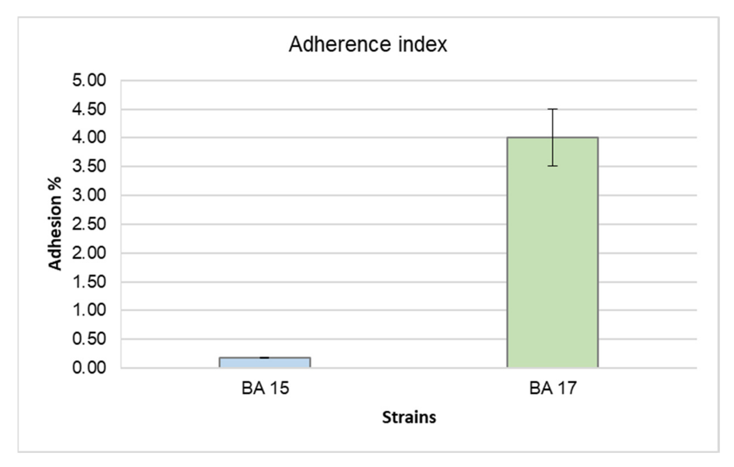
3.3. Adhesion to Abiotic Surfaces

Figure 2 shows the ability of B. asteroides BA15 and BA17 strains to adhere on abiotic surfaces (expressed as adherence index). Overall, both tested strains showed adhesion abilities and the highest adherence index was exhibited by the BA17 strain (4.00).

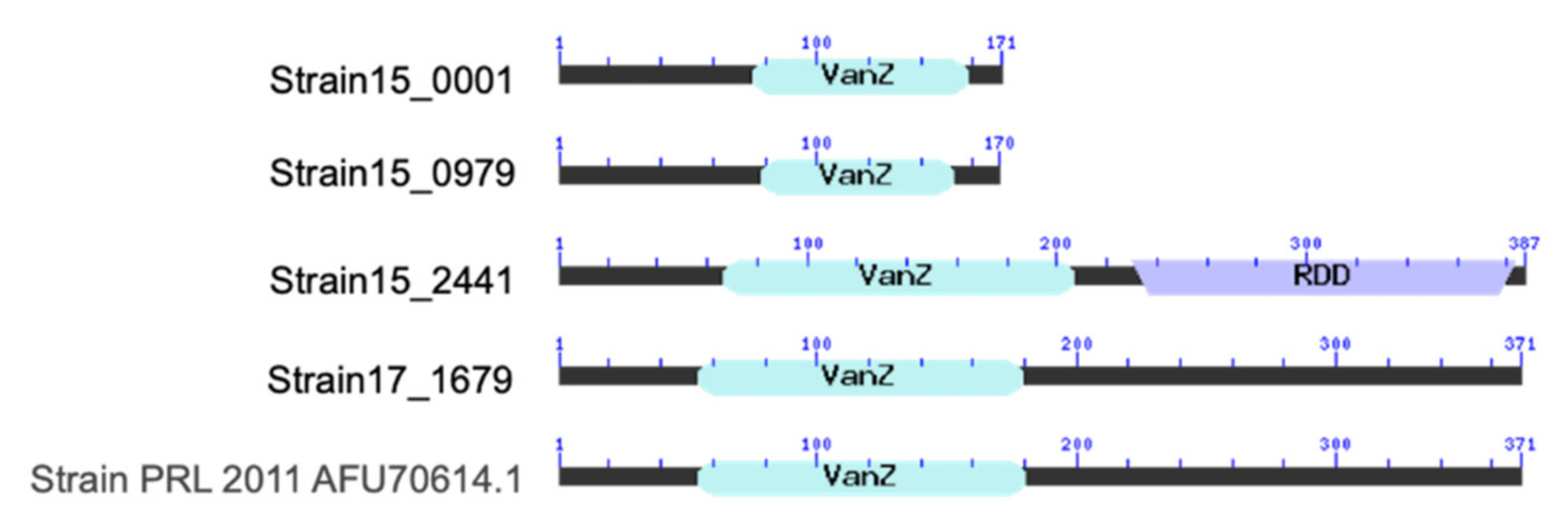
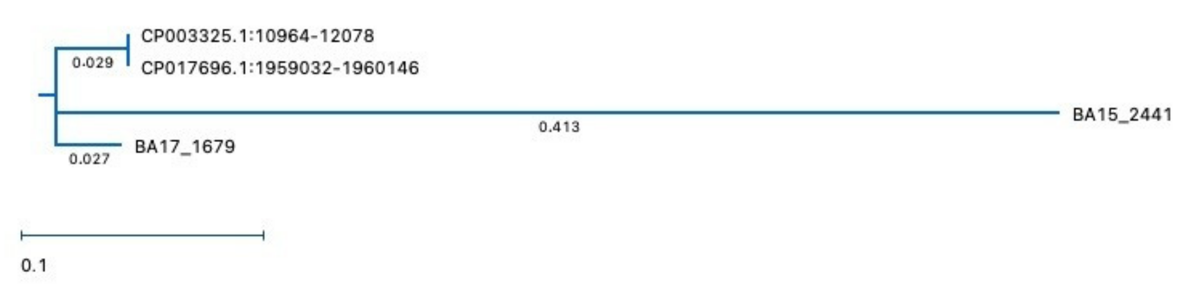
3.4. Alignment for VanZ Putative Gene

The vanZ gene is an orthologous gene belonging to the glycopeptide resistance protein family (vanZ-A, vanZ-F, vanZ-Pt and vanZ-1). Both tested stains showed the presence of a putative vanZ gene. More specifically, vanZ was detected in the genome of the B. asteroides BA15 strain in three different positions (Strain15_0001: 457-972; Strain15_0979: 1060104-1060616; Strain15_2441: 2576152-2577315), whereas it was only identified once in the B. asteroides BA17 genome (17_1679: 2161147-2162262). On the basis of vanZ nucleotide analysis using blastn, the Strain15_0001, Strain15_0979 and Strain15_2441 genes showed a high (100% identity) homology with the genes present in the Lactobacillus plantarum genomes. The gene 17_1679 showed a high (93.9% identity) homology with the gene present in the Bifidobacterium asteroides genomes.
Figure 3 shows the putative proteins encoded by vanZ from B. asteroides BA15 and B. asteroides BA17. The number of amino acids of the putative protein encoded by the vanZ gene results as being 171 for gene Strain15_0001, 170 for gene Strain15_0979, 387 for gene Strain15_2441 and 371 for gene Strain17_1679, which is equal to those for the control strain PRL 2011. All proteins showed the conserved domain vanZ, which belongs to the vanZ-like family and contains several examples of the vanZ protein, as well as examples of phosphotransbutyrylases; however, they differ in amino acid length. Moreover, the putative protein encoded by vanZ from gene Strain15_2441 also showed the RDD domain, which is a family of proteins that contains three highly conserved amino acids (one arginine and two aspartates). This region contains two predicted transmembrane regions: the arginine occurs at the N-terminus of the first helix and the first aspartate occurs in the middle of this helix. The molecular function of the RDD region is unknown; however, this region may be involved in the transport of a set of ligands that are still not well identified.
Table 6 shows the ratio between percentage identity and distance of the vanZ genes of BA15 and BA17 in comparison with the genes present in the genome of the reference strain PRL2011. The MUSCLE analysis algorithm showed a high homology (94%) between the putative vanZ gene from B. asteroides BA17 and the reference strain B. asteroides PRL2011 (CP003325.1:10964-12078). Instead, a lower homology percentage (from 46.5% to 49.8%) was shown by VanZ genes from the Bifidobacterium asteroides BA15 genome (Table 6).
Figure 4 shows the unrooted phylogenetic tree relating the vanZ genes of BA15 and BA17 strains and their distance with the PRL2011 strains. Moreover, the nucleotide sequences of the putative vanZ gene of BA15 and BA17 strains, aligned using MUSCLE (MegAlign Pro of DNAstar), compared with DSM 20089 and PRL2011 strains, are shown in the Supplementary Materials (Figure S1).

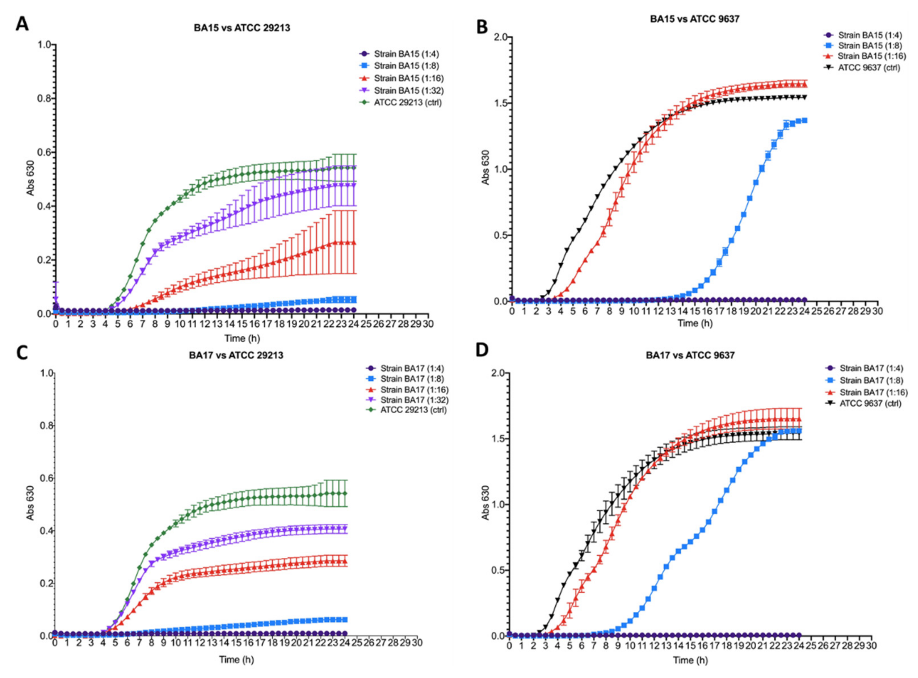
3.5. Antibacterial Activity

Figure 5 shows the antibacterial effect of the BA15 and BA17 strains against S. aureus ATCC 29213 and E. coli ATCC 9637. Both tested strains showed high activity against the Gram-positive S. aureus ATCC 29213, with a dilution factor of wild supernatant from 1:4 to 1:32 ratio (Figure 5A,C). Both tested supernatants resulted as being active towards the Gram-negative E. coli ATCC 9637 in a dilution range from 1:4 to 1:8 (Figure 5B,D). The antibacterial activity against S. aureus ATCC 29213 was exerted in the dilution range from 1:4 to 1:8, whereas, against E. coli ATCC 9637, it was only exerted at the higher tested concentration (dilution 1:4 of the wild supernatants). The supernatant obtained from BA15 and BA17 showed an inhibitory activity towards S. aureus ATCC 29213 when it was tested at the dilution range from 1:16 to 1:32, and towards E. coli ATCC 9637 when it was tested at a dilution of 1:8. In particular, both BA15 and BA17 appeared to reduce the growth of S. aureus ATCC 29213 and E. coli ATCC 9637, which after 24 h of incubation reported an OD630 lower than the untreated control. The growth of E. coli ATCC 9637 was delayed by about 14 h from the time of incubation when treated with BA15 and by about 9 h (from the time of incubation) when treated with BA17, although after 24 h, the OD630 values were the same as those of the untreated control.

3.6. Auto-Aggregation, Co-Aggregation and Hydrophobicity Abilities

Table 7 summarizes the surface characteristics (hydrophobicity, auto-aggregation and co-aggregation) of the B. asteroides BA15 and BA17 tested strains, compared to those exhibited by the Bifidobacterium animalis BB12 strain. The BA15 and BA17 strains showed an auto-aggregation percentage, similar to that displayed by the BB12 reference strain. Both BA15 and BA17 strains also exhibited the ability to co-aggregate with the tested pathogens. In particular, both the B. asteroides BA15 and BA17 strains showed the highest co-aggregation percentage with Listeria monocytogenes ATCC 12466 (Table 7) when compared to the B. animalis BB12 strain. A variable degree of hydrophobicity was observed, with the highest percentage exhibited by the BB12 and BA17 strains.

4. Discussion

The gut microbiota of honeybees and insect pollinators is still an unexplored ecosystem. Recent findings on human gut microbiota have paved the way for a better understanding of its role in other living species on Earth. The evolutionary role of bifidobacteria among humans and animals seems to be related to their ability to ferment complex non-digestible carbohydrates and to modulate the host immune system through changes in innate and/or adaptive immune responses [44,45]. The distribution of specific species of bifidobacteria across the human lifetime has been recently studied [46] and a high transfer level of the species belonging to the main phyla of gut microbiota between host family members has been demonstrated [46,47]. A small number of bifidobacterial subspecies (Bifidobacterium pseudolongum, Bifidobacterium adolescentis, Bifidobacterium pseudocatenulatum and Bifidobacterium bifidum) have been recognized as cosmopolitan because they have been isolated from various animal and mammalian hosts, unlike other taxa which appear to be much less widely distributed [47]. A study of primate-associated bifidobacteria demonstrated the phylosymbiosis between the Hominidae family and bifidobacterial species isolated from humans, on the basis of observed bifidobacterial–host co-phylogeny [48]. Despite the social relevance of bees, their gut microbiota is still far from being completely understood. Recent ecological surveys on gut microbiota of insects have revealed that in the same way as mammals, they rely on a mutualistic gut microbial community [49]. Differently from other insects, such as ants, whose microbiota is acquired from food and the environment, honeybees, similarly to humans, have a gut microbiota with a stable core that, after the early developmental stages, remains relatively stable through most of their adult lifetime [50]. It has recently been discovered that the phyla that constitute the core of honeybee gut microbiota are three of the most important components of human gut microbiota (Firmicutes, Proteobacteria, Actinobacteria) [15]. Several studies using 16S rDNA surveys and metagenomic of the total DNA, highlighted that Bifidobacterium asteroides, along with Lactobacillus FIRM4 and Lactobacillus FIRM5, represent the so-called core-bacteria, as the most essential microorganism in the honeybee gut and these evidence could be related to a possible probiotic potential of Bifidobacterium asteroides strains [49,50,51,52]. The Bifidobacterium asteroides PRL2011 strain, isolated from the hindgut of Apis mellifera, represents the first reported case of the presence of a respiratory chain, which means that this strain may be able to grow in aerobic conditions. This species is phylogenetically distant from other bifidobacterial species, and its ability to tolerate oxygen has been lost in bifidobacteria that inhabit the mammalian gut [7,51,52]. The present study characterizes two strains of Bifidobacterium asteroides isolated from honeybees, with the aim of contributing to a better understanding of the properties of this species and their possible applications.
The strains were typed and characterized by using both phenotypic and genotypic tests, following the FAO/WHO working group [53] guidelines, to identify the strains at phenotypic/genotypic levels. In addition, stepwise in vitro procedures were carried out to investigate safety and other properties. The EFSA has suggested that more research on bacterial genomes should be carried out to provide an adequate characterization of new isolates [54]. Genome analysis can define the safety profile and is useful for the characterization of specific properties, such as the production of metabolites, polysaccharides and compounds with antimicrobial activity [7,55,56,57].
In accordance with former studies, our results confirm that the combination of genotypic and phenotypic methods is a powerful tool for strain discrimination [30,58].
Safety concerns represent one of the main requirements that should be addressed for the selection of new functional strains. Other requirements are the absence of potential pathogenic traits (hemolytic, DNase and gelatinase activities) and the study of the antibiotic resistance profile.
It is well known that strains able to transfer resistance to certain antibiotics are of great interest because they can be co-administrated with antibiotics, avoiding side-effects [59,60]. Therefore, in the present study, the B. asteroides strains were tested for antimicrobial resistance, following the EFSA guidelines [9]. In accordance with previous studies, the phenotypic approach highlighted that both BA15 and BA17 stains were susceptible to gentamicin, streptomycin, erythromycin, clindamycin and tetracycline [60,61,62,63,64].
Even though the tested B. asteroides BA15 and BA17 strains showed phenotypic resistance to ampicillin, vancomycin and chloramphenicol, genome analysis discarded the risk of transferability to the host. Moreover, in the bifidobacterial genome, the vanZ genes could be expected to confer low-level resistance to glycopeptide antibiotics, which act by preventing the incorporation of D-Alanine into peptidoglycan precursors. Specific strains of bifidobacteria contain a vanZ homolog flanked by a predicted transposase encoding gene (transposon family IS256) [19].
The Bifidobacterium bifidum Yakult strain YIT4007 is a mutant of Bifidobacterium bifidum Yakult strain YIT4001, showing enhanced resistance to neomycin, erythromycin and streptomycin, due to a chromosomal mutation on genes rluD and rspL, which increases the resistance to aminoglycosides. In silico analysis has revealed the presence of putative genes for β-lactamase resistance in the Bifidobacterium spp. Genome; however, laboratory-based investigations have demonstrated that non-representative strains are resistant to β-lactam antibiotics [65]. Based on collated data from worldwide sources, the European Committee on Antimicrobial Susceptibility testing (EUCAST) [66] software displays the distribution of MIC-values (generated by methods calibrated to broth microdilution or agar dilution) and zone diameters (generated with EUCAST disk diffusion methodology), together with EUCAST epidemiological cut-off values (ECOFFs), and the species Bifidobacterium asteroides is not reported among those species subjected to surveillance, such as B. adolescentis, B. angulatum, B. animalis, B. bifidum, B. breve, B. catenulatum, B. dentium, B. longum, B. pseudocatenulatum, B. pseudolongum, B. ruminantium and B. thermophilum. Thus, considering the generic indications for Bifidobacterium spp., it is reasonable surveilling the streptomycin and tetracycline resistance for these strains. The range of MIC susceptibility values for these two antibiotics (streptomycin and tetracycline) reported by EUCAST are from 4 to 512 µg/mL and from 0.025 to 512 µg/mL, respectively. These differ from the range values reported by the EFSA (2012) (from 8 to 256 µg/mL for streptomycin and from 2 to 64 µg/mL for tetracycline). According to EUCAST, the highest MIC distribution percentage for streptomycin was 30.77%, with a MIC value of 64 µg/mL and for tetracycline the highest MIC distribution percentage was 25.27%, with a MIC value of 0.5 µg/mL (EUCAST 2019). According to these suggestions, the MIC value of 8 µg/mL shown by the B. asteroides BA15 strain for streptomycin is lower than the values reported by EUCAST, for most Bifidobacterium spp. (64 µg/mL); in fact, the MIC of the streptomycin was found to be 8 µg/mL in only 3.5% of Bifidobacterium spp. The MIC value of 2 µg/mL shown by the strain B. asteroides BA15 for tetracycline is higher than that reported by EUCAST for most Bifidobacterium spp. (0.5 µg/mL); in fact, the MIC of the tetracycline was found to be 2 µg/mL in only 7.6% of Bifidobacterium spp.
The Actinobacteria phylum harbors antibiotic-producing bacteria and carries a large number of resistance genes. In particular, bifidobacteria can harbor resistance genes to macrolide, lincosamide, streptogramin, ketolide, oxazolidinone (MLSKO) and tetracycline, with the following genes: tetM, tetS, tetW, tetO, tetQ, tetL specific for tetracycline, aph(E) specific for aminoglycosides erm(A), erm(X), erm(CD) and erm(Y) specific for erythromycin [67,68]. Even though the tested Bifidobacterium asteroides BA 15 and BA 17 strains showed phenotypic resistance to ampicillin, vancomycin and chloramphenicol, genome analysis discarded the risk of transferability to the host. Genome analysis reveals the absence of antibiotic genes associated to these antibiotics.
The informatic analysis performed on all putative proteins encoded by the vanZ gene showed a well-organized conserved domain. In particular, all putative proteins show the presence of the vanZ domain. The proteins encoded by vanZ 0001 from the B. asteroides BA15 genome are the only ones showing two domains, vanZ and RDD. The vanZ proteins family may be involved in the transport of a still unknown set of ligands because it contains two predicted transmembrane regions; the arginine occurs at the N-terminus of the first helix and the first aspartate occurs in the middle of this helix [69].
The presence of a putative metabolic pathway for vancomycin synthesis in the genomes of both strains is noteworthy. Moreover, in the bifidobacterial genome, the vanZ genes could be expected to confer low-level resistance to glycopeptide antibiotics, which acts by preventing the incorporation of D-Alanine into peptidoglycan precursors. Specific strains of bifidobacteria contain a vanZ homolog flanked by a predicted transposase encoding gene (transposon family IS256) [19].
The ability to adhere to surfaces, hydrophobicity and auto-aggregation [70] are considered a prerequisite for different applications, such as food for honeybees and antibiotic production. In the present study, in which xylene was chosen as an apolar solvent able to reflect cell surface hydrophobicity [71], the B. asteroides BA17 strains exhibited good adhesion to hydrocarbons. In addition, both tested strains showed an auto-aggregation ability similar to that exhibited by the B. animalis BB12 reference strain. This feature is essential for epithelium cell colonization preventing elimination by peristalses [72]. Antimicrobial activity against pathogens has been the subject of numerous investigations [59,73,74]. In this study, a broad range of antagonistic activity was displayed by both BA17 and BA15 strains, confirming the ability to inhibit and displace pathogens, as previously reported [75,76]. Several mechanisms have been suggested to explain this inhibitory activity of bifidobacteria towards both Gram-positive and Gram-negative pathogens [74,77,78], such as the decrease in local pH via the production of organic acids, as well as the production of bacteriocins or bacteriocin-like compounds [79,80].

5. Conclusions

The present study provides evidence that honeybee gut can be considered a reservoir of bacteria with safety features for human use. This suggests that honeybees could be exploited as an almost unexplored source of isolates for application in different fields, such as food for precious pollinator insects exposed to pesticides and toxic products at sublethal concentrations, and also for antibiotic production.

Supplementary Materials

The following supporting information can be downloaded at: https://www.mdpi.com/article/10.3390/microorganisms10030655/s1. Figure S1: Nucleotide sequences of the putative vanZ gene of BA15 and BA17 strains and their distance with the DSM 20089 and PRL2011 strains aligned using MUSCLE.

Author Contributions

Conceptualization, A.P. and R.I.; methodology, C.C. and C.L.R.; software, G.A.; formal analysis, A.P., B.B., R.I., N.R. and A.V.; data curation, A.P., R.I. and C.L.R.; writing—original draft preparation, A.P., A.V. and G.A.; writing—review and editing, A.P., R.I., P.A., S.C.V., H.M., C.L.R. and C.C. All authors have read and agreed to the published version of the manuscript.

Funding

This research received no external funding.

Institutional Review Board Statement

Not applicable.

Informed Consent Statement

Not applicable.

Data Availability Statement

Not applicable.

Acknowledgments

We thank Giovanna Blandino (Department of Biomedical and Biotechnological Sciences, University of Catania, Italy) for her scientific contribution.

Conflicts of Interest

The authors declare no conflict of interest.

References

  1. Cunningham, M.; Azcarate-Peril, M.A.; Barnard, A.; Benoit, V.; Grimaldi, R.; Guyonnet, D.; Holscher, H.D.; Hunter, K.; Manurung, S.; Obis, D.; et al. Shaping the Future of Probiotics and Prebiotics. Trends Microbiol. 2021, 29, 667–685. [Google Scholar] [CrossRef] [PubMed]
  2. Biavati, B.; Matterelli, P. The Family Bifidobacteriaceae in The Prokaryotes, 3rd ed.; Springer: New York, NY, USA, 2006; Volume 3, Chapter 1.1.2; pp. 331–336. [Google Scholar]
  3. Okamoto, M.; Benno, Y.; Leung, K.-P.; Maeda, N. Bifidobacterium tsurumiense sp. nov., from hamster dental plaque. Int. J. Syst. Evol. Microbiol. 2008, 58, 144–148. [Google Scholar] [CrossRef] [Green Version]
  4. Ventura, M.; Turroni, F.; Zomer, A.; Foroni, E.; Giubellini, V.; Bottacini, F.; Canchaya, C.; Claesson, M.; He, F.; Mantzourani, M.; et al. The Bifidobacterium dentium Bd1 Genome Sequence Reflects Its Genetic Adaptation to the Human Oral Cavity. PLoS Genet. 2009, 5, e1000785. [Google Scholar] [CrossRef] [PubMed]
  5. Watanabe, K.; Makino, H.; Sasamoto, M.; Kudo, Y.; Fujimoto, J.; Demberel, S. Bifidobacterium mongoliense sp. nov., from airag, a traditional fermented mare’s milk product from Mongolia. Int. J. Syst. Evol. Microbiol. 2009, 59, 1535–1540. [Google Scholar] [CrossRef]
  6. Arzamasov, A.A.; Van Sinderen, D.; Rodionov, D.A. Comparative Genomics Reveals the Regulatory Complexity of Bifidobacterial Arabinose and Arabino-Oligosaccharide Utilization. Front. Microbiol. 2018, 9, 776. [Google Scholar] [CrossRef] [PubMed]
  7. Bottacini, F.; Milani, C.; Turroni, F.; Sánchez, B.; Foroni, E.; Duranti, S.; Serafini, F.; Viappiani, A.; Strati, F.; Ferrarini, A.; et al. Bifidobacterium asteroides PRL2011 Genome Analysis Reveals Clues for Colonization of the Insect Gut. PLoS ONE 2012, 7, e44229. [Google Scholar] [CrossRef] [PubMed] [Green Version]
  8. Alberoni, D.; Gaggìa, F.; Baffoni, L.; Modesto, M.M.; Biavati, B.; Di Gioia, D. Bifidobacterium xylocopae sp. nov. and Bifidobacterium aemilianum sp. nov., from the carpenter bee (Xylocopa violacea) digestive tract. Syst. Appl. Microbiol. 2019, 42, 205–216. [Google Scholar] [CrossRef] [PubMed]
  9. EFSA. Opinion of the Scientific Committee on a request from EFSA on the introduction of a Qualified Presumption of Safety (QPS) approach for assessment of selected microorganisms referred to EFSA. EFSA J. 2007, 587, 1–16. [Google Scholar] [CrossRef]
  10. Sornplang, P.; Piyadeatsoontorn, S. Probiotic isolates from unconventional sources: A review. J. Anim. Sci. Technol. 2016, 58, 26. [Google Scholar] [CrossRef] [Green Version]
  11. Pennacchia, C.; Vaughan, E.; Villani, F. Potential probiotic Lactobacillus strains from fermented sausages: Further investigations on their probiotic properties. Meat Sci. 2006, 73, 90–101. [Google Scholar] [CrossRef]
  12. Chang, J.-H.; Shim, Y.Y.; Cha, S.-K.; Chee, K.M. Probiotic characteristics of lactic acid bacteria isolated from kimchi. J. Appl. Microbiol. 2010, 109, 220–230. [Google Scholar] [CrossRef] [PubMed]
  13. Vitali, B.; Minervini, G.; Rizzello, C.G.; Spisni, E.; Maccaferri, S.; Brigidi, P.; Gobbetti, M.; Di Cagno, R. Novel probiotic candidates for humans isolated from raw fruits and vegetables. Food Microbiol. 2012, 31, 116–125. [Google Scholar] [CrossRef] [PubMed]
  14. Hill, C.; Guarner, F.; Reid, G.; Gibson, G.R.; Merenstein, D.J.; Pot, B.; Morelli, L.; Canani, R.B.; Flint, H.J.; Salminen, S.; et al. Expert consensus document: The International Scientific Association for Probiotics and Prebiotics consensus statement on the scope and appropriate use of the term probiotic. Nat. Rev. Gastroenterol. Hepatol. 2014, 11, 506–514. [Google Scholar] [CrossRef] [PubMed] [Green Version]
  15. Daisley, B.A.; Chmiel, J.A.; Pitek, A.P.; Thompson, G.J.; Reid, G. Missing Microbes in Bees: How Systematic Depletion of Key Symbionts Erodes Immunity. Trends Microbiol. 2020, 28, 1010–1021. [Google Scholar] [CrossRef] [PubMed]
  16. Bonilla-Rosso, G.; Engel, P. Functional roles and metabolic niches in the honey bee gut microbiota. Curr. Opin. Microbiol. 2018, 43, 69–76. [Google Scholar] [CrossRef] [Green Version]
  17. European Safety Authority Home Page. Available online: https://www.efsa.europa.eu/en/science/scientific-committee-and-panels/feedap (accessed on 7 February 2022).
  18. Janashia, I.; Carminati, D.; Rossetti, L.; Zago, M.; Fornasari, M.E.; Haertlé, T.; Chanishvili, N.; Giraffa, G. Characterization of fructophilic lactic microbiota of Apis mellifera from the Caucasus Mountains. Ann. Microbiol. 2016, 66, 1387–1395. [Google Scholar] [CrossRef]
  19. Mancino, W.; Duranti, S.; Mancabelli, L.; Longhi, G.; Anzalone, R.; Milani, C.; Lugli, G.A.; Carnevali, L.; Statello, R.; Sgoifo, A.; et al. Bifidobacterial Transfer from Mother to Child as Examined by an Animal Model. Microorganisms 2019, 7, 293. [Google Scholar] [CrossRef] [Green Version]
  20. Caggia, C.; De Angelis, M.; Pitino, I.; Pino, A.; Randazzo, C. Probiotic features of Lactobacillus strains isolated from Ragusano and Pecorino Siciliano cheeses. Food Microbiol. 2015, 50, 109–117. [Google Scholar] [CrossRef]
  21. Inturri, R.; Stivala, A.; Blandino, G. Microbiological characteristics of the probiotic strains B. longum BB536 and L. rhamnosus HN001 used in combination. Minerva Gastroenterol. Dietol. 2015, 61, 191–197. [Google Scholar]
  22. Blandino, G.; Fazio, D.; Petronio, G.P.; Inturri, R.; Tempera, G.; Furneri, P.M. Labeling quality and molecular characterization studies of products containing Lactobacillus spp. strains. Int. J. Immunopathol. Pharmacol. 2016, 29, 121–128. [Google Scholar] [CrossRef] [Green Version]
  23. Inturri, R.; Stivala, A.; Furneri, P.M.; Blandino, G. Growth and adhesion to HT-29 cells inhibition of Gram-negatives by Bifidobacterium longum BB536 e Lactobacillus rhamnosus HN001 alone and in combination. Eur. Rev. Med. Pharmacol. Sci. 2016, 20, 4943–4949. [Google Scholar] [PubMed]
  24. Inturri, R.; Trovato, L.; Volti, G.L.; Oliveri, S.; Blandino, G. In vitro inhibitory activity of Bifidobacterium longum BB536 and Lactobacillus rhamnosus HN001 alone or in combination against bacterial and Candida reference strains and clinical isolates. Heliyon 2019, 5, e02891. [Google Scholar] [CrossRef] [PubMed] [Green Version]
  25. Ruas-Madiedo, P.; Gueimonde, M.; Margolles, A.; de los Reyes-Gavilan, C.; Salminen, S. Exopolysaccharides Produced by Probiotic Strains Modify the Adhesion of Probiotics and Enteropathogens to Human Intestinal Mucus. J. Food Prot. 2006, 69, 2011–2015. [Google Scholar] [CrossRef]
  26. Foroni, E.; Serafini, F.; Amidani, D.; Turroni, F.; He, F.; Bottacini, F.; O’Connell Motherway, M.; Viappiani, A.; Zhang, Z.; Rivetti, C.; et al. Genetic analysis and morphological identification of pilus-like structures in members of the genus Bifidobacterium. Microb. Cell Fact. 2011, 10, S16. [Google Scholar] [CrossRef] [PubMed] [Green Version]
  27. Killer, J.; Kopečný, J.; Mrázek, J.; Rada, V.; Dubná, S.; Marounek, M. Bifidobacteria in the digestive tract of bumblebees. Anaerobe 2010, 16, 165–170. [Google Scholar] [CrossRef]
  28. Pino, A.; Bartolo, E.; Caggia, C.; Cianci, A.; Randazzo, C.L. Detection of vaginal lactobacilli as probiotic candidates. Sci. Rep. 2019, 9, 3355. [Google Scholar] [CrossRef]
  29. Ruas-Madiedo, P.; Hernández-Barranco, A.; Margolles, A.; de los Reyes-Gavilán, C.G. A Bile Salt-Resistant Derivative of Bifidobacterium animalis Has an Altered Fermentation Pattern When Grown on Glucose and Maltose. Appl. Environ. Microbiol. 2005, 71, 6564–6570. [Google Scholar] [CrossRef] [Green Version]
  30. Arboleya, S.; Ruas-Madiedo, P.; Margolles, A.; Solís, G.; Salminen, S.; de los Reyes-Gavilán, C.G.; Gueimonde, M. Characterization and in vitro properties of potentially probiotic Bifidobacterium strains isolated from breast-milk. Int. J. Food Microbiol. 2011, 149, 28–36. [Google Scholar] [CrossRef] [Green Version]
  31. ISO 10932/IDF 233; Milk and Milk Products—Determination of the Minimal Inhibitory Concentration (MIC) of Antibiotics Applicable to Bifidobacteria and Non-Enterococcal Lactic Acid Bacteria (LAB). ISO: International Organization for Standardization: Geneva, Switzerland, 2010.
  32. Russo, N.; Caggia, C.; Pino, A.; Coque, T.M.; Arioli, S.; Randazzo, C.L. Enterococcus spp. in Ragusano PDO and Pecorino Siciliano cheese types: A snapshot of their antibiotic resistance distribution. Food Chem. Toxicol. 2018, 120, 277–286. [Google Scholar] [CrossRef]
  33. Wattam, A.R.; Abraham, D.; Dalay, O.; Disz, T.L.; Driscoll, T.; Gabbard, J.L.; Gillespie, J.J.; Gough, R.; Hix, D.; Kenyon, R.; et al. PATRIC, the bacterial bioinformatics database and analysis resource. Nucleic Acids Res. 2014, 42, D581–D591. [Google Scholar] [CrossRef] [Green Version]
  34. Vankerckhoven, V.; Van Autgaerden, T.; Vael, C.; Lammens, C.; Chapelle, S.; Rossi, R.; Jabes, D.; Goossens, H. Development of a Multiplex PCR for the Detection of asa1, gelE, cylA, esp, and hyl Genes in Enterococci and Survey for Virulence Determinants among European Hospital Isolates of Enterococcus faecium. J. Clin. Microbiol. 2004, 42, 4473–4479. [Google Scholar] [CrossRef] [PubMed] [Green Version]
  35. Depardieu, F.; Perichon, B.; Courvalin, P. Detection of the van Alphabet and Identification of Enterococci and Staphylococci at the Species Level by Multiplex PCR. J. Clin. Microbiol. 2004, 42, 5857–5860. [Google Scholar] [CrossRef] [PubMed] [Green Version]
  36. Pino, A.; Russo, N.; Van Hoorde, K.; De Angelis, M.; Sferrazzo, G.; Randazzo, C.L.; Caggia, C. Piacentinu Ennese PDO Cheese as Reservoir of Promising Probiotic Bacteria. Microorganisms 2019, 7, 254. [Google Scholar] [CrossRef] [Green Version]
  37. Pino, A.; Rapisarda, A.M.C.; Vitale, S.G.; Cianci, S.; Caggia, C.; Randazzo, C.L.; Cianci, A. A clinical pilot study on the effect of the probiotic Lacticaseibacillus rhamnosus TOM 22.8 strain in women with vaginal dysbiosis. Sci. Rep. 2021, 11, 2592. [Google Scholar] [CrossRef]
  38. Gross, M.; Cramton, S.E.; Götz, F.; Peschel, A. Key Role of Teichoic Acid Net Charge in Staphylococcus aureus Colonization of Artificial Surfaces. Infect. Immun. 2001, 69, 3423–3426. [Google Scholar] [CrossRef] [Green Version]
  39. Edgar, R.C. MUSCLE: A multiple sequence alignment method with reduced time and space complexity. BMC Bioinform. 2004, 5, 113. [Google Scholar] [CrossRef] [Green Version]
  40. Randazzo, C.L.; Russo, N.; Pino, A.; Mazzaglia, A.; Ferrante, M.; Conti, G.O.; Caggia, C. Effects of selected bacterial cultures on safety and sensory traits of Nocellara Etnea olives produced at large factory scale. Food Chem. Toxicol. 2018, 115, 491–498. [Google Scholar] [CrossRef]
  41. Argyri, A.A.; Zoumpopoulou, G.; Karatzas, K.A.G.; Tsakalidou, E.; Nychas, G.-J.E.; Panagou, E.Z.; Tassou, C.C. Selection of potential probiotic lactic acid bacteria from fermented olives by in vitro tests. Food Microbiol. 2013, 33, 282–291. [Google Scholar] [CrossRef]
  42. Clinical and Laboratory Standards Institute. Methods for Dilution Antimicrobial Susceptibility Tests for Bacteria That Grow Aerobically, 7th ed.; CLSI Document M7-A7; Clinical and Laboratory Standards Institute: Wayne, PA, USA, 2006; ISBN 1-56238-587-9. [Google Scholar]
  43. EFSA. Technical guidance prepared by the Panel on Additives and Products or Substances used in Animal Feed (FEEDAP) on the update of the criteria used in the assessment of bacterial resistance to antibiotics of human or veterinary importance. EFSA J. 2008, 732, 1–15. [Google Scholar] [CrossRef]
  44. Alessandri, G.; Ossiprandi, M.C.; Mac Sharry, J.; Van Sinderen, D.; Ventura, M. Bifidobacterial Dialogue with Its Human Host and Consequent Modulation of the Immune System. Front. Immunol. 2019, 10, 2348. [Google Scholar] [CrossRef] [Green Version]
  45. Pan, M.; Nethery, M.A.; Hidalgo-Cantabrana, C.; Barrangou, R. Comprehensive Mining and Characterization of CRISPR-Cas Systems in Bifidobacterium. Microorganisms 2020, 8, 720. [Google Scholar] [CrossRef] [PubMed]
  46. Odamaki, T.; Bottacini, F.; Kato, K.; Mitsuyama, E.; Yoshida, K.; Horigome, A.; Xiao, J.-Z.; Van Sinderen, D. Genomic diversity and distribution of Bifidobacterium longum subsp. longum across the human lifespan. Sci. Rep. 2018, 8, 85. [Google Scholar] [CrossRef] [PubMed] [Green Version]
  47. Turroni, F.; Duranti, S.; Milani, C.; Lugli, G.A.; Van Sinderen, D.; Ventura, M. Bifidobacterium bifidum: A Key Member of the Early Human Gut Microbiota. Microorganisms 2019, 7, 544. [Google Scholar] [CrossRef] [Green Version]
  48. Lugli, G.A.; Alessandri, G.; Milani, C.; Mancabelli, L.; Ruiz, L.; Fontana, F.; Borragán, S.; González, A.; Turroni, F.; Ossiprandi, M.C.; et al. Evolutionary development and co-phylogeny of primate-associated bifidobacteria. Environ. Microbiol. 2020, 22, 3375–3393. [Google Scholar] [CrossRef] [PubMed]
  49. Kešnerová, L.; Emery, O.; Troilo, M.; Liberti, J.; Erkosar, B.; Engel, P. Gut microbiota structure differs between honeybees in winter and summer. ISME J. 2020, 14, 801–814. [Google Scholar] [CrossRef] [Green Version]
  50. Kwong, W.K.; Moran, N.A. Gut microbial communities of social bees. Nat. Rev. Microbiol. 2016, 14, 374–384. [Google Scholar] [CrossRef]
  51. Leahy, S.; Higgins, D.; Fitzgerald, G.; Van Sinderen, D. Getting better with bifidobacteria. J. Appl. Microbiol. 2005, 98, 1303–1315. [Google Scholar] [CrossRef]
  52. Nowak, A.; Szczuka, D.; Górczyńska, A.; Motyl, I.; Kręgiel, D. Characterization of Apis mellifera Gastrointestinal Microbiota and Lactic Acid Bacteria for Honeybee Protection—A Review. Cells 2021, 10, 701. [Google Scholar] [CrossRef]
  53. FAO; WHO. Probiotics in food. In Health and Nutritional Properties and Guidelines for Evaluation; Food and Agriculture Organization: Rome, Italy, 2006; ISBN 92-5-105513-0. [Google Scholar]
  54. EFSA. Panel on Additives and Products or Substances used in Animal Feed (FEEDAP). Guidance on the Assessment of Bacterial Susceptibility to Antimicrobials of Human and Veterinary Importance. EFSA J. 2012, 10, 2740. [Google Scholar] [CrossRef]
  55. Inturri, R.; Ventura, M.; Ruas-Madiedo, P.; Lugli, G.A.; Blandino, G. Complete Genome Sequence of Bifidobacterium longum W11 (LMG P-21586), Used as a Probiotic Strain. Genome Announc. 2017, 5, e01659-16. [Google Scholar] [CrossRef] [Green Version]
  56. Inturri, R.; Mangano, K.; Santagati, M.; Intrieri, M.; Di Marco, R.; Blandino, G. Immunomodulatory Effects of Bifidobacterium longum W11 Produced Exopolysaccharide on Cytokine Production. Curr. Pharm. Biotechnol. 2017, 18, 883–889. [Google Scholar] [CrossRef] [PubMed]
  57. Marras, L.; Caputo, M.; Bisicchia, S.; Soato, M.; Bertolino, G.; Vaccaro, S.; Inturri, R. The Role of Bifidobacteria in Predictive and Preventive Medicine: A Focus on Eczema and Hypercholesterolemia. Microorganisms 2021, 9, 836. [Google Scholar] [CrossRef] [PubMed]
  58. Gueimonde, M.; Delgado, S.; Mayo, B.; Ruas-Madiedo, P.; Margolles, A.; de los Reyes-Gavilán, C.G. Viability and diversity of probiotic Lactobacillus and Bifidobacterium populations included in commercial fermented milks. Food Res. Int. 2004, 37, 839–850. [Google Scholar] [CrossRef]
  59. Awasti, N.; Tomar, S.; Pophaly, S.D.; Poonam; Lule, V.K.; Singh, T.P.; Anand, S. Probiotic and functional characterization of bifidobacteria of Indian human origin. J. Appl. Microbiol. 2016, 120, 1021–1032. [Google Scholar] [CrossRef] [PubMed] [Green Version]
  60. Ku, S.; Yang, S.; Lee, H.H.; Choe, D.; Johnston, T.V.; Ji, G.E.; Park, M.S. Biosafety assessment of Bifidobacterium animalis subsp. lactis AD011 used for human consumption as a probiotic microorganism. Food Control 2019, 117, 106985. [Google Scholar] [CrossRef]
  61. Gueimonde, M.; Sánchez, B.; de Los Reyes-Gavilán, C.G.; Margolles, A. Antibiotic resistance in probiotic bacteria. Front. Microbiol. 2013, 4, 202. [Google Scholar] [CrossRef] [PubMed] [Green Version]
  62. Campedelli, I.; Mathur, H.; Salvetti, E.; Clarke, S.; Rea, M.C.; Torriani, S.; Ross, R.P.; Hill, C.; O’Toole, P.W. Genus-Wide Assessment of Antibiotic Resistance in Lactobacillus spp. Appl. Environ. Microbiol. 2019, 85, e01738-18. [Google Scholar] [CrossRef] [Green Version]
  63. Kim, M.J.; Ku, S.; Kim, S.Y.; Lee, H.H.; Jin, H.; Kang, S.; Li, R.; Johnston, T.V.; Park, M.S.; Ji, G.E. Safety Evaluations of Bifidobacterium bifidum BGN4 and Bifidobacterium longum BORI. Int. J. Mol. Sci. 2018, 19, 1422. [Google Scholar] [CrossRef] [Green Version]
  64. Sirichoat, A.; Flórez, A.B.; Vázquez, L.; Buppasiri, P.; Panya, M.; Lulitanond, V.; Mayo, B. Antibiotic Susceptibility Profiles of Lactic Acid Bacteria from the Human Vagina and Genetic Basis of Acquired Resistances. Int. J. Mol. Sci. 2020, 21, 2594. [Google Scholar] [CrossRef] [Green Version]
  65. Fouhy, F.; O’Connell Motherway, M.; Fitzgerald, G.F.; Ross, R.; Stanton, C.; Van Sinderen, D.; Cotter, P.D. In Silico Assigned Resistance Genes Confer Bifidobacterium with Partial Resistance to Aminoglycosides but Not to Β-Lactams. PLoS ONE 2013, 8, e82653. [Google Scholar] [CrossRef] [Green Version]
  66. European Committee on Antimicrobial Susceptibility Testing. EUCAST 2019. The European Committee on Antimicrobial Susceptibility Testing. Breakpoint Tables for Interpretation of MICs and Zone Diameters. Version 9.0, 2019. Last Accession MIC Distributions for Bifidobacterium spp. 2021. Available online: http://www.eucast.org (accessed on 7 February 2022).
  67. Gueimonde, M.; Flórez, A.B.; van Hoek, A.H.A.M.; Stuer-Lauridsen, B.; Strøman, P.; de los Reyes-Gavilán, C.G.; Margolles, A. Genetic Basis of Tetracycline Resistance in Bifidobacterium animalis subsp. lactis. Appl. Environ. Microbiol. 2010, 76, 3364–3369. [Google Scholar] [CrossRef] [PubMed] [Green Version]
  68. Fatahi-Bafghi, M. Antibiotic resistance genes in the Actinobacteria phylum. Eur. J. Clin. Microbiol. Infect. Dis. 2019, 38, 1599–1624. [Google Scholar] [CrossRef] [PubMed]
  69. Marchler-Bauer, A.; Bo, Y.; Han, L.; He, J.; Lanczycki, C.J.; Lu, S.; Chitsaz, F.; Derbyshire, M.K.; Geer, R.C.; Gonzales, N.R.; et al. CDD/SPARCLE: Functional classification of proteins via subfamily domain architectures. Nucleic Acids Res. 2017, 45, D200–D203. [Google Scholar] [CrossRef] [PubMed] [Green Version]
  70. Dixon, A.; Robertson, K.; Yung, A.; Que, M.; Randall, H.; Wellalagodage, D.; Cox, T.; Robertson, D.; Chi, C.; Sun, J. Efficacy of Probiotics in Patients of Cardiovascular Disease Risk: A Systematic Review and Meta-analysis. Curr. Hypertens. Rep. 2020, 22, 74. [Google Scholar] [CrossRef] [PubMed]
  71. Collado, M.C.; Meriluoto, J.; Salminen, S. Adhesion and aggregation properties of probiotic and pathogen strains. Eur. Food Res. Technol. 2008, 226, 1065–1073. [Google Scholar] [CrossRef]
  72. Begum, S.B.; Roobia, R.R.; Karthikeyan, M.; Murugappan, R. Validation of nutraceutical properties of honey and probiotic potential of its innate microflora. LWT 2015, 60, 743–750. [Google Scholar] [CrossRef]
  73. Rahman, M.; Kim, W.-S.; Kumura, H.; Shimazaki, K.-I. Autoaggregation and surface hydrophobicity of bifidobacteria. World J. Microbiol. Biotechnol. 2008, 24, 1593–1598. [Google Scholar] [CrossRef]
  74. Kesen, M.A.; Aiyegoro, O.A. Beneficial Characteristics and Evaluation Criteria of Probiotics. Int. J. Food Biosci. 2018, 1, 19–26. [Google Scholar]
  75. Collado, M.; Meriluoto, J.; Salminen, S. Role of commercial probiotic strains against human pathogen adhesion to intestinal mucus. Lett. Appl. Microbiol. 2007, 45, 454–460. [Google Scholar] [CrossRef]
  76. Monteagudo-Mera, A.; Rastall, R.A.; Gibson, G.R.; Charalampopoulos, D.; Chatzifragkou, A. Adhesion mechanisms mediated by probiotics and prebiotics and their potential impact on human health. Appl. Microbiol. Biotechnol. 2019, 103, 6463–6472. [Google Scholar] [CrossRef] [Green Version]
  77. De Vuyst, L.; Avonts, L.; Makras, L. Probiotics, prebiotics and gut health. In Functional Foods, Ageing and Degenerative Disease; Remacle, C., Reusens, B., Eds.; Woodhead Publishing: Cambridge, UK, 2004; Chapter 17; pp. 416–482. [Google Scholar]
  78. Cheikhyoussef, A.; Pogori, N.; Chen, W.; Zhang, H. Antimicrobial proteinaceous compounds obtained from bifidobacteria: From production to their application. Int. J. Food Microbiol. 2008, 125, 215–222. [Google Scholar] [CrossRef]
  79. Igbafe, J.; Kilonzo-Nthenge, A.; Nahashon, S.; Mafiz, A.; Nzomo, M. Probiotics and Antimicrobial Effect of Lactiplantibacillus plantarum, Saccharomyces cerevisiae, and Bifidobacterium longum against Common Foodborne Pathogens in Poultry. Agriculture 2020, 10, 368. [Google Scholar] [CrossRef]
  80. Randazzo, C.L.; Pino, A.; Ricciardi, L.; Romano, C.; Comito, D.; Arena, E.; Saitta, S.; Caggia, C. Probiotic supplementation in systemic nickel allergy syndrome patients: Study of its effects on lactic acid bacteria population and on clinical symptoms. J. Appl. Microbiol. 2015, 118, 202–211. [Google Scholar] [CrossRef] [PubMed]
Figure 1. Metabolic pathway responsible for vancomycin biosynthesis. Analysis performed by Patric 3.6.9.
Microorganisms 10 00655 g001
Figure 2. Adherence index of the Bifidobacterium asteroides BA15 and Bifidobacterium asteroides BA17 strains on an abiotic surface.
Microorganisms 10 00655 g002
Figure 3. Domain of the putative protein encoded by the vanZ gene in the BA15 and BA17 genome, obtained by CDART (domain architectures) from the NCBI database.
Microorganisms 10 00655 g003
Figure 4. Unrooted phylogenetic tree (constructed by maximum likelihood: RAxML) relating the vanZ of BA15 and BA17 and their distance with the DSM 20089 and PRL2011 strains.
Microorganisms 10 00655 g004
Figure 5. Killing curves of the cells free supernatants (CFS) obtained from 96 h broth cultures of Bifidobacterium asteroides BA15 (CFSBA15) and Bifidobacterium asteroides BA17 (CFSBA17) at different dilution factors: 1:4 (purple curve); 1:8 (light blue curve); 1:16 (red curve); 1:32 (light purple curve), against Escherichia coli ATCC 9637 (black curve) and Staphylococcus aureus ATCC 29213 (green curve). Killing curves of CFSBA15 (A) against E. coli ATCC 9637; (B) against S. aureus ATCC 29213. Killing curves of CFSBA17 (C) against S. aureus ATCC 29213; (D) against E. coli ATCC 9637.
Microorganisms 10 00655 g005
Table 1. PCR primers used for the detection of virulence genes.
GeneVirulence FactorPrimer NameOligonucleotide Sequence (5′ to 3′)Product Size (bp)
asa1aggregation substanceASAfw
ASArw
GCACGCTATTACGAACTATGA
TAAGAAAGAACATCACCACGA
375
cylAcytolysinCYTfw
CYTrw
TATGACAATGCTTTTTGGGAT
AGATGCACCCGAAATAATATA
213
gelEgelatinaseGELfw
GELrw
ATAGACAATGCTTTTTGGGAT
AGATGCACCCGAAATAATATA
213
hylhyaluronidaseHYLfw
HYLrw
ACAGAAGAGCTGCAGGAAATG
GACTGACGTCCAAGTTTCCAA
276
espsurface proteinSPfw
SPrw
AGATTTCATCTTTGATTCTTGG
AATTGATTCTTTAGCATCTGG
688
Table 2. Enzymatic profile exhibited by the Bifidobacterium asteroides BA15 and BA17 strains.
Reaction/EnzymeBA15BA17Bifidobacterium spp.
(% of Positive Reaction)
Urease0
Arginine dehydrolase ++100
α-galactosidase++100
β-galactosidase9
β-galactosidase-6-phosphate++100
α-glucosidase++91
β-glucosidase+45
α-arabinosidase0
β-glucuronidase++64
N-acetyl-β-glucosaminidase++99
Mannose fermentation++93
Raffinose fermentation0
Glutamic acid decarboxylase0
α-fucosidase9
Reduction of nitrates1
Indole production5
Alkaline phosphatase++100
Arginine arylamidase++99
Proline arylamidase27
Leucyl glycine arylamidase++99
Phenylalanine arylamidase++91
Leucine arylamidase9
Pyroglutamic acid arylamidase++99
Tyrosine arylamidase+64
Alanine arylamidase++99
Glycine arylamidase++91
Histidine arylamidase1
Glutamyl Glutamic Acid Arylaminidase++91
Serine arylaminidase0
Legend: positive reaction (+); negative reaction (−).
Table 3. Biochemical profile exhibited by the Bifidobacterium asteroides BA15 and BA17 strains.
Biochemical ReactionsBA15BA17
NH3 from arginine
Gelatin liquefaction
Indole production
Glucosidase ++
Xylose
D-Fructose ++
D-Galactose ++
Maltose
Trehalose
D-Melibiose ++
Mannitol
Salicin
Sorbitol
L-Arabinose +
Raffinose
D-Ribose
Lactose ++
Inulin
Cellobiose
Melezitose
Legend: positive reaction (+); negative reaction (−).
Table 4. Antibiotic resistance pattern of the Bifidobacterium asteroides BA15 and BA17 strains.
AMP (4) *VAN (2) *GEN (16) *STRE (32) *ERY (1) *CLI (1) *TET (8) *CHL (4) *
Tested Range (µg/mL)
STRAINS(0.5–16)(0.5–16)(4–128)(8–256)(0.25–8)(0.25–8)(2–64)(1–64)
BB12<0.50.5128 R128 R0.25<0.2516 R2
BA1516 R>16 R480.250.25264 R
BA1716 R>16 R<4<8<0.25<0.25<264 R
*: Microbiological cut-off according to the EFSA Journal, 2008. AMP: ampicillin; GEN: gentamicin; STRE: streptomycin; ERY: erythromycin; CLI: clindamycin; TET: tetracycline; CHL: chloramphenicol; R: resistant.
Table 5. Antimicrobial activity against pathogenic bacteria.
StrainsE. coli
ATCC 25922
E. coli
ATCC 9637
S. typhimurium
ATCC 14028
S. aureus
ATCC 6538
S. aureus
ATCC 29213
BB12++++++++++
BA15+++++-+
BA17++++++++
(-) no inhibition zone; (+) inhibition zone <5 mm; (++) inhibition zone >5 mm.
Table 6. Results of single alignment (Pairwise) between nucleotides of the vanZ gene from reference strain PRL2011 (CP003325.1:10964-12078) vs. Strain15_0001, Strain15_0979, Strain15_2441, Strain17_1679.
Strains% Identity% GapsIdenticalGap CountGap LengthScoreLength
PRL2011Strain15_000146.536.930355240348651
Strain15_097949.032.832962220379671
Strain15_244149.831.56491094098721297
Strain17_167994.00.010480049721115
Table 7. Auto-Aggregation, Co-Aggregation and Hydrophobicity Abilities of the tested strains.
Auto-A%Co-A%H%
Strains S. aureus
ATCC 6538
S. typhimurium
ATCC 14028
E. coli
ATCC 25922
S. enteritidis
ATCC 13076
Listeria monocytogenes
ATCC 12466
BB 1236.70 ± 0.11 a40.05 ± 0.17 b10.00 ± 0.19 a23.50 ± 0.13 b34.50 ± 0.12 c15.60 ± 0.13 a84.50 ± 0.13 c
BA 1534.13 ± 0.13 a14.22 ± 0.12 a19.25 ± 0.17 b15.97 ± 0.12 a15.97 ± 0.15 a19.18 ± 0.15 b59.67 ± 0.14 a
BA 1733.11 ± 0.17 a16.67 ± 0.11 a13.33 ± 0.18 a28.31 ± 0.18 b26.25 ± 0.17 b19.73 ± 0.12 b79.15 ± 0.11 b
Results are expressed as average value and standard deviation of three separate experiments. The different letters (a–c) in the same column indicate significant differences by one-way ANOVA test, followed by Tukey’s post hoc test (p < 0.05). Auto-A%: Auto-aggregation; CoA%: Co-aggregation; H%: Hydrophobicity.
Publisher’s Note: MDPI stays neutral with regard to jurisdictional claims in published maps and institutional affiliations.

