Review

Deciphering the Role of Protein Phosphatases in Apicomplexa: The Future of Innovative Therapeutics?

by
Aline Fréville
1,2,*,
Bénédicte Gnangnon
1,3,
Asma S. Khelifa
1,
Mathieu Gissot
1,
Jamal Khalife
1,† and
Christine Pierrot
1,*,†
1
Univ. Lille, CNRS, Inserm, CHU Lille, Institut Pasteur de Lille, U1019-UMR 9017-CIIL-Centre d’Infection et d’Immunité de Lille, 59000 Lille, France
2
Department of Infection Biology, Faculty of Infectious and Tropical Diseases, London School of Tropical Medicine and Hygiene, Keppel Street, London WC1E 7HT, UK
3
Department of Epidemiology, Center for Communicable Diseases Dynamics, Harvard TH Chan School of Public Health, Boston, MA 02115, USA
*
Authors to whom correspondence should be addressed.
These authors contributed equally to this work.
Microorganisms 2022, 10(3), 585; https://doi.org/10.3390/microorganisms10030585
Submission received: 25 January 2022 / Revised: 4 March 2022 / Accepted: 5 March 2022 / Published: 8 March 2022
(This article belongs to the Special Issue Phosphatases: Their Functions and Druggability in Pathogens)

Abstract

:
Parasites belonging to the Apicomplexa phylum still represent a major public health and world-wide socioeconomic burden that is greatly amplified by the spread of resistances against known therapeutic drugs. Therefore, it is essential to provide the scientific and medical communities with innovative strategies specifically targeting these organisms. In this review, we present an overview of the diversity of the phosphatome as well as the variety of functions that phosphatases display throughout the Apicomplexan parasites’ life cycles. We also discuss how this diversity could be used for the design of innovative and specific new drugs/therapeutic strategies.

1. The Impact of Apicomplexa in Human Health: An Overview

Apicomplexa correspond to a large and diverse phylum of more than 6000 eukaryotic protozoa that live as obligate parasites in humans and animals [1]. Several major human pathogens such as Plasmodium spp. (causing Malaria), Toxoplasma gondii (causing Toxoplasmosis), Cryptosporidium spp. (causing Cryptosporidiosis) and Babesia spp. (causing Babesiosis) belong to this phylum. Apicomplexa are characterized by the presence of a complex of secretory organelles essential to the establishment of host cell infection, namely rhoptries and micronemes [2], and by the presence of a vestigial chloroplast-like organelle called the apicoplast, which is involved in metabolic processes crucial for parasite survival. Cryptosporidium is an exception, being the only Apicomplexa lacking an apicoplast [3,4,5].
Plasmodium, the causative agent of human malaria, is transmitted by Anopheles mosquitoes. It affects about half of the world population and has led to an estimated 229 million cases worldwide, mainly in Africa (93%) but also in Southeast Asia (3.4%) and the Eastern Mediterranean region (2.1%), causing major social, economic and health problems [6]. The clinical manifestations of the disease are linked to the parasite cyclic development in red blood cells and the amplitude of the host immune response. They include fever and flu-like symptoms and can lead to fatal complications such as chronic anemia or cerebral malaria. In 2019, an estimated 409,000 people died from malaria. Despite recent progress linked to local elimination campaigns launched in endemic areas and promises of the RTS,S vaccine recently recommended by the WHO, the capacity of Plasmodium to escape protective host immune responses and the continual emergence of resistance to current treatments and insecticides significantly impede eradication [6].
T. gondii is a widespread opportunistic pathogen affecting between 30% and 60% of the world population. It can infect any nucleated cell in virtually any warm-blooded animal, and its definitive hosts belong to the Felidae family [7,8]. The fetus of newly infected women during pregnancy may be subjected to severe birth defects (e.g., encephalitis and ocular diseases). T. gondii is also an opportunistic pathogen and reactivation of the latent forms in immunocompromised patients can lead to deadly infections [8,9]. Additionally, a growing body of evidence has recently emerged about the possible link between chronic toxoplasmosis and neurological and psychiatric conditions such as schizophrenia or bipolar disorder [10,11,12,13].
Babesia spp. is an intra-erythrocytic parasite transmitted by ticks. Babesia microti and Babesia divergens are the two species predominantly affecting humans, causing flu-like illness and hemolytic anemia. The elderly, immunocompromised or asplenic patients are the most at risk of developing this symptomatic disease [14].
Cryptosporidium is a common pathogen causing moderate-to-severe diarrhea in humans. Two species, Cryptosporidium parvum and Cryptosporidium hominis, are responsible for most cryptosporidiosis cases [15]. The disease can lead to developmental delays and malnourishment, and even death, in children aged 6 to 18 months [16,17]. Cryptosporidiosis is the leading protozoan cause of diarrheal mortality worldwide. Patients with immunocompromised systems are also at risk of developing chronic infection leading to debilitating diarrhea, which may eventually be fatal [18]. Further, cryptosporidiosis may have a causal link with digestive cancers. However, this remains under scrutiny [19].
Apicomplexa are characterized by their diversity, which is tightly linked to the way they evolved to infect specific hosts [20]. Their life cycles are complex, as they require transitions through multiple life stages. These processes can only be achieved through their ability to adjust to changing environments, escape the host defense mechanisms, and undergo massive morphological and metabolic changes [21]. A growing number of studies have recently highlighted the essential role played by phosphatases in the regulation of these processes.
In this review, we discuss these recent advances and how Apicomplexan phosphatases could prove essential to refueling research strategies and providing innovative ways of inhibiting the growth and/or the virulence of these life-threatening parasites.

2. Apicomplexan Serine/Threonine and Tyrosine Phosphatome

Protein phosphorylation is an ancient-in-origin post-translational modification that is probably universal across phyla. Protein kinases and phosphatases cover between 2% and 4% of a typical eukaryote’s proteome (reviewed in [22]). Unlike protein kinases that catalyze the formation of a covalent bond between a protein substrate and a phosphate group, protein phosphatases catalyze the removal of that phosphate group by hydrolysis. Another key difference between these two types of enzymes is linked to their structure; while kinases fold according to a single structure model [23], the folding of phosphatases differs according to their catalytic activities. Hence, phosphatases can be grouped into superfamilies [24]. Here, we compare the serine/threonine (S/T) and tyrosine (Y) phosphatome of several Apicomplexa of interest (namely, Plasmodium falciparum, Babesia divergens, Toxoplasma gondii and Cryptosporidium parvum) to the S/T/Y human phosphatome. We chose to focus on the enzymes capable of dephosphorylating the S/T/Y residues of proteins, as they represent most of the phosphatome of interest and are well characterized. By comparing the host and parasite phosphatome, we aim to highlight the divergences in these enzymes, which are promising sources of potential drug targets.

2.1. Key Differences between the Apicomplexan and Human Phosphatome

When compared to the human S/T/Y phosphatome (140 enzymes), the size of the Apicomplexan S/T/Y phosphatome is much smaller. T. gondii has the biggest phosphatome among the Apicomplexa considered (77 enzymes) and is followed by Plasmodium falciparum (40 enzymes), Cryptosporidium parvum (35 enzymes) and finally Babesia divergens (20 enzymes, see Figure 1). This is in accordance with studies led on the kinomes of these parasites, which found that Apicomplexa parasites presented the smallest kinomes among eukaryotes (from 35 sequences in Babesia bovis up to 135 sequences in Toxoplasma gondii, reviewed in [25]). The reduced number of phosphatases in Apicomplexa as compared to mammalian species is thought to result from the adaptation to a parasitic lifestyle, as parasites can live on nutrients provided by their hosts and thus require less complex metabolic regulation networks (discussed in [26]).
The composition of the protein phosphatome also differs dramatically between the parasites and their intermediate host. The most striking difference is the fact that serine/threonine phosphatases constitute around 80% of the Apicomplexan protein phosphatome but represent only around 30% of the human phosphatome, which contains mostly tyrosine phosphatases (Figure 1). It is also striking that some families of phosphatases are well conserved, while others are parasite specific. These discrepancies are detailed below.

2.2. General Characteristics of the Apicomplexan Serine/Threonine Phosphatome

According to the classification published by Chen et al. [24], serine/threonine phosphatases can be categorized into three major folds or superfamilies in Apicomplexa, PPPL (phosphoprotein phosphatases-like), PPM/PP2C (protein phosphatase Mn2+ or Mg2+ dependent) and HAD (haloacid dehydrogenase) (see Table 1, Figure 1, and Supplementary Materials, Table S1).

2.2.1. PPPL Superfamily

Members of the PPPL fold can be sub-divided between the PPP (phosphoprotein phosphatases) and PAP (purple acid phosphatase) families.
In Apicomplexa, the PPPL superfamily is relatively well conserved across the phyla. The two members of the PAP family have been well characterized in Plasmodium (see later in this review). PPP members are relatively well characterized as well since they are extremely well-conserved across eukaryotes. These phosphatases tend to be expressed as catalytic subunits capable of interacting with many protein partners regulating their localization and/or activity. PPP phosphatases can be grouped into seven subfamilies: PP1, PP2A, PP2B, PP4, PP5, PP6 and PP7. T. gondii and P. falciparum possess at least a member in each of these subfamilies. Conversely, C. parvum does not possess any PP6 and PP7 members, and B. divergens lacks PP2B and PP6 (see Supplementary Materials, Table S1 [27]).
Two additional classes that are absent from the human host can be observed in Apicomplexa: PPKL (Kelch-like domain-containing protein phosphatase) and SHLP (Shewanella-like protein phosphatase). P. falciparum, T. gondii and C. parvum possess members belonging to PPKL and SHLP classes. However, B. divergens only possesses one PPKL (see Supplementary Materials, Table S1).

2.2.2. PPM (Protein Phosphatase Mn2+ or Mg2+-Dependent or PP2C) Superfamily

While many members of the PPP family tend to be expressed as holoenzymes, members of the PPM family rely on regulatory domains and motifs that modulate substrate specificity (reviewed in [22]) and on the binding of manganese/magnesium ions (Mn2+/Mg2+) for activity. PPM and PPPL families differ at the primary sequence level but share similarities in the way that their catalytic domains fold and catalyze the dephosphorylation of substrates (reviewed in [27]). In Apicomplexa, P. falciparum possesses 13 PPM members, C. parvum 10, and B. divergens 4. T. gondii possesses 34 members, which represents the biggest group of S/T phosphatases in this organism and exceeds in number its human host (20 PPM). Strikingly, an important proportion of the PPM family members do not have any human ortholog (i.e., 77% in P. falciparum, 88% in T. gondii, 80% in C. parvum and 50% in B. divergens, Supplementary Materials, Table S1, [28]), targeting them as potential drug targets.

2.2.3. HAD Superfamily

Members of the HAD superfamily can be sub-categorized into FCP (TFIIF-associating C-terminal domain (CTD) phosphatase) and NIF (NLI interacting factor-like phosphatase) families (more families exist in non-Apicomplexan eukaryotes, see [29] for a detailed classification). Studies led on human and yeast members of these families have shown that they are involved in the regulation of highly specific dephosphorylation processes, as they target a unique substrate, RNA Polymerase II, and are also involved in interactions with transcription factor TFIIF (reviewed in [28]).

2.3. General Characteristics of the Apicomplexan Tyrosine Phosphatome

While the proportion of tyrosine phosphatases in the human phosphatome is greater than 65%, it is only about 10–16% in Apicomplexa. This is a rather striking observation. Although phosphorylated tyrosines have been detected in Plasmodium and Toxoplasma by mass spectrometry [30], no tyrosine kinases have been found in Apicomplexa [25]. It is thought that tyrosine kinases have emerged in unicellular eukaryotes from serine/threonine kinases, and that tyrosine phosphorylation was a means for these organisms to better sense their environment (reviewed in [31]). It is likely that tyrosine phosphatases have emerged alongside their kinase counterparts and have allowed the eukaryotic to set up new cell–cell signaling pathways [24]. It is therefore not surprising that deregulations of the expression or catalytic activity of these phosphatases can lead to disruption of immunity [32,33], organ dysfunction such as heart failure [34], or diseases such as cancer [35,36,37]. It is thus astonishing to observe that Apicomplexa have found ways to minimize the use of tyrosine phosphorylation.
Tyrosine phosphatases are categorized into four superfamilies/folds: CC1 (cysteine-based Class I), CC2 (cysteine-based Class II), CC3 (cysteine-based Class III) and some enzymes of the HAD folds (EYA family–EYes Absent; see Table 1, Figure 1 and Supplementary Materials, Table S1). Contrary to the four human EYA phosphatases whose catalytic activity is aspartate-based, the members of the CC1-3 folds are defined by a conserved CX5R motif located in a phosphate-binding pocket (reviewed in [38]).

2.3.1. CC1 Superfamily

CC1 represents the biggest group of phosphatases in humans (Figure 1) and can be sub-divided into PTP (protein tyrosine phosphatase) and the DSP (dual-specificity phosphatase) families. P. falciparum has two PTPs, one DSP and two RHOD DSPs (DSPs with a Rhodanese domain). T. gondii has one PTP, seven DSPs and four RHOD DSPs. C. parvum does not have any PTP but has five DSPs. Finally, B. divergens has one PTP and one DSP.
In Apicomplexa, little is known about tyrosine phosphatases. To date, only two of them have been enzymatically characterized. PfPRL (protein of regenerating liver), a member of the Plasmodium PTPs, was shown in vitro to be inhibited by a tyrosine phosphatase inhibitor [39], and a similar in vitro study conducted on PfYVH1 demonstrated its dual-specificity nature (i.e., its ability to dephosphorylate both S/T and Y residues) [40]. Finally, the atypical DSP PfMKP1 was found to regulate the S/M transition during the life cycle of P. falciparum ([41], see later in the review). It therefore appears critical to explore, in more detail, the function of Apicomplexan tyrosine phosphatases to decipher their biological role and to identify differences with their host counterpart that could be exploited for drug design.

2.3.2. CC2 and CC3 Superfamilies

The CC2 superfamily comprises LMWPTP (low molecular weight protein tyrosine phosphatase) and SSU72 (RNA polymerase II subunit A C-terminal domain phosphatase) phosphatases in humans. One or two LMWPTP can be found in the phosphatome of P. falciparum (Pf3D7_1127000), T. gondii (TGME49_305790, TGME49_232620) and C. parvum (cgd6_3570, cgd8_1490), and there is no CC2 in B. divergens’ phosphatome. CC3, which includes one family of phosphatases in humans, CDC25 (M-phase inducer phosphatase), is absent from the Apicomplexan phosphatome (Figure 1, Table 1).

2.3.3. Other Tyrosine Phosphatases

Besides tyrosine phosphatases, all Apicomplexa possess one PTPL (protein tyrosine phosphatase-like), named PTPLA, which is completely absent from the host’s phosphatome (Figure 1, Table 1). This protein, localized at the endoplasmic reticulum and proved essential for mitotic division at the oocyst stage, has recently been re-annotated as DEH (3-hydroxyacyl-CoA dehydratase) and may no longer be considered as a phosphatase [42]. This highlights the need for a better characterization of all the phosphatases of Apicomplexa.

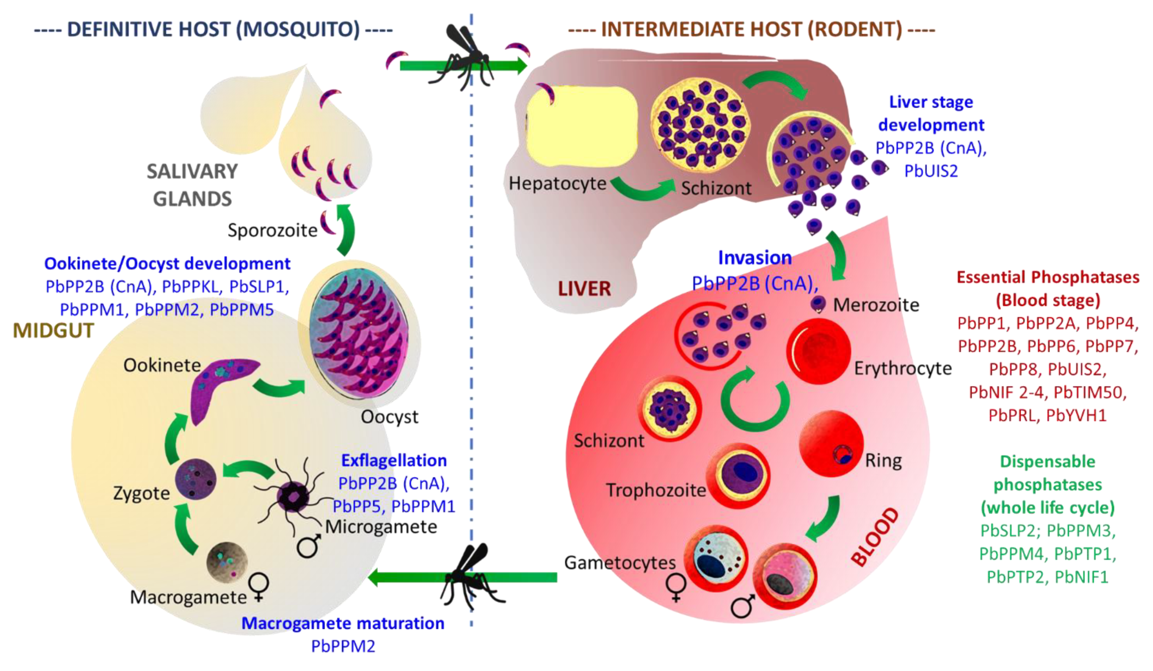
3. The Function of Protein Phosphatases in Apicomplexa: Where Do We Stand?

In recent years, the massive improvement of reverse genetics and multi-omics technologies has allowed for the development of ambitious projects based on global functional approaches, targeting Apicomplexa of medical importance, such as Plasmodium falciparum and Toxoplasma gondii, as well as the model organism Plasmodium berghei.
Using genome-wide saturation mutagenesis (P. falciparum, [43]), knock-out strategies (P. berghei, [44]) or CRISPR screens (T. gondii, [45]), the contribution of each gene during Plasmodium asexual development or T. gondii development in human fibroblasts was assessed. More than 50% of P. falciparum and 30% of P. berghei phosphatases seem essential for parasite survival in blood cells, highlighting how critical these enzymes are (Figure 2, Supplementary Materials, Tables S2 and S3). These studies, along with the characterization of engineered parasite mutant strains, drastically improved our understanding of the critical role played by protein phosphatases over the life cycle of these parasites.
In the following section of this review, we summarized our current knowledge regarding the functions of the S/T/Y phosphatases in Plasmodium and Toxoplasma.

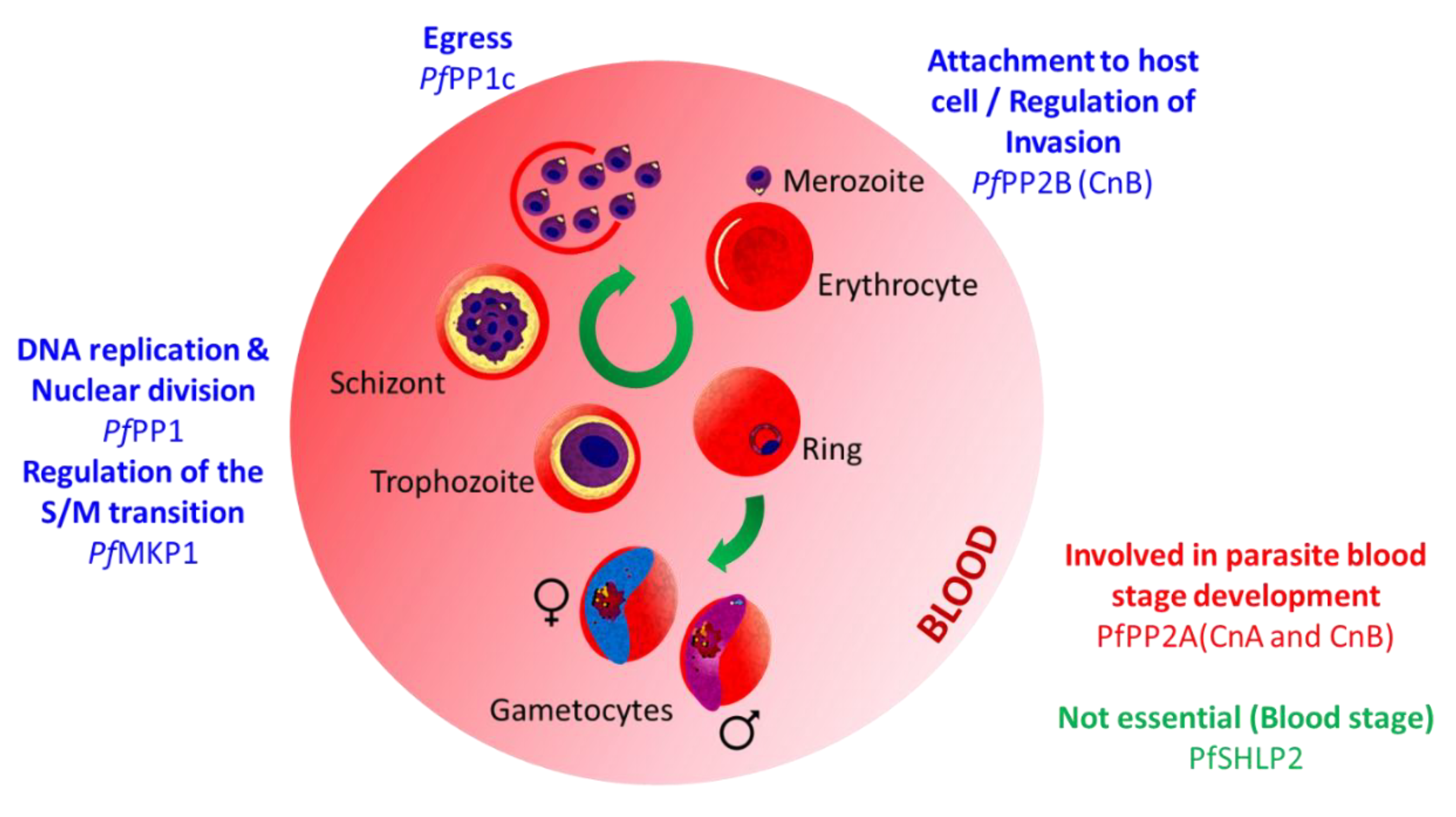
3.1. Functional Characterization of Plasmodium Proteins Phosphatases

3.1.1. PPPL Fold

  • Phosphoprotein Phosphatases (PPP) family

Protein Phosphatase Type 1 (PP1)

Protein phosphatase type 1 is an extremely well-conserved phosphatase known to be involved in a myriad of biological processes such as protein synthesis, transcription regulation, and cell division [46]. The PP1 catalytic domain possesses the distinct characteristic of interaction capacity with hundreds of regulatory subunits, thus leading to the formation of numerous holoenzymes, and allowing the enzyme to be involved in the regulation of a wide range of biological processes [47]. In Plasmodium falciparum, PfPP1 is expressed throughout the intraerythrocytic development cycle (IDC), mainly from the trophozoite to the schizont stage [48,49,50]. PfPP1 has been localized at the parasite’s nucleo-cytoplasmic compartment [48], but also at the Maurer’s clefts where it was suggested to modulate the phosphorylation of PfSBP1, an erythrocyte skeletal-binding protein [51]. The first evidence of the enzyme’s massive impact on the phosphorylation levels in the parasite came from early studies showing the drastic effect of phosphatase inhibitors on parasite development [52]. These observations were later confirmed by a reverse genetics screen, suggesting its essentiality for IDC completion [43]. Recently, inducible knockdown (iKD) approaches were used to refine PfPP1 function. These studies revealed a role in DNA replication, the formation of multinucleate schizonts, and egress from the host red blood cell. This last observation was linked to the parasite’s inability to secrete PfSUB1, a protease essential to the orchestration of the early steps of egress when PfPP1 is absent [50] (Figure 3, Supplementary Materials, Table S2).
To assess the function of PP1 during development in mosquito and liver cells, various reverse genetics strategies were implemented using the Plasmodium berghei model of rodent infection. When a reduction of pbpp1 expression in gametocytes was engineered following a promotor swap approach, drastic effects on male exflagellation, ookinete conversion, and a complete inability to transmit the infection were observed [53]. When pbpp1 was conditionally disrupted from the sporozoite stage (FlpL/FRT site-specific recombination system, [54]), no effect was observed on sporozoite formation or development in liver cells. However, merozoites exiting hepatocytes lost their ability to spread the infection, thus confirming PP1 essentiality for parasite development in the blood ([43], Figure 4, Supplementary Materials, Table S3).
Comparative sequence analysis, in silico motifs, and yeast two-hybrid screening of cDNA libraries shed light on the existence of hundreds of potential Plasmodium PP1 regulators [55]. Among them, four conserved regulators previously described in mammals, as well as three PfPP1 regulators restricted to the Plasmodium genus, have been characterized for their ability to bind and regulate the phosphatase activity [48,49,56,57,58,59,60,61,62] (for more details on PP1 and its interactors in Plasmodium, see also review [63]).

Calcineurin (PP2B; CnA, CnB)

Calcineurin has been previously described to be involved in numerous processes related to calcium signal transduction [64]. PP2B exists as a holoenzyme formed by the association of CnA, a catalytic subunit with CnB, its regulatory subunit [65]. In P. falciparum, the phosphatase was first purified from the cytosolic extract and identified due to its susceptibility to cyclosporin A (CsA) and FK506 [66], two calcineurin inhibitors described in mammalian cells [67]. Later, these two compounds were shown to inhibit the secretion of microneme proteins, suggesting a link between the phosphatase and calcium-dependent microneme exocytosis [68]. However, when using a conditional protein expression system targeting CnB, in a study carried out by Paul AS et al., it was demonstrated that, although calcineurin is essential to the parasite development in erythrocytes, its function is independent of microneme exocytosis but is rather related to a poorly understood form of strong attachment to the red blood cell before invasion [69]. Accordingly, by using an auxin-inducible degron (AID) system in P. berghei, calcineurin’s (CnA) critical function on merozoite attachment and invasion was confirmed. Further analysis during mosquito development revealed essential functions for gamete development and fertilization, but also in ookinete infectivity and sporozoite invasion ([70], Figure 3 and Figure 4, Supplementary Materials, Tables S2 and S3).

PP5

Unlike other phosphatases belonging to the PPP family, PP5 is a monomeric phosphatase containing both catalytic and regulatory subunits. This class of enzyme is also characterized by the presence of N-terminal tetratricopeptide repeat (TRP) domains known to be involved in autoregulation and in protein–protein interactions [71,72,73]. In P. falciparum, PP5 possesses structural and functional similarities with its orthologs. Its N-terminal TPR region seems to be required for the phosphatase’s auto-inhibition, and similar to mammalian cells, can interact with heat shock protein 90 (hsp90) [73,74,75]. A series of reverse genetic analyses leading to the GFP tagging of P. berghei PP5 and its gene deletion revealed that the enzyme is highly expressed in male gametocytes and is critical for the regulation of male fertility ([76,77], Figure 4, Supplementary Materials, Table S3).

Protein Phosphatase Containing Kelch-like Domains (PPKL)

The protein phosphatase containing Kelch-like domains (or PPKL) is intriguing as it belongs to a family that can only be found in plants and alveolates [78]. Therefore, PPKL appears as an interesting drug target. The phosphatase was initially identified in P. falciparum (under the name PPα) and, early on, was suggested to be involved in sexual stages development, as its transcript could only be detected in gametocytes [79]. This observation was later confirmed using the P. berghei model. Knock-out reverse genetics analyses revealed that pbppkl deletion severally impacts ookinete morphology and the ability to glide, as it resulted in parasites that could not develop in the mosquito midgut and, therefore, could not be transmitted ([80,81], Figure 4, Supplementary Materials, Table S3).

Schewanella-like Phosphatases

Schewanella-like phosphatases” (or Shelphs/SHLPs) belong to a class of enzymes closely related to phosphatases found in the bacteria genus Schewanella [82]. Besides in bacteria, Shelphs proteins can be found in fungi, plantae, and Chromoalveolata and Excavata parasites [83]. Therefore, they represent an interesting target for drug design. In Plasmodium, two genes encoding Schewanella-like phosphatases have been identified: shlp1 and shlp2. In P. falciparum, shlp1 was described as likely essential for erythrocyte development by a functional screen analysis [43]. On the contrary, in P. berghei, the gene is dispensable at that stage, but its knock-out impacts the parasite’s ability to produce proper ookinetes, ultimately resulting in a drastic loss of the oocyst formation [83]. Conversely, shlp2 knock-out does not seem to disturb the parasite erythrocyte development in either species [44,76,84] (Figure 3 and Figure 4, Supplementary Materials, Tables S2 and S3), although the early observation of its apical localization in P. falciparum merozoites first suggested a role in invasion [85].
2.
Purple acid phosphatase (PAP) family

GAP50/SAP (Secreted Acid Phosphatase)

In P. falciparum, GAP50 (or glideosome-associated protein 50) was first identified as part of the MyoA–MTIP–GAP motor complex or glideosome: an Apicomplexan complex involved in gliding and invasion [86]. In Plasmodium merozoites, the protein was, as expected, localized to the IMC (inner membrane complex), where it participates as a membrane anchor linking the glideosome to the IMC and was suggested to take part in erythrocyte invasion [87,88]. Subsequently, the protein was also detected on the surface of gametocytes [89,90]. In the mosquito midgut, GAP50 is involved in the co-opt of Human Factor H, allowing the parasite to evade complement-mediated lysis [90].
The resolution of the PfGAP50 crystal structure revealed similarity to purple acid phosphatases (PAPs) [91]. This confirmed an earlier study describing the protein as an acidic phosphatase (renamed SAP) with a broad substrate profile, secreted to the parasite periphery where it was proposed to be involved in host nutrient acquisition through dephosphorylation [92].

UIS2

In Plasmodium salivary gland sporozoites, protein synthesis is inhibited by the phosphorylation of a translational factor: Eif2α. Any further development into the host liver requires its dephosphorylation. In mammalian cells, Eif2α is regulated by PP1 [93], but it does not seem to be the case in Plasmodium. Instead, the parasite relies on UIS2, a phosphatase restricted to Apicomplexa, to ensure the proper regulation of Eif2α and the timely translation of genes essential to the sporozoite transformation into exo-erythrocytic stages. UIS2 activity can be enhanced by Mn2+ and inhibited by Cd2+ but remains unchanged by the okadaic acid (PP1/PP2A phosphatase inhibitor). Thus, the phosphatase was first classified as a PP2C/PPM family member ([94], (Figure 4, Supplementary Materials, Table S3). However, the protein does not possess a canonical PP2C catalytic domain but shares some similarity with purple acid phosphatases and, therefore, was classified as a PAP in this review.

3.1.2. Protein Phosphatase Mn2+ or Mg2+ Dependent (PPM) Fold

3.
Protein Phosphatases Mn2+ or Mg2+ dependent (PPM) family
Most of the information related to the function of Plasmodium PPM came from genome-wide functional studies carried out using the Plasmodium berghei model. In an earlier analysis aiming to target the parasite protein phosphatome, the knock-out of 9 of the 13 existing PbPPMs (Table 1, Supplementary Materials, Tables S1 and S3) was attempted [76]. According to this study, two genes (ppm6 and ppm9) were described as essential for the completion of parasite development in blood, and four genes (ppm3, ppm6, ppm7, and ppm8) were described as dispensable for development in both the murine host and mosquito vector. Finally, three genes (ppm1, ppm2 and ppm5) were analyzed for their implication in the development in mosquitos. Ppm1 knock-out mutants formed defective, non-exflagellating male gametocytes. Ppm2 mutants, conversely, produced fewer female gametocytes. The ookinete conversion, blocked at early retort stage II, was also drastically impacted. Finally, Ppm5 knock-out parasites produced few oocysts that were unable to form sporozoites, suggesting a role in the regulation of the oocyst development. Ookinete ultrastructure analysis revealed a partial defect in microneme formation, which could be responsible for the observed phenotype [76].
More recently, a genome-wide functional screen analysis nuanced the phenotype data described above. According to this study, PbPPM1 and PbPPM2 may have a mild role in parasite growth, and PbPPM9 may be dispensable at this stage. Additionally, the remaining members of the PPM family: PbPPM10, PbPPM12 and PbPPMX1, were suggested to be dispensable for the development in erythrocytes ([44], see the comparison of both analysis outcomes in Supplementary Materials, Table S3). In Plasmodium falciparum, PfPPM2 contains two catalytic domains whose dimerization is required for optimal activity [95]. Further analyses were performed to identify the phosphatase substrates. The translation elongation factor 1-beta was identified among other components of the translation and transcription machinery [96], suggesting that PfPPM2 may play a role in the regulation of those mechanisms. Using a saturation mutagenesis approach, Zhang et al. carried out a functional screen revealing that PfPPM2, PfPPM6, PfPPM8 and PfPPM9 could be essential for the parasite blood development stage when PfPPM3 and PfPPM10 play a mild role [43]. On the contrary, the remaining members of the family (PbPPM1, PbPPM4, PbPPM5, PbPPM7, PbPPM11, PbPPM12, and PbPPMX1) were described as dispensable (Figure 3 and Figure 4, Supplementary Materials, Tables S2 and S3).
The comparison of the functional screening performed in both falciparum and berghei models revealed some discrepancies suggesting that species specificity may be at play here.
4.
PfMKP1
PfMKP1 was identified and classified based on some structural similarities with mitogen-activated protein kinase (MAPK) phosphatases (or MKPs) which, in other organisms, have been described in cell cycle progression, cell growth and proliferation [97]. PfMKP1 sequence analysis revealed the presence of a non-catalytic N-terminal rhodanese (RDH)-like domain upstream of a catalytic dual-specificity phosphatases (DUSP)-like region, an association characteristic of MKPs [98,99,100]. However, the poor conservation of some critical catalytic residues suggests that the protein may be of low activity or may be a pseudo-phosphatase. Using random insertional mutagenesis, Balu et al. observed that the presence of a single transposon downstream of the mkp1 start codon of a P. falciparum cloned line was responsible for a significant attenuation of the parasite growth. Subsequent analysis revealed that the phenotype was due to a prolonged pre-S phase (trophozoite stage) causing a delayed entry into the S/M phase (schizont stage) ([41], Figure 3, Supplementary Materials, Table S2).

3.2. Functional Characterization of Toxoplasma gondii Proteins Phosphatases

3.2.1. PPPL Fold

5.
Phosphoprotein Phosphatases (PPP) family
Few protein phosphatases belonging to the phosphoprotein phosphatase family (PPP) have been characterized in Toxoplasma gondii thus far. However, the presence of members belonging to the PPP family has been established. Relatively comparing the genome of eukaryotes to that of T. gondii has demonstrated that the latter possesses a minimum of one member belonging to each of the seven PPP sub-families. Thus, the T. gondii genome encodes a complete set of PPP sub-families including PP1, PP2A, PP2B (calcineurin), PP4, PP5, PP6, and PP7 [27].

Protein Phosphatase Type 1 (PP1)

T. gondii PP1 (TgPP1) has been studied to a lesser extent compared to PP1 from other eukaryotes and the Apicomplexan parasite Plasmodium falciparum. TgPP1 was initially identified through experiments in which T. gondii tachyzoites were exposed to specific inhibitors of PP1, including tautomycin (TAU) and okadaic acid (OA). Tachyzoites treated with TAU and OA demonstrated a defect in invasion by a percentage of 50%. The impairment of invasiveness of T. gondii tachyzoites suggested that TgPP1 has an important role in the invasion of the host cell [101]. However, these inhibitors could target other phosphatases, and the effect demonstrated may be due to the inhibition of multiple phosphatases. Studies carried out on TgPP1 involving phosphatase assays on extracts of T. gondii have indicated the presence of TgPP1 dephosphorylation function, which is sensitive to specific TgPP1 inhibitors such as human PP1 Inhibitor 2 and okadaic acid [101]. Attempts to identify specific protein targets of TgPP1 were carried out by means of immunoprecipitation assays as well as in vitro phosphate labeling assays [101] but yielded few potential targets. Among TgPP1 interactors, TgLRR1, a homolog of sds22 in yeast, was identified to form a complex with TgPP1. The association of TgPP1 with TgLRR1 was demonstrated to occur within the nucleus of the tachyzoite [102]. TgLRR1 was identified as an inhibitor of TgPP1 by using the Xenopus oocyte model during which germinal vesicle breakdown takes place once the okadaic acid inhibitor or TgPP1 antibodies are micro-injected within the oocytes [102]. Furthermore, the micro-injection of oocytes with cRNA of TgLRR1 led to nearly a total germinal vesicle breakdown of oocytes, suggesting that TgLRR1 can inhibit the Xenopus PP1. A more recent study regarding inhibitors of TgPP1characterized inhibitor-2 (TgI2) that it functions similarly to its eukaryotic homologs exhibiting inhibitory activity [103]. The TgI2 inhibitor has three different motifs (SILK-like, RVxF, and FKK/HYNE). TgPP1 associates with TgI2 through the RVxF motif, whereas other motifs play a more minor role in the binding to TgPP1 [103]. However, both SILK-like and RVxF motifs are critical for regulating the activity of TgPP1, a feature that is common with the higher eukaryote’s Inhibitor-2 protein but was not observed for PfPP1 [103]. Relatively little has been described regarding TgPP1 biological function. Although it has been suggested to be essential during infection of human cells ([45], Supplementary Materials, Table S4), many questions remain unanswered, such as its specific mechanism of action and its specific targets.

PP2A and PP2B (Calcineurin)

Another protein belonging to this family of phosphoprotein phosphatases is PP2A. PP2A has been shown to have a key role in regulating the cell cycle in eukaryotes and remains crucial for regulating the progression from the G2 phase to the M phase. The deletion of PP2A led to a delayed cell cycle and an impairment of the spindle assembly checkpoint [104]. Despite the central role of PP2A in the cell cycle of mammals and higher eukaryotes and its existence within Toxoplasma, not much is known about its function within the Apicomplexan parasite T. gondii and remains to be characterized in future studies.
Recently, conditional knock-down approaches have been implemented to decipher the role of calcineurin in Toxoplasma gondii. The phosphatase seems, similar to its Plasmodium falciparum counterpart, to be involved in the regulation of host cell attachment independently of microneme exocytosis ([69], Figure 5, Supplementary Materials, Table S4).
6.
Purple acid Phosphatase (PAP) family

GAP50/SAP (Secreted Acid Phosphatase)

Similar to Plasmodium falciparum, TgGAP50 is a critical element of the glideosome. Harding et al. demonstrated that gap50 conditionally depleted the parasites’ present defects in the IMC morphology and the localization of the glideosome’s components, suggesting a role in IMC biogenesis and stability during parasite intracellular replication ([105], Figure 5, Supplementary Materials, Table S4).

GRA44 (TGGT1_228170 Secreted Acid Phosphatase)

GRA44 was initially identified as a secreted acid phosphatase based on a bioinformatics analysis of proteins within the T. gondii genome, containing predicted phosphatase domains and signal peptides. Localization studies of GRA44 identified that this acid phosphatase localized within the PV lumen and at the PV membrane, where two specific amino acids were demonstrated as necessary for its proper processing, which is not essential for its function [106]. Additionally, the conditional knockdown of GRA44 allowed it to demonstrate significantly impaired growth in the mutant parasites compared to the parental strain. GRA44 was demonstrated to associate with components of the putative translocon complex, which includes MYR1, MYR2, and MYR3 and induces c-Myc expression, which is crucial for the translocation of the parasite’s effector proteins [106], Figure 5, Supplementary Materials, Table S4).

3.2.2. Protein Phosphatase Mn2+ or Mg2+ Dependent (PPM) Fold

7.
Protein Phosphatases Mn2+ or Mg2+ dependent (PPM) family
Most of the work related to the functional analysis of Toxoplasma gondii phosphatases has been performed on proteins belonging to the PPM family. Recently, Yang et al. carried out the analysis of the sub-cellular localization of five of them (PPM2A, PPM2B, PPM3D, PPM5 and PPM11), revealing an intriguing diversity of localizations [107]. TgPPM2A was found to be associated with the nucleocytoplasmic compartment. TgPPM2B, conversely, is expressed in the cytoplasm but is excluded from the nucleus. TgPPM3D, albeit predicted to contain a transmembrane domain, was found to be expressed in the endoplasmic reticulum. A more detailed investigation carried out on TgPPM5C revealed that the protein, expressed at the plasma membrane, is involved in host cell attachment. TgPPM5C possesses some putative myristoylation and palmitoylation sites, specifically glycine residue 2 and cysteine residue 4, which are both required for the proper localization and function of the TgPPM5C protein at the plasma membrane. Studies carried out on TgPPM5C knock-out parasites demonstrated that these mutant parasites possess a defect in parasite propagation, which is linked to an impairment of the mutant parasite to invade the host cell ([107], Supplementary Materials, Table S4). Adhesion assays indicated that the defect in TgPPM5C knock-out parasites is in the parasite’s ability to attach to the host cell. Additionally, the protein phosphatase activity, although not directly linked with TgPPM5C localization, was proven essential to its function. Moreover, TgPPM5C was shown to modulate the phosphorylation status of multiple signaling proteins such as Rab-GTPases, multiple kinases, a guanylyl cyclase, and phosphatases. Finally, TgPPM11 is secreted in the parasitophorous vacuole. This observation will require further study, as an error in the protein annotation may be accountable for the result ([107], Figure 5, Supplementary Materials, Table S4).
More recently, TgPPM3C was identified as a phosphatase secreted into the lumen of the parasitophorous vacuole. It has been shown to interact with MYR1, an essential protein belonging to a vacuolar transposon involved in export to the host cell [108]. Tgppm3c knock-out led to the production of parasites exhibiting a mild growth defect in vitro but with a profound loss of virulence during in vivo acute infection. Electron microscopy revealed an absence of major differences between the wild-type and TgPPM3C deficient parasites [109]. Furthermore, a phospho-proteomics analysis was carried out to identify potential substrates of TgPPM3C. In total, 118 phospho-peptides were more abundant in the TgPPM3C knock-out strain than in the corresponding wild-type. Alternatively, merely 10 phospho-peptides were identified as significantly less abundant [109]. The phospho-peptides belonging to significantly abundant proteins were determined to be part of the rhoptry, dense granule, and microneme compartments. A significant decrease in the abundance of TgGRA16 and TgGRA28 export to the host nucleus was observed in cells infected with the TgPPM3C knock-out strain when compared to those infected with its wild-type counterpart, suggesting an impairment in the export of these two effector proteins ([109], Figure 5, Supplementary Materials, Table S4).
This was confirmed by carrying out phospho-mimetic mutation experiments on TgGRA16, where a decreased accumulation of this protein within the host nucleus was observed, suggesting a reduction in the export of the GRA16 effector protein. The reduction of TgGRA16 and TgGRA28 export may be linked to the virulence defect observed in vivo. Furthermore, during chronic infection, these defective parasites formed less cysts in the mouse brain that could be linked to an alteration of the phosphorylation and export status of GRA16 and GRA28 [109].
TgPPM13 (also named TgPP2C) is a nucleocytoplasmic protein that, when overexpressed, impacts parasite cytokinesis, suggesting a role in the regulation of cell cycle control [110]. TgPPM13 was also shown to regulate the phosphorylation of Toxofilin [111], a secreted protein that is known to bind host actin [112]. TgPPM13 carries out its function in accordance with a kinase known by the host casein kinase II (CKII). CKII phosphorylates Toxofilin specifically by targeting Serine 53. The association between TgPPM13 and CKII results in the sequestering of actin monomers as well as the regulation of binding between Toxofilin and host actin impacting the parasite’s movement [111,113].
Finally, TgPPM20 (also referred to as PP2C-hn) displays a mild role in parasite growth. During invasion, it is secreted from the rhoptries and is directed to the host cell nucleus, suggesting a role in host modulation [114]. Recently, this secreted phosphatase-lacking homolog in its host has triggered interest and was successfully tested as a potential vaccine candidate using a murine model [115]. However, further studies are needed in order to identify the direct targets of this PP2C protein (Figure 5, Supplementary Materials, Table S4).
8.
Aspartate-based phosphatase family (FCP/SCP)
Phosphatases belonging to this family vary from members of other phosphatase families due to their characteristic aspartate catalytic activity. Most of the T. gondii aspartate-based phosphatases consist of a conserved motif known by DxDT/V. T. gondii possesses eight of these aspartate-based phosphatases. According to the literature, none of these proteins in T. gondii have been functionally characterized. FCP1, a single protein previously characterized in humans, belongs to this family and functions in modulating the phosphorylation status of the carboxy-terminal region of the RNA polymerase II enzyme [116]. T. gondii possesses the FCP1 gene as other Apicomplexa. However, within the T. gondii parasite, the regular CTD function observed in other Apicomplexa is absent and is most likely due to its characteristic low number of heptapeptide repeat pairs [116].
In sum, in Apicomplexa, S/T/Y phosphatase roles are diverse and seem to impact every aspect of the parasite’s asexual/sexual development and interaction with its host and environment. Therefore, these enzymes must be considered as a great potential for the design of innovative and specific treatment. In the next section of this review, we will highlight and discuss opportunities supporting this idea in Plasmodium.

4. Therapeutic Potential of Protein Phosphatases as Drug Targets: The Case of Plasmodium

Over the last decades, most research and development companies have focused on targeting protein kinomes (comprising more than 350 kinases) [117] to treat various human diseases, especially cancer. The search for inhibitors against kinases has been tremendously developed since these enzymes are considered specific targets, thus contributing to selective treatments decreasing the risks of adverse drug effects.
With respect to protein phosphatases, their validation as drug targets first appeared less captivating and more challenging. They were considered less specific, given the conserved nature of their catalytic subunits, and more challenging to target, due to the level of uncertainty and the paucity of data regarding their molecular functions along with some major technical bottlenecks hampering the identification of their substrates. Nevertheless, it is now well known that protein phosphatases encompass diverse types of enzymes that are involved in the control of dephosphorylation processes in an extremely precise way and coordinated manner in a broad range of vital cellular functions. Hence, it is not surprising to uncover that phosphatases, when dysregulated, are found to be associated with a plethora of human diseases, including diabetes, cancer, or neurodegeneration [118]. Therefore, phosphatases could be considered attractive therapeutic drug targets. This is supported by recent reviews reporting why and how protein phosphatases should be included in the list of druggable enzymes or proposed as clinically pertinent targets [38,119,120,121].

4.1. Targeting Phosphatases to Treat Human Diseases: Two Decisive Cases

In recent studies, considerable and significant progress has been made to better define the functions and contribution of phosphatase signaling in normal development and pathology. This led to breakthroughs demonstrating the feasibility of targeting those enzymes with concrete impact on human diseases. The first evidence emerged in 2016 when Chen et al. showed that a small molecule named SHP099 was able to bind and selectively block the function of the protein tyrosine phosphatase SHP2, which was reported as the first oncogenic phosphatase linked to multiple cancers [122]. This molecule has been shown to be a potent inhibitor of the growth of human cancer cells in vitro and of xenograft tumors in a mouse model when administered via the oral route. Current clinical trials are still under investigation to validate SH099 as a viable drug for the treatment of human diseases. The second example came from a study in which a specific inhibitor of a regulator of PP1 (PPP1R15B) was shown to control translation and improve proteostasis as well as deficiencies linked to Huntington’s disease in a mouse model [123]. These successful strategies have certainly contributed in 2021 to the creation of the first research and development company (Anavo Therapeutics, [124]) intending to develop drug leads targeting phosphatases with the hope to accelerate the research in the field and provide additional means for the treatment of human diseases.

4.2. Is Targeting Phosphatases the Future of Anti-Plasmodium Therapeutics?

More than 95% of Plasmodium proteins are Ser/Thr phosphorylated [125], suggesting that their dephosphorylation is a major process in the regulation of protein activities. Additionally, Plasmodium not only expresses conserved phosphatases whose actions may be adapted and distinct from humans but also specific phosphatases that can also be targeted with minimal adverse effects.
In this perspective, along with the fact that the identification of drugs with novel modes of action becomes decisive, we will highlight and discuss opportunities for targeting Plasmodium protein phosphatases for the development of drugs for malaria treatment.

4.3. Targeting the PPP Family

4.3.1. PP1 and PP2A

While there are about 38 human Ser/Thr phosphatases, almost 90% of the cellular Ser/Thr phosphatase activities are covered by PP1 and PP2A. Although these two enzymes share a conserved catalytic subunit, they have been shown to exhibit distinct cellular activities at specific times of the cell cycle, thus indicating that they dephosphorylate specific substrates [126]. This is further substantiated by the fact that they function as holoenzymes with a great number of different regulatory subunits (transport, specificity, and control of activity of the catalytic subunits [127,128]), which could pave the way for new means to selectively interfere with phosphatases activities for effective therapy.
In Plasmodium falciparum, the first report highlighting the importance of phosphatases and their potential to control parasite growth came from the use of two natural toxins: okadaic acid and calyculin A. These toxins are well-known potent inhibitors of Ser/Thr mammalian phosphatases PP1 and PP2A, albeit not at the same concentration. If PP2A can be inhibited by okadaic acid and calyculin A at approximatively the same concentration, the amount of okadaic acid required to inhibit PP1 is about 100 times higher than the amount required for calyculin A in humans (reviewed in [129]). In their study, Yokoyama et al. demonstrated that, in vitro, the addition of okadaic acid or calyculin A led to a drastic inhibition of blood-stage parasite growth with an IC50 of 9.9 and 38.5 nM, respectively [130]. This was recently confirmed by our analysis of these toxins from which we added cantharidic acid (CA), which showed an IC50 of about 4.3 µM (personal communication). Based on these observations, it is likely that the inhibition of in vitro parasite growth following the use of natural toxins is due to a drastic blockage of PP1 activities.
Over the last decade, there has been a particular interest and rebound in targeting the catalytic subunit of PP1 alone or as a holoenzyme. Given the low probability to develop a selective lead or drug acting against PP1 because of the high conservation of its active site within its family and across species, we and others examined strategies aiming to target PP1 regulatory sub-units to interrupt the formation of PP1 holoenzymes [56,58,61,121,131,132,133]. The feasibility of those strategies was supported by several studies carried out on viruses showing that the use of small molecules designed to bind to the non-catalytic RVxF binding site of PP1 [134] was able to prevent viral replication in vitro of viruses such as HIV1 and Ebola [135,136,137]. One small molecule showed anti-HIV1 activity in a humanized mouse model [136]. In P. falciparum, we observed the potent inhibitory effect on the parasite’s in vitro growth of binding peptides derived from PP1 partners mimicking interaction sites with the phosphatase [56,58,61,63,138]. From these studies, it becomes obvious that the interference with the assembly PP1 holoenzymes can constitute a novel and major pathway to be drug targeted in Plasmodium (and other apicomplexan parasites).

4.3.2. Calcineurin (PP2B)

Indirect evidence has demonstrated that cyclosporin A (CsA) is a potent inhibitor of Plasmodium calcineurin (PP2B) when found associated with Plasmodium cyclophilin 19A and 19B [66,139]. Cyclosporin A has been shown to inhibit P. falciparum growth in vitro [140,141,142]. For P. berghei, its administration in mice (5 mg/kg) induced a reduction in blood parasitemia associated with an increase in host survival, which may be linked to an increase in suicidal erythrocyte death (eryptosis) [143]. More recent studies have shown the presence of human cyclophilin B on the surface of red blood cells, which along with additional parasitic cyclophilins, could be possible targets of cyclosporin A, contributing to the inhibition of Plasmodium growth [144,145]. However, the impact of cyclosporin A on Plasmodium calcineurin activity requires further investigation.

4.3.3. PP4 and PP6 In Silico Analysis of Druggability

Recently, Ali et al. created a list of anti-malaria potential drug targets to be prioritized for further investigation. P. falciparum proteins previously identified as essential for parasite blood development were subjected to in silico analysis to assess their druggability potential [146]. From this screening, two Ser/Thr phosphatases: PP6 and PP4, were identified based on their sub-cellular localization, their structure, and the absence of host homologs. PP4 was even shortlisted as one of the most promising potential targets due to some promising structural features such as druggable pockets or the fact that it may be an important component of the Plasmodium metabolic network [146].

4.4. Targeting the Tyrosine Phosphatases

4.4.1. In Silico Docking Analysis of PRL

In a recent study, Pandey et al. carried out an in silico virtual screening of the ChEMBL-NTD library using the Plasmodium falciparum phosphatase of regenerating liver (PfPRL) as a drug target [147]. Among the identified potential hits, they demonstrated that the Novartis_003209 compound was able to inhibit the phosphatase activity of recombinant PfPRL and was able to prevent the growth of intraerythrocytic P. falciparum parasites in vitro (IC50: 0.273 µM) [148]. This compound does not display any cytotoxicity against human cells [149] and therefore should be considered as a potential antimalarial. Further in silico docking analysis performed on other Plasmodium tyrosine phosphatases (PfYVH1, Pf3D7_112700 and PfPTP1) suggested that Novartis_003209 can bind to their conserved catalytic domain. Based on these data, it will be interesting to use the structural scaffold of this hit, and to pharmacomodulate it to design a compound library that will allow for the identification of specific tyrosine phosphatases inhibitors with improved potency and selectivity against Plasmodium in vitro and in vivo development.

4.4.2. In Silico Docking Analysis of PfMKP1

A homology model of PfMKP1 was created using a crystal of human MKP3 [150], with whom it shares about 21% similarity. The model was then used to screen antimalarial compounds from the ChEMBL-NTD library. The IC50 of seven compounds showing a high binding affinity with PfMKP1 in silico was measured in vitro against WT and ∆PfMKP1 P. falciparum parasites. For three of them, the IC50 was significantly higher in the ∆PfMKP1 parasites, suggesting that the phosphatase may be a potential target for these compounds. As a whole, this study represents a good example of the use of structure-based drug discovery strategies as a new means to identify new classes of antimalarial drugs [151].

5. Conclusions and Future Perspectives

Despite recent progress, diseases caused by Apicomplexan parasites remain of human and veterinary importance. The rapid spread of emerging resistance to current anti-malaria front-line drugs has only urged for innovative therapeutics. In this review, we attempted to summarize the current knowledge regarding Apicomplexan phosphatase diversity in terms of their structure, regulation, and function. We also discussed how their most distinct features could prove essential in the design of innovative drugs. As an example, we pointed out the combinatory capacity of some phosphatases’ catalytic sub-units to create potent interfering peptides.
In recent years, tremendous progress has been made to elucidate Apicomplexan phosphatase function. These studies, mostly carried out in Plasmodium, highlighted the crucial role of some specific phosphatases, revealing opportunities for interventions. In-depth analysis will be necessary to explore them further. Nevertheless, several research groups have already based their recent studies on both functional analyses and subsequent in silico structural analyses to successfully screen some compound libraries for anti-malarial purposes. In this context, the recent availability of the new AlphaFold Protein Structure Database version (https://alphafold.ebi.ac.uk, accessed on 20 January 2022), allowing for highly reliable prediction of a protein structure, will undoubtedly accelerate the in silico based drug design. This highlights, once again, the importance of multidisciplinary joint efforts in the fight against diseases caused by Apicomplexa.

Supplementary Materials

The following supporting information can be downloaded at: https://www.mdpi.com/article/10.3390/microorganisms10030585/s1, Table S1: Table combining the accession numbers of the serine/threonine and tyrosine protein phosphatases identified in the five apicomplexans analyzed: Plasmodium falciparum, Plasmodium berghei, Toxoplasma gondii, Cryptosporidium parvum and Babesia divergens. Table S2: List of Plasmodium falciparum Protein Phosphatases with functional characterization. Table S3: List of Plasmodium berghei Protein Phosphatases with functional characterization. Table S4: List of Toxoplasma gondii Protein Phosphatases with functional characterization. Table S5: Supplementary Materials References.

Author Contributions

Conceptualization, A.F., B.G., J.K., and C.P.; Investigation, A.F., B.G., and A.S.K.; Supervision, A.F., J.K., and C.P.; writing—original draft preparation, A.F., B.G., A.S.K., M.G., J.K., and C.P.; writing—review and editing, A.F., B.G., A.S.K., M.G., J.K., and C.P. All authors have read and agreed to the published version of the manuscript.

Funding

This research received no external funding.

Data Availability Statement

Not applicable.

Acknowledgments

We would like to thank “La region Hauts-de-France” for funding A.F. (“Accueil de Jeunes Chercheurs” award, 2017–2019).

Conflicts of Interest

The authors declare no conflict of interest.

References

  1. Adl, S.M.; Leander, B.S.; Simpson, A.G.B.; Archibald, J.M.; Anderson, O.R.; Bass, D.; Bowser, S.S.; Brugerolle, G.; Farmer, M.A.; Karpov, S.; et al. Diversity, Nomenclature, and Taxonomy of Protists. Syst. Biol. 2007, 56, 684–689. [Google Scholar] [CrossRef] [PubMed] [Green Version]
  2. Blackman, M.J.; Bannister, L.H. Apical organelles of Apicomplexa: Biology and isolation by subcellular fractionation. Mol. Biochem. Parasitol. 2001, 117, 11–25. [Google Scholar] [CrossRef]
  3. Fichera, M.E.; Roos, D.S. A plastid organelle as a drug target in apicomplexan parasites. Nature 1997, 390, 407–409. [Google Scholar] [CrossRef]
  4. McFadden, G.I. Apicoplast. Curr. Biol. 2014, 24, R262–R263. [Google Scholar] [CrossRef] [PubMed] [Green Version]
  5. Salomaki, E.D.; Kolisko, M. There Is Treasure Everywhere: Reductive Plastid Evolution in Apicomplexa in Light of Their Close Relatives. Biomolecules 2019, 9, 378. [Google Scholar] [CrossRef] [PubMed] [Green Version]
  6. World Health, O. World Malaria Report 2020: 20 Years of Global Progress and Challenges; World Health Organization: Geneva, Switzerland, 2020. [Google Scholar]
  7. Hajj, R.E.; Tawk, L.; Itani, S.; Hamie, M.; Ezzeddine, J.; El Sabban, M.; El Hajj, H. Toxoplasmosis: Current and Emerging Parasite Druggable Targets. Microorganisms 2021, 9, 2531. [Google Scholar] [CrossRef]
  8. Montoya, J.G.; Liesenfeld, O. Toxoplasmosis. Lancet 2004, 363, 1965–1976. [Google Scholar] [CrossRef]
  9. Djurković-Djaković, O.; Dupouy-Camet, J.; Van der Giessen, J.; Dubey, J.P. Toxoplasmosis: Overview from a One Health perspective. Food Waterborne Parasitol. 2019, 15, e00054. [Google Scholar] [CrossRef]
  10. Kezai, A.M.; Lecoeur, C.; Hot, D.; Bounechada, M.; Alouani, M.L.; Marion, S. Association between schizophrenia and Toxoplasma gondii infection in Algeria. Psychiat Res. 2020, 291, 113293. [Google Scholar] [CrossRef]
  11. Fuglewicz, A.J.; Piotrowski, P.; Stodolak, A. Relationship between toxoplasmosis and schizophrenia: A review. Adv. Clin. Exp. Med. 2017, 26, 1031–1036. [Google Scholar] [CrossRef]
  12. Fabiani, S.; Pinto, B.; Bonuccelli, U.; Bruschi, F. Neurobiological studies on the relationship between toxoplasmosis and neuropsychiatric diseases. J. Neurol. Sci 2015, 351, 3–8. [Google Scholar] [CrossRef] [PubMed] [Green Version]
  13. Ngoungou, E.B.; Bhalla, D.; Nzoghe, A.; Dardé, M.-L.; Preux, P.-M. Toxoplasmosis and epilepsy--systematic review and meta analysis. PLoS Negl. Trop. Dis. 2015, 9, e0003525. [Google Scholar] [CrossRef]
  14. Vannier, E.; Krause, P.J. Human Babesiosis. N. Eng. J. Med. 2012, 366, 2397–2407. [Google Scholar] [CrossRef] [PubMed] [Green Version]
  15. Feng, Y.; Ryan, U.M.; Xiao, L. Genetic Diversity and Population Structure of Cryptosporidium. Trends Parasitol 2018, 34, 997–1011. [Google Scholar] [CrossRef] [PubMed]
  16. Platts-Mills, J.A.; Babji, S.; Bodhidatta, L.; Gratz, J.; Haque, R.; Havt, A.; McCormick, B.J.; McGrath, M.; Olortegui, M.P.; Samie, A.; et al. Pathogen-specific burdens of community diarrhoea in developing countries: A multisite birth cohort study (MAL-ED). Lancet Glob. Health 2015, 3, e564–e575. [Google Scholar] [CrossRef] [Green Version]
  17. Kotloff, K.L.; Nataro, J.P.; Blackwelder, W.C.; Nasrin, D.; Farag, T.H.; Panchalingam, S.; Wu, Y.; Sow, S.O.; Sur, D.; Breiman, R.F.; et al. Burden and aetiology of diarrhoeal disease in infants and young children in developing countries (the Global Enteric Multicenter Study, GEMS): A prospective, case-control study. Lancet 2013, 382, 209–222. [Google Scholar] [CrossRef]
  18. O’Connor, R.M.; Shaffie, R.; Kang, G.; Ward, H.D. Cryptosporidiosis in patients with HIV/AIDS. AIDS 2011, 25, 549–560. [Google Scholar] [CrossRef]
  19. Sawant, M.; Baydoun, M.; Creusy, C.; Chabé, M.; Viscogliosi, E.; Certad, G.; Benamrouz-Vanneste, S. Cryptosporidium and Colon Cancer: Cause or Consequence? Microorganisms 2020, 8, 1665. [Google Scholar] [CrossRef]
  20. Van Dooren, G.G.; Striepen, B. The Algal Past and Parasite Present of the Apicoplast. Annu. Rev. Microbiol. 2013, 67, 271–289. [Google Scholar] [CrossRef]
  21. Gubbels, M.-J.; Coppens, I.; Zarringhalam, K.; Duraisingh, M.T.; Engelberg, K. The Modular Circuitry of Apicomplexan Cell Division Plasticity. Front. Cell Infect. Microbiol. 2021, 11, 670049. [Google Scholar] [CrossRef]
  22. Moorhead, G.B.G.; De Wever, V.; Templeton, G.; Kerk, D. Evolution of protein phosphatases in plants and animals. Biochem. J. 2009, 417, 401–409. [Google Scholar] [CrossRef] [PubMed] [Green Version]
  23. Manning, G.; Whyte, D.B.; Martinez, R.; Hunter, T.; Sudarsanam, S. The protein kinase complement of the human genome. Science 2002, 298, 1912–1934. [Google Scholar] [CrossRef] [PubMed] [Green Version]
  24. Chen, M.J.; Dixon, J.E.; Manning, G. Genomics and evolution of protein phosphatases. Sci. Signal. 2017, 10, eaag1796. [Google Scholar] [CrossRef] [PubMed]
  25. Miranda-Saavedra, D.; Gabaldón, T.; Barton, G.J.; Langsley, G.; Doerig, C. The kinomes of apicomplexan parasites. Microbes Infect. 2012, 14, 796–810. [Google Scholar] [CrossRef] [PubMed] [Green Version]
  26. Miranda-Saavedra, D.; Barton, G.J. Classification and functional annotation of eukaryotic protein kinases. Proteins 2007, 68, 893–914. [Google Scholar] [CrossRef]
  27. Yang, C.; Arrizabalaga, G. The serine/threonine phosphatases of apicomplexan parasites. Mol. Microbiol. 2017, 106, 1–21. [Google Scholar] [CrossRef] [Green Version]
  28. Wilkes, J.M.; Doerig, C. The protein-phosphatome of the human malaria parasite Plasmodium falciparum. BMC Genom. 2008, 9, 412. [Google Scholar] [CrossRef] [Green Version]
  29. Phosphatase Wiki—Protein Phosphatase Classification and Evolution. Available online: http://phosphatome.net/wiki (accessed on 1 September 2021).
  30. Treeck, M.; Sanders, J.L.; Elias, J.E.; Boothroyd, J.C. The phosphoproteomes of Plasmodium falciparum and Toxoplasma gondii reveal unusual adaptations within and beyond the parasites’ boundaries. Cell Host Microbe 2011, 10, 410–419. [Google Scholar] [CrossRef] [Green Version]
  31. Hunter, T. The genesis of tyrosine phosphorylation. Cold Spring Harb. Perspect. Biol. 2014, 6, a020644. [Google Scholar] [CrossRef] [Green Version]
  32. Spalinger, M.R.; Lang, S.; Gottier, C.; Dai, X.; Rawlings, D.J.; Chan, A.C.; Rogler, G.; Scharl, M. PTPN22 regulates NLRP3-mediated IL1B secretion in an autophagy-dependent manner. Autophagy 2017, 13, 1590–1601. [Google Scholar] [CrossRef] [Green Version]
  33. Penafuerte, C.; Perez-Quintero, L.A.; Vinette, V.; Hatzihristidis, T.; Tremblay, M.L. Mining the Complex Family of Protein Tyrosine Phosphatases for Checkpoint Regulators in Immunity. In Emerging Concepts Targeting Immune Checkpoints in Cancer and Autoimmunity; Yoshimura, A., Ed.; Springer International Publishing: Cham, Switzerland, 2017; pp. 191–214. [Google Scholar]
  34. Wade, F.; Belhaj, K.; Poizat, C. Protein tyrosine phosphatases in cardiac physiology and pathophysiology. Heart Fail. Rev. 2018, 23, 261–272. [Google Scholar] [CrossRef] [PubMed] [Green Version]
  35. Gao, P.-P.; Qi, X.-W.; Sun, N.; Sun, Y.-Y.; Zhang, Y.; Tan, X.-N.; Ding, J.; Han, F.; Zhang, Y. The emerging roles of dual-specificity phosphatases and their specific characteristics in human cancer. Biochim. Biophys. Acta 2021, 1876, 188562. [Google Scholar] [CrossRef] [PubMed]
  36. Ruckert, M.T.; de Andrade, P.V.; Santos, V.S.; Silveira, V.S. Protein tyrosine phosphatases: Promising targets in pancreatic ductal adenocarcinoma. Cell. Mol. Life Sci. CMLS 2019, 76, 2571–2592. [Google Scholar] [CrossRef] [PubMed]
  37. Frankson, R.; Yu, Z.-H.; Bai, Y.; Li, Q.; Zhang, R.-Y.; Zhang, Z.-Y. Therapeutic Targeting of Oncogenic Tyrosine Phosphatases. Cancer Res. 2017, 77, 5701–5705. [Google Scholar] [CrossRef] [Green Version]
  38. Alonso, A.; Sasin, J.; Bottini, N.; Friedberg, I.; Friedberg, I.; Osterman, A.; Godzik, A.; Hunter, T.; Dixon, J.; Mustelin, T. Protein tyrosine phosphatases in the human genome. Cell 2004, 117, 699–711. [Google Scholar] [CrossRef] [Green Version]
  39. Pendyala, P.R.; Ayong, L.; Eatrides, J.; Schreiber, M.; Pham, C.; Chakrabarti, R.; Fidock, D.A.; Allen, C.M.; Chakrabarti, D. Characterization of a PRL protein tyrosine phosphatase from Plasmodium falciparum. Mol. Biochem. Parasitol. 2008, 158, 1–10. [Google Scholar] [CrossRef]
  40. Kumar, R.; Musiyenko, A.; Cioffi, E.; Oldenburg, A.; Adams, B.; Bitko, V.; Krishna, S.S.; Barik, S. A zinc-binding dual-specificity YVH1 phosphatase in the malaria parasite, Plasm.modium falciparum, and its interaction with the nuclear protein, pescadillo. Mol. Biochem. Parasitol. 2004, 133, 297–310. [Google Scholar] [CrossRef]
  41. Balu, B.; Campbell, C.; Sedillo, J.; Maher, S.; Singh, N.; Thomas, P.; Zhang, M.; Pance, A.; Otto, T.D.; Rayner, J.C.; et al. Atypical mitogen-activated protein kinase phosphatase implicated in regulating transition from pre-S-Phase asexual intraerythrocytic development of Plasmodium falciparum. Eukaryot. Cell 2013, 12, 1171–1178. [Google Scholar] [CrossRef] [Green Version]
  42. Guttery, D.S.; Pandey, R.; Ferguson, D.J.; Wall, R.J.; Brady, D.; Gupta, D.; Holder, A.A.; Tewari, R. Plasmodium DEH is ER-localized and crucial for oocyst mitotic division during malaria transmission. Life Sci. Alliance 2020, 3, e202000879. [Google Scholar] [CrossRef]
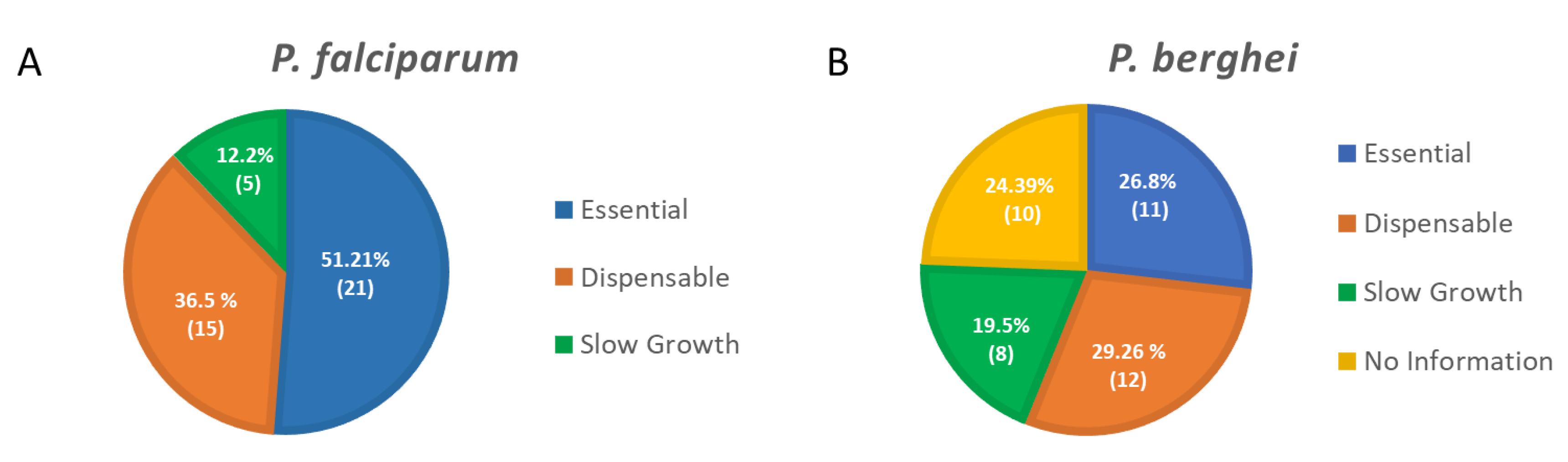
  43. Zhang, M.; Wang, C.; Otto, T.D.; Oberstaller, J.; Liao, X.; Adapa, S.R.; Udenze, K.; Bronner, I.F.; Casandra, D.; Mayho, M.; et al. Uncovering the essential genes of the human malaria parasite Plasmodium falciparum by saturation mutagenesis. Science 2018, 360, eaap7847. [Google Scholar] [CrossRef] [Green Version]
  44. Bushell, E.; Gomes, A.R.; Sanderson, T.; Anar, B.; Girling, G.; Herd, C.; Metcalf, T.; Modrzynska, K.; Schwach, F.; Martin, R.E.; et al. Functional Profiling of a Plasmodium Genome Reveals an Abundance of Essential Genes. Cell 2017, 170, 260–272.e8. [Google Scholar] [CrossRef] [PubMed] [Green Version]
  45. Sidik, S.M.; Huet, D.; Ganesan, S.M.; Huynh, M.H.; Wang, T.; Nasamu, A.S.; Thiru, P.; Saeij, J.P.J.; Carruthers, V.B.; Niles, J.C.; et al. A Genome-wide CRISPR Screen in Toxoplasma Identifies Essential Apicomplexan Genes. Cell 2016, 166, 1423–1435.e12. [Google Scholar] [CrossRef] [PubMed] [Green Version]
  46. Ceulemans, H.; Bollen, M. Functional diversity of protein phosphatase-1, a cellular economizer and reset button. Physiol. Rev. 2004, 84, 1–39. [Google Scholar] [CrossRef] [PubMed] [Green Version]
  47. Verbinnen, I.; Ferreira, M.; Bollen, M. Biogenesis and activity regulation of protein phosphatase 1. Biochem. Soc. Trans. 2017, 45, 89–99. [Google Scholar] [CrossRef]
  48. Daher, W.; Browaeys, E.; Pierrot, C.; Jouin, H.; Dive, D.; Meurice, E.; Dissous, C.; Capron, M.; Tomavo, S.; Doerig, C.; et al. Regulation of protein phosphatase type 1 and cell cycle progression by PfLRR1, a novel leucine-rich repeat protein of the human malaria parasite Plasmodium falciparum. Mol. Microbiol. 2006, 60, 578–590. [Google Scholar] [CrossRef]
  49. Hollin, T.; De Witte, C.; Freville, A.; Guerrera, I.C.; Chhuon, C.; Saliou, J.M.; Herbert, F.; Pierrot, C.; Khalife, J. Essential role of GEXP15, a specific Protein Phosphatase type 1 partner, in Plasmodium berghei in asexual erythrocytic proliferation and transmission. PLoS Pathog. 2019, 15, e1007973. [Google Scholar] [CrossRef] [Green Version]
  50. Paul, A.S.; Miliu, A.; Paulo, J.A.; Goldberg, J.M.; Bonilla, A.M.; Berry, L.; Séveno, M.; Braun-Breton, C.; Kosber, A.L.; Elsworth, B.; et al. Co-option of Plasmodium falciparum PP1 for egress from host erythrocytes. Nat. Commun. 2020, 11, 3532. [Google Scholar] [CrossRef]
  51. Blisnick, T.; Vincensini, L.; Fall, G.; Braun-Breton, C. Protein phosphatase 1, a Plasmodium falciparum essential enzyme, is exported to the host cell and implicated in the release of infectious merozoites. Cell. Microbiol. 2006, 8, 591–601. [Google Scholar] [CrossRef]
  52. Bhattacharyya, M.K.; Hong, Z.; Kongkasuriyachai, D.; Kumar, N. Plasmodium falciparum protein phosphatase type 1 functionally complements a glc7 mutant in Saccharomyces cerevisiae. Int. J. Parasitol. 2002, 32, 739–747. [Google Scholar] [CrossRef]
  53. Zeeshan, M.; Pandey, R.; Subudhi, A.K.; Ferguson, D.J.P.; Kaur, G.; Rashpa, R.; Nugmanova, R.; Brady, D.; Bottrill, A.R.; Vaughan, S.; et al. Protein phosphatase 1 regulates atypical mitotic and meiotic division in Plasmodium sexual stages. Commun. Biol. 2021, 4, 760. [Google Scholar] [CrossRef]
  54. Combe, A.; Giovannini, D.; Carvalho, T.G.; Spath, S.; Boisson, B.; Loussert, C.; Thiberge, S.; Lacroix, C.; Gueirard, P.; Ménard, R. Clonal Conditional Mutagenesis in Malaria Parasites. Cell Host Microbe 2009, 5, 386–396. [Google Scholar] [CrossRef] [PubMed] [Green Version]
  55. Hollin, T.; De Witte, C.; Lenne, A.; Pierrot, C.; Khalife, J. Analysis of the interactome of the Ser/Thr Protein Phosphatase type 1 in Plasmodium falciparum. BMC Genom. 2016, 17, 246. [Google Scholar] [CrossRef] [PubMed] [Green Version]
  56. Freville, A.; Cailliau-Maggio, K.; Pierrot, C.; Tellier, G.; Kalamou, H.; Lafitte, S.; Martoriati, A.; Pierce, R.J.; Bodart, J.F.; Khalife, J. Plasmodium falciparum encodes a conserved active inhibitor-2 for Protein Phosphatase type 1: Perspectives for novel anti-plasmodial therapy. BMC Biol. 2013, 11, 80. [Google Scholar] [CrossRef] [PubMed] [Green Version]
  57. Freville, A.; Landrieu, I.; Garcia-Gimeno, M.A.; Vicogne, J.; Montbarbon, M.; Bertin, B.; Verger, A.; Kalamou, H.; Sanz, P.; Werkmeister, E.; et al. Plasmodium falciparum inhib.bitor-3 homolog increases protein phosphatase type 1 activity and is essential for parasitic survival. J. Biol. Chem. 2012, 287, 1306–1321. [Google Scholar] [CrossRef] [Green Version]
  58. Freville, A.; Tellier, G.; Vandomme, A.; Pierrot, C.; Vicogne, J.; Cantrelle, F.X.; Martoriati, A.; Cailliau-Maggio, K.; Khalife, J.; Landrieu, I. Identification of a Plasmodium falciparum inhibitor-2 motif involved in the binding and regulation activity of protein phosphatase type 1. FEBS J. 2014, 281, 4519–4534. [Google Scholar] [CrossRef]
  59. Gnangnon, B.; Fréville, A.; Cailliau, K.; Leroy, C.; De Witte, C.; Tulasne, D.; Martoriarti, A.; Jung, V.; Guerrera, I.C.; Marion, S.; et al. Plasmodium pseudo-Tyrosine Kinase-like binds PP1 and SERA5 and is exported to host erythrocytes. Sci. Rep. 2019, 9, 8120. [Google Scholar] [CrossRef] [Green Version]
  60. Lenne, A.; De Witte, C.; Tellier, G.; Hollin, T.; Aliouat, E.M.; Martoriati, A.; Cailliau, K.; Saliou, J.-M.; Khalife, J.; Pierrot, C. Characterization of a Protein Phosphatase Type-1 and a Kinase Anchoring Protein in Plasmodium falciparum. Front. Microbiol. 2018, 9, 2617. [Google Scholar] [CrossRef]
  61. Pierrot, C.; Zhang, X.; Zhangi, G.; Freville, A.; Rebollo, A.; Khalife, J. Peptides derived from Plasmodium falciparum leucine-rich repeat 1 bind to serine/threonine phosphatase type 1 and inhibit parasite growth in vitro. Drug Des. Dev. Ther. 2018, 12, 85–88. [Google Scholar] [CrossRef] [Green Version]
  62. Tellier, G.; Lenne, A.; Cailliau-Maggio, K.; Cabezas-Cruz, A.; Valdes, J.J.; Martoriati, A.; Aliouat el, M.; Gosset, P.; Delaire, B.; Freville, A.; et al. Identification of Plasmodium falciparum Translation Initiation eIF2beta Subunit: Direct Interaction with Protein Phosphatase Type 1. Front. Microbiol. 2016, 7, 777. [Google Scholar] [CrossRef]
  63. Khalife, J.; Fréville, A.; Gnangnon, B.; Pierrot, C. The Multifaceted Role of Protein Phosphatase 1 in Plasmodium. Trends Parasitol. 2021, 37, 154–164. [Google Scholar] [CrossRef]
  64. Rusnak, F.; Mertz, P. Calcineurin: Form and Function. Physiol. Rev. 2000, 80, 1483–1521. [Google Scholar] [CrossRef] [PubMed]
  65. Klee, C.B.; Ren, H.; Wang, X. Regulation of the Calmodulin-stimulated Protein Phosphatase, Calcineurin. J. Biol. Chem. 1998, 273, 13367–13370. [Google Scholar] [CrossRef] [PubMed] [Green Version]
  66. Dobson, S.; May, T.; Berriman, M.; Del Vecchio, C.; Fairlamb, A.H.; Chakrabarti, D.; Barik, S. Characterization of protein Ser/Thr phosphatases of the malaria parasite, Plasmodium falciparum: Inhibition of the parasitic calcineurin by cyclophilin-cyclosporin complex. Mol. Biochem. Parasitol. 1999, 99, 167–181. [Google Scholar] [CrossRef]
  67. Liu, J.; Farmer, J.D.; Lane, W.S.; Friedman, J.; Weissman, I.; Schreiber, S.L. Calcineurin is a common target of cyclophilin-cyclosporin A and FKBP-FK506 complexes. Cell 1991, 66, 807–815. [Google Scholar] [CrossRef]
  68. Singh, S.; More, K.R.; Chitnis, C.E. Role of calcineurin and actin dynamics in regulated secretion of microneme proteins in Plasmodium falciparum merozoites during erythrocyte invasion. Cell. Microbiol. 2014, 16, 50–63. [Google Scholar] [CrossRef] [PubMed]
  69. Paul, A.S.; Saha, S.; Engelberg, K.; Jiang, R.H.Y.; Coleman, B.I.; Kosber, A.L.; Chen, C.-T.; Ganter, M.; Espy, N.; Gilberger, T.W.; et al. Parasite Calcineurin Regulates Host Cell Recognition and Attachment by Apicomplexans. Cell Host Microbe 2015, 18, 49–60. [Google Scholar] [CrossRef] [PubMed] [Green Version]
  70. Philip, N.; Waters, A.P. Conditional Degradation of Plasmodium Calcineurin Reveals Functions in Parasite Colonization of both Host and Vector. Cell Host Microbe 2015, 18, 122–131. [Google Scholar] [CrossRef] [Green Version]
  71. Chinkers, M. Targeting of a distinctive protein-serine phosphatase to the protein kinase-like domain of the atrial natriuretic peptide receptor. Proc. Natl. Acad. Sci. USA 1994, 91, 11075–11079. [Google Scholar] [CrossRef] [Green Version]
  72. Becker, W.; Kentrup, H.; Klumpp, S.; Schultz, J.E.; Joost, H.G. Molecular cloning of a protein serine/threonine phosphatase containing a putative regulatory tetratricopeptide repeat domain. J. Biol. Chem. 1994, 269, 22586–22592. [Google Scholar] [CrossRef]
  73. Hinds, T.D., Jr.; Sánchez, E.R. Protein phosphatase 5. Int. J. Biochem. Cell Biol. 2008, 40, 2358–2362. [Google Scholar] [CrossRef]
  74. Dobson, S.; Kar, B.; Kumar, R.; Adams, B.; Barik, S. A novel tetratricopeptide repeat (TPR) containing PP5 serine/threonine protein phosphatase in the malaria parasite, Plasmodium falciparum. BMC Microbiol. 2001, 1, 31. [Google Scholar] [CrossRef] [PubMed]
  75. Lindenthal, C.; Klinkert, M.Q. Identification and biochemical characterisation of a protein phosphatase 5 homologue from Plasmodium falciparum. Mol. Biochem. Parasitol 2002, 120, 257–268. [Google Scholar] [CrossRef]
  76. Guttery, D.S.; Poulin, B.; Ramaprasad, A.; Wall, R.J.; Ferguson, D.J.; Brady, D.; Patzewitz, E.M.; Whipple, S.; Straschil, U.; Wright, M.H.; et al. Genome-wide functional analysis of Plasmodium protein phosphatases reveals key regulators of parasite development and differentiation. Cell Host Microbe 2014, 16, 128–140. [Google Scholar] [CrossRef] [PubMed] [Green Version]
  77. Zhu, X.; Sun, L.; He, Y.; Wei, H.; Hong, M.; Liu, F.; Liu, Q.; Cao, Y.; Cui, L. Plasmodium berghei serine/threonine protein phosphatase PP5 plays a critical role in male gamete fertility. Int. J. Parasitol Drug 2019, 49, 685–695. [Google Scholar] [CrossRef] [PubMed]
  78. Kutuzov, M.A.; Andreeva, A.V. Protein Ser/Thr phosphatases with kelch-like repeat domains. Cell. Signal. 2002, 14, 745–750. [Google Scholar] [CrossRef]
  79. Li, J.L.; Baker, D.A. A putative protein serine/threonine phosphatase from Plasmodium falciparum contains a large N-terminal extension and five unique inserts in the catalytic domain. Mol. Biochem. Parasitol. 1998, 95, 287–295. [Google Scholar] [CrossRef]
  80. Philip, N.; Vaikkinen, H.J.; Tetley, L.; Waters, A.P. A unique Kelch domain phosphatase in Plasmodium regulates ookinete morphology, motility and invasion. PLoS ONE 2012, 7, e44617. [Google Scholar] [CrossRef] [Green Version]
  81. Guttery, D.S.; Poulin, B.; Ferguson, D.J.; Szoor, B.; Wickstead, B.; Carroll, P.L.; Ramakrishnan, C.; Brady, D.; Patzewitz, E.M.; Straschil, U.; et al. A unique protein phosphatase with kelch-like domains (PPKL) in Plasmodium modulates ookinete differentiation, motility and invasion. PLoS Pathog. 2012, 8, e1002948. [Google Scholar] [CrossRef]
  82. Andreeva, A.V.; Kutuzov, M.A. Widespread presence of “bacterial-like” PPP phosphatases in eukaryotes. BMC Evol. Biol. 2004, 4, 47. [Google Scholar] [CrossRef] [Green Version]
  83. Patzewitz, E.M.; Guttery, D.S.; Poulin, B.; Ramakrishnan, C.; Ferguson, D.J.; Wall, R.J.; Brady, D.; Holder, A.A.; Szoor, B.; Tewari, R. An ancient protein phosphatase, SHLP1, is critical to microneme development in Plasmodium ookinetes and parasite transmission. Cell Rep. 2013, 3, 622–629. [Google Scholar] [CrossRef] [Green Version]
  84. Miliu, A.; Lebrun, M.; Braun-Breton, C.; Lamarque, M.H. Shelph2, a bacterial-like phosphatase of the malaria parasite Plasmodium falciparum, is dispensable during asexual blood stage. PLoS ONE 2017, 12, e0187073. [Google Scholar] [CrossRef] [PubMed]
  85. Fernandez-Pol, S.; Slouka, Z.; Bhattacharjee, S.; Fedotova, Y.; Freed, S.; An, X.; Holder, A.A.; Campanella, E.; Low, P.S.; Mohandas, N.; et al. A bacterial phosphatase-like enzyme of the malaria parasite Plasmodium falciparum possesses tyrosine phosphatase activity and is implicated in the regulation of band 3 dynamics during parasite invasion. Eukaryot. Cell 2013, 12, 1179–1191. [Google Scholar] [CrossRef] [PubMed] [Green Version]
  86. Soldati-Favre, D. Molecular dissection of host cell invasion by the Apicomplexans: The glideosome. Parasite 2008, 15, 197–205. [Google Scholar] [CrossRef] [PubMed] [Green Version]
  87. Baum, J.; Richard, D.; Healer, J.; Rug, M.; Krnajski, Z.; Gilberger, T.W.; Green, J.L.; Holder, A.A.; Cowman, A.F. A conserved molecular motor drives cell invasion and gliding motility across malaria life cycle stages and other apicomplexan parasites. J. Biol. Chem. 2006, 281, 5197–5208. [Google Scholar] [CrossRef] [PubMed] [Green Version]
  88. Yeoman, J.A.; Hanssen, E.; Maier, A.G.; Klonis, N.; Maco, B.; Baum, J.; Turnbull, L.; Whitchurch, C.B.; Dixon, M.W.; Tilley, L. Tracking Glideosome-associated protein 50 reveals the development and organization of the inner membrane complex of Plasmodium falciparum. Eukaryot. Cell 2011, 10, 556–564. [Google Scholar] [CrossRef] [PubMed] [Green Version]
  89. Dearnley, M.K.; Yeoman, J.A.; Hanssen, E.; Kenny, S.; Turnbull, L.; Whitchurch, C.B.; Tilley, L.; Dixon, M.W.A. Origin, composition, organization and function of the inner membrane complex of Plasmodium falciparum gametocytes. J. Cell Sci. 2012, 125, 2053–2063. [Google Scholar] [CrossRef] [Green Version]
  90. Simon, N.; Lasonder, E.; Scheuermayer, M.; Kuehn, A.; Tews, S.; Fischer, R.; Zipfel, P.F.; Skerka, C.; Pradel, G. Malaria Parasites Co-opt Human Factor H to Prevent Complement-Mediated Lysis in the Mosquito Midgut. Cell Host Microbe 2013, 13, 29–41. [Google Scholar] [CrossRef] [Green Version]
  91. Bosch, J.; Paige, M.H.; Vaidya, A.B.; Bergman, L.W.; Hol, W.G. Crystal structure of GAP50, the anchor of the invasion machinery in the inner membrane complex of Plasmodium falciparum. J. Struct. Biol. 2012, 178, 61–73. [Google Scholar] [CrossRef] [Green Version]
  92. Muller, I.B.; Knockel, J.; Eschbach, M.L.; Bergmann, B.; Walter, R.D.; Wrenger, C. Secretion of an acid phosphatase provides a possible mechanism to acquire host nutrients by Plasmodium falciparum. Cell. Microbiol. 2010, 12, 677–691. [Google Scholar] [CrossRef]
  93. Harding, H.P.; Zhang, Y.; Scheuner, D.; Chen, J.-J.; Kaufman, R.J.; Ron, D. Ppp1r15 gene knockout reveals an essential role for translation initiation factor 2 alpha (eIF2alpha) dephosphorylation in mammalian development. Proc. Natl. Acad. Sci. USA 2009, 106, 1832–1837. [Google Scholar] [CrossRef] [Green Version]
  94. Zhang, M.; Mishra, S.; Sakthivel, R.; Fontoura, B.M.; Nussenzweig, V. UIS2: A Unique Phosphatase Required for the Development of Plasmodium Liver Stages. PLoS Pathog. 2016, 12, e1005370. [Google Scholar] [CrossRef] [PubMed] [Green Version]
  95. Mamoun, C.B.; Sullivan, D.J., Jr.; Banerjee, R.; Goldberg, D.E. Identification and characterization of an unusual double serine/threonine protein phosphatase 2C in the malaria parasite Plasmodium falciparum. J. Biol. Chem. 1998, 273, 11241–11247. [Google Scholar] [CrossRef] [PubMed] [Green Version]
  96. Mamoun, C.B.; Goldberg, D.E. Plasmodium protein phosphatase 2C dephosphorylates translation elongation factor 1beta and inhibits its PKC-mediated nucleotide exchange activity in vitro. Mol. Microbiol. 2001, 39, 973–981. [Google Scholar] [CrossRef] [PubMed]
  97. Kondoh, K.; Nishida, E. Regulation of MAP kinases by MAP kinase phosphatases. Biochim. Biophys. Acta 2007, 1773, 1227–1237. [Google Scholar] [CrossRef] [PubMed] [Green Version]
  98. Farooq, A.; Chaturvedi, G.; Mujtaba, S.; Plotnikova, O.; Zeng, L.; Dhalluin, C.; Ashton, R.; Zhou, M.-M. Solution Structure of ERK2 Binding Domain of MAPK Phosphatase MKP-3: Structural Insights into MKP-3 Activation by ERK2. Mol. Cell 2001, 7, 387–399. [Google Scholar] [CrossRef]
  99. Theodosiou, A.; Ashworth, A. MAP kinase phosphatases. Genome Biol. 2002, 3, 3009.3001–3009.3010. [Google Scholar] [CrossRef] [Green Version]
  100. Farooq, A.; Zhou, M.-M. Structure and regulation of MAPK phosphatases. Cell. Signal. 2004, 16, 769–779. [Google Scholar] [CrossRef]
  101. Delorme, V.; Garcia, A.; Cayla, X.; Tardieux, I. A role for Toxoplasma gondii type 1 ser/thr protein phosphatase in host cell invasion. Microbes Infect. 2002, 4, 271–278. [Google Scholar] [CrossRef]
  102. Daher, W.; Oria, G.; Fauquenoy, S.; Cailliau, K.; Browaeys, E.; Tomavo, S.; Khalife, J. A Toxoplasma gondii leucine-rich repeat protein binds phosphatase type 1 protein and negatively regulates its activity. Eukaryot. Cell 2007, 6, 1606–1617. [Google Scholar] [CrossRef] [Green Version]
  103. Deveuve, Q.; Lesage, K.; Mouveaux, T.; Gissot, M. The Toxoplasma gondii inhibitor-2 regulates protein phosphatase 1 activity through multiple motifs. Parasitol. Res. 2017, 116, 2417–2426. [Google Scholar] [CrossRef]
  104. Jiang, Y. Regulation of the cell cycle by protein phosphatase 2A in Saccharomyces cerevisiae. Microbiol. Mol. Biol. Rev. 2006, 70, 440–449. [Google Scholar] [CrossRef] [PubMed] [Green Version]
  105. Harding, C.R.; Egarter, S.; Gow, M.; Jiménez-Ruiz, E.; Ferguson, D.J.P.; Meissner, M. Gliding Associated Proteins Play Essential Roles during the Formation of the Inner Membrane Complex of Toxoplasma gondii. PLoS Pathog. 2016, 12, e1005403. [Google Scholar] [CrossRef] [PubMed] [Green Version]
  106. Blakely, W.J.; Holmes, M.J.; Arrizabalaga, G. The Secreted Acid Phosphatase Domain-Containing GRA44 from Toxoplasma gondii Is Required for c-Myc Induction in Infected Cells. mSphere 2020, 5, e00877-00819. [Google Scholar] [CrossRef] [PubMed] [Green Version]
  107. Yang, C.; Broncel, M.; Dominicus, C.; Sampson, E.; Blakely, W.J.; Treeck, M.; Arrizabalaga, G. A plasma membrane localized protein phosphatase in Toxoplasma gondii, PPM5C, regulates attachment to host cells. Sci. Rep. 2019, 9, 5924. [Google Scholar] [CrossRef] [Green Version]
  108. Cygan, A.M.; Theisen, T.C.; Mendoza, A.G.; Marino, N.D.; Panas, M.W.; Boothroyd, J.C. Coimmunoprecipitation with MYR1 Identifies Three Additional Proteins within the Toxoplasma gondii Parasitophorous Vacuole Required for Translocation of Dense Granule Effectors into Host Cells. mSphere 2020, 5, e00858-00819. [Google Scholar] [CrossRef] [Green Version]
  109. Mayoral, J.; Tomita, T.; Tu, V.; Aguilan, J.T.; Sidoli, S.; Weiss, L.M. Toxoplasma gondii PPM3C, a secreted protein phosphatase, affects parasitophorous vacuole effector export. PLoS Pathog. 2020, 16, e1008771. [Google Scholar] [CrossRef]
  110. Jan, G.; Delorme, V.; Saksouk, N.; Abrivard, M.; Gonzalez, V.; Cayla, X.; Hakimi, M.-A.; Tardieux, I. A Toxoplasma type 2C serine-threonine phosphatase is involved in parasite growth in the mammalian host cell. Microbes Infect. 2009, 11, 935–945. [Google Scholar] [CrossRef]
  111. Delorme, V.; Cayla, X.; Faure, G.; Garcia, A.; Tardieux, I. Actin dynamics is controlled by a casein kinase II and phosphatase 2C interplay on Toxoplasma gondii Toxofilin. Mol. Biol. Cell 2003, 14, 1900–1912. [Google Scholar] [CrossRef] [Green Version]
  112. Delorme-Walker, V.; Abrivard, M.; Lagal, V.; Anderson, K.; Perazzi, A.; Gonzalez, V.; Page, C.; Chauvet, J.; Ochoa, W.; Volkmann, N.; et al. Toxofilin upregulates the host cortical actin cytoskeleton dynamics, facilitating Toxoplasma invasion. J. Cell Sci. 2012, 125, 4333–4342. [Google Scholar] [CrossRef] [Green Version]
  113. Jan, G.; Delorme, V.; David, V.; Revenu, C.; Rebollo, A.; Cayla, X.; Tardieux, I. The toxofilin-actin-PP2C complex of Toxoplasma: Identification of interacting domains. Biochem. J. 2007, 401, 711–719. [Google Scholar] [CrossRef] [Green Version]
  114. Gilbert, L.A.; Ravindran, S.; Turetzky, J.M.; Boothroyd, J.C.; Bradley, P.J. Toxoplasma gondii targets a protein phosphatase 2C to the nuclei of infected host cells. Eukaryot. Cell 2007, 6, 73–83. [Google Scholar] [CrossRef] [PubMed] [Green Version]
  115. Song, P.X.; Yao, S.H.; Yao, Y.; Zhou, J.; Li, Q.F.; Cao, Y.H.; He, S.Y. Epitope Analysis and Efficacy Evaluation of Phosphatase 2C (PP2C) DNA Vaccine Against Toxoplasma gondii Infection. J. Parasitol. 2020, 106, 513–521. [Google Scholar] [CrossRef] [PubMed]
  116. Stiller, J.W.; Cook, M.S. Functional unit of the RNA polymerase II C-terminal domain lies within heptapeptide pairs. Eukaryot. Cell 2004, 3, 735–740. [Google Scholar] [CrossRef] [PubMed] [Green Version]
  117. Braconi Quintaje, S.; Orchard, S. The annotation of both human and mouse kinomes in UniProtKB/Swiss-Prot: One small step in manual annotation, one giant leap for full comprehension of genomes. Mol. Cell. Proteom. MCP 2008, 7, 1409–1419. [Google Scholar] [CrossRef] [Green Version]
  118. Zhang, M.; Yogesha, S.D.; Mayfield, J.E.; Gill, G.N.; Zhang, Y. Viewing serine/threonine protein phosphatases through the eyes of drug designers. FEBS J. 2013, 280, 4739–4760. [Google Scholar] [CrossRef] [Green Version]
  119. Hendriks, W.J.A.J.; Elson, A.; Harroch, S.; Pulido, R.; Stoker, A.; den Hertog, J. Protein tyrosine phosphatases in health and disease. FEBS J. 2013, 280, 708–730. [Google Scholar] [CrossRef]
  120. Khalife, J.; Pierrot, C. Phosphatases are emerging as novel druggable targets in Plasmodium. Future Microbiol. 2016, 11, 603–606. [Google Scholar] [CrossRef] [Green Version]
  121. Köhn, M. Turn and Face the Strange: A New View on Phosphatases. ACS Cent. Sci. 2020, 6, 467–477. [Google Scholar] [CrossRef] [Green Version]
  122. Chen, Y.-N.P.; LaMarche, M.J.; Chan, H.M.; Fekkes, P.; Garcia-Fortanet, J.; Acker, M.G.; Antonakos, B.; Chen, C.H.-T.; Chen, Z.; Cooke, V.G.; et al. Allosteric inhibition of SHP2 phosphatase inhibits cancers driven by receptor tyrosine kinases. Nature 2016, 535, 148–152. [Google Scholar] [CrossRef]
  123. Krzyzosiak, A.; Sigurdardottir, A.; Luh, L.; Carrara, M.; Das, I.; Schneider, K.; Bertolotti, A. Target-Based Discovery of an Inhibitor of the Regulatory Phosphatase PPP1R15B. Cell 2018, 174, 1216–1228.e19. [Google Scholar] [CrossRef] [Green Version]
  124. Anavo Therapeutics. Available online: https://www.anavotx.com/ (accessed on 1 September 2021).
  125. Pease, B.N.; Huttlin, E.L.; Jedrychowski, M.P.; Talevich, E.; Harmon, J.; Dillman, T.; Kannan, N.; Doerig, C.; Chakrabarti, R.; Gygi, S.P.; et al. Global analysis of protein expression and phosphorylation of three stages of Plasmodium falciparum intraerythrocytic development. J. Proteome Res. 2013, 12, 4028–4045. [Google Scholar] [CrossRef] [PubMed] [Green Version]
  126. Fardilha, M.; Esteves, S.L.; Korrodi-Gregorio, L.; da Cruz e Silva, O.A.; da Cruz e Silva, F.F. The physiological relevance of protein phosphatase 1 and its interacting proteins to health and disease. Curr. Med. Chem. 2010, 17, 3996–4017. [Google Scholar] [CrossRef] [PubMed]
  127. Shi, Y. Serine/Threonine Phosphatases: Mechanism through Structure. Cell 2009, 139, 468–484. [Google Scholar] [CrossRef] [PubMed] [Green Version]
  128. Terrak, M.; Kerff, F.; Langsetmo, K.; Tao, T.; Dominguez, R. Structural basis of protein phosphatase 1 regulation. Nature 2004, 429, 780–784. [Google Scholar] [CrossRef] [Green Version]
  129. Takai, A.; Eto, M.; Hirano, K.; Takeya, K.; Wakimoto, T.; Watanabe, M. Protein phosphatases 1 and 2A and their naturally occurring inhibitors: Current topics in smooth muscle physiology and chemical biology. J. Physiol. Sci 2018, 68, 1–17. [Google Scholar] [CrossRef] [Green Version]
  130. Yokoyama, D.; Saito-Ito, A.; Asao, N.; Tanabe, K.; Yamamoto, M.; Matsumura, T. Modulation of the growth of Plasmodium falciparum in vitro by protein serine/threonine phosphatase inhibitors. Biochem. Biophys. Res. Commun. 1998, 247, 18–23. [Google Scholar] [CrossRef]
  131. Chatterjee, J.; Köhn, M. Targeting the untargetable: Recent advances in the selective chemical modulation of protein phosphatase-1 activity. Curr. Opin. Chem. Biol. 2013, 17, 361–368. [Google Scholar] [CrossRef]
  132. Fontanillo, M.; Köhn, M. Microcystins: Synthesis and structure–activity relationship studies toward PP1 and PP2A. Bioorg Med. Chem. 2018, 26, 1118–1126. [Google Scholar] [CrossRef]
  133. Woydziak, Z.R.; Yucel, A.J.; Chamberlin, A.R. Tautomycetin Synthetic Analogues: Selective Inhibitors of Protein Phosphatase I. ChemMedChem 2021, 16, 839–850. [Google Scholar] [CrossRef]
  134. Ammosova, T.; Platonov, M.; Ivanov, A.; Kont, Y.S.; Kumari, N.; Kehn-Hall, K.; Jerebtsova, M.; Kulkarni, A.A.; Uren, A.; Kovalskyy, D.; et al. 1E7-03, a low MW compound targeting host protein phosphatase-1, inhibits HIV-1 transcription. Br. J. Pharm. 2014, 171, 5059–5075. [Google Scholar] [CrossRef] [Green Version]
  135. Lin, X.; Ammosova, T.; Choy, M.S.; Pietzsch, C.A.; Ivanov, A.; Ahmad, A.; Saygideğer, Y.; Kumari, N.; Kovalskyy, D.; Üren, A.; et al. Targeting the Non-catalytic RVxF Site of Protein Phosphatase-1 With Small Molecules for Ebola Virus Inhibition. Front. Microbiol. 2019, 10, 2145. [Google Scholar] [CrossRef] [PubMed] [Green Version]
  136. Lin, X.; Kumari, N.; DeMarino, C.; Kont, Y.S.; Ammosova, T.; Kulkarni, A.; Jerebtsova, M.; Vazquez-Meves, G.; Ivanov, A.; Dmytro, K.; et al. Inhibition of HIV-1 infection in humanized mice and metabolic stability of protein phosphatase-1-targeting small molecule 1E7-03. Oncotarget 2017, 8, 76749–76769. [Google Scholar] [CrossRef] [PubMed]
  137. Lin, X.; Sajith, A.M.; Wang, S.; Kumari, N.; Choy, M.S.; Ahmad, A.; Cadet, D.R.; Gu, X.; Ivanov, A.I.; Peti, W.; et al. Structural Optimization of 2,3-Dihydro-1H-cyclopenta[b]quinolines Targeting the Noncatalytic RVxF Site of Protein Phosphatase 1 for HIV-1 Inhibition. ACS Infect. Dis. 2020, 6, 3190–3211. [Google Scholar] [CrossRef] [PubMed]
  138. Trebacz, M.; Wang, Y.; Makotta, L.; Henschke, L.; Köhn, M. Development of a Photoactivatable Protein Phosphatase-1-Disrupting Peptide. J. Org. Chem. 2020, 85, 1712–1717. [Google Scholar] [CrossRef] [Green Version]
  139. Kumar, R.; Musiyenko, A.; Barik, S. Plasmodium falciparum calcineurin and its association with heat shock protein 90: Mechanisms for the antimalarial activity of cyclosporin A and synergism with geldanamycin. Mol. Biochem. Parasitol. 2005, 141, 29–37. [Google Scholar] [CrossRef]
  140. Biswas, S.; Saxena, Q.B.; Upender, M. Antimalarial effect of cyclosporin-A on murine P. berghei and human P. falciparum. Indian J. Malariol. 1991, 28, 1–8. [Google Scholar]
  141. Uadia, P.O.; Ezeamuzie, I.C.; Ladan, M.J.; Gerrets, R. Antimalarial activity of cyclosporins A, C and D. Afr. J. Med. Med. Sci. 1994, 23, 47–51. [Google Scholar]
  142. Thommen-Scott, K. Antimalarial activity of cyclosporin A. Agents Actions 1981, 11, 770–773. [Google Scholar] [CrossRef]
  143. Bobbala, D.; Koka, S.; Lang, C.; Boini, K.M.; Huber, S.M.; Lang, F. Effect of cyclosporine on parasitemia and survival of Plasmodium berghei infected mice. Biochem. Biophys. Res. Commun. 2008, 376, 494–498. [Google Scholar] [CrossRef]
  144. Marin-Menendez, A.; Bell, A. Identification and characterization of novel Plasmodium falciparum cyclophilins and their roles in the antimalarial actions of cyclosporin A and derivatives. Malar. J. 2010, 9, O3. [Google Scholar] [CrossRef] [Green Version]
  145. Prakash, P.; Zeeshan, M.; Saini, E.; Muneer, A.; Khurana, S.; Kumar Chourasia, B.; Deshmukh, A.; Kaur, I.; Dabral, S.; Singh, N.; et al. Human Cyclophilin B forms part of a multi-protein complex during erythrocyte invasion by Plasmodium falciparum. Nat. Commun. 2017, 8, 1548. [Google Scholar] [CrossRef] [PubMed]
  146. Ali, F.; Wali, H.; Jan, S.; Zia, A.; Aslam, M.; Ahmad, I.; Afridi, S.G.; Shams, S.; Khan, A. Analysing the essential proteins set of Plasmodium falciparum PF3D7 for novel drug targets identification against malaria. Malar. J. 2021, 20, 335. [Google Scholar] [CrossRef] [PubMed]
  147. Pandey, R.; Kumar, R.; Gupta, P.; Mohmmed, A.; Tewari, R.; Malhotra, P.; Gupta, D. High throughput in silico identification and characterization of Plasmodium falciparum PRL phosphatase inhibitors. J. Biomol. Struct. Dyn. 2018, 36, 3531–3540. [Google Scholar] [CrossRef]
  148. Pandey, R.; Gupta, P.; Mohmmed, A.; Malhotra, P.; Gupta, D. A Plasmodium falciparum protein tyrosine phosphatase inhibitor identified from the ChEMBL-NTD database blocks parasite growth. FEBS Open Bio. 2021, 11, 1921–1929. [Google Scholar] [CrossRef] [PubMed]
  149. PubChem—CID 2928525. Available online: https://pubchem.ncbi.nlm.nih.gov/compound/2928525 (accessed on 1 September 2021).
  150. RCSB Protein Data Bank—Crystal Structure of PYST1 (MKP3). Available online: https://www.rcsb.org/structure/1MKP (accessed on 1 September 2021).
  151. Campbell, C.O.; Santiago, D.N.; Guida, W.C.; Manetsch, R.; Adams, J.H. In silico characterization of an atypical MAPK phosphatase of Plasmodium falciparum as a suitable target for drug discovery. Chem. Biol. Drug Des. 2014, 84, 158–168. [Google Scholar] [CrossRef] [PubMed] [Green Version]
Figure 1. Comparison of the human S/T/Y phosphatome percent composition to those of Plasmodium falciparum, Toxoplasma gondii, Cryptosporidium parvum and Babesia divergens. The total number of phosphatases included for each organism is indicated on the right-hand side of each bar. PTPL, protein tyrosine phosphatase-like; PPPL, phosphoprotein phosphatase like; PPM, protein phosphatases Mn2+ or Mg2+ dependent; HAD, haloacid dehydrogenase; CC1-3, cysteine-based Class I-III. The figure was made using plotly in R version 3.6.3.
Microorganisms 10 00585 g001
Figure 2. (A). Pie chart of the protein phosphatases’ essentiality in P. falciparum for the parasite blood stage development as determined in the genome-wide saturation mutagenesis [43] (See also Supplementary Materials, Table S2). (B). Pie chart of the protein phosphatases’ essentiality in P. berghei for the parasite blood stage development as determined in the PlasmoGEM study [44] (see also Supplementary Materials, Table S3). Essential, Dispensable, and Slow Growth represent relative growth rate of 0.1, 1.0, and between 0.1 and 1.0, respectively. The overall percentage and corresponding number of protein phosphatases identified as essential, dispensable or with a slow growth phenotype are represented in the pie charts.
Microorganisms 10 00585 g002
Figure 3. Schematic illustration of P. falciparum development in red blood cell showing the protein phosphatases with known stage specific function (see Supplementary Materials, Table S2 for details and references). This scheme was created using PowerPoint from Microsoft Office 365.
Microorganisms 10 00585 g003
Figure 4. Schematic illustration of P. berghei life cycle showing the protein phosphatases with known stage specific function (see Supplementary Materials, Table S3 for details and references). This scheme was created using PowerPoint from Microsoft Office 365.
Microorganisms 10 00585 g004
Figure 5. Schematic illustration of T. gondii life cycle showing the protein phosphatases with known stage-specific function (see Supplementary Materials, Table S4 for details and references). This scheme has been created using PowerPoint from Microsoft Office 365.
Microorganisms 10 00585 g005
Table 1. Comparison of the human (Hs) S/T/Y phosphatome composition to those of Plasmodium falciparum (Pf), Babesia divergens (Bd), Toxoplasma gondii (Tg) and Cryptosporidium parvum (Cp). Phosphatases are grouped in families following the classification from Chen et al. [24] (see Supplementary Materials, Table S1).
Fold/SuperfamilyFamilySubstrateNumber in Each Organism
HsPfPbBdTgCp
PPPL
(Phosphoprotein phosphatases-like)
PPPpSer/pThr13141361510
PAPunknown222142
PPM (Protein phosphatase Mn2+ or Mg2+-dependent)PPMpSer/pThr20131343410
CC1
(Cysteine-based Class I)
PTPpTyr3723110
DSPpTyr, pSer/pThr4011175
DSP (RHOD)pTyr, pSer/pThr1122040
CC2
(Cysteine-based Class II)
LMWPTPpTyr11102 2
SSU72pSer100000
CC3
(Cysteine-based Class III)
CDC25pTyr, pThr300000
PTPL
(Protein tyrosine phosphatase-like)
PTPLA * 011111
HAD
(Haloacid dehydrogenase)
EYApTyr400000
FCP & NIF-likepSer845695
Total 1404041207735
(*) Plasmodium PTPLA has been recently re-annotated as DEH (3-hydroxyacyl-CoA dehydratase) and may no longer be considered as a phosphatase (see below in the text).
Publisher’s Note: MDPI stays neutral with regard to jurisdictional claims in published maps and institutional affiliations.

