Article

New Therapeutic Options in Mild Moderate COVID-19 Outpatients

1
Clinic of Infectious Diseases, Department of Medicine and Science of Aging, University “G. d’Annunzio” Chieti-Pescara, 66100 Chieti, Italy
2
Laboratory of Biostatistics, Department of Medical, Oral and Biotechnological Sciences, University “G. d’Annunzio” Chieti-Pescara, 66100 Chieti, Italy
*
Author to whom correspondence should be addressed.
Microorganisms 2022, 10(11), 2131; https://doi.org/10.3390/microorganisms10112131
Submission received: 30 September 2022 / Revised: 14 October 2022 / Accepted: 26 October 2022 / Published: 27 October 2022

Abstract

:
Background: In recent years, the therapeutic options for COVID have significantly improved; however, the therapies are expensive with restricted access to drugs, and expeditious and difficult to manage at home. We investigated the effect of pidotimod in preventing hospitalization in patients with mild-moderate COVID-19. Methods: A total of 1231 patients between January and June 2021 were screened. A total of 184 patients with mild-moderate COVID-19 were enrolled and divided into two groups: group-A (97) had undergone therapy with pidotimod 800 mg bid for 7–10 days and group-B (87) had other therapies. We excluded those who had undergone complete vaccination course, monoclonal anti-spike/antivirals or the co-administration of pidotimod-steroid. The primary outcome chosen was the emergency room, hospitalization, and deaths for COVID-related causes; the secondary outcome chosen was the duration of COVID-19 illness. Results: A total of 34 patients (18.5%) required hospital treatment, 11 in group-A and 23 in group-B (11.3% vs. 26.4%, p = 0.008). The median disease duration in group-A was 21 days (IQR 17–27) vs. 23 (IQR 20–31) in group-B (p = 0.005). Patients in the pidotimod group had higher SpO2 in the walking test (IQR 96–99% vs. IQR 93–98%, p = 0.01) and a lower need for steroid rescue therapy (11.5% vs. 60.9%, p < 0.001). Conclusions: In the first phase of disease, pidotimod can represent an effective, low-cost, weapon, without restrictions of use, that is able to prevent a second aggressive phase and promote faster virological recovery.

1. Introduction

Severe acute respiratory syndrome coronavirus 2 (SARS-CoV-2) is responsible for COVID-19, a disease that in a few months become a pandemic starting at the end of 2019. As of 31 July 2022, the COVID-19 pandemic is still raging all around the world with more than 500 million cases and several million deaths. Despite more than 2 years since its start, SARS-CoV-2 infections continue to pose a public health threat in many countries [1].
Symptoms and signs of SARS-CoV-2 infection are not always specific and range from fever, dry cough, fatigue, headache, dysgeusia, anosmia, acute lung injury with shortness of breath to acute respiratory distress syndrome (ARDS), which can lead to patient death [2,3].
COVID-disease is classically divided into two pathogenic phases: the first is characterized by viral replication, the second, instead, is linked to the activation of an inflammatory response with the possible appearance of a cytokine storm. A rapid and well-coordinated innate immune response is the first line of defense against viral infections, but dysregulated and excessive immune responses may cause immunopathology [4,5].
Different therapeutic strategies have been adopted in order to moderate the cytokine storm in critical patients such as interleukin 1 (IL-1), interleukin 6 (IL-6) inhibitors [6] or Janus kinase inhibitor (JAK inhibitor) [7]. About 20% of patients present COVID with a disease that requires hospitalization and treatments on the above statements [8]. The aim of the most recent research is to try to reduce the hospitalization and therefore the mortality of patients. However, such options are expensive and are not always accessible and available. In light of this, it is necessary to develop less expensive strategies that allow for improvements in the outcome of COVID-19 patients, in order to allow them to be practicable for all patients. Pidotimod (3-L-pyroglutamyl-L-thiaziolidine-4 carboxylic acid), a synthetic dipeptide molecule with immunomodulatory properties [9], seems to be an ideal candidate [10].
In vivo and in vitro studies show that pidotimod’s immunomodulatory activity is focused on both adaptive and innate immunity: this dipeptide induces dendritic cell (DC) maturation, upregulates the expression of HLADR and co-stimulatory molecules, stimulates DCs to release pro-inflammatory molecules that drive T-cell proliferation and differentiation toward a Th1 phenotype, enhances natural killer (NK) cell functions, and promotes phagocytosis [11,12]. This mechanism seems to also play a role in COVID-19: recent studies have shown an anti-inflammatory effect on COVID-19 pneumonia [13] and a rapid rection in symptoms in paucisintomatic COVID patients when treated with pidotimod [14].
We investigated the effect of pidotimod in mild moderate COVID-19 outpatients. The aims of this study were as follows. The first objective was to evaluate the efficacy of pidotimod administration in COVID-19 infection in non-vaccinated subjects, in order to reduce the numbers in accessing the emergency room, hospitalization, and deaths. The secondary outcome was the number of days between the onset of disease and the date of the first negative swab.

2. Materials and Methods

The study design was by the Clinic of Infectious Diseases of the University ‘G. D’Annunzio’—PO SS Annunziata of Chieti, which enrolled patients by Special Units of Continuity of Care (USCA) for COVID-19 outpatients in Lanciano and Guardiagrele, Italy, between January and June 2021 with mild or moderate COVID-19. We considered patients from January to June 2021. All patients were proposed treatment by USCA based on different therapy options according to the local guidelines for home management of the COVID-19 patient redacted by the local health authority. The diagnosis was confirmed by PCR in a nasopharyngeal swab and mild/moderate disease was considered present when the basal oxygen saturation was >93%, there was no dyspnea at rest, and the modified early warning score (MEWS) was <6.
Exclusion criteria were: Severe/critical COVID-19 disease at time of treatment enrolment, treatment with anti-spike monoclonal antibodies, complete vaccination for SARS-CoV-2, incorrect pidotimod administration schedule (one daily dose, not on an empty stomach, incorrect dosage, treatment duration of less than 7 days) or timing (administration after the first 7 days of disease), co-administration of pidotimod and corticosteroids, and aged <18 years old.
The study was approved by the local institutional review board at the University “G. d’Annunzio” Chieti-Pescara, and all patients provided written informed consent to participate in the study.

2.1. Study Procedures

On admission, the demographic characteristics (age, gender), comorbidities (like hypertension, diabetes, obesity, COPD, etc.) and the history of COVID-19 disease (presence, type and onset of symptoms, date of positive swab, date of any close contact with infected subjects) were recorded; clinical data (respiration rate, peripherical oxygen saturation at rest and during the 6 min walking test, heart rate, blood pressure, body temperature, chest auscultation, ecografic lung ultrasound score, LUS) were collected during home medical examination and (partially) during daily telephone monitoring of the patient.
Pidotimod therapy consisted of administrations of 800 mg bid effervescent granules, to be taken on an empty stomach, for 10 days. The time indication for the administration of this molecule was within the first 7 days from the onset of the disease (identified as the earliest between the date of onset of symptoms and the date of first positive swab). For the control group, all patients who met the enrolment criteria who had not administered pidotimod or steroid were considered.
We considered the introduction of rescue therapy with corticosteroids in patients who, during the therapeutic course, experienced desaturation (SpO2 <94%), and therefore needed support with oxygen therapy at home, or in whom there was a persistent fever, resistant to any other therapy. When rescue therapy was introduced, pidotimod was discontinued.

2.2. Statistical Analysis

Descriptive analysis was carried out using median and interquartile range (IQR) for the quantitative variables and percentage values for the qualitative ones. The normality distribution for the quantitative variables was assessed by the Shapiro–Wilk test. The association between endpoint variables (outcome of ER access/hospitalizations and illness duration) and explicative variables was investigated by the Pearson χ2 test and non-parametric Wilcoxon rank-sum test for unpaired two-samples. Correlations among variables were tested using Spearman’s rho coefficients. Crude odds ratio (OR) and corresponding 95% CI were calculated in order to quantify the risk associated with the outcome of hospitalizations considered explicative variables using the Wald test. Multivariable logistic regression model was conducted to identify the mutually adjusted effect among the outcome of ER access/hospitalizations and the independent variables chosen on the basis of (1) the statistical significance (univariate analysis, p ≤ 0.05); and (2) the clinical judgment and their contribution to the model fit (likelihood-ratio test). The goodness of fit of the multivariable logistic regression model was assessed by the Hosmer–Lemeshow test. A multiple linear regression model was implemented to verify the relationship between the illness duration and the independent variables considered. Statistical significance was set at the level of ≤0.05. All analyses were performed using Stata software v17.1 (StataCorp, College Station, TX, USA).

3. Results

Of the 1231 patients managed by the USCA between January and June 2021, after the application of the exclusion criteria, 615 were enrolled and divided: 310 in the “pidotimod” group and 305 in the “not-pidotimod” group.
In the pidotimod group, 223 patients were excluded due to treatment discontinuation, incorrect dosage/duration of treatment, simultaneous administration of pidotimod and corticosteroids, and incomplete data. In the not-pidotimod group, 218 patients were excluded due to the incompleteness of the collected data. Finally, the study population consisted of 184 patients, 97 in the pidotimod group, and 87 in the non-pidotimod group (Figure 1).
The study population consisted of 46% male subjects with a median age of 53.0 (IQR 42.0–62.5) years. A total of 113 (65.7%) patients had at least one comorbidity. The median of disease duration was 22.0 (IQR 18.0–28.0), the BMI was 27.1 (IQR 23.8–29.4), and the median of fever duration was 3.0 (IQR 0.0–6.0).
The median number of unique symptoms developed during the disease course was 4.0 (IQR 3.0–6.0) symptoms (the incidence of the various symptoms is described in Table 1), the median SpO2 at enrolment was 98% (IQR 97.0–99.0), and the median minimum SpO2 during the course of disease was 96% (IQR 94.0–97.0). The median MEWS score at enrolment was 1 (IQR 0–3), and the median LUS at lung ultrasound at enrolment was 3 (IQR 0–6). The need for steroid use became necessary in 35% of the population (Table 1).
In the group treated with pidotimod (vs. who were treated with other therapy) there was a higher number of patients with comorbidities (59 vs. 54, p = 0.012); there was a greater number of symptoms during the course (median = 5.0 vs. median = 4.0, p < 0.001), with a predominance of mild symptoms (myalgia 70 vs. 50, p = 0.037; headache 50 vs. 30, p = 0.02), a higher minimum SpO2 [median = 96.0% (IQR 94.0–97.0) vs. median = 95.0% (IQR 93.0–97.0), p = 0.046], and a higher SpO2 in the 6-min walking test [median = 97.0% (IQR 96.0–99.0) vs. median = 97.0% (IQR 93.0–98.0), p = 0.01].
In this group, there was a lower need for rescue therapy with corticosteroids (11 (11.5%) vs. 53 (60.9%), p < 0.001), a lower need for ED access/hospitalization for COVID-related causes [11 (11.3%) vs. 23 (26.4%), p = 0.008], and shorter disease duration [median 21.0 days (IQR 17.0–27.0) vs. median = 23.0 days (IQR 20.0–31.0), p = 0.005] than in the group not treated with pidotimod (Table 2).
A total of 34 (18.48%) patients (52.9% female) needed hospital care: three were treated in the emergency room and then returned home, 31 were hospitalized and, of these, seven died. Hospitalized versus non-hospitalized patients were older [66 (IQR 54–79) vs. 50.5 (IQR 39–60) years old, p < 0.001], had a longer duration of illness [28 (IQR 23–38) vs. 22 (IQR 18–27) days, p < 0.01], a higher BMI [33.3 (IQR 27.1–40.8) vs. 26.2 (IQR 23.5–29.1), p < 0.02], frequently had more comorbidities (87.9% vs. 60.4% p < 0.01), more frequently had dyspnea (47.1% vs. 19.3% p < 0.001), a longer duration of fever [4 (IQR 2–7) vs. 3 (IQR 0–5) days, p < 0.05], a worse saturation at baseline [96% (IQR 95–98) vs. 98% (IQR 97–100), p < 0.001] and nadir [93% (IQR 90–94) vs. 96% (IQR 95–97), p < 0.001], a worse SpO2 at 6 min walking-test at nadir [93% (IQR 90.5–97) vs. 97% (IQR 95–99), p < 0.001], a higher MEWS at baseline [3 (IQR 1–5) vs. 0 (IQR 0–2), p < 0.001] and nadir [5 (IQR 3–7) vs. 1 (IQR 0–2), p < 0.001], worse LUS score at baseline [6 (IQR 2–10) vs. 2 (IQR 0–6), p < 0.001] and nadir [12 (IQR 8–14) vs. 4.5 (IQR 1.5–6.5), p < 0.001], the more frequently patients used steroids (50% vs. 31.5% p < 0.05), with earlier steroid use [7 (IQR 4.5–8) vs. 8,5 (IQR 8–11) day of disease, p < 0.001]. Instead, they used pidotimod less frequently (32.4% vs. 57.3% p < 0.01), and less often had headache (23.5% vs. 48% p < 0.01) and ageusia or anosmia (32.4% vs. 56.7% p < 0.01) as symptoms (Table 3).
The secondary outcome chosen in our study was disease duration. It has been observed that the longer duration of illness correlates with age (rho 0.184, p = 0.014), the presence of comorbidities [median = 23.0 (IQR 19.0–30.0) days vs. median = 20.5 (IQR 16.0–26.0) days, p = 0.026], a minor SpO2 at enrolment (rho −0.1831, p = 0.015), a minor minimum SpO2 during the course of illness (rho −0.266, p < 0.001), a minor SpO2 in the 6-min walking test (rho −0.1757, p = 0.030), and the need for access to the emergency room/hospitalization [median = 28.0 (IQR 23.0–38.0) days vs. median = 22.0 (IQR 18.0–27.0) days, p = 0.002]. The use of pidotimod, on the other hand, was correlated with a shorter illness duration [median = 21.0 (IQR 17.0–27.0) vs. median = 23.0 (IQR 20.0–31.0), p = 0.005]. No other statistically significant differences were detected.
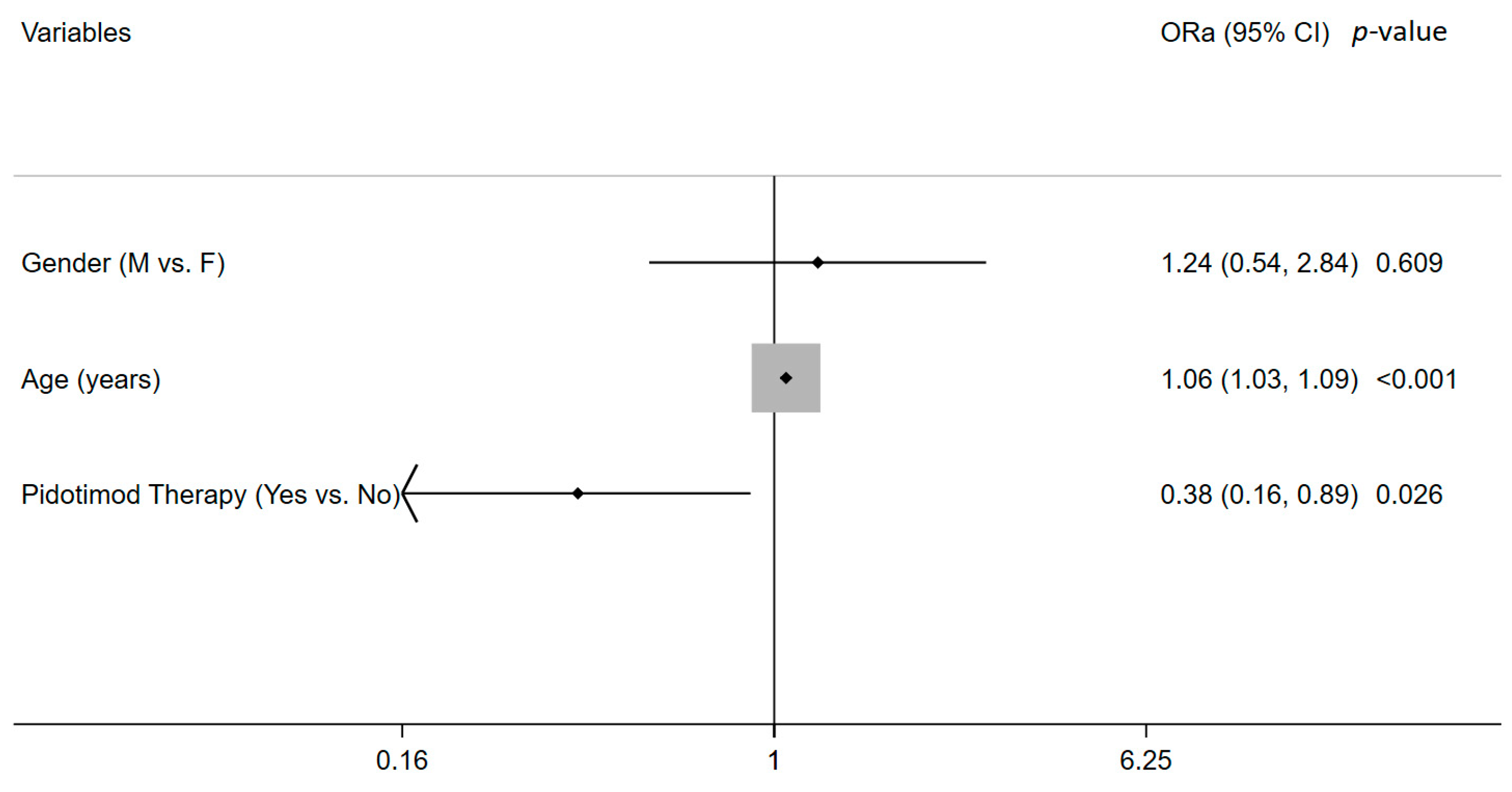
Table 4 and Figure 2 show the results of the crude and adjusted ORs for ER access/hospitalization. Crude OR confirmed the association, as shown in Table 3, while the logistic regression model showed that the risk of ER access/hospitalization increased with age (OR 1.06, 95 CI% 1.03–1.09, p < 0.0001) and decreased in patients undergoing pidotimod therapy (OR 0.38, 95 CI% 0.16–0.89, p = 0.026).
The Hosmer–Lemeshow goodness-of-fit test indicates that the model describes the data well (χ2(129) = 107.36, p = 0.917).
Finally, the multiple linear model for the illness duration showed that as 1 year of age increased the duration of illness (p < 0.0001), it decreased for patients who had been given pidotimod therapy (p = 0.004) (Table 5).

4. Discussion

Since its beginning, the SARS-CoV-2 pandemic has put welfare systems under pressure, causing the proliferation of inhomogeneous and often very different home therapeutic strategies. A critical point in the home management of the COVID-19 patient was the lack of effective treatment strategies in the first week of illness.
The arrival of monoclonal antibodies (bamlanivimab-etesevimab, casirivimab-imdevimab, sotrovimab, etc.) and antivirals (molnupiravir, nirmatrelvir-ritonavir) has, in part, responded to this need by providing effective, but expensive and restricted access (from temporal and patient characteristics points of view), weapons [15,16,17,18]. Part of this arsenal has also been subject to a loss of efficacy linked to the emergence of new viral variants (for example, most of the available antibody combinations are currently unusable for this reason) [19,20,21,22,23]. We evaluated an alternative approach based on early moderation of the inflammatory response, directing it toward a more efficient, coordinated and appropriate response. On the basis of previous literature experiences, the drug that best suited this aim was pidotimod, a polypeptide with chemical and functional similarities to imiquimod and with immunomodulating properties, expressed through multiple actions on innate and acquired immunity.
Pidotimod favors the expression of TLR2 and TLR7 (over TLR4) in the respiratory tract with a correct antigenic presentation [20,24,25], stimulates the production of INF 1 and gamma with the establishment of the antiviral state and the predominance of a Th1 phenotype (over Th2 and Th17) [11,21,22] with inflammation control, and stimulates mucosal IgA production with a reduction in the amount of antigen available for ADE [26]. Finally, a population of M2-type alveolar macrophages is favored, which is found to be prevalent in subjects who do not develop severe symptoms [23,27,28].
The data that emerged from our study seems to be in agreement with what has been said thus far. In the pidotimod group, hospital access, hospitalizations, and deaths were lower with a statistically significant difference compared to the control group. These data could indicate a better control of the inflammatory response in these patients by an immune system that is adequately modulated and prepared, in the early stages of the infection, to give a better response. Consequently, less lung damage, less development of respiratory failure, ARDS, and finally, less need for care in a hospital setting would result.
Two other results that emerged from our study reinforce this hypothesis: the minimum SpO2 value found during the course of the disease and the SpO2 value found in the 6-min walking test were higher in patients in the pidotimod group. A higher SpO2 minimum value could indicate less cumulative lung injury in patients treated with pidotimod. The SpO2 value in the walking test is an index of the residual pulmonary respiratory reserve; the alteration in this index is earlier than the alteration of the SpO2 at rest in the case of lung damage. It is a pulmonary functional index, an indirect reflection of the state of damage to this organ [29,30]; then the difference between the two groups supports the hypothesis of a lower functional damage (probably linked to a lower anatomical damage) in the group treated with pidotimod.
This lower degree of lung inflammation is probably related to the reduced homing of inflammatory cells in this organ, also linked to the expression of Toll-like receptors 2 and 7, which correctly process viral antigens, and to the M2 phenotype of alveolar macrophages (producers of fewer chemokines and pro-inflammatory cytokines), both events stimulated by pidotimod [20,27].
Another finding in this direction is a minor need for rescue therapy with corticosteroids in the pidotimod group. The need to use corticosteroids indicates, on one hand, greater lung damage (reflected in a lower SpO2), and on the other hand, the presence of a more marked systemic inflammation (persistent fever), which, left to itself, probably would have caused lung damage and the need for hospital treatment (often the steroid was not able to reverse these processes): the early immunomodulation implemented by pidotimod might be able to prevent these advanced disease states, thus reducing the need to use corticosteroids as a last attempt of home therapy before hospital treatment. Symptomatologically, patients treated with pidotimod more often developed symptoms such as headache and myalgia, which could be part of less severe disease subsets, less associated with complications compared with subsets including respiratory symptoms and dyspnea (as emerged also in our analysis). From a clinical point of view, a better control of the inflammatory state could be reflected in a systemic symptomatology of minor importance and, in particular, in a lower percentage of relevant respiratory symptoms. In the pidotimod group, disease duration was also significantly shorter. This difference could be linked to the aforementioned result of a less severe disease with less need for hospitalization. An organized immune response, addressed by an early immunomodulation, could have produced a faster viral clearance: in particular, the stimulatory effect of pidotimod on the interferon 1 system could have earlier produced that so-called intracellular “antiviral state” necessary for the correct elimination of viral infections [22].
In our study, it emerged that the duration of the disease is directly correlated with the age of the patient and the presence of comorbidities, as is already known in the literature. It was highlighted how a clinically more severe disease is associated with a longer duration of positivity to SARS-CoV-2: this is probably linked to a more disorganized and harmful immune response that fails to obtain an efficient viral clearance. It can also be noted, with a value close to statistical significance (p = 0.06), how the use of the corticosteroid can be associated with a longer disease duration [31,32]: this could be linked to the more severe clinical picture that is usually present in patients who required the use of the steroid [33]. Additionally, in the sub-analysis of this secondary outcome, the role of pidotimod in reducing the number of days of positivity to SARS-CoV-2 emerged compared to patients who had undergone other therapies.
From the data of our study, it emerges that pidotimod, in a patient with mild/moderate COVID-19 and the right timing of the disease, can be an effective therapy in preventing the development of severe disease and the need for hospital care. At the time, we had different options for the viral replication phase (with stringent inclusion criteria and time frames) and for the inflammatory phase; we have no effective options to treat patients in the viral phase without risk factors, outside of the fifth day of disease, younger, or in therapy with drugs that interact with antivirals. All of these categories cannot access antivirals and pidotimod can represent a valid therapeutic option. Indeed, it is a low-cost alternative, compared to others that can be used in this setting, which can be used in almost any type of home patient without access restrictions and relevant drug interactions.
Another purpose of our work was to highlight a series of factors (clinical and non-clinical) related to the patient that could be associated with an unfavorable outcome or a longer duration of illness to provide additional patient assessment tools outside the hospital.
In addition to age, the presence of comorbidities and BMI, which are already known in the literature [34,35,36], other factors were significantly associated with a greater risk of hospital access: number of days with fever, presence of dyspnea, initial SpO2, SpO2 on walking test, MEWS score, LUS score, corticosteroid use, and day of illness on which they were introduced in therapy.
In patients with a worse outcome, the presence of fever was observed, on average, for a higher number of days. This could indicate a more pronounced and aggressive inflammatory process responsible for the lung damage, which then leads to hospitalization.
In this same direction goes the relief, in this group of patients, of a more frequent presence of dyspnea, evidence of lung damage already underway [37]. It could therefore be identified, within the multiform presentations of the COVID-19 disease, a subset with a greater risk of hospitalization characterized by fever and marked respiratory symptoms, which would deserve greater attention when it appears. On the other hand, symptoms such as ageusia, anosmia, headache, and myalgia could constitute a subset with a lower risk of nefarious evolution. For some of these, a protective effect has already been highlighted in the literature [38,39,40]. Patients who undergo hospitalization also showed fewer days without symptoms and a longer disease course, underlining the biggest burden of disease with a slower and longer recovery and therefore a longer positivity to SARS-CoV-2.
Although within the normal limits, saturation at rest was lower, right from enrolment, in patients who subsequently underwent hospitalization. The walking test was confirmed, even in our study, to be a useful tool capable of highlighting (even before SpO2 at rest) the presence of desaturation. An SpO2 below 94% during the walking test should be a wake-up call for any physician working with COVID patients outside the hospital.
MEWS is a clinical score, part of routine clinical practice from 1999, with the aim of the early identification of patients at high risk of admission to ICU; this has been validated for a general medical population and for a surgical population [41,42]. It is a quick score composed of parameters that can be easily collected and therefore is applicable at home, by the bedside. In previous studies on COVID-19 patients, this score has not been shown to be able to predict the evolution toward severe forms of the disease [43]; this is probably linked to its late use (application in Emergency Room). In our study, this score was also useful in predicting hospital admission for COVID-19 patients, identifying a value greater than 2 as deserving particular attention.
In our study, we identified a marked difference in the ultrasound findings of those who were hospitalized and those who did not need.
LUS is a validated ultrasound score based on the “ring-down” artifact that indicates the presence of fluid inside the lung parenchyma (edema, alveolar or interstitial), which is directly proportional to the severity of the lung inflammation [44,45]. Lung ultrasound is shown to be an adequate tool to identify early deterioration of the lung parenchyma. Previous studies have attempted to identify a minimum LUS value to indicate patients who may need hospitalization [44]. In our study, this value was found to be 8 points.
Corticosteroid use was associated with an increased risk of hospitalization [45,46]. These data could be interpreted in light of the fact that these drugs have been reserved, in our area, for patients who are on average more serious (hyperpyrexia resistant to NSAIDs or peripheral SpO2 <94% with the need for home oxygen therapy) [33]: this also highlights the inability of this therapeutic tool to recover most of the advanced clinical situations not yet in need for hospital treatment. A portion of the patients treated with corticosteroids may have started them in an untimely manner. As is known, these drugs promote viral replication; using them in the first week of the onset of the disease could fuel the replication of SARS-CoV-2, inducing greater lung damage in the second week of illness [31]. Our data show that the use of the steroid before the eighth day of illness is more associated with the need for hospitalization.
COVID-19 is a disease that is very variable in clinical aspects and courses; some symptom subsets can predict a less aggressive disease than others and provide useful information in the home management of patients with SARS-CoV-2. Likewise, LUS, MEWS, and the walking test can represent effective evaluation tools in identifying early clinical deterioration that requires hospital care, representing important weapons in the arsenal of the physician.

5. Conclusions

SARS-CoV-2 infection causes an aggressive, off-scale immune reaction that results in systemic and lung damage. The use of pidotimod proved able to prevent this phenomenon by directing the immune system toward a more appropriate and effective response, managing to prevent the evolution of the disease from mild-moderate to severe forms, which require hospitalization.
Furthermore, the use of pidotimod is able to speed up the negativization of the infection from SARS-CoV-2. Pidotimod is configured as an effective therapeutic option, inexpensive and usable in almost all categories of patients. Acting on the immune response may also represent a valid strategy in light of the emergence of new variants and resistance that can reduce the effectiveness of monoclonal antibodies and antivirals.

Author Contributions

Conceptualization, C.U. and K.F.; Methodology, M.D.N.; Software, P.B.; Validation C.U. and K.F.; Formal analysis, M.D.N. and P.B.; Investigation, A.D.G.; Writing—original draft preparation, C.U., A.D.G. and K.F.; Writing—review and editing, C.U., M.D.N. and J.V. All authors have read and agreed to the published version of the manuscript.

Funding

This research received no external funding.

Data Availability Statement

Data available upon request.

Acknowledgments

We acknowledge the USCA Berdini Giuseppe, Della Penna Nicola, Dente Giuseppe, Di Lanzo Sandra, Garzarella Vera, Mininni Fabrizia, Primante Mariella Valentina, Pugliese Maria Costanza, and Valentina.

Conflicts of Interest

The authors declare no conflict of interest.

References

  1. Available online: www.coronavirus.jhu.edu/map.html (accessed on 29 September 2022).
  2. Yang, X.; Yu, Y.; Xu, J.; Shu, H.; Xia, J.; Liu, H.; Wu, Y.; Zhang, L.; Yu, Z.; Fang, M.; et al. Clinical course and outcomes of critically ill patients with SARS-CoV-2 pneumonia in Wuhan, China: A single-centered, retrospective, observational study. Lancet Respir. Med. 2020, 8, 475–481. [Google Scholar] [CrossRef] [Green Version]
  3. Lai, C.C.; Shih, T.P.; Ko, W.C.; Tang, H.J.; Hsueh, P.R. Severe acute respiratory syndrome coronavirus 2 (SARS-CoV-2) and coronavirus disease-2019 (COVID-19): The epidemic and the challenges. Int. J. Antimicrob. Agents 2020, 55, 105924. [Google Scholar] [CrossRef] [PubMed]
  4. Soy, M.; Keser, G.; Atagündüz, P.; Tabak, F.; Atagündüz, I.; Kayhan, S. Cytokine storm in COVID-19: Pathogenesis and overview of anti-inflammatory agents used in treatment. Clin. Rheumatol. 2020, 39, 2085–2094. [Google Scholar] [CrossRef] [PubMed]
  5. Song, P.; Li, W.; Xie, J.; Hou, Y.; You, C. Cytokine storm induced by SARS-CoV-2. Clin. Chim. Acta 2020, 509, 280–287. [Google Scholar] [CrossRef] [PubMed]
  6. Ucciferri, C.; Auricchio, A.; Di Nicola, M.; Potere, N.; Abbate, A.; Cipollone, F.; Vecchiet, J.; Falasca, K. Canakinumab in a subgroup of patients with COVID-19. Lancet Rheumatol. 2020, 2, e457–e458. [Google Scholar] [CrossRef]
  7. Vannucchi, A.M.; Mortara, A.; D’Alessio, A.; Morelli, M.; Tedeschi, A.; Festuccia, M.B.; Monforte, A.D.; Capochiani, E.; Selleri, C.; Simonetti, F.; et al. JAK Inhibition with Ruxolitinib in Patients with COVID-19 and Severe Pneumonia: Multicenter Clinical Experience from a Compassionate Use Program in Italy. J. Clin. Med. 2021, 10, 3752. [Google Scholar] [CrossRef]
  8. Wu, Z.; McGoogan, J.M. Characteristics of and Important Lessons From the Coronavirus Disease 2019 (COVID-19) Outbreak in China: Summary of a Report of 72 314 Cases From the Chinese Center for Disease Control and Prevention. JAMA 2020, 323, 1239–1242. [Google Scholar] [CrossRef]
  9. Zuccotti, G.V.; Mameli, C. Pidotimod: The past and the present. Ital. J. Pediatr. 2013, 39, 75. [Google Scholar] [CrossRef] [Green Version]
  10. Chatterjee, A.N.; Al Basir, F. A Model for SARS-CoV-2 Infection with Treatment. Comput. Math. Methods Med. 2020, 2020, 1352982. [Google Scholar] [CrossRef]
  11. Giagulli, C.; Noerder, M.; Avolio, M.; Becker, P.D.; Fiorentini, S.; Guzman, C.A.; Caruso, A. Pidotimod promotes functional maturation of dendritic cells and displays adjuvant properties at the nasal mucosa level. Int. Immunopharmacol. 2009, 9, 1366–1373. [Google Scholar] [CrossRef]
  12. Zhao, N.; Liu, C.; Zhu, C.; Dong, X.; Liu, X. Pidotimod: A review of its pharmacological features and clinical effectiveness in respiratory tract infections. Expert Rev. Anti-Infect. Ther. 2019, 17, 803–818. [Google Scholar] [CrossRef] [PubMed]
  13. Santus, P.; Radovanovic, D.; Garziano, M.; Pini, S.; Croce, G.; Fuccia, G.; Spitaleri, D.; Biasin, M.; Clerici, M.; Trabattoni, D. Anti-Inflammatory Effects of Immunostimulation in Patients with COVID-19 Pneumonia. J. Clin. Med. 2021, 10, 5765. [Google Scholar] [CrossRef] [PubMed]
  14. Ucciferri, C.; Mirko, B.; Vecchiet, J.; Falasca, K. Pidotimod in paucisymptomatic SARS-CoV2 infected patients. Mediterr. J. Hematol. Infect. Dis. 2020, 12, e2020048. [Google Scholar] [CrossRef] [PubMed]
  15. Wong, C.K.; Au, I.C.; Lau, K.T.; Lau, E.; Cowling, B.J.; Leung, G.M. Real-world effectiveness of molnupiravir and nirmatrelvir/ritonavir among COVID-19 inpatients during Hong Kong’s Omicron BA.2 wave: An observational study. medRxiv 2022. [Google Scholar] [CrossRef]
  16. Vena, A.; Traman, L.; Bavastro, M.; Limongelli, A.; Dentone, C.; Magnè, F.; Giacobbe, D.R.; Mikulska, M.; Taramasso, L.; Di Biagio, A.; et al. Early Clinical Experience with Molnupiravir for Mild to Moderate Breakthrough COVID-19 among Fully Vaccinated Patients at Risk for Disease Progression. Vaccines 2022, 10, 1141. [Google Scholar] [CrossRef]
  17. Gentile, I.; Scotto, R.; Moriello, N.S.; Pinchera, B.; Villari, R.; Trucillo, E.; Ametrano, L.; Fusco, L.; Castaldo, G.; Buonomo, A.R.; et al. Nirmatrelvir/ritonavir and molnuipiravir in the treatment of mild/moderate COVID-19: Results of a real-life study. medRxiv 2022. [Google Scholar] [CrossRef]
  18. Hung, Y.-P.; Lee, J.-C.; Chiu, C.-W.; Lee, C.-C.; Tsai, P.-J.; Hsu, I.-L.; Ko, W.-C. Oral Nirmatrelvir/Ritonavir Therapy for COVID-19: The Dawn in the Dark? Antibiotics 2022, 11, 220. [Google Scholar] [CrossRef]
  19. Takashita, E.; Yamayoshi, S.; Simon, V.; van Bakel, H.; Sordillo, E.M.; Pekosz, A.; Fukushi, S.; Suzuki, T.; Maeda, K.; Halfmann, P.; et al. Efficacy of Antibodies and Antiviral Drugs against Omicron BA.2.12.1, BA.4, and BA.5 Subvariants. N. Engl. J. Med. 2022, 387, 468–470. [Google Scholar] [CrossRef]
  20. Carta, S.; Silvestri, M.; Rossi, G.A. Modulation of airway epithelial cell functions by Pidotimod: NF-kB cytoplasmatic expression and its nuclear translocation are associated with an increased TLR-2 expression. Ital. J. Pediatr. 2013, 39, 29. [Google Scholar] [CrossRef] [Green Version]
  21. Zuccotti, G.V.; Mameli, C.; Trabattoni, D.; Beretta, S.; Biasin, M.; Guazzarotti, L.; Clerici, M. Immunomodulating activity of Pidotimod in children with Down syndrome. J. Biol. Regul. Homeost. Agents 2013, 27, 253–258. Available online: https://pubmed.ncbi.nlm.nih.gov/23489705/ (accessed on 30 August 2022).
  22. Ferrario, B.E.; Garuti, S.; Braido, F.; Canonica, G.W. Pidotimod: The state of art. Clin. Mol. Allergy 2015, 13, 8. [Google Scholar] [CrossRef] [PubMed]
  23. Lv, J.; Wang, Z.; Qu, Y.; Zhu, H.; Zhu, Q.; Tong, W.; Bao, L.; Lv, Q.; Cong, J.; Li, D.; et al. Distinct uptake, amplification, and release of SARS-CoV-2 by M1 and M2 alveolar macrophages. Cell Discov. 2021, 7, 24. [Google Scholar] [CrossRef]
  24. Trabattoni, D.; Clerici, M.; Centanni, S.; Mantero, M.; Garziano, M.; Blasi, F. Immunomodulatory effects of pidotimod in adults with community-acquired pneumonia undergoing standard antibiotic therapy. Pulm. Pharmacol. Ther. 2017, 44, 24–29. [Google Scholar] [CrossRef] [PubMed]
  25. Esposito, S.; Garziano, M.; Rainone, V.; Trabattoni, D.; Biasin, M.; Senatore, L.; Marchisio, P.G.; Rossi, M.; Principi, N.; Clerici, M. Immunomodulatory activity of pidotimod administered with standard antibiotic therapy in children hospitalized for community-acquired pneumonia. J. Transl. Med. 2015, 13, 288. [Google Scholar] [CrossRef] [PubMed] [Green Version]
  26. Rossi, G.A.; Peri, C.; Raynal, M.E.; Defilippi, A.-C.; Risso, F.M.; Schenone, G.; Pallestrini, E.; Melioli, G. Naturally occurring immune response against bacteria commonly involved in upper respiratory tract infections: Analysis of the antigen-specific salivary IgA levels. Immunol. Lett. 2003, 86, 85–91. [Google Scholar] [CrossRef]
  27. Hu, S.; Fu, X.; Fu, A.; Du, W.; Ji, J.; Li, W. The regulatory peptide pidotimod facilitates M2 macrophage polarization and its function. Amino Acids 2014, 46, 1177–1185. [Google Scholar] [CrossRef] [PubMed]
  28. Lee, J.-W.; Chun, W.; Lee, H.; Min, J.-H.; Kim, S.-M.; Seo, J.-Y.; Ahn, K.-S.; Oh, S.-R. The Role of Macrophages in the Development of Acute and Chronic Inflammatory Lung Diseases. Cells 2021, 10, 897. [Google Scholar] [CrossRef]
  29. Mantha, S.; Tripuraneni, S.L.; Roizen, M.F.; Fleisher, L.A. Proposed Modifications in the 6-Minute Walk Test for Potential Application in Patients With Mild COVID-19: A Step to Optimize Triage Guidelines. Anesthesia Analg. 2020, 131, 398–402. [Google Scholar] [CrossRef]
  30. Singh, S.J.; Puhan, M.; Andrianopoulos, V.; Hernandes, N.A.; Mitchell, K.E.; Hill, C.J.; Lee, A.L.; Camillo, C.A.; Troosters, T.; Spruit, M.A.; et al. An official systematic review of the European Respiratory Society/American Thoracic Society: Measurement properties of field walking tests in chronic respiratory disease. Eur. Respir. J. 2014, 44, 1447–1478. [Google Scholar] [CrossRef] [Green Version]
  31. Theoharides, T.C.; Conti, P. Dexamethasone for COVID-19? Not so fast. J. Biol. Regul. Homeost. Agents 2020, 34, 1241–1243. [Google Scholar] [CrossRef]
  32. Spagnuolo, V.; Guffanti, M.; Galli, L.; Poli, A.; Querini, P.R.; Ripa, M.; Clementi, M.; Scarpellini, P.; Lazzarin, A.; Tresoldi, M.; et al. Viral clearance after early corticosteroid treatment in patients with moderate or severe COVID-19. Sci. Rep. 2020, 10, 21291. [Google Scholar] [CrossRef] [PubMed]
  33. RECOVERY Collaborative Group; Horby, P.; Lim, W.S.; Emberson, J.R.; Mafham, M.; Bell, J.L.; Linsell, L.; Staplin, N.; Brightling, C.; Ustianowski, A.; et al. Dexamethasone in Hospitalized Patients with COVID-19. N. Engl. J. Med. 2021, 384, 693–704. [Google Scholar] [CrossRef] [PubMed]
  34. Nasir, N.; Tajuddin, S.; Khaskheli, S.; Khan, N.; Niamatullah, H.; Nasir, N. Clinical outcomes of immunomodulatory therapies in the management of COVID-19: A tertiary-care experience from Pakistan. PLoS ONE 2022, 17, e0262608. [Google Scholar] [CrossRef] [PubMed]
  35. Grasselli, G.; Greco, M.; Zanella, A.; Albano, G.; Antonelli, M.; Bellani, G.; Bonanomi, E.; Cabrini, L.; Carlesso, E.; Castelli, G.; et al. Risk Factors Associated With Mortality Among Patients With COVID-19 in Intensive Care Units in Lombardy, Italy. JAMA Intern. Med. 2020, 180, 1345–1355. [Google Scholar] [CrossRef]
  36. Du, Y.; Lv, Y.; Zha, W.; Zhou, N.; Hong, X. Association of body mass index (BMI) with critical COVID-19 and in-hospital mortality: A dose-response meta-analysis. Metabolism 2020, 117, 154373. [Google Scholar] [CrossRef]
  37. Alhazzani, W.; Møller, M.H.; Arabi, Y.M.; Loeb, M.; Gong, M.N.; Fan, E.; Oczkowski, S.; Levy, M.M.; Derde, L.; Dzierba, A.; et al. Acute Respiratory Distress Syndrome: Diagnosis and Management. Am. Fam. Physician 2020, 101, 730–738. [Google Scholar] [CrossRef] [Green Version]
  38. Trigo, J.; García-Azorín, D.; Planchuelo-Gómez, Á.; Martínez-Pías, E.; Talavera, B.; Hernández-Pérez, I.; Valle-Peñacoba, G.; Simón-Campo, P.; De Lera, M.; Chavarría-Miranda, A.; et al. Factors associated with the presence of headache in hospitalized COVID-19 patients and impact on prognosis: A retrospective cohort study. J. Headache Pain 2020, 21, 94. [Google Scholar] [CrossRef]
  39. Shapiro, R.E.; Gallardo, V.J.; Caronna, E.; Pozo-Rosich, P. The impact of headache disorders on COVID-19 survival: A world population-based analysis. medRxiv 2021. [Google Scholar] [CrossRef]
  40. Gil-Rodrigo, A.; Miró, Ò.; Piñera, P.; Burillo-Putze, G.; Jiménez, S.; Martín, A.; Martín-Sánchez, F.J.; Jacob, J.; Guardiola, J.M.; García-Lamberechts, E.J.; et al. Analysis of clinical characteristics and outcomes in patients with COVID-19 based on a series of 1000 patients treated in Spanish emergency departmen. Emergencias 2020, 32, 233–241. Available online: https://pubmed.ncbi.nlm.nih.gov/32692000/ (accessed on 13 October 2022).
  41. Subbe, C.P.; Kruger, M.; Rutherford, P.; Gemmel, L. Validation of a modified Early Warning Score in medical admission |Read by QxMD. Qjm 2001, 94, 521–526. Available online: https://read.qxmd.com/read/11588210/validation-of-a-modified-early-warning-score-in-medical-admissions (accessed on 23 August 2022). [CrossRef] [Green Version]
  42. Stenhouse, C.; Coates, S.; Tivey, M.; Allsop, P.; Parker, T. Prospective evaluation of a modified Early Warning Score to aid earlier detection of patients developing critical illness on a general surgical ward. Br. J. Anaesth. 2000, 84, 663P. [Google Scholar] [CrossRef]
  43. Özdemir, S.; Algın, A.; Akça, H.; Altunok, I.; Kokulu, K.; Eroğlu, S.E.; Aksel, G. Predictive Ability of the MEWS, REMS, and RAPS in Geriatric Patients With SARS-CoV-2 Infection in the Emergency Department. Disaster Med. Public Health Prep. 2022, 1–4. [Google Scholar] [CrossRef] [PubMed]
  44. Boccatonda, A.; Grignaschi, A.; Lanotte, A.M.G.; Cocco, G.; Vidili, G.; Giostra, F.; Schiavone, C. Role of Lung Ultrasound in the Management of Patients with Suspected SARS-CoV-2 Infection in the Emergency Department. J. Clin. Med. 2022, 11, 2067. [Google Scholar] [CrossRef] [PubMed]
  45. Nasir, N.; Habib, K.; Khanum, I.; Khan, N.; Muhammad, Z.A.; Mahmood, S.F. Clinical characteristics and outcomes of COVID-19: Experience at a major tertiary care center in Pakistan. J. Infect. Dev. Ctries. 2021, 15, 480–489. [Google Scholar] [CrossRef] [PubMed]
  46. Rommasi, F.; Nasiri, M.J.; Mirsaeidi, M. Immunomodulatory agents for COVID-19 treatment: Possible mechanism of action and immunopathology features. Mol. Cell Biochem. 2022, 477, 711–726. [Google Scholar] [CrossRef]
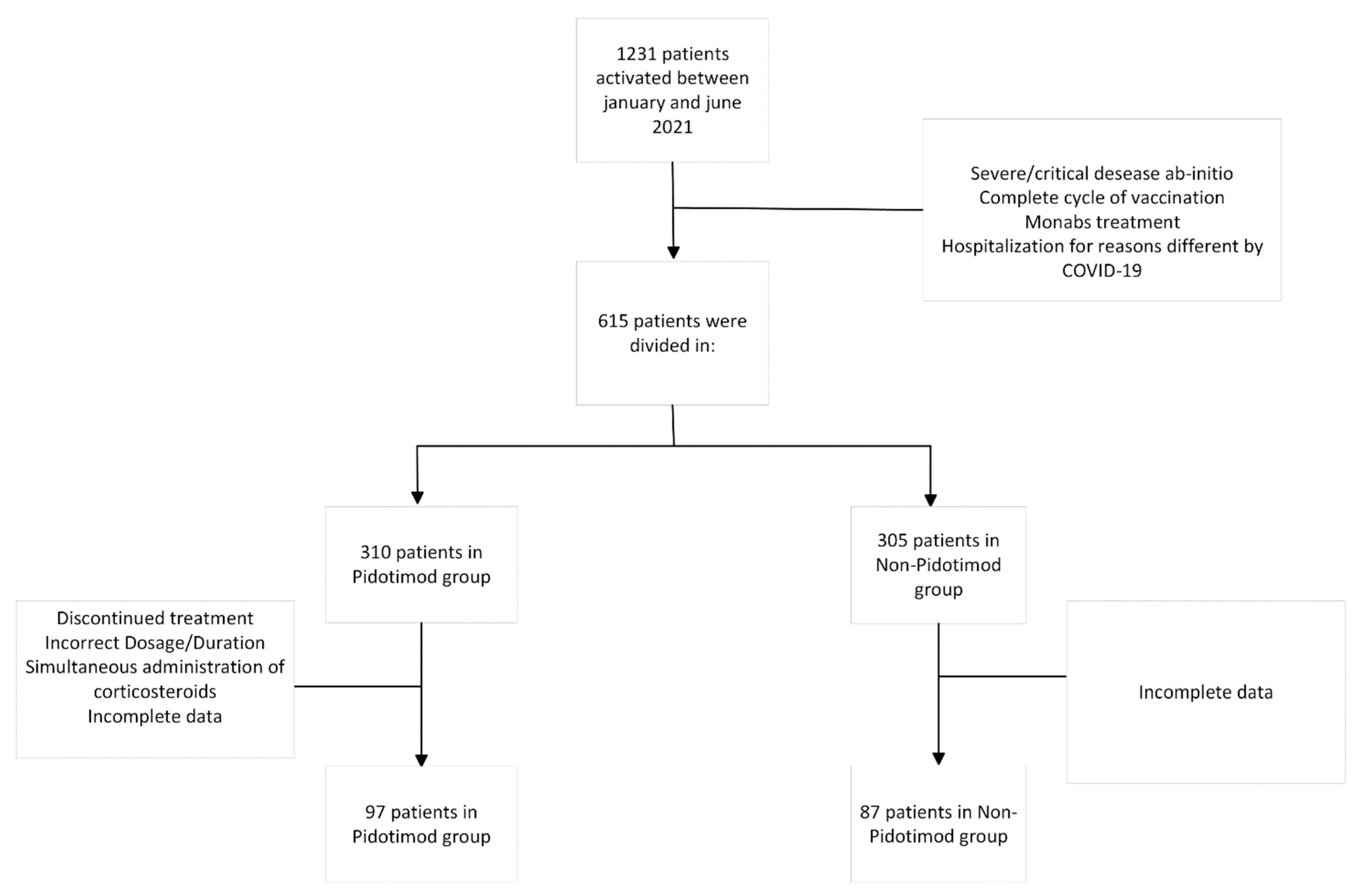
Figure 1. Study enrolment.
Microorganisms 10 02131 g001
Figure 2. Logistic regression for ER access/hospitalization; Ora = adjusted odds ratio; 95% CI = 95% interval confidence.
Microorganisms 10 02131 g002
Table 1. The patients’ socio-demographic and clinical characteristics (n = 184).
Gender
F100 (54.3%)
M84 (45.7%)
Age (years)53.0 (42.0–62.5)
Illness Duration (days)22.0 (18.0–28.0)
Comorbidity
No59 (34.3%)
Yes113 (65.7%)
BMI (kg/m2)27.1 (23.8–29.4)
Fever
No51 (27.9%)
Yes132 (72.1%)
Maximum Temperature38.0 (37.6–38.5)
Days of Fever3.0 (0.0–6.0)
Cough
No24 (13.0%)
Yes160 (87.0%)
Dyspnea
No139 (75.5%)
Yes45 (24.5%)
Ageusia or Anosmia
No88 (47.8%)
Yes96 (52.2%)
Fatigue
No41 (22.3%)
Yes143 (77.7%)
Myalgia
No64 (34.8%)
Yes120 (65.2%)
Headache
No104 (56.5%)
Yes80 (43.5%)
Others
No86 (47.3%)
Yes96 (52.7%)
Number of Symptoms4.0 (3.0–6.0)
SpO2 at Enrolment98.0 (97.0–99.0)
Minimum SpO296.0 (94.0–97.0)
Mews1.0 (0.0–3.0)
LUS6.0 (2.0–10.0)
Walking test97.0 (95.0–99.0)
Pidotimod Therapy
No87 (47.3%)
Yes97 (52.7%)
Pidotimod Start Day3.0 (2.0–6.0)
Pidotimod Duration3.0 (0.0–10.0)
Corticosteroid Therapy
No119 (65.0%)
Yes64 (35.0%)
Corticosteroid Start Day8.0 (7.0–10.0)
ER Access/Hospitalization
No150 (81.5%)
Yes34 (18.5%)
N (%) or median and IQR are shown when appropriate.
Table 2. The patients’ socio-demographic and clinical characteristics for pidotimod therapy (n = 184).
Pidotimod Therapy
No (n = 87)Yes (n = 97)* p-Value
Gender
F47 (54.0%)53 (54.6%)0.526
M40 (46.0%)44 (45.4%)
Age (years)53.0 (44.0–64.0)53.0 (40.0–61.0)0.240
Illness Duration (days)23.0 (20.0–31.0)21.0 (17.0–27.0)0.005
Comorbidity
No19 (24.4%)40 (42.6%)0.012
Yes59 (75.6%)54 (57.4%)
BMI (kg/m2)27.1 (26.0–29.4)26.9 (23.5–29.4)0.508
Fever during the course
No26 (29.9%)25 (26.0%)0.563
Yes61 (70.1%)71 (74.0%)
Maximum Temperature38.0 (37.5–38.5)38.0 (37.6–38.5)0.914
Days of Fever3.0 (0.0–6.0)3.0 (0.0–6.0)0.438
Cough **
No14 (16.1%)10 (10.3%)0.245
Yes73 (83.9%)87 (89.7%)
Dyspnea **
No64 (73.6%)75 (77.3%)0.753
At rest and in motion11 (12.6%)9 (9.3%)
Only in motion12 (13.8%)13 (13.4%)
Ageusia or Anosmia **
No46 (52.9%)42 (43.3%)0.194
Yes41 (47.1%)55 (56.7%)
Fatigue **
No24 (27.6%)17 (17.5%)0.102
Yes63 (72.4%)80 (82.5%)
Myalgia **
No37 (42.5%)27 (27.8%)0.037
Yes50 (57.5%)70 (72.2%)
Headache **
No57 (65.5%)47 (48.5%)0.020
Yes30 (34.5%)50 (51.5%)
Others **
No49 (56.3%)37 (38.9%)0.113
Yes28 (43.7%)57 (61.1%)
Number of Symptoms **4.0 (3.0–5.0)5.0 (3.0–6.0)<0.001
SpO2 at Enrolment98.0 (96.0–99.0)98.0 (97.0–99.0)0.103
Minimum SpO2 during the course95.0 (93.0–97.0)96.0 (94.0–97.0)0.046
Mews at Enrolment1.0 (0.0–3.0)1.0 (0.0–2.0)0.062
Maximum Mews during the course3.0 (0.0–5.0)1.0 (0.0–3.0)0.770
Maximum LUS during the course7.0 (2.0–12.0)4.5 (1.5–8.0)0.128
Minimum SpO2 Walking Test97.0 (93.0–98.0)97.0 (96.0–99.0)0.010
Corticosteroid Therapy
No34 (39.1%)85 (88.5%)<0.001
Yes53 (60.9%)11 (11.5%)
Corticosteroid Start Day8.0 (7.0–9.0)8.0 (7.0–12.0)0.282
Corticosteroid Duration3.5 (0.0–11.0)0.0 (0.0–0.0)<0.001
ER Access/Hospitalization
No64 (73.6%)86 (88.7%)0.008
Yes23 (26.4%)11 (11.3%)
N (%) or median (IQR) are shown when appropriate; * p-values are for the Wilcoxon rank-sum test or Pearson’s chi-square test; ** Symptoms present during the course of disease.
Table 3. The patients’ socio-demographic and clinical characteristics for ER access/hospitalization (n = 184).
ER Access/Hospitalization
No (n = 150)Yes (n = 34)* p-Value
Gender
F84 (56.0%)16 (47.1%)0.225
M66 (44.0%)18 (52.9%)
Age (years)50.5 (39.0–60.0)66.0 (54.0–79.0)<0.001
Illness Duration (days)22.0 (18.0–27.0)28.0 (23.0–38.0)0.002
Comorbidity
No55 (39.6%)4 (12.1%)0.003
Yes84 (60.4%)29 (87.9%)
BMI (kg/m2)26.2 (23.5–29.1)33.3 (27.1–40.8)0.010
Fever during the course
No46 (30.9%)5 (14.7%)0.058
Yes103 (69.1%)29 (85.3%)
Maximum Temperature38.0 (37.5–38.5)38.2 (37.8–38.9)0.183
Days of Fever3.0 (0.0–5.0)4.0 (2.0–7.0)0.031
Cough **
No21 (14.0%)3 (8.8%)0.418
Yes129 (86.0%)31 (91.2%)
Dyspnea **
No121 (80.7%)18 (52.9%)<0.001
At rest and in motion9 (6.0%)11 (32.4%)
Only in motion20 (13.3%)5 (14.7%)
Ageusia or Anosmia **
No65 (43.3%)23 (67.6%)0.010
Yes85 (56.7%)11 (32.4%)
Fatigue **
No30 (20.0%)11 (32.4%)0.118
Yes120 (80.0%)23 (67.6%)
Myalgia **
No49 (32.7%)15 (44.1%)0.206
Yes101 (67.3%)19 (55.9%)
Headache **
No78 (52.0%)26 (76.5%)0.009
Yes72 (48.0%)8 (23.5%)
Others **
No67 (45.3%)19 (55.9%)0.336
Yes81 (54.7%)21 (44.1%)
Number of Symptoms **4.0 (3.0–6.0)4.0 (3.0–5.0)0.127
SpO2 at Enrolment98.0 (97.0–100.0)96.0 (95.0–98.0)<0.001
Minimum SpO2 during the course96.0 (95.0–97.0)93.0 (90.0–94.0)<0.001
Maximum Mews during the course1.0 (0.0–2.0)5.0 (3.0–7.0)0.001
Maximum LUS during the course4.5 (1.5–6.5)12.0 (8.0–14.0)0.003
Minimum SpO2 at Walking Test97.0 (95.0–99.0)93.0 (90.5–97.0)<0.001
Pidotimod Therapy
No64 (42.7%)23 (67.6%)0.008
Yes86 (57.3%)11 (32.4%)
Pidotimod Start Day3.0 (2.0–6.0)4.0 (1.0–6.0)0.792
Pidotimod Duration7.0 (0.0–10.0)0.0 (0.0–2.0)<0.001
Corticosteroid Therapy
No102 (68.5%)17 (50.0%)0.042
Yes47 (31.5%)17 (50.0%)
Corticosteroid Start Day8.5 (8.0–11.0)7.0 (4.5–8.0)<0.001
N (%) or median (IQR) are shown when appropriate; * p-values are for the Wilcoxon rank-sum test or Pearson’s chi-square test; ** Symptoms present during the course of disease.
Table 4. Crude odds ratio and corresponding 95% CI for ER access/hospitalization.
ORc95%CIp-Value
Gender (M vs. F)1.431[0.678, 3.020]0.346
Age (years)1.064[1.036, 1.092]<0.0001
BMI (kg/m2)1.220[1.059, 1.427]0.007
Illness Duration (days)1.073[1.031, 1.116]<0.0001
Comorbidity (Yes vs. No)4.747[1.581, 14.250]0.005
Dyspnea (Yes vs. No)1.648[1.034, 2.624]0.035
Ageusia or Anosmia (Yes vs. No)0.365[0.166, 0.804]0.012
Headache (Yes vs. No)0.333[0.141, 0.783]0.012
Pidotimod Therapy (Yes vs. No)0.355[0.161, 0.782]0.010
Corticosteroid Therapy (Yes vs. No)2.170[1.019, 4.621]0.045
ORc = crude odds ratio; 95% CI = 95% interval confidence.
Table 5. Multiple linear model for the illness duration.
Coeff95%CIp-Value
Gender−0.970[−3.825, 1.879]0.502
Age (years)0.163[0.078, 0.248]<0.0001
Pidotimod Therapy (Yes)−4.190[−7.041, −1.340]0.004
Coeff = coefficients; 95% CI = 95% interval confidence.
Publisher’s Note: MDPI stays neutral with regard to jurisdictional claims in published maps and institutional affiliations.