Article

Detection of Tick-Borne Pathogens in Ticks from Cattle in Western Highlands of Cameroon

by
Yannick Ngnindji-Youdje
1,2,3,
Adama Zan Diarra
1,2,
Michel Lontsi-Demano
3,
Timoléon Tchuinkam
3 and
Philippe Parola
1,2,*
1
Aix Marseille Univ, IRD, AP-HM, SSA, VITROME, 13005 Marseille, France
2
IHU-Méditerranée Infection, 19-21 Boulevard Jean Moulin, 13005 Marseille, France
3
Vector-Borne Diseases Laboratory of the Applied Biology and Ecology Research Unit (VBID-URBEA), Department of Animal Biology, Faculty of Science of the University of Dschang, Dschang P.O. Box 067, Cameroon
*
Author to whom correspondence should be addressed.
Microorganisms 2022, 10(10), 1957; https://doi.org/10.3390/microorganisms10101957
Submission received: 12 August 2022 / Revised: 22 September 2022 / Accepted: 27 September 2022 / Published: 30 September 2022
(This article belongs to the Section Veterinary Microbiology)

Abstract

:
This study aimed to detect and identify microorganisms in ticks collected in the Western Highlands of Cameroon. Quantitative real-time and standard PCR assays, coupled with sequencing, were used. A total of 944 ticks collected from cattle in five distinct sites in Cameroon were selected for the analyses. They belonged to five genera (Amblyomma, Hyalomma, Rhipicephalus, Haemaphysalis, and Ixodes) and twelve species. Real-time PCR revealed that 23% (n = 218) of the ticks were positive for Rickettsia spp., 15% (n = 141) for bacteria of the Anaplasmataceae family, 3% (n = 29) for Piroplasmida, 0.5% (n = 5) for Coxiella burnetii, 0.4% (n = 4) for Borrelia spp., and 0.2% (n = 2) for Bartonella spp. The co-infection rate (3.4%, n = 32) involved mainly Rickettsia spp. and Anaplasmataceae. Of the Rickettsia spp. positive ticks, the targeted PCR and sequencing yielded Rickettsia africae (78.9%), Rickettsia aeschlimannii (6.4%), Rickettsia massiliae (7.8%), Candidatus Rickettsia barbariae (0.9%), and Rickettsia sp. (0.9%). Anaplasmataceae included Anaplasma marginale (4.3%), Anaplasma platys (1.4%), Anaplasma centrale (0.7%), Ehrlichia ruminantium (0.7%), Wolbachia sp., Candidatus Ehrlichia rustica (13.5%), Candidatus Ehrlichia urmitei (7%), and an uncultured Ehrlichia sp. (4.2%). Borrelia theileri was identified in one Rhipicephalus microplus tick. Unfortunately, Piroplasmida could not be identified to the species level. This study demonstrates that in Cameroon, ticks harbour a wide variety of microorganisms and present a risk of zoonotic diseases.

1. Introduction

Ticks are obligate haematophagous arachnids, which are distributed globally and parasitise a wide range of vertebrates [1]. On a global scale, ticks are currently considered as the leading vectors of animal diseases and second biggest vectors of human diseases after mosquitoes [2,3]. Only about 20% of the world’s cattle population remains unaffected by ticks and tick-borne diseases. However, the annual global cost losses are estimated to range from USD 14–19 billion [4,5]. With an estimated population of 6.5 million, cattle are regarded in Cameroon as the main source of animal protein in most households, according to the National Institute of the Statistic [6]. However, one of the most important constraints of small- and large-scale cattle production is the high prevalence of infectious diseases [7]. Of the about 900 currently known tick species, only about 10% are of significant veterinary and/or medical importance [8]. In sub-Saharan Africa, ticks of economic importance in livestock belong mainly to four genera, according to the previous studies, namely Amblyomma, Hyalomma, Haemaphysalis, and Rhipicephalus [9,10].
The direct effects of tick on their hosts are irritation, anaemia, inflammation, paralysis, abscesses, allergies, hypersensitivity, and skin deterioration at the biting site, which often leads to reduction in weight gain and milk yield [11]. Ticks are also responsible for indirect effects through the transmission of pathogens responsible for bacterial, viral, and protozoan diseases worldwide [12,13,14].
In certain major livestock production zones in Cameroon, particularly the Sudano-Sahelian zone, the high Guinean savannah zone, and the Western Highlands zone, ticks are less studied than other disease vectors. There is also little information on the biodiversity of ticks and microorganisms transmitted, although some studies have been conducted in the country [15,16,17]. There is a lack of support for livestock farmers from veterinarians, which leads to an absence of care and, therefore, the ineffectiveness of control strategies undertaken by livestock farmers. However, in the light of recent information, the prevalence of ticks and tick-borne diseases in livestock is constantly increasing [18]. Furthermore, according to a study conducted in 1982 in the principal cattle-rearing agro-ecological zone of Cameroon (Wakwa research station, Adamawa), approximately 63% of animal mortality was attributed to ticks and tick-borne diseases [19]. The situation was made worse by the recent introduction of the invasive cattle tick (Rhipicephalus microplus) into the country whose presence was doubtful prior to 2019 [20,21]. This cattle tick is known as the most significant parasite and disease vector of livestock worldwide. It is known to be the main vector of Babesia bovis, Babesia bigemina, and Anaplasma marginale, causing bovine babesiosis and anaplasmosis in cattle [10,22,23,24]. Currently, the control of tick-borne diseases relies mainly on tick control. One of the difficulties that prevents the eradication of tick-borne diseases is that there is no vaccine, despite enormous efforts employed in chemical vector control. However, as some studies show, very little progress has been made to control ectoparasite infestations in animals using the vaccine approach [25].
This study aimed to detect certain classic tick-associated bacteria in ticks species collected from the Western Highlands of Cameroon using molecular tools.

2. Materials and Methods
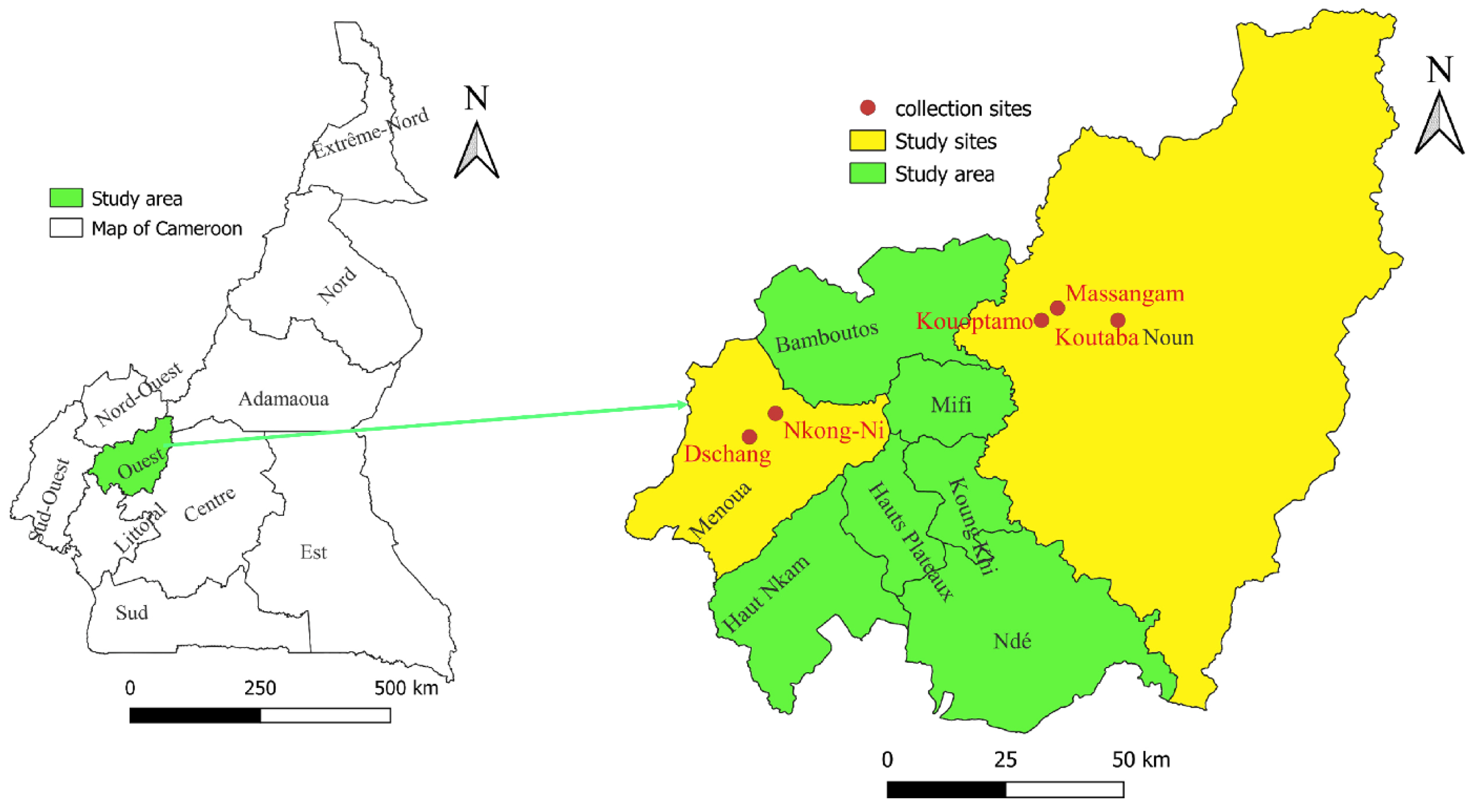
2.1. Study Area

This study was done in the Western Highlands of Cameroon, which are one of the three major agro-ecological zones for livestock production (Figure 1). It is in the mid- and high-latitude zones of the country. The annual average temperature is 20.6 °C and annual precipitation ranges from 1300 to 3000 mm. Two seasons can be distinguished as follows: the rainy season lasts eight months (March to October) and the dry season from November to February [20]. The choice of this zone is justified by the potential for the development of breeding livestock, which are the main host of ticks in Cameroon and Central Africa. In addition, there is a significant livestock trade network between this area and the Adamawa region, which is the primary livestock-producing region in Cameroon. It should also be noted that this area is one of the entry points for animals from West Africa, particularly neighbouring Nigeria [20,26].

2.2. Tick Collection and Morphological Identification

Cattle dwellings were visited in five sites in the Western Highlands (zone III), namely Dschang and Nkong-Ni (Bafou) in the Menoua division and Kouoptamo, Massangam, and Koutaba in the Noun division. Ticks were collected from cattle using blunt steel forceps and placed inside a collection tube containing 70% ethanol.
Ticks were first identified morphologically to the species by PhD-trained entomologists from the Vector Borne Disease Laboratory (VBID) at the University of Dschang in Dschang, Cameroon [10,27]. The samples were sent to France after obtaining import authorisation (number FR13-2020 from the French Ministry of Agriculture, Food and Forestry). At the IHU Méditerranée Infection in Marseille (France), the tick morphology was then rechecked using a Leica binocular lens (Leica Camera, Wetzlar, Germany) with an LED light source, by entomologists using previously established taxonomic identification keys [10]. Due to their engorged status and to morphological similarities between the ticks, some specimens in the Rhipicephalus genus were identified to the genus level only. The identification of tick specimens was then confirmed by molecular biology and further refined by MALDI-TOF mass spectrometry (Ngnindji et al., under review).

2.3. DNA Extraction and Molecular Detection of Microorganisms in Ticks Using Real-Time PCR

DNA was extracted from half of the ticks, as described previously [28]. To investigate the presence of pathogens using primers and probes targeting Anaplasmataceae bacteria, Bartonella spp., Borrelia spp., Piroplasmida, Rickettsia spp., and Coxiella burnetii, quantitative PCR (qPCR) was performed on the extracted DNA, using a CFX96 touch detection system (Bio-Rad, Marnes-la-Coquette, France). The reaction mix quantity and the programme for qPCR was the same as previously described [29]. For each qPCR run, DNA from Rickettsia montanensis, Bartonella elizabethae, Ehrlichia canis, C. burnetii, Borrelia crocidurae, and Theileria orientalis were used as a positive control. DNA from Rhipicephalus sanguineus s.l. raised in our laboratory, which were free of microorganisms, were used as negative controls. The qPCR tests were considered positive when the cycle threshold (Ct) was lower than 36 [28]. Positive samples for Rickettsia spp. were then submitted to the qPCR system, specifically for detecting Rickettsia africae [30]. Samples which were positive for C. burnetii using IS1111 gene were submitted to the second gene (IS30A) for confirmation. In our reference centre for Q fever and Rickettsial infections, only samples positive for both genes will be considered positive for C. burnetii [31]. The primers and probes used for quantitative real-time and conventional PCRs are summarised in Table 1.

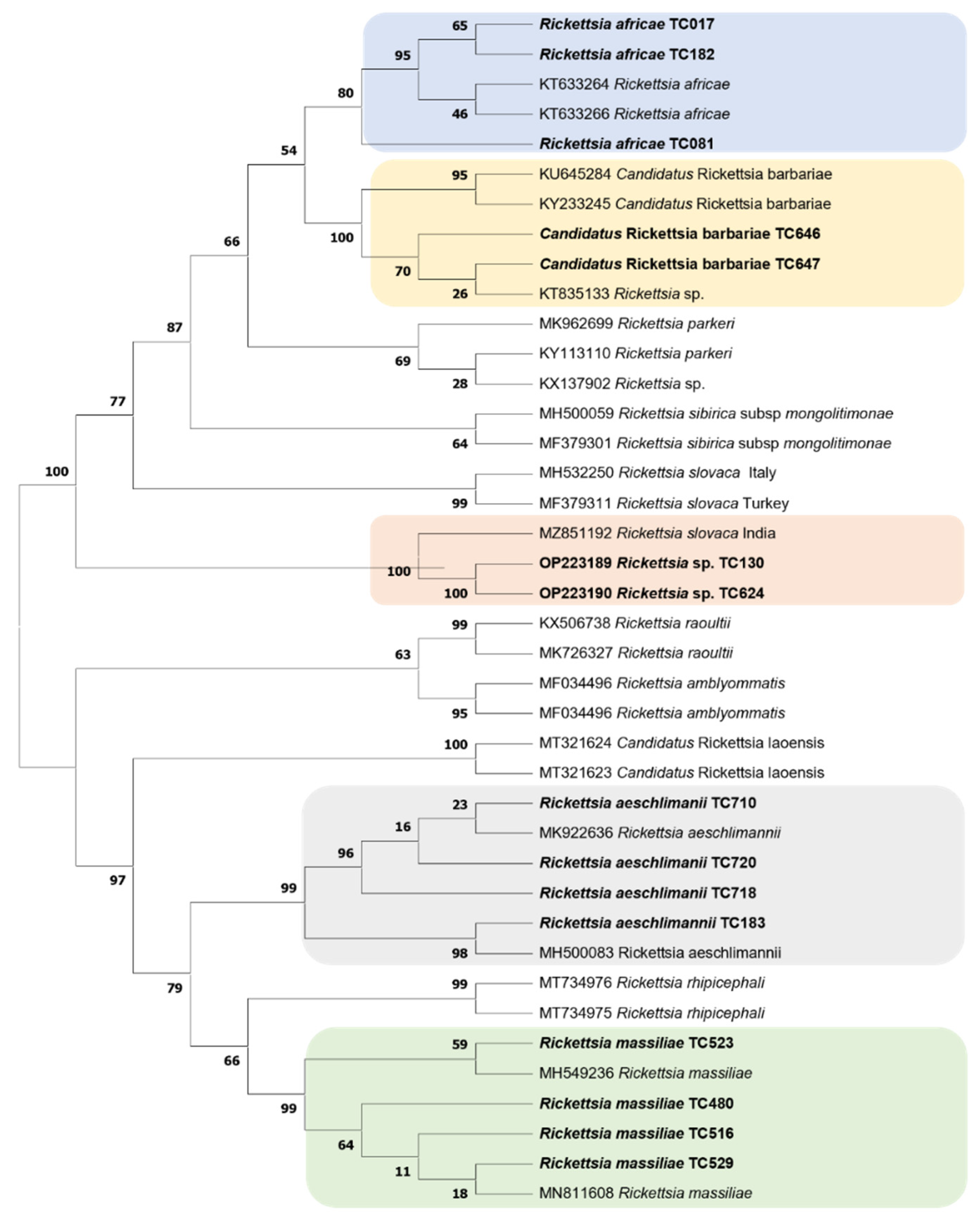
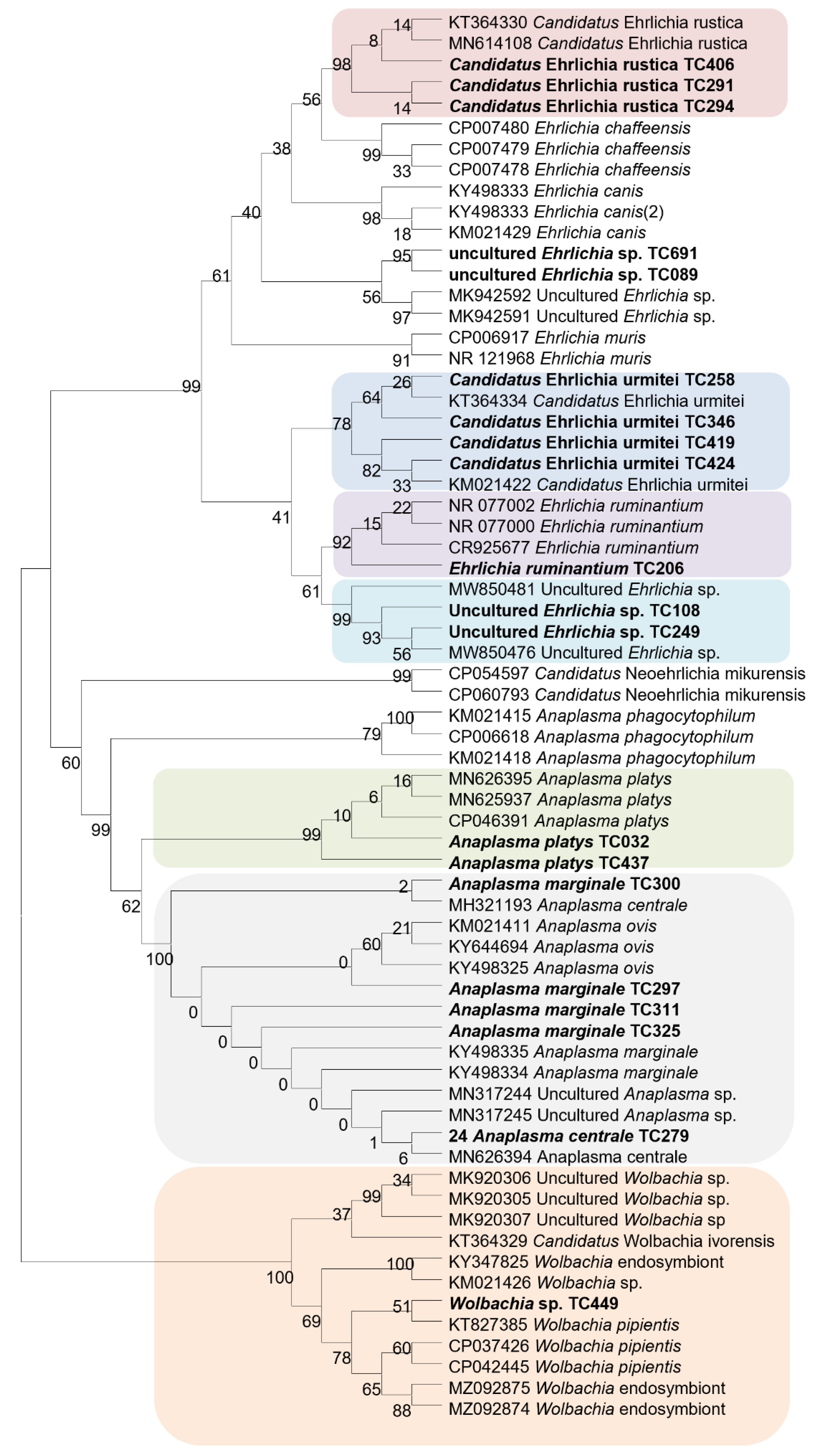
2.4. Standard PCR, Sequencing, and Phylogenetic Analysis

Standard PCR was performed using a thermal cycler (Applied Biosystems, Paris, France) on the samples that were positive in qPCR, and then sequencing to identify the microorganism species. Samples that were negative for R. africae but positive for Rickettsia spp. were subjected to standard PCR to amplify a 632-bp fragment of the ompA gene [34,36]. The amplicons were sequenced, assembled, and compared to GenBank entries sequences by a BLAST search to identify the Rickettsia species. For sequences with a low percentage identity with the corresponding sequences in GenBank, PCR targeting a second gene (gltA) was used to amplify a 700 bp DNA fragment, followed by sequencing. Samples that were positive for the Anaplasmataceae family were subjected to amplifying and sequencing of 520 bp fragment of the 23S rRNA gene [32]. The samples positive for Borrelia spp. under qPCR were subjected to amplifying and sequencing of a 300 bp fragment of the flagellin (FlaB) gene [40]. Samples that were Piroplasmida-positive following qPCR were subjected to amplifying and sequencing of a 969 bp fragment of the 18S rRNA gene [29]. The obtained sequences were assembled and analysed using the CromasPro software (version 1.7.7) (Technelysium Pty. Ltd., Tewantin, Australia) and were then blasted against the reference sequences available in GenBank (http://blast.ncbi.nlm.nih.gov/ accessed on 20 July 2021).
Sequences from microorganisms were aligned using the BioEdit v 7.2.5.0 software (University of North Texas, Denton, TX, USA). The aligned sequences were imported into TOPALi v2.5 software (Biomathematics and Statistics Scotland, Edinburgh, UK) and phylogenetic trees were constructed using TOPALi v2.5 software [41]. The maximum likelihood (ML) phylogenetic tree model proposed by default by the software was used to construct the phylogenetic tree. Node numbers are percentages of bootstrap values obtained by repeating 100 interactions of the analysis to generate a majority consensus tree (only those with values equal to greater than 80 were retained).

3. Results

3.1. Ticks

A total of 944 of the 1483 ticks collected from cattle in five sites of the Western Highlands of Cameroon were randomly selected for the analyses. The combination of the three identification methods allowed us to classify the ticks into 5 genera and 12 species. These included 299 (31.7%) Rhipicephalus microplus, 272 (29%) Rhipicephalus lunulatus, 217 (23%) Amblyomma variegatum, 48 (5%) Rhipicephalus sanguineus s.l, 43 (4.5%) Haemaphysalis leachi group specimens, 25 (2.6%) Hyalomma truncatum, 16 (1.7%) Hyalomma rufipes, 12 (1.3%) Rhipicephalus muhsamae, 6 (0.6%) Rhipicephalus annulatus, 3 (0.3%) Rhipicephalus decoloratus, and 3 (0.3%) Ixodes rasus (Table 2).

3.2. Detection of Microorganisms in Ticks

Of the 944 ticks, 399 (42.6%) were qRT-PCR positive for at least one of the microorganisms tested. Among them were 218 (54.6%) Rickettsia spp., 141 (35.3%) Anaplasmataceae, 29 (7.3%) Piroplasmida, 5 (1.2%) C. burnetii, 4 (1%) Borrelia spp., and 2 (0.5%) Bartonella spp. (Table 3).
Among the ticks infected by Rickettsia spp., R. africae was found in 172 samples (78.9%). Rickettsia africae was detected in Am. variegatum, Hy. Truncatum, and Rh. microplus. The remaining 46 tick samples, which were positive for Rickettsia spp. but negative for R. africae, were subjected to amplification of the ompA gene fragment to identify these Rickettsia species. Of these 46 positive tick samples, amplification and sequencing provided sequences for 35 (78.2%) samples. The BLAST analyses showed that 17 (48.6%) sequences were 99–100% identical to Rickettsia massiliae (MH549236, MN811608), and 14 (40%) sequences were 99–100% identical to the Rickettsia aeschlimannii (MH932060, MK922621). Similarly, two (5.7%) sequences were 99.49% identical to Candidatus Rickettsia barbariae (KU645284). In contrast, two (5.7%) sequences of Rickettsia sp. were 97.88% and 97.92% identical to Rickettsia slovaca (MZ851192) using the ompA gene. When the citrate synthase (gltA) gene was targeted for the same samples, the obtained sequences were 98.09% identical to those of Rickettsia parkeri (CP040325). The sequences of the latter species have been deposited in GenBank as Rickettsia sp. under the numbers: OP223189, OP223190, OP223191, and OP223192.
Rickettsiamassiliae was detected in Rh. lunulatus and Rh. muhsamae. Rickettsia aeschlimannii was observed in Hy. rufipes, Hy. Truncatum, and Rh. sanguineus. Candidatus Rickettsia barbariae was detected in Rh. muhsamae. Finally, Rickettsia sp. was detected in two Rh. lunulatus ticks (Table 4).
A total of 141 (35.3%) ticks tested positive by qPCR for bacteria from the Anaplasmataceae family. The amplification of the 23S rRNA gene and sequencing were successful for only 46 (32.6%) samples. The BLAST analysis showed that 19 (41.3%) of the obtained sequences were 98–100% identical to Candidatus Ehrlichia rustica (KT364330, MN614109), ten (21.7%) sequences were 99–100% identical to Candidatus Ehrlichia urmitei (GenBank KT364334). Six (13%) sequences were 99–100% identical to sequence of A. marginale (CP023731), six (13%) sequences were 99–100% identical to an uncultured Ehrlichia sp. (MW850476, MK942565), and two (4.3%) sequences were 98.72% and 98.74% identical to A. platys (MN626395). Similarly, two sequences were 100% and 99.58% identical to E. ruminantium (CR925677) and A. centrale (MH321193), respectively. One sequence of Wolbachia sp. from Ha. leachi was 99.78% identical to Wolbachia pipientis (KT827385) for the first hit and other Wolbachia from Mycopsylla fici deposited in GenBank as “endosymbiont” (KT273261). Candidatus Ehrlichia rustica was detected in Rh. lunulatus, Rh. Microplus, and Haemaphysalis sp.; Candidatus Ehrlichia urmitei were found in Rh. microplus. Uncultured Ehrlichia sp. were detected in Rh. microplus, Rh. lunulatus, Rh. Sanguinues, and Haemaphysalis sp.; Anaplasma marginale were detected in Rh. microplus and Haemaphysalis sp.; A. platys in Rh. microplus and Rh. sanguineus (Table 4).
Coxiella burnetii was detected in five (0.5%) of the samples with IS1111 and IS30A genes. The tick species Hy. truncatum, Hy. Rufipes, and R. lunulatus carried C. burnetii DNA.
DNA of Borrelia spp. and Bartonella spp. were found by qPCR for four and two ticks, respectively. For Borrelia spp., we succeeded in amplifying the flaB gene sequence only in one of four ticks. A BLAST search showed that sequence was 100% identical with Borrelia theileri (MK984606). This Borrelia specie was detected in a Ha. leachi specimen. However, all the standard PCR to identify the Bartonella species failed.
DNA from the Piroplamida was detected in 7.3% (29/399) of positive ticks (17 Rh. microplus; 7 Rh. lunulatus; 3 Am. variegatum; 1 Hy. rufipes and 1 Ha. leachi.) by qPCR. Unfortunately, we could not amplify these positive samples using standard PCR.
Finally, 29 co-infections (7.3%, 29/399) were detected. Most co-infections (72.4%, 21/29) involved the presence of Rickettsia spp. in Am. variegatum ticks. Eleven co-infections (38%, 11/29) were observed with R. africae, of which nine of the eleven were R. africae with Anaplasma spp. and two of the eleven were R. africae and Piroplasmida. Other co-infections were Rickettsia sp. plus Anaplasma spp. (17.2%, 5/29) in Rh. lunulatus, Rh. microplus and Rh. sanguineus. Rickettsia africae plus C. burnetii (17.2%, 5/29) in Hy. truncatum and Hy. rufipes. Anaplasma spp. plus Piroplasmida (17.2%, 5/29) in Rh. microplus, Rh. Lunulatus, and Hy. truncatum.
No bacterial DNA was identified in I. rasus, Rh. Annulatus, and Rh. decoloratus ticks.
Two phylogenetic trees of Rickettsiae and Anaplasmataceae were constructed from the partial sequences of the ompA gene and the 23S rRNA gene sequences of our amplicons, respectively. These phylogenetic trees showed that sequences of the microorganisms detected in this study are close to their homologues available is GenBank, except for two sequences of Rickettsia sp. (Figure 2 and Figure 3).

4. Discussion

Several species of microorganisms were detected in ticks from Cameroon. DNA of Rickettsia spp. was found in 23% of the tested ticks, 78.8% of which was found in Am. variegatum and 92.2% was R. africae. Rickettsia africae is the aetiological agent of African tick-bite fever in humans [42,43]. The results show that the R. africae infection rate is high among Am. variegatum in the Western Highlands zone of Cameroon. This high prevalence of R. africae had already been identified in Cameroon [16,36]. This finding confirms those of previous studies conducted in other African countries [28,44]. Rickettsia africae was identified in 7 (6%) of 118 patients with acute fever of unknown aetiology in clinics along the coastal region of Cameroon [16]. Amblyomma variegatum is a vector of R. africae in sub-Saharan Africa. These ticks are not only vectors but also reservoirs of rickettsiae in sub-Saharan Africa with transstadial and transovarial transmission of R. africae infection in Am. variegatum ticks [42,45]. Rickettsia africae was also detected in 0.7% of Rh. microplus and 4.7% of Hy. truncatum in this study. These proportions are very low, compared to those obtained in Am. variegatum. However, the detection of a microorganism in an arthropod does not mean that this arthropod can act as a vector of the microorganism. This bacterium may have infected Rh. microplus and Hy. truncatum during co-feeding. Indeed, co-feeding transmission is the transmission, which can occur when ticks (infected and uninfected) feed in close spatial and temporal proximity on the same host. During this form of transmission, the host acts as a belt, bringing together infected and uninfected ticks to facilitate pathogen exchange [46]. Furthermore, it has been shown that in co-feeding transmission (or non-systemic transmission), vector-to-vector transmission on the vertebrate host is essentially immediate [47]. This finding was shown in a previous study in Côte d’Ivoire [44]. In this study, other rickettsial DNA was also found in Hyalomma and Rhipicephalus tick species. Of the remaining 46 tick samples positives for Rickettsia spp., amplification and sequencing yielded sequences for 35 (78.2%) samples. The 11 positive Rickettsia qPCR samples for which amplification with the ompA gene was not possible may be due to a higher sensitivity of qPCR, compared to standard PCR [48]. This difference in sensitivity often results in qPCR positive samples with a high Ct (low bacterial load) not being amplified.
Rickettsia aeschlimannii was also identified in 75% of Hy. rufipes and 4% of Hy. truncatum. This microorganism is a recognised human pathogen, causing spotted fever and has been detected in many countries in sub-Saharan Africa, including Cameroon [28,36,42,44]. Rickettsia aeschlimannii was also found in 2.2% of Rh. sanguineus sl. This tick species is, however, not known to be a competent vector of R. aeschlimannii, although it is a rickettsia that has been frequently associated with Hyalomma spp. Co-feeding could, therefore, be the cause of infection of this tick species, as mentioned above.
In Rh. lunulatus (5.6%) and Rh. muhsamae (10%) samples, rickettsial sequences with homology to R. massiliae were identified. Rickettsia massiliae is a pathogenic rickettsia that is associated with Rhipicephalus spp. ticks. It has been described as a human pathogen in Europe and South America, but there has never been reports of human infections in Africa [42]. This is the second finding of R. massiliae in Cameroon, following a previous study conducted by Vanegas and collaborators in 2018 [36], and is the first in the Western Highlands area of the country. It was previously reported in Rh. lunulatus from Cameroon, in Rh. senegalensis (33%) from Côte d’Ivoire and in Rh. guilhoni (22%) from Senegal [36,37,44].
In this study, Candidatus Rickettsia barbariae, a SFG rickettsiae of unknown pathogenicity, was detected in 20% (2/10) of Rh. muhsamae. This Rickettsia has previously been reported in Sardinia, Italy, where it was detected in a Rh. turanicus ticks and named Candidatus Rickettsia barbariae [49]. Several other studies have reported the presence of Candidatus Rickettsia barbariae in ticks from livestock in some African countries, including Cameroon [36,50,51].
Two sequences of Rickettsia sp. from Rh. lunulatus were close to the corresponding sequence of R. slovaca (97.92 and 97.88%) using the ompA gene, and close to the corresponding sequences of R. parkeri at 98.09% similarity with citrate synthase (gltA) gene. Furthermore, these two species are not known in sub-Saharan Africa [42]. However, in order to be classified as known species, an isolate should exhibit more than one of the following degrees of nucleotide similarity with the most homologous validated species: 99.9% for the gltA gene and, when amplifiable, 98.8% for the ompA gene [52]. In this case, it might be a new species of Rickettsia and more work would be needed to characterise it properly.
Several bacteria from the Anaplasmataceae family were detected in our study. Some bacteria from this family are known to be pathogens of human and veterinary importance [53].
We found a Wolbachia sp., which was 99.78% identical to Wolbachia pipientis (KT827385) and Wolbachia endosymbiont at the same percentage identity (KT273261) deposited by Fromont and collaborators in 2015 (unpublished data). Wolbachia spp. are known to be obligate intracellular endosymbionts of arthropods [54]. Some studies have shown that environmental factors can influence the presence of Wolbachia in arthropods, such as mosquitoes [55]. Furthermore, Wolbachia may impact the reproductive biology of their hosts, through a wide range of interactions [56]. The presence of Wolbachia sp. has already been reported in several mosquito species in Cameroon [57,58]. This study reports, for the first time in the country, the presence of Wolbachia sp. in ticks. The presence of Wolbachia spp. in one Ix. ricinus has previously been reported in Algeria [59]. However, the mechanism of the transmission of Wolbachia in ticks and their consequences on tick biology remains elusive [60,61].
We also identified the bacteria named Candidatus Ehrlichia urmitei and Candidatus Ehrlichia rustica. The DNA of these two bacterial species were found mainly in Rh. microplus and Rh. lunulatus ticks in this study. These potential new species have previously been found in Am. variegatum and Rh. microplus collected from cattle in Côte d’Ivoire [44] and in Rh. microplus in Mali [28]. Candidatus Ehrlichia urmitei has been also identified in Corsica R. bursa ticks [62]. These bacteria have not been characterised to date. Further studies are needed to identify the pathogenicity of these bacteria.
Anaplasma marginale and A. centrale were detected in Rh. microplus. These two are obligate intracellular bacteria responsible for bovine anaplasmosis (gallsickness) worldwide, transmitted by tick species, mainly belonging to the Rhipicephalus genus. Anaplasma marginale would also be transmitted by Hy. Rufipes [10]. The presence of the bacteria A. marginale in Rh. Microplus is obvious, as this tick species is its principal vector. Rhipicephalus microplus is also known to be a competent vector of various tick-borne livestock pathogens, as previously described [20]. It has been shown that the introduction of this invasive tick is most often accompanied by the greatest economic losses in cattle breeding [63]. Several other studies have been conducted in Cameroon showing the presence of these Anaplasma spp. Both in ticks and their hosts [15,64,65]. The presence of A. marginale has previously been reported in cattle from south-western Ethiopia [66], in Rhipicephalus spp. In western Kenya [67], and in Malian Rh. Microplus ticks [28].
Anaplasma platys was also detected in one Rh. Microplus and one Rh. Sanguineus. This is a canine anaplasmosis agent, which exclusively infects platelets and causes cyclic thrombocytopenia in dogs. The main known vector is Rh. sanguineus s.l. Anaplasma platys has also been identified in other mammals, including cattle and humans, and ticks worldwide [68]. This Anaplasma has already been detected from blood samples from cattle in Cameroon [17], but our study marks the first time it has been discovered in Rh. sanguineus s.l. ticks from Cameroon. On the scale of Africa as a whole, A. platys is a species frequently found in the northern part of the continent. However, it had been previously detected in cattle from Nigeria [69]. The presence of A. platys in main vector in this area poses a risk to potential host dogs.
Ehrlichia ruminantium is the aetiological agent of heartwater or cowdriosis, which particularly affects domestic and wild ruminants. It is transmitted by the ticks of the genus Amblyomma, primarily Am. variegatum and Am. hebraeum [70]. The prevalence of 0.7% in main vector, Am. variegatum, was significantly lower in comparison to recently published data (8.3%) from Mali [28]. Ehrlichia ruminantium had already been reported in several studies conducted in Cameroon, both in ticks and on their hosts [16,17,71,72]. These bacteria were previously described in Am. variegatum from several countries in western Africa [28,44].
We found Borrelia theileri, a member of the tick-borne relapsing fever group [45]. This study reports, for the first time, the presence of B. theileri in ticks from Cameroon. This had already been detected in cattle blood in the country [17]. Rhipicephalus microplus, in which this bacterium was found in this study, is genetically related and overlaps in distribution with Rh. decoloratus and Rh. annulatus, the main known vectors of B. theileri [73]. The infection rate (0.3%) reported here is comparable to the study in Mali, which found 0.5% infected with B. theileri in Rh. geigyi collected from cattle. Nevertheless, the vector capacity of Rh. geigyi is unknown [74]. Other studies have revealed the presence of species of this genus in different tick species in Algeria [59], Mali [28], and Côte d’Ivoire [44]. Reported cases of tick-borne relapsing fever have proven to be responsible for economic losses in livestock [75].
For some of our tick samples, which were positive for Bartonella spp. by qPCR using the ITS2 genes and Piroplasmida using 18S Piro gene, no amplification was observed after standard PCR, despite the fact that our DNA quantification gave satisfactory results. This issue may be due to a higher sensitivity of qPCR, compared to conventional PCR [48]. It could also be due to DNA degradation or to PCR inhibition by cattle blood. However, several other microorganisms in the Piroplasmida order have already been found in cattle blood samples from Cameroon [17,64,76].
Coxiella burnetii, a strict intracellular Gram-negative bacterium, is responsible for Q fever affecting humans and a variety of animals [77]. Although Q fever is far more frequently airborne, at least seven hard and soft tick species, including Hyalomma spp., have formally been shown to be competent vectors of C. burnetii [78]. Our study is the first to detect C. burnetii in tick samples (0.5%) in Cameroon. The prevalence rate is comparable to the 0.6% rate reported in ticks from Côte d’Ivoire [44]. In addition, a study conducted in Mali and Nigeria reported a high prevalence of 37.6% and 14% in ticks, respectively [28,79]. Coxiella burnetii represents a real, albeit underappreciated threat to human and animal health throughout Africa [77]
Finally, it has been reported that ticks are often co-infected after taking a blood meal from a host carrying several infectious agents [80]. Several studies have reported mixed infections in feeding ticks caused mainly by Rickettsia spp. and C. burnetii [44,79]. We also reported, for the first time, mixed infections in ticks from Cameroon involving mainly R. africae and Ehrlichia spp. The co-infected rate here (3.4%) is comparable to that (1.3%) observed in Nigeria [79].

5. Conclusions

This study demonstrates that ticks in Cameroon harbour a wide variety of tick-borne pathogens of veterinary and medical importance, in particular R. africae, the agent of ATBF. Humans are thus at risk of infection with R. africae, and Africa tick-bite fever should also be considered in patients presenting with febrile illnesses. This study also reports, for the first time in ticks from Cameroon, the presence of various agents, such as Borrelia theileri. Nevertheless, further studies are needed to ascertain the origin and zoonotic potential of the strains and their significance for animals and human health. These data can contribute towards future research, for example by providing an avenue for larger studies of ticks and the pathogens they harbour in Cameroon.

Author Contributions

Conceptualisation, P.P. and T.T.; methodology, P.P., T.T. and A.Z.D.; formal analysis, Y.N.-Y. and A.Z.D.; investigation, Y.N.-Y. and M.L.-D.; resource, P.P.; data curation, P.P.; writing original draft preparation, Y.N.-Y.; critically revised the paper, all the authors; supervision, P.P. and T.T.; funding acquisition, P.P. All authors have read and agreed to the published version of the manuscript.

Funding

This study was supported by the Institut Hospitalo-Universitaire (IHU) Méditerranée Infection, the French National Research Agency under the “Investissements d’avenir” programme, reference ANR-10-IAHU-03, the Région Provence Alpes Côte d’Azur and European ERDF PRIMI funding. Y.N.-Y. received a Ph.D. scholarship grant from IHU Méditerranée Infection. The funders had no role in the study design, data collection, and analysis; decision to publish; or preparation of the manuscript.

Institutional Review Board Statement

Permission to undertake the study was obtained from the Western Regional Delegation of the Ministry of Livestock, Animal Husbandry and Fisheries in Cameroon, reference number 21/20/L/RO/DREPIA-O/SRAG.

Informed Consent Statement

The farmers provided informed consent for the use of their cattle and information for this study. For informed consent it was given verbally by the livestock owners.The collection of ticks from cattle did not involve national parks or other protected areas or endangered or protected species.

Data Availability Statement

Not applicable.

Acknowledgments

The authors would like to thank all shepherds and horse owners for giving us access to their ranches and helping us to contain the animals in order to collect the ticks on the field.

Conflicts of Interest

The authors declare no conflict of interest.

References

  1. Dantas-Torres, F.; Chomel, B.B.; Otranto, D. Ticks and Tick-Borne Diseases: A One Health Perspective. Trends Parasitol. 2012, 28, 437–446. [Google Scholar] [CrossRef]
  2. Parola, P.; Raoult, D. Ticks and Tickborne Bacterial Diseases in Humans: An Emerging Infectious Threat. Clin. Infect. Dis. 2001, 32, 897–928. [Google Scholar] [CrossRef]
  3. Jongejan, F.; Uilenberg, G. The Global Importance of Ticks. Parasitology 2004, 129, S3–S14. [Google Scholar] [CrossRef]
  4. Kopp, N.; Diaz, D.; Amacker, M.; Odongo, D.O.; Beier, K.; Nitsch, C.; Bishop, R.P.; Daubenberger, C.A. Identification of a Synthetic Peptide Inducing Cross-Reactive Antibodies Binding to Rhipicephalus (Boophilus) decoloratus, Rhipicephalus (Boophilus) microplus, Hyalomma anatolicum anatolicum and Rhipicephalus appendiculatus BM86 Homologues. Vaccine 2009, 28, 261–269. [Google Scholar] [CrossRef]
  5. de Castro, J.J. Long-Term Studies on the Economic Impact of Ticks on Sanga Cattle in Zambia. Exp. Appl. Acarol. 1997, 21, 3–19. [Google Scholar] [CrossRef]
  6. INS, Elevage et Peche au Cameroun. Annuaire Statistique du Cameroun. 2017. Available online: https://books.google.fr/books?id=NLu9FwEXG90C (accessed on 24 April 2021).
  7. Raboloko, O.O.; Ramabu, S.S.; Guerrini, L.; Jori, F. Seroprevalence of Selected Tick Borne Pathogens and Diversity and Abundance of Ixodid Ticks (Acari: Ixodidae) at the Wildlife-Livestock Interface in Northern Botswana. Front. Vet. Sci. 2020, 7, 187. [Google Scholar] [CrossRef] [PubMed]
  8. de la Fuente, J.; Antunes, S.; Bonnet, S.; Cabezas-Cruz, A.; Domingos, A.G.; Estrada-Peña, A.; Johnson, N.; Kocan, K.M.; Mansfield, K.L.; Nijhof, A.M.; et al. Tick-Pathogen Interactions and Vector Competence: Identification of Molecular Drivers for Tick-Borne Diseases. Front. Cell. Infect. Microbiol. 2017, 7, 114. [Google Scholar] [CrossRef]
  9. Okello-Onen, J.; Hassan, S.M.; Essuman, S. Taxonomy of African Ticks: An Identification Manual; International Centre of Insect Physiology and Ecology (ICIPE): Nairobi, Kenya, 1999. [Google Scholar]
  10. Walker, A.R.; Bouattour, A.; Camicas, J.-L.; Estrada-Peña, A.; Horak, I.G.; Latif, A.A.; Pegram, R.G.; Preston, P.M. Ticks in Domestic Animals in Africa: A Guide to Identification of Species; Springer: New York, NY, USA, 2003. [Google Scholar]
  11. Laamari, A.; Kharrim, K.E.; Mrifag, R.; Boukbal, M.; Belghyti, D. Dynamique des populations de tiques parasites des bovins de la région du Gharb au Maroc. Rev. D’élevage Médecine Vét. Pays Trop. 2012, 65, 57–62. [Google Scholar] [CrossRef]
  12. Parola, P.; Inokuma, H.; Camicas, J.-L.; Brouqui, P.; Raoult, D. Detection and Identification of Spotted Fever Group Rickettsiae and Ehrlichiae in African Ticks. Emerg. Infect. Dis. 2001, 7, 1014–1017. [Google Scholar] [CrossRef]
  13. Estrada-Pena, A. Tick-Borne Pathogens, Transmission Rates and Climate Change. Front. Biosci. Landmark Ed. 2009, 14, 2674–2687. [Google Scholar] [CrossRef] [Green Version]
  14. Gondard, M.; Cabezas-Cruz, A.; Charles, R.A.; Vayssier-Taussat, M.; Albina, E.; Moutailler, S. Ticks and Tick-Borne Pathogens of the Caribbean: Current Understanding and Future Directions for More Comprehensive Surveillance. Front. Cell. Infect. Microbiol. 2017, 7, 490. [Google Scholar] [CrossRef]
  15. Ndi, C.; Bayemi, P.H.; Ekue, F.N.; Tarounga, B. Preliminary Observations on Ticks and Tick-Borne Diseases in the North West Province of Cameroon. I. Babesiosis and Anaplasmosis. Rev. Elev. Med. Vet. Pays Trop. 1991, 44, 263–265. [Google Scholar] [CrossRef]
  16. Ndip, L.M.; Titanji, V.P.K.; Ndip, R.N.; Mcbride, J.W.; Bouyer, D.H.; Walker, D.H.; Fokam, E.B. Detection of Rickettsia africae in patients and ticks along the coastal region of Cameroon. Am. J. Trop. Med. Hyg. 2004, 71, 363–366. [Google Scholar] [CrossRef]
  17. Abanda, B.; Paguem, A.; Abdoulmoumini, M.; Kingsley, M.T.; Renz, A.; Eisenbarth, A. Molecular Identification and Prevalence of Tick-Borne Pathogens in Zebu and Taurine Cattle in North Cameroon. Parasit. Vectors 2019, 12, 448. [Google Scholar] [CrossRef]
  18. Lontsi-Demano, M.; Laroche, M.; Ngnindji, Y.C.; Djikolmbairangar, J.-E.; Mamoudou, A.; Tchuinkam, T. Breeder’s Knowledge on Ticks and Tick-Borne Diseases and Management Strategies in Menoua Division (Western Region of Cameroon). Int. J. Vet. Sci. Anim. Husb. 2021, 6, 12–21. [Google Scholar] [CrossRef]
  19. Achukwi, M.D.; Tanya, V.N.; Messiné, O.; Njongmeta, L.M. Etude comparative de l’infestation des bovins Namchi (Bos taurus) et Goudali de Ngaoundéré (Bos indicus) par la tique adulte Amblyomma variegatum. Rev. élev Med. Vet. Pays Trop. 2001, 54, 37. [Google Scholar] [CrossRef]
  20. Silatsa, B.A.; Kuiate, J.-R.; Njiokou, F.; Simo, G.; Feussom, J.-M.K.; Tunrayo, A.; Amzati, G.S.; Bett, B.; Bishop, R.; Githaka, N.; et al. A Countrywide Molecular Survey Leads to a Seminal Identification of the Invasive Cattle Tick Rhipicephalus (Boophilus) microplus in Cameroon, a Decade after It Was Reported in Cote d’Ivoire. Ticks Tick-Borne Dis. 2019, 10, 585–593. [Google Scholar] [CrossRef]
  21. Lontsi-Demano, M.; Ngnindji-Youdje, Y.; Laroche, M.; Bamou, R.; Talom, A.D.; Abah, S.; Fopa, F.; Mamoudou, A.; Tchuinkam, T. Cattle Trading Favors the Introduction and Establishment of the Invasive Tick Rhipicephalus (Boophilus) microplus in Menoua Division, West Region of Cameroon. J. Entomol. Zool. Stud. 2020, 8, 207–214. [Google Scholar]
  22. Guerrero, F.D.; Nene, V.M.; George, J.E.; Barker, S.C.; Willadsen, P. Sequencing a New Target Genome: The Boophilus microplus (Acari: Ixodidae) Genome Project. J. Med. Entomol. 2006, 43, 9–16. [Google Scholar] [CrossRef]
  23. Madder, M.; Adehan, S.; De Deken, R.; Adehan, R.; Lokossou, R. New Foci of Rhipicephalus microplus in West Africa. Exp. Appl. Acarol. 2012, 56, 385–390. [Google Scholar] [CrossRef] [PubMed]
  24. Adakal, H.; Biguezoton, A.; Zoungrana, S.; Courtin, F.; De Clercq, E.M.; Madder, M. Alarming Spread of the Asian Cattle Tick Rhipicephalus microplus in West Africa-Another Three Countries Are Affected: Burkina Faso, Mali and Togo. Exp. Appl. Acarol. 2013, 61, 383–386. [Google Scholar] [CrossRef]
  25. Ribeiro, H.S.; Pereira, D.F.S.; Melo-Junior, O.; Mariano, R.M.d.S.; Leite, J.C.; da Silva, A.V.; de Oliveira, D.S.; Gonçalves, A.A.M.; Lair, D.F.; Soares, I.d.S.; et al. Vaccine Approaches Applied to Controlling Dog Ticks. Ticks Tick-Borne Dis. 2021, 12, 101631. [Google Scholar] [CrossRef]
  26. Motta, P.; Porphyre, T.; Handel, I.; Hamman, S.M.; Ngu Ngwa, V.; Tanya, V.; Morgan, K.; Christley, R.; Bronsvoort, B.M. deC. Implications of the Cattle Trade Network in Cameroon for Regional Disease Prevention and Control. Sci. Rep. 2017, 7, 43932. [Google Scholar] [CrossRef]
  27. Estrada-Peña, A.; Bouattour, A.; Camicas, J.; Walker, A. Tiques d’importance Médicale et Vétérinaire: Le Bassin Méditerranéen. ICTTD CD-ROM Mediterr. Ticks 2004, 12, 3–12. [Google Scholar]
  28. Diarra, A.Z.; Almeras, L.; Laroche, M.; Berenger, J.-M.; Koné, A.K.; Bocoum, Z.; Dabo, A.; Doumbo, O.; Raoult, D.; Parola, P. Molecular and MALDI-TOF Identification of Ticks and Tick-Associated Bacteria in Mali. PLoS Negl. Trop. Dis. 2017, 11, e0005762. [Google Scholar] [CrossRef]
  29. Dahmana, H.; Amanzougaghene, N.; Davoust, B.; Normand, T.; Carette, O.; Demoncheaux, J.-P.; Mulot, B.; Fabrizy, B.; Scandola, P.; Chik, M.; et al. Great Diversity of Piroplasmida in Equidae in Africa and Europe, Including Potential New Species. Vet. Parasitol. Reg. Stud. Rep. 2019, 18, 100332. [Google Scholar] [CrossRef]
  30. Kernif, T. Rickettsia africae in Hyalomma dromedarii Ticks from Sub-Saharan Algeria. Ticks Tick-Borne Dis. 2012, 3, 377–379. [Google Scholar] [CrossRef]
  31. Mediannikov, O.; Fenollar, F.; Socolovschi, C.; Diatta, G.; Bassene, H.; Molez, J.-F.; Sokhna, C.; Trape, J.-F.; Raoult, D. Coxiella burnetii in Humans and Ticks in Rural Senegal. PLoS Negl. Trop. Dis. 2010, 4, e654. [Google Scholar] [CrossRef]
  32. Dahmani, M.; Davoust, B.; Sambou, M.; Bassene, H.; Scandola, P.; Ameur, T.; Raoult, D.; Fenollar, F.; Mediannikov, O. Molecular Investigation and Phylogeny of Species of the Anaplasmataceae Infecting Animals and Ticks in Senegal. Parasit. Vectors 2019, 12, 495. [Google Scholar] [CrossRef]
  33. Djiba, M.L.; Mediannikov, O.; Mbengue, M.; Thiongane, Y.; Molez, J.-F.; Seck, M.T.; Fenollar, F.; Raoult, D.; Ndiaye, M. Survey of Anaplasmataceae Bacteria in Sheep from Senegal. Trop. Anim. Health Prod. 2013, 45, 1557–1561. [Google Scholar] [CrossRef]
  34. Mura, A.; Socolovschi, C.; Ginesta, J.; Lafrance, B.; Magnan, S.; Rolain, J.-M.; Davoust, B.; Raoult, D.; Parola, P. Molecular Detection of Spotted Fever Group Rickettsiae in Ticks from Ethiopia and Chad. Trans. R. Soc. Trop. Med. Hyg. 2008, 102, 945–949. [Google Scholar] [CrossRef]
  35. Rolain, J.-M.; Stuhl, L.; Maurin, M.; Raoult, D. Evaluation of Antibiotic Susceptibilities of Three Rickettsial Species Including Rickettsia felis by a Quantitative PCR DNA Assay. Antimicrob. Agents Chemother. 2002, 46, 2747–2751. [Google Scholar] [CrossRef] [Green Version]
  36. Vanegas, A.; Keller, C.; Krüger, A.; Manchang, T.K.; Hagen, R.M.; Frickmann, H.; Veit, A.; Achukwi, M.D.; Krücken, J.; Poppert, S. Molecular Detection of Spotted Fever Group Rickettsiae in Ticks from Cameroon. Ticks Tick-Borne Dis. 2018, 9, 1049–1056. [Google Scholar] [CrossRef]
  37. Mediannikov, O.; Diatta, G.; Fenollar, F.; Sokhna, C.; Trape, J.-F.; Raoult, D. Tick-Borne Rickettsioses, Neglected Emerging Diseases in Rural Senegal. PLoS Negl. Trop. Dis. 2010, 4, e821. [Google Scholar] [CrossRef]
  38. Rolain, J.-M.; Franc, M.; Davoust, B.; Raoult, D. Molecular Detection of Bartonella quintana, B. koehlerae, B. henselae, B. clarridgeiae, Rickettsia felis, and Wolbachia pipientis in Cat Fleas, France. Emerg. Infect. Dis. 2003, 9, 339–342. [Google Scholar] [CrossRef]
  39. Rolain, J.-M.; Raoult, D. Molecular Detection of Coxiella burnetii in Blood and Sera during Q Fever. QJM Int. J. Med. 2005, 98, 615–620. [Google Scholar] [CrossRef]
  40. Aubry, C.; Socolovschi, C.; Raoult, D.; Parola, P. Bacterial Agents in 248 Ticks Removed from People from 2002 to 2013. Ticks Tick-Borne Dis. 2016, 7, 475–481. [Google Scholar] [CrossRef]
  41. Ngoy, S.; Diarra, A.Z.; Laudisoit, A.; Gembu, G.-C.; Verheyen, E.; Mubenga, O.; Mbalitini, S.G.; Baelo, P.; Laroche, M.; Parola, P. Using MALDI-TOF Mass Spectrometry to Identify Ticks Collected on Domestic and Wild Animals from the Democratic Republic of the Congo. Exp. Appl. Acarol. 2021, 84, 637–657. [Google Scholar] [CrossRef]
  42. Parola, P.; Paddock, C.D.; Socolovschi, C.; Labruna, M.B.; Mediannikov, O.; Kernif, T.; Abdad, M.Y.; Stenos, J.; Bitam, I.; Fournier, P.-E.; et al. Update on Tick-Borne Rickettsioses around the World: A Geographic Approach. Clin. Microbiol. Rev. 2013, 26, 657–702. [Google Scholar] [CrossRef]
  43. Raoult, D.; Fournier, P.E.; Fenollar, F.; Jensenius, M.; Prioe, T.; de Pina, J.J.; Caruso, G.; Jones, N.; Laferl, H.; Rosenblatt, J.E.; et al. Rickettsia africae, a Tick-Borne Pathogen in Travelers to Sub-Saharan Africa. N. Engl. J. Med. 2001, 344, 1504–1510. [Google Scholar] [CrossRef]
  44. Ehounoud, C.B.; Yao, K.P.; Dahmani, M.; Achi, Y.L.; Amanzougaghene, N.; N’Douba, A.K.; N’Guessan, J.D.; Raoult, D.; Fenollar, F.; Mediannikov, O. Multiple Pathogens Including Potential New Species in Tick Vectors in Cote d’Ivoire. PLoS Negl. Trop. Dis. 2016, 10, e0004367. [Google Scholar] [CrossRef] [PubMed]
  45. Socolovschi, C.; Mediannikov, O.; Raoult, D.; Parola, P. Update on Tick-Borne Bacterial Diseases in Europe. Parasite Paris Fr. 2009, 16, 259–273. [Google Scholar] [CrossRef] [PubMed] [Green Version]
  46. Randolph, S.E. Transmission of Tick-Borne Pathogens between Co-Feeding Ticks: Milan Labuda’s Enduring Paradigm. Ticks Tick-Borne Dis. 2011, 2, 179–182. [Google Scholar] [CrossRef] [PubMed]
  47. Belli, A.; Sarr, A.; Rais, O.; Rego, R.O.M.; Voordouw, M.J. Ticks Infected via Co-Feeding Transmission Can Transmit Lyme Borreliosis to Vertebrate Hosts. Sci. Rep. 2017, 7, 5006. [Google Scholar] [CrossRef]
  48. Kidd, L.; Maggi, R.; Diniz, P.P.V.P.; Hegarty, B.; Tucker, M.; Breitschwerdt, E. Evaluation of Conventional and Real-Time PCR Assays for Detection and Differentiation of Spotted Fever Group Rickettsia in Dog Blood. Vet. Microbiol. 2008, 129, 294–303. [Google Scholar] [CrossRef]
  49. Mura, A.; Masala, G.; Tola, S.; Satta, G.; Fois, F.; Piras, P.; Rolain, J.-M.; Raoult, D.; Parola, P. First Direct Detection of Rickettsial Pathogens and a New Rickettsia, “Candidatus Rickettsia Barbariae”, in Ticks from Sardinia, Italy. Clin. Microbiol. Infect. Off. Publ. Eur. Soc. Clin. Microbiol. Infect. Dis. 2008, 14, 1028–1033. [Google Scholar] [CrossRef]
  50. Sadeddine, R.; Diarra, A.Z.; Laroche, M.; Mediannikov, O.; Righi, S.; Benakhla, A.; Dahmana, H.; Raoult, D.; Parola, P. Molecular Identification of Protozoal and Bacterial Organisms in Domestic Animals and Their Infesting Ticks from North-Eastern Algeria. Ticks Tick-Borne Dis. 2020, 11, 101330. [Google Scholar] [CrossRef] [PubMed]
  51. Abdelkadir, K.; Palomar, A.M.; Portillo, A.; Oteo, J.A.; Ait-Oudhia, K.; Khelef, D. Presence of Rickettsia aeschlimannii, “Candidatus Rickettsia barbariae” and Coxiella burnetii in Ticks from Livestock in Northwestern Algeria. Ticks Tick-Borne Dis. 2019, 10, 924–928. [Google Scholar] [CrossRef]
  52. Fournier, P.-E.; Dumler, J.S.; Greub, G.; Zhang, J.; Wu, Y.; Raoult, D. Gene Sequence-Based Criteria for Identification of New Rickettsia Isolates and Description of Rickettsia heilongjiangensis sp. nov. J. Clin. Microbiol. 2003, 41, 5456–5465. [Google Scholar] [CrossRef]
  53. Inokuma, H. Vectors and Reservoir Hosts of Anaplasmataceae. In Rickettsial Diseases; CRC Press: Boca Raton, FL, USA, 2007; ISBN 978-0-429-13200-1. [Google Scholar]
  54. Werren, J.H. Biology of Wolbachia. Annu. Rev. Entomol. 1997, 42, 587–609. [Google Scholar] [CrossRef]
  55. Inácio da Silva, L.M.; Dezordi, F.Z.; Paiva, M.H.S.; Wallau, G.L. Systematic Review of Wolbachia Symbiont Detection in Mosquitoes: An Entangled Topic about Methodological Power and True Symbiosis. Pathogens 2021, 10, 39. [Google Scholar] [CrossRef]
  56. Landmann, F. The Wolbachia Endosymbionts. Microbiol. Spectr. 2019, 7, BAI-0018-2019. [Google Scholar] [CrossRef]
  57. Bamou, R.; Diarra, A.Z.; Mayi, M.P.A.; Djiappi-Tchamen, B.; Antonio-Nkondjio, C.; Parola, P. Wolbachia Detection in Field-Collected Mosquitoes from Cameroon. Insects 2021, 12, 1133. [Google Scholar] [CrossRef]
  58. Walker, T.; Quek, S.; Jeffries, C.L.; Bandibabone, J.; Dhokiya, V.; Bamou, R.; Kristan, M.; Messenger, L.A.; Gidley, A.; Hornett, E.A.; et al. Stable High-Density and Maternally Inherited Wolbachia Infections in Anopheles moucheti and Anopheles demeilloni Mosquitoes. Curr. Biol. 2021, 31, 2310–2320.e5. [Google Scholar] [CrossRef]
  59. Boucheikhchoukh, M.; Laroche, M.; Aouadi, A.; Dib, L.; Benakhla, A.; Raoult, D.; Parola, P. MALDI-TOF MS Identification of Ticks of Domestic and Wild Animals in Algeria and Molecular Detection of Associated Microorganisms. Comp. Immunol. Microbiol. Infect. Dis. 2018, 57, 39–49. [Google Scholar] [CrossRef]
  60. Chao, L.-L.; Castillo, C.T.; Shih, C.-M. Molecular Detection and Genetic Identification of Wolbachia Endosymbiont in Rhipicephalus sanguineus (Acari: Ixodidae) Ticks of Taiwan. Exp. Appl. Acarol. 2021, 83, 115–130. [Google Scholar] [CrossRef]
  61. Plantard, O.; Bouju-Albert, A.; Malard, M.-A.; Hermouet, A.; Capron, G.; Verheyden, H. Detection of Wolbachia in the Tick Ixodes ricinus Is Due to the Presence of the Hymenoptera Endoparasitoid Ixodiphagus hookeri. PLoS ONE 2012, 7, e30692. [Google Scholar] [CrossRef]
  62. Cicculli, V.; DeCarreaux, D.; Ayhan, N.; Casabianca, F.; de Lamballerie, X.; Charrel, R.; Falchi, A. Molecular Screening of Anaplasmataceae in Ticks Collected from Cattle in Corsica, France. Exp. Appl. Acarol. 2020, 81, 561–574. [Google Scholar] [CrossRef]
  63. Frisch, J.E. Towards a Permanent Solution for Controlling Cattle Ticks. Int. J. Parasitol. 1999, 29, 57–71. [Google Scholar] [CrossRef]
  64. Lontsi-Demano, M.; Djikolbairangar, J.E.; Laroche, M.; Ngnindji-Youdje, Y.C.; Luogbou, N.D.D.; Abah, S.; Mamoudou, A.; Tchuinkam, T. Tick-Borne Haemoparasites of Veterinary Importance in Cattle in Menoua Division, Western Highlands of Cameroon. J. Fish. Livest. Prod. 2021, 9, 10. [Google Scholar]
  65. Abanda, B.; Paguem, A.; Achukwi, M.D.; Renz, A.; Eisenbarth, A. Development of a Low-Density DNA Microarray for Detecting Tick-Borne Bacterial and Piroplasmid Pathogens in African Cattle. Trop. Med. Infect. Dis. 2019, 4, 64. [Google Scholar] [CrossRef]
  66. Hailemariam, Z.; Krücken, J.; Baumann, M.; Ahmed, J.S.; Clausen, P.-H.; Nijhof, A.M. Molecular Detection of Tick-Borne Pathogens in Cattle from Southwestern Ethiopia. PLoS ONE 2017, 12, e0188248. [Google Scholar] [CrossRef] [PubMed]
  67. Chiuya, T.; Masiga, D.K.; Falzon, L.C.; Bastos, A.D.S.; Fèvre, E.M.; Villinger, J. Tick-Borne Pathogens, Including Crimean-Congo Haemorrhagic Fever Virus, at Livestock Markets and Slaughterhouses in Western Kenya. Transbound. Emerg. Dis. 2021, 68, 2429–2445. [Google Scholar] [CrossRef] [PubMed]
  68. Maggi, R.G.; Mascarelli, P.E.; Havenga, L.N.; Naidoo, V.; Breitschwerdt, E.B. Co-Infection with Anaplasma platys, Bartonella henselae and Candidatus Mycoplasma haematoparvum in a Veterinarian. Parasit. Vectors 2013, 6, 103. [Google Scholar] [CrossRef]
  69. Elelu, N.; Ferrolho, J.; Couto, J.; Domingos, A.; Eisler, M.C. Molecular Diagnosis of the Tick-Borne Pathogen Anaplasma marginale in Cattle Blood Samples from Nigeria Using QPCR. Exp. Appl. Acarol. 2016, 70, 501–510. [Google Scholar] [CrossRef]
  70. Peter, T.F.; Burridge, M.J.; Mahan, S.M. Ehrlichia ruminantium Infection (Heartwater) in Wild Animals. Trends Parasitol. 2002, 18, 214–218. [Google Scholar] [CrossRef]
  71. Esemu, S.N.; Ndip, R.N.; Ndip, L.M. Detection of Ehrlichia ruminantium Infection in Cattle in Cameroon. BMC Res. Notes 2018, 11, 388. [Google Scholar] [CrossRef]
  72. Awa, D.N. Serological Survey of Heartwater Relative to the Distribution of the Vector Amblyomma variegatum and Other Tick Species in North Cameroon. Vet. Parasitol. 1997, 68, 165–173. [Google Scholar] [CrossRef]
  73. Estrada-Peña, A.; Bouattour, A.; Camicas, J.-L.; Guglielmone, A.; Horak, I.; Jongejan, F.; Latif, A.; Pegram, R.; Walker, A.R. The Known Distribution and Ecological Preferences of the Tick Subgenus Boophilus (Acari: Ixodidae) in Africa and Latin America. Exp. Appl. Acarol. 2006, 38, 219–235. [Google Scholar] [CrossRef]
  74. McCoy, B.N.; Maïga, O.; Schwan, T.G. Detection of Borrelia theileri in Rhipicephalus geigyi from Mali. Ticks Tick-Borne Dis. 2014, 5, 401–403. [Google Scholar] [CrossRef]
  75. Sharma, S.P.; Amanfu, W.; Losho, T.C. Bovine Borreliosis in Botswana. Onderstepoort J. Vet. Res. 2000, 67, 221–223. [Google Scholar]
  76. Silatsa, B.A.; Simo, G.; Githaka, N.; Kamga, R.; Oumarou, F.; Keambou Tiambo, C.; Machuka, E.; Domelevo, J.; Odongo, D.; Bishop, R.; et al. First Detection of Theileria parva in Cattle from Cameroon in the Absence of the Main Tick Vector Rhipicephalus appendiculatus. Transbound. Emerg. Dis. 2020, 67, 68–78. [Google Scholar] [CrossRef] [PubMed]
  77. Vanderburg, S.; Rubach, M.P.; Halliday, J.E.B.; Cleaveland, S.; Reddy, E.A.; Crump, J.A. Epidemiology of Coxiella burnetii Infection in Africa: A OneHealth Systematic Review. PLoS Negl. Trop. Dis. 2014, 8, e2787. [Google Scholar] [CrossRef]
  78. Duron, O.; Sidi-Boumedine, K.; Rousset, E.; Moutailler, S.; Jourdain, E. The Importance of Ticks in Q Fever Transmission: What Has (and Has Not) Been Demonstrated? Trends Parasitol. 2015, 31, 536–552. [Google Scholar] [CrossRef] [PubMed]
  79. Reye, A.L.; Arinola, O.G.; Hübschen, J.M.; Muller, C.P. Pathogen Prevalence in Ticks Collected from the Vegetation and Livestock in Nigeria. Appl. Environ. Microbiol. 2012, 78, 2562–2568. [Google Scholar] [CrossRef]
  80. Levin, M.L.; Fish, D. Acquisition of Coinfection and Simultaneous Transmission of Borrelia burgdorferi and Ehrlichia phagocytophila by Ixodes scapularis Ticks. Infect. Immun. 2000, 68, 2183–2186. [Google Scholar] [CrossRef] [Green Version]
Figure 1. Map of the Menoua and Noun Divisions in the Western Region of Cameroon showing tick collection sites from cattle.
Microorganisms 10 01957 g001
Figure 2. Phylogenetic tree of Rickettsia species detected in ticks from the Western Highlands of Cameroon and constructed by maximum likelihood method. Partial sequence of ompA gene from ticks collected in 2018 were aligned. Microorganisms sequenced in the current study are in bold.
Microorganisms 10 01957 g002
Figure 3. Phylogenetic tree of Anaplasmataceae detected in ticks from the Western Highlands of Cameroon Maximum likelihood method. The partial sequence of 23S rRNA gene from ticks collected in 2018 were aligned. Microorganisms sequenced in the current study are in bold.
Microorganisms 10 01957 g003
Table 1. Primers and probes used for quantitative real-time and standard PCR in this study.
MicroorganismsTargeted Sequence Primers (5′-3′) and Probes (Used for qPCR Screening or Sequencing)References
Anaplasmataceae23S (TtAna)f_TGACAGCGTACCTTTTGCAT
r_GTAACAGGTTCGGTCCTCCA
p_6FAM-GGATTAGACCCGAAACCAAG
[32,33]
23S (520-bp)f_ATAAGCTGCGGGGAATTGTC
r_TGCAAAAGGTACGCTGTCAC
Piroplasmida5.8Sf_AYYKTYAGCGRTGGATGTC
r_TCGCAGRAGTCTKCAAGTC
p_FAM-TTYGCTGCGTCCTTCATCGTTGT-MGB
[32]
18S (969-bp)f1_GCGAATGGCTCATTAIAACA
f4_CACATCTAAGGAAGGCAGCA
f3_GTAGGGTATTGGCCTACCG *
r4_AGGACTACGACGGTATCTGA *
RickettsiagltA (RKND03)f_GTGAATGAAAGATTACACTATTTAT
r_GTATCTTAGCAATCATTCTAATAGC
p_6FAM-CTATTATGCTTGCGGCTGTCGGTTC
[34,35]
ITS (Rafricae)f_TGCAACACGAAGCACAAAAC
r_CCTCTTGCGAAACTCTACTTTTGA
6FAM-CGTGTGGATTCGAGCACCGGA
[30]
OmpA (630-bp)70_ATGGCGAATATTTCTCCAAAA
701_GTTCCGTTAATGGCAGCATCT
180_GCAGCGATAATGCTGAGTA *
[12,36]
gltA (400-bp)f_ATGACCAATGAAAATAATAAT
r_CTTATACTCTCTATGTACA
BorreliaITS4f_GGCTTCGGGTCTACCACATCTA
r_CCGGGAGGGGAGTGAAATAG
p_6FAM-TGCAAAAGGCACGCCATCACC
[37]
flaB (344-bp)f_TGGTATGGGAGTTTCTGG
r_TAAGCTGACTAATACTAATTACCC
BartonellaITS2f_GATGCCGGGGAAGGTTTTC
r_GCCTGGGAGGACTTGAACCT
p_GCGCGCGCTTGATAAGCGTG
[38]
Correlia burnetiiIS30Af_CGCTGACCTACAGAAATATGTCC
r_GGGGTAAGTAAATAATACCTTCTGG
p_CATGAAGCGATTTATCAATACGTGTATG
[39]
IS1111Af_CAAGAAACGTATCGCTGTGGC
r_CACAGAGCCACCGTATGAATC
6FAM-CCGAGTTCGAAACAATGAGGGCTG
[31]
Abbreviation *, used for sequencing only.
Table 2. Tick species and numbers of tick specimens collected from cattle in the five study sites in Western Highlands of Cameroon. For each site, the number of female tick specimens is indicated in parentheses.
Tick GenusTick SpeciesNo of Ticks CollectedSexMenoua DivisionNoun Division
MaleFemaleNkong-NiDschangKouoptamoMassangamKoutaba
AmblyommaAm. variegatum35324510880 (30)87 (42)35 (6)67 (16)84 (14)
RhipicephalusRh. microplus552168384207 (153)101 (62)104 (83)88 (56)52 (30)
Rh. annulatus66006000
Rh. decoloratus33030000
Rh. lunulatus38723914870 (20)105 (50)45 (15)92 (41)75 (22)
Rh. sanguineus4840838 (8)10 (0)000
Rh. muhsamae1010073000
Rhipicephalus spp.350355 (5)26 (26)04 (4)0
HaemaphysalisHa. leachi45351035 (10)10 (0)000
HyalommaHy. rufipes1610616 (6)0000
Hy. truncatum25141125 (11)0000
IxodesIx. rasus3033 000
Total 1483770713489348184251211
Table 3. Percentage of positive tick species by quantitative real-time PCR.
Tick Species
MicroorganismTarget SequenceAm. variegatumRh. microplusRh. sanguineusHa. leachiRh. lunulatusRh. muhsamaeHy. rufipesHy. truncatum(%) Pos/Total
Rickettsia spp.gltA (RKND03)78.8% (171/217)0.6% (2/308)4.3% (2/46)-8.8% (24/272)35% (3/12)39% (16/41)23% (218/944)
R. africaepoT15-dam277.4% (168/217)0.6% (2/308)-- ---8% (2/25)18.2% (172/944)
Anaplasmatacae 23SrRNA(TtAna)7.4% (16/217)25% (77/308) 8.3% (4/48)14% (6/43)14% (38/272) ---14.9% (141/944)
Piroplasmida 5.8S/Piro 18S1.4% (3/217)5.5% (17/308)-2.3% (1/43)2.6% (7/272)-6.3% (1/16)-3% (29/944)
Bartonela spp.(Barto ITS2)/gltA-0.6% (2/308)------0.2% (2/944)
Borrelia spp.(Bor ITS4)0.5% (1/217)0.6% (2/308) -2.3% (1/43) ----0.4% (4/944)
C. burnetii(IS1111)/ITS30A----0.3% (1/272)-6.3% (1/16)12% (3/25)0.5% (5/944)
Table 4. Tick species collected in Cameroon and studied for microorganisms.
Tick Species
MicroorganismTarget SequencePer. Ident (%)Am. variegatumRh. microplusRh. sanguineusHa. leachiRh. lunulatusRh. muhsamaeHy. rufipesHy. truncatum(%) Sequences Obtained/Pos qPCR
Rickettsia aeschlimanniiompA99.49–100--50% (1/2)---75% (12/16)4% (1/25)6.4% (14/218)
Rickettsia massiliae99.83–100----59.2% (16/27)10% (1/10) -7.8% (17/218)
Candidatus Rickettsia barbariae99.49-----20% (2/10) -0.9% (2/218)
Rickettsia sp.97.88–97.92----7.4% (2/27)- -0.9% (2/218)
Anaplasma centrale23S Ana100-1.3% (1/77)- ----0.7% (1/141)
Ehrlichia ruminantium1006.2% (1/16)-- ----0.7% (1/141)
uncultured Ehrlichia sp.98.32–100-3.9% (3/77)25% (1/4)16.7% (1/6)2.6% (1/38)---4.2% (6/141)
Candidatus Ehrlichia urmitei99.16–100-13% (10/77)- ----7% (10/141)
Anaplasma marginale99.79–100-6.5% (5/77)-16.7% (1/6)- --4.3% (6/141)
Candidatus Ehrlichia rustica98.6–100-9% (7/77)-16.7% (1/6)28.9% (11/38)---13.5% (19/141)
Anaplasma platys98.72–98.74-1.3% (1/77)25% (1/4)-----1.4% (2/141)
Wolbachia pipientis99.78---16.7% (1/6)----0.7% (1/141)
Borrelia theileriflaB100 1.3% (1/77) 25% (1/4)
Publisher’s Note: MDPI stays neutral with regard to jurisdictional claims in published maps and institutional affiliations.

