Article

Gre Factors Are Required for Biofilm Formation in Salmonella enterica Serovar Typhimurium by Targeting Transcription of the csgD Gene

by
Tania Gaviria-Cantin
1,†,
Andrés Felipe Vargas
1,
Youssef El Mouali
1,‡,
Carlos Jonay Jiménez
1,
Annika Cimdins-Ahne
2,
Cristina Madrid
1,
Ute Römling
2 and
Carlos Balsalobre
1,*
1
Department of Genetics, Microbiology and Statistics, School of Biology, University of Barcelona, Av. Diagonal 643, 08028 Barcelona, Spain
2
Department of Microbiology, Tumor and Cell Biology, Karolinska Institutet, 17177 Stockholm, Sweden
*
Author to whom correspondence should be addressed.
Current address: Bacterial Cell Cycle & Development (BCCD), Biology of Microorganisms Research Unit (URBM), University of Namur, 5000 Namur, Belgium.
Current address: Department of Microbial Immune Regulation, Helmholtz Centre for Infection Research (HZI), 38124 Braunschweig, Germany.
Microorganisms 2022, 10(10), 1921; https://doi.org/10.3390/microorganisms10101921
Submission received: 29 July 2022 / Revised: 19 September 2022 / Accepted: 23 September 2022 / Published: 27 September 2022
(This article belongs to the Special Issue Feature Papers in Microbial Biofilm Formation)

Abstract

:
Rdar biofilm formation of Salmonella typhimurium and Escherichia coli is a common ancient multicellular behavior relevant in cell–cell and inter-organism interactions equally, as in interaction with biotic and abiotic surfaces. With the expression of the characteristic extracellular matrix components amyloid curli fimbriae and the exopolysaccharide cellulose, the central hub for the delicate regulation of rdar morphotype expression is the orphan transcriptional regulator CsgD. Gre factors are ubiquitously interacting with RNA polymerase to selectively overcome transcriptional pausing. In this work, we found that GreA/GreB are required for expression of the csgD operon and consequently the rdar morphotype. The ability of the Gre factors to suppress transcriptional pausing and the 147 bp 5′-UTR of csgD are required for the stimulatory effect of the Gre factors on csgD expression. These novel mechanism(s) of regulation for the csgD operon might be relevant under specific stress conditions.

1. Introduction

The ubiquitous transcription elongation factors GreA and GreB, henceforth jointly named Gre factors, small cytoplasmic proteins of <20 kDa, have been extensively studied in Escherichia coli. Several roles have been attributed to those proteins: (i) stimulating transcription initiation by likely facilitation of promoter escape, (ii) promoting release of stalling RNA polymerases during transcription elongation by inducing the intrinsic endoribonucleolytic activity of the RNA polymerases, and (iii) chaperone activity promoting refolding of denatured proteins [1,2,3]. In E. coli we have previously described that the Gre factors affect expression of biofilm promoting factors such as type 1 fimbriae and flagella [4,5]. Moreover, transcriptome studies using E. coli K-12-derivative strains allowed the conclusion that genes upregulated by GreA are mostly involved in energy metabolism and protein synthesis, whereas most of the genes downregulated by GreA belong to the cell metabolism and stress response [6]. Of note, a more recent report systematically characterized the promoter proximal pauses occurring during transcription in E. coli. It has been suggested that transcriptional pausing represents a widespread regulatory mechanism aimed at modulation of transcriptional output from specific operons in response to changing environmental conditions in a Gre factor dependent manner [3].
In Salmonella enterica serovar Typhimurium (S. Typhimurium), while explored to a lesser extent, it has been shown that the Gre factors play a major role in S. Typhimurium pathogenic lifestyle. We showed that a strain lacking the Gre factors was avirulent both in vitro and in vivo. Specifically, the expression of the SPI-1 genes, mediating host cell invasion, was importantly down regulated in the absence of the Gre factors due to a direct effect on the transcription elongation of the hilD gene, coding for the master regulator of the SPI-1 genes. A transcriptional pause in the 3′-UTR of the hilD gene was the target for the Gre factors [7]. S. Typhimurium has the ability to form biofilm on diverse biotic and abiotic surfaces, such as interacting with epithelial cells during the infection process [8] and adhering to abiotic surfaces such as plastic, glass and steel, leading to transmission and outbreaks of S. Typhimurium during food processing [9]. Most studies on S. Typhimurium biofilm have been performed upon growth on/in rich media, low osmolarity and low temperature (25–28 °C). Under these conditions, S. Typhimurium produces a rdar colony morphotype on agar plates and a pellicle air-liquid interface biofilm [10]. Two pivotal interaction-promoting factors are the main components of the extracellular matrix of described biofilms: curli, an amyloid proteinaceous surface appendage, and cellulose, an exopolysaccharide. Curli production is encoded in the divergently transcribed biogenesis operons csgBAC and csgDEFG. The biosynthesis of cellulose requires proteins encoded by the divergently transcribed operons bcsQRABZC and bcsEFG. Production of c-di-GMP by AdrA, a diguanylate cyclase encoded in the adrA gene, is necessary to eventually post-transcriptionally activate biosynthesis of cellulose. CsgD, an orphan transcriptional regulator encoded in the csgDEFG operon, regulates production of both curli and cellulose by stimulating transcription of the csgBAC and adrA genes [11,12]. CsgD expression is subject to complex regulation at the transcriptional level mediated by a 521 bp long intergenic region in combination with sequences downstream of the translational start site [13,14]. A 147 bp long 5′-UTR required for optimal expression is targeted by small regulatory RNA regulation [15,16,17,18]. The genes and basic regulation of rdar morphotype expression are conserved between S. Typhimurium and E. coli [19].
The putative widespread role of Gre factors on transcription elongation and its previously described key role in fundamental regulatory pathways for both E. coli and S. Typhimurium indicated that Gre factors might play a role in the regulation of the ability of S. Typhimurium to form biofilms. Here, the role of the Gre factors in regulating the process of biofilm formation in S. Typhimurium has been explored. We found that biofilm production was impaired in strains lacking the Gre factors. Consistently, the production of curli and cellulose was found to be strictly dependent of the Gre factors being abolished in strains lacking both factors. Expression studies showed that the Gre factors are required for the transcriptional expression of the master regulator CsgD. Gre-mediated regulation of csgD does not occur at transcription initiation but on the level of transcription elongation within the 5′-UTR of csgD. Our data add new players to the complex regulation of the biofilm formation process in S. Typhimurium and highlight the impact of 5′-UTRs of mRNAs as key regulatory hubs, not only at the posttranscriptional level but also at the level of Gre-mediated transcriptional elongation.

2. Materials and Methods

2.1. The Bacterial Strains and Culture Media

The Salmonella enterica serovar Typhimurium (S. Typhimurium) wild-type strains used in this work are UMR1, a rdar28+ nalidixic acid resistant derivative of ATCC14028 [20], and SV5015, a His+ derivate of SL1344 [21,22]. All strains and plasmids used in this work are described in Table S1 (Supplementary material). Cultures were routinely grown on either LB (10 g/L NaCl, 10 g/L tryptone, 5 g/L yeast extract and 15 g/L agar), LB0 (10 g/L tryptone, 5 g/L yeast extract and 15 g/L agar) or CFA plates (10 g/L casamino acids, 1.5 g/L yeast extract, 0.4 mM MgSO4 and 0.4 mM MnCl2). When required, ampicillin (50 μg/mL), kanamycin (50 μg/mL), chloramphenicol (25 μg/mL), spectinomycin (25 μg/mL), arabinose (0.02%) and calcofluor (Fluorescent Brightener 28, Sigma-Aldrich, Saint Louis, MO, USA) (0.2%) were added.

2.2. Genetic Manipulations

Chromosomal lacZ fusions were generated in two subsequent steps. First, homologous recombination by one-step gene replacement [23] was applied to partially delete the csgD gene from different positions (+9 and +147). Subsequently, a promoter-less lacZ gene was integrated by site-specific recombination via a suicide plasmid [24] (see File S1 in Supplementary material). Briefly, hybrid primers containing nucleotide sequences homologous to the 5′ (csgD+9 and csgD+147) and 3′-end (csgD lac Rev) and sequences flanking the antibiotic marker were used to amplify the kanamycin resistance cassette encoded by plasmid pKD4. Gene replacement and removal of the antibiotic cassette in S. Typhimurium SV5015 was performed by the standard approach [23]. Constructs were PCR-verified with primers csgD up/csgD down located in the sequences flanking the deleted open reading frame (Table S2). The suicide plasmid pKG136, bearing a FRT site upstream of a promoter less lacZ gene, was electroporated into the resulting strains transformed with plasmid pCP20. The pKG136 plasmid was integrated into the chromosome by FLP-mediated recombination aided by plasmid pCP20. Recombinant clones were selected on LB agar containing kanamycin and X-Gal. Primers used in this work are listed in Table S2 (Supplementary Material).
Mutant alleles were transduced into a novel strain background by phage P22 HT/int4 [25]. Transductants were streaked twice on EBU LB-agar plates (0.25% (w/v) glucose, 0.25% (w/v) K2HPO4, 0.0125 g/L Evans blue (Sigma-Aldrich, Saint Louis, MO, USA) and 0.0250 g/L fluorescein (Sigma-Aldrich, Saint Louis, MO, USA)) [26] supplemented with the corresponding antibiotics.
The 5′UTR of csgD was PCR amplified using primers csgD UTR Fw/csgD UTR Rv carrying restriction sites for NcoI and SalI, respectively. The amplified fragment was cloned in pGEM-T easy (Promega corporation, Madison, WI, USA). The cloned fragment was sequenced and subcloned into the NcoI and SalI restriction sites of the pTT68 plasmid.

2.3. Biofilm Formation, Detection and Quantification

For biofilm on glass, qualitative assays were performed as described [27]. For quantitative assays on plastic, CFA cultures were grown using flat-bottomed 96-well polystyrene plates (Nalge Nunc International, Roskilde, Denmark). The inoculum was achieved by diluting a S. Typhimurium suspension to an OD600nm of 0.02. Plates were incubated for 72 h at 28 °C in static conditions inside a plastic bag with wet cellulose paper to maintain the humidity levels. Liquid culture was removed and the OD600nm determined. Quantitative measurement of the biofilm biomass was performed by crystal violet (CV) staining as described [28], with minor modifications. Briefly, plates were rinsed twice with distilled water and biofilms were fixed by heating at 80 °C for 30 min. Then, 0.2 mL of a 1% (w/v) CV solution (Panreac Applichem, Darmstadt, Germany), was added to the wells and incubated for 15 min, rinsed with water and air-dried. 0.2 mL of acetic acid 30% (v/v) was added to the wells. The OD570nm of the resulting solution was determined. The signal detected in control wells containing only culture media was subtracted from the values obtained for all the samples. The data are mean values with standard deviations of six independent cultures.

2.4. β-Galactosidase Assay

Cultures were grown on LB0 plates at 28 °C for 24 or 48 h. After incubation, bacterial suspensions in LB were obtained, its OD600nm determined and kept on ice until β-galactosidase assay was performed, as described by Miller [29]. Enzymatic activity was determined from three independent cultures with duplicate values and the mean values with standard deviations were plotted.

2.5. Cellulose Quantification

Qualitative assessment of bacterial cellulose production was determined on LB0 plates supplemented with calcofluor. Plates were incubated at 28 °C for 48 h and fluorescence was observed under a UV light source.

2.6. Rdar Morphotype

The rdar morphotype was judged visually on Congo red agar plates (LB0 agar plates supplemented with Congo red (Fluka Chemie GmbH, Buchs, Switzerland) (40 mg/L) and Coomassie brilliant blue G-250 (Sigma-Aldrich, Saint Louis, MO, USA) (20 mg/L)). Five microliters of a bacterial suspension in water (OD600nm of 5.0) from a fresh LB-agar plate were spotted onto the plate and incubated at 28 °C.

2.7. Immunodetection of CsgD

Immunodetection of CsgD was performed as described [7], using a polyclonal peptide antiserum against CsgD [12].

2.8. Expression Analysis by qPCR

RNA was isolated from cultures grown on LB0 plates at 28 °C for 24 h, as described previously [30]. qPCR assays were performed as described [7]. The primer pair used for csgD transcript detection was qCsgDfw/qCsgDrev. Expression was normalized against the recA gene as endogenous control using the primer pair qrecAFW/qrecAREV.

2.9. Statistical Analysis

Differences between average values were tested for significance by performing an unpaired two-sided Student’s test. The levels of significance of the resulting p values are indicated in the figure legends.

3. Results and Discussion

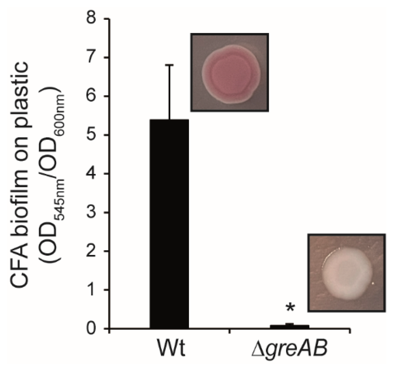
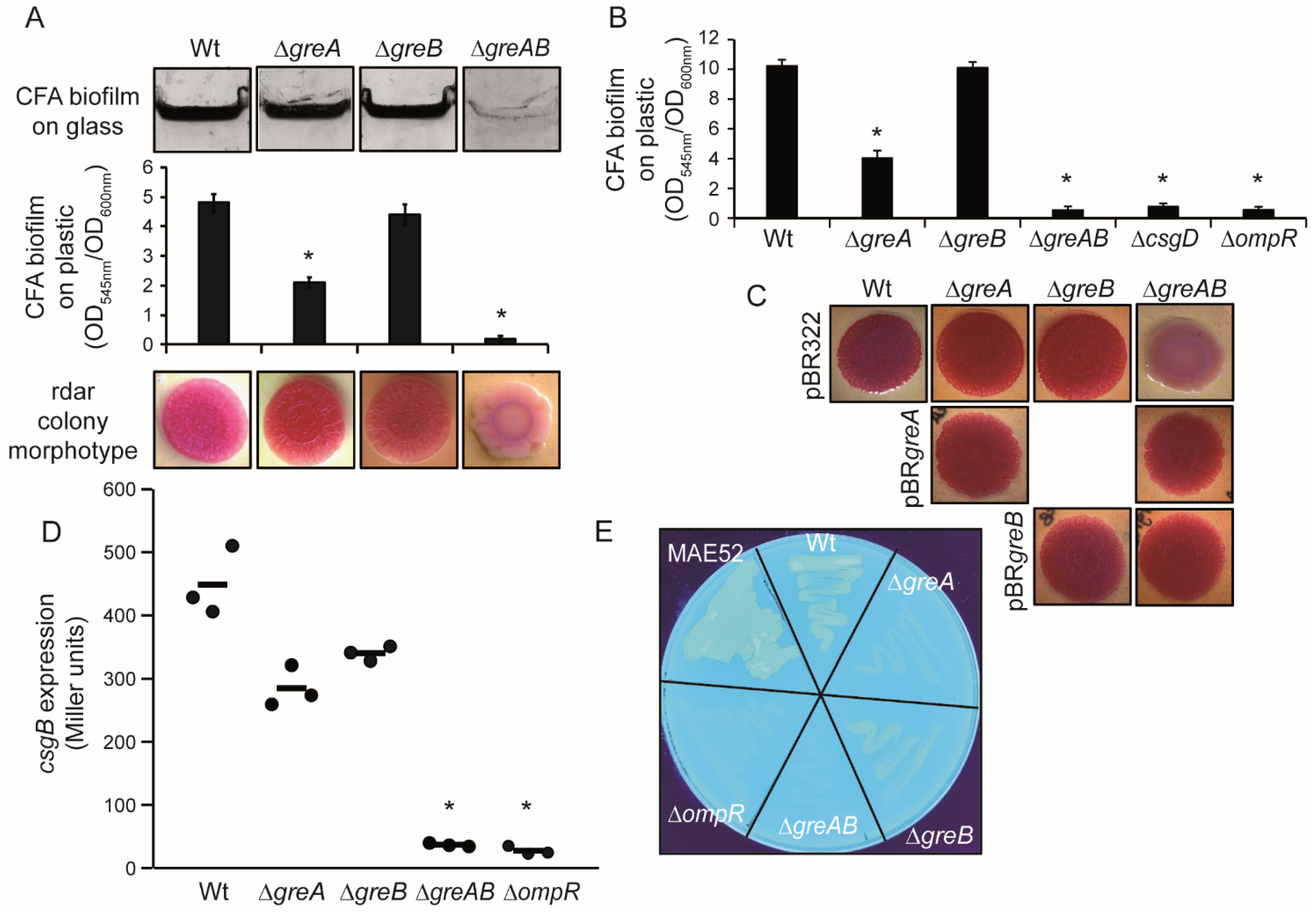
3.1. Rdar Biofilm Formation Is Impaired in S. Typhimurium When Deficient for the Transcription Elongation Factors GreA and GreB

S. Typhimurium is able to form a csgD-dependent rdar biofilm on a plethora of surfaces. In order to address the potential requirement of the Gre factors for the ability of S. Typhimurium to form biofilm, we determined the capacity of Salmonella enterica SL1344 his+ derivative SV5015 (Wt) strain and its mutants ∆greA, ∆greB and ∆greAgreB (from now referred as ∆greAB) to form biofilm on glass and plastic. Both the qualitative biofilm assay on glass and quantitative determination of biofilm formation on plastic indicated that biofilm production was impaired in the absence of both Gre factors (Figure 1A). The absence of either greA or greB did not abolish biofilm formation, although surface-growth of cells was reduced to some extent with a more pronounced drop in biofilm biomass for ∆greA than for ∆greB. The percentage of biofilm biomass compared to Wt was 42% and 87% for ∆greA and ∆greB, respectively. To corroborate the involvement of the Gre factors in the ability of S. Typhimurium to form biofilm, similar experiments were performed with the strain UMR1, a rdar28+ single colony derivative of ATCC14028 resistant to nalidixic acid, used as a model in biofilm studies [20]. A similar decrease in the ability to form biofilm on polystyrene was observed in the ∆greA, ∆greB and ∆greAB mutant derivatives of UMR1 as described for the SV5015 strains. Consistent with previous reports, the ATCC14028 derivative forms a more pronounced biofilm compared with SV5015, whereas no biofilm was detected from cultures of the ∆ompR and ∆csgD strains [20] (Figure 1B).
The production of the two extracellular appendages, curli and cellulose, which are promoting cell–cell, cell–interface and cell–surface interactions in S. Typhimurium biofilms, can be phenotypically scored by growing bacteria on Congo red agar plates [31] (Figure 1A,C). As expected, Wt strains, both SV5015 and UMR1, showed a rdar (red, dry and rough) colony morphotype, indicative of curli and cellulose production. Consistent with the qualitative and quantitative biofilm assays, the single mutants—∆greA and ∆greB—also showed a rdar morphotype, whereas the ∆greAB mutant strains showed saw (smooth and white) morphotype, indicating that they do not produce curli and cellulose. The requirement of the Gre factors for the production of the biofilm-promoting factors was further characterized. The transcriptional expression of csgB, coding the major subunit of curli, was monitored using a chromosomal csgB::lacZ fusion (Figure 1D). Downregulation of csgB expression was detected to some extent in the single mutants, being more pronounced in the ∆greA mutant. In the absence of both Gre factors, the expression was apparently fully abolished, reaching levels similar to the ∆ompR mutant strain. The production of cellulose was determined by growing the different strains in the presence of calcofluor, a fluorochrome that binds to β-1,4-polysaccharides, including cellulose (Figure 1E). Visual examination of the strains growing on LB0 supplemented with calcofluor under UV light showed that the Wt strain emitted substantially more fluorescence than the ∆greAB mutant, clearly indicating that the double mutant is impaired in cellulose production. Consistently, the single mutants, ∆greA and ∆greB, showed intermediate calcofluor binding phenotype. As a control, no fluorescence was detected in the ∆ompR strain. High emission of fluorescence was observed with the strain MAE52, which semi-constitutively overexpresses csgD and the biofilm extracellular matrix.
Notably, the Gre-mediated control of curli and cellulose expression was further confirmed by successful restoration of the rdar morphotype in a ∆greAB strain by trans-complementation with expression of either greA or greB from a plasmid (Figure 1C). In conclusion, GreA and GreB additively stimulates expression of cellulose and curli in S. Typhimurium, resulting in the production of a rdar biofilm.

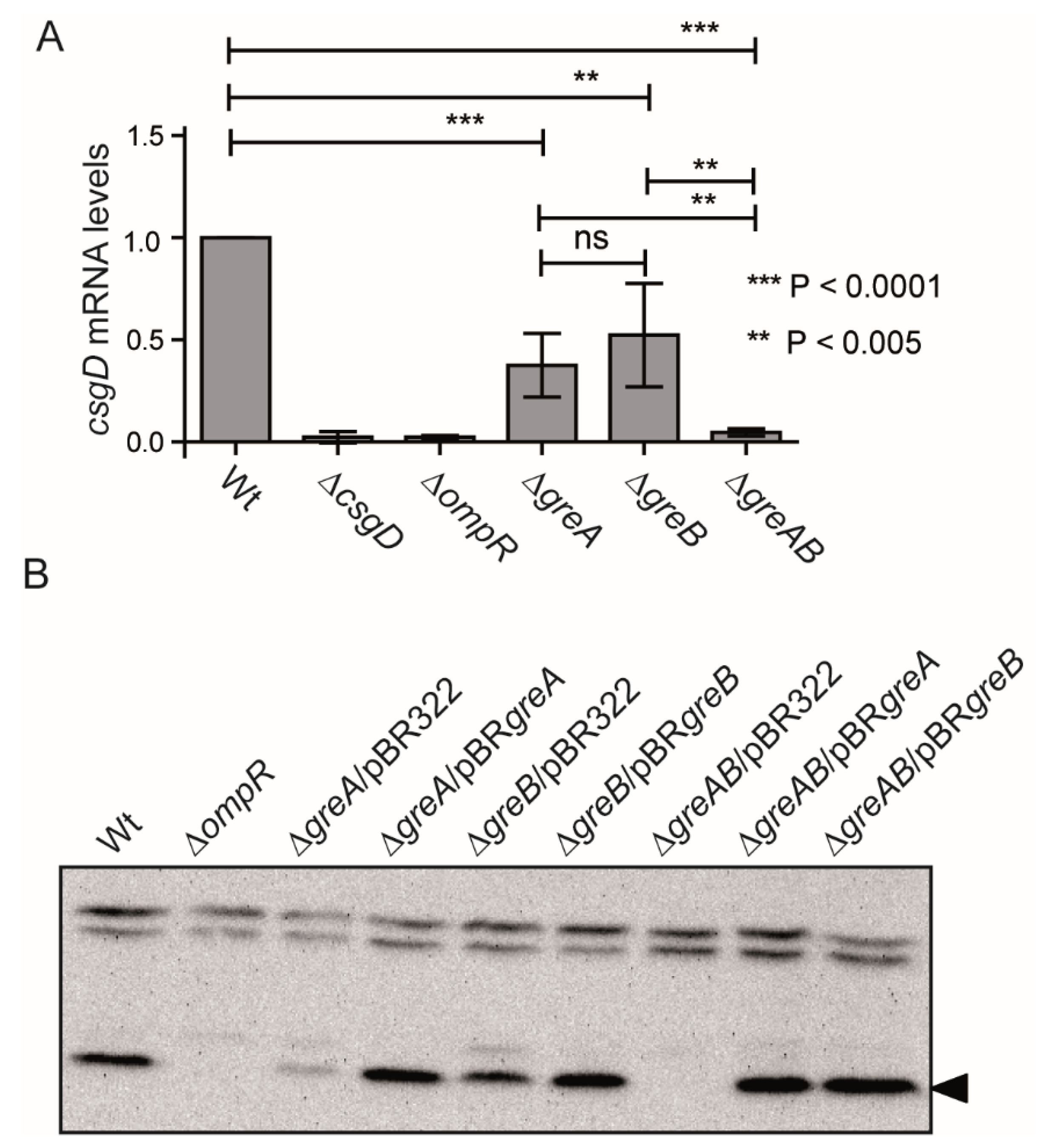
3.2. The Expression of CsgD, the Master Regulator of Biofilm Formation in S. Typhimurium Is Impaired in Strains Lacking the Gre Factors

In S. Typhimurium, the expression of curli and cellulose is under the control of CsgD. CsgD binds to the csgBAC promoter, stimulating curli expression, and to the adrA promoter, encoding a diguanylate cyclase that promotes cellulose production from the BCS proteins post-transcriptionally through the synthesis of cyclic di-GMP [12]. Since the production of both, curli and cellulose, was impaired in the ∆greAB mutant, a feasible explanation is that the Gre factors are required for the expression of CsgD and in turn for the production of the two biofilm-promoting factors. The transcriptional expression of csgD was studied by qPCR determination of the csgD mRNA levels (Figure 2A). Consistent with previous reports, csgD expression was not detected in a ∆ompR strain [11]. In agreement with the effect described on the rdar biofilm promoting factors, csgD expression was impaired in the ∆greAB, whereas the single mutants display partial reduction of csgD expression. Furthermore, the production of CsgD was determined by immunodetection of the CsgD protein in whole cell extracts of strains grown on LB0 agar plates (Figure 2B). No CsgD was produced when the bacterial cells lack both Gre factors while partial reduction was observed in the absence of ∆greA and to a lesser extent on ∆greB. Of note, both greA and greB can complement the drop in CsgD production in a ∆greAB strain. Altogether, our data indicated that the Gre factors are required for the expression of csgD, the master regulator of rdar biofilm formation in S. Typhimurium. Gre factors, as well as the curli operon, are conserved between S. Typhimurium and E. coli. Not unexpectedly, the absence of the Gre factors caused a severe drop in biofilm production in liquid culture and in the expression of biofilm promoting factors, as depicted by the rdar morphotypes, also in E. coli (Figure 3). In summary, greA and greB upregulate the central hub of the rdar biofilm, csgD, on the steady state mRNA level in S. Typhimurium and presumably also in E. coli.

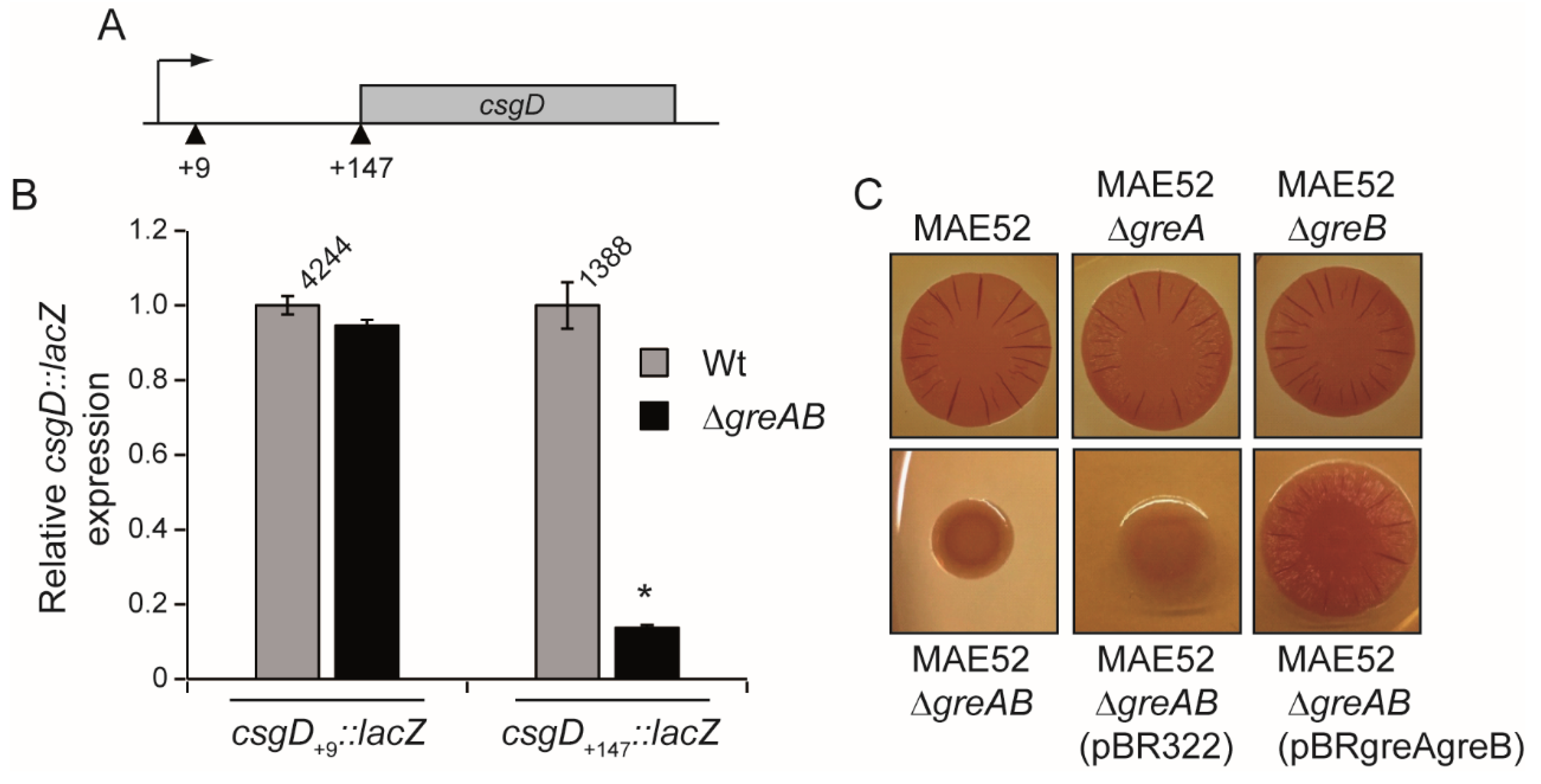
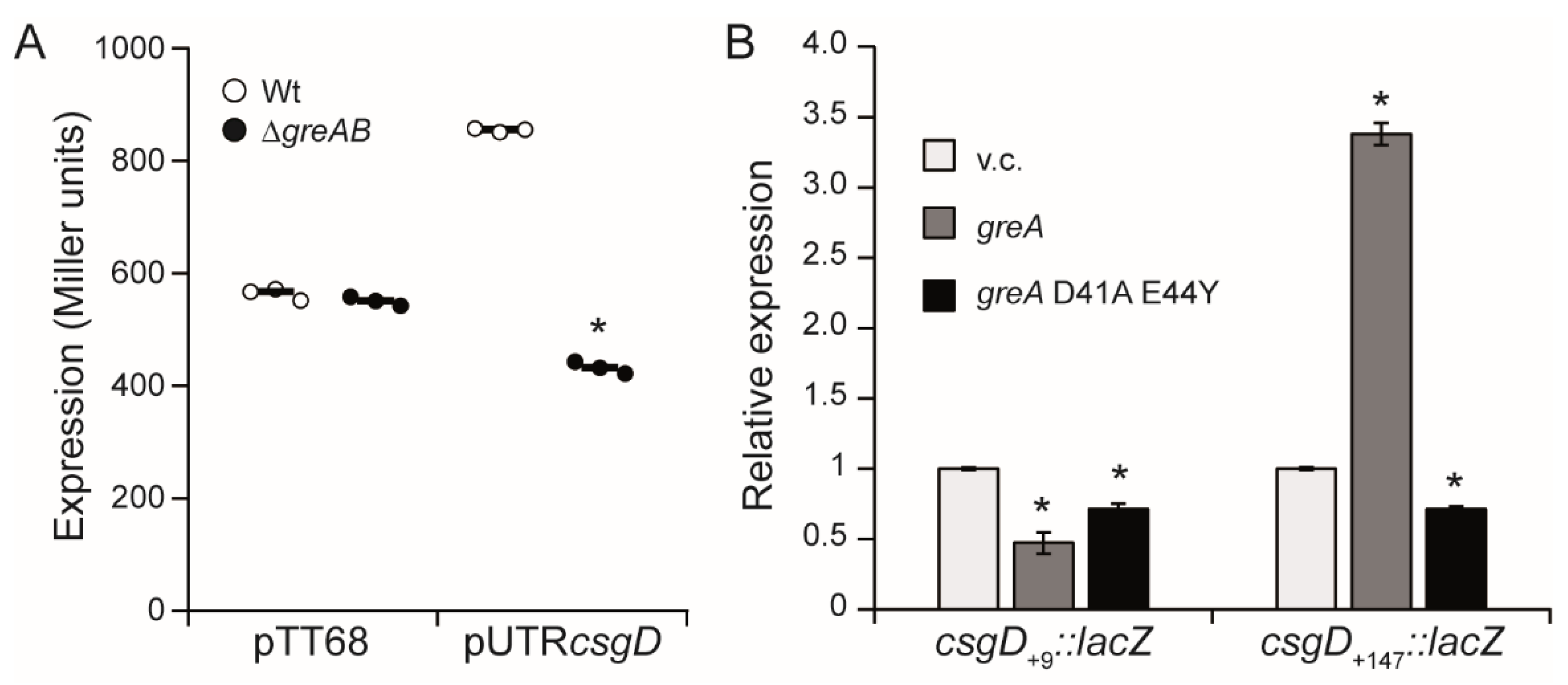
3.3. The 5′-UTR of the csgD Gene Is Required for Stimulation of csgD Expression by the Gre Factors

In order to determine if the Gre factors are required for csgD expression at the level of transcription initiation, transcriptional studies in Wt and ∆greAB strains were performed with a chromosomal lacZ fusions to the csgD gene at position +9 (csgD+9::lacZ, Figure 4A, see materials and methods for details). The down regulation of the csgD transcript in the ∆greAB mutant, detected by qPCR quantification, was not observed when csgD expression was monitored using the csgD+9::lacZ construct (Figure 4B). The csgD mRNA carries a long 5′-UTR of 147 bp which is required for enhanced transcription [17] and it is absent in the csgD+9::lacZ construct. In order to determine if the 5′-UTR region is important for the stimulatory role of the Gre factors in csgD expression, a csgD::lacZ construct carrying the entire 5′-UTR (csgD+147::lacZ, Figure 4A, see materials and methods for details) was generated. When transcription of the csgD+147::lacZ was assessed, a substantial downregulation of lacZ activity in the absence of the Gre factors was detected indicating that the 5′-UTR of csgD is subject to regulation by greA/greB (Figure 4B). Thus, we can speculate that Gre factors relieve transcriptional pause(s) in the 5′-untranslated region (5′-UTR) of the csgD gene, a mechanism of action occurring in most cases in 5′-UTR regions as described in the literature [3].
These results also clearly indicate that the 5′-UTR is required for Gre-mediated stimulation of csgD expression, while regulation does not occur at the level of transcription initiation. The latest statement was further corroborated using strain MAE52 carrying a single point mutation in the csgD promoter and causing an approximately three-fold higher transcriptional expression of the csgD gene. In MAE52, csgD is transcribed temperature independently with the aid of the housekeeping sigma factor RpoD instead of the stress sigma factor RpoS as in strain UMR1 [11,32]. Independently of the fact that the csgD gene was expressed from a semi-constitutive promoter in MAE52, mutations in greA and greB lead to downregulation of the expression of the rdar morphotype, which further supports a regulatory mechanism of action by Gre factors downstream of transcription initiation (Figure 4C).
These data indicate that the 5′-UTR of the csgD gene is the target of the Gre factors. The csgD 5′-UTR was cloned into the pTT68 plasmid, downstream of a pBAD promoter and upstream of a promoter less lacZ gene. Expression of the construct was induced by the addition of arabinose and monitored in both Wt and ∆greAB genetic backgrounds. The expression dropped in the ∆greAB mutant with the construct carrying the csgD 5′-UTR (Figure 5A), further indicating that the 5′-UTR is the target of the Gre factors.
Gre factors promote transcription elongation by resolving backtracking of paused transcription complexes [1]. In E. coli, the D41 and E44 GreA residues are essential for the ability of GreA to alleviate backtracked complexes and thereby to suppress transcriptional pauses [33]. E. coli and S. Typhimurium GreA share a high percentage of identity (97%) comprising the 41 and 44 residues (Figure S1). Previously, we demonstrated that E. coli GreA complements the ∆greAB mutant of S. Typhimurium and that the D41 and E44 residues are required to suppress transcriptional pausing [7]. To determine whether downregulation of csgD expression in the Gre-factor deficient strain is due to a transcriptional pause, two GreA variants of E. coli were used, the GreAWt and the GreAD41AE44Y, being the latter impaired to resolve paused transcription complexes. The effect of the presence of GreAWt and GreAD41AE44Y on the expression of the csgD gene was monitored using the csgD+9::lacZ and csgD+147::lacZ constructs (Figure 5B). As expected referring to the previous results, the production of GreAWt in a ∆greAB mutant complemented the absence of the Gre factors as monitored by transcription of the csgD fusion carrying the 5′-UTR (csgD+147::lacZ), whereas no change in expression of the csgD fusion lacking the 5′-UTR (csgD+9::lacZ). Remarkably, no induction of the csgD+147::lacZ construct was detected when GreAD41AE44Y was expressed, indicating that the reported effect of the Gre factors on csgD expression can be due to the resolution of a transcriptional pause occurring in the 5′-UTR of the csgD gene. Although, in vitro transcription experiments will be required to confirm the direct involvement of the Gre factors in csgD transcription, our data suggest that GreA and GreB alleviate transcriptional arrest in the 5′-UTR of csgD mediated by the residues participating in transcript cleavage by the RNA polymerase. Whether additional GreA and GreB checkpoints exist upon transcription of the csgD open reading frame and/or of the csgDEFG operon needs to be investigated in future studies.

4. Concluding Remarks

Rdar and alternative modes of biofilm formation are delicately regulated. The switch between planktonic and sessile (biofilm) cells denotes not only a radical change in the single cell versus population behavior, but also in the physiology and metabolism of the organism. In S. Typhimurium, the activity of the CsgD regulator is pivotal in the stimulation of rdar biofilm formation including modulation of the synthesis of two biofilm promoting factors, curli and cellulose. The synthesis of those biofilm matrix components requires a relevant number of the assets available for the cells. Different environmental stresses including nutrient starvation trigger biofilm formation in S. Typhimurium.
In this report, we describe the Gre factors as new players in the array of regulators affecting csgD expression. A potential role for the Gre factors in bacterial adaptation to different environmental stresses has been proposed [3,7]. Hence, expression of greA and greB occurs under different environmental conditions in E. coli [3]. The expression of the greA gene is σE-dependent in E. coli and S. Typhimurium, is induced under hypoxia in Mycobacterium avium and under acid stress in Streptococcus mutans [34,35,36,37,38]. Mutation in genes encoding Gre factors cause sensitivity to different stresses such as heat shock, osmotic shock and presence of detergents [39,40,41]. A role in stress response for the Gre factor proteins by altering transcriptional expression was suggested for the stress resistant bacterium Deinococcus radiodurans [42].
In a previous report, we described in S. Typhimurium that the Gre factors are required for the expression of hilD encoding the main regulator of the SPI-1, a genomic island encoding the type three secretion system 1 needed for invasion of epithelial cells. The the 3′-UTR of the hilD gene was identified as the target of the Gre factors [7]. Here, we propose that Gre-mediated alleviation of pausing during 5′-UTR transcription may be pivotal to generate a fully functional csgD transcript through nucleolytic cleavage. Such mRNA processing might even influence the effect of additional known post-transcriptional regulators that target the 5′-UTR of csgD and can constitute an additional regulatory mechanism in the multilayered regulation of the csgD gene. Transcriptional pausing causes gene repression that can be alleviated by the Gre factors as a response to specific environmental stimuli.
Long 5′UTR are regulatory motifs often involved in post-transcriptional regulation by very diverse mechanisms and targets for regulation at the level of transcription elongation [43]. The RNA chaperone Hfq and small RNAs are among the global regulators that directly target the 5′-UTR of the csgD operon to negatively or positively affect transcription [15,16,18]. It has been shown that genes encoding for regulators are especially prone to suffer from transcriptional pausing [3], therefore transcriptional pausing consequently affects the expression of all genes under the control of a specific regulator. Further studies will be required to fully dissect the role of the csgD 5′-UTR and the impact of the Gre-dependent pausing on the regulatory pathways that convey on the 5′-UTR of csgD.

Supplementary Materials

The following supporting information can be downloaded at: https://www.mdpi.com/article/10.3390/microorganisms10101921/s1, Figure S1: Alignment of the GreA proteins from the E. coli strain MG1655 (GreA MG1655) and from the S. Typhimurium strains ATCC14028 and SL1433 (GreA SALM); Table S1: Strains and plasmids used in this study. Table S2: Primers used in this study; File S1: Information regarding construction of the csgD::lacZ chromosomal fusions.

Author Contributions

Conceptualization, T.G.-C., Y.E.M., U.R., C.M. and C.B.; methodology, T.G.-C., Y.E.M., A.C.-A., A.F.V. and C.J.J.; formal analysis, T.G.-C., Y.E.M., A.C.-A., A.F.V., C.J.J., U.R., C.M. and C.B.; investigation, T.G.-C., Y.E.M., A.C.-A., A.F.V. and C.J.J.; writing—original draft preparation, C.B.; writing—review and editing, T.G.-C., Y.E.M., A.C.-A., A.F.V., C.J.J., U.R., C.M. and C.B.; supervision, U.R., C.M. and C.B.; project administration, U.R., C.M. and C.B.; funding acquisition, U.R., C.M. and C.B. All authors have read and agreed to the published version of the manuscript.

Funding

This research was funded by the Spanish Ministry of Economy and Competitiveness [grant AGL2013-45339R, C.B.], the Spanish Ministry of Science, Innovation and Universities (MCIU), State Bureau of Investigation (AIE), the European Regional Development Fund (FEDER) [grant PGC2018-096958-B-I00, C.B.], and the Catalonian Government [grant 2017SGR499, C.M.], the Swedish Research Council for Natural Sciences and Engineering [621-2013-4809 and 2017-04465, U.R.], and the German Research Foundation [Deutsche Forschungsgemeinschaft; CI 239/1-1 and CI 239/2-1, A.C.-A.]. T.G.-C. and Y.E.M. were recipient of predoctoral fellowships from the University of Barcelona.

Data Availability Statement

Not applicable.

Conflicts of Interest

The authors declare no conflict of interest.

References

  1. Laptenko, O.; Lee, J.; Lomakin, I.; Borukhov, S. Transcript Cleavage Factors GreA and GreB Act as Transient Catalytic Components of RNA Polymerase. EMBO J. 2003, 22, 6322–6334. [Google Scholar] [CrossRef] [PubMed]
  2. Li, K.; Jiang, T.; Yu, B.; Wang, L.; Gao, C.; Ma, C.; Xu, P.; Ma, Y. Transcription Elongation Factor GreA Has Functional Chaperone Activity. PLoS ONE 2012, 7, e47521. [Google Scholar] [CrossRef] [PubMed]
  3. Sun, Z.; Yakhnin, A.V.; Fitzgerald, P.C.; Mclntosh, C.E.; Kashlev, M. Nascent RNA Sequencing Identifies a Widespread Sigma70-Dependent Pausing Regulated by Gre Factors in Bacteria. Nat Commun. 2021, 12, 906. [Google Scholar] [CrossRef] [PubMed]
  4. Aberg, A.; Fernandez-Vazquez, J.; Cabrer-Panes, J.D.; Sanchez, A.; Balsalobre, C. Similar and Divergent Effects of PpGpp and DksA Deficiencies on Transcription in Escherichia coli. J. Bacteriol. 2009, 191, 3226–3236. [Google Scholar] [CrossRef] [PubMed]
  5. Åberg, A.; Shingler, V.; Balsalobre, C. Regulation of the fimB Promoter: A Case of Differential Regulation by PpGpp and DksA in vivo. Mol. Microbiol. 2008, 67, 1223–1241. [Google Scholar] [CrossRef] [PubMed]
  6. Stepanova, E.; Lee, J.; Ozerova, M.; Semenova, E.; Datsenko, K.; Wanner, B.L.; Severinov, K.; Borukhov, S. Analysis of Promoter Targets for Escherichia coli Transcription Elongation Factor GreA In Vivo and In Vitro. J. Bacteriol. 2007, 189, 8772–8785. [Google Scholar] [CrossRef]
  7. Gaviria-Cantin, T.; El Mouali, Y.; Le Guyon, S.; Römling, U.; Balsalobre, C. Gre Factors-Mediated Control of hilD Transcription Is Essential for the Invasion of Epithelial Cells by Salmonella enterica Serovar Typhimurium. PLoS Pathog. 2017, 13, e1006312. [Google Scholar] [CrossRef]
  8. Steenackers, H.; Hermans, K.; Vanderleyden, J.; De Keersmaecker, S.C.J. Salmonella Biofilms: An Overview on Occurrence, Structure, Regulation and Eradication. Food Res. Int. 2012, 45, 502–531. [Google Scholar] [CrossRef]
  9. Harrell, J.E.; Hahn, M.M.; D’Souza, S.J.; Vasicek, E.M.; Sandala, J.L.; Gunn, J.S.; McLachlan, J.B. Salmonella Biofilm Formation, Chronic Infection, and Immunity Within the Intestine and Hepatobiliary Tract. Front. Cell. Infect. Microbiol. 2021, 10, 910. [Google Scholar] [CrossRef]
  10. Römling, U.; Rohde, M. Flagella Modulate the Multicellular Behavior of Salmonella typhimurium on the Community Level. FEMS Microbiol. Lett. 1999, 180, 91–102. [Google Scholar] [CrossRef]
  11. Römling, U.; Sierralta, W.D.; Eriksson, K.; Normark, S. Multicellular and Aggregative Behaviour of Salmonella typhimurium Strains Is Controlled by Mutations in the AgfD Promoter. Mol. Microbiol. 1998, 28, 249–264. [Google Scholar] [CrossRef] [PubMed]
  12. Römling, U.; Rohde, M.; Olsén, A.; Normark, S.; Reinköster, J. agfD, the Checkpoint of Multicellular and Aggregative Behaviour in Salmonella typhimurium Regulates at Least Two Independent Pathways. Mol. Microbiol. 2000, 36, 10–23. [Google Scholar] [CrossRef] [PubMed]
  13. Ogasawara, H.; Ishizuka, T.; Hotta, S.; Aoki, M.; Shimada, T.; Ishihama, A. Novel Regulators of the csgD Gene Encoding the Master Regulator of Biofilm Formation in Escherichia coli K-12. Microbiology 2020, 166, 880–890. [Google Scholar] [CrossRef] [PubMed]
  14. Gerstel, U.; Römling, U. The csgD Promoter, a Control Unit for Biofilm Formation in Salmonella typhimurium. Res. Microbiol. 2003, 154, 659–667. [Google Scholar] [CrossRef] [PubMed]
  15. Andreassen, P.R.; Pettersen, J.S.; Szczerba, M.; Valentin-Hansen, P.; Møller-Jensen, J.; Jørgensen, M.G. SRNA-Dependent Control of Curli Biosynthesis in Escherichia coli: McaS Directs Endonucleolytic Cleavage of csgD mRNA. Nucleic Acids Res. 2018, 46, 6746–6760. [Google Scholar] [CrossRef] [PubMed]
  16. Bordeau, V.; Felden, B. Curli Synthesis and Biofilm Formation in Enteric Bacteria Are Controlled by a Dynamic Small RNA Module Made up of a Pseudoknot Assisted by an RNA Chaperone. Nucleic Acids Res. 2014, 42, 4682–4696. [Google Scholar] [CrossRef]
  17. Gerstel, U.; Kolb, A.; Römling, U. Regulatory Components at the CsgD Promoter--Additional Roles for OmpR and Integration Host Factor and Role of the 5’ Untranslated Region. FEMS Microbiol. Lett. 2006, 261, 109–117. [Google Scholar] [CrossRef]
  18. Holmqvist, E.; Reimegård, J.; Sterk, M.; Grantcharova, N.; Römling, U.; Wagner, E.G.H. Two Antisense RNAs Target the Transcriptional Regulator CsgD to Inhibit Curli Synthesis. EMBO J. 2010, 29, 1840–1850. [Google Scholar] [CrossRef]
  19. Römling, U. Characterization of the Rdar Morphotupe, a Multicellular Behaviour in Enterobacteriaceae. CMLS, Cell. Mol. Life. Sci. 2005, 62, 1234–1246. [Google Scholar] [CrossRef]
  20. Römling, U.; Bian, Z.; Hammar, M.; Sierralta, W.D.; Normark, S. Curli Fibers Are Highly Conserved between Salmonella typhimurium and Escherichia coli with Respect to Operon Structure and Regulation. J. Bacteriol. 1998, 180, 722–731. [Google Scholar] [CrossRef] [Green Version]
  21. Hoiseth, S.K.; Stocker, B.A. Aromatic-Dependent Salmonella typhimurium Are Non-Virulent and Effective as Live Vaccines. Nature 1981, 291, 238–239. [Google Scholar] [CrossRef] [PubMed]
  22. Vivero, A.; Banos, R.C.; Mariscotti, J.F.; Oliveros, J.C.; Garcia-del Portillo, F.; Juarez, A.; Madrid, C. Modulation of Horizontally Acquired Genes by the Hha-YdgT Proteins in Salmonella enterica Serovar Typhimurium. J. Bacteriol. 2008, 190, 1152–1156. [Google Scholar] [CrossRef] [PubMed]
  23. Datsenko, K.A.; Wanner, B.L. One-Step Inactivation of Chromosomal Genes in Escherichia coli K-12 Using PCR Products. Proc. Natl. Acad. Sci. USA 2000, 97, 6640–6645. [Google Scholar] [CrossRef] [PubMed]
  24. Ellermeier, C.D.; Janakiraman, A.; Slauch, J.M. Construction of Targeted Single Copy Lac Fusions Using Lambda Red and FLP-Mediated Site-Specific Recombination in Bacteria. Gene 2002, 290, 153–161. [Google Scholar] [CrossRef]
  25. Sternberg, N.L.; Maurer, R. Bacteriophage-Mediated Generalized Transduction in Escherichia Coli and Salmonella Typhimurium. Methods Enzymol. 1991, 204, 18–43. [Google Scholar] [PubMed]
  26. Maloy, S.; Stewart, R. Genetic Analysis of Pathogenic Bacteria; Cold Spring Harbor Laboratory Press: Cold Spring Harbor, NY, USA, 1996. [Google Scholar]
  27. Paytubi, S.; Guirado, P.; Balsalobre, C.; Madrid, C. An Improved and Versatile Methodology to Quantify Biofilms Formed on Solid Surfaces and Exposed to the Air-Liquid Interphase. J. Microbiol. Methods 2014, 103. [Google Scholar] [CrossRef]
  28. Paytubi, S.; Cansado, C.; Madrid, C.; Balsalobre, C. Nutrient Composition Promotes Switching between Pellicle and Bottom Biofilm in Salmonella. Front. Microbiol. 2017, 8, 2160. [Google Scholar] [CrossRef]
  29. Miller, J.H. A Short Course in Bacterial Genetics: A Laboratory Manual and Handbook for Escherichia coli and Related Bacteria; Cold Spring Harbor Laboratory Press: Cold Spring Harbor, NY, USA, 1992; ISBN 0879693495. [Google Scholar]
  30. Zafar, M.; Jahan, H.; Shafeeq, S.; Nimtz, M.; Jänsch, L.; Römling, U.; Iqbal Choudhary, M. Clarithromycin Exerts an Antibiofilm Effect against Salmonella enterica Serovar Typhimurium Rdar Biofilm Formation and Transforms the Physiology towards an Apparent Oxygen-Depleted Energy and Carbon Metabolism. Infect. Immun. 2020, 88, e00510-20. [Google Scholar] [CrossRef]
  31. Lamprokostopoulou, A.; Römling, U. Yin and Yang of Biofilm Formation and Cyclic Di-GMP Signaling of the Gastrointestinal Pathogen Salmonella enterica Serovar Typhimurium. J. Innate Immun. 2022, 14, 275–292. [Google Scholar] [CrossRef]
  32. Zakikhany, K.; Harrington, C.R.; Nimtz, M.; Hinton, J.C.D.; Römling, U. Unphosphorylated CsgD Controls Biofilm Formation in Salmonella enterica Serovar Typhimurium. Mol. Microbiol. 2010, 77, 771–786. [Google Scholar] [CrossRef]
  33. Vinella, D.; Potrykus, K.; Murphy, H.; Cashel, M. Effects on Growth by Changes of the Balance between GreA, GreB, and DksA Suggest Mutual Competition and Functional Redundancy in Escherichia coli. J. Bacteriol. 2012, 194, 261–273. [Google Scholar] [CrossRef] [PubMed]
  34. Nogales, J.; Campos, R.; BenAbdelkhalek, H.; Olivares, J.; Lluch, C.; Sanjuan, J. Rhizobium tropici Genes Involved in Free-Living Salt Tolerance Are Required for the Establishment of Efficient Nitrogen-Fixing Symbiosis with Phaseolus vulgaris. Mol. Plant. Microbe. Interact. 2002, 15, 225–232. [Google Scholar] [CrossRef] [PubMed]
  35. Rhodius, V.A.; Suh, W.C.; Nonaka, G.; West, J.; Gross, C.A. Conserved and Variable Functions of the SigmaE Stress Response in Related Genomes. PLoS Biol. 2006, 4, e2. [Google Scholar] [CrossRef]
  36. Skovierova, H.; Rowley, G.; Rezuchova, B.; Homerova, D.; Lewis, C.; Roberts, M.; Kormanec, J. Identification of the SigmaE Regulon of Salmonella enterica Serovar Typhimurium. Microbiology 2006, 152, 1347–1359. [Google Scholar] [CrossRef] [PubMed]
  37. Li, J.; Overall, C.C.; Johnson, R.C.; Jones, M.B.; McDermott, J.E.; Heffron, F.; Adkins, J.N.; Cambronne, E.D. ChIP-Seq Analysis of the σE Regulon of Salmonella enterica Serovar Typhimurium Reveals New Genes Implicated in Heat Shock and Oxidative Stress Response. PLoS ONE 2015, 10, e0138466. [Google Scholar] [CrossRef] [PubMed]
  38. Gumber, S.; Taylor, D.L.; Marsh, I.B.; Whittington, R.J. Growth Pattern and Partial Proteome of Mycobacterium avium Subsp. Paratuberculosis during the Stress Response to Hypoxia and Nutrient Starvation. Vet. Microbiol. 2009, 133, 344–357. [Google Scholar] [CrossRef] [PubMed]
  39. Len, A.C.L.; Harty, D.W.S.; Jacques, N.A. Stress-Responsive Proteins Are Upregulated in Streptococcus mutans during Acid Tolerance. Microbiology 2004, 150, 1339–1351. [Google Scholar] [CrossRef]
  40. Campbell, G.R.O.; Sharypova, L.A.; Scheidle, H.; Jones, K.M.; Niehaus, K.; Becker, A.; Walker, G.C. Striking Complexity of Lipopolysaccharide Defects in a Collection of Sinorhizobium meliloti Mutants. J. Bacteriol. 2003, 185, 3853–3862. [Google Scholar] [CrossRef]
  41. Orlova, M.; Newlands, J.; Das, A.; Goldfarb, A.; Borukhov, S. Intrinsic Transcript Cleavage Activity of RNA Polymerase. Proc. Natl. Acad. Sci. USA 1995, 92, 4596–4600. [Google Scholar] [CrossRef]
  42. Agapov, A.; Esyunina, D.; Kulbachinskiy, A. Gre-Family Factors Modulate DNA Damage Sensing by Deinococcus radiodurans RNA Polymerase. RNA Biol. 2019, 16, 1711–1720. [Google Scholar] [CrossRef]
  43. Kaberdin, V.R.; Bläsi, U. Translation Initiation and the Fate of Bacterial MRNAs. FEMS Microbiol. Rev. 2006, 30, 967–979. [Google Scholar] [CrossRef] [PubMed] [Green Version]
Figure 1. The Gre factors are required for expression of rdar biofilm promoting factors in S. Typhimurium. (A). Effect of deletion mutations of Gre factor encoding genes on the biofilm formed by the SV5015 strain. Upper panel, crystal violet stained biofilm formed by SV5015 (Wt) and the indicated mutant derivatives strains on glass slides after static growth for 72 h at 25 °C in CFA medium in Hellendahl jars. Middle panel, quantification of crystal violet stained biofilm formed on polystyrene walls of 96 well plates in CFA medium after 72 h at 25 °C. Lower panel, colony morphotype of the indicated strains on Congo red plates. (B). Effect of deletion mutations of the Gre factor encoding genes on biofilm formation by the UMR1 strain formed on polystyrene walls of 96 well plates. Cultures and quantification were performed as in (A). Biofilm quantification, in both (A,B), is based on calculation of the mean and standard deviations of six samples (three independent cultures with two technical replicas each). (C). Trans-complementation of the UMR1∆greAB mutant strain monitored by colony morphotype on Congo red plates carrying either the pBR322 (control vector), pBRgreA or pBRgreB. (D). Transcriptional expression of a csgB::lacZ chromosomal fusion was monitored in the indicated strains by β-galactosidase determination. Mean and standard deviations calculated from three independent cultures are shown. (E). The production of cellulose is impaired in the ∆greAB mutant strain. Observed fluorescence under UV of the indicated strains grown at 25 °C for 48 h on a LB0 plate supplemented with calcofluor. Statistically significant difference compared to Wt is shown by an asterisk (p < 0.0005).
Microorganisms 10 01921 g001
Figure 2. The expression of csgD encoding the master regulator of rdar biofilm formation is impaired in the absence of the Gre factors. (A). csgD mRNA levels were determined by qPCR in RNA samples from cultures of the indicated strains grown on LB0 plates at 28 °C for 24 h. Results are normalized to expression of recA as an endogenous control and given as relative values to Wt, arbitrarily set as 1.0. The results are mean and standard deviation from three biological replicates. Statistical significance is indicated by *** p < 0.0001, ** p < 0.005, ns: non-significant. (B). Immunodetection of CsgD (indicated by an arrowhead). Cell extracts from cells grown on LB0 plates at 28 °C for 24 h were separated by PAGE and analyzed by Western blot analysis.
Microorganisms 10 01921 g002
Figure 3. The Gre factors are required for expression of rdar biofilm promotion in Escherichia coli. A. Effect of deletion mutants of Gre factor encoding genes on biofilm formation by E. coli MG1655 strain. Quantification of crystal violet stained biofilm formed on polystyrene walls of 96 well plates by MG1655 (Wt) and its ∆greAB mutant grown in CFA medium for 72 h at 25 °C. Insets show the colony morphotype of the indicated strains on Congo red plates. Biofilm quantification values are mean and standard deviations of six samples. Statistical significance to Wt is shown by an asterisk (p < 0.0005).
Microorganisms 10 01921 g003
Figure 4. The 5′UTR of the csgD gene is the target of the Gre factors. (A). Schematic representation of the generated chromosomal csgD::lacZ fusion: the csgD+9::lacZ lacks the 5′-UTR and csgD+147::lacZ carries the 5′-UTR. Arrowheads indicate the relative position of the gene fusions. (B). Relative expression of csgD in Wt and ∆greAB in the absence (csgD+9::lacZ) and presence (csgD+147::lacZ) of the 5′UTR. Cultures were grown on LB0 plates and incubated at 28 °C for 24 h. A bar shows the arithmetic mean of experimental results and the error bar indicates the standard deviation from three biological replicates. The mean in Miller unit is indicated for the Wt. Statistical significance to Wt is shown by an asterisk (p < 0.0005). (C). Colony morphotype of strain MAE52 and its derivatives on Congo red agar plate. Plates were incubated at 28 °C for 24 h.
Microorganisms 10 01921 g004
Figure 5. A Gre dependent transcriptional pause is located in the 5′-UTR of the csgD gene. (A). lacZ expression from plasmid construct (pUTRcsgD) carrying the 5′-UTR of csgD cloned in pTT68 vector downstream of the PBAD promoter and upstream of a promoter less lacZ gene in both Wt and ∆greAB strains. Cultures were grown in LB supplemented with ampicillin and arabinose at 37 °C up to an OD600nm of 2.0. (B). Expression from the chromosomal fusions csgD+9::lacZ (lacking the 5′-UTR) and csgD+147::lacZ (carrying the 5′-UTR) in the ∆greAB strain carrying the plasmids pHM1883 (vector control, v.c.), pHM1873 (pHM1883 + greAMG1655) and pHM1854 (pHM1883 + greAMG1655(D41AE44Y). Cultures were grown on LB0 plates supplemented with spectinomycin and incubated at 28 °C for 48 h. A bar shows the arithmetic mean of experimental results and the error bar indicate the standard deviation from three biological replicates. Statistical significance to Wt is shown by an asterisk (p < 0.0005).
Microorganisms 10 01921 g005
Publisher’s Note: MDPI stays neutral with regard to jurisdictional claims in published maps and institutional affiliations.

