Article

Whole-Transcriptome Sequencing Reveals Characteristics of Cancer Microbiome in Korean Patients with GI Tract Cancer: Fusobacterium nucleatum as a Therapeutic Target

Department of Biomedical Science and Engineering, Gwangju Institute of Science and Technology (GIST), Gwangju 61005, Korea
*
Author to whom correspondence should be addressed.
Microorganisms 2022, 10(10), 1896; https://doi.org/10.3390/microorganisms10101896
Submission received: 7 September 2022 / Revised: 21 September 2022 / Accepted: 21 September 2022 / Published: 23 September 2022
(This article belongs to the Special Issue Gut Microbiota in Disease)

Abstract

:
Remarkable progress has occurred over the past two decades in identifying microbiomes affecting the human body in numerous ways. The microbiome is linked to gastrointestinal (GI) tract cancer. The purpose of this study was to determine if there is a common microbiome among GI tract cancers and how the microbiome affects the disease. To ensure ethnic consistency, Korean patients with GI tract cancer were selected. Fusobacterium nucleatum is an enriched bacteria in all cancer tissues. F. nucleatum is a Gram-negative obligate anaerobe that promotes colorectal cancer. Through Gene Set Enrichment Analysis (GSEA) and Differentially Expressed Genes (DEG) analyses, the upregulation of the G2M checkpoint pathway was identified in the F. nucleatum-high group. Cell viability and G2M checkpoint pathway genes were examined in MC 38 cells treated with F. nucleatum. F. nucleatum upregulated the expression of G2M checkpoint pathway genes and the cell proliferation of MC 38 cells. F. nucleatum facilitated cancer’s use of G2M checkpoint pathways and F. nucleatum could be a therapeutic target in Korean GI tract cancer.

1. Introduction

Over the past two decades, there has been outstanding progress in the identification of the microbiome that broadly affects the human body. The human body hosts a variety of microbes, and the influences of the gut microbiome on cancer and cancer immunity are well-known. These microbes have been detected even in tumor tissues and can immensely affect tumors [1]. With the development of high-throughput sequencing, also known as next-generation sequencing (NGS), such as 16 s rRNA and whole-transcriptome sequencing (WTS), cancer microbiome research has been actively conducted [2]. The 16 s rRNA sequencing method is a widely used NGS platform upon which to identify taxonomic composition in cancer research. Moreover, the WTS of tumor tissues shows an association between genes and the microbiome. The microbiome is closely related to the mechanisms, genes, and pathways that cancer cells utilize to proliferate [3].
Recent studies have demonstrated that bacteria, such as Helicobacter pylori, Escherichia coli, Bacteroides fragilis, Salmonella enterica, and F. nucleatum are key players in cancer [4]. It is estimated that approximately half of the global population suffers from an H. pylori infection. Through immune response and inflammation, H. pylori contributes to the development of gastric adenocarcinoma [5]. By causing inflammation and oxidative stress, E. coli strains promote the development of colorectal cancer (CRC). Additionally, genotoxin secreted from E. coli is known to generate DNA damage in eukaryotic epithelial cells [6]. B. fragilis is common in the entire colon, and enterotoxigenic B. fragilis (ETBF) produces metalloprotease Bacteroides fragilis toxin (BFT), leading to inflammation and tissue damage in CRC, thereby promoting colon tumorigenesis [7]. S. enterica infection causes the activation of the MAPK and AKT pathways, which provoke cellular transformation related to gallbladder cancer [8]. F. nucleatum is a Gram-negative obligate anaerobe bacterium and is usually acknowledged as a CRC-promoting bacterium [9]. Cancer is promoted by F. nucleatum through the enhancement of host responses that initiate and promote tumors and by encouraging tumor invasion and metastasis [10]. To examine whether the microbiome is associated with gastrointestinal (GI) tract cancer, we looked for a common microbiome among the GI tract cancers and sought to understand how the microbiome is associated with cancer. Additionally, Korean patients were selected for ethnic consistency, which might cause a discrepancy in microbial abundance among different ethnic groups.
After the recognition of the bacterial effect on cancer, there have been many attempts to target the microbiome for cancer therapy [11]. There are a few studies that target the common microbiome in Korean GI tract cancers. Here, F. nucleatum was identified as a highly expressed bacteria in Korean GI tract tumor tissues and possible pathways that F. nucleatum use to promote cancer. In this study, through WTS analysis by in vitro assay, F. nucleatum was demonstrated to encourage cancer cells to proliferate through the G2M checkpoint pathway. Understanding the F. nucleatum mechanism and its effect on cancer cells could lead to the discovery of potential therapeutic targets for GI tract cancers.

2. Materials and Methods

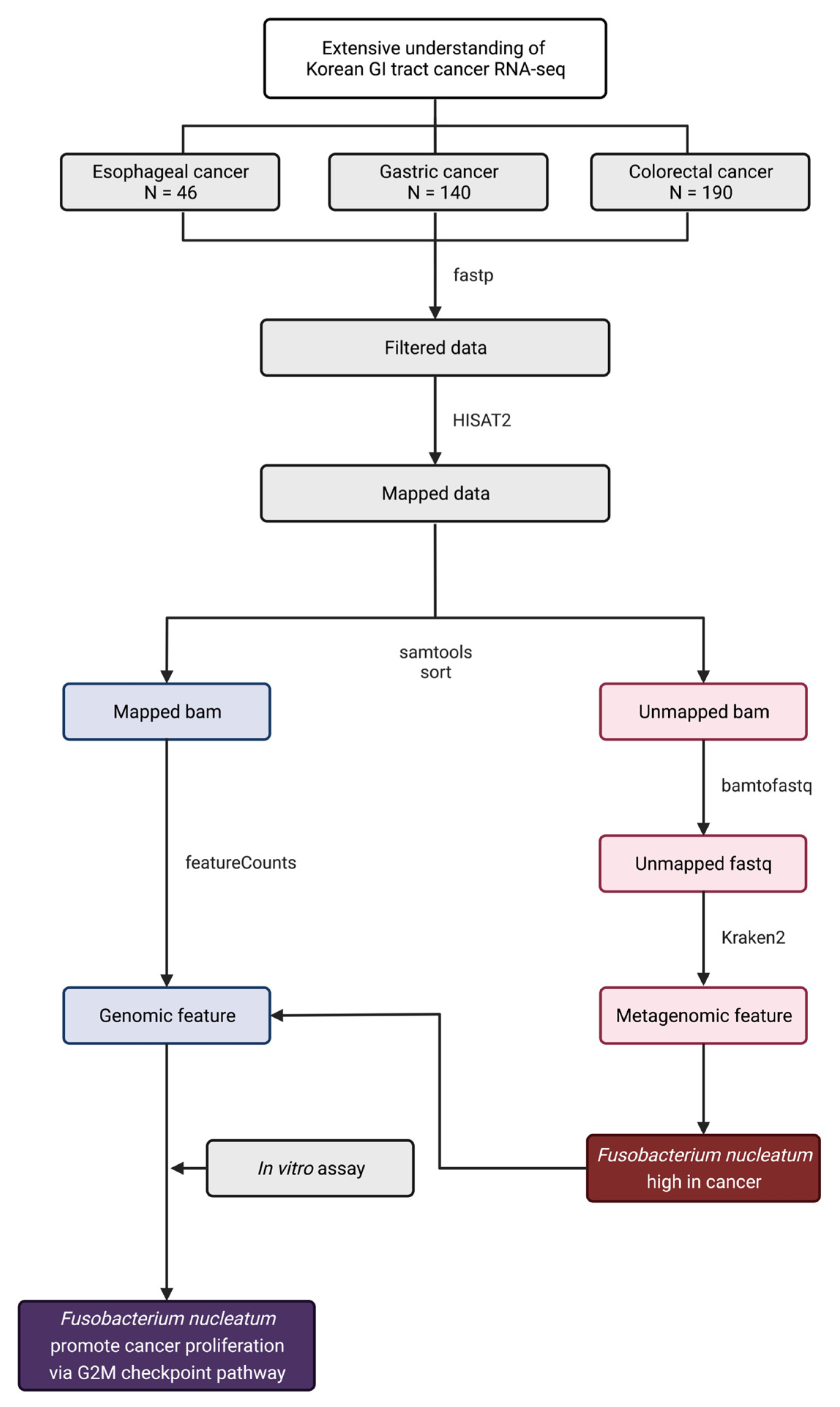
2.1. Public Whole Transcriptome Dataset Analysis

Gastric, esophageal, and CRC WTS data used in this study were obtained from accession numbers GSE113255, GSE130078, and GSE180440 in the Gene Expression Omnibus (GEO) repository [12,13,14]. Quality control and adapter trimming were processed with a fastp pipeline [15]. After filtering the data, HISAT2 was used to align the filtered data to the human database (GRCh38) [16]. HISAT2-aligned bam files were separated into mapped and unmapped bam files with the SAMtools “view” and “sort” commands. Unmapped bam files were converted into fastq formats with the SAMtools “bam2fastq” command [17]. Unmapped fastq files were processed with Kraken2 and the minikraken database to assign taxonomic labels to metagenomic sequences [18]. To quantify reads by genomic features, we used featureCounts [19]. The “DESeq2” package in R version 4.1.2 (Vienna, Austria) was used to normalize the genomic and metagenomic data and extract DEGs and microbiome differential abundance [20]. We used the GSEA platform version 4.1.0 with the MSigDB hallmark gene set for GSEA [21]. The “pheatmap” package in R was used to draw a heatmap plot with z-score-transformed expression data [22]. The “VennDiagram” package in R was used to find overlapped microbiomes in GI tract cancer [23]. The “ggplot2” package in R was used to draw volcano plots, barplots, boxplots, and PCoA plots [24].

2.2. Bacterial Culture Growth and Supernatants

We inoculated F. nucleatum subsp. nucleatum (KCTC 2640) on Reinforced Clostridial Medium (RCM) culture plates and grew it in anaerobic jars in 37 °C incubators for 3 d. We picked isolated strains from the culture plate and grew them in an anaerobic liquid RCM vial in a 37 °C incubator for 2 d. The optical density of F. nucleatum supernatants was adjusted to 1.0 at 600 nm in 1 mL RCM broth. The supernatants were obtained at 6000× g for 15 min at 4 °C and stored at 4 °C.

2.3. Cell Culture Growth, siRNA Transfection, and Treatment of Bacterial Supernatants

The MC 38 cell line was purchased from Kerafast. MC 38 cells were cultured in Dulbecco’s Modified Eagle’s Medium (DMEM) with 10% heat-inactivated fetal bovine serum (FBS) and antibiotic–antimycotic solution (100 units/mL penicillin, 100 μg/mL streptomycin, and 0.25 μg/mL amphotericin B) (Gibco, Life Technologies Co. Ltd., Waltham, MA, USA) in a 37 °C/5% CO2 incubator. MC 38 cells (1 × 10⁵) were seeded in 6-well plates with 2.5 mL of DMEM without antibiotics and incubated in a 37 °C/5% CO2 incubator until they reached 60% confluency. LipofectamineTM RNAiMAX (Invitrogen, Carlsbad, CA, USA) was used to transfect siRNA oligos. siRNA for mouse Chek1 mRNA were synthesized commercially at Bioneer: siChek1 (forward: 5′-UCUCAAGUCAUGAUUGCUU-3′; reverse: 5′-AAGCAAUCAUGACUUGAGA-3′). The siRNA duplex and lipofectamine mixture were treated in each well for 24 h in a 37 °C/5% CO2 incubator. The supernatants of F. nucleatum were treated after discarding the siRNA-treated DMEM and incubated for 36 h.

2.4. Total RNA Extraction and qRT-PCR Analysis

Total RNA was extracted from MC 38 cells using an RNeasy mini kit (Hilden, Germany, Qiagen) according to the manufacturer’s instructions. cDNA was synthesized using TOPscript RT DryMIX dT18plus (Daejeon, Korea, Enzynomics) according to the manufacturer’s instructions. Gene expression was measured using qRT-PCR, and expression data were handled using StepOne PlusTM software (Waltham, MA, USA, Applied Biosystems). qRT-PCR amplification was achieved using TOPrealTM SYBR Green qPCR PreMIX (Enzynomics). Primers were synthesized by Macrogen: Cdk1 (forward: 5′-AAGGTACTTACGGTGTGGTG-3′; reverse: 5′-CAGGTACTTCTTGAGGTCCA-3′), Cdc25a (forward: 5′-TGGACCTGTCTCCTACACTC-3′; reverse: 5′-GCTCAGTGAGAGCAGCTAAC-3ʹ), Cdc25c (forward: 5ʹ-CTACAGGACCTATCCCACCT-3′; reverse: 5′-CTCTCCACTGCTAAGATTCG-3′), Chek1 (forward: 5′-GGAGTAAGGAAATGCAGGAG-3′; reverse: 5′-GGAGAGTTAAGTGGGTGACA-3′), and Ccnb1 (forward: 5′-GCACCTGGCTAAGAATGTAG-3′; reverse: 5′-GAGCAAGTAAACACGGTAGG-3′). Mouse beta-actin was used as an internal control.

2.5. WST-1 Assay

We incubated 100 μL of (5 × 10³ cells) MC 38 cells per well in a 37 °C/5% CO2 incubator for 24 h. We transfected Chek1 siRNA for 6 h and treated the supernatants of F. nucleatum overnight. We treated Quanti-MaxTM (Seoul, Korea, BIOMAX) with 10 μL for 2 h and measured the absorbance at 450 nm absorbance.

2.6. Statistical Analysis

All data were tested for normality, and datasets were analyzed using one-way ANOVAs. Post-hoc analysis was conducted using the Bonferroni test. All data results are presented as the mean ± SD. All statistical analyses were performed using GraphPad Prism 9 (La Jolla, CA, USA) and R-4.2.1 (Vienna, Austria) for Mac OS. To compare the two groups (cancer and normal), we used the Wilcoxon test. Statistical significance was set at p < 0.05.

3. Results and Discussion

To investigate the common bacteria in GI tract cancer, the microbial composition of cancer tissues was compared with those of normal tissues. Open datasets from studies by Kim et al., You et al., and Park et al. in the GEO database were used for identifying the genomic and metagenomic features of GI tract cancer [12,13,14]. An in vitro assay was performed to validate the results of the WTS analysis (Figure 1).

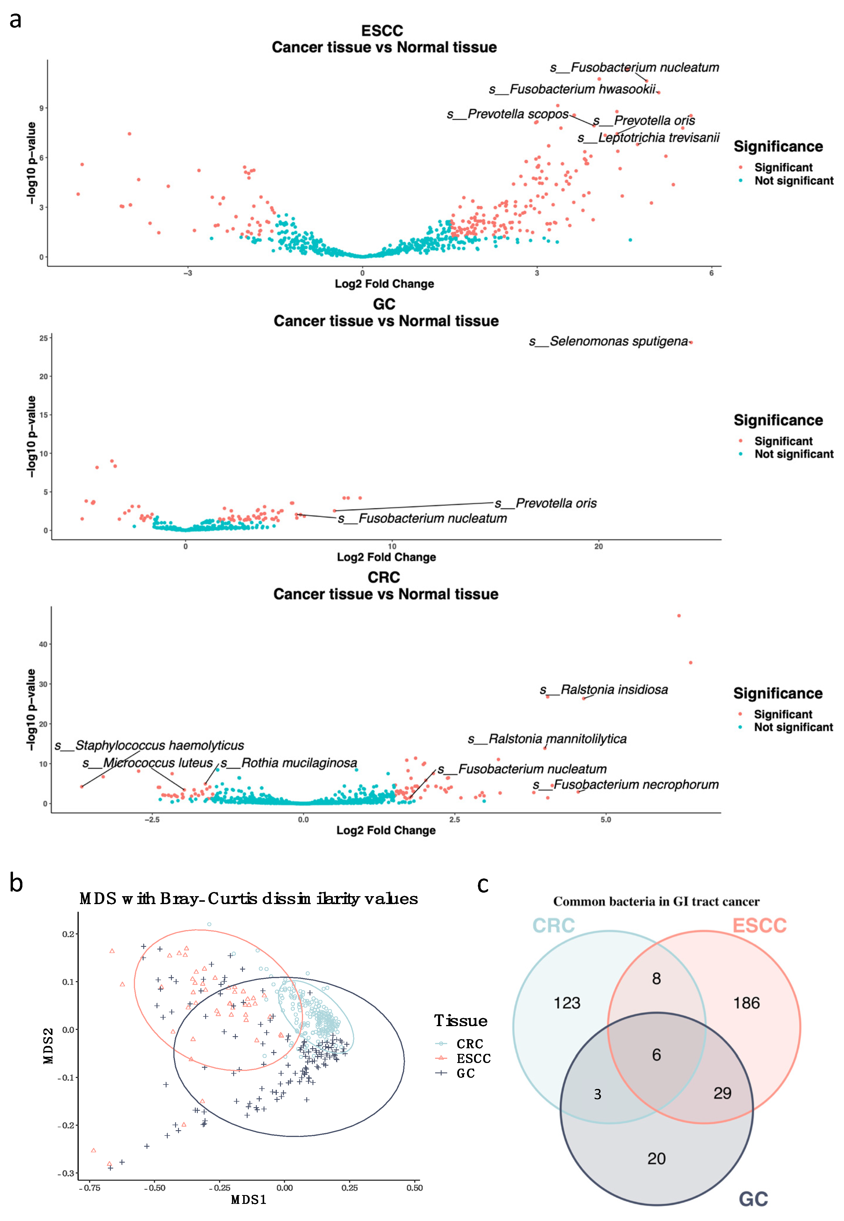
3.1. Fusobacterium nucleatum Is Enriched in Gastrointestinal Tract Cancer Tissues

The GI tract is affected by many bacteria. F. nucleatum is a well-known bacteria that promotes colorectal carcinogenesis. A recent study showed that it could originate from the oral cavity through the circulatory system [25]. Additionally, there is evidence that F. nucleatum plays a role in oral, esophageal, gastric, head and neck, breast, and pancreatic cancers [25,26,27,28]. F. nucleatum plays a role in carcinogenesis by inducing a host response for tumor initiation and promotion, and encouraging tumor invasion and metastasis [10]. Humans are highly susceptible to H. pylori infection, with approximately half of the global population suffering from this infection. H. pylori is related to gastric adenocarcinoma through immune response and inflammation mechanisms. However, H. pylori is associated with a decreased risk of esophageal adenocarcinoma [5,29]. Based on previous studies regarding the effects of bacteria on the GI tract, we examined common bacteria as therapeutic targets in GI tract cancer. Korean GI tract cancer has not yet been studied, and there are applicable cohorts in which to examine common bacteria in GI tract tumor tissues. The CRC dataset consisted of 190 WTS data points for 145 tumor tissues and 45 normal colon tissues. The WTS data for esophageal cancer were composed of 23 cancerous tissues and 23 normal adjacent tissues. There were 130 gastric cancer tissues and 10 normal intestinal mucosae in gastric cancer WTS data.
Using a microbiome abundance analysis, it is shown that the expression of Staphylococcus haemolyticus, Micrococcus luteus, and Rothia mucilaginosa in normal colorectal tissues was higher, whereas the expression of Ralstonia insidiosa, R. mannitolilytica, F. necrophorum, and F. nucleatum was higher in CRC tissues. In esophageal cancer tissues, F. nucleatum, F. hwasookii, Prevotella oris, and Leptotrichia trevisanii were highly expressed. Furthermore, in gastric cancer tissues, Selenomonas sputigena, P. oris, and F. nucleatum were more highly expressed (Figure 2a). F. nucleatum is a commonly highly expressed bacteria in all three types of cancer tissues. P. oris is a bacteria that is known to originate from the oral cavity and is enriched in gastric cancer samples. Our research showed it was enriched in the ESCC and GC cancer groups [30]. To determine any differences between the relationship of F. nucleatum abundance in cancer and normal tissues, the expression count was normalized using DESeq2, which indicated a significant difference (CRC LogFC = 1.755, Adj. p value = 0.037; ESCC LogFc = 4.880, Adj. p value = 2.36 × 10¹¹; GC LogFC = 5.358, Adj. p value = 0.009). To specify whether the batch effect of each cohort contributed to this outcome, a principal coordinate analysis (PCoA) was performed with Bray-Curtis distances. The PCoA plot showed that there were few community dissimilarities (Figure 2b). There were six commonly highly expressed bacteria in GI tract cancer. In particular, F. nucleatum was the only bacteria that was defined at the species level (Figure 2c, Supplementary Table S1). With the microbiome overlap Venn Diagram, it was found that the gastric cancer cohort shared more than 60% of the enriched microbiome with other cohorts. This is thought to be a result of the stomach being a passageway between the esophagus and colon.

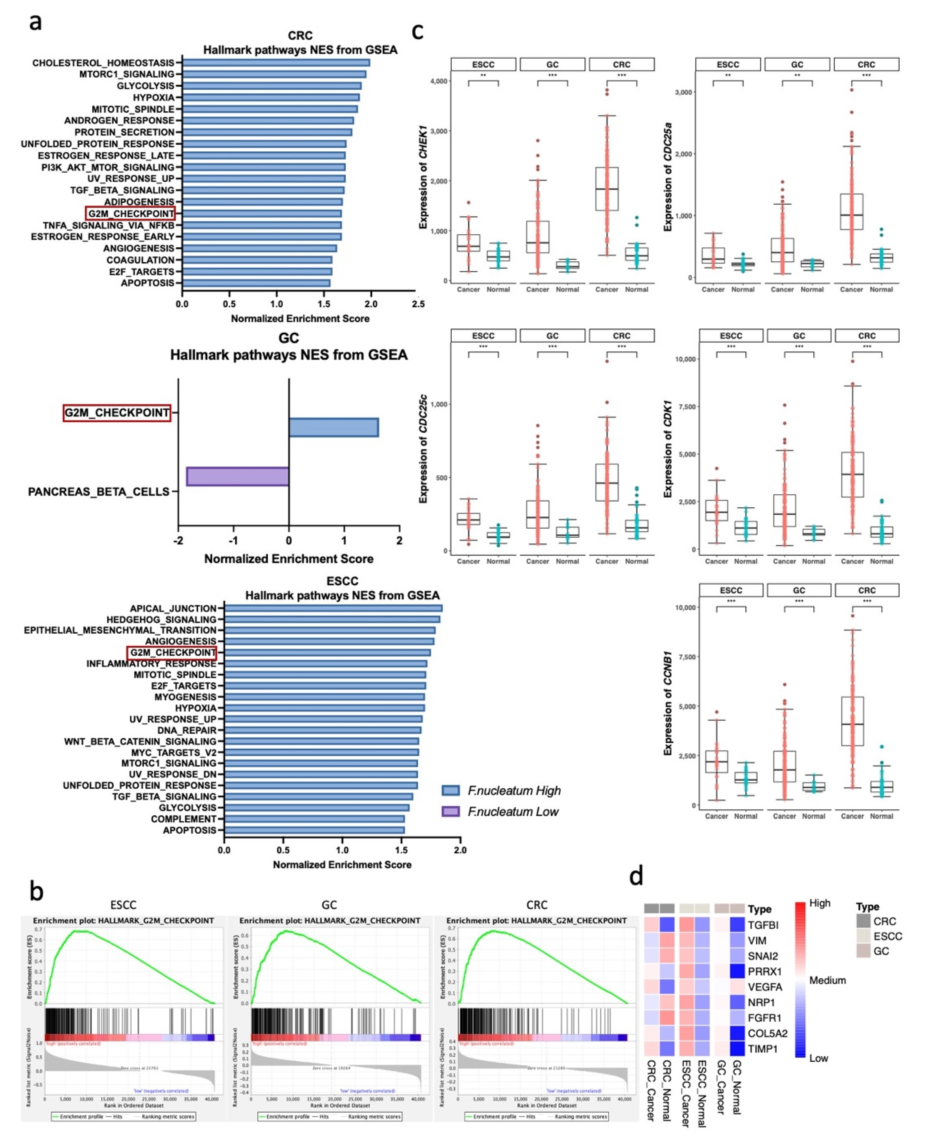
3.2. G2M Checkpoint Pathway Is Associated with F. nucleatum Abundance in GI Tract Cancer

To determine the possible mechanism of how F. nucleatum works, each dataset is divided into two groups, the F. nucleatum-high and F. nucleatum-low groups, using median values. Using the DESeq2 normalized gene count, Gene Set Enrichment Analysis (GSEA) was performed. Through GSEA analysis, possible pathways with a normalized p-value < 0.05 was selected (Figure 3a). We examined whether hallmark genes of the G2M checkpoint were commonly upregulated in all cancer types in the F. nucleatum high group (Figure 3b, CRC NES = 1.692, Nom p-value = 0.042, FDR q-value = 0.024; ESCC NES = 1.754, Nom p-value = 0.008, FDR q-value = 0.039; GC NES = 1.630, Nom p-value = 0.039, FDR q-value = 0.452). Major anti-cancer therapies target DNA or cell-division mechanisms. These therapies elicit the activation of cell-cycle checkpoints. The G2M checkpoint pathway is used by cancer cells to avoid apoptosis [31]. Then, the expression of genes related to the G2M checkpoint pathways in cancer and normal tissues was compared. Higher expression of G2M checkpoint pathway genes (CHEK1, CDK1, CDC25A, CDC25B, CDC25C, CCNB1) was observed in cancer tissues, with all comparisons having a p-value less than 0.01 (Figure 3c). CHEK1 and its downstream genes appeared to be related to a common cancerous pathway, and CHEK1 is a key factor in the checkpoint control of the G2M checkpoint pathway [32]. CHEK1 is known as a tumor repressor, but recent studies have demonstrated that CHEK1 can promote cancer cell proliferation. Furthermore, the inhibition of CHEK1 can be a therapeutic target for cancer by sensitizing cells to DNA-damaging agents, such as ionizing radiation (IR) and antimetabolites, or when used as single agents [32,33,34]. The suppression of CHEK1 can downregulate CCNB1, and this downregulation can impair CRC proliferation. The repression of CCNB1 can invoke G2M phase arrest and interfere with the expression of CDC25c and CDK1 [35]. Furthermore, genes related to angiogenesis and epithelial–mesenchymal transition (EMT) were investigated, and meaningful significance was found in the GSEA analysis of CRC and ESCC cohorts (Supplementary Figure S1a, CRC angiogenesis NES = 1.641, Nom p-value = 0.019, FDR q-value = 0.038; CRC EMT NES = 1.46, Nom p-value = 0.128, FDR q-value = 0.101; ESCC angiogenesis NES = 1.780, Nom p-value = 0.008, FDR q-value = 0.035; ESCC EMT NES = 1.795, Nom p-value = 0.004, FDR q-value = 0.038). Additionally, angiogenesis and EMT are pathways that contribute to cancer proliferation [36]. However, angiogenesis- and EMT-related genes were not always highly expressed in the cancer group, such as VIM and VEGFA (Figure 3d). To determine if the abundance of F. nucleatum affects the expression of the G2M pathway, a correlation analysis was performed between the expression of F. nucleatum and G2M pathway-related genes. A weak positive correlation was identified between F. nucleatum abundance and the expression of G2M pathway genes (Supplementary Figure S1b).

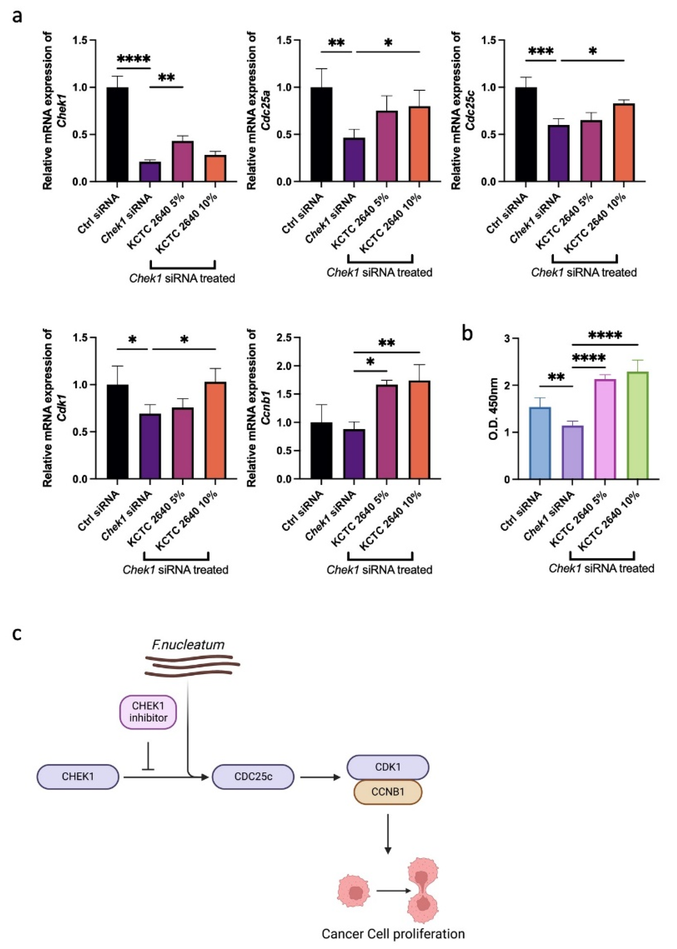
3.3. F. nucleatum Promotes Cancer Cell Proliferation through the G2M Checkpoint Pathway

MC 38 cells, derived from murine colon adenocarcinoma cells, were used to verify the WTS results by in vitro assay, especially for CRC patients [37]. To inhibit the cell proliferation of MC 38 cells, Chek1 siRNA was treated. We expected that F. nucleatum might counteract the activity of Chek1 siRNA. F. nucleatum subsp. nucleatum, which is well-known for causing infections in humans, was used [38]. We differed the concentration of supernatants to determine the optimal density. The expression of Chek1, Cdk1, Cdc25a, Cdc25c, and Ccnb1 was examined to confirm the activity of Chek1 siRNA and the supernatants of F. nucleatum. The qRT-PCR results demonstrated that treatment with F. nucleatum in a 5% concentration best offset the effects of Chek1 knockdown. Chek1 siRNA successfully knocked down the expression of Chek1 in MC 38 cells, and the supernatant of F. nucleatum could not decrease the effect of the knockdown on Chek1.
However, the supernatants increased the expression of Cdc25 components, which resulted in the upregulation of Cdk1 (Figure 4a). In particular, Cdc25c is known to be overexpressed in various types of cancer and promotes tumorigenesis. Furthermore, through the interaction between Cdc25c and Cdk1, cancer cell proliferation can be repressed [35,39]. Through the WST-1 assay, the increased proliferation of MC 38 cells treated with the supernatants of F. nucleatum was identified (Figure 4b). These results demonstrated that the knockdown of Chek1 could inhibit cancer cell proliferation through Cdc25c and Cdk1-Ccnb1, and the treatment of F. nucleatum supernatants can overcome this suppression.
In conclusion, GI tract cancers frequently recur, especially GC and CRC, in Koreans [40]. There are known diagnostic biomarkers for GI tract cancers, such as genetic aberrations and molecules [41]. F. nucleatum is one of the well-known bacteria that negatively affects the prognosis of cancers by the activation of chemokines that lead to aggressive tumor behavior [25,28]. In our study, it is shown that F. nucleatum is enriched in Korean GI tract cancers, and F. nucleatum facilitated cancer’s use of the G2M checkpoint pathway. In particular, the expression of CDC25c and CDK1-CCNB1 was investigated, which was increased by F. nucleatum among diverse paths of the G2M checkpoint. This study suggests that F. nucleatum is a potential therapeutic target not only for CRC but also for ESCC and GC in Korean patients. The prevention of F. nucleatum from proliferating could be used as a clinical strategy for GI tract cancer patients. Metronidazole, an antibiotic for anaerobic infections, could be used for targeting the broad spectrum of anaerobic organisms, including F. nucleatum, in CRC [42].To further investigate how F. nucleatum acts in GI tract cancers, metabolites associated with F. nucleatum, and the G2M checkpoint pathways, additional examination is needed. Furthermore, additional microbiome studies of the GI tracts of other ethnic populations could elucidate whether enriched F. nucleatum in GI tract cancers is the consequence of ethnic attributes.

Supplementary Materials

The following supporting information can be downloaded at: https://www.mdpi.com/article/10.3390/microorganisms10101896/s1, Figure S1: GSEA and correlation analysis of GI tract cancer RNA-seq data; Table S1: Common bacteria between cancer groups.

Author Contributions

Conceptualization, H.P.; data curation, H.A.; formal analysis, H.A.; funding acquisition, S.K., Y.K. (Yunjae Kim), B.C. and C.J.; investigation, H.A., K.M., E.L. and H.K.; methodology, H.A. and K.M.; supervision, H.P.; visualization, H.A., G.K. and Y.K. (Yeongmin Kim); writing—original draft, H.A.; writing—review and editing, K.M. All authors have read and agreed to the published version of the manuscript.

Funding

This work was supported by the GIST Research Institute (GRI) grant funded by GIST in 2022. This work was supported by the National Research Foundation of Korea (NRF) with a grant funded by the Korean government (MSIT) (No. 2022R1A2C2008976).

Data Availability Statement

This study used data processed from the Gene Expression Omnibus, accession numbers GSE113255, GSE130078, and GSE180440, for the analysis of gene expression. These data are freely available at https://www.ncbi.nlm.nih.gov/geo/query/acc.cgi?acc=GSE113255 (accessed on 22 February 2022), https://www.ncbi.nlm.nih.gov/geo/query/acc.cgi?acc=GSE130078 (accessed on 9 January 2022), https://www.ncbi.nlm.nih.gov/geo/query/acc.cgi?acc=GSE180440 (accessed on 5 May 2022).

Acknowledgments

The illustrations used in Figure 1 and Figure 4c were created with BioRender.com (Toronto, Canada).

Conflicts of Interest

The authors declare no conflict of interest.

References

  1. Bhatt, A.P.; Redinbo, M.R.; Bultman, S.J. The role of the microbiome in cancer development and therapy. CA A Cancer J. Clin. 2017, 67, 326–344. [Google Scholar] [CrossRef] [PubMed]
  2. Hong, M.; Tao, S.; Zhang, L.; Diao, L.-T.; Huang, X.; Huang, S.; Xie, S.-J.; Xiao, Z.-D.; Zhang, H. RNA sequencing: New technologies and applications in cancer research. J. Hematol. Oncol. 2020, 13, 166. [Google Scholar] [CrossRef] [PubMed]
  3. Allen, J.; Sears, C.L. Impact of the gut microbiome on the genome and epigenome of colon epithelial cells: Contributions to colorectal cancer development. Genome Med. 2019, 11, 11. [Google Scholar] [CrossRef]
  4. Cullin, N.; Azevedo Antunes, C.; Straussman, R.; Stein-Thoeringer, C.K.; Elinav, E. Microbiome and cancer. Cancer Cell 2021, 39, 1317–1341. [Google Scholar] [CrossRef] [PubMed]
  5. Doorakkers, E.; Lagergren, J.; Engstrand, L.; Brusselaers, N. Eradication of Helicobacter pylori and Gastric Cancer: A Systematic Review and Meta-analysis of Cohort Studies. J. Natl. Cancer Inst. 2016, 108. [Google Scholar] [CrossRef] [PubMed]
  6. Buc, E.; Dubois, D.; Sauvanet, P.; Raisch, J.; Delmas, J.; Darfeuille-Michaud, A.; Pezet, D.; Bonnet, R. High prevalence of mucosa-associated E. coli producing cyclomodulin and genotoxin in colon cancer. PLoS ONE 2013, 8, e56964. [Google Scholar] [CrossRef]
  7. Haghi, F.; Goli, E.; Mirzaei, B.; Zeighami, H. The association between fecal enterotoxigenic B. fragilis with colorectal cancer. BMC Cancer 2019, 19, 1–4. [Google Scholar] [CrossRef]
  8. Scanu, T.; Spaapen, R.M.; Bakker, J.M.; Pratap, C.B.; Wu, L.-E.; Hofland, I.; Broeks, A.; Shukla, V.K.; Kumar, M.; Janssen, H. Salmonella manipulation of host signaling pathways provokes cellular transformation associated with gallbladder carcinoma. Cell Host Microbe 2015, 17, 763–774. [Google Scholar] [CrossRef] [PubMed]
  9. Sánchez-Alcoholado, L.; Ramos-Molina, B.; Otero, A.; Laborda-Illanes, A.; Ordóñez, R.; Medina, J.A.; Gómez-Millán, J.; Queipo-Ortuño, M.I. The Role of the Gut Microbiome in Colorectal Cancer Development and Therapy Response. Cancers 2020, 12, 1406. [Google Scholar] [CrossRef] [PubMed]
  10. Zhao, T.; Wang, X.; Fu, L.; Yang, K. Fusobacterium nucleatum: A new player in regulation of cancer development and therapeutic response. Cancer Drug Resist. 2022, 5, 436–450. [Google Scholar] [CrossRef] [PubMed]
  11. Suraya, R.; Nagano, T.; Kobayashi, K.; Nishimura, Y. Microbiome as a Target for Cancer Therapy. Integr. Cancer Ther. 2020, 19, 1534735420920721. [Google Scholar] [CrossRef]
  12. Kim, S.K.; Kim, H.J.; Park, J.L.; Heo, H.; Kim, S.Y.; Lee, S.I.; Song, K.S.; Kim, W.H.; Kim, Y.S. Identification of a molecular signature of prognostic subtypes in diffuse-type gastric cancer. Gastric. Cancer 2020, 23, 473–482. [Google Scholar] [CrossRef]
  13. You, B.H.; Yoon, J.H.; Kang, H.; Lee, E.K.; Lee, S.K.; Nam, J.W. HERES, a lncRNA that regulates canonical and noncanonical Wnt signaling pathways via interaction with EZH2. Proc. Natl. Acad. Sci. USA 2019, 116, 24620–24629. [Google Scholar] [CrossRef] [PubMed]
  14. Park, D.Y.; Choi, C.; Shin, E.; Lee, J.H.; Kwon, C.H.; Jo, H.J.; Kim, H.R.; Kim, H.S.; Oh, N.; Lee, J.S.; et al. NTRK1 fusions for the therapeutic intervention of Korean patients with colon cancer. Oncotarget 2016, 7, 8399–8412. [Google Scholar] [CrossRef] [PubMed]
  15. Chen, S.; Zhou, Y.; Chen, Y.; Gu, J. fastp: An ultra-fast all-in-one FASTQ preprocessor. Bioinformatics 2018, 34, i884–i890. [Google Scholar] [CrossRef]
  16. Kim, D.; Paggi, J.M.; Park, C.; Bennett, C.; Salzberg, S.L. Graph-based genome alignment and genotyping with HISAT2 and HISAT-genotype. Nat. Biotechnol. 2019, 37, 907–915. [Google Scholar] [CrossRef]
  17. Li, H.; Handsaker, B.; Wysoker, A.; Fennell, T.; Ruan, J.; Homer, N.; Marth, G.; Abecasis, G.; Durbin, R. The Sequence Alignment/Map format and SAMtools. Bioinformatics 2009, 25, 2078–2079. [Google Scholar] [CrossRef] [PubMed]
  18. Wood, D.E.; Lu, J.; Langmead, B. Improved metagenomic analysis with Kraken 2. Genome Biol. 2019, 20, 257. [Google Scholar] [CrossRef]
  19. Liao, Y.; Smyth, G.K.; Shi, W. featureCounts: An efficient general purpose program for assigning sequence reads to genomic features. Bioinformatics 2013, 30, 923–930. [Google Scholar] [CrossRef] [PubMed]
  20. Love, M.I.; Huber, W.; Anders, S. Moderated estimation of fold change and dispersion for RNA-seq data with DESeq2. Genome Biol. 2014, 15, 550. [Google Scholar] [CrossRef] [PubMed] [Green Version]
  21. Subramanian, A.; Tamayo, P.; Mootha, V.K.; Mukherjee, S.; Ebert, B.L.; Gillette, M.A.; Paulovich, A.; Pomeroy, S.L.; Golub, T.R.; Lander, E.S.; et al. Gene set enrichment analysis: A knowledge-based approach for interpreting genome-wide expression profiles. Proc. Natl. Acad. Sci. USA 2005, 102, 15545–15550. [Google Scholar] [CrossRef]
  22. Kolde, R. Pheatmap: Pretty heatmaps. R Package Version 2012, 1, 726. [Google Scholar]
  23. Chen, H.; Boutros, P.C. VennDiagram: A package for the generation of highly-customizable Venn and Euler diagrams in R. BMC Bioinform. 2011, 12, 35. [Google Scholar] [CrossRef]
  24. Wickham, H. Data analysis. In ggplot2; Springer: Germany, 2016; pp. 189–201. [Google Scholar]
  25. Yamamura, K.; Baba, Y.; Nakagawa, S.; Mima, K.; Miyake, K.; Nakamura, K.; Sawayama, H.; Kinoshita, K.; Ishimoto, T.; Iwatsuki, M.; et al. Human Microbiome Fusobacterium Nucleatum in Esophageal Cancer Tissue Is Associated with Prognosis. Clin. Cancer Res. 2016, 22, 5574–5581. [Google Scholar] [CrossRef]
  26. Irfan, M.; Delgado, R.Z.R.; Frias-Lopez, J. The Oral Microbiome and Cancer. Front. Immunol. 2020, 11, 591088. [Google Scholar] [CrossRef] [PubMed]
  27. Mitsuhashi, K.; Nosho, K.; Sukawa, Y.; Matsunaga, Y.; Ito, M.; Kurihara, H.; Kanno, S.; Igarashi, H.; Naito, T.; Adachi, Y.; et al. Association of Fusobacterium species in pancreatic cancer tissues with molecular features and prognosis. Oncotarget 2015, 6, 7209–7220. [Google Scholar] [CrossRef] [PubMed]
  28. Boehm, E.T.; Thon, C.; Kupcinskas, J.; Steponaitiene, R.; Skieceviciene, J.; Canbay, A.; Malfertheiner, P.; Link, A. Fusobacterium nucleatum is associated with worse prognosis in Lauren’s diffuse type gastric cancer patients. Sci. Rep. 2020, 10, 16240. [Google Scholar] [CrossRef]
  29. Khatoon, J.; Rai, R.P.; Prasad, K.N. Role of Helicobacter pylori in gastric cancer: Updates. World J. Gastrointest. Oncol. 2016, 8, 147–158. [Google Scholar] [CrossRef]
  30. Könönen, E.; Gursoy, U.K. Oral Prevotella Species and Their Connection to Events of Clinical Relevance in Gastrointestinal and Respiratory Tracts. Front. Microbiol. 2022, 12. [Google Scholar] [CrossRef] [PubMed]
  31. Visconti, R.; Della Monica, R.; Grieco, D. Cell cycle checkpoint in cancer: A therapeutically targetable double-edged sword. J. Exp. Clin. Cancer Res. 2016, 35, 153. [Google Scholar] [CrossRef]
  32. Zhang, Y.; Hunter, T. Roles of Chk1 in cell biology and cancer therapy. Int. J. Cancer 2014, 134, 1013–1023. [Google Scholar] [CrossRef] [Green Version]
  33. Qiu, Z.; Oleinick, N.L.; Zhang, J. ATR/CHK1 inhibitors and cancer therapy. Radiother. Oncol. 2018, 126, 450–464. [Google Scholar] [CrossRef] [PubMed]
  34. Albiges, L.; Goubar, A.; Scott, V.; Vicier, C.; Lefèbvre, C.; Alsafadi, S.; Commo, F.; Saghatchian, M.; Lazar, V.; Dessen, P.; et al. Chk1 as a new therapeutic target in triple-negative breast cancer. Breast 2014, 23, 250–258. [Google Scholar] [CrossRef]
  35. Fang, Y.; Yu, H.; Liang, X.; Xu, J.; Cai, X. Chk1-induced CCNB1 overexpression promotes cell proliferation and tumor growth in human colorectal cancer. Cancer Biol. Ther. 2014, 15, 1268–1279. [Google Scholar] [CrossRef]
  36. Ribatti, D. Epithelial-mesenchymal transition in morphogenesis, cancer progression and angiogenesis. Exp. Cell Res. 2017, 353, 1–5. [Google Scholar] [CrossRef] [PubMed]
  37. Newsome, R.C.; Yang, Y.; Jobin, C. The microbiome, gastrointestinal cancer, and immunotherapy. J. Gastroenterol. Hepatol. 2022, 37, 263–272. [Google Scholar] [CrossRef] [PubMed]
  38. Lee, S.A.; Liu, F.; Riordan, S.M.; Lee, C.S.; Zhang, L. Global Investigations of Fusobacterium nucleatum in Human Colorectal Cancer. Front. Oncol. 2019, 9, 566. [Google Scholar] [CrossRef] [PubMed]
  39. Lee, M.H.; Cho, Y.; Kim, D.H.; Woo, H.J.; Yang, J.Y.; Kwon, H.J.; Yeon, M.J.; Park, M.; Kim, S.H.; Moon, C.; et al. Menadione induces G2/M arrest in gastric cancer cells by down-regulation of CDC25C and proteasome mediated degradation of CDK1 and cyclin B1. Am. J. Transl. Res. 2016, 8, 5246–5255. [Google Scholar]
  40. Jung, K.-W.; Won, Y.-J.; Kong, H.-J.; Lee, E.S. Cancer Statistics in Korea: Incidence, Mortality, Survival, and Prevalence in 2015. Cancer Res. Treat. 2018, 50, 303–316. [Google Scholar] [CrossRef] [PubMed]
  41. Vedeld, H.M.; Goel, A.; Lind, G.E. Epigenetic biomarkers in gastrointestinal cancers: The current state and clinical perspectives. In Proceedings of the Seminars in Cancer Biology; Academic Press: Cambridge, MA, USA, 2018; pp. 36–49. [Google Scholar]
  42. Bullman, S.; Pedamallu, C.S.; Sicinska, E.; Clancy, T.E.; Zhang, X.; Cai, D.; Neuberg, D.; Huang, K.; Guevara, F.; Nelson, T.; et al. Analysis of Fusobacterium persistence and antibiotic response in colorectal cancer. Science 2017, 358, 1443–1448. [Google Scholar] [CrossRef] [PubMed] [Green Version]
Figure 1. Flow of WTS data processing. Overall flow of WTS data processing for genomic feature and metagenomic feature analysis.
Microorganisms 10 01896 g001
Figure 2. Microbiome differential abundance analysis of GI tract cancer WTS data. (a) Volcano plot demonstrating the differential prevalence of the microbiome between cancer and normal tissues from the esophageal (GSE130078), gastric (GSE180440), and colorectal (GSE113255) samples. The microbiome was colored when it surpassed the significance threshold (p-value < 0.05, fold change > 1.5). Names of species levels in the microbiome are annotated. (b) Principal coordinate analysis (PCoA) plot on the Bray−Curtis dissimilarity indexes between microbiome profiles of different cancer types. (c) Venn Diagram showing numbers of overlapping microbiomes between GI tract cancer types.
Microorganisms 10 01896 g002
Figure 3. DEGs and GSEA analysis of GI tract cancer WTS data. (a) Barplot indicating normalized enrichment score (NES) in F. nucleatum−high vs. F. nucleatum−low groups for every cancer type. (b) Gene set enrichment analysis (GSEA) demonstrating the common pathway (G2M checkpoint) in the F. nucleatum−high group of every cancer type (CRC NES = 1.692, Nom p-value = 0.042, FDR q-value = 0.024; ESCC NES = 1.754, Nom p-value = 0.008, FDR q-value = 0.039; GC NES = 1.630, Nom p-value = 0.039, FDR q-value = 0.452). (c) Boxplot representing the expression of the G2M pathway−related genes between cancer and normal tissues. ** p < 0.01, *** p < 0.001. Statistical significance was calculated using the Wilcoxon test. (d) Heatmap for the z−score−converted expression of the EMT pathway- and angiogenesis pathway-related genes.
Microorganisms 10 01896 g003
Figure 4. qRT-PCR and WST-1 assay of MC 38 cell lines. (a) qRT-PCR assay representing the G2M checkpoint pathway-related genes in MC 38 cell lines treated with Chek1 siRNA and F. nucleatum. (b) WST-1 assay demonstrating the cell proliferation of MC 38 cell lines treated with Chek1 siRNA and F. nucleatum. (c) Scheme of the G2M checkpoint pathway and effect of F. nucleatum. * p < 0.05, ** p < 0.01, *** p < 0.001, **** p < 0.0001. Statistical significance was calculated using Bonferroni tests.
Microorganisms 10 01896 g004
Publisher’s Note: MDPI stays neutral with regard to jurisdictional claims in published maps and institutional affiliations.

